Autophagy and TGF-Beta Antagonist Signaling in … · Award Number: W81XWH-13-1-0109 TITLE:...
15
Award Number: W81XWH-13-1-0109 TITLE: Autophagy and TGF-Beta Antagonist Signaling in Breast Cancer Dormancy at Premetastatic Sites PRINCIPAL INVESTIGATOR: Xuejun Jiang, PhD CONTRACTING ORGANIZATION: Sloan-Kettering Institute for Cancer Research New York, NY 10065-6007 REPORT DATE: June 2015 TYPE OF REPORT: Final PREPARED FOR: U.S. Army Medical Research and Materiel Command Fort Detrick, Maryland 21702-5012 DISTRIBUTION STATEMENT: X Approved for public release; distribution unlimited The views, opinions and/or findings contained in this report are those of the author(s) and should not be construed as an official Department of the Army position, policy or decision unless so designated by other documentation.
Transcript of Autophagy and TGF-Beta Antagonist Signaling in … · Award Number: W81XWH-13-1-0109 TITLE:...
Award Number: W81XWH-13-1-0109
TITLE: Autophagy and TGF-Beta Antagonist Signaling in Breast Cancer Dormancy at Premetastatic Sites
PRINCIPAL INVESTIGATOR: Xuejun Jiang, PhD
CONTRACTING ORGANIZATION: Sloan-Kettering Institute for Cancer Research New York, NY 10065-6007
REPORT DATE: June 2015
TYPE OF REPORT: Final
PREPARED FOR: U.S. Army Medical Research and Materiel Command Fort Detrick, Maryland 21702-5012
DISTRIBUTION STATEMENT:
X Approved for public release; distribution unlimited
The views, opinions and/or findings contained in this report are those of the author(s) and should not be construed as an official Department of the Army position, policy or decision unless so designated by other documentation.
REPORT DOCUMENTATION PAGE Form Approved
OMB No. 0704-0188 Public reporting burden for this collection of information is estimated to average 1 hour per response, including the time for reviewing instructions, searching existing data sources, gathering and maintaining the data needed, and completing and reviewing this collection of information. Send comments regarding this burden estimate or any other aspect of this collection of information, including suggestions for reducing this burden to Department of Defense, Washington Headquarters Services, Directorate for Information Operations and Reports (0704-0188), 1215 Jefferson Davis Highway, Suite 1204, Arlington, VA 22202-4302. Respondents should be aware that notwithstanding any other provision of law, no person shall be subject to any penalty for failing to comply with a collection of information if it does not display a currently valid OMB control number. PLEASE DO NOT RETURN YOUR FORM TO THE ABOVE ADDRESS. 1. REPORT DATE (DD-MM-YYYY)June 2015
2. REPORT TYPEFinal
3. DATES COVERED (From - To)1May2013 - 31Mar2015
4. TITLE AND SUBTITLE 5a. CONTRACT NUMBER W81XWH-13-1-0109
Autophagy and TGF-Beta Antagonist Signaling in Breast Cancer
Dormancy at Premetastatic Sites 5b. GRANT NUMBER BC121829P1 5c. PROGRAM ELEMENT NUMBER
6. AUTHOR(S)
5d. PROJECT NUMBER
Xuejun Jiang 5e. TASK NUMBER
5f. WORK UNIT NUMBER
7. PERFORMING ORGANIZATION NAME(S) AND ADDRESS(ES)
8. PERFORMING ORGANIZATION REPORTNUMBER
Sloan-Kettering Institute for Cancer Research 1275 York Avenue New York, NY 10065-6007
9. SPONSORING / MONITORING AGENCY NAME(S) AND ADDRESS(ES) 10. SPONSOR/MONITOR’S ACRONYM(S)
U.S. Army Medical Research and Materiel Command Fort Detrick, MD 21702-5012 11. SPONSOR/MONITOR’S REPORT
NUMBER(S)
12. DISTRIBUTION / AVAILABILITY STATEMENT
Approved for Public Release; Distribution Unlimited
13. SUPPLEMENTARY NOTES
14. ABSTRACTThe majority of breast cancer mortality is caused by metastatic relapse, which requires the reactivation and outgrowth of tumor cells after they have disseminated to pre-metastatic sites and undergone prolonged dormancy. Understanding the mechanisms underpinning dormancy, reactivation, and outgrowth will provide novel approaches for the prevention and treatment of metastatic diseases. We discovered the TGFβ signaling antagonist Coco as a mediator of the reactivation of lung-disseminated breast cancer cells. We also made contributions to the mechanistic understanding and cancer relevance of autophagy, a catabolic response sustaining cancer cell survival under stress and therapeutic treatment. In this proposal, we investigate the role of autophagy and its interplay with Coco signaling in breast cancer dormancy and metastatic relapse, and the therapeutic potential of targeting autophagy for the treatment of breast cancer metastasis. Over the period supported by this grant, we have further deepened our understanding of the Coco signaling in dormancy reactivation, and the mechanisms and tumorigenic role of autophagy. We have developed a series of genetically controlled cancer cell models for in vivo mouse studies to define the roles and interplay of Coco signaling with autophagy in breast cancer dormancy and reactivation. Monoclonal antibodies against Coco have also been developed for future Coco-targeted therapies. Experiments utilizing mouse models to determine the in vivo effect of autophagy on breast cancer dormancy at lung premetastatic sites are ongoing.
15. SUBJECT TERMS
Metastasis, dormancy, reactivation, Coco signaling, autophagy, cancer therapy 16. SECURITY CLASSIFICATION OF: 17. LIMITATION
OF ABSTRACT 18. NUMBEROF PAGES
19a. NAME OF RESPONSIBLE PERSONUSAMRMC
a. REPORT
Unclassified
b. ABSTRACT
Unclassified
c. THIS PAGE
Unclassified Unclassified
19b. TELEPHONE NUMBER (include area code)
Standard Form 298 (Rev. 8-98) Prescribed by ANSI Std. Z39.18
email: [email protected]
15
2
Table of Contents
Page
Introduction…………………………………………………………….………..….. 3
Body………………………………………………………………………………….. 4
Key Research Accomplishments………………………………………….…….. 11
Reportable Outcomes……………………………………………………………… 12
Conclusion…………………………………………………………………………… 13
References……………………………………………………………………………. 14
3
Introduction
The goal of this project is to determine the role of autophagy and its interplay with Coco signaling in breast cancer dormancy and metastatic relapse, and the therapeutic potential of targeting autophagy and Coco for the treatment of breast cancer metastasis. The majority of breast cancer mortality is caused by metastatic relapse, which requires the reactivation and outgrowth of tumor cells after they have disseminated to pre-metastatic sites and undergone prolonged dormancy. Therefore, understanding the mechanisms underpinning dormancy, reactivation, and outgrowth will provide novel approaches for the prevention and treatment of metastatic disease. Recently, we developed a novel mouse model for breast tumor dormancy, and using this model we discovered that a signaling molecule called Coco mediates the reactivation of lung-disseminated breast cancer cells. Analysis of breast cancer patient samples revealed a significant correlation of Coco expression with lung metastasis and poor prognosis, suggesting that Coco expression can be used as a diagnostic tool. Furthermore, Coco signaling may regulate autophagy, a survival response that has been frequently implicated in cancer cell survival under stress and therapeutic treatment. In this research project, we not only investigate fundamental questions about the biology of breast cancer metastasis, but also test our clinically-relevant hypothesis that targeting autophagy is a viable strategy for eradicating the disseminated breast cancer cells that are responsible for metastatic relapse, and that combined inhibition of autophagy and Coco can induce the death of dormant cells and prevent their reactivation. Over the two years of funding period, we have made notable progress, with all in vitro work accomplished and mouse modeling studies ongoing. Further, to translate this research into breast cancer treatment, the two laboratories involved in this project are also actively developing small molecule inhibitors targeting specific autophagy proteins and humanized antibodies blocking Coco and other players involved in reactivation of dormant legions. These inhibitors will be the drug leads for further medicinal chemistry and preclinical/clinical studies.

4
Body Overview Over the first year of the grant period, we have made significant progress largely in keeping with the approved Statement of Work (SOW). In addition to the original plan, we have also made significant progress in (1) generating monoclonal antibodies for Coco, which may be a promising tool for future Coco-targeted therapies; and (2) Identification and mechanistic investigation of TM4SF1 as a positive regulator for metastatic reactivation of breast cancer at multiple organ sites. There is one area of experiments that are lagging though, which is the area of xenograft mouse model experiments for autophagy, largely due to unexpected half-year delay of the official approval of our ACURO animal protocol. Below we report our progress, starting with the SOW check list below followed by detailed description.
Statement of Work (SOW):
Task-1: To determine the effect of autophagy in the dormancy of breast cancer cells in xenograft mouse models (months 1-12)
1a. To establish 4T07 cell lines harboring triple modality reporter gene and (1) tetracycline (tet)-inducible LacZ shRNA as control, (2) tet-inducible ATG7-shRNA (two constructs), or (3) tet-inducible ULK1-shRNA (two constructs); confirm the knockdown effect on autophagy-block (months 1-3; Jiang Lab).
1b. To establish breast cancer cell dormancy at the lung in nude mice using the cell lines established in task-1a, induce autophagy-RNAi by doxycycline-feeding, and prepare lung sections. Each condition needs 10 mice, 50 mice total (months 4-8; Giancotti Lab).
1c. To determine the role of autophagy in dormancy by immuno-histological analysis of the lung sections (months 9-12; Giancotti Lab).
Task-2: To examine the interplay of BMP signaling, autophagy, and cell death in breast cancer cells in cultured cells (months 4-14)
2a. To establish 4T07 cell lines expressing GFP-LC3 that can be used for monitoring autophagy by immunofluorescence. These cell lines will also harbor (1) tet-inducible LacZ shRNA as control, (2) tet-inducible ATG7-shRNA, or (3) tet-inducible ULK1-shRNA (Jiang Lab, months 4-5).
2b. To establish the potential role of the canonical TGFβ signaling pathway in BMP-induced autophagy in 4T07 cells by Smad6 overexpression and Smad1 RNAi (Jiang Lab, months 5-8).
2c. To determine whether Coco can antagonize BMP-induced autophagy (Jiang Lab, months 9-10).
2d. To determine whether tet-inducible, RNAi-mediated autophagy-block can result in an increase of cell death upon BMP treatment. Both apoptotic and non-apoptotic cell death, as well as long-term viability of the cells will be assessed (Jiang Lab, months 11-14).
Task-3: To determine the role of autophagy in Coco-induced reactivation of lung-disseminated breast cancer cells in mouse models (months 1-15)
3a. To establish 4T07 cell lines expressing triple modality reporter gene, tet-inducible Coco expression, and tet-inducible shRNA against autophagy genes or LacZ control. Totally 5 cell lines (Jiang Lab, months 1-3).
3b. To establish breast cancer cell dormancy at the lung in nude mice using the cell lines established in task-3a; induce Coco expression (thus reactivation) and/or autophagy-RNAi by doxycycline-feeding; and prepare lung sections. Each condition needs 10 mice, 50 mice total (Giancotti Lab, months 4-9).
3c. To determine the role of autophagy in Coco-induced exit from dormancy by immuno-histological analysis of the lung sections (Giancotti Lab and Jiang Lab, months 10-15).
Continued in the next page Progress/Status
Completed
Completed
In progress
Completed
Completed
Completed
Completed
Completed
In progress
In progress
5
Continued --- Statement of Work (SOW):
Task-4: To determine the effect of autophagy on BMP/Coco-regulated cancer cell stemness/self-renewal capability (months 6-15)
4a. In a 4T07 cell culture model, determine the role of autophagy in BMP4/Coco-regulated tumor sphere formation (Jiang Lab, months 6-12).
4b. In a 4T07 cell culture model, determine the role of autophagy in BMP4/Coco-regulated expression of multiple stem cell markers (Jiang Lab, months 6-12).
4c. Using 4T07 cell lines harboring tet-inducible Coco expression and autophagy-RNAi or control-RNAi (5 genetic conditions, each with or without tet induction), determine the role of Coco and autophagy in tumor initiation ability of the cancer cells in vivo. Each condition needs 10 mice, 100 mice total (Giancotti Lab and Jiang Lab, months 9-15).
Task-5: To study the function of autophagy in lung-metastatic breast cancer outgrowth (months 13-20)
5a. To establish 4T1 cell lines harboring triple modality reporter gene and (1) tet-inducible LacZ shRNA as control, (2) tet-inducible ATG7-shRNA, or (3) tet-inducible ULK1-shRNA (month 13; Jiang Lab).
5b. Using cell lines established in 5a and xenograft models, determine the role of autophagy in lung-metastatic breast cancer cell outgrowth at the pre-angiogenesis stage. Each condition needs 10 mice, 50 mice total (months 14-20; Jiang Lab).
5C. Determine the role of autophagy in lung-metastatic breast cancer cell outgrowth after exiting dormancy but at the post-angiogenesis stage in xenograft mouse models. Each condition needs 10 mice, 50 mice total (months 14-20; Giancotti Lab).
Task-6: To examine the therapeutic potential of combination targeting of autophagy and Coco (months 13-24)
6a. To establish a collection of 4T1 cell lines harboring triple modality reporter gene, and/or tet-inducible Coco-shRNA, and/or tet-inducible autophagy-shRNA (months 13-14; Jiang Lab).
6b. In xenograft mouse models, establish breast cancer lung metastasis using these cell lines. Upon dormancy establishment or reactivation/outgrowth from dormancy, induce autophagy-RNAi, Coco-RNAi, or their combination by doxycycline-feeding. Subsequently, assess the therapeutic effect of autophagy-targeting, Coco-targeting, or their combination, by monitoring metastatic tumor growth, immunohistology, and Kaplan-Meyer animal survival. Each condition needs 10 mice, 80 mice total (months 15-24; Giancotti Lab and Jiang Lab).
Progress/Status
In progress
In progress
In progress
Completed
In progress
In progress
Completed
In progress
6
Detailed description This project aims to dissect the potential role of autophagy in three distinctive stages of Coco-induced breast cancer metastatic relapse at the lung: dormancy, reactivation from dormancy, and outgrowth. We are using inducible RNAi to regulate autophagy in breast cancer cells with defined Coco expression, thus dormancy status. Using these cells and our newly developed mouse model for breast tumor
dormancy and outgrowth, we will also determine the therapeutic potential of targeting autophagy and Coco.
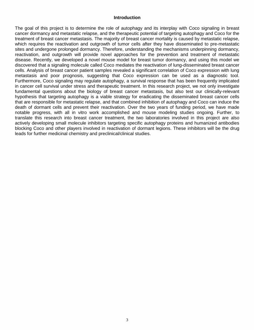
(1) Genetic engineer of multiple tool tumor cell lines. Up to now, we have accomplished the development of various breast cancer cell lines with defined Coco expression status that harbor GFP-LC3 (to monitor autophagy) and doxycycline (DOX)-inducible shRNA of autophagy genes (ULK1 and ATG7, as well as LacZ control). In these cells, we can manipulate autophagy in a DOX-induced manner. Further, we introduced triple modality reporter gene into these cells, enable live animal imaging of tumor cells in the in vivo experiments. These cells are currently used in xenograft experiments to examine the role of autophagy in Coco-mediated reactivation of dormant breast cancer in lung. Fig. 1 gives an example of such genetically engineered cell line in which autophagy can be controlled by DOX. In these experiments, autophagy was monitored by LC3-I/LC3-II conversion (Fig. 1A-C) and GFP-LC3 translocation (Fig. 1D).
(2) The potential interplay between TGFβ signaling and autophagy We examined the possibility that TGFβ signaling and its antagonist Coco are important regulators of autophagy in breast cancer cells (Tasks-2b, 2c). However, the effect of TGFβ signaling on nutrient starvation-induced autophagy is rather modest, if any, thus ruling out the possibility (data not shown). We will examine the opposite possibility: since pre-angiogenic stress triggers autophagy in the metastatic tumor cells, is autophagy able to modulate TGFβ signaling and thus reactivation of breast cancer
cells from dormancy? This new line of work is ongoing. (3) The role of autophagy in metastatic dormancy, reactivation
from dormancy, and outgrowth. This study is ongoing using the novel Coco-dictated xenograft mouse models and the inducible autophagy RNAi breast cancer cell lines we have developed.
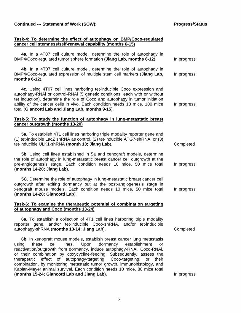
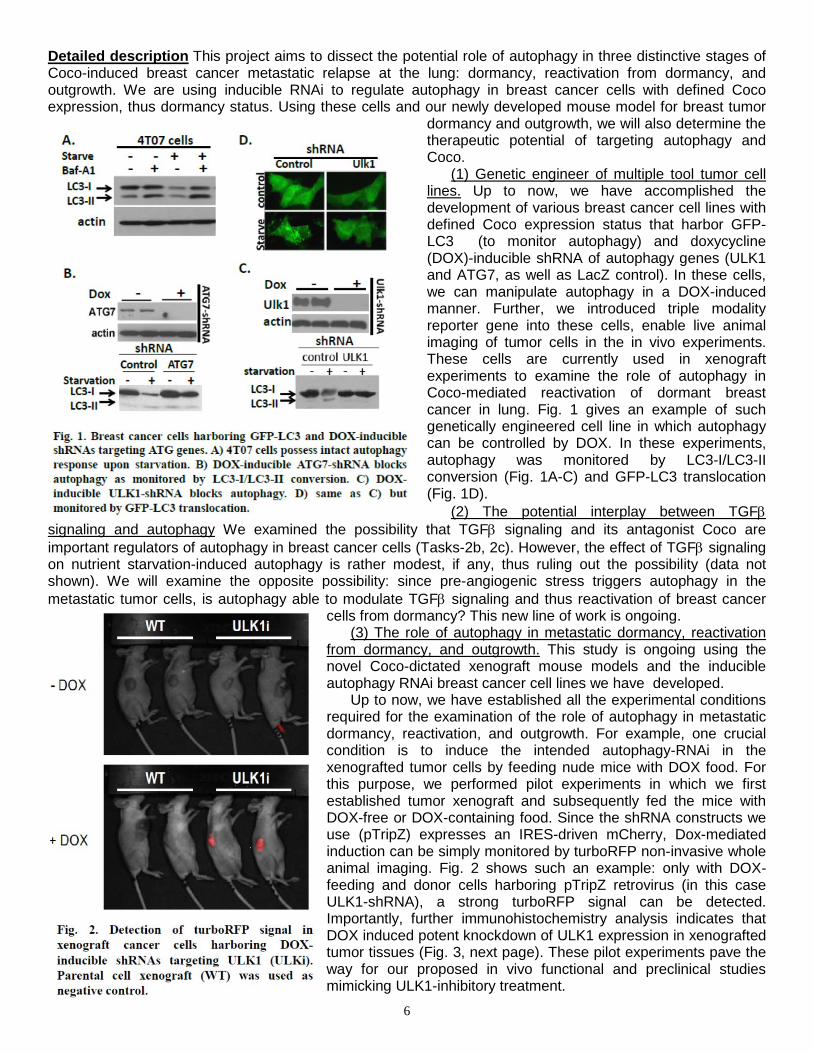
Up to now, we have established all the experimental conditions required for the examination of the role of autophagy in metastatic dormancy, reactivation, and outgrowth. For example, one crucial condition is to induce the intended autophagy-RNAi in the xenografted tumor cells by feeding nude mice with DOX food. For this purpose, we performed pilot experiments in which we first established tumor xenograft and subsequently fed the mice with DOX-free or DOX-containing food. Since the shRNA constructs we use (pTripZ) expresses an IRES-driven mCherry, Dox-mediated induction can be simply monitored by turboRFP non-invasive whole animal imaging. Fig. 2 shows such an example: only with DOX-feeding and donor cells harboring pTripZ retrovirus (in this case ULK1-shRNA), a strong turboRFP signal can be detected. Importantly, further immunohistochemistry analysis indicates that DOX induced potent knockdown of ULK1 expression in xenografted tumor tissues (Fig. 3, next page). These pilot experiments pave the way for our proposed in vivo functional and preclinical studies mimicking ULK1-inhibitory treatment.
7
(4) Monoclonal Antibodies Blocking Coco and their Preclinical Efficacy Because Coco promotes dormant breast cancer cell outgrowth from lung metastatic sites, it is a possible therapeutic target. As an extracellular factor, Coco can be targeted by specific monoclonal antibodies. For this reason, we developed monoclonal antibodies that block Coco’s binding to BMP proteins and evaluate their preclinical efficacy, as single agents or in combination with Lapatinib, in mouse models of ErbB2-mediated mammary tumorigenesis. Our preliminary results are exciting. We are aware that this direction is not in our original plan, but it is in line with our ultimate goal of developing Coco- and
autophagy-targeted therapies to treat lung-metastatic breast cancer. Therefore, below we describe the progress as well as the immediate future plan of this direction.
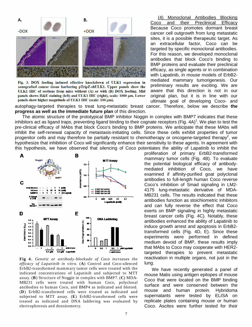
The atomic structure of the prototypical BMP inhibitor Noggin in complex with BMP7 indicates that these inhibitors act as ligand traps, preventing ligand binding to their cognate receptors (Fig. 4A)1. We plan to test the pre-clinical efficacy of MAbs that block Coco’s binding to BMP proteins. We anticipate that these MAbs will inhibit the self-renewal capacity of metastasis-initiating cells. Since these cells exhibit properties of tumor progenitor cells and may therefore be partially resistant to chemotherapy or oncogene-targeted therapy2, we hypothesize that inhibition of Coco will significantly enhance their sensitivity to these agents. In agreement with this hypothesis, we have observed that silencing of Coco potentiates the ability of Lapatinib to inhibit the
proliferation of primary ErbB2-transformed mammary tumor cells (Fig. 4B). To evaluate the potential biological efficacy of antibody-mediated inhibition of Coco, we have examined if affinity-purified goat polyclonal antibodies to full-length human Coco reverse Coco’s inhibition of Smad signaling in LM2-4175 lung-metastatic derivative of MDA-MB231 cells. The results indicated that these antibodies function as stoichiometric inhibitors and can fully reverse the effect that Coco exerts on BMP signaling in highly metastatic breast cancer cells (Fig. 4C). Notably, these antibodies enhanced the ability of Lapatinib to induce growth arrest and apoptosis in ErbB2-transformed cells (Fig. 4D, E). Since these experiments were performed in defined medium devoid of BMP, these results imply that MAbs to Coco may cooperate with HER2-targeted therapies to prevent metastatic reactivation in multiple organs, not just in the lung.
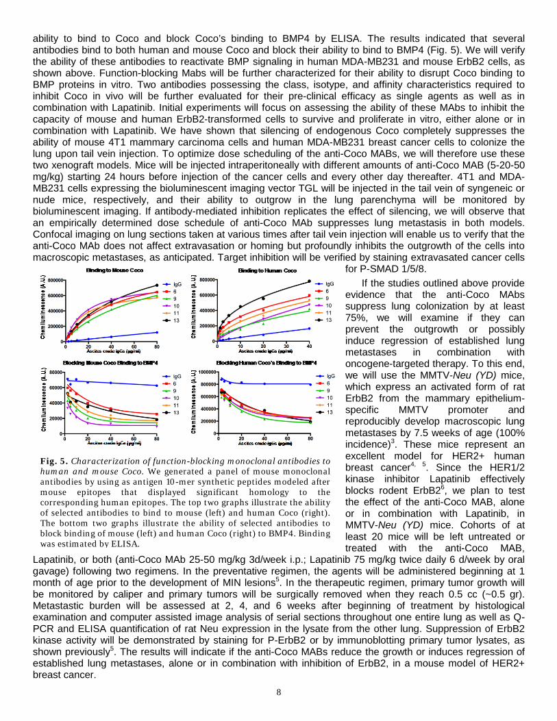
We have recently generated a panel of mouse Mabs using antigen epitopes of mouse Coco that were located on the BMP binding surface and were conserved between the mouse and human protein. Hybridoma supernatants were tested by ELISA on replicate plates containing mouse or human Coco. Ascites were further tested for their
Fig 4. Genetic or antibody-blockade of Coco increases the efficacy of Lapatinib in vitro. (A) Control and Coco-silenced ErbB2-transformed mammary tumor cells were treated with the indicated concentrations of Lapatinib and subjected to MTT assay. (B) Structure of Noggin in complex with BMP7. (C) MDA-MB231 cells were treated with human Coco, polyclonal antibodies to human Coco, and BMP4 as indicated and blotted. (D) ErbB2-transformed cells were treated as indicated and subjected to MTT assay. (E) ErbB2-transformed cells were treated as indicated and DNA laddering was evaluated by electrophoresis and densitometry.
8
ability to bind to Coco and block Coco’s binding to BMP4 by ELISA. The results indicated that several antibodies bind to both human and mouse Coco and block their ability to bind to BMP4 (Fig. 5). We will verify the ability of these antibodies to reactivate BMP signaling in human MDA-MB231 and mouse ErbB2 cells, as shown above. Function-blocking Mabs will be further characterized for their ability to disrupt Coco binding to BMP proteins in vitro. Two antibodies possessing the class, isotype, and affinity characteristics required to inhibit Coco in vivo will be further evaluated for their pre-clinical efficacy as single agents as well as in combination with Lapatinib. Initial experiments will focus on assessing the ability of these MAbs to inhibit the capacity of mouse and human ErbB2-transformed cells to survive and proliferate in vitro, either alone or in combination with Lapatinib. We have shown that silencing of endogenous Coco completely suppresses the ability of mouse 4T1 mammary carcinoma cells and human MDA-MB231 breast cancer cells to colonize the lung upon tail vein injection. To optimize dose scheduling of the anti-Coco MABs, we will therefore use these two xenograft models. Mice will be injected intraperitoneally with different amounts of anti-Coco MAB (5-20-50 mg/kg) starting 24 hours before injection of the cancer cells and every other day thereafter. 4T1 and MDA-MB231 cells expressing the bioluminescent imaging vector TGL will be injected in the tail vein of syngeneic or nude mice, respectively, and their ability to outgrow in the lung parenchyma will be monitored by bioluminescent imaging. If antibody-mediated inhibition replicates the effect of silencing, we will observe that an empirically determined dose schedule of anti-Coco MAb suppresses lung metastasis in both models. Confocal imaging on lung sections taken at various times after tail vein injection will enable us to verify that the anti-Coco MAb does not affect extravasation or homing but profoundly inhibits the outgrowth of the cells into macroscopic metastases, as anticipated. Target inhibition will be verified by staining extravasated cancer cells
for P-SMAD 1/5/8. If the studies outlined above provide
evidence that the anti-Coco MAbs suppress lung colonization by at least 75%, we will examine if they can prevent the outgrowth or possibly induce regression of established lung metastases in combination with oncogene-targeted therapy. To this end, we will use the MMTV-Neu (YD) mice, which express an activated form of rat ErbB2 from the mammary epithelium-specific MMTV promoter and reproducibly develop macroscopic lung metastases by 7.5 weeks of age (100% incidence)3. These mice represent an excellent model for HER2+ human breast cancer4, 5. Since the HER1/2 kinase inhibitor Lapatinib effectively blocks rodent ErbB26, we plan to test the effect of the anti-Coco MAB, alone or in combination with Lapatinib, in MMTV-Neu (YD) mice. Cohorts of at least 20 mice will be left untreated or treated with the anti-Coco MAB,
Lapatinib, or both (anti-Coco MAb 25-50 mg/kg 3d/week i.p.; Lapatinib 75 mg/kg twice daily 6 d/week by oral gavage) following two regimens. In the preventative regimen, the agents will be administered beginning at 1 month of age prior to the development of MIN lesions5. In the therapeutic regimen, primary tumor growth will be monitored by caliper and primary tumors will be surgically removed when they reach 0.5 cc (~0.5 gr). Metastastic burden will be assessed at 2, 4, and 6 weeks after beginning of treatment by histological examination and computer assisted image analysis of serial sections throughout one entire lung as well as Q-PCR and ELISA quantification of rat Neu expression in the lysate from the other lung. Suppression of ErbB2 kinase activity will be demonstrated by staining for P-ErbB2 or by immunoblotting primary tumor lysates, as shown previously5. The results will indicate if the anti-Coco MABs reduce the growth or induces regression of established lung metastases, alone or in combination with inhibition of ErbB2, in a mouse model of HER2+ breast cancer.
Fig. 5. Characterization of function-blocking monoclonal antibodies to human and mouse Coco. We generated a panel of mouse monoclonal antibodies by using as antigen 10-mer synthetic peptides modeled after mouse epitopes that displayed significant homology to the corresponding human epitopes. The top two graphs illustrate the ability of selected antibodies to bind to mouse (left) and human Coco (right). The bottom two graphs illustrate the ability of selected antibodies to block binding of mouse (left) and human Coco (right) to BMP4. Binding was estimated by ELISA.
9
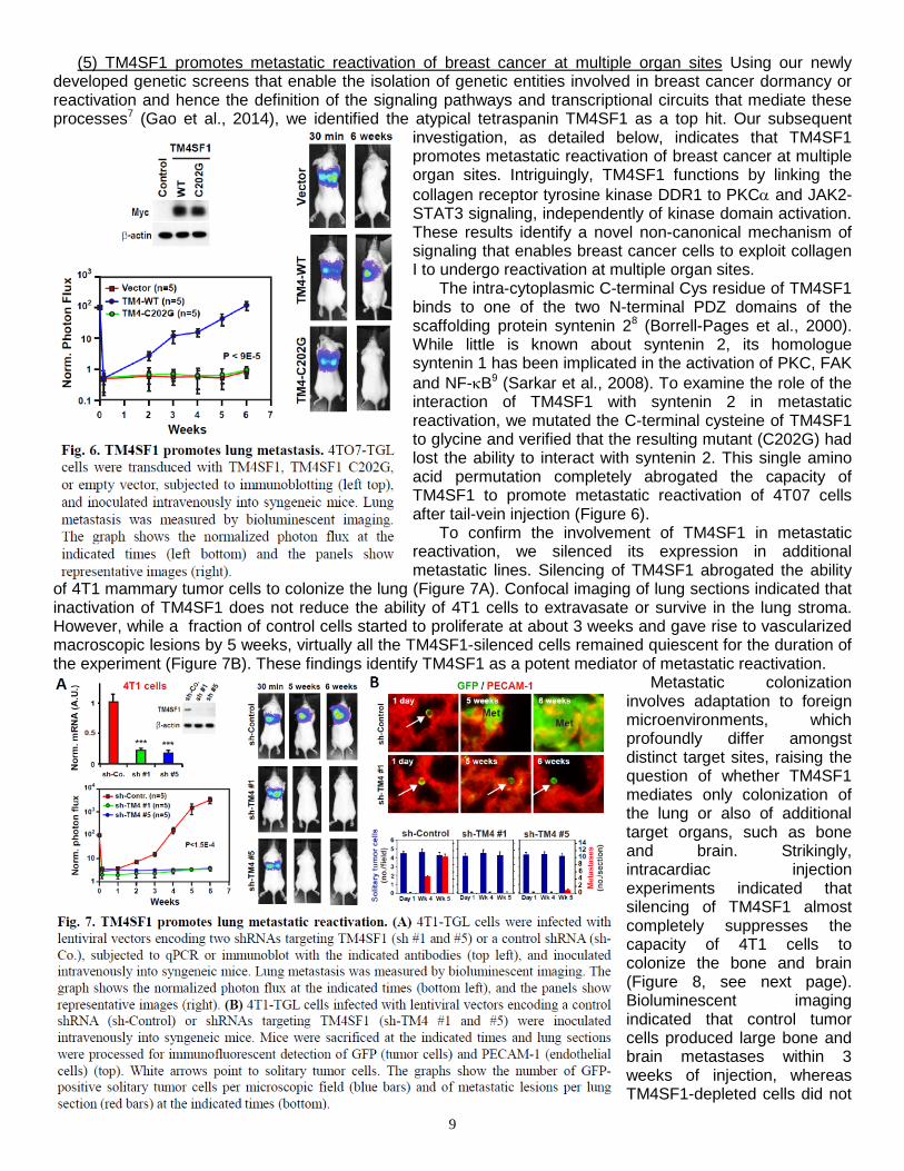
(5) TM4SF1 promotes metastatic reactivation of breast cancer at multiple organ sites Using our newly developed genetic screens that enable the isolation of genetic entities involved in breast cancer dormancy or reactivation and hence the definition of the signaling pathways and transcriptional circuits that mediate these processes7 (Gao et al., 2014), we identified the atypical tetraspanin TM4SF1 as a top hit. Our subsequent
investigation, as detailed below, indicates that TM4SF1 promotes metastatic reactivation of breast cancer at multiple organ sites. Intriguingly, TM4SF1 functions by linking the collagen receptor tyrosine kinase DDR1 to PKCα and JAK2-STAT3 signaling, independently of kinase domain activation. These results identify a novel non-canonical mechanism of signaling that enables breast cancer cells to exploit collagen I to undergo reactivation at multiple organ sites.
The intra-cytoplasmic C-terminal Cys residue of TM4SF1 binds to one of the two N-terminal PDZ domains of the scaffolding protein syntenin 28 (Borrell-Pages et al., 2000). While little is known about syntenin 2, its homologue syntenin 1 has been implicated in the activation of PKC, FAK and NF-κB9 (Sarkar et al., 2008). To examine the role of the interaction of TM4SF1 with syntenin 2 in metastatic reactivation, we mutated the C-terminal cysteine of TM4SF1 to glycine and verified that the resulting mutant (C202G) had lost the ability to interact with syntenin 2. This single amino acid permutation completely abrogated the capacity of TM4SF1 to promote metastatic reactivation of 4T07 cells after tail-vein injection (Figure 6).
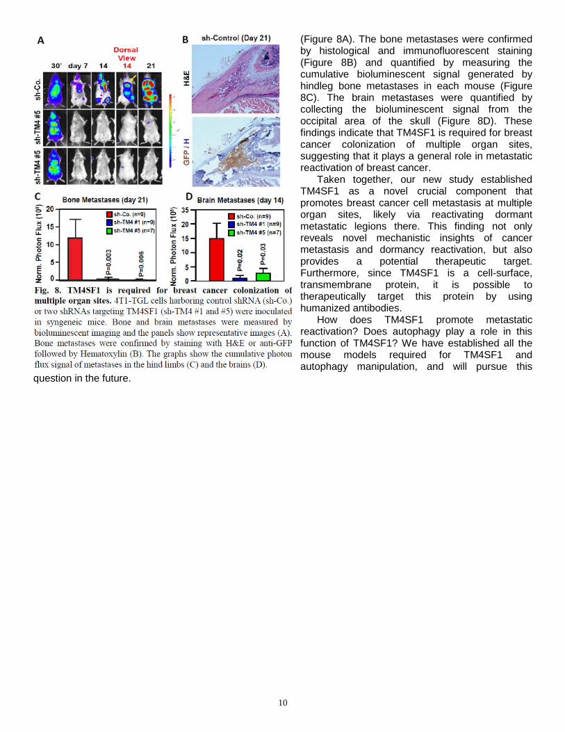
To confirm the involvement of TM4SF1 in metastatic reactivation, we silenced its expression in additional metastatic lines. Silencing of TM4SF1 abrogated the ability
of 4T1 mammary tumor cells to colonize the lung (Figure 7A). Confocal imaging of lung sections indicated that inactivation of TM4SF1 does not reduce the ability of 4T1 cells to extravasate or survive in the lung stroma. However, while a fraction of control cells started to proliferate at about 3 weeks and gave rise to vascularized macroscopic lesions by 5 weeks, virtually all the TM4SF1-silenced cells remained quiescent for the duration of the experiment (Figure 7B). These findings identify TM4SF1 as a potent mediator of metastatic reactivation.
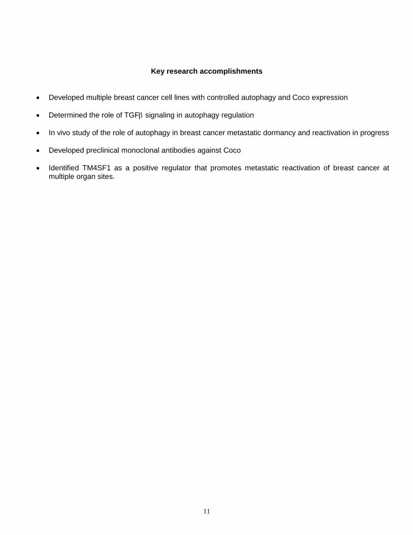
Metastatic colonization involves adaptation to foreign microenvironments, which profoundly differ amongst distinct target sites, raising the question of whether TM4SF1 mediates only colonization of the lung or also of additional target organs, such as bone and brain. Strikingly, intracardiac injection experiments indicated that silencing of TM4SF1 almost completely suppresses the capacity of 4T1 cells to colonize the bone and brain (Figure 8, see next page). Bioluminescent imaging indicated that control tumor cells produced large bone and brain metastases within 3 weeks of injection, whereas TM4SF1-depleted cells did not
10
(Figure 8A). The bone metastases were confirmed by histological and immunofluorescent staining (Figure 8B) and quantified by measuring the cumulative bioluminescent signal generated by hindleg bone metastases in each mouse (Figure 8C). The brain metastases were quantified by collecting the bioluminescent signal from the occipital area of the skull (Figure 8D). These findings indicate that TM4SF1 is required for breast cancer colonization of multiple organ sites, suggesting that it plays a general role in metastatic reactivation of breast cancer.
Taken together, our new study established TM4SF1 as a novel crucial component that promotes breast cancer cell metastasis at multiple organ sites, likely via reactivating dormant metastatic legions there. This finding not only reveals novel mechanistic insights of cancer metastasis and dormancy reactivation, but also provides a potential therapeutic target. Furthermore, since TM4SF1 is a cell-surface, transmembrane protein, it is possible to therapeutically target this protein by using humanized antibodies.
How does TM4SF1 promote metastatic reactivation? Does autophagy play a role in this function of TM4SF1? We have established all the mouse models required for TM4SF1 and autophagy manipulation, and will pursue this
question in the future.
11
Key research accomplishments
• Developed multiple breast cancer cell lines with controlled autophagy and Coco expression
• Determined the role of TGFβ signaling in autophagy regulation
• In vivo study of the role of autophagy in breast cancer metastatic dormancy and reactivation in progress
• Developed preclinical monoclonal antibodies against Coco
• Identified TM4SF1 as a positive regulator that promotes metastatic reactivation of breast cancer atmultiple organ sites.
12
Reportable Outcomes
• Developed multiple breast cancer cell lines with controlled autophagy and Coco expression
• Developed preclinical monoclonal antibodies against Coco
• One postdoctoral fellow involved in this project have obtained a faculty job
• One graduate student involved in this project is planning thesis defense
• Multiple research seminars presenting work partially supported by this grant
13
Conclusion
Metastatic relapse requires reactivation of the disseminated tumor cells after they survive prolonged dormancy. We posit that the survival of metastatic cells, both during dormancy and upon Coco-induced reactivation, requires regulated autophagy. Our experiments will put these potential mechanisms under test and thereby answer a fundamental question about the biology of breast cancer metastasis. Validation of our hypothesis has significant therapeutic impact: it will indicate that targeting autophagy is a viable strategy for eradicating the disseminated breast cancer cells that are responsible for metastatic relapse; and combined inhibition of autophagy and Coco could induce the death of dormant cells and prevent their reactivation.
The experiments are going smoothly as planned, with various genetically controllable breast cancer cells already developed as critical research tools and subsequent xenograft mouse modeling experiments ongoing to determine the role of autophagy and Coco (and thus therapies targeting them) in metastatic breast cancer dormancy and reactivation. In addition to these cell culture and animal model studies as originally planned, we have also (1) developed pre-clinical anti-Coco monoclonal antibodies, for the ultimate goal of the development of a novel Coco-targeted therapeutic product for breast cancer treatment; and (2) identified a new factor, TM4SF1, which is crucial for metastatic reactivation of breast cancer cells at multiple target organ sites. TM4SF1 is also a potential therapeutic target for the treatment of metastatic breast cancer.
14
REFERENCES
1. Groppe, J. et al. Structural basis of BMP signalling inhibition by the cystine knot protein Noggin. Nature420, 636-642 (2002).
2. Pardal, R., Clarke, M.F. & Morrison, S.J. Applying the principles of stem-cell biology to cancer. Naturereviews. Cancer 3, 895-902 (2003).
3. Dankort, D. et al. Grb2 and Shc adapter proteins play distinct roles in Neu (ErbB-2)-induced mammarytumorigenesis: implications for human breast cancer. Molecular and cellular biology 21, 1540-1551(2001).
4. Siegel, P.M., Shu, W., Cardiff, R.D., Muller, W.J. & Massague, J. Transforming growth factor betasignaling impairs Neu-induced mammary tumorigenesis while promoting pulmonary metastasis.Proceedings of the National Academy of Sciences of the United States of America 100, 8430-8435(2003).
5. Guo, W. et al. Beta 4 integrin amplifies ErbB2 signaling to promote mammary tumorigenesis. Cell 126,489-502 (2006).
6. Strecker, T.E. et al. Effect of lapatinib on the development of estrogen receptor-negative mammarytumors in mice. Journal of the National Cancer Institute 101, 107-113 (2009).
7. Borrell-Pages, M., Fernandez-Larrea, J., Borroto, A., Rojo, F., Baselga, J., and Arribas, J. (2000). Thecarboxy-terminal cysteine of the tetraspanin L6 antigen is required for its interaction with SITAC, anovel PDZ protein. Molecular biology of the cell 11, 4217-4225.
8. Gao, H., Chakraborty, G., Lee-Lim, A. P., Mavrakis, K. J., Wendel, H. G., and Giancotti, F. G. (2014).Forward genetic screens in mice uncover mediators and suppressors of metastatic reactivation.Proceedings of the National Academy of Sciences of the United States of America 111, 16532-16537.
9. Sarkar, D., Boukerche, H., Su, Z. Z., and Fisher, P. B. (2008). mda-9/Syntenin: more than just a simpleadapter protein when it comes to cancer metastasis. Cancer research 68, 3087-3093.