Automated flow-based anion-exchange method for high-throughput isolation and real-time monitoring of...
8
Talanta 84 (2011) 1259–1266 Contents lists available at ScienceDirect Talanta journal homepage: www.elsevier.com/locate/talanta Automated flow-based anion-exchange method for high-throughput isolation and real-time monitoring of RuBisCO in plant extracts Ruth Suárez a , Manuel Miró a,∗ , Víctor Cerdà a , Juan Alejandro Perdomo b , Jeroni Galmés b a Department of Chemistry, University of the Balearic Islands, Carretera de Valldemossa km 7.5, E-07122 Palma de Mallorca, Illes Balears, Spain b Grup de Biologia de les Plantes en Condicions Mediterrànies, Department of Biology, University of the Balearic Islands, Carretera de Valldemossa km 7.5, E-07122 Palma de Mallorca, Illes Balears, Spain article info Article history: Available online 19 January 2011 Keywords: Anion-exchange separation Multi-syringe flow analysis Plant extracts RuBisCO abstract In this work, a miniaturized, completely enclosed multisyringe-flow system is proposed for high- throughput purification of RuBisCO from Triticum aestivum extracts. The automated method capitalizes on the uptake of the target protein at 4 ◦ C onto Q-Sepharose Fast Flow strong anion-exchanger packed in a cylindrical microcolumn (105 × 4 mm) followed by a stepwise ionic-strength gradient elution (0–0.8 mol/L NaCl) to eliminate concomitant extract components and retrieve highly purified RuBisCO. The manifold is furnished downstream with a flow-through diode-array UV/vis spectrophotometer for real-time monitoring of the column effluent at the protein-specific wavelength of 280 nm to detect the elution of RuBisCO. Quantitation of RuBisCO and total soluble proteins in the eluate fractions were undertaken using polyacrylamide gel electrophoresis (PAGE) and the spectrophotometric Bradford assay, respectively. A comprehensive investigation of the effect of distinct concentration gradients on the isolation of RuBisCO and experimental conditions (namely, type of resin, column dimensions and mobile- phase flow rate) upon column capacity and analyte breakthrough was effected. The assembled set-up was aimed to critically ascertain the efficiency of preliminary batchwise pre-treatments of crude plant extracts (viz., polyethylenglycol (PEG) precipitation, ammonium sulphate precipitation and sucrose gradient cen- trifugation) in terms of RuBisCO purification and absolute recovery prior to automated anion-exchange column separation. Under the optimum physical and chemical conditions, the flow-through column sys- tem is able to admit crude plant extracts and gives rise to RuBisCO purification yields better than 75%, which might be increased up to 96 ± 9% with a prior PEG fractionation followed by sucrose gradient step. © 2011 Elsevier B.V. All rights reserved. 1. Introduction Ribulose-1,5-bisphosphate carboxylase/oxygenase (RuBisCO) is the most abundant protein in the biosphere constituting up to half of the soluble protein in plant leaves [1]. RuBisCO catalyses the fix- ation of atmospheric CO 2 to ribulose-1,5-bisphosphate to yield two Abbreviations: Bicine, N,N-bis(2-hydroxyethyl)glycine; BSA, bovine serum albu- min; CC, communication channel; DIECA, diethyldithiocarbamate; DLLs, dynamic link libraries; DTT, dithiothreitol; EDTA, ethylenediaminetetraacetic acid; FC, fraction collector; FI, flow injection; HC, holding coil; LSU, large subunits of RuBisCO; MPV, multiposition selection valve; MSFI, multisyringe flow injection; MSP, multisyringe piston pump; P, purification yields; PAGE, polyacrylamide gel electrophoresis; PEG, polyethylenglycol; PMMA, poly(methyl methacrylate); PMSF, phenylmethylsulphonyl fluoride; PS-DVB, poly(styrenedivinylbenzene); PTFE, poly(tetrafluoroethylene); PVC, poly (vinyl chloride); PVP, poly(vinylpyrrolidine); R, absolute recoveries; RuBisCO, ribulose-1,5-bisphosphate carboxylase/oxygenase; SDS, sodium dodecylsulphate; SI, sequential injection; SSU, small subunits of RuBisCO; Tris, tris(hydroxymethyl)aminomethane. ∗ Corresponding author. Tel.: +34 971172746; fax: +34 971173426. E-mail address: [email protected] (M. Miró). molecules of 3-phosphoglycerate. By doing this function, RuBisCO constitutes the unique conversion point of inorganic to organic car- bon and its activity amounts to more than 10 11 tons of CO 2 per year [1]. In spite of its biological importance, RuBisCO presents a num- ber of catalytic inefficiencies, the most important of which is its failure to distinguish between CO 2 and O 2 . In addition, RuBisCO is a notoriously slow enzyme [2], which obligates plants to divert huge amounts of nitrogen to RuBisCO in order to achieve acceptable rates of photosynthesis. The low affinity for CO 2 and the production of miss-products during catalysis are other well known limitations of the enzyme [3]. All these inefficiencies greatly limit the photosyn- thetic capacity of plants, decreasing the efficiencies by which water and nitrogen are used in agriculture. It is therefore not surprising that RuBisCO has been historically targeted as a major gateway to increase plant productivity, and therefore food and energy produc- tion [4,5]. The expectative to improve the catalytic performance of RuBisCO has been enhanced by both the discovery of remarkably different versions of the enzyme among higher plants [6,7] and the progress in the production of transplantomic lines where the native RuBisCO is replaced by foreign forms [8,9]. Yet, to sustain the pos- 0039-9140/$ – see front matter © 2011 Elsevier B.V. All rights reserved. doi:10.1016/j.talanta.2011.01.024
-
Upload
ruth-suarez -
Category
Documents
-
view
215 -
download
2
Transcript of Automated flow-based anion-exchange method for high-throughput isolation and real-time monitoring of...

Aa
Ra
b
E
a
AA
KAMPR
1
toa
mlfRMeppRSR
0d
Talanta 84 (2011) 1259–1266
Contents lists available at ScienceDirect
Talanta
journa l homepage: www.e lsev ier .com/ locate / ta lanta
utomated flow-based anion-exchange method for high-throughput isolationnd real-time monitoring of RuBisCO in plant extracts
uth Suáreza, Manuel Miróa,∗, Víctor Cerdàa, Juan Alejandro Perdomob, Jeroni Galmésb
Department of Chemistry, University of the Balearic Islands, Carretera de Valldemossa km 7.5, E-07122 Palma de Mallorca, Illes Balears, SpainGrup de Biologia de les Plantes en Condicions Mediterrànies, Department of Biology, University of the Balearic Islands, Carretera de Valldemossa km 7.5,-07122 Palma de Mallorca, Illes Balears, Spain
r t i c l e i n f o
rticle history:vailable online 19 January 2011
eywords:nion-exchange separationulti-syringe flow analysis
lant extractsuBisCO
a b s t r a c t
In this work, a miniaturized, completely enclosed multisyringe-flow system is proposed for high-throughput purification of RuBisCO from Triticum aestivum extracts. The automated method capitalizeson the uptake of the target protein at 4 ◦C onto Q-Sepharose Fast Flow strong anion-exchanger packedin a cylindrical microcolumn (105 × 4 mm) followed by a stepwise ionic-strength gradient elution(0–0.8 mol/L NaCl) to eliminate concomitant extract components and retrieve highly purified RuBisCO.The manifold is furnished downstream with a flow-through diode-array UV/vis spectrophotometer forreal-time monitoring of the column effluent at the protein-specific wavelength of 280 nm to detectthe elution of RuBisCO. Quantitation of RuBisCO and total soluble proteins in the eluate fractions wereundertaken using polyacrylamide gel electrophoresis (PAGE) and the spectrophotometric Bradford assay,respectively. A comprehensive investigation of the effect of distinct concentration gradients on theisolation of RuBisCO and experimental conditions (namely, type of resin, column dimensions and mobile-phase flow rate) upon column capacity and analyte breakthrough was effected. The assembled set-up was
aimed to critically ascertain the efficiency of preliminary batchwise pre-treatments of crude plant extracts(viz., polyethylenglycol (PEG) precipitation, ammonium sulphate precipitation and sucrose gradient cen-trifugation) in terms of RuBisCO purification and absolute recovery prior to automated anion-exchangecolumn separation. Under the optimum physical and chemical conditions, the flow-through column sys-tem is able to admit crude plant extracts and gives rise to RuBisCO purification yields better than 75%,up to
which might be increased. Introduction
Ribulose-1,5-bisphosphate carboxylase/oxygenase (RuBisCO) ishe most abundant protein in the biosphere constituting up to halff the soluble protein in plant leaves [1]. RuBisCO catalyses the fix-tion of atmospheric CO2 to ribulose-1,5-bisphosphate to yield two
Abbreviations: Bicine, N,N-bis(2-hydroxyethyl)glycine; BSA, bovine serum albu-in; CC, communication channel; DIECA, diethyldithiocarbamate; DLLs, dynamic
ink libraries; DTT, dithiothreitol; EDTA, ethylenediaminetetraacetic acid; FC,raction collector; FI, flow injection; HC, holding coil; LSU, large subunits ofuBisCO; MPV, multiposition selection valve; MSFI, multisyringe flow injection;SP, multisyringe piston pump; P, purification yields; PAGE, polyacrylamide gel
lectrophoresis; PEG, polyethylenglycol; PMMA, poly(methyl methacrylate); PMSF,henylmethylsulphonyl fluoride; PS-DVB, poly(styrenedivinylbenzene); PTFE,oly(tetrafluoroethylene); PVC, poly (vinyl chloride); PVP, poly(vinylpyrrolidine);, absolute recoveries; RuBisCO, ribulose-1,5-bisphosphate carboxylase/oxygenase;DS, sodium dodecylsulphate; SI, sequential injection; SSU, small subunits ofuBisCO; Tris, tris(hydroxymethyl)aminomethane.∗ Corresponding author. Tel.: +34 971172746; fax: +34 971173426.
E-mail address: [email protected] (M. Miró).
039-9140/$ – see front matter © 2011 Elsevier B.V. All rights reserved.oi:10.1016/j.talanta.2011.01.024
96 ± 9% with a prior PEG fractionation followed by sucrose gradient step.
© 2011 Elsevier B.V. All rights reserved.
molecules of 3-phosphoglycerate. By doing this function, RuBisCOconstitutes the unique conversion point of inorganic to organic car-bon and its activity amounts to more than 1011 tons of CO2 per year[1]. In spite of its biological importance, RuBisCO presents a num-ber of catalytic inefficiencies, the most important of which is itsfailure to distinguish between CO2 and O2. In addition, RuBisCO is anotoriously slow enzyme [2], which obligates plants to divert hugeamounts of nitrogen to RuBisCO in order to achieve acceptable ratesof photosynthesis. The low affinity for CO2 and the production ofmiss-products during catalysis are other well known limitations ofthe enzyme [3]. All these inefficiencies greatly limit the photosyn-thetic capacity of plants, decreasing the efficiencies by which waterand nitrogen are used in agriculture. It is therefore not surprisingthat RuBisCO has been historically targeted as a major gateway toincrease plant productivity, and therefore food and energy produc-
tion [4,5]. The expectative to improve the catalytic performance ofRuBisCO has been enhanced by both the discovery of remarkablydifferent versions of the enzyme among higher plants [6,7] and theprogress in the production of transplantomic lines where the nativeRuBisCO is replaced by foreign forms [8,9]. Yet, to sustain the pos-
1 nta 84
skenpoap
adsfocpaidaoicdy
ecetbcwcspoesssa
tho[dmfrtre
2
2
scgt
260 R. Suárez et al. / Tala
ibilities for an improved RuBisCO, it is necessary to gain improvednowledge upon the structure and regulatory processes of thenzyme and to deeply explore the variability of the kinetic traits inature [5,10]. Increasing the knowledge on the enzyme would alsootentially benefit other related subjects, namely, improvementf plant responses to raising atmospheric CO2 concentration [11],nd open new prospects for artificial photosynthesis and biofuelroduction [12].
RuBisCO needs to be purified from extracts for appropri-te biochemical characterization. The degree of required purityepends on the specific analysis, with crystallization and mea-urements of some kinetic constants, such as the specificityactor, demanding high degrees of purification [7,13]. A surveyf the literature revealed that a number of purification proto-ols and variants thereof for RuBisCO purification from severallant sources have been reported including fractionation withmmonium sulphate [14–16] or polyethylenglycol (PEG) [17–19],mmunoprecipitation and immunoadsorption [20], sucrose gra-ient centrifugation [21,22] size-exclusion or dialysis [8,23],nion-exchange chromatography [7,8,16,24,25] and a plethoraf combinations thereof [8,16,18,22,23,25]. However, the exper-mental results and conclusions drawn are in several instancesontroversial and debatable, and no universal analytical proce-ure for isolation of RuBisCO from plant sources is available as ofet.
This work is aimed at the development of an automated,ntirely enclosed multisyringe flow injection (MSFI)-based analyti-al method for high-throughput purification of RuBisCO from plantxtracts. The flow network capitalizes on the uptake of the nega-ively charged protein at pH ≥ 8.0 and 4 ◦C onto anion-exchangeeads followed by stepwise ionic-strength elution as preciselyontrolled by flow programming. The flow manifold is equippedith a flow-through diode-array spectrophotometer and fraction
ollector for real-time monitoring of column effluent at the protein-pecific wavelength of 280 nm and automated retrieval of highlyurified RuBisCO fractions, respectively. The inherent versatilityf the assembled flow device is exploited upon optimization tondorse a simplified automated procedure capable of admittingample extracts regardless of the preceding sample processingteps (direct crude extracts, precipitation with PEG or ammoniumulphate or sucrose gradient centrifugation), and of giving rise tocceptable recoveries and purification yields of RuBisCO.
The first two generations of flow analysis, that is, flow injec-ion (FI) and sequential injection (SI), and microfluidic devicesave drawn much attention as platforms for automated assaysf soluble proteins [26] and individual determinations of albumin27–29] and/or creatinine [30,31] by resorting to highly sensitiveye-binding spectrophotometric assays (e.g., Eosin Y or tetrabro-ophenolphthalein ethyl ester for albumin and Jaffe’s reaction
or creatinine), beside the exploitation of SI-affinity chromatog-aphy for monitoring the binding of drugs on albumin [30]. Tohe best of our knowledge, however, no FI or SI method has beeneported so far for retrieval and quantitation of RuBisCO in plantxtracts.
. Experimental
.1. Plant material
Wheat (Triticum aestivum L., T. aestivum in the following) waselected as has been largely used as a model plant for RuBisCOharacterization [7,32]. Wheat seeds were germinated and plantsrown under controlled conditions (20–25 ◦C night–day tempera-ures, 16 h photoperiod and 700 �mol photons m−2 s−1).
(2011) 1259–1266
2.2. Reagents and solutions
All chemicals and reagents used in this work were of analyt-ical reagent grade and used as purchased. A stock standard ofhighly purified RuBisCO from T. aestivum for method develop-ment was obtained according to the batchwise purification protocolby Galmés et al. [7]. The high purity of the stock RuBisCO fromT. aestivum was corroborated by sodium dodecylsulphate (SDS)-polyacrylamide gel electrophoresis (PAGE), revealing only the twobands corresponding to the large (LSU) and small subunits (SSU)of RuBisCO. Doubly de-ionised water (resistivity = 18.2 M�·cm)obtained from a Milli-Q system (Milllipore Synthesis A10, MilliporeCorporation, Billerica, MA, USA) was used throughout.
Q-Sepharose Fast Flow (GE Healthcare, Bio-Sciences AB,Sweden), with a particle size range of 40–165 �m, was used in theflow network as strong anion exchanger, with no need for any addi-tional swelling protocol. Q-Sepharose Fast Flow is composed of ahighly cross-linked, bead-formed 6% agarose matrix furnished withdiethyl-(2-hydroxypropyl)aminoethyl as functional moiety.
A tris(hydroxymethyl)aminomethane (Tris) column buffer at pH8.0, used as a carrier solution as well, contained 10 mmol/L Tris–HCl(acid salt) + NaOH, 10 mmol/L MgCl2, 10 mmol/L NaHCO3, 1 mmol/LNa2EDTA, 1 mmol/L KH2PO4. MgCl2, NaHCO3 and KH2PO4 main-tained RuBisCO activated [33,34], whilst the role of EDTA was todecrease the activity of proteases by chelation of transition metals[35].
2.3. Extraction and purification of RuBisCO
Three different RuBisCO purification procedures involving PEGprecipitation, ammonium sulphate precipitation and sucrose gra-dient separation besides direct analysis of crude extracts have beenevaluated prior to anion-exchange chromatography in terms ofRuBisCO yield and purity, and sample throughput as well. Theanalytical procedures for extraction and preliminary processing ofplant extracts performed in triplicate are summarized below andillustrated in Fig. 1.
2.3.1. Preparation of crude extractAll the extraction and purification steps were carried out
at 4 ◦C. 60 g of leaf material was collected under illuminatedconditions, ground to a powder in a mortar and imme-diately extracted with 250 mL of protein extraction buffer(pH 8.2) composed of 100 mmol/L N,N-Bis(2-hydroxyethyl)glycine(bicine), 6% (w/v) PEG-4000, 2 mmol/L MgCl2, 1 mmol/L Na2EDTA,10 mmol/L NaHCO3, 10 mmol/L sodium diethyldithiocarbamate(Na-DIECA), 1 mmol/L benzamidine, 1 mmol/L �-amino-n-caproicacid, 50 mmol/L 2-mercaptoethanol, 10 mmol/L dithiothreitol(DTT), 2 mmol/L phenylmethylsulphonyl fluoride (PMSF) and 3%(w/v) poly(vinylpyrrolidine) (PVP) as per the procedure reported byGalmés et al. [7]. DTT and 2-mercaptoethanol worked as reducingagents, PVP was used for precipitation of polyphenolic compoundsand benzamidine, �-amino-n-caproic acid, Na-DIECA and PMSFwere used as protease inhibitors. The plant extract was filteredthrough 2 layers of butter muslin and then centrifuged at 22,100 × gfor 20 min. The supernatant liquid was sieved through 50 �m meshnylon and injected into the MSFI-anion exchange separation systemwithout further purification.
2.3.2. Purification based on selective precipitation using PEGIn this protocol, the crude extract resulting from the abovemen-
tioned sample treatment was subjected to a selective precipitationwith PEG. It should be noted that the PEG concentration should bethoroughly selected for selective precipitation of macromoleculeson the basis of molecular weight. To this end, 60% (w/v) PEG-4000was added to the supernatant to give rise to a final concentration of

R. Suárez et al. / Talanta 84 (2011) 1259–1266 1261
igated
2agacmb2
2
tls5tttse
2a
fwrthf2wbA2cmwac
2
Muub
Fig. 1. Schematic diagram of the analytical procedures invest
0% (w/v) for RuBisCO precipitation. In addition, 1 mol/L MgCl2 wasdded dropwise to a final concentration of 20 mmol/L followed byentle mixing for 10 min. Thereafter, the mixture was centrifugedt 22,100 × g for 20 min. The pellet was resuspended in 10 mL ofolumn buffer (pH 8.0) containing 1 mmol/L each of PMSF, benza-idine and �-amino-n-caproic acid. The suspension was clarified
y centrifugation to remove insoluble material at 48,400 × g for0 min, prior to anion-exchange separation.
.3.3. Purification based on sucrose gradientThis protocol is exploited as a further extension of PEG precipi-
ation. A metered volume of supernatant liquid, namely, 6 mL, wasayered onto a linear-step density gradient from 0.2 to 1.2 mol/Lucrose of 2.5 mL each, in 0.1 mol/L bicine, 20 mmol/L MgCl2,0 mmol/L 2-mercaptoethanol and 11 mmol/L Na-DIECA and cen-rifuged at 103,900 × g for 14 h at 4 ◦C. Each 2.5-mL fraction ofhe sucrose gradient was subjected to Bradford assay (see below)o detect the layers containing the highest concentration of totaloluble protein, which were pooled prior loading onto the anion-xchange column.
.3.4. Purification based on selective precipitation usingmmonium sulphate
Sample processing using ammonium sulphate as a molecularractionation reagent was performed following RuBisCO extractionith bicine buffer. The supernatant was made up to 35% satu-
ated (at 0 ◦C) with respect to ammonium sulphate, correspondingo 16.4% (w/v), for selective precipitation of macromolecules ofigher molecular size than RuBisCO. The mixture was gentle stirred
or 20 min whereupon was again clarified by centrifugation at2,100 × g for 20 min. The resulting supernatant was further treatedith ammonium sulphate to collect the material precipitating
etween 35 and 55% saturation, corresponding to 16.4–25.7% (w/v).fter 20 min, the suspension was again centrifuged at 22,100 × g for0 min. The pellet containing RuBisCO was resuspended in 10 mL ofolumn buffer (pH 8.0) containing 1 mmol/L each of PMSF, benza-idine and �-amino acid-n-caproic. Removal of insoluble materialas accomplished by centrifugation at 48,400 × g for 20 min, to
fford a supernatant solution ready for injection into the MSFI-hromatographic setup.
.4. Automatic flow setup
A multisyringe piston pump with programmable speed (MSP,icroBu 2030, Crison Instruments, Alella, Barcelona, Spain) was
sed as a liquid driver for automation of the anion-exchange col-mn separation method. It was equipped with four high-precisionidirectional syringes (Hamilton, Bonaduz, Switzerland), S1–S4,
for RuBisCO purification. MSFI: multi-syringe flow injection.
with a capacity of 2.5 mL each and connected in block to a 16,000-step motor. A schematic diagram of the MSFI setup is illustratedin Fig. 2. S1, S2, S3 and S4 contained column buffer, 0.1 mol/LNaCl; 0.17 mol/L NaCl; and 0.8 mol/L NaCl, respectively, for step-wise ionic-strength gradient elution. A three-way solenoid valve(N-Research, Caldwell, NJ) was placed at the head of each syringeenabling automatic connection with either the liquid reservoirs(OFF) or the flow network (ON). The MSP module was coupled toan 8-port multiposition selection valve (MPV, Crison) operating assample-processing unit. This valve encompasses a central port anda communication channel (CC) that can be programmed to addresseach of the peripheral ports. It was connected via a 2.0 mL-holdingcoil (HC, 1.5 mm i.d. PTFE tubing) to S1 for fluidic handling of sam-ple and solutions for column rinsing and regeneration. The flowmanifold was constructed from PTFE tubing (0.5 mm i.d.) usingpoly(vinyl chloride) (PVC) or poly(methyl methacrylate) (PMMA)fittings.
A PMMA cylindrical column (105 × 4 mm) packed with194 ± 4 mg Q-Sepharose Fast Flow (void volume of 300 �L) wasexploited for in-line anion-exchange separation of RuBisCO. Glasswool was used for bead trapping in lieu of porous frits becauseof decrease of pressure drop. The outlet of the column was cou-pled via a 12-cm long PTFE tubing (0.5 mm i.d.) to a flow-throughdiode-array spectrophotometer (Hewlett-Packard HP8452A, Van-couver, WA, USA) equipped with a quartz cell (18 �L, 10 mm pathlength) for real-time monitoring of column effluent and recordingof the fiagram at the protein selective wavelength of 280 nm using320 nm as a reference wavelength. Peak area was used as analyti-cal signal for quantitation of eluted RuBisCO. Automated collectionof protein containing fractions was accomplished by resorting toa 45-position rack fraction collector (FC, Crison). The entire flowsetup (namely, MSP, MSV, column and FC) was refrigerated at 4 ◦C(Marecos Olitrem refrigerator, Tremês, Portugal) for undertakingRuBisCO purification at 4 ◦C.
Instrumental control, data acquisition, and processing of thechromatographic readouts were accomplished by means of thesoftware package AutoAnalysis 5.0 (Sciware, Palma de Mallorca,Spain) [36]. The software based on dynamic link libraries (DLLs) iscomposed of a main protocol wherein the DLLs of MSP, MPV, FCand detector are in this work attached.
2.5. Analytical procedure
The MSFI-based anion-exchange separation procedure was uti-lized for further purification of the extracts resulting from thepreliminary sample processing protocols detailed above. The entireprocedure was monitored at real-time by in-line recording of spec-tra within the wavelength range of 190–500 nm and the fiagram at

1262 R. Suárez et al. / Talanta 84 (2011) 1259–1266
F ationc
2eps
ow1(nb5ts2w0te
TA
MA
ig. 2. Schematic illustration of the miniaturised flow-based anion-exchange separhannel; S: Syringe; Saline solutions: 0.1 M NaCl, 0.17 M NaCl, and 0.8 M NaCl.
80 nm against 320 nm all at 1 Hz. The overall analytical sequencencompassing the volumes and flow rates of handled solutions andositions of MPV and FC is compiled in Table 1 (shown for PEG anducrose gradient procedures) and summarized as follows:
The anion exchanger was first conditioned by dispensing 2.5 mLf column buffer at 1.0 mL/min, followed by perfusing the columnith 500 �L of RuBisCO standard or plant extract containing ca.
000 �g of soluble protein as determined by Bradford’s methodsee below) previously loaded into the holding coil. Removal ofon-retained matrix ingredients or components of the extractionuffer was accomplished by flushing the anion exchanger with.0 mL of column buffer at 1.0 mL/min. The stepwise gradient elu-ion protocol of increasing ionic strength involves two preliminaryteps, namely, the delivery of 2.0 mL of 0.1 mol/L NaCl followed by
.0 mL of 0.17 mol/L NaCl both at 1.0 mL/min, in order to wash awayeakly retained pigments and proteins prior to RuBisCO elution at.4 mol/L NaCl by concurrent activation of S1 and S4. As soon ashe RuBisCO peak was in-line detected (a volume of 1250 �L ofluent is needed) the MSP and FC are synchronized to collect 4
able 1nalytical procedure for automated MSFI-anion exchange separation of RuBisCO from pla
Step Instrumentation Operation
FC Move to position 1 (waste)MPV Move to port 4
Conditioning of anion-exchanger MSP Dispense 2.5 mLMPV Move to port 8
Loading of sample MSP Aspirate 500 �LData acquisition Detector Get spectral range from 19Start gradient MPV Move to port 4
MSP Dispense 5.0 mLMSP Dispense 2.0 mLMSP Dispense 2.0 mLMSP Dispense 1.25 mL
Start loopFC Move to next position (col
Collectionof RuBisCO
MSP Dispense 350 �LEnd loop: repeat 4 times
FC Move to position 1 (waste)MSP Dispense 2.35 mLMSP Dispense 3.50 mLMSP Dispense 750 mLDetector Stop measurement
SP, multisyringe pump; MPV, multiposition valve, FC, fraction collector.dditional steps for refilling of syringes are not shown in the table.a Used in combination with PEG and PEG + sucrose gradient pre-treatment protocols.
system for automatic purification of RuBisCO in plant extracts. CC: communication
individual fractions of 350 �L eluate each for further exploration ofRuBisCO content and retrieval of highly purified protein fraction(s).The eluted column fractions were stored at −80 ◦C after quick freez-ing in liquid nitrogen. To eliminate potential interfering species thatmight become strongly trapped onto the beads, two final gradi-ent steps were programmed involving the subsequent perfusion of3.5 mL of 0.49 mol/L NaCl (simultaneous activation of S3 and S4)and 750 �L of 0.8 mol/L NaCl through the column followed by con-ditioning of the anion exchanger with 5.0 mL of column buffer priorto next chromatographic run. Each extract was assayed in triplicate.
In the analytical protocol for purification of plant extracts fol-lowing sulphate ammonium precipitation, the anion-exchangerwas rinsed with 5 mL of 1 mol/L NaOH prior to the next assay aimedto wash away precipitated or denatured proteins accumulatedonto the beads. This regeneration protocol was repeated 2-foldwhen handling crude extracts. Remains of sodium hydroxide were
removed by flushing the column with 25-fold column void volumeof column buffer and monitoring the conductivity of the effluentuntil matching that of the buffer solution.nt extracts.a
Flow rate (mL min−1) S1 S2 S3 S4
1.0 On Off Off Off
1.0 On Off Off Off0 to 500 nm at 1 Hz
1.0 On Off Off Off1.0 Off On Off Off1.0 Off Off On Off1.0 On Off Off On
lector)1.0 On Off Off On
1.0 On Off Off On1.0 Off Off On On1.0 Off Off Off On

nta 84
2
sos2id[iifmfdi
ibedAsuG1paum
2
fw5btwipififccfGw(NbsU
3
3
fie
R. Suárez et al. / Tala
.6. Determination of total soluble proteins
The Bradford protein assay is the most currently used spectro-copic analytical procedure for measurement of the concentrationf soluble proteins [37]. This method is based on the absorbancehift observed in an acidic solution of Coomassie Brillant Blue G-50 when added to a solution of proteins. The chromogenic reagent
nteracts primarily with arginine residues, but also binds to a lesseregree to hystidine, lysine, tyrosine, tryptophan and phenylalanine38]. The dye has been assumed to bind to proteins via electrostaticnteractions between dye’s sulphonic groups and cationic moietiesn proteins. The absorbance peak of the acidic dye solution shiftsrom 465 to 595 nm when binding to protein occurs. Therefore,
easuring the absorbance of the protein–dye complex at 595 nmosters quantitation of the protein content. This assay is very repro-ucible and rapid with the dye binding process virtually complete
n 4 min.Quantitation of total soluble protein using Bradford’s method
s usually effected via external calibration against standards ofovine serum albumin (BSA) [24,39]. Yet, in our study, we havevaluated external calibration against BSA and RuBisCO stan-ards to ascertain the reliability of the conventional procedure.six-level calibration plot based on least-squares linear regres-
ion was established within the range of 0–10 �g RuBisCO or BSAsing 500 �L of commercially available Coomassie Brillant Blue-250 (Sigma–Aldrich, Steinheim, Germany) in a final volume of000 �L for subsequent quantitation of total protein content inlant extracts and eluates of the automated anion-exchange sep-ration method. Absorbance readouts of protein–dye associatesnder steady-state were recorded at 595 nm by resorting to a Beck-an DU-730 UV/vis spectrophotometer.
.7. Polyacrylamide gel electrophoresis
Quantitation of RuBisCO in the extracts and eluates was per-ormed using SDS-PAGE. The samples were appropriately dilutedith 0.5 mol/L Tris (basic form) + HCl (pH 6.8) buffer containing
% (w/v) sodium dodecylsulphate (SDS), 1% (v/v) bromophenollue used as a marker dye, 5% (v/v) 2-mercaptoethanol for pro-ein denaturation and 10% (w/v) sucrose. The sample preparationsere loaded onto slab gels (10 mm × 8.2 mm × 0.75 mm) contain-
ng 5% (w/v) polyacrylamide stacking gel in 0.5 mol/L Tris buffer atH 6.8 with a 12.5% (w/v) polyacrylamide resolving gel prepared
n 3 mol/L Tris at pH 8.8. Samples were electrophoresed at 200 Vor 1 h using 0.012 mol/L Tris in glycine buffer (pH 8.3) contain-ng 0.1% (w/v) SDS. Different amounts of RuBisCO standard rangingrom 75 to 675 ng were loaded on five lanes serving for externalalibration purposes. Proteins in the gel were fixed by 33% (v/v)oncentrated acetic acid in methanol for 1 h (first 15 min at 65 ◦C)ollowed by staining in Coomassie Brilliant Blue R-250 (EZBlueTM
el Staining, Sigma, Steinheim, Germany). Gels were destainedith distilled water for 1 h and then scanned using image analysis
Odyssey infrared imaging system, Li-COR Biotechnology, Lincoln,E, US). Identification and quantitation of RuBisCO was performedy densitometric analysis of the gel on the basis of the enzyme largeubunit with the aid of the TotalLab software (Newcastle upon Tyne,K).
. Results and discussion
.1. Sorbent material and column dimensions
This work is aimed at high-throughput retrieval of highly puri-ed RuBisCO in plant extracts. To expedite conventional anionxchange chromatographic methods using large-sized columns
(2011) 1259–1266 1263
(e.g., 880 × 4 mm) packed with Q-Sepharose [7] and lasting as longas 16 h, downscaling of the separation procedure in a flow-basedformat was explored using small sized packed columns (namely,50 × 4 mm or 105 × 4 mm). The former was inappropriate becauseof limited sorbent bed capacity for RuBisCO uptake in harsh matri-ces (e.g., crude extracts). On the other hand, the latter was provensuitable for in-line processing of up to ca 1000 �g RuBisCO in lessthan 1 h without analyte breakthrough nor column saturation, andthus selected for the remainder of the work.
A survey of the literature revealed that anion exchangers bearingpoly(styrenedivinylbenzene) (PS-DVB) [40] or polysaccharide-based [7,8] matrixes have been so far exploited for batchwiseseparation of RuBisCO, proteins or organic anions. Preliminaryinvestigations resorting to AG 1-X8 (PS-DVB; Bio-Rad Laborato-ries) for in-line separations revealed a poor distribution constantfor RuBisCO, which coeluted in the void volume. This is mostlikely a consequence of the slow transfer kinetics of the tar-get protein from aqueous media onto the anion exchanger ofhydrophobic polymer matrix. On the contrary, narrow peak widthsfor RuBisCO were attained at increasing ionic strength of themobile phase whenever AG 1-X8 was replaced by Q-SepharoseFast Flow. This observation is in good agreement with recent lit-erature on RuBisCO purification by resorting to anion-exchangechromatography, where Q-Sepharose type exchangers are almostexclusively employed [7,8,25]. A matrix of cross-linked agarose fea-tures improved hydrodynamic conditions for fast anion exchangeand negligible swelling/shrinking upon changing of the ionicstrength of perfused solutions.
3.2. Gradient elution
A survey of the literature has revealed that RuBisCO is retrievedfrom anion-exchangers at electrolyte concentrations ≥0.3 mol/LNaCl [16,24] in column buffer. Preliminary studies were con-ducted to determine the minimum concentration of salt forquantitative elution of the target protein preceded by removal ofweakly retained proteins, pigments and other matrix ingredients.Two stepwise gradient elution protocols involving automatic col-umn perfusion of NaCl concentrations of 0.10 mol/L, 0.23 mol/L,0.35 mol/L, 0.45 mol/L and 0.80 mol/L; or 0.10 mol/L, 0.30 mol/L,0.40 mol/L, 0.60 mol/L and 0.80 mol/L, by synchronous activa-tion of solenoid valves of MSP, were initially explored. In eithercase, experimental results revealed lack of quantitative elution ofRuBisCO along with broad asymmetric peaks at 0.30–0.35 mol/LNaCl. In fact, as much as 21% (w/w) of injected RuBisCO was elutedin the subsequent step of 0.4 mol/L NaCl in the latter gradient pro-cedure. Aimed to concentrate the target protein in a small numberof highly purified eluate fractions, a 5-step ionic-strength gradientelution affording sharp RuBisCO peaks at 0.4 mol/L NaCl and involv-ing increasing concentrations of NaCl in the order of 0.1 mol/L,0.17 mol/L, 0.40 mol/L, 0.49 mol/L and 0.80 mol/L was selected forthe remainder of the work.
The effect of column perfusion flow rate on the separa-tion efficiency of RuBisCO was investigated within the range of0.5–2.0 mL/min for 500 �L of 1.6 mg/mL RuBisCO standard. Unduepressure drop was observed at nominal flow rates ≥1.5 mL/minwith the consequent pre-elution of the target protein well below0.4 mol/L NaCl. Hence, we do suggest the use of a perfusate flowrate of 1.0 mL/min within the overall column separation procedurefor reproducible elution profiles of RuBisCO without appreciablebuild-up of flow backpressure.
3.3. Analytical figures of merit
Analytical performance of the flow-through anion-exchangeseparation method was explored on the basis of column capac-
1 nta 84 (2011) 1259–1266
ibRat(woafRg
peGmb2BtbctiitelptRcs
3a
csirt
P
R
womcib
sps≥Rtogt
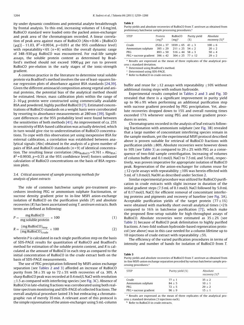
Table 2Purity yields and absolute recoveries of RuBisCO from T. aestivum as obtained frompreliminary batchwise sample processing procedures.a
STEP Protein(mg)b
RuBisCO(mg)c
Purity yield(%)
Absoluterecovery (%)d
Crude 2524 ± 37 1039 ± 65 41 ± 3 100 ± 6Ammonium sulphate 395 ± 29 211 ± 25 54 ± 2 20 ± 2PEG 893 ± 50 516 ± 44 58 ± 3 50 ± 4PEG + sucrose gradient 396 ± 42 304 ± 25 77 ± 12 29 ± 2
a Results are expressed as the mean of three replicates of the analytical pro-
col (see above) was in this case needed for a column lifetime up to10 injections of crude extract with repeatability ≤5%.
The efficiency of the varied purification procedures in terms ofintensity and number of bands for isolation of RuBisCO from T.
Table 3Purity yields and absolute recoveries of RuBisCO from T. aestivum as obtained fromin-line MSFI-anion exchange separation preceded by various batchwise sample pro-cessing procedures.a
STEP Purity yield (%) Absoluterecovery (%)b
Crude 77 ± 1 35 ± 2Ammonium sulphate 84 ± 5 10 ± 1
264 R. Suárez et al. / Tala
ty under dynamic conditions and potential analyte breakthroughy frontal analysis. To this end, increasing volumes of 1.6 mg/mLuBisCO standard were loaded onto the packed anion-exchangernd peak area of the chromatogram recorded. A linear correla-ion of peak area against mass of RuBisCO (Abs = 0.06 × [RuBisCO�g)] − 11.03, R2 = 0.9934, p = 0.051 at the 95% confidence level)ith repeatability < 6% (n = 8) within the overall dynamic range
f 348–930 �g RuBisCO was encountered. For real-life samplessays, the soluble protein content as determined by Brad-ord’s method should not exceed 1000 �g per run to preventuBisCO pre-elution in the early stages of the ionic-strengthradient.
A common practice in the literature to determine total solublerotein via Bradford’s method involves the use of least-squares lin-ar regression plots of absorbance against BSA standards [24,39].iven the different aminoacid composition among vegetal and ani-al proteins, the potential bias of the analytical method should
e estimated. Hence, mass calibration plots within the range of–10 �g protein were constructed using commercially availableSA and powdered, highly purified RuBisCO [7]. Estimated concen-rations of RuBisCO standards on a weight basis were corroboratedy resorting to absorbance measurements at 280 nm [39]. Signifi-ant differences at the 95% probability level were found betweenhe sensitivities of both methods [41]. An improvement of ca. 25%n sensitivity of BSA-based calibration was actually detected, whichn turn would give rise to underestimation of RuBisCO concentra-ions. To cope with this observation yet using inexpensive BSA forxternal calibration, a correlation was established between ana-ytical signals (Abs) obtained in the analysis of a given number ofairs of BSA and RuBisCO standards (n = 9) of identical concentra-ion. The resulting linear correlation (AbsRuBisCO = 0.761 × AbsBSA;2 = 0.9930, p = 0.35 at the 95% confidence level) fosters unbiasedalculation of RuBisCO concentrations on the basis of BSA regres-ion plots.
.4. Critical assessment of sample processing methods fornalysis of plant extracts
The role of common batchwise sample pre-treatment pro-edures involving PEG or ammonium sulphate fractionation, orucrose density gradient prior to in-line MSFI anion-exchangesolation of RuBisCO on the purification yields (P) and absoluteecoveries (R) has been ascertained using T. aestivum extracts. Botherms are defined as following:
= mg RuBisCOmg soluble protein
× 100 (1)
= (mg RuBisCO)step
(mg RuBisCO)crude× 100 (2)
herein P is calculated in each single purification step on the basisf SDS-PAGE results for quantitation of RuBisCO and Bradford’sethod for estimation of the soluble protein content, and R is cal-
ulated as the amount of RuBisCO in each step as compared to thenitial concentration of RuBisCO in the crude extract both on theasis of SDS-PAGE measurements.
The use of PEG precipitation followed by MSFI-anion exchangeeparation (see Tables 2 and 3) afforded an increase of RuBisCOurity from 58 ± 3% up to 72 ± 5% with recoveries of ca. 30%. Aharp RuBisCO peak was recorded at 0.4 mol/L NaCl with resolution1.5 as compared with interfering species (see Fig. 3C). Absence of
uBisCO in late-eluting fractions was corroborated using both real-ime spectrum monitoring and SDS-PAGE of collected fractions. Theverall analytical procedure lasted 3 h but embracing a chromato-raphic run of merely 35 min. A relevant asset of this protocol ishe simple rejuvenation of the anion-exchanger using 5 mL-columncess ± standard deviation.b Deternined using Bradford’s method.c Determined using SDS-PAGE.d Refer to RuBisCO in crude extracts.
buffer and reuse for ≥12 assays with repeatability ≤10% withoutadditional rinsing steps with sodium hydroxide.
Experimental results compiled in Tables 2 and 3 and Fig. 3Drevealed that there is a significant improvement in purity yieldup to 96 ± 9% when performing an additional purification stepwith sucrose gradient preceded by PEG precipitation. Yet, abso-lute recoveries dropped down to 15% and overall running timesexceeded 17 h whenever using PEG and sucrose gradient proce-dures in series.
Chromatograms recorded in the analysis of leaf extracts follow-ing fractionation with ammonium sulphate (see Fig. 3B) revealedthat a large number of concomitant interfering species remain inthe sample medium, yet the experimental MSFI protocol describedabove is proven suitable for retrieval of RuBisCO fractions withpurification yields ≥80%. Absolute recoveries were however downto 10% (see Table 3) as compared to 29 ± 2% with PEG as a conse-quence of two-fold sample centrifugation. Increasing of volumesof column buffer and 0.1 mol/L NaCl to 7.5 mL and 5.0 mL, respec-tively, was proven imperative for appropriate isolation of RuBisCOpeak. Regeneration of the anion-exchanger for column reuse for≥12 cycle assays with repeatability ≤10% was herein effected with5 mL of 1.0 mol/L NaOH as described under Section 2.
Similar experimental procedure was utilized for RuBisCO purifi-cation in crude extracts with slight increase in duration of theinitial gradient steps (7.5 mL of 0.1 mol/L NaCl followed by 5.0 mLof 0.17 mol/L NaCl) for efficient removal of concomitant interfer-ing proteins and pigments and recovery of baseline (see Fig. 3A).Acceptable purification yields of the target protein (77 ± 1%)were obtained with markedly short overall analytical times (<2 hcompared to 16 h in batchwise purification [7]), which makethe proposed flow-setup suitable for high-throughput assays ofRuBisCO. Absolute recoveries were estimated as 35 ± 2% (seeTable 3) because of RuBisCO peak delimitation to highly purifiedfractions. A two-fold sodium hydroxide-based regeneration proto-
PEG 72 ± 5 29 ± 2PEG + sucrose gradient 96 ± 9 15 ± 1
a Results are expressed as the mean of three replicates of the analytical pro-cess ± standard deviation (3 injections each).
b Refer to RuBisCO in crude extracts.

R. Suárez et al. / Talanta 84 (2011) 1259–1266 1265
Fig. 3. (A) Close-up of the chromatogram of Triticum aestivum as obtained by direct injection of crude extract into the MSFI-anion exchange manifold. (B) Close-up of thechromatogram of Triticum aestivum extract as obtained by MSFI-anion exchange preceded by precipitation with ammonium sulphate. (C) Close-up of the chromatogram ofTriticum aestivum extract as obtained by MSFI-anion exchange preceded by precipitation with PEG. (D) Close-up of the chromatogram of Triticum aestivum extract as obtainedby MSFI-anion exchange preceded by precipitation with PEG and sucrose gradient separation. Injected samples contained ca. 1000 mg of soluble protein as determined byBradford’s method. RuBisCO peak eluted at 0.4 mol/L NaCl.

1266 R. Suárez et al. / Talanta 84
Fig. 4. SDS-PAGE image of different steps of purification of RuBisCO from Triticumaestivum. Molecular weight standards (kDa) used as marker (Precision Plus Pro-tein Standards, Bio-Rad) are given on the left. LSU and SSU represent RuBisCO largeand small subunits, respectively. Lanes C and C* were loaded with crude extractand crude extract after MSFI-anion exchange separation, respectively. Lanes P andP* were loaded with extracts obtained from PEG precipitation, and PEG precipita-tion followed by MSFI-anion exchange separation, respectively. Lanes S and S* wereloaded with extracts obtained from sucrose gradient, and sucrose gradient followedby MSFI-anion exchange separation, respectively. Lanes A and A* were loaded withextracts obtained from ammonium sulphate precipitation, and ammonium sulphateplS
aTs[N
4
stpenrgranmbe
pbetep
sopf
[[[
[[
[[
[[
[
[[
[[
[[
[
[
[
[
[
[
[[[[[[[
recipitation followed by MSFI-anion exchange separation, respectively. Lane R wasoaded with RuBisCO standard. Electrophoresis was carried out as described underection 2. Soluble protein in the extracts ranged from 600 to 700 ng.
estivum is illustrated in the close up of SDS-PAGE gel in Fig. 4.he high molecular weight bands revealed by the gel above largeubunits of RuBisCO (LSU) are most likely LSU-binding proteins42] eluting at the chromatograms (see Fig. 3A–C) at ≥0.49 mol/LaCl.
. Conclusion
An automated and versatile multisyringe-flow anion-exchangeeparation assembly is herein proposed for expeditious isola-ion and real-time monitoring of RuBisCO in plant extracts withurification yields within the range of 72–96%. Handling of crudextracts as well as pre-treated samples following PEG and ammo-ium sulphate precipitation is proven feasible without manifoldeconfiguration by optimization of the stepwise ionic-strengthradient elution and regeneration protocols. As compared withobotic stations for protein purification dedicated to routine sep-rations (e.g., Äktapurifier, GE Healthcare) the MSFI-set up doesot merely offer a more open architecture and flexibility forethod development relying upon flow programming but should
e regarded as a cost-effective technique with 5-fold reducedquipment costs.
The approach presented in this work represents significantrogress towards the development of a rugged and automatic flow-ased manifold for high-throughput purification of RuBisCO asssential tool for further exploration of the variability of kineticraits of RuBisCO in crops and identification of more enzymaticallyfficient forms to be exploited for genetic transformations of croplants to increase productivity.
Current efforts in the authors’ lab are focused on methodcaling up using tandem-column anion-exchange separation, andrthogonal anion-exchange × size-exclusion chromatography forreparative separations emcompassing in-line desalting of eluateractions.
[[[
[
(2011) 1259–1266
Acknowledgements
Manuel Miró and Víctor Cerdà acknowledge financial sup-port from Spanish Ministry of Science and Innovation throughprojects CTM2010-17214 and CTQ2010-15541, respectively. JeroniGalmés extends his appreciation to the Government of the BalearicIslands and Spanish Ministry of Science and Innovation for finan-cial support through projects AAEE0031/08 and AGL2009-07999,respectively.
References
[1] G. Schneider, Y. Lindqvist, C.I. Branden, Annu. Rev. Biophys. Biomater. 21 (1992)119–143.
[2] S. von Caemmerer, Biochemical Model of Leaf Photosynthesis, CSIRO Publish-ing, Canberra, Australia, 2000.
[3] F.G. Pearce, Biochem. J. 399 (2006) 525–534.[4] M.A.J. Parry, P.J. Andralojc, R.A.C. Mitchell.R., P.J. Madgwick, A.J. Keys, J. Exp. Bot.
54 (2003) 1321–1333.[5] C. Peterhansel, M. Niessen, R.M. Kebeish, Photochem. Photobiol. 84 (2008)
1317–1323.[6] H. Yeoh, M.R. Badger, L. Watson, Plant Physiol. 66 (1980) 1110–1112.[7] J. Galmés, J. Flexas, A.J. Keys, J. Cifre, R.A.C. Mitchell, P.J. Madgwick, R.P. Haslam,
H. Medrano, M.A.J. Parry, Plant Cell Environ. 28 (2005) 571–579.[8] R.E. Sharwood, S. von Caemmerer, P. Maliga, S.M. Whitney, Plant Physiol. 146
(2007) 83–96.[9] S.M. Whitney, R.E. Sharwood, J. Exp. Bot. 59 (2008) 1909–1921.10] T.J. Andrews, S.M. Whitney, Arch. Biochem. Biophys. 414 (2003) 159–169.11] A.E. Ainsworth, A. Rogers, A.D.B. Leakey, Plant Physiol. 147 (2008) 13–19.12] A. Bar-Even, E. Noor, N.E. Lewis, R. Milo, Proc. Natl. Acad. Sci. U.S.A. 107 (2010)
8889–8894.13] I. Andersson, C.-I. Brändén, J. Mol. Biol. 172 (1984) 363–366.14] H.J. Kane, H. Vill, B. Entsch, K. Paul, M.K. Morell, T.J. Andrews, Plant Physiol. 21
(1994) 61–449.15] D.S. Kubien, S.M. Whitney, P.V. Moore, L.K. Jesson, J. Exp. Bot. 60 (2008) 1–11.16] D. Wang, S.L. Naidu Jr., A.R. Portis, S.P. Moose, S.P. Long, J. Exp. Bot. 59 (2008)
1779–1787.17] N.P. Hall, N.E. Tolbert, FEBS Lett. 96 (1978) 167–169.18] G. Zhu, R.G. Jensen, H.J. Bohnert, G.F. Wildner, J. Schlitter, Photosynth. Res. 57
(1998) 71–79.19] E. Mizohata, H. Matsumura, Y. Okano, M. Kumei, H. Takuma, J. Onodera, K. Kato,
N. Shibata, T. Inoue, A. Yokota, Y. Kai, J. Mol. Biol. 316 (2002) 679–691.20] P.D. Toman, J.J. Lynch Jr., R.R. Schmidt, Plant Physiol. 79 (1985) 806–814.21] S.J. Crafts-Brandner, M.E. Salvucci, Proc. Natl. Acad. Sci. U.S.A. 97 (2000)
13430–13435.22] S.M. Whitney, P. Baldet, G.S. Hudson, T.J. Andrews, Plant J. 26 (2001) 535–547.23] K. Uemura, Y. Suzuki, T. Shikanai, A. Wadano, R.G. Jensen, Plant Cell Physiol. 37
(1996) 325–331.24] M.E. Salvucci, A.R. Portis, W.L. Ogren, Anal. Biochem. 153 (1986) 97–101.25] J. Bota, J. Flexas, A.J. Keys, J. Loveland, M.A.J. Parry, H. Medrano, Vitis 41 (2002)
163–168.26] A. Recktenwald, K.H. Kroner, M.R. Kula, Enzyme Microb. Technol. 7 (1985)
607–612.27] T. Sakai, Y. Kito, N. Teshima, S. Katoh, K. Watla-Iad, K. Grudpan, J. Flow Inject.
Anal. 24 (2007) 23–26.28] K. Watla-Iad, T. Sakai, N. Teshima, S. Katoh, K. Grudpan, Anal. Chim. Acta 604
(2007) 139–146.29] W. Siangproh, N. Teshima, T. Sakai, S. Katoh, O. Chailapakul, Talanta 79 (2009)
1111–1117.30] C.K. Zacharis, E.A. Kalaitzantonakis, A. Podgornik, G. Theodoridis, J. Chromatogr.
B 1144 (2007) 126–134.31] T. Songjaroen, T. Maturos, A. Sappat, A. Tuantranont, W. Laiwattanapaisal, Anal.
Chim. Acta 647 (2009) 78–83.32] E. Delgado, H. Medrano, A.J. Keys, M.A.J. Parry, J. Exp. Bot. 46 (1995) 1775–1777.33] F.A. Boyle, A.J. Keys, Photsynth. Res. 11 (1987) 97–108.34] A.J. Keys, M.A.J. Parry, Methods Plant Biochem. 3 (1990) 1–14.35] K. Nishina, S. Jenks, S. Supattapone, J. Biol. Chem. 24 (2004) 40788–40794.36] E. Becerra, A. Cladera, V. Cerdà, Lab. Robot. Autom. 11 (1999) 131–140.37] M.M. Bradford, Anal. Biochem. 72 (1976) 248–254.38] S.J. Compton, C.G. Jones, Anal Biochem. 151 (1985) 369–374.
39] D. Afif, D. Gérant, G. Cavalié, P. Dizengremel, Physiol. Plant. 88 (1993) 113–122.40] J.M. Redja, S. Johal, R. Chollet, Arch. Biochem. Biophys. 210 (1981) 617–624.41] D.L. Massart, B.G.M. Vandeginste, L.M.C. Buydens, S. De Jong, P.J. Lewi, J.Smeyers-Verbeke, Handbook of Chemometrics and Qualimetrics: Part A, Else-vier, Amsterdam, 1997, ch. 8, pp. 208–210, 544.
42] J.E. Musgrove, R.J. Ellis, Phil. Trans. R. Soc. Lond. B 313 (1986) 419–428.