
Automated Detection of Pain in Horses through Facial...
32
Automated Detection of Pain in Horses through Facial Expression Analysis Master’s thesis in Complex Adaptive Systems KASHIF BHATTI Department of Applied Mechanics CHALMERS UNIVERSITY OF TECHNOLOGY Göteborg, Sweden 2016
Transcript of Automated Detection of Pain in Horses through Facial...
Automated Detection of Pain in Horsesthrough Facial Expression AnalysisMaster’s thesis in Complex Adaptive Systems
KASHIF BHATTI
Department of Applied MechanicsCHALMERS UNIVERSITY OF TECHNOLOGYGöteborg, Sweden 2016


MASTER’S THESIS IN COMPLEX ADAPTIVE SYSTEMS
Automated Detection of Pain in Horses through Facial Expression Analysis
KASHIF BHATTI
Department of Applied Mechanics
CHALMERS UNIVERSITY OF TECHNOLOGY
Göteborg, Sweden 2016
Automated Detection of Pain in Horses through Facial Expression AnalysisKASHIF BHATTI
© KASHIF BHATTI, 2016
Master’s thesis 2016:86ISSN 1652-8557Department of Applied MechanicsChalmers University of TechnologySE-412 96 GöteborgSwedenTelephone: +46 (0)31-772 1000
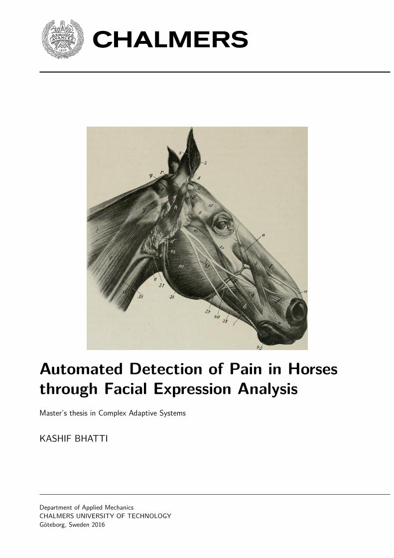
Cover:
Ellenburger-Baum (1914) Muscles of Head of Horse; Lateral View. The M. cutaneus is Removed In: Sisson,Septimus, The anatomy of the domestic animals. p. 256. Philadelphia and London: W. B. Saunders Company.
a, Levator labii superioris proprius; b, levator nasolabialis; c, brachiocephalicus; d, sterno-cephalicus; d’,tendons of d; e, omo-hyoideus; f, dilatator naris lateralis; g, zygomaticus; h, buccinator; i, depressor labiiinferioris; k, orbicularis oris; l, lateralis nasi, dorsal part; m, masseter; n, parotido-auricularis; o, zygomatico-auricularis; p, interscutularis; p’, fronto-scutularis, pars temporalis; q, cervico-auricularis profundus major; r,cervico-auricularis superficialis; s, obliquus capitis anterior; t, splenius; v, occipito-mandibularis; y, mastoidtendon of brachiocephalicus; 2, posterior, 3, anterior, border of external ear; 8, scutiform cartilage; 9, zygomaticarch; 10, orbital fat; 18, temporo-mandibular articulation; 27, facial crest; 30’, angle of jaw; 37, externalmaxillary vein; 38, jugular vein; 39, facial vein; 40, parotid duct; 41, transverse facial vein; 42, massetericvein; 43, facial nerve; 44, parotid gland; 45, chin; x, wing of atlas. By an oversight the superior buccal branchof the facial nerve is shown crossing over instead of under the zygomaticus.
Chalmers ReproserviceGöteborg, Sweden 2016

Automated Detection of Pain in Horses through Facial Expression AnalysisMaster’s thesis in Complex Adaptive SystemsKASHIF BHATTIDepartment of Applied MechanicsChalmers University of Technology
Abstract
A method for automated pain-assessment in horses through facial-expression analysis is proposed. The methodis based on supervised linear classification of a feature stack of Gabor filters and has the desirable quality ofnot requiring expert knowledge or specialized equipment to make an assessment. The method is evaluated byapplying it to images of horses from two clinical trials where the horses were (ethically) subjected to pain.The resulting accuracy of 78% compares favorably to an alternate method of pain assessment based on facialexpression cues that requires expertise to administer.
Keywords: facial-expressions, machine-learning, horse, equine, artificial intelligence, pain assessment
i

ii

Acknowledgements
I thank Krister Wolff for connecting me with the opportunity to work on this project and for taking on therole of examiner.
I am grateful to Pia Andersson (Swedish University of Agricultural Sciences) and Emanuella Dalla Costa(Università degli Studi di Milano) for their excellent clinical investigations on equine pain expression, forallowing me access to their photographic data without which this thesis would not be possible and for theirvaluable advice and consultation.
iii

iv
Contents
Abstract i
Acknowledgements iii
Contents v
1 Introduction 11.1 Background . . . . . . . . . . . . . . . . . . . . . . . . . . . . . . . . . . . . . . . . . . . . . . . . . 11.2 Goals . . . . . . . . . . . . . . . . . . . . . . . . . . . . . . . . . . . . . . . . . . . . . . . . . . . . 1
2 Theory 22.1 Facial Expression Coding . . . . . . . . . . . . . . . . . . . . . . . . . . . . . . . . . . . . . . . . . 22.2 Automation in Facial Expression Analysis . . . . . . . . . . . . . . . . . . . . . . . . . . . . . . . . 22.3 An Overview of Automated Facial Expression Analysis . . . . . . . . . . . . . . . . . . . . . . . . . 32.3.1 Face Acquisition . . . . . . . . . . . . . . . . . . . . . . . . . . . . . . . . . . . . . . . . . . . . . 32.3.2 Normalization . . . . . . . . . . . . . . . . . . . . . . . . . . . . . . . . . . . . . . . . . . . . . . 32.3.3 Segmentation . . . . . . . . . . . . . . . . . . . . . . . . . . . . . . . . . . . . . . . . . . . . . . . 32.3.4 Feature Extraction . . . . . . . . . . . . . . . . . . . . . . . . . . . . . . . . . . . . . . . . . . . . 42.3.5 Facial Expression Classification . . . . . . . . . . . . . . . . . . . . . . . . . . . . . . . . . . . . . 42.4 Feature Extraction Techniques . . . . . . . . . . . . . . . . . . . . . . . . . . . . . . . . . . . . . . 42.4.1 Active Appearance Models . . . . . . . . . . . . . . . . . . . . . . . . . . . . . . . . . . . . . . . 42.4.2 2D Gabor Filters . . . . . . . . . . . . . . . . . . . . . . . . . . . . . . . . . . . . . . . . . . . . . 6
3 Method 83.1 Challenges . . . . . . . . . . . . . . . . . . . . . . . . . . . . . . . . . . . . . . . . . . . . . . . . . . 83.2 Experimental design of clinical trials and image acquisition . . . . . . . . . . . . . . . . . . . . . . 83.2.1 An equine pain face . . . . . . . . . . . . . . . . . . . . . . . . . . . . . . . . . . . . . . . . . . . 93.2.2 Development of the Horse Grimace Scale (HGS) . . . . . . . . . . . . . . . . . . . . . . . . . . . 93.3 System design . . . . . . . . . . . . . . . . . . . . . . . . . . . . . . . . . . . . . . . . . . . . . . . . 113.3.1 Choice of pose . . . . . . . . . . . . . . . . . . . . . . . . . . . . . . . . . . . . . . . . . . . . . . 113.3.2 Preprocessing . . . . . . . . . . . . . . . . . . . . . . . . . . . . . . . . . . . . . . . . . . . . . . 113.3.3 System design . . . . . . . . . . . . . . . . . . . . . . . . . . . . . . . . . . . . . . . . . . . . . . 123.4 Feature extraction . . . . . . . . . . . . . . . . . . . . . . . . . . . . . . . . . . . . . . . . . . . . . 123.5 Classification . . . . . . . . . . . . . . . . . . . . . . . . . . . . . . . . . . . . . . . . . . . . . . . . 13
4 Results and discussion 164.1 Comparison to human based trials . . . . . . . . . . . . . . . . . . . . . . . . . . . . . . . . . . . . 16
5 Conclusions and recommendations 195.1 Proposals for future work . . . . . . . . . . . . . . . . . . . . . . . . . . . . . . . . . . . . . . . . . 195.1.1 Animal facial expression database . . . . . . . . . . . . . . . . . . . . . . . . . . . . . . . . . . . 195.1.2 Comparative study of automated facial expression approaches . . . . . . . . . . . . . . . . . . . . 20
References 20
v

vi
1 Introduction
Pain symptomizes many medical conditions and its presence can have a powerful negative influence on ananimal’s wellbeing and quality of life. Unless they are prevented from doing so due to factors like disability,adults are able to communicate the presence of pain and the qualities associated with it—such as duration,location and intensity—with clarity and specificity. The challenge of identifying pain in human infants issignificantly greater, but even there, humans are able to detect the presence of distress through an innateunderstanding of vocalizations, facial expressions and other associated indicators of pain.
Detecting pain in animals presents a much larger challenge. Without training and experience, it is not possibleto reliably detect the presence of pain in animals. This thesis proposes an approach to detecting pain in largefarm animals, applies it to horses and evaluates its performance compared to trained humans.
1.1 Background
The ability to quickly and inexpensively detect pain in large animals has implications for ethics, animal welfareand veterinary medicine. The means of detecting pain in farm animals found in the scientific literature tendto focus on physiological and behavioral changes. Training and experience of the individual carrying out theassessment can influence the results of the evaluation as can his/her knowledge of the species, breed andindividual animal. Human empathy and emotions can also effect the results of the evaluation [1]. Methods ofpain detection that are automated and do not suffer from the aforementioned drawbacks can be of value.
Facial expressions of pain are commonly used indicators of acute procedural pain in clinical and researchenvironments [2]. Similarities exist between detecting pain in animals and in human infants as both may notbe able to communicate feelings of pain and both may display behavioral and psychological changes due toa variety of reasons besides pain. These could include, illness, dejection, general distress etc. Pal, Pritam etal. [3], and others, have been successful in devising systems that can identify pain in infants using images andthis motivates an exploration of image-based approaches for animals.
Veterinary literature exists that catalogs and correlates the presence of equine facial expressions with pain.Gleerup et al., [4] have documented behavioral and physiological expressions of pain in six adult horses exposedto to two noxious stimuli; E Dalla Costa et al. [5] have developed a three-step Horse Grimace Scale based onclinical study of forty-six adult horses (of which six formed the control group).
1.2 Goals
The thesis aims to develop and evaluate the performance of an image-based automated system for detectingpain based on observations made by and empirical data collected by Gleerup et al., E Dalla Costa et al. andsimilar trials.
Recommendations will be made on future work in this direction, and improvements will be suggested to clinicaltrial procedures and cataloging efforts to facilitate future research efforts.
1
2 Theory
2.1 Facial Expression Coding
The universality of facial expression in animals has been demonstrated by Darwin in 1872. One of his claimswas that there are specific inborn emotions which originated in serviceable associated habits [6]. These areinherited behaviors that are useful responses to certain mental states, such as the furrowing of the eyes toprevent too much light from entering or raising of eye-brows when accessing memory, which is associated withsearching ones surroundings where raised eye-brows are useful in increasing one’s field of view.
Before attracting broader attention, research interest in human face identification and facial expression detec-tion existed primarily amongst psychologists. Ekman and Friesen conducted seminal research on human facialexpression analysis, postulating, in 1971 [7] six prototypic emotional displays consisting of happiness, sadness,fear, disgust, surprise and anger that are universal across human ethnicities and cultures (as cited in [8]).
Ekman and Friesen went on to develop a comprehensive Facial Action Coding System (FACS) thatencodes and classifies all human facial expressions and associates them with the six prototypic emotionaldisplays [9]. Expressions are broken down into groups of facial muscle actions that produce a feature on theface, such as raised inner eye-brow or tightened lips. These components are called Action Units (AUs).FACS is heavily used in detecting and analysing human facial expressions, both manually and automatically;and in recreating them—such as through computer animations.
Facial expression in non-human animals have been studied to a markedly lesser extent. The pain face ofmice [10], horses [11, 5] and primates [12] have been investigated. No universal coding system for any non-human animal has so far been developed.
Dalla Costa et al. have developed a Horse Grimace Scale [5] consisting of 6 AUs for pain assessment. EachAU is scored with three levels of intensity from ”not present”, through ”moderately present” to ”obviouslypresent” (intensity scores 0-3). This may be contrasted against the five scale intensity scoring used in humanFACS.
2.2 Automation in Facial Expression Analysis
The availability of inexpensive and easy access to large amounts of computational power has spurred interest inautomatic human face detection and to a lesser degree facial expression analysis. Most systems today attemptto classify facial expressions into FACS AUs. So far only marker based systems are able to reliably detect allFACS AUs (citation), although image and video based approaches such as Ref. [13] are able to detect a subsetof AUs with high reliability.
Very little research effort has been directed towards automation in detecting animal facial expressions. Onesuch instance is the Rodent Face Finder® developed by Susana G Sotocinal et. al. [10] which automaticallydetects mice eyes and ears in still-images using boosted cascades of Haar classifiers and estimates the bounds
2
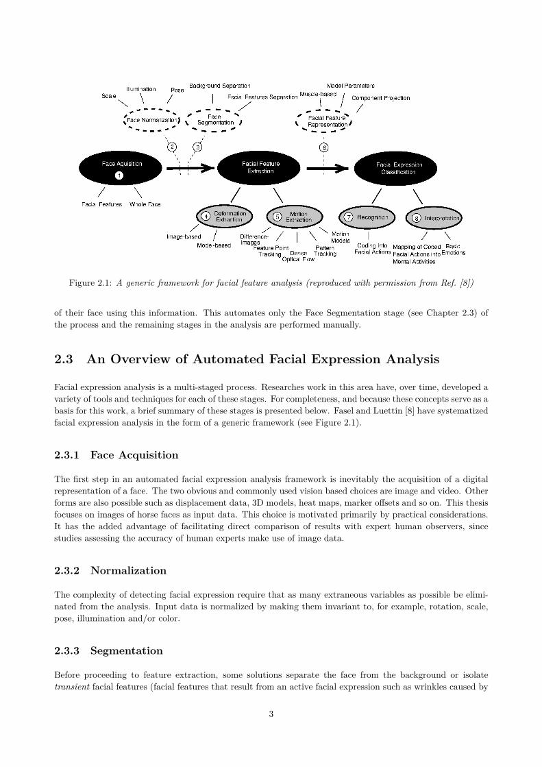
Figure 2.1: A generic framework for facial feature analysis (reproduced with permission from Ref. [8])
of their face using this information. This automates only the Face Segmentation stage (see Chapter 2.3) ofthe process and the remaining stages in the analysis are performed manually.
2.3 An Overview of Automated Facial Expression Analysis
Facial expression analysis is a multi-staged process. Researches work in this area have, over time, developed avariety of tools and techniques for each of these stages. For completeness, and because these concepts serve as abasis for this work, a brief summary of these stages is presented below. Fasel and Luettin [8] have systematizedfacial expression analysis in the form of a generic framework (see Figure 2.1).
2.3.1 Face Acquisition
The first step in an automated facial expression analysis framework is inevitably the acquisition of a digitalrepresentation of a face. The two obvious and commonly used vision based choices are image and video. Otherforms are also possible such as displacement data, 3D models, heat maps, marker offsets and so on. This thesisfocuses on images of horse faces as input data. This choice is motivated primarily by practical considerations.It has the added advantage of facilitating direct comparison of results with expert human observers, sincestudies assessing the accuracy of human experts make use of image data.
2.3.2 Normalization
The complexity of detecting facial expression require that as many extraneous variables as possible be elimi-nated from the analysis. Input data is normalized by making them invariant to, for example, rotation, scale,pose, illumination and/or color.
2.3.3 Segmentation
Before proceeding to feature extraction, some solutions separate the face from the background or isolatetransient facial features (facial features that result from an active facial expression such as wrinkles caused by
3
Holistic methods Local methods
Deformation extractionImage-based Neural Network Intensity profiles
Gabor wavelets High gradient componentsPCA + Neural Networks
Model-based Active appearance model Geometric face modelPoint distribution model Two view point-based modelsLabeled graphs
Motion extractionDense optical flow Dense flow fields Region-based flows
Motion models 3D motion models Parametric motion models3D deformable models 3D motion models
Feature point tracking Feature trackingDifference-images Holistic diff.-imgs Region-based difference-imagesMarker-based Highlighted facial features
Dot markers
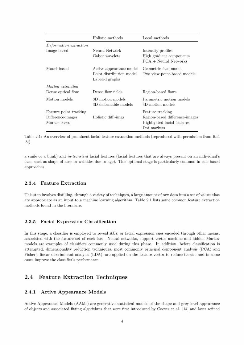
Table 2.1: An overview of prominent facial feature extraction methods (reproduced with permission from Ref.[8])
a smile or a blink) and in-transient facial features (facial features that are always present on an individual’sface, such as shape of nose or wrinkles due to age). This optional stage is particularly common in rule-basedapproaches.
2.3.4 Feature Extraction
This step involves distilling, through a variety of techniques, a large amount of raw data into a set of values thatare appropriate as an input to a machine learning algorithm. Table 2.1 lists some common feature extractionmethods found in the literature.
2.3.5 Facial Expression Classification
In this stage, a classifier is employed to reveal AUs, or facial expression cues encoded through other means,associated with the feature set of each face. Neural networks, support vector machine and hidden Markovmodels are examples of classifiers commonly used during this phase. In addition, before classification isattempted, dimensionality reduction techniques, most commonly principal component analysis (PCA) andFisher’s linear discriminant analysis (LDA), are applied on the feature vector to reduce its size and in somecases improve the classifier’s performance.
2.4 Feature Extraction Techniques
2.4.1 Active Appearance Models
Active Appearance Models (AAMs) are generative statistical models of the shape and grey-level appearanceof objects and associated fitting algorithms that were first introduced by Cootes et al. [14] and later refined
4
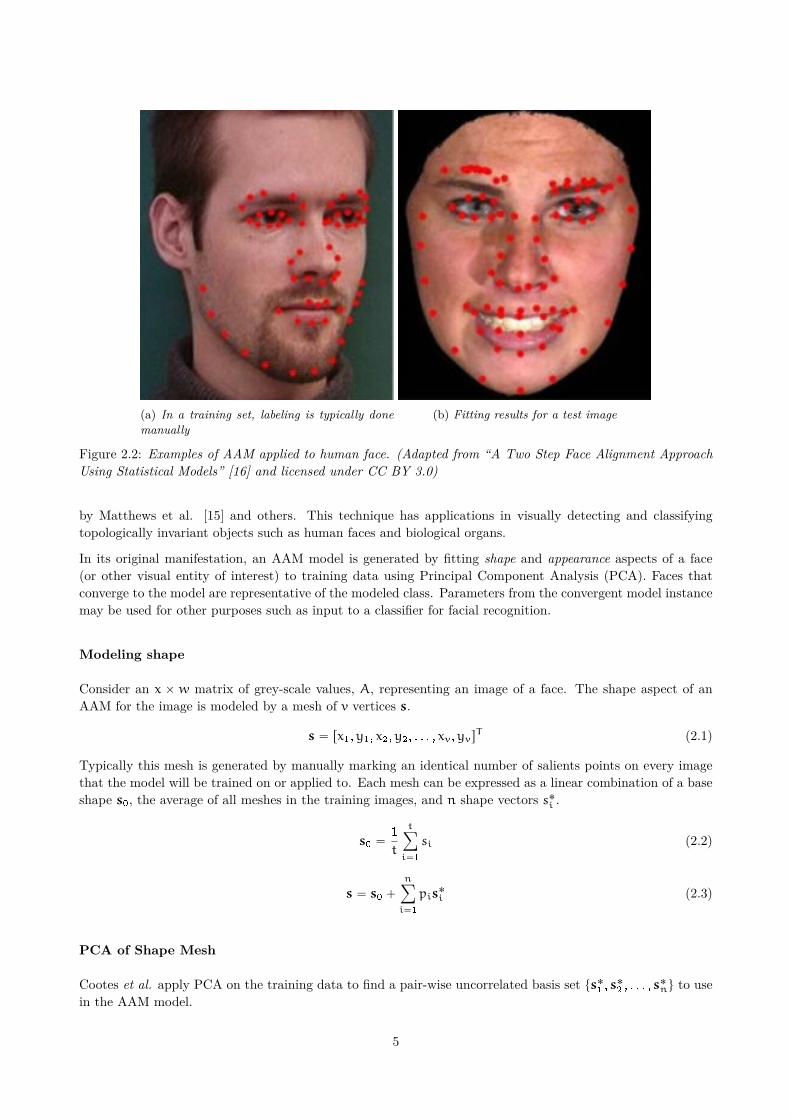
(a) In a training set, labeling is typically donemanually
(b) Fitting results for a test image
Figure 2.2: Examples of AAM applied to human face. (Adapted from “A Two Step Face Alignment ApproachUsing Statistical Models” [16] and licensed under CC BY 3.0)
by Matthews et al. [15] and others. This technique has applications in visually detecting and classifyingtopologically invariant objects such as human faces and biological organs.
In its original manifestation, an AAM model is generated by fitting shape and appearance aspects of a face(or other visual entity of interest) to training data using Principal Component Analysis (PCA). Faces thatconverge to the model are representative of the modeled class. Parameters from the convergent model instancemay be used for other purposes such as input to a classifier for facial recognition.
Modeling shape
Consider an x ˆ w matrix of grey-scale values, A, representing an image of a face. The shape aspect of anAAM for the image is modeled by a mesh of v vertices s.
s “ rx1,y1, x2,y2, . . . , xv,yvsT (2.1)
Typically this mesh is generated by manually marking an identical number of salients points on every imagethat the model will be trained on or applied to. Each mesh can be expressed as a linear combination of a baseshape s0, the average of all meshes in the training images, and n shape vectors s˚
i .
s0 “1
t
tÿ
i“1
si (2.2)
s “ s0 `
nÿ
i“1
pis˚i (2.3)
PCA of Shape Mesh
Cootes et al. apply PCA on the training data to find a pair-wise uncorrelated basis set ts˚1, s˚
2, . . . , s˚
nu to usein the AAM model.
5

First the covariance matrix C P R2vˆ2v is computed using ∆si “ si ´ s0.
Cij “ Erpsi ´ s0qpsj ´ s0qs (2.4)
C “1
t
tÿ
i“1
∆si∆sTi (2.5)
A linearly independent basis for C may now be determined through eigenvalue decomposition by computingQ such that C “ PΛP´1. Here the i-th column in P is pi and Λ is a diagonal matrix of eigenvalues λi. It isknown that for symmetric matrices of real values, the eigenvectors are orthogonal, therefore since Cij “ Cji,C “ PΛPT . The vectors with the largest values in this orthogonal basis constitutes the principal componentsof the shape mesh.
Modeling Appearance
A similar approach is used to model the appearance of faces i.e. the grey-level textural information embeddedin each image.
First all pixels that lie outside of the boundaries of the facial mesh are discarded from A and the resultingmatrix vectorized, yielding �A. This may also be represented as a linear combination of a base shape and mappearance vectors �A
˚
i
�A “ �A0 `
mÿ
i“0
λi �A˚ (2.6)
Similarly, �A˚ may be computed through PCA on the training texture data.
Fitting algorithms
A test image may be described by an AAM by locating the closes model instance in its parameter space (piand λi). The search for this instance is an optimization problem is solved using gradient descent like methods.
2.4.2 2D Gabor Filters
A Gabor filter can be expressed as a complex sinusoidal, referred to as the carrier modulated by a Gaussianshaped function, called the envelope.
gpx,yq “ spx,yqloomoon
carrier
wpx,yqloomoon
envelope
(2.7)
The following parametric form is used when implementing the solution.
gpx,yq “ exp
ˆ
´x 12 ` γ2y 12
2σ2
˙
exp
ˆ
i
ˆ
2πx 1
λ`ψ
˙˙
x 1 “ x cos θ` y sin θ
y 1 “ ´x sin θ` y cos θ
(2.8)
where λ is the wavelength of the sinusoidal factor, θ is the orientation of the normal to the parallel stripes ofa Gabor function, γ is the spatial aspect ratio, σ is the standard deviation of the Gaussian envelop, and ψ isthe phase offset.
Gabor filters work in the same was as the vision system in humans and animals (improve this text and adda reference) [17]. Unlike some classic approaches such as eigenfaces, Gabor filters can also be made imageillumination invariant.
6
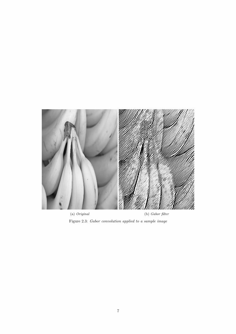
(a) Original (b) Gabor filter
Figure 2.3: Gabor convolution applied to a sample image
7

3 Method
3.1 Challenges
Human Focus of Existing Research
Research in the area of animal facial expression detection is limited as reviewed in Section 2.2. While inspirationmay be drawn from the more active field of human facial expression detection, care must be taken to addressthe anatomical idiosyncrasies of horses: ears that can point in different direction, fur, patches of colors, lackof a well defined frontal view and so on.
Cataloged Experimental Data
Researchers interested in automation of human facial detection and facial expression analysis have at theirdisposal various large, high-quality, open-access, labeled data-sets such as the Japanese Female Facial Ex-pression Database (JAFFE) [18], the AR database [19], and the Cohn-Kanade AU-Coded facial expressiondatabase [20]. The availability of these databases relieve researchers from the time-consuming, administration-heavy task of collecting and cataloging data. Open-access also eases verification of results and performancecomparisons.
As no comparable database for horses is available, a labeled catalog must first be prepared based on inputfrom the two studies discussed in the next section (Section 3.2).
Universal Vocabulary for Equine Facial-Expressions
Pioneering work by Ekman and Friesen (see Section 2.1) has let to a well-developed and widely adaptedvocabulary of human AUs. No such comprehensive vocabulary exists for any non-human animal and theseAUs cannot be translated to them. Limited AU vocabularies, in the form of grimace scales, for pain haverecently been developed for rodents, rabbits and horses (see Section 2.1), however their adaption so far islimited.
3.2 Experimental design of clinical trials and image acquisition
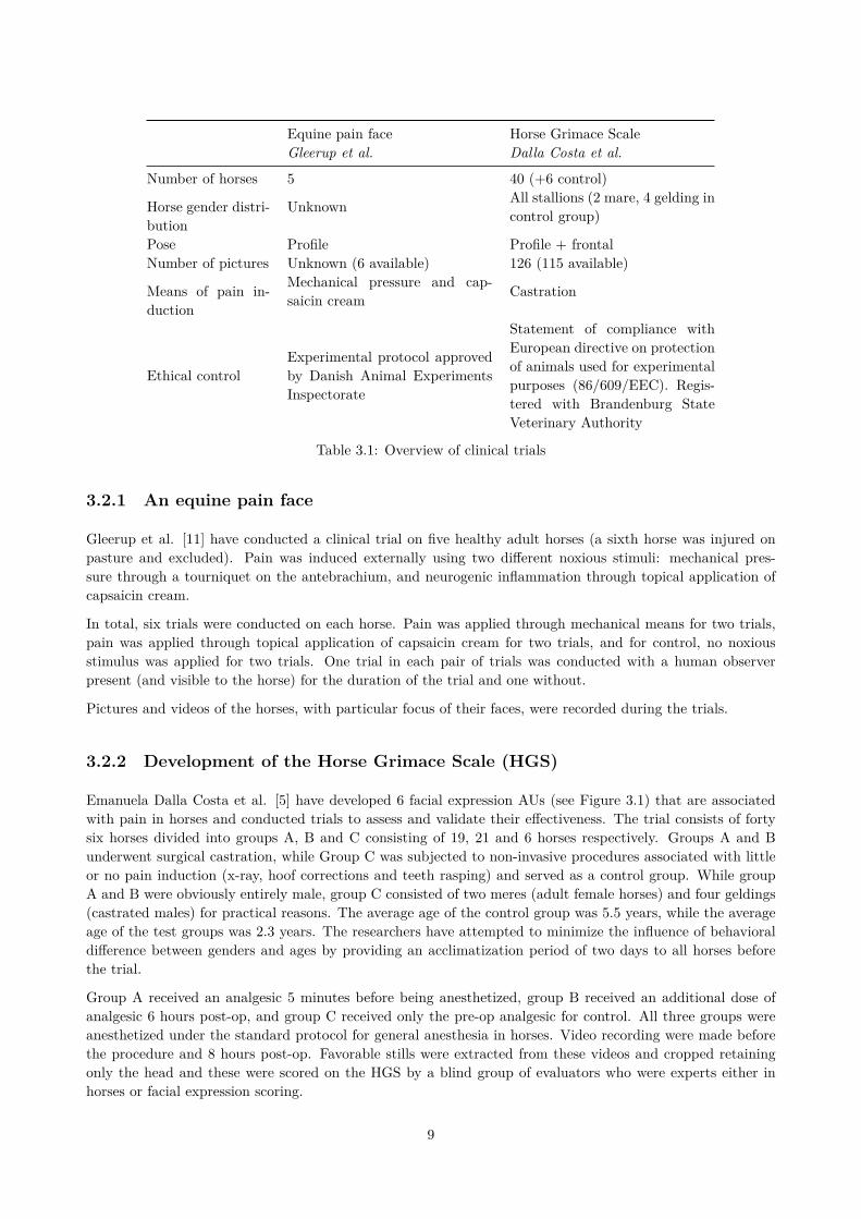
The image data for this study has been collected during two clinical trials on horses. The experimental designof these trials is briefly summarized in Table 3.1, accompanied by details of the facial expression data collectedand the conditions under which they were collected.
8
Equine pain face Horse Grimace ScaleGleerup et al. Dalla Costa et al.
Number of horses 5 40 (+6 control)
Horse gender distri-bution
Unknown All stallions (2 mare, 4 gelding incontrol group)
Pose Profile Profile + frontalNumber of pictures Unknown (6 available) 126 (115 available)
Means of pain in-duction
Mechanical pressure and cap-saicin cream Castration
Ethical controlExperimental protocol approvedby Danish Animal ExperimentsInspectorate
Statement of compliance withEuropean directive on protectionof animals used for experimentalpurposes (86/609/EEC). Regis-tered with Brandenburg StateVeterinary Authority
Table 3.1: Overview of clinical trials
3.2.1 An equine pain face
Gleerup et al. [11] have conducted a clinical trial on five healthy adult horses (a sixth horse was injured onpasture and excluded). Pain was induced externally using two different noxious stimuli: mechanical pres-sure through a tourniquet on the antebrachium, and neurogenic inflammation through topical application ofcapsaicin cream.
In total, six trials were conducted on each horse. Pain was applied through mechanical means for two trials,pain was applied through topical application of capsaicin cream for two trials, and for control, no noxiousstimulus was applied for two trials. One trial in each pair of trials was conducted with a human observerpresent (and visible to the horse) for the duration of the trial and one without.
Pictures and videos of the horses, with particular focus of their faces, were recorded during the trials.
3.2.2 Development of the Horse Grimace Scale (HGS)
Emanuela Dalla Costa et al. [5] have developed 6 facial expression AUs (see Figure 3.1) that are associatedwith pain in horses and conducted trials to assess and validate their effectiveness. The trial consists of fortysix horses divided into groups A, B and C consisting of 19, 21 and 6 horses respectively. Groups A and Bunderwent surgical castration, while Group C was subjected to non-invasive procedures associated with littleor no pain induction (x-ray, hoof corrections and teeth rasping) and served as a control group. While groupA and B were obviously entirely male, group C consisted of two meres (adult female horses) and four geldings(castrated males) for practical reasons. The average age of the control group was 5.5 years, while the averageage of the test groups was 2.3 years. The researchers have attempted to minimize the influence of behavioraldifference between genders and ages by providing an acclimatization period of two days to all horses beforethe trial.
Group A received an analgesic 5 minutes before being anesthetized, group B received an additional dose ofanalgesic 6 hours post-op, and group C received only the pre-op analgesic for control. All three groups wereanesthetized under the standard protocol for general anesthesia in horses. Video recording were made beforethe procedure and 8 hours post-op. Favorable stills were extracted from these videos and cropped retainingonly the head and these were scored on the HGS by a blind group of evaluators who were experts either inhorses or facial expression scoring.
9
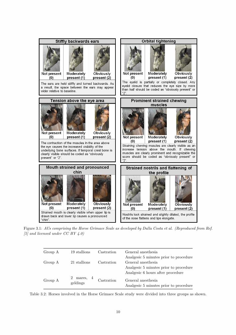
Figure 3.1: AUs comprising the Horse Grimace Scale as developed by Dalla Costa et al. (Reproduced from Ref.[5] and licensed under CC BY 4.0)
Group A 19 stallions Castration General anesthesiaAnalgesic 5 minutes prior to procedure
Group A 21 stallions Castration General anesthesiaAnalgesic 5 minutes prior to procedureAnalgesic 6 hours after procedure
Group A 2 mares, 4geldings Castration General anesthesia
Analgesic 5 minutes prior to procedure
Table 3.2: Horses involved in the Horse Grimace Scale study were divided into three groups as shown.
10
3.3 System design
This thesis focuses on deformation based facial feature extraction of available image data and its interpretation.This corresponds to stages (4) and (8) in Fasel and Luettin’s generic framework (Figure 2.1).
3.3.1 Choice of pose
A good choice of pose for automated facial expression analysis will facilitate capturing of as much facialexpression information as possible. In humans this choice is obvious and indeed a common characteristic ofall (non-pose invariant) human facial recognition studies is that they rely on the frontal face view.
Due to the anatomy of a horse’s head, a frontal projection has a smaller area than a side projection. Thenarrow, elongated profile obscures details around the mouth, cheeks and eyes. This frontal view is dominatedby a large forehead that runs down to the muzzle and, unlike humans, offer no expressiveness. These factorsmake a frontal view less than ideal for capturing facial expression details.
A profile view may be a superior choice as it captures more expressive facial features in plane. Not allfacial expressions are symmetric, however, and the asymmetry of the facial expressions will contain valuableinformation which is lost if only one profile view is used. This may be addressed by taking multiple profileviews or combining a profile views with a frontal view.
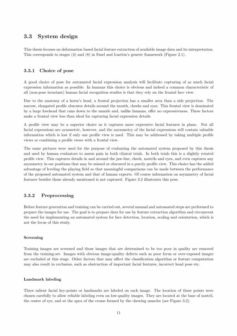
The same pictures were used for the purpose of evaluating the automated system proposed by this thesisand used by human evaluators to assess pain in both clinical trials. In both trials this is a slightly rotatedprofile view. This captures details in and around the jaw-line, cheek, nostrils and eyes, and even captures anyasymmetry in ear positions that may be missed or obscured in a purely profile view. This choice has the addedadvantage of leveling the playing field so that meaningful comparisons can be made between the performanceof the proposed automated system and that of human experts. Of course information on asymmetry of facialfeatures besides those already mentioned is not captured. Figure 3.2 illustrates this pose.
3.3.2 Preprocessing
Before feature generation and training can be carried out, several manual and automated steps are performed toprepare the images for use. The goal is to prepare data for use by feature extraction algorithm and circumventthe need for implementing an automated system for face detection, location, scaling and orientation, which isnot the focus of this study.
Screening
Training images are screened and those images that are determined to be too poor in quality are removedfrom the training-set. Images with obvious image-quality defects such as poor focus or over-exposed imagesare excluded at this stage. Other factors that may affect the classification algorithm or feature computationmay also result in exclusion, such as obstruction of important facial features, incorrect head pose etc.
Landmark labeling
Three salient facial key-points or landmarks are labeled on each image. The location of these points werechosen carefully to allow reliable labeling even on low-quality images. They are located at the base of nostril,the center of eye, and at the apex of the crease formed by the chewing muscles (see Figure 3.2).
11
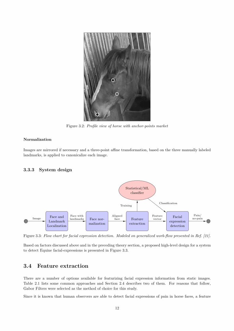
Figure 3.2: Profile view of horse with anchor-points market
Normalization
Images are mirrored if necessary and a three-point affine transformation, based on the three manually labeledlandmarks, is applied to canonicalize each image.
3.3.3 System design
Face andLandmark
Localization
Face nor-malization
Featureextraction
Facialexpressiondetection
Statistical/MLclassifier
ImageFace withlandmarks
Alignedface
Featurevector
TrainingClassification
Pain/no-pain
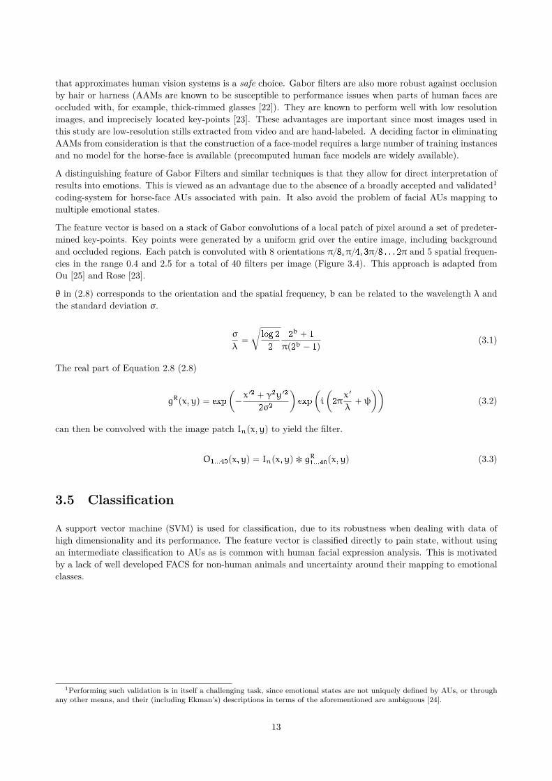
Figure 3.3: Flow chart for facial expression detection. Modeled on generalized work-flow presented in Ref. [21].
Based on factors discussed above and in the preceding theory section, a proposed high-level design for a systemto detect Equine facial-expressions is presented in Figure 3.3.
3.4 Feature extraction
There are a number of options available for featurizing facial expression information from static images.Table 2.1 lists some common approaches and Section 2.4 describes two of them. For reasons that follow,Gabor Filters were selected as the method of choice for this study.
Since it is known that human observers are able to detect facial expressions of pain in horse faces, a feature
12
that approximates human vision systems is a safe choice. Gabor filters are also more robust against occlusionby hair or harness (AAMs are known to be susceptible to performance issues when parts of human faces areoccluded with, for example, thick-rimmed glasses [22]). They are known to perform well with low resolutionimages, and imprecisely located key-points [23]. These advantages are important since most images used inthis study are low-resolution stills extracted from video and are hand-labeled. A deciding factor in eliminatingAAMs from consideration is that the construction of a face-model requires a large number of training instancesand no model for the horse-face is available (precomputed human face models are widely available).
A distinguishing feature of Gabor Filters and similar techniques is that they allow for direct interpretation ofresults into emotions. This is viewed as an advantage due to the absence of a broadly accepted and validated1
coding-system for horse-face AUs associated with pain. It also avoid the problem of facial AUs mapping tomultiple emotional states.
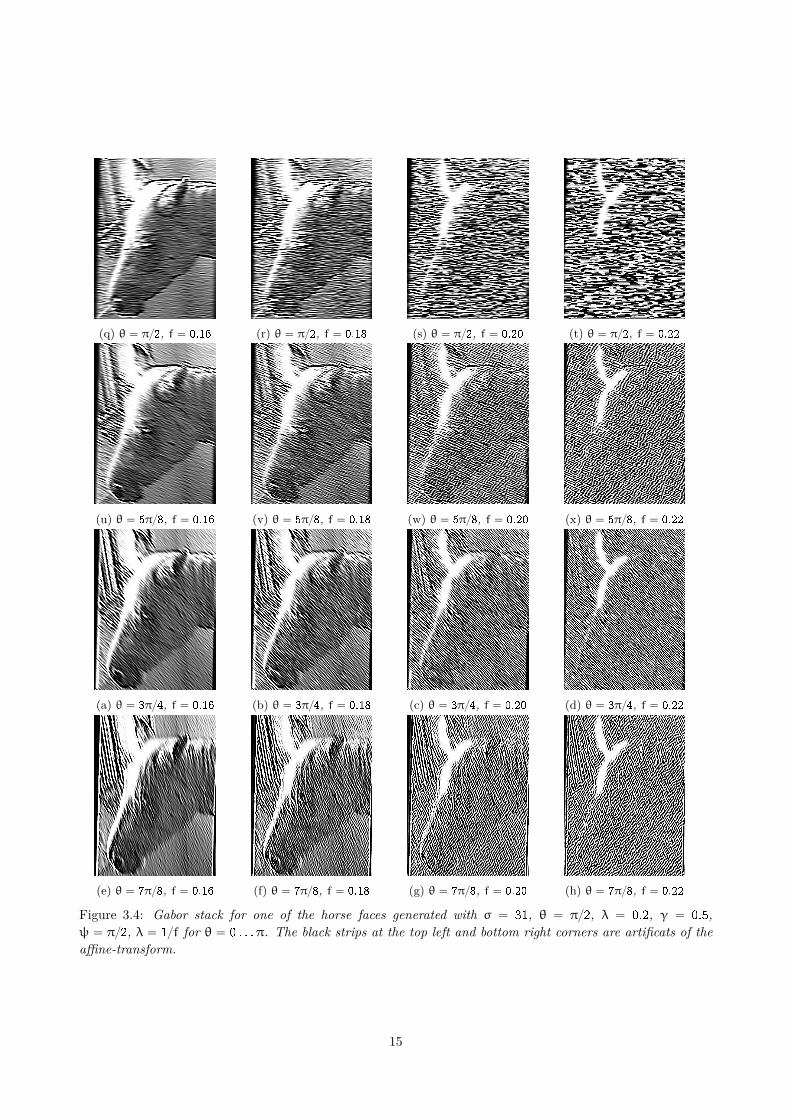
The feature vector is based on a stack of Gabor convolutions of a local patch of pixel around a set of predeter-mined key-points. Key points were generated by a uniform grid over the entire image, including backgroundand occluded regions. Each patch is convoluted with 8 orientations π{8,π{4, 3π{8 . . . 2π and 5 spatial frequen-cies in the range 0.4 and 2.5 for a total of 40 filters per image (Figure 3.4). This approach is adapted fromOu [25] and Rose [23].
θ in (2.8) corresponds to the orientation and the spatial frequency, b can be related to the wavelength λ andthe standard deviation σ.
σ
λ“
c
log 2
2
2b ` 1
πp2b ´ 1q(3.1)
The real part of Equation 2.8 (2.8)
gRpx,yq “ exp
ˆ
´x 12 ` γ2y 12
2σ2
˙
exp
ˆ
i
ˆ
2πx 1
λ`ψ
˙˙
(3.2)
can then be convolved with the image patch Inpx,yq to yield the filter.
O1...40px,yq “ Inpx,yq ˚ gR1...40
px,yq (3.3)
3.5 Classification
A support vector machine (SVM) is used for classification, due to its robustness when dealing with data ofhigh dimensionality and its performance. The feature vector is classified directly to pain state, without usingan intermediate classification to AUs as is common with human facial expression analysis. This is motivatedby a lack of well developed FACS for non-human animals and uncertainty around their mapping to emotionalclasses.
1Performing such validation is in itself a challenging task, since emotional states are not uniquely defined by AUs, or throughany other means, and their (including Ekman’s) descriptions in terms of the aforementioned are ambiguous [24].
13

(a) θ “ 0, f “ 0.16 (b) θ “ 0, f “ 0.18 (c) θ “ 0, f “ 0.20 (d) θ “ 0, f “ 0.22
(e) θ “ π{8, f “ 0.16 (f) θ “ π{8, f “ 0.18 (g) θ “ π{8, f “ 0.20 (h) θ “ π{8, f “ 0.22
(i) θ “ π{4, f “ 0.16 (j) θ “ π{4, f “ 0.18 (k) θ “ π{4, f “ 0.20 (l) θ “ π{4, f “ 0.22
(m) θ “ 3π{8, f “ 0.16 (n) θ “ 3π{8, f “ 0.18 (o) θ “ 3π{8, f “ 0.20 (p) θ “ 3π{8, f “ 0.22
14
(q) θ “ π{2, f “ 0.16 (r) θ “ π{2, f “ 0.18 (s) θ “ π{2, f “ 0.20 (t) θ “ π{2, f “ 0.22
(u) θ “ 5π{8, f “ 0.16 (v) θ “ 5π{8, f “ 0.18 (w) θ “ 5π{8, f “ 0.20 (x) θ “ 5π{8, f “ 0.22
(a) θ “ 3π{4, f “ 0.16 (b) θ “ 3π{4, f “ 0.18 (c) θ “ 3π{4, f “ 0.20 (d) θ “ 3π{4, f “ 0.22
(e) θ “ 7π{8, f “ 0.16 (f) θ “ 7π{8, f “ 0.18 (g) θ “ 7π{8, f “ 0.20 (h) θ “ 7π{8, f “ 0.22
Figure 3.4: Gabor stack for one of the horse faces generated with σ “ 31, θ “ π{2, λ “ 0.2, γ “ 0.5,ψ “ π{2, λ “ 1{f for θ “ 0 . . .π. The black strips at the top left and bottom right corners are artificats of theaffine-transform.
15
4 Results and discussion
The procedure has been applied on a combined set of 64 images from both clinical trials. Using 95% of theimages as a test-set, an average prediction accuracy of 78% has been achieved with a small bias towards falsepositive results (12.5% versus 9.5% for false negatives).
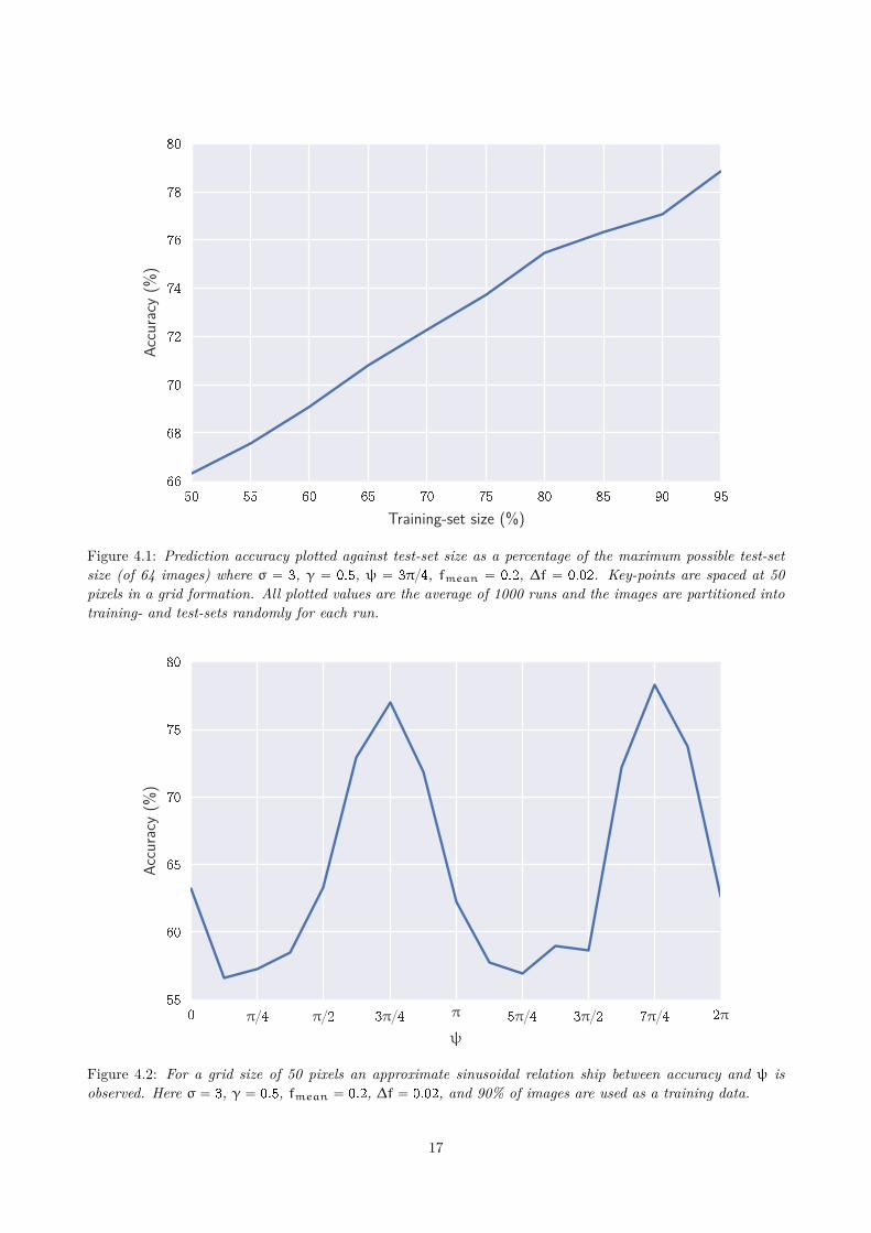
The effect of test-set size on prediction accuracy has been investigated and the results summarize in Figure 4.1.Prediction accuracy is expected to increases with increasing test-set size until it asymptotically approaches apeak value. Since the increase in accuracy is approximately linear for the entire range of test-set sizes tested, weconclude that the classifier has not been adequately trained. An increase in performance is therefore expectedwith a larger training-set size.
4.1 Comparison to human based trials
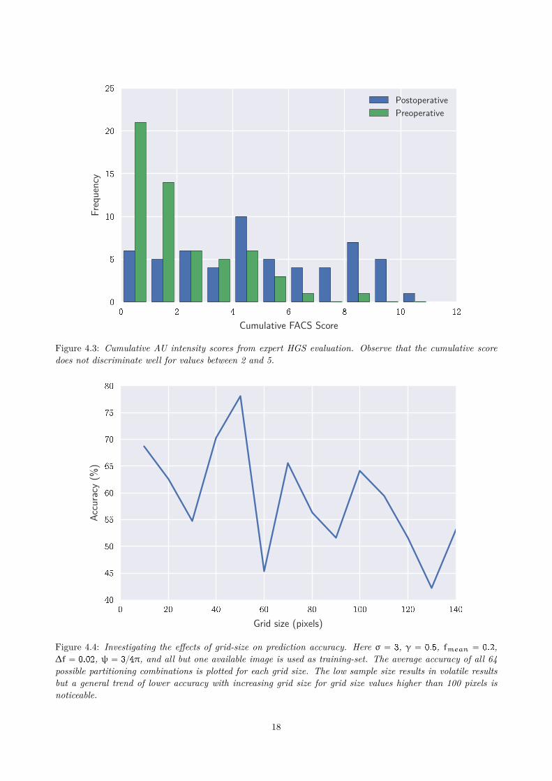
Dalla Costa et al. have carried out assessment of the prediction accuracy of the HGS (see Section 3.2.2).Five individuals with expertise in either horses or facial-expression assess and assign intensity scores to AUsdescribed as stiffly backwards ears, orbital tightening, tension above the ear area, prominent strained chewingmuscles, mouth strained and pronounced chin, and strained nostrils and flattening of the profile. Figure 3.1presents examples of these AUs. The values are then used by the experts to make an overall pain assessment.The study reports a prediction accuracy ranging from 67.5 to 77.8%, with an average of 73.3%. 17.0%of incorrect evaluations were false positives and 9.8% false negatives. We observe that raw score (sum ofintensity values for all AUs) have poor discrimination power for values between 2 and 5 which corresponds toapproximately 33% of the spectrum of values (see Figure 4.3).
While the results of the proposed automated approach compare well with those reported in the study, it isworth noting that there are systematic differences in both performance analyses. Dalla Costa’s evaluationincludes both front and lateral pictures of horses, while those used in the automated evaluation employed onlylateral views; and their pictures were not screened for image-quality, orientation or obstruction, while thoseused in the automated evaluation were (see Section 3.3.2). The effect of screening is not expected to have alarge impact on the performance of human evaluators since human vision is more tolerant to obstruction andout of plane rotation compared a computer algorithm.
16
50 55 60 65 70 75 80 85 90 95
Training-set size (%)
66
68
70
72
74
76
78
80Ac
cura
cy(%
)
Figure 4.1: Prediction accuracy plotted against test-set size as a percentage of the maximum possible test-setsize (of 64 images) where σ “ 3, γ “ 0.5, ψ “ 3π{4, fmean “ 0.2, ∆f “ 0.02. Key-points are spaced at 50pixels in a grid formation. All plotted values are the average of 1000 runs and the images are partitioned intotraining- and test-sets randomly for each run.
0 π{4 π{2 3π{4 π 5π{4 3π{2 7π{4 2π
ψ
55
60
65
70
75
80
Accu
racy
(%)
Figure 4.2: For a grid size of 50 pixels an approximate sinusoidal relation ship between accuracy and ψ isobserved. Here σ “ 3, γ “ 0.5, fmean “ 0.2, ∆f “ 0.02, and 90% of images are used as a training data.
17
0 2 4 6 8 10 12
Cumulative FACS Score
0
5
10
15
20
25Fr
eque
ncy
PostoperativePreoperative
Figure 4.3: Cumulative AU intensity scores from expert HGS evaluation. Observe that the cumulative scoredoes not discriminate well for values between 2 and 5.
0 20 40 60 80 100 120 140
Grid size (pixels)
40
45
50
55
60
65
70
75
80
Accu
racy
(%)
Figure 4.4: Investigating the effects of grid-size on prediction accuracy. Here σ “ 3, γ “ 0.5, fmean “ 0.2,∆f “ 0.02, ψ “ 3{4π, and all but one available image is used as training-set. The average accuracy of all 64possible partitioning combinations is plotted for each grid size. The low sample size results in volatile resultsbut a general trend of lower accuracy with increasing grid size for grid size values higher than 100 pixels isnoticeable.
18

5 Conclusions and recommendations
Based on the preceding findings, detecting pain through automated facial expression analysis in horses is viable.It has potential as an addition to the toolbox of non-invasive equine pain assessment and can compliment expertphysiological, behavioral and facial-expression based assessments. The findings of this limited study suggestthat it may even offer a superior alternative to an expert evaluated grimace scale, in accuracy, speed andconvenience.
The method proposed has the advantage of not requiring specialized equipment (besides cameras which areubiquitous in this age) or training. Once a classifier has been trained, pain evaluation can be made in a matterof minutes.
Since the proposed automation approach is agnostic to the emotion under assessment, it may have applicationsfor other emotional states. The challenge will be in constructing experiments where images can be capturedwith the with the emotion under test can be reliably captured. This is easy to achieve with humans, butrequire careful design and domain specific knowledge.
5.1 Proposals for future work
5.1.1 Animal facial expression database
A key challenge in this thesis has been the lack of a high-quality, expertly labeled image database. Researchersworking with human face detection and recognition and human facial expression detection have access toseveral databases for research purposes (see Section 3.1). There are strong arguments for creating such adatabase for horses (and indeed other mammals).
Compared to collecting humans facial expression data, compiling labeled data on equine facial recognition is avastly more complex endeavor, requiring expertise in veterinary sciences, animal behaviorally, clinical research,anesthesiology as well as data and computer sciences.
Such database can allow researchers in the ”analysis” aspect of facial expression analysis—such as codificationof facial expressions (into AUs or similar), performing comparative studies between animals, modeling exercisesor investigations into automation—to avoid diverting time and resources into this necessary preparatory step.Researchers and practitioners may easily compare results of various techniques by using these databases asbenchmarks (this is common with human face and facial expression databases). They also ease verification ofresults which may be challenging to do otherwise.
For these reasons, we recommend that an image database modeled on the aforementioned databases be devel-oped for animal facial expressions to spur and facilitate research in this area.
19

5.1.2 Comparative study of automated facial expression approaches
This approach for automated facial expression analysis has been developed around the constraints of availabledata. It is by no means is the only possible approach. A study evaluating the suitability for automated equinefacial-expression analysis of other approaches, such as those mentioned in Table 2.1 and their comparativeperformance may be a natural next step. The database of animal facial expressions proposed in the precedingsection will certainly help.
20
References
[1] V. Molony and J. E. Kent. Assessment of Acute Pain in Farm Animals Using Behavioral and Physiolog-ical Measurementsˆ 1ˆ,ˆ 2. JOURNAL OF ANIMAL SCIENCE-MENASHA THEN ALBANY THENCHAMPAIGN ILLINOIS- 75 (1997), 266–272.
[2] A. Desrosiers, D. Harrison, and A. Letham. Use of facial expressions for pain assessment in infants duringacute painful procedures. 17.1 (2015).
[3] N. Streets. EMOTION DETECTION FROM INFANT FACIAL EXPRESSIONS AND CRIES PritamPal , Ananth N . Iyer and Robert E . Yantorno , (2006), 721–724.
[4] K. B. Gleerup et al. An equine pain face. Veterinary anaesthesia and analgesia 42.1 (Jan. 2015), 103–14.issn: 1467-2995. doi: 10.1111/vaa.12212. url: http://www.ncbi.nlm.nih.gov/pubmed/25082060%20http://www.pubmedcentral.nih.gov/articlerender.fcgi?artid=PMC4312484.
[5] E. Dalla Costa et al. Development of the Horse Grimace Scale (HGS) as a pain assessment tool in horsesundergoing routine castration. PloS one 9.3 (2014), e92281. issn: 1932-6203. doi: 10.1371/journal.pone.0092281.
[6] C. Darwin, P. Ekman, and P. Prodger. The expression of the emotions in man and animals. OxfordUniversity Press, USA, 1998.
[7] P. Ekman and W. V. Friesen. Constants across cultures in the face and emotion. Journal of personalityand social psychology 17.2 (1971), 124.
[8] B. Fasel and J. Luettin. Automatic facial expression analysis: a survey. Pattern Recognition 36.1 (Apr.2003), 259–275. issn: 0031-3203. doi: 10.1016/S0031-3203(02)00052-3.
[9] P. Ekman and W. V. Friesen. Facial action coding system (1977).[10] S. G. Sotocinal et al. The Rat Grimace Scale: A partially automated method for quantifying pain in the
laboratory rat via facial expressions. Molecular Pain 7.1 (2011), 55. issn: 1744-8069. doi: 10.1186/1744-8069-7-55. url: http://www.molecularpain.com/content/7/1/55.
[11] K. B. Gleerup and C. Lindegaard. Recognition and quantification of pain in horses: A tutorial review.Equine Veterinary Education (2015), n/a–n/a. issn: 09577734. doi: 10.1111/eve.12383. url: http://doi.wiley.com/10.1111/eve.12383.
[12] S. Chevalier-Skolnikoff. Facial expression of emotion in nonhuman primates. Darwin and facial expres-sion: A century of research in review (1973), 11–89.
[13] J. Hamm et al. Automated Facial Action Coding System for dynamic analysis of facial expressions inneuropsychiatric disorders. Journal of Neuroscience Methods 200.2 (2011), 237–256. issn: 01650270.doi: 10.1016/j.jneumeth.2011.06.023. url: http://linkinghub.elsevier.com/retrieve/pii/S016502701100358X.
[14] T. F. Cootes, G. J. Edwards, and C. J. Taylor. Active appearance models. IEEE Transactions on PatternAnalysis and Machine Intelligence 23.6 (2001), 681–685. issn: 01628828. doi: 10.1007/BFb0054760. url:http://ieeexplore.ieee.org/xpls/abs%7B%5C_%7Dall.jsp?arnumber=927467$%5Cbackslash$nhttp://link.springer.com/chapter/10.1007/BFb0054766.
[15] I. Matthews et al. Active appearance models revisited. International Journal of Computer Vision 60.2(2004), 135–164. issn: 0920-5691. doi: 10.1023/B:VISI.0000029666.37597.d3. url: http://www.springerlink.com/index/K1N57K0228074X08.pdf.
[16] Y. Cui, Z. Jin, and W. Yang. A two step face alignment approach using statistical models. InternationalJournal of Advanced Robotic Systems 9 (2012), 1. issn: 17298806. doi: 10.5772/52207. url: http:
21

//www.intechopen. com/journals/international%7B%5C_%7Djournal%7B%5C_%7Dof%7B%5C_%7Dadvanced%7B%5C_%7Drobotic%7B%5C_%7Dsystems/a- two- step- face- alignment- approach-using-statistical-models.
[17] J. Saragih and R. Göcke. Learning AAM fitting through simulation. Pattern Recognition 42.11 (2009),2628–2636. issn: 00313203. doi: 10.1016/j.patcog.2009.04.014.
[18] M. J. Lyons et al. The Japanese female facial expression (JAFFE) database (1998).[19] A. Martinez and R. Benavente. The AR face database, 1998. Computer Vision Center, Technical Report
3 (2007).[20] P. Lucey et al. “The Extended Cohn-Kanade Dataset (CK+): A complete dataset for action unit and
emotion-specified expression”. Computer Vision and Pattern Recognition Workshops (CVPRW), 2010IEEE Computer Society Conference on. IEEE. 2010, pp. 94–101.
[21] A. K. Jain and S. Z. Li. Handbook of face recognition. Vol. 1. Springer, 2005. Chap. 1.[22] Y.-K. W. Y.-K. Wang et al. Improvement of Face Recognition by Eyeglass Removal. Intelligent Infor-
mation Hiding and Multimedia Signal Processing (IIH-MSP), 2010 Sixth International Conference on(2010). doi: 10.1109/IIHMSP.2010.64.
[23] N. Rose. “Facial Expression Classification using Gabor and Log-Gabor Filters”. 7th International Con-ference on Automatic Face and Gesture Recognition, 2006. FGR 2006. IEEE, 2006, pp. 346–350. isbn:0-7695-2503-2. doi: 10.1109/FGR.2006.49. url: http://ieeexplore.ieee.org/ielx5/10739/33863/01613044.pdf?tp=%7B%5C&%7Darnumber=1613044%7B%5C&%7Disnumber=33863%20http://ieeexplore.ieee.org/xpls/abs%7B%5C_%7Dall.jsp?arnumber=1613044%20http://ieeexplore.ieee.org/lpdocs/epic03/wrapper.htm?arnumber=1613044.
[24] M. Pantic and L. J. M. Rothkrantz. Automatic analysis of facial expressions: the state of the art. IEEETransactions on Pattern Analysis and Machine Intelligence 22.12 (2000), 1424–1445. issn: 0162-8828.doi: 10.1109/34.895976. url: %7B%3C%7DGo%20to%20ISI%7B%3E%7D://WOS:000165901900006$%5Cbackslash$nhttp://ieeexplore.ieee.org/ielx5/34/19391/00895976.pdf?tp=%7B%5C&%7Darnumber=895976%7B%5C&%7Disnumber=19391%20http://ieeexplore.ieee.org/ielx5/34/19391 / 00895976 . pdf ? tp = %7B % 5C & %7Darnumber = 895976 % 7B % 5C & %7Disnumber = 19391 % 20http ://ieeexplore.ieee.org/xpl/articleDe.
[25] J. O. J. Ou et al. “Automatic Facial Expression Recognition Using Gabor Filter and Expression Analysis”.Second International Conference on Computer Modeling and Simulation, 2010. ICCMS ’10. Vol. 2. 2010,pp. 215–218. isbn: 978-1-4244-5642-0. doi: 10.1109/ICCMS.2010.45. url: http://ieeexplore.ieee.org/ielx5/5420948/5421056/05421091.pdf?tp=%7B%5C&%7Darnumber=5421091%7B%5C&%7Disnumber=5421056%20http://ieeexplore.ieee.org/Xplore/cookiedetectresponse.jsp.
22