Autoimmune oophoritis: clinical presentation of an … report Page 1 of 5 Compe ... was located both...
5
Case report Page 1 of 5 Compe ng interests: none declared. Conflict of Interests: none declared. All authors contributed to the concep on, design, and prepara on of the manuscript, as well as read and approved the final manuscript. All authors abide by the Associa on for Medical Ethics (AME) ethical rules of disclosure. Licensee OA Publishing London 2013. Creative Commons Attribution Licence (CC-BY) FĔė ĈĎęĆęĎĔē ĕĚėĕĔĘĊĘ: Varras M, Anastasiadis A, Panelos J, Balassi E, Demou A, Akrivis CH. Autoimmune oophoritis: clinical presentation of an unusual clinical entity. OA Case Reports 2013 Jan 31;2(1):7. Autoimmune oophoritis: clinical presentation of an unusual clinical entity M Varras 1 *, A Anastasiadis 2 , J Panelos 3 , E Balassi 3 , A Demou 3 , CH Akrivis 4 Abstract Introduction Autoimmune oophoritis is a rare co- ndition, which provokes ovarian fail- ure with either primary amenorrhea or secondary amenorrhea and a sub- sequent loss of fertility and ovarian hormonal function. The purpose of t- his report is to document the clinical findings from two patients with aut- oimmune oophoritis. Cases report Two cases of autoimmune oophorit- is are presented whose histopathol- ogical findings were consistent with international literature. Both cases were histopathologically characteri- sed by lymphocytic and plasmacytic inflammatory infiltrations around t- he cystic follicles. The inflammation was located both in the theca and gr- anular layers. Conclusion Patients with autoimmune oophorit- is should be recognised by the histo- pathology of the ovarian biopsies as they are at an increased risk of deve- loping other autoimmune disorders. Introduction Premature ovarian failure (POF) is a condition characterised by amen- orrhea, some hot flushes, elevated serum gonadotropin levels and hypo-oestrogenism with associated infertility in women before the age of 40 years 1-4 . The aetiologies of this condition include chromosomal anomalies (such as X chromosome monosomy, translocations or partial deletions), genetic predisposition (such as fragile X pre-mutations, BMP15 or DIAPH2 mutations), infec- tious diseases, complications of chem- otherapy, pelvic radiotherapy, surgical interventions or surgery, enzymatic disorders and endometriosis. Prema- ture ovarian failure might also be idio- pathic or autoimmune 2 . Among a total of 266 patients with spontaneous POF, 4% were diagnosed to have autoim- mune oophoritis 3,4 . Ovarian autoimmunity was first reported and serologically docu- mented by Vallotton and Forbes in 1966 5 . Autoimmune oophoritis is a distinct clinical entity and one of the causes of POF, particularly in women with secondary amenorrhea 6-8 . Auto- immune oophoritis generally occurs in the setting of autoimmune poly- endocrine syndromes and is asso- ciated commonly with other major endocrine failures such as diabetes mellitus, Addison’s disease, hypopar- athyroidism or hypothyroidism 8-10 . A wide clinical spectrum has also been demonstrated 11 . The aim of this report was to describe the clinical spectrum and the interesting pathological findings originating from small ovarian biop- sies from two patients with autoim- mune oophoritis. Case report Case 1 A 33-year-old woman presented with a 10-month history of secondary amenorrhoea and hot flushes. Menarche had occurred at an age of 14 years, she had developed secondary sex characteristics appropriately and had regular menses on a 28-day cycle. She had five full-term normal pregnancies without any miscar- riages. On pelvic bimanual examination, an anteverted uterus was palpated, while both fallopian tubes and ovaries were impalpable. Pelvic ultrasonog- raphy disclosed normal-sized ovaries with bilateral multicystic structures, with the largest follicle measuring 1.2 cm. The thickness of the endo- metrium was 3.5 mm. Initial inves- tigation showed her serum gonado- tropin concentrations to be elevated: follicle stimulating hormone (FSH) at 40 mIU/ml and luteinizing hormone (LH) at 56 mIU/ml. 17-beta oestra- diol was at 30 pg/ml (normal range: 30–100 pg/ml). Serum prolactin levels were normal and there was no evidence of hypoparathyroidism. Thyroid function tests were normal. Also, kidneys and liver function tests were normal. Serum electrolytes were normal. Proteinuria was nega- tive. Serum adrenocorticotropin hormone levels were normal. The progesterone challenge test was negative. At exploratory laparoscopy, her ovaries appeared small and inactive. There were no signs of abdominal or pelvic inflammatory processes. Biop- sies from both ovaries were obtained and endometrial curettage was performed as well. Grossly, two specimens had been obtained from the right ovary with a whitish-grey colour, measuring 0.7 cm and 1.2 cm in their greatest dimension, respectively, and one specimen had been obtained from the left ovary with white tan to grey colour measuring 1.6 cm in its greatest dimension. Also, the spec- imen from the endometrial biopsy measured 3.5 × 3 × 0.2 cm. At micro- * Corresponding author Email: [email protected] 1 Third Department of Obstetrics and Gynecol- ogy, ‘Elena Venizelou’ General Maternity Hos- pital, Athens, Greece 2 Fourth Department of Obstetrics and Gyne- cology, ‘Elena Venizelou’ General Maternity Hospital, Athens, Greece 3 Department of Pathology, ‘G. Chatzikosta’ General State Hospital, Ioannina, Greece 4 Department of Obstetrics and Gynecology, ‘G. Chatzikosta’ General State Hospital, Ioannina, Greece Obstetrics & Gynecology
Transcript of Autoimmune oophoritis: clinical presentation of an … report Page 1 of 5 Compe ... was located both...
Case report
Page 1 of 5
Com
pe n
g in
tere
sts:
non
e de
clar
ed. C
onfl i
ct o
f Int
eres
ts: n
one
decl
ared
. A
ll au
thor
s co
ntrib
uted
to th
e co
ncep
on,
des
ign,
and
pre
para
on
of th
e m
anus
crip
t, a
s w
ell a
s re
ad a
nd a
ppro
ved
the fi n
al m
anus
crip
t. A
ll au
thor
s ab
ide
by th
e A
ssoc
ia o
n fo
r Med
ical
Eth
ics
(AM
E) e
thic
al ru
les
of d
iscl
osur
e.
Licensee OA Publishing London 2013. Creative Commons Attribution Licence (CC-BY)
F : Varras M, Anastasiadis A, Panelos J, Balassi E, Demou A, Akrivis CH. Autoimmune oophoritis: clinical presentation of an unusual clinical entity. OA Case Reports 2013 Jan 31;2(1):7.
Autoimmune oophoritis: clinical presentation of an unusual clinical entity
M Varras1*, A Anastasiadis2, J Panelos3, E Balassi3, A Demou3, CH Akrivis4
AbstractIntroductionAutoimmune oophoritis is a rare co-ndition, which provokes ovarian fail-ure with either primary amenorrhea or secondary amenorrhea and a sub-sequent loss of fertility and ovarian hormonal function. The purpose of t-his report is to document the clinical findings from two patients with aut-oimmune oophoritis.Cases reportTwo cases of autoimmune oophorit-is are presented whose histopathol-ogical findings were consistent with international literature. Both cases were histopathologically characteri-sed by lymphocytic and plasmacytic inflammatory infiltrations around t-he cystic follicles. The inflammation was located both in the theca and gr-anular layers.ConclusionPatients with autoimmune oophorit-is should be recognised by the histo-pathology of the ovarian biopsies as they are at an increased risk of deve-loping other autoimmune disorders.
IntroductionPremature ovarian failure (POF) is a condition characterised by amen-orrhea, some hot flushes, elevated serum gonadotropin levels and hypo- oestrogenism with associated
infertility in women before the age of 40 years1-4. The aetiologies of this condition include chromosomal anomalies (such as X chromosome monosomy, translocations or partial deletions), genetic predisposition (such as fragile X pre-mutations, BMP15 or DIAPH2 mutations), infec-tious diseases, complications of chem-otherapy, pelvic radiotherapy, surgical interventions or surgery, enzymatic disorders and endometriosis. Prema-ture ovarian failure might also be idio-pathic or autoimmune2. Among a total of 266 patients with spontaneous POF, 4% were diagnosed to have autoim-mune oophoritis3,4.
Ovarian autoimmunity was first reported and serologically docu-mented by Vallotton and Forbes in 19665. Autoimmune oophoritis is a distinct clinical entity and one of the causes of POF, particularly in women with secondary amenorrhea6-8. Auto-immune oophoritis generally occurs in the setting of autoimmune poly-endocrine syndromes and is asso-ciated commonly with other major endocrine failures such as diabetes mellitus, Addison’s disease, hypopar-athyroidism or hypothyroidism8-10. A wide clinical spectrum has also been demonstrated11.
The aim of this report was to describe the clinical spectrum and the interesting pathological findings originating from small ovarian biop-sies from two patients with autoim-mune oophoritis.
Case report Case 1A 33-year-old woman presented with a 10-month history of secondary amenorrhoea and hot flushes. Menarche had occurred at an age of 14 years, she had developed secondary sex characteristics appropriately
and had regular menses on a 28-day cycle. She had five full-term normal pregnancies without any miscar-riages.
On pelvic bimanual examination, an anteverted uterus was palpated, while both fallopian tubes and ovaries were impalpable. Pelvic ultrasonog-raphy disclosed normal-sized ovaries with bilateral multicystic structures, with the largest follicle measuring 1.2 cm. The thickness of the endo-metrium was 3.5 mm. Initial inves-tigation showed her serum gonado-tropin concentrations to be elevated: follicle stimulating hormone (FSH) at 40 mIU/ml and luteinizing hormone (LH) at 56 mIU/ml. 17-beta oestra-diol was at 30 pg/ml (normal range: 30–100 pg/ml). Serum prolactin levels were normal and there was no evidence of hypoparathyroidism. Thyroid function tests were normal. Also, kidneys and liver function tests were normal. Serum electrolytes were normal. Proteinuria was nega-tive. Serum adrenocorticotropin hormone levels were normal. The progesterone challenge test was negative.
At exploratory laparoscopy, her ovaries appeared small and inactive. There were no signs of abdominal or pelvic inflammatory processes. Biop-sies from both ovaries were obtained and endometrial curettage was performed as well.
Grossly, two specimens had been obtained from the right ovary with a whitish-grey colour, measuring 0.7 cm and 1.2 cm in their greatest dimension, respectively, and one specimen had been obtained from the left ovary with white tan to grey colour measuring 1.6 cm in its greatest dimension. Also, the spec-imen from the endometrial biopsy measured 3.5 × 3 × 0.2 cm. At micro-
* Corresponding authorEmail: [email protected] Third Department of Obstetrics and Gynecol-
ogy, ‘Elena Venizelou’ General Maternity Hos-pital, Athens, Greece
2 Fourth Department of Obstetrics and Gyne-cology, ‘Elena Venizelou’ General Maternity Hospital, Athens, Greece
3 Department of Pathology, ‘G. Chatzikosta’ General State Hospital, Ioannina, Greece
4 Department of Obstetrics and Gynecology, ‘G. Chatzikosta’ General State Hospital, Ioannina, Greece
Obst
etr
ics
& G
ynecolo
gy

Case report
Page 2 of 5
Com
pe n
g in
tere
sts:
non
e de
clar
ed. C
onfl i
ct o
f int
eres
ts: n
one
decl
ared
.A
ll au
thor
s co
ntrib
uted
to th
e co
ncep
on,
des
ign,
and
pre
para
on
of th
e m
anus
crip
t, a
s w
ell a
s re
ad a
nd a
ppro
ved
the fi n
al m
anus
crip
t.A
ll au
thor
s ab
ide
by th
e A
ssoc
ia o
n fo
r Med
ical
Eth
ics
(AM
E) e
thic
al ru
les
of d
iscl
osur
e.
Licensee OA Publishing London 2013. Creative Commons Attribution Licence (CC-BY)
F : Varras M, Anastasiadis A, Panelos J, Balassi E, Demou A, Akrivis CH. Autoimmune oophoritis: clinical presentation of an unusual clinical entity. OA Case Reports 2013 Jan 31;2(1):7.
scopic examination, the ovarian biop-sies contained primordial, developing and atretic follicles. Dense inflamma-tion of the ovary was found (Figure 1). The most striking feature was the destruction of the developing follicles by lymphocytic and plasma-cytic infiltration (Figure 2), while the primordial follicles were apparently and entirely spared from this process (Figure 3). The inflammation was located both in the theca and granular layers. The ovarian stroma was unre-markable. Immunohistochemically, the inflammatory population was composed mainly from T-lympho-cytes (CD4+, CD8+) (Figures 4 and 5) and plasma cells (CD138+) (Figure 6). The endometrium had an oedematous appearance with focal haemorrhagic infiltration of the stroma. The endometrial glands were small, round, coated with cylinder epithelium and without any remark-able mitotic activity. No inflammation
was i dentified in the endometrium. The final pathological diagnosis was autoimmune oophoritis.
The patient was diagnosed as having POF due to autoimmune oophoritis, and she was started on oestrogen and progesterone replace-ment therapy. Laboratory tests were negative for rheumatoid factor, anti-nuclear antibodies (ANA), compo-nents 3 and 4 of the complement (C3 and C4), immunoglobulin (Ig) G and IgM alpha cardiolipin antibodies and C-reactive protein (CRP). The patient was missed from further follow-up.
Case 2 A 32-year-old woman presented with acute lower abdominal pain. Last menstrual period was three months previous to the date of presentation. Menarche had occurred at the age of 15 years and her menstrual cycle had been regular previously on a 29-day cycle. She had two previous caesarean sections and a medical
history of hypothyroidism; she was stable on thyroxine 0.1 mg daily.
On pelvic bimanual examination, an anteverted uterus was palpated and a painful cystic mass in the right adnexa was palpated as well. Pelvic ultrasonography revealed a well-defined cystic mass with a hypoechogenic appearance in the right adnexa uteri, measuring 8 cm in its maximum diameter and a small multicystic structure in the left adnexa. Free liquid in the posterior space of Douglas was not found.
Initial investigation showed normal blood cell counts, as well as normal function tests for liver, kidneys and thyroid. The INR and activated partial thromboplastin time ratios were normal. Elevated levels of serum carbohydrate antigen (CA)-19.9 (81 IU/ml, normal range <37) and CA-125 (39.7 U/ml, normal range: <37) were found, while the serum levels of CA-15.3, carcinoem-bryonic antigen and alpha fetoprotein
Figure 1: Case 1: dense inflammation of the ovary (haematoxylin-eosin × 100).
Figure 2: Case 1: dense inflamma-tory infiltrate destroys the follicle (haematoxylin-eosin × 200).
Figure 3: Case 1: primordial folli-cles are spared by the inflammation (haematoxylin-eosin × 400).
Figure 4: Case 1: T4 positive lympho-cytes (CD4 stain × 200).
Figure 5: Case 1: T8 positive lympho-cytes (CD8 stain × 200).
Figure 6: Case 1: CD138 positive plasma cells (× 100).
Case report
Page 3 of 5
Com
pe n
g in
tere
sts:
non
e de
clar
ed. C
onfl i
ct o
f Int
eres
ts: n
one
decl
ared
. A
ll au
thor
s co
ntrib
uted
to th
e co
ncep
on,
des
ign,
and
pre
para
on
of th
e m
anus
crip
t, a
s w
ell a
s re
ad a
nd a
ppro
ved
the fi n
al m
anus
crip
t. A
ll au
thor
s ab
ide
by th
e A
ssoc
ia o
n fo
r Med
ical
Eth
ics
(AM
E) e
thic
al ru
les
of d
iscl
osur
e.
Licensee OA Publishing London 2013. Creative Commons Attribution Licence (CC-BY)
F : Varras M, Anastasiadis A, Panelos J, Balassi E, Demou A, Akrivis CH. Autoimmune oophoritis: clinical presentation of an unusual clinical entity. OA Case Reports 2013 Jan 31;2(1):7.
were in normal ranges. The patient underwent exploratory laparoscopy with an ovarian cystectomy and a wedge resection of the right ovary. The left ovary appeared small and inactive. There were no signs of abdominal or pelvic inflammatory processes.
Grossly, the largest specimen had dimensions of 6 × 3 × 0.5 cm. Also, the other two histological pieces measuring 2.5 cm and 3.5 cm in their maximum diameter were sent for frozen biopsy. Cut sections revealed two cystic follicles measuring 0.4 cm and 1.0 cm in their maximum diam-eter, respectively.
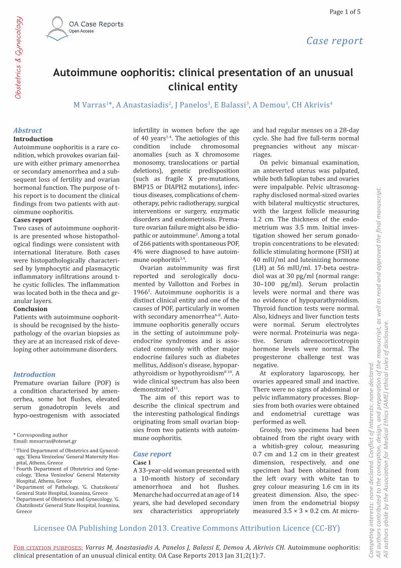
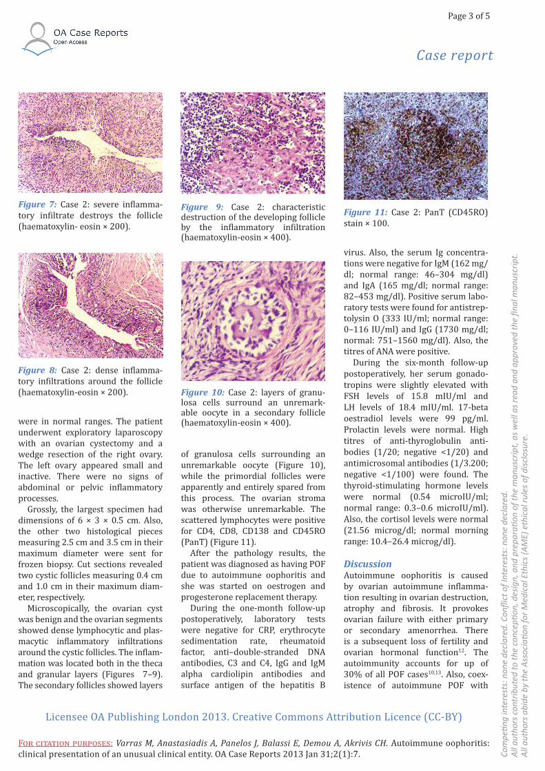
Microscopically, the ovarian cyst was benign and the ovarian segments showed dense lymphocytic and plas-macytic inflammatory infiltrations around the cystic follicles. The inflam-mation was located both in the theca and granular layers (Figures 7–9). The secondary follicles showed layers
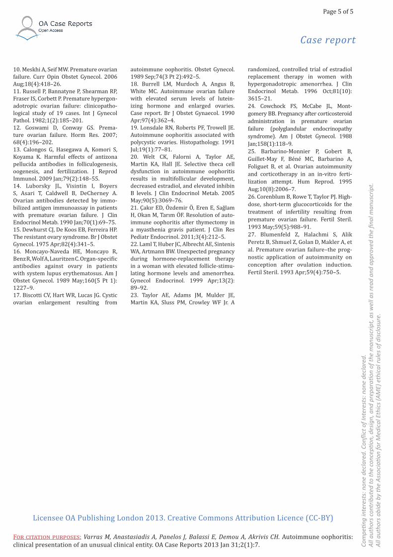
of granulosa cells surrounding an unremarkable oocyte (Figure 10), while the primordial follicles were apparently and entirely spared from this process. The ovarian stroma was otherwise unremarkable. The scattered lymphocytes were positive for CD4, CD8, CD138 and CD45RO (PanT) (Figure 11).
After the pathology results, the patient was diagnosed as having POF due to autoimmune oophoritis and she was started on oestrogen and progesterone replacement therapy.
During the one-month follow-up postoperatively, laboratory tests were negative for CRP, erythrocyte sedimentation rate, rheumatoid factor, anti–double-stranded DNA antibodies, C3 and C4, IgG and IgM alpha cardiolipin antibodies and surface antigen of the hepatitis B
virus. Also, the serum Ig concentra-tions were negative for IgM (162 mg/dl; normal range: 46–304 mg/dl) and IgA (165 mg/dl; normal range: 82–453 mg/dl). Positive serum labo-ratory tests were found for antistrep-tolysin O (333 IU/ml; normal range: 0–116 IU/ml) and IgG (1730 mg/dl; normal: 751–1560 mg/dl). Also, the titres of ANA were positive.
During the six-month follow-up postoperatively, her serum gonado-tropins were slightly elevated with FSH levels of 15.8 mIU/ml and LH levels of 18.4 mIU/ml. 17-beta oestradiol levels were 99 pg/ml. Prolactin levels were normal. High titres of anti-thyroglobulin anti-bodies (1/20; negative <1/20) and antimicrosomal antibodies (1/3.200; negative <1/100) were found. The thyroid-stimulating hormone levels were normal (0.54 microIU/ml; normal range: 0.3–0.6 microIU/ml). Also, the cortisol levels were normal (21.56 microg/dl; normal morning range: 10.4–26.4 microg/dl).
DiscussionAutoimmune oophoritis is caused by ovarian autoimmune inflamma-tion resulting in ovarian destruction, atrophy and fibrosis. It provokes ovarian failure with either primary or secondary amenorrhea. There is a subsequent loss of fertility and ovarian hormonal function12. The autoimmunity accounts for up of 30% of all POF cases10,13. Also, coex-istence of autoimmune POF with
Figure 7: Case 2: severe inflamma-tory infiltrate destroys the follicle (haematoxylin- eosin × 200).
Figure 8: Case 2: dense inflamma-tory infiltrations around the follicle (haematoxylin-eosin × 200).
Figure 9: Case 2: characteristic destruction of the developing follicle by the inflammatory infiltration (haematoxylin-eosin × 400).
Figure 10: Case 2: layers of granu-losa cells surround an unremark-able oocyte in a secondary follicle (haematoxylin-eosin × 400).
Figure 11: Case 2: PanT (CD45RO) stain × 100.

Case report
Page 4 of 5
Com
pe n
g in
tere
sts:
non
e de
clar
ed. C
onfl i
ct o
f int
eres
ts: n
one
decl
ared
.A
ll au
thor
s co
ntrib
uted
to th
e co
ncep
on,
des
ign,
and
pre
para
on
of th
e m
anus
crip
t, a
s w
ell a
s re
ad a
nd a
ppro
ved
the fi n
al m
anus
crip
t.A
ll au
thor
s ab
ide
by th
e A
ssoc
ia o
n fo
r Med
ical
Eth
ics
(AM
E) e
thic
al ru
les
of d
iscl
osur
e.
Licensee OA Publishing London 2013. Creative Commons Attribution Licence (CC-BY)
F : Varras M, Anastasiadis A, Panelos J, Balassi E, Demou A, Akrivis CH. Autoimmune oophoritis: clinical presentation of an unusual clinical entity. OA Case Reports 2013 Jan 31;2(1):7.
other autoimmune diseases such as those associated with the thyroid gland (e.g. Hashimoto’s and Graves’s disease) and adrenal gland (e.g. Addi-son’s disease) is often observed14, as in our case 2. Approximately 60% of POF cases without adrenal autoim-mune disease lack ovarian follicles, and in these cases, fibrotic ovaries are found15. Moreover, ANA and rheuma-toid factors have been reported with a higher frequency in POF patients than normal4. Moncayo-Naveda et al. reported the presence of anti-ovarian antibodies in 84% of the cases with system lupus erythematosus16.
The patient of our case 2 presented with a large ovarian cyst measuring 8 cm in its maximum diameter and this is in agreement with the observations of other studies with autoimmune POF17-21. The possible mechanism for the development of ovarian cysts in patients with auto-immune POF is due to the elevated gonadotropin levels because of the impaired negative feedback and the subsequent overstimulation of the ovarian tissues21. Both of our cases were amenorrheic for 10 months (case 1) and three months (case 2) with increased FSH levels compared to women with regular cycles. In patients with autoimmune oophor-itis, hormone replacement should be used as in our patients. During hormone-replacement therapy, elevated gonadotropin levels usually return to physiologically normal ranges22. As a result, folliculogen-esis may occur and less frequently is followed by ovulation and rarely by pregnancy23. Both of our patients had completed their families. However, for patients who desire a child, there are multiple case reports in which corticosteroid treatment has resulted in pregnancies in women with auto-immune oophoritis and amenor-rhea20. Cowchock et al. described a patient who had been treated with oestrogen replacement therapy because of POF for more than 15 years. However, the patient devel-oped Addison’s disease and was
treated with corticosteroid replace-ment therapy. An uneventful preg-nancy was achieved one year after commencement of corticosteroid replacement therapy24. Luborsky et al. described two patients with documented POF who became preg-nant, and each patient delivered a healthy infant after treatment with high doses of corticoster-oids. However, in both cases, POF has resumed after delivery14. Also, Barbarino-Monnier et al. reported a pregnancy and delivery after in vitro fertilisation in a patient with anti-ovarian autoimmunity treated with corticosteroids25. Apart from the case reports, Corenblum et al. studied 11 chromosomally normal patients with POF who received high doses of corticosteroids for 15 days and found that two of them had resumed ovarian function and became pregnant26. In addition, Blumenfeld et al. studied 15 patients with autoimmune POF treated with human menopausal gonadotropins and corticosteroids after pituitary desensitisation with a gonadotropin-releasing hormone agonist. Eight of 15 patients had become pregnant at least once, and in total, 14 pregnancies were achieved. All of these pregnancies were obtained within the first three months after the onset of treatment27.
ConclusionAutoimmune oophoritis is diagnosed by laparoscopic ovarian biopsy, which on microscopic examination reveals a folliculotropic, lymphoid infiltrate that affects developing folli-cles with a theca layer, corpora lutea and atretic follicles, and it indicates the patient’s risk of developing other autoimmune diseases. Autoimmune oophoritis should be kept in mind by gynaecologists when treating women with POF. These patients should be treated with hormone replacement therapy. For young patients with autoimmune POF who desire a preg-nancy, high doses of corticosteroids might help in the achievement of pregnancy.
ConsentWritten informed consent was obtained from all patients for publi-cation of this series study and accom-panying images. A copy of the written consents is available for review by the Editor-in-Chief of this journal.
Abbreviations listANA, antinuclear antibodies; CA, carbohydrate antigen; CRP, C-reactive protein; C3 and C4, components 3 and 4 of the complement; FSH, follicle stimulating hormone; Ig, immuno-globulin; LH, luteinizing hormone; POF, premature ovarian failure.
References1. Forges T, Monnier- Barbarino P, FaureGC, Bene MC. Autoimmunity and antigenic targets in ovarian pathology. Hum Reprod Update. 2004 Mar–Apr;10(2):163–75.2. Bats AS, Barbarino PM, Bene MC,Faure GC, Forges T. Local lymphocytic and epithelial activation in a case of auto-immune oophoritis. Fertil Steril. 2008 Sep;90(3):849.e5–8. 3. Bakalov VK, Anasti JN, Calis KA, Vander-hoof VH, Premkumar A, Chen S, et al. Auto-immune oophoritis as a mechanism of follicular dysfunction in women with 46,XX spontaneous premature ovarian failure. Fertil Steril. 2005 Oct;84(4):958–65.4. La Marca A, Brozzetti A, Sighinolfi G, Marzotti S, Volpe A, Falorni A. Primary ovarian insufficiency: autoimmune causes. Curr Opin Obstet Gynecol. 2010 Aug;22(4):277–82. 5. Vallotton MB, Forbes AP. Antibodies to cytoplasm of ova. Lancet. 1966 Jul;2(7457):67–85.6. Sedmak DD, Hart WR, Tubbs RR. Auto-immune oophoritis: a histopathologic study of involved ovaries with immuno-logic characterization of the mononu-clear cell infiltrate. Int J Gynecol Pathol. 1987;6(1):73–81.7. Suh SL. Autoimmune oophoritis–a case report. J Korean Med Sci. 1992 Sep;7(3):284–90.8. Welt CK. Autoimmune oophoritis in the adolescent. Ann N Y Acad Sci. 2008;1135:118–22.9. Friedman CI, Gurgen-Varol F, Lucas J, Neff J. Persistent progesterone produc-tion associated with autoimmune oophoritis. A case report. J Reprod Med. 1987 Apr;32(4):293–6.
Case report
Page 5 of 5
Com
pe n
g in
tere
sts:
non
e de
clar
ed. C
onfl i
ct o
f Int
eres
ts: n
one
decl
ared
. A
ll au
thor
s co
ntrib
uted
to th
e co
ncep
on,
des
ign,
and
pre
para
on
of th
e m
anus
crip
t, a
s w
ell a
s re
ad a
nd a
ppro
ved
the fi n
al m
anus
crip
t. A
ll au
thor
s ab
ide
by th
e A
ssoc
ia o
n fo
r Med
ical
Eth
ics
(AM
E) e
thic
al ru
les
of d
iscl
osur
e.
Licensee OA Publishing London 2013. Creative Commons Attribution Licence (CC-BY)
F : Varras M, Anastasiadis A, Panelos J, Balassi E, Demou A, Akrivis CH. Autoimmune oophoritis: clinical presentation of an unusual clinical entity. OA Case Reports 2013 Jan 31;2(1):7.
10. Meskhi A, Seif MW. Premature ovarian failure. Curr Opin Obstet Gynecol. 2006 Aug;18(4):418–26.11. Russell P, Bannatyne P, Shearman RP, Fraser IS, Corbett P. Premature hypergon-adotropic ovarian failure: clinicopatho-logical study of 19 cases. Int J Gynecol Pathol. 1982;1(2):185–201.12. Goswami D, Conway GS. Prema-ture ovarian failure. Horm Res. 2007;68(4):196–202.13. Calongos G, Hasegawa A, Komori S, Koyama K. Harmful effects of antizona pellucida antibodies in folliculogenesis, oogenesis, and fertilization. J Reprod Immunol. 2009 Jan;79(2):148–55.14. Luborsky JL, Visintin I, BoyersS, Asari T, Caldwell B, DeCherney A. Ovarian antibodies detected by immo-bilized antigen immunoassay in patients with premature ovarian failure. J Clin Endocrinol Metab. 1990 Jan;70(1):69–75.15. Dewhurst CJ, De Koos EB, Ferreira HP. The resistant ovary syndrome. Br J Obstet Gynecol. 1975 Apr;82(4):341–5.16. Moncayo-Naveda HE, Moncayo R,Benz R, Wolf A, Lauritzen C. Organ-specific antibodies against ovary in patients with system lupus erythematosus. Am J Obstet Gynecol. 1989 May;160(5 Pt 1):1227–9.17. Biscotti CV, Hart WR, Lucas JG. Cystic ovarian enlargement resulting from
autoimmune oophoritis. Obstet Gynecol. 1989 Sep;74(3 Pt 2):492–5.18. Burrell LM, Murdoch A, Angus B, White MC. Autoimmune ovarian failure with elevated serum levels of lutein-izing hormone and enlarged ovaries. Case report. Br J Obstet Gynaecol. 1990 Apr;97(4):362–4.19. Lonsdale RN, Roberts PF, Trowell JE. Autoimmune oophoritis associated with polycystic ovaries. Histopathology. 1991 Jul;19(1):77–81.20. Welt CK, Falorni A, Taylor AE, Martin KA, Hall JE. Selective theca cell dysfunction in autoimmune oophoritis results in multifollicular development, decreased estradiol, and elevated inhibin B levels. J Clin Endocrinol Metab. 2005 May;90(5):3069–76.21. Çakır ED, Özdemir Ö, Eren E, Sağlam H, Okan M, Tarım ÖF. Resolution of auto-immune oophoritis after thymectomy in a myasthenia gravis patient. J Clin Res Pediatr Endocrinol. 2011;3(4):212–5. 22. Laml T, Huber JC, Albrecht AE, Sintenis WA, Artmann BW. Unexpected pregnancy during hormone-replacement therapy in a woman with elevated follicle-stimu-lating hormone levels and amenorrhea. Gynecol Endocrinol. 1999 Apr;13(2):89–92.23. Taylor AE, Adams JM, Mulder JE, Martin KA, Sluss PM, Crowley WF Jr. A
randomized, controlled trial of estradiol replacement therapy in women with hypergonadotropic amenorrhea. J Clin Endocrinol Metab. 1996 Oct;81(10):3615–21.24. Cowchock FS, McCabe JL, Mont-gomery BB. Pregnancy after corticosteroid administration in premature ovarian failure (polyglandular endocrinopathy syndrome). Am J Obstet Gynecol. 1988 Jan;158(1):118–9.25. Barbarino-Monnier P, Gobert B, Guillet-May F, Béné MC, Barbarino A, Foliguet B, et al. Ovarian autoimmunity and corticotherapy in an in-vitro ferti-lization attempt. Hum Reprod. 1995 Aug;10(8):2006–7.26. Corenblum B, Rowe T, Taylor PJ. High-dose, short-term glucocorticoids for the treatment of infertility resulting from premature ovarian failure. Fertil Steril. 1993 May;59(5):988–91.27. Blumenfeld Z, Halachmi S, AlikPeretz B, Shmuel Z, Golan D, Makler A, et al. Premature ovarian failure–the prog-nostic application of autoimmunity on conception after ovulation induction. Fertil Steril. 1993 Apr;59(4):750–5.


![Marco Mendeni Selected Artworks - Theca Gallery Milano · BRUXELLES, Theca Gallery Lugano, curated by Matteo Bittanti, 2014 Mendeni*Kalinka, Drome, [.Box] Milano with Theca Gallery](https://static.fdocuments.net/doc/165x107/5c687ba209d3f2e4258b5e54/marco-mendeni-selected-artworks-theca-gallery-bruxelles-theca-gallery-lugano.jpg)


![Krishna Yajur Veda newest complete - MIU...Krishna Yajur Veda newest complete - MIU ... Yajur Veda]](https://static.fdocuments.net/doc/165x107/5e9132f90107e60dc30af5c7/krishna-yajur-veda-newest-complete-miu-krishna-yajur-veda-newest-complete.jpg)