
Author's personal copy - Cardiovascular Research Institutekornberg/Tom_Kornberg_Lab/pdfs/98. Guha,...
10
Author's personal copy Regulation of Drosophila matrix metalloprotease Mmp2 is essential for wing imaginal disc:trachea association and air sac tubulogenesis Arjun Guha 1 , Li Lin, Thomas B. Kornberg ⁎ Department of Biochemistry and Biophysics, University of California, San Francisco, CA 94143, USA abstract article info Article history: Received for publication 31 July 2009 Accepted 1 September 2009 Available online 12 September 2009 Keywords: Drosophila air sacs Tracheal morphogenesis FGF Basal lamina Matrix metalloproteinase The Drosophila Dorsal Air Sac Primordium (ASP) is a tracheal tube that grows toward Branchless FGF- expressing cells in the wing imaginal disc. We show that the ASP arises from a tracheal branch that invades the basal lamina of the disc to juxtapose directly with disc cells. We examined the role of matrix metalloproteases (Mmps), and found that reducing Mmp2 activity perturbed disc-trachea association, altered peritracheal distributions of collagen IV and Perlecan, misregulated ASP growth, and abrogated development of the dorsal air sacs. Whereas the function of the membrane-tethered Mmp2 in the ASP is non-cell autonomous we find that it may have distinct tissue-specific roles in the ASP and disc. These findings demonstrate a critical role for Mmp2 in tubulogenesis post-induction, and implicate Mmp2 in regulating dynamic and essential changes to the extracellular matrix. © 2009 Elsevier Inc. All rights reserved. Introduction Organogenesis integrates signaling, proliferation, patterning and structural processes to generate the variety of forms and functions essential to viability. Many organs develop from tubular outgrowths that assemble, grow and mature by a variety of mechanisms (reviewed in Hogan and Kolodziej, 2002). The only identified example of tubulogenesis in Drosophila that involves both cell proliferation and directed extension is the Dorsal Air Sac Primordium (ASP; Sato and Kornberg, 2002). This tubular outgrowth of the larval tracheal system is induced during the third instar (L3) period by Bnl/FGF expressed in the wing imaginal disc. Within 16 h of its induction, the ASP grows from a simple epithelial bud to a sac with many cells and with a distinct stalk and tip. During the subsequent pupal period, further growth and branching morphogenesis lead to its forming the Dorsal Air Sacs, the major tracheal organs of the adult fly that deliver oxygen to the thoracic flight muscles (Cabernard and Affolter, 2005; Guha and Kornberg, 2005; Sato and Kornberg, 2002). Although ASP development, structure and physiology are largely unexplored, the ASP promises to be an excellent system for investigating basic mechanisms of organogenesis. Here we report that regulation of the extracellular matrix (ECM) is essential for both cell proliferation and morphogenesis of the ASP and that the matrix metalloprotease Mmp2 plays a critical role. Epithelial tissues are typically associated with an ECM layered over their basal surface. The ECM is a meshwork of proteins and carbohydrate polymers that when viewed with an electron micro- scope appears as a basal lamina (BL) that has a tissue-proximal lamina lucida and an outer dense sheet, the lamina densa. BL thickness and composition vary in different tissues and during development. An example is the mammary ducts, whose BL appearance and compo- sition change during branching morphogenesis (Fata et al., 2004). The importance of BL assembly, disassembly and architecture during development and for tissue homeostasis is underscored by changes in the BL that accompany many disorders. Metastasis of tumors is thought to require BL-disassembly. While there are many candidate genes that potentially could regulate the ECM, the essential functions for maintenance and remodeling have not been definitively identified. Mmps constitute a large family of Zn-containing endopeptidases that proteolyze ECM proteins. Although in vivo evidence for Mmp roles is limited and most Mmp mutant mice develop without apparent consequence (Andrews et al., 2000; Brinckerhoff and Matrisian, 2002), there are several notable exceptions. Ectopic expression of Mmp3/Stromelysin perturbs both mammary gland development and influences breast cancer (Sternlicht et al., 1999). The phenotype of mice deficient for Gelatinase B/Mmp9 establishes a role for this proteinase in angiogenesis (Vu et al., 1998). Evidence from mice deficient for the membrane-tethered MT1-Mmp implicates this enzyme in connective tissue metabolism and white adipose tissue development, and suggests a role in degrading and remodeling pericellular collagen I (Chun et al., 2006; Holmbeck et al., 1999). Since the principal constituents of ECM (collagen IV, Laminin, Perlecan, Nidogen/Entactin; LeBleu et al., 2007) are conserved in metazoa (Hopf et al., 2001), it seems likely that the mechanisms for ECM-remodeling are also shared. Indeed, Mmps are conserved (Page- McCaw, 2008), and Drosophila has two—Mmp1 and Mmp2 (Llano et al., 2002; Llano et al., 2000; Page-McCaw et al., 2003). The Drosophila Developmental Biology 335 (2009) 317–326 Abbreviations: ASP, Air Sac Primordium; Mmp, Matrix metalloprotease; BL, Basal lamina; Bnl/FGF, Branchless-Fibroblast Growth Factor; TIMP, Tissue Inhibitor of Mmp. ⁎ Corresponding author. Fax: +1 415 476 2489. E-mail address: [email protected] (T.B. Kornberg). 1 Present address: Pulminary Center, Department of Medicine, Boston University School of Medicine, Boston, MA 02118, USA. 0012-1606/$ – see front matter © 2009 Elsevier Inc. All rights reserved. doi:10.1016/j.ydbio.2009.09.005 Contents lists available at ScienceDirect Developmental Biology journal homepage: www.elsevier.com/developmentalbiology
Transcript of Author's personal copy - Cardiovascular Research Institutekornberg/Tom_Kornberg_Lab/pdfs/98. Guha,...

Author's personal copy
Regulation of Drosophila matrix metalloprotease Mmp2 is essential for wingimaginal disc:trachea association and air sac tubulogenesis
Arjun Guha 1, Li Lin, Thomas B. Kornberg ⁎Department of Biochemistry and Biophysics, University of California, San Francisco, CA 94143, USA
a b s t r a c ta r t i c l e i n f o
Article history:Received for publication 31 July 2009Accepted 1 September 2009Available online 12 September 2009
Keywords:Drosophila air sacsTracheal morphogenesisFGFBasal laminaMatrix metalloproteinase
The Drosophila Dorsal Air Sac Primordium (ASP) is a tracheal tube that grows toward Branchless FGF-expressing cells in the wing imaginal disc. We show that the ASP arises from a tracheal branch that invadesthe basal lamina of the disc to juxtapose directly with disc cells. We examined the role of matrixmetalloproteases (Mmps), and found that reducing Mmp2 activity perturbed disc-trachea association,altered peritracheal distributions of collagen IV and Perlecan, misregulated ASP growth, and abrogateddevelopment of the dorsal air sacs. Whereas the function of the membrane-tethered Mmp2 in the ASP isnon-cell autonomous we find that it may have distinct tissue-specific roles in the ASP and disc. Thesefindings demonstrate a critical role for Mmp2 in tubulogenesis post-induction, and implicate Mmp2 inregulating dynamic and essential changes to the extracellular matrix.
© 2009 Elsevier Inc. All rights reserved.
Introduction
Organogenesis integrates signaling, proliferation, patterning andstructural processes to generate the variety of forms and functionsessential to viability. Many organs develop from tubular outgrowthsthat assemble, grow andmature by a variety ofmechanisms (reviewedin Hogan and Kolodziej, 2002). The only identified example oftubulogenesis in Drosophila that involves both cell proliferation anddirected extension is the Dorsal Air Sac Primordium (ASP; Sato andKornberg, 2002). This tubular outgrowthof the larval tracheal system isinducedduring the third instar (L3) period by Bnl/FGF expressed in thewing imaginal disc. Within 16 h of its induction, the ASP grows from asimple epithelial bud to a sac with many cells and with a distinct stalkand tip. During the subsequent pupal period, further growth andbranching morphogenesis lead to its forming the Dorsal Air Sacs, themajor tracheal organsof the adultfly that deliver oxygen to the thoracicflight muscles (Cabernard and Affolter, 2005; Guha and Kornberg,2005; Sato and Kornberg, 2002). Although ASP development, structureand physiology are largely unexplored, the ASP promises to be anexcellent system for investigating basic mechanisms of organogenesis.Here we report that regulation of the extracellular matrix (ECM) isessential for both cell proliferation and morphogenesis of the ASP andthat the matrix metalloprotease Mmp2 plays a critical role.
Epithelial tissues are typically associated with an ECM layered overtheir basal surface. The ECM is a meshwork of proteins and
carbohydrate polymers that when viewed with an electron micro-scope appears as a basal lamina (BL) that has a tissue-proximal laminalucida and an outer dense sheet, the lamina densa. BL thickness andcomposition vary in different tissues and during development. Anexample is the mammary ducts, whose BL appearance and compo-sition change during branchingmorphogenesis (Fata et al., 2004). Theimportance of BL assembly, disassembly and architecture duringdevelopment and for tissue homeostasis is underscored by changes inthe BL that accompany many disorders. Metastasis of tumors isthought to require BL-disassembly. While there are many candidategenes that potentially could regulate the ECM, the essential functionsfor maintenance and remodeling have not been definitively identified.
Mmps constitute a large family of Zn-containing endopeptidasesthat proteolyze ECM proteins. Although in vivo evidence for Mmproles is limited andmostMmpmutantmice developwithout apparentconsequence (Andrews et al., 2000; Brinckerhoff and Matrisian,2002), there are several notable exceptions. Ectopic expression ofMmp3/Stromelysin perturbs both mammary gland development andinfluences breast cancer (Sternlicht et al., 1999). The phenotype ofmice deficient for Gelatinase B/Mmp9 establishes a role for thisproteinase in angiogenesis (Vu et al., 1998). Evidence from micedeficient for the membrane-tethered MT1-Mmp implicates thisenzyme in connective tissue metabolism and white adipose tissuedevelopment, and suggests a role in degrading and remodelingpericellular collagen I (Chun et al., 2006; Holmbeck et al., 1999).
Since the principal constituents of ECM (collagen IV, Laminin,Perlecan, Nidogen/Entactin; LeBleu et al., 2007) are conserved inmetazoa (Hopf et al., 2001), it seems likely that the mechanisms forECM-remodeling are also shared. Indeed, Mmps are conserved (Page-McCaw, 2008), and Drosophila has two—Mmp1 and Mmp2 (Llano etal., 2002; Llano et al., 2000; Page-McCaw et al., 2003). The Drosophila
Developmental Biology 335 (2009) 317–326
Abbreviations: ASP, Air Sac Primordium; Mmp, Matrix metalloprotease; BL, Basallamina; Bnl/FGF, Branchless-Fibroblast Growth Factor; TIMP, Tissue Inhibitor of Mmp.⁎ Corresponding author. Fax: +1 415 476 2489.
E-mail address: [email protected] (T.B. Kornberg).1 Present address: Pulminary Center, Department of Medicine, Boston University
School of Medicine, Boston, MA 02118, USA.
0012-1606/$ – see front matter © 2009 Elsevier Inc. All rights reserved.doi:10.1016/j.ydbio.2009.09.005
Contents lists available at ScienceDirect
Developmental Biology
j ourna l homepage: www.e lsev ie r.com/deve lopmenta lb io logy
Author's personal copy
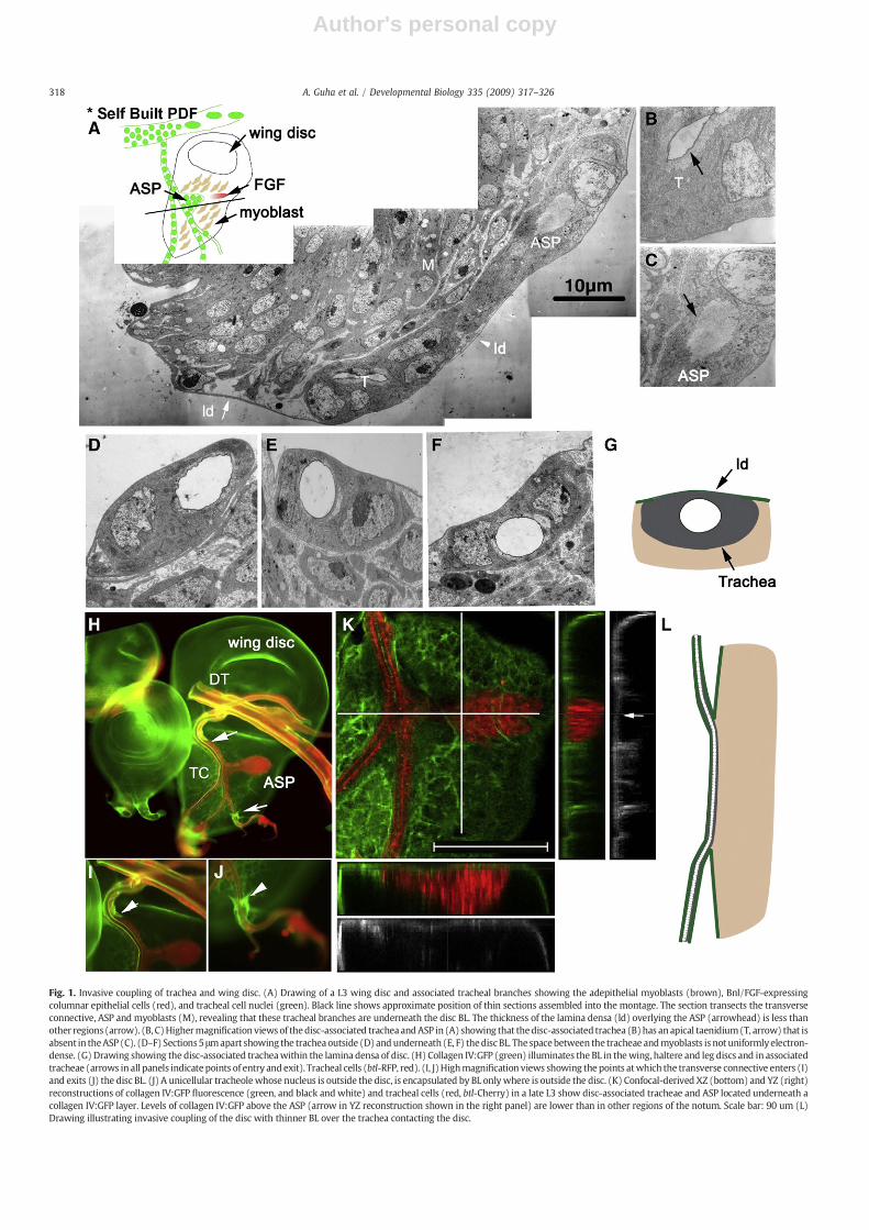
Fig. 1. Invasive coupling of trachea and wing disc. (A) Drawing of a L3 wing disc and associated tracheal branches showing the adepithelial myoblasts (brown), Bnl/FGF-expressingcolumnar epithelial cells (red), and tracheal cell nuclei (green). Black line shows approximate position of thin sections assembled into the montage. The section transects the transverseconnective, ASP and myoblasts (M), revealing that these tracheal branches are underneath the disc BL. The thickness of the lamina densa (ld) overlying the ASP (arrowhead) is less thanother regions (arrow). (B, C)Highermagnification views of thedisc-associated tracheaandASP in (A) showing that thedisc-associated trachea (B)has anapical taenidium(T, arrow) that isabsent in theASP (C). (D–F) Sections5 μmapart showing the tracheaoutside (D) andunderneath (E, F) thediscBL. The spacebetween the tracheae andmyoblasts is not uniformlyelectron-dense. (G) Drawing showing the disc-associated tracheawithin the lamina densa of disc. (H) Collagen IV:GFP (green) illuminates the BL in thewing, haltere and leg discs and in associatedtracheae (arrows in all panels indicate points of entry andexit). Tracheal cells (btl-RFP, red). (I, J)Highmagnification views showing the points atwhich the transverse connective enters (I)and exits (J) the disc BL. (J) A unicellular tracheolewhose nucleus is outside the disc, is encapsulated by BL onlywhere is outside the disc. (K) Confocal-derived XZ (bottom) and YZ (right)reconstructions of collagen IV:GFP fluorescence (green, and black and white) and tracheal cells (red, btl-Cherry) in a late L3 show disc-associated tracheae and ASP located underneath acollagen IV:GFP layer. Levels of collagen IV:GFP above the ASP (arrow in YZ reconstruction shown in the right panel) are lower than in other regions of the notum. Scale bar: 90 um (L)Drawing illustrating invasive coupling of the disc with thinner BL over the trachea contacting the disc.
318 A. Guha et al. / Developmental Biology 335 (2009) 317–326

Author's personal copy
Mmps (Dm-Mmp1 and Dm-Mmp2) are required for the morphogen-esis of various tissues (Miller et al., 2008; Page-McCaw et al., 2003)and appear to play roles in tumor invasion (Beaucher et al., 2007;Page-McCaw et al., 2003; Pastor-Pareja et al., 2004; Srivastava et al.,2007; Uhlirova and Bohmann, 2006). The sequences of the twoMmpssuggest that they are structurally distinct: Dm-Mmp1 is predicted tobe secreted and Dm-Mmp2 to be membrane-tethered by a glycosyl-phosphatidyl inositol anchor (Llano et al., 2000, 2002; Page-McCaw etal., 2003).
We were alerted to a possible role for ECM-remodeling in themorphogenesis of the ASP when we examined the disc:trachealinterface. We found that the ASP arises from a region of a trachealbranch that is directly juxtaposed to the wing disc. This trachealsegment lies underneath the disc BL and lacks a conspicuous,tracheal-specific BL. We investigated the role of Mmps in theregulation of this architecture to find that Dm-Mmp2 regulateslevels of collagen IV and Perlecan in the ECM around disc-associatedtrachea and the ASP, and this function is essential to properlyjuxtapose tracheal and disc cells, for growth of the ASP and fordevelopment of the Dorsal Air Sacs.
Materials and methods
Fly strains
btl-Gal4 (Shiga et al., 1996), ap-Gal4 (Flybase) are promoter- orenhancer trap Gal4 transgenes that express Gal4 in trachea (btl), thedorsal compartment of the wing disc but not in trachea (ap) and wereused to drive the expression of UAS transgenes. Tracheal cells wereilluminated by the expression of GAP-GFP (UAS GAP-GFP, Sato andKornberg, 2002), UAS-RFP and UAS-Cherry (gift from S. Roy). BL
distribution was observed using Perlecan protein traps (Flytrap; GFPProtein Trap Database: http://flytrap.med.yale.edu; G00022,ZCL1700, ZCL1973) and Viking (collagen IV; G00454). Mmp2W307,UAS TIMP, and UAS Mmp2 (Page-McCaw et al., 2003), FRTMmp2K07511 FRT Mmp2W307Mmp1Q112 (gift from A. Page-McCaw),UAS Mmp1RNAi, UAS Mmp2RNAi (Uhlirova and Bohmann, 2006), andMmp2 Gal4 (Srivastava et al., 2007) were used for the analysis ofMmp function. Bnl/FGF clones and ectopic expression were inducedas described in Sato and Kornberg (2002).
Fluorochromes and histochemistry
Nuclei were stained with DRAQ5 (Biostatus, Ltd.). Immunostain-ings of discs with mouse α-Discs large (1:50, DSHB) and mouse α-Dp-ERK (1:100, Sigma) were performed with a Cy3-conjugateddonkey anti-mouse secondary antibodies (1:750, Jackson Labs). RNAin situ hybridization was performed according to O'Neill and Bier(1994) and Klebes et al. (2002). DIG labeled antisense probe wasgenerated by in vitro transcription (Megascript) from HindIII digestedBnl cDNA (pBSKII, Sutherland et al., 1996)) and fragmentation at highpH. Alkaline phosphatase conjugated α-DIG antibody (Roche) wasused to detect the DIG labeled probe in situ.
Mounting and imaging fixed and unfixed imaginal discs
All discs were mounted using the “hanging drop” method (Satoand Kornberg, 2002) or in flat preparations (Vectashield) and wereimaged at room temperature. All epifluorescence images of collagenIV:GFP and Perlecan-GFP were acquired using the “hanging drop”method on a Zeiss Axioplan 2 microscope with a 10X Fluar, 0.5 NAobjective, a PCO Sensicam CCD camera (Cooke Corporation, USA)
Fig. 2. Tracheal tunneling does not restrict responsiveness to Bnl/FGF. Clones expressing ectopic Bnl/FGF (dark areas) attract outgrowths of the trachea from a region outside the BL(green; GFP). Region of disc in (A) delineated by white box is shown at high magnification in (B) to reveal the multicellular composition of the outgrowth. (C) Gap:GFP expressed intracheal cells illuminates the ASP and, from an adjacent tunneled region, an ectopic outgrowth (arrow) that extends toward Bnl-expressing cells (encircled by white line). (D)Collagen IV:GFP marks enlarged ASP and ectopic outgrowth. Arrowheads indicate “points of entry/exit” through the disc BL. (E) Collagen IV:GFP (arrows) labels extensions thatbridge the transverse connective and disc. (F, upper) Drawing showing normal association of transverse connective (green) with the wing disc (gray), and identifying Bnl/FGF-expressing cells (blue) and adepithelial myoblasts (red). BL (brown) encapsulates both trachea and disc except regions of invasive coupling. (F, lower) Drawing showing ectopicoutgrowths from transverse connective, penetrating the disc BL to contact disc clones expressing Bnl/FGF.
319A. Guha et al. / Developmental Biology 335 (2009) 317–326

Author's personal copy
using Slidebook acquisition software (Intelligent Imaging Innova-tions). The 20× Plan-NeoFluar, 0.5NA and 40× Plan-NeoFluarobjectives were used for other epifluorescence images. Opticalsections were acquired on Leica TCS SP2 confocal microscope systemusing a Leica DMRXE microscope with an HC PL APO 20×, 0.7 NAobjective. Identical acquisition settings were used for wild type andmutant tissue; all confocal images were acquired using a pinhole sizeof 1.0 Airy Unit. Brightfield images were acquired on a Leica DMRmicroscope equipped with SPOT CCD Camera (Diagnostics Instru-ments) and SPOT acquisition software. Adult flies were mounted inhalocarbon oil for imaging dorsal air sacs.
Electron microscopy
Larvae were dissected in 0.12 M sodium cacodylate buffer (CaCo;pH7.0) andfixed either overnight at 4°C or at room temperature for 1 hin 0.12 M CaCo, 2% glutaraldehyde. Following primary fixation, wingdiscs were dissected and stained with tannic acid (1%) and post-fixedin osmium tetroxide (2%). Samples were dehydrated in an ascendingseries of ethyl alcohol and embedded in a medium mixture of Embed821 resin. Ultra-thin sections were stained for contrast using 1%aqueous uranyl acetate and Reynold's lead citrate and examined andphotographed using either a JEOL 100CX transmission electronmicroscope or a FEI Technai Spirit transmission electron microscope.
Results
The transverse connective, ASP and wing imaginal disc areinvasively coupled
The ASP originates near a point of bifurcation of the transverseconnective and extends over the notum primordium of the wing disc
in an oblique, dorsoventral orientation where myoblasts overly thecolumnar epithelial cells (Fig. 1A). Contact between the transverseconnective and disc is sufficiently strong that the transverseconnective frequently accompanies wing discs when wing discs aredissected from larvae. Although association of the disc and transverseconnective can also be observed in intact larva, the nature of thecontact between the disc and tracheal tube has not been characterizedpreviously. Our finding that Bnl/FGF signals from the disc to thetransverse connective to induce the outgrowth of the ASP (Sato andKornberg, 2002) suggested that contact between the transverseconnective and the disc has a functional role. We therefore undertooka fine structure analysis to gain insight into the nature of theirassociation.
Tracheae are tripartite tubes with an ECM on the outer basalsurface, a tube consisting of epithelial cells, and apically, a cuticle(taenidium) that lines the lumen. Serial ultra-thin sections of discsand associated trachea revealed that the ASP differs from othertracheal tubes in two respects (number of specimens examined=2).The ASP lacks a taenidium (compare Figs. 1B, C). In addition, althoughthe BL that lines segments of the tracheal branch that do not contactthe disc has a thick lamina densa that was similar in appearance to theone that encapsulates the disc (Fig. 1D), the portions of the transverseconnective that are directly apposed to the disc does not (Figs. 1A, E, F).Instead, they are situated underneath the BL of the disc and weredirectly juxtaposed to the disc (shown schematically in Fig. 1G). Thesurfaces of tracheal cells situated underneath the disc BL are associatedwith lamina densa that is significantly thinner and less uniform thanthe lamina densa elsewhere in the disc. In some sections (not shown),tracheal cells of the transverse connective are directly juxtaposed tothe columnar epithelium, the source of Bnl/FGF. We refer to theplacement of the trachea underneath and inside the disc BL as“invasive coupling”.
Fig. 3. ECM dynamics during ASP morphogenesis. Collagen IV:GFP fluorescence marks ECM in wing disc and trachea in preparations isolated from early L3 larvae prior to ASPinduction (left panels, A, B) and at successively later stages (middle and right panels). Red: cherry expressed in btl domain in trachea. (A) Arrowheads indicate “points of entry/exit” through the disc BL; arrows indicate regions of dynamic changes in collagen IV levels. (B) Confocal images with adjacent XZ (bottom) and YZ (right) reconstructions showingthat the abundance of collagen IV:GFP around the trachea and ASP (solid lines) is lower (arrows) than in other regions (dashed lines). Number of preparations examined: pre-ASP: 5; early: 6; late 5.
320 A. Guha et al. / Developmental Biology 335 (2009) 317–326

Author's personal copy
Fig. 4. ECM abnormalities in animals expressing TIMP and inMmp2mutants. (A–C) In contrast to normal animals (A, ASP outlined in white), btl-TIMP (B) and ap-TIMP (C) specimenshave elevated collagen IV:GFP levels at edges of disc-associated transverse connective and ASP (arrows). (D) XZ (bottom) and YZ (right) reconstructions of collagen IV:GFPdistribution in ap-TIMP disc showing that collagen IV:GFP accumulates around the distal tip of the ASP (XZ, arrow) and that disc-associated trachea and ASP extrude from the disc(YZ, arrows). (E–G) Ultrastructure of ap-TIMP discs reveals defects in the ECM. Trachea is under the lamina densa (arrow), but ectopic lamina densa is detected around tunneledtrachea. (F) Higher resolution views of left boxed region in (E) showing disc lamina densa (arrow) and ectopic lamina densa (arrowheads), and extrusion from the disc (G, rightboxed region in (E)). (H) RNAi knockdown of mmp2 in the trachea recapitulates the phenotypes in (B) and (C). In mmp2W307 (I, J), ectopic collagen IV:GFP is detected around disc-associated tracheae. Note both extrusion of the trachea (J, XZ arrows) and accumulation of collagen-GFP along the lateral margins (right arrow in XZ, arrows in YZ) of the ASP (K, L)Ultrastructure of mmp2W307 discs at two positions along the disc–trachea interface showing the trachea underneath the disc lamina densa but extruded from the disc proper. (M)Diagram depicting defects in disc–tracheal association and lamina densa in ap-TIMP and Mmp2W307 animals. (N) Diagram showing the distribution and levels of collagen IV andPerlecan in specimens with wild type and reduced Mmp2 activity.
321A. Guha et al. / Developmental Biology 335 (2009) 317–326

Author's personal copy
322 A. Guha et al. / Developmental Biology 335 (2009) 317–326

Author's personal copy
To further characterize the tracheal-wing disc association, weexamined protein trap strains that express GFP in the ECM. Weexamined a line that tags collagen IV (Figs. 1H-K) and three lines thattag Perlecan (not shown). In specimens dissected from L3 larvae of allfour lines, we detected similar patterns of strong fluorescence aroundthe disc and aroundmost of the tracheal branches (Fig. 1H). However,we did not detect strong fluorescence from the regions of the tracheathat contact the disc (Figs. 1H–J). In addition to the absence of strongfluorescence around the trachea in this region of contact, the intensityof fluorescence in this region was lower than in other regions of thewing imaginal disc. The pattern of fluorescence correlated well withthe appearance of the lamina densa in ultrastructural studies—wedetected a prominent, thick layer over the disc and around trachealbranches, except the portion of the transverse connective that contactsthe disc. In these regions, the lamina densa overlying the trachea wassignificantly thinner. Confocal microscopy of collagen IV:GFP expres-sing discs also showed that both disc-associated trachea and the ASPwere underneath the disc BL, directly juxtaposed to the disc (Fig. 1K).In addition, both XZ and YZ reconstructions showed that the levels ofCollagen IV:GFP overlying the ASP were significantly lower than otherregions of the notum (Figs. 1K, YZ). Epi-fluorescence images clearlyrevealed the points of tracheal “entry” and “exit” (Figs. 1H–J) thatdefine the extent of “invasion”; these points were similar in allspecimens we examined. Fig. 1L summarizes the disposition of thetransverse connective with respect to the disc and BL.
Based on these observations we conclude that the wing disc andtrachea are invasively coupled and that the ASP grows out from thisinvasively coupled tracheal segment. Since wewere unable to detect acontinuous BL around the tracheal segments that are coupled to thedisc (no lamina densa was present at the transverse connective:discinterface, it may be that disc-derived cues suppress BL assemblylocally. We note that the unicellular tracheole that emanates from thetransverse connective at a point distal to the ASP (Figs. 1H, J) hasportions both inside and outside the disc BL. Since only the portion ofthe tube that is outside the disc BL had collagen IV, BL assembly by thistracheole is not cell autonomous.
The BL is functionally transparent to FGF signaling
Growth of the ASP from a region of the trachea that invasivelycouples with the disc raises the question: is direct contact necessaryfor FGF signaling to induce the ASP? Would interposition of BLbetween the disc and tracheal cells abrogate signaling? In order todetermine if direct contact is essential for FGF signaling, we inducedclones of Bnl/FGF-expressing cells and monitored responses oftrachea by examining fluorescence of either btl-GAP:GFP or btl-collagen IV:GFP. Sincewe observed association of disc and trachea anddetected collagen IV:GFP in the ECM around these tissues inpreparations from first instar larvae (data not shown), induction ofBnl/FGF expression in first and second instar larvae would allow us toaddress the responses of the trachea after initial BL assembly. EctopicBnl/FGF expression induced outgrowths from the transverse connec-tive, both from the portions that had invasively coupled with the discas well as outlying regions that were outside the disc BL (Figs. 2A, C,D). The latter type of outgrowths were multi-cellular (Fig. 2B) andoriented toward Bnl/FGF-expressing disc cells, and some had thincollagenous structures that connected trachea and disc that could be
discerned (Fig. 2E). Since these ectopic structures suggested thatsignaling is not blocked by the disc BL (schematized in Fig. 2F), wesuggest that direct juxtaposition of disc and trachea is not essential forFGF signaling and tubulogenesis, and that the BL is functionallytransparent to FGF signaling.
Matrix metalloprotease is required during ASP outgrowth for ECMremodeling at the disc:trachea junction
The disc-associated trachea and ASP are situated beneath alayer of collagen IV:GFP-containing ECM from the earliest stages ofASP induction (Figs. 3A, B). However, during the early stages ofgrowth, collagen IV-GFP distribution accumulated along the lateralmargins of the disc-associated trachea and along the lateral anddistal margins of ASP. These bands of fluorescence were variable,but at late stages, most specimens had no apparent pericellularcollagen IV:GFP (Fig. 3A). The levels of collagen IV:GFP in the BLthat overlies both the invasively coupled TC and ASP were lower atall stages of growth (Fig. 3B). We interpret both the lower levels ofcollagen IV:GFP in the BL overlying the ASP and the transientelevation of pericellular collagen IV:GFP fluorescence along thelateral and distal edges of the ASP at early stages of ASP growth asevidence of active ECM remodeling during growth and extension ofthe ASP.
The invasive nature of the disc–tracheal association, together withthe dynamic changes in the ECM around disc-associated tracheae,suggested that enzymes involved in ECM remodeling might playimportant roles. The Drosophila genome encodes two MMPs, Dm-Mmp1 and Dm-Mmp2 (Llano et al., 2000, 2002; Page-McCaw et al.,2003), as well as a tissue inhibitor of MMPs (TIMP; Brew et al., 2000).Previous studies reported that Dm-Mmp1 is expressed in the tracheaand the ASP, and that Dm-Mmp2 is expressed in the trachea, ASP andwing disc (Llano et al., 2002; Page-McCaw et al., 2003; Srivastava etal., 2007; Uhlirova and Bohmann, 2006). Drosophila TIMP is thoughtto inhibit both Dm-Mmp1 and Dm-Mmp2 in the extracellular milieu,although its endogenous distribution and roles are not wellunderstood.
To assess the roles of Mmps, we examined animals in which TIMPwas ectopically expressed, mutant animals defective forMMP activity,as well as animals in which RNAi was expressed to reduce Mmp1 andMmp2 levels. As detailed in the subsequent paragraphs, theseapproaches led us to focus on the role of Mmp2. While ectopic TIMPexpression, which reduces both Mmp1 and Mmp2 activities per-turbed disc:tracheal association and ASP growth, the presence ofMmp1RNAi in the btl expression domain had no apparent effect. Andalthough most Mmp1 mutants exhibited early larval lethality, someMmp1Q273 mutant animals grew slowly and reached the pupal stage,but the imaginal discs, trachea and ASP in these mutants appearednormal. Thus, we found no role for Mmp1 in disc:tracheal association,ASP growth, or ECM remodeling (data not shown).
When TIMP was expressed in the apterous (ap-GAL4 UAS-TIMP,(ap-TIMP); number of specimens examined=12) or btl domains (btl-GAL4 UAS-TIMP, (btl-TIMP); number of specimens examined=8),the general level of collagen IV:GFP fluorescence was comparable towild type, as was the fluorescence of tracheal branches not in contactwith the disc (Figs. 4A-D). However, collagen IV:GFP fluorescenceincreased at specific locations at which the invasively coupled
Fig. 5. Role of Mmp2 in FGF-dependent ASP growth. (A) Disc from WT animal (48–50 h, L3) stained with α-Dlg. Similar preparations from ap-TIMP (B) and Mmp2W307 (C) animals(48–50 h, L3) revealed ASP induction, but no directional growth. A requirement for Mmp2 expression in the trachea was revealed in preparations from btl- Mmp2RNAi-expressinganimals (D). Mmp2 knockdown in the disc (ap-Mmp2RNAi) resulted in hyperplastic ASP growth (E). Knockdown of Mmp2 did not diminish Bnl/FGF expression (arrow in (F)) in thedisc (F–I) or FGF signaling in the ASP (K-O). Knockdown of Mmp2 in the disc (ap-Mmp2RNAi) resulted in expansion of the domain of Bnl/FGF expression (arrows, J). (P) Expression ofmmp2GAL4 (Mmp2-nls GFP) was detected at higher levels at the distal edge of the ASP. (Q) Animals in which FGF expression was induced by heat shock showed hypertrophy of theASP (outlined with a white line) and increased levels of Mmp2-nls GFP expression. (R) Model for the role of Mmp2 in ASP morphogenesis. Proliferation and growth of the ASP isaccompanied by ECM remodeling (black arrows), mediated, in part, by FGF-dependent expression of Mmp2 in the ASP and consequent modulation of collagen IV/Perlecan levels(red arrows).
323A. Guha et al. / Developmental Biology 335 (2009) 317–326

Author's personal copy
transverse connective and the ASP make contact (Fig. 4D). ap-TIMP-expressing discs had a similar pattern of Perlecan distribution, asindicated by Perlecan:GFP fluorescence (see Supplementary Figure)or immunolabeling with anti-Perlecan antibody (not shown). Ectopicbasal lamina around the tunneled tracheae was also apparent in serialthin sections of five ap-TIMP-expressing discs (Figs. 4E–G). In additionto increasing BL where the invasively coupled tracheal tubes and discmake contact, TIMP expression changed the depth at which the disc-associated transverse connective and ASP position within the disc. Inwild type, both the transverse connective and ASP tunnel so that theplane of disc BL is relatively flat (Figs. 1, 4B). In ap-TIMP animals,however, slight extrusion was apparent both in fluorescence images(Fig. 4D, YZ plane) and in thin sections (Fig. 4G). These results suggestthat MMP is required to limit synthesis of BL that would otherwiseform around the tunneled tracheal tubes.
Mmp2W307 mutant larvae had similar phenotypes. Although theirtracheal systems were without detectable structural or cell prolifer-ation defects, mutant wing discs and associated transverse connectiveand ASP had phenotypes comparable to btl-TIMP and ap-TIMP. Highlevels of both collagen IV:GFP (Figs. 4I, J; number of specimensexamined=8) and Perlecan-GFP (see supplementary figure) accu-mulated around the ASP. Confocal images (number of specimensexamined=3) and serial ultra-thin sections (number of specimensexamined =2) of mmp2W307 also revealed invasive coupling of thetransverse connective and ASP, and showed that the mutant tracheaextruded from the plane of the disc BL throughout the length of theircontact (Figs. 4K, L and summarized in Figs. 4M, N). Knockdown ofMmp2 in the trachea (btl-GAL4 UAS-Mmp2RNAi (btl-Mmp2RNAi))increased levels of collagen IV:GFP in patterns that were comparableto ectopic expression of TIMP and Mmp2W307 (Fig. 4H; number ofspecimens examined=5).
In addition to the increases in BL, reduced levels of Mmp2 in ap-TIMP, btl-TIMP, btl-Mmp2RNAi and Mmp2W307 animals stunted ASPgrowth (Figs. 4B, C, H, I; Figs. 5A-D). These stunted ASP tubes werepopulated by polarized epithelial cells (Figs. 5A-E), but extension andgrowth was arrested beyond the stages shown in these figures. Viableadults that expressed TIMP in the trachea (btl-TIMP) lacked normalDorsal Air Sacs (Fig. 6), showing that Mmp2 function is required tomake these adult organs.
Study of Mmp2 mutant clones revealed the non-autonomy of itsfunction within the ASP. We generated clones of mmp2K07511 andmmp2W307mmp1Q112 cells (number of clones=10 and 8, respectively)in the ASP using the MARCM system, and found no change in the
distribution, or reduction in the number or size of the clones whencompared with control (number of specimens examined=20). Thus,our data show that in the ASP, Mmp2 has a tissue-specific but non-autonomous role.
In sum, reduction of Mmp2 function resulted in three distinctiveand consistent mutant phenotypes: (1) ECM proteins collagen IV andPerlecan accumulated to abnormally high levels at the junctionalinterfaces of tunneled trachea and disc; (2) tunneled trachea and ASPextruded from the plane of the disc BL; and (3) development of theASP and dorsal airs sacs were abnormal. As described below, ASPdevelopment depends upon Mmp2 function in both the ASP andwing disc.
Autonomy of Mmp2 function for ASP development
The experiments described in the previous section show thatreduction ofMmp2 function in trachea alone (btl-Mmp2RNAi) or in bothtrachea and disc (ap-TIMP andmmp2W307) have similar effects on thethe BL distribution, the topology of the trachea within the disc BL andon ASP growth. These results establish that the Mmp2RNAi–inducedphenotypes are specific to knockdown ofMmp2 and are not due to off-target effects. Expression ofMmp2RNAi to reduceMmp2 function in thedisc alone (ap-Mmp2RNAi) had an unexpected and strikingly differentphenotype. RNAi knockdown of Mmp2 in the ap domain did not affectdisc development (not shown), but caused hyperplastic growth of theASP (Fig. 5E). This effect shows that expression of Mmp2 in both discand ASP is essential for ASP development and that Mmp2 has tissue-specific roles. During normal development presumably, proteolysis ofBL components by ASP-produced Mmp2 enables extension andgrowth of the ASP, whereas proteolysis of BL components by disc-produced Mmp2 suppresses ASP growth.
Bnl/FGF signaling drives Mmp2 expression
Since ASP induction and growth are dependent on Bnl/FGFproduced in the disc, we monitored discs with altered levels ofMmp2 for Bnl/FGF expression. In late L3, Bnl/FGF RNA was expressedin a discrete group of cells in the posterior compartment of the disc(Fig. 5F). Neither its abundance nor its distribution changed in ap-TIMP,Mmp2W307, and btl-Mmp2RNAi discs (Figs. 5G–I). Discs were alsoprobed for evidence of FGF signal transduction with α-DpERKantibody. We detected nuclear Dp-ERK at the leading edge of theASP in specimens from all backgrounds (Figs. 5K–O). Although these
Fig. 6. Dorsal Air Sacs development is inhibited by TIMP. Red lines outline the medioscuteal (upper) and scutellar (lower) air sacs in the thorax of adults imaged with brightfield optics by mounting flies in halocarbon oil. Arrows indicate macrochaete. Medioscutal and scutellar lobes are stunted or missing in animals expressing TIMP in tracheal (btldomain) cells.
324 A. Guha et al. / Developmental Biology 335 (2009) 317–326

Author's personal copy
assays for FGF expression and pathway activation are not quantitative,we conclude that the growth defects were not a consequence of aqualitative change in FGF signaling. In contrast, both the levels andarea of Bnl/FGF expression increased in ap-Mmp2RNAi discs (Fig. 5J).This increase is consistent with the ASP hyperplasia (Fig. 5E).
As shown above, Mmp2 must be expressed in the trachea topromote ASP growth. Previous studies of its expression patternrevealed that it is expressed at elevated levels in the ASP (Llano et al.,2002; Srivastava et al., 2007). Since growth of the ASP is dependentupon Bnl/FGF signal transduction (Sato and Kornberg, 2002), wetested if Bnl/FGF drives Mmp2 expression. We over-expressed Bnl/FGF with a heat shock-Bnl/FGF construct in an Mmp2 GAL4 UAS-nls-GFP background, and observed both increased proliferation andelevated levels of GFP in the ASP (Figs. 5P, Q). Elevated Dm-Mmp2expression is consistent with the hypothesis that Dm-Mmp2expression in the ASP is an outcome of Bnl/FGF signaling.
Discussion
The invasive coupling of the wing disc with the Tr2 transverseconnective and the ECM remodeling that accompanies ASP growthled us to investigate how the presence of a BL impacts FGF signalingand the roles of MMPs. Based on the capacity of Bnl/FGF to signalthrough the disc and tracheal BL, we conclude that these BL arefunctionally transparent to FGF. Based on the effects of btl-TIMP,ap-TIMP, btl-MMP2 RNAi and ap-MMP2RNAi, and on the pheno-types of Mmp2 mutants, we conclude that Dm-MMP2 has essentialroles sculpting the disc-trachea association and remodeling the ECMduring ASP induction and growth. We now consider the mechanismsunderlying disc–trachea association, disc to trachea FGF signaling andthe role of Dm-Mmp2 in tissue contact, ECM remodeling and organmorphogenesis.
Invasive coupling of wing disc and trachea and the role of Dm-MMP2
Invasive coupling of the wing disc and trachea must involveseveral distinct processes. First, progenitor disc and trachea cells,which originate independently in the embryo, must establish contact.Studies by Inoue et al. (2007) revealed that disc cells migrate towardsthe trachea during embryogenesis, leading to direct juxtaposition.Collagen IV was not detected at this stage (Inoue and Hayashi, 2007),so it seems unlikely that the BL had fully formed. Our experiments didnot address whether Mmps are required for the early association ofdisc and trachea. However, we found that L3 discs remain associatedwith trachea in Mmp2w307 (Fig. 4) and Mmp1Q273 (data not shown)mutants. Since the Mmp2w307 allele harbors a nonsense mutation andis a genetic null, our findings suggest thatMmp2 is not required to jointhese tissues.
Second, the arrangement of BL over the invasively coupled trachealsegment requires precise position-specific synthesis as well ascontinuous remodeling as the disc and trachea grow. Since coreproteins of the ECM (e.g. collagen IV) are expressed by only a few disccells (Butler et al., 2003), most BL components are presumablyrecruited from circulating stores in the hemolymph; little is known ofthe processes that bring these components to appropriate locations orregulate their assembly. We do not know, for instance, whether theabsence of a distinct tracheal BL where the transverse connectivecontacts the disc is a consequence of insufficient levels of components.Alternatively, if availability of BL components is not limiting, whetherthe enzymes that synthesize BL are absent from these locations, orwhether proteases that degrade the core components might beactivated there. The changes in levels of Mmp2 that we engineeredhad significant effects on the ECM and its components. The presenceof ectopic BL around disc-associated trachea in ap-TIMP animalssuggests that neither components nor synthetic enzymes are limiting.Moreover, the accumulation of collagen IV and Perlecan in Mmp2
mutants revealed that proteolysis of ECM components is dependentupon Mmp2 and regulates BL assembly around disc-associatedtracheae. collagen IV and Perlecan are either substrates of Mmp2 ortheir levels are dependent on another component that is. The distincteffects of reducingMmp2 activity in the trachea (increased collagen IVand stunted ASP growth) or in the disc (hypertrophic ASP growth)show that location and level of Mmp2 activity is critically importantfor normal association and growth of the ASP. They also imply that thelocation and level of Mmp2 expression must be precisely regulatedand that Mmp2, a membrane-tethered enzyme, might have differentsubstrates in the disc and trachea extracellular milieu. A possibleexplanation is that despite the absence of a lamina densa separatingthe ASP and disc, distinct collagen-containing layers overly each tissue(see Fig. 7).
Third, the invasively coupled transverse connective and ASP nestlewithin the plane of the disc such that the overlying ECM forms arelatively flat sheet. However, reducing Mmp2 activity led to thepartial extrusion of the disc-associated trachea (Fig. 4). Thisphenotype revealed that the character of the disc:trachea associationis sensitive to the composition of the ECM, and is impaired if thesystem's capacity to remodel the ECM is reduced.
The BL is not a barrier to FGF signaling
The BL is rich with proteins that bind and sequester growth factors(LeBleu et al., 2007), and it therefore has the potential to blockmovement of proteins such as FGF. However, ectopic expression ofBnl/FGF in the disc induces invasive outgrowths from regions of thetrachea that do not contact the disc and are separated from disc cellsby two layers of BL (Fig. 2). Invasive coupling and direct contact arenot therefore prerequisites for signaling and growth. Our ectopicexpression assay is a qualitative measure of FGF signaling and doesnot ascertain whether direct apposition facilitates signaling; however,our results suggest that the BL is functionally transparent to FGFsignaling. Tunneling may be needed for other purposes, for example,for the ASP to interact with the disc and to develop together withother thoracic structures during pupal development. We speculatethat the functional transparency of the BL is likely to be generalproperty, that the BL may be generally transparent to signalingproteins and growth factors. Such transparency would be relevant tothe mechanisms that distribute signaling proteins, since constraining
Fig. 7. Fine substructure may underlie the tissue specificity of Mmp2. Two genericpossibilities are that both the invasively coupled trachea and the disc have distinctcollagen IV-containing layers that are regulated independently by tracheal- or disc-generated Mmp2 (above), or that there is a single layer that both enzymes proteolyze(below).
325A. Guha et al. / Developmental Biology 335 (2009) 317–326
Author's personal copy
signaling proteins to restrict their influence to only their intendedtargets would seem to be an essential feature (Kornberg and Guha,2007).
ASP morphogenesis and the role of Mmp2
Although tube formation is essential to generate many vertebrateorgans, Drosophila offers few relevant models. Strategies for makingtubes have been classified according to the apical-basal polarity of thefounding cells (Hogan and Kolodziej, 2002). Some, such as thevertebratemammary gland, hair follicle and early pancreas, form fromclusters of cells that initially lack polarity but acquire apical-basalpolarity as they coalesce around a central lumen. Others, such as thevertebrate liver, lung and neural tube and the Drosophila salivaryglands, form directly from morphogenetic movements of polarizedepithelial sheets. The progenitors of these tubes retain their apical-basal polarity as they generate tubular extrusions.
The ASP is an example of the latter type of tubulogenesis. Thecells of the ASP retain the apical-basal polarity of the trachealepithelium from which they emerge (Cabernard and Affolter, 2005).Many of the cells in the ASP are mitotically active, distinguishingthe ASP from the Drosophila salivary gland, whose cells invaginatefrom an epithelial sheet but do not divide. The process of ASPtubulogenesis is therefore more like that of the vertebrate liver,lung and neural tube, which also grow by coupling cell division toinvagination and morphogenesis.
Mmps have been implicated in organ morphogenesis in a varietyof contexts (Page-McCaw et al., 2007). A relevant example is HGF-induced tubulogenesis by MDCK cells cultured in 3D-matricies. Initialstages of tube morphogenesis required ERK activation, after whichtube growth was dependent on Mmps but independent of ERK(O'Brien et al., 2004). Since the Drosophila ASP was induced but itsgrowth was stunted in genetic backgrounds that reduced Mmpfunction, Mmps also appear to have a stage-specific role in ASPmorphogenesis.
Acknowledgments
We thank members of the Kornberg lab for the help andsuggestions, the Jan lab for use of their confocal microscope, N.Khare, Andrea Page-McCaw, and Dirk Bohmann for fly strains, StefanBaumgartner for the α-Perlecan antiserum, and Larry Ackerman, RickFetter, Stefanie Hopkins, Mei Lie Wong and Henry (Pete) Ralston forassistance and advice with electron microscopy. This work wassupported by grants from the NSF and NIH to T.B.K.
Appendix A. Supplementary data
Supplementary data associated with this article can be found, inthe online version, at doi:10.1016/j.ydbio.2009.09.005.
References
Andrews, K.L., Betsuyaku, T., Rogers, S., Shipley, J.M., Senior, R.M., Miner, J.H., 2000.Gelatinase B (MMP-9) is not essential in the normal kidney and does not influenceprogression of renal disease in a mouse model of Alport syndrome. Am. J. Pathol.157, 303–311.
Beaucher, M., Hersperger, E., Page-McCaw, A., Shearn, A., 2007. Metastatic ability ofDrosophila tumors depends on MMP activity. Dev. Biol. 303, 625–634.
Brew, K., Dinakarpandian, D., Nagase, H., 2000. Tissue inhibitors of metalloproteinases:evolution, structure and function. Biochim. Biophys. Acta 1477, 267–283.
Brinckerhoff, C.E., Matrisian, L.M., 2002. Matrix metalloproteinases: a tail of a frog thatbecame a prince. Nat. Rev. Mol. Cell Biol. 3, 207–214.
Butler, M.J., Jacobsen, T.L., Cain, D.M., Jarman, M.G., Hubank, M., Whittle, J.R., Phillips, R.,Simcox, A., 2003. Discovery of genes with highly restricted expression patterns inthe Drosophila wing disc using DNA oligonucleotide microarrays. Development130, 659–670.
Cabernard, C., Affolter, M., 2005. Distinct roles for two receptor tyrosine kinases inepithelial branching morphogenesis in Drosophila. Dev. Cell 9, 831–842.
Chun, T.H., Hotary, K.B., Sabeh, F., Saltiel, A.R., Allen, E.D., Weiss, S.J., 2006. A pericellularcollagenase directs the 3-dimensional development of white adipose tissue. Cell125, 577–591.
Fata, J.E., Werb, Z., Bissell, M.J., 2004. Regulation of mammary gland branchingmorphogenesis by the extracellular matrix and its remodeling enzymes. BreastCancer Res. 6, 1–11.
Guha, A., Kornberg, T.B., 2005. Tracheal branch repopulation precedes induction of theDrosophila dorsal air sac primordium. Dev. Biol. 287, 192–200.
Hogan, B.L., Kolodziej, P.A., 2002. Organogenesis:molecularmechanisms of tubulogenesis.Nat. Rev. Genet. 3, 513–523.
Holmbeck, K., Bianco, P., Caterina, J., Yamada, S., Kromer, M., Kuznetsov, S.A., Mankani,M., Robey, P.G., Poole, A.R., Pidoux, I., Ward, J.M., Birkedal-Hansen, H., 1999. MT1-MMP-deficient mice develop dwarfism, osteopenia, arthritis, and connective tissuedisease due to inadequate collagen turnover. Cell 99, 81–92.
Hopf, M., Gohring, W., Ries, A., Timpl, R., Hohenester, E., 2001. Crystal structure andmutational analysis of a perlecan-binding fragment of nidogen-1. Nat. Struct. Biol.8, 634–640.
Inoue, Y., Hayashi, S., 2007. Tissue-specific laminin expression facilitates integrin-dependent association of the embryonic wing disc with the trachea in Drosophila.Dev. Biol. 304, 90–101.
Klebes, A., Biehs, B., Cifuentes, F., Kornberg, T.B., 2002. Expression profiling ofDrosophila imaginal discs. Genome Biol. 3, 1–16.
Kornberg, T.B., Guha, A., 2007. Understanding morphogen gradients: a problem ofdispersion and containment. Curr. Opin. Genet. Dev. 17, 264–271.
LeBleu, V.S., Macdonald, B., Kalluri, R., 2007. Structure and function of basementmembranes. Exp. Biol. Med. (Maywood) 232, 1121–1129.
Llano, E., Pendas, A.M., Aza-Blanc, P., Kornberg, T.B., Lopez-Otin, C., 2000. Dm1-MMP, amatrix metalloproteinase from Drosophila with a potential role in extracellularmatrix remodeling during neural development. J. Biol. Chem. 275, 35978–35985.
Llano, E., Adam, G., Pendas, A.M., Quesada, V., Sanchez, L.M., Santamaria, I., Noselli, S.,Lopez-Otin, C., 2002. Structural and enzymatic characterization of Drosophila Dm2-MMP, a membrane-bound matrix metalloproteinase with tissue-specific expres-sion. J. Biol. Chem. 277, 23321–23329.
Miller, C.M., Page-McCaw, A., Broihier, H.T., 2008. Matrix metalloproteinases promotemotor axon fasciculation in the Drosophila embryo. Development 135, 95–109.
O'Brien, L.E., Tang, K., Kats, E.S., Schutz-Geschwender, A., Lipschutz, J.H., Mostov, K.E.,2004. ERK and MMPs sequentially regulate distinct stages of epithelial tubuledevelopment. Dev. Cell. 7, 21–32.
O'Neill, J.W., Bier, E., 1994. Double-label in situ hybridization using biotin anddigoxigenin-tagged RNA probes. Biotechniques 17 (870), 874–875.
Page-McCaw, A., 2008. Remodeling the model organism: matrix metalloproteinasefunctions in invertebrates. Semin. Cell Dev. Biol. 19, 14–23.
Page-McCaw, A., Serano, J., Sante, J.M., Rubin, G.M., 2003. Drosophila matrix metal-loproteinases are required for tissue remodeling, but not embryonic development.Dev. Cell. 4, 95–106.
Page-McCaw, A., Ewald, A.J., Werb, Z., 2007. Matrix metalloproteinases and theregulation of tissue remodelling. Nat. Rev. Mol. Cell Biol. 8, 221–233.
Pastor-Pareja, J.C., Grawe, F., Martin-Blanco, E., Garcia-Bellido, A., 2004. Invasive cellbehavior during Drosophila imaginal disc eversion is mediated by the JNK signalingcascade. Dev. Cell. 7, 387–399.
Sato, M., Kornberg, T.B., 2002. FGF is an essential mitogen and chemoattractant for theair sacs of the Drosophila tracheal system. Dev. Cell. 3, 195–207.
Shiga, Y., Tanaka-Matakatsu, M., Hayashi, S., 1996. A nuclear GFP beta-galactosidasefusion protein as a marker for morphogenesis in living Drosophila. Dev. GrowthDiffn. 38, 99–106.
Srivastava, A., Pastor-Pareja, J.C., Igaki, T., Pagliarini, R., Xu, T., 2007. Basement mem-brane remodeling is essential for Drosophila disc eversion and tumor invasion. Proc.Natl. Acad. Sci. U. S. A. 104, 2721–2726.
Sternlicht, M.D., Lochter, A., Sympson, C.J., Huey, B., Rougier, J.P., Gray, J.W., Pinkel, D.,Bissell, M.J., Werb, Z., 1999. The stromal proteinase MMP3/stromelysin-1 promotesmammary carcinogenesis. Cell 98, 137–146.
Sutherland, D., Samakovlis, C., Krasnow, M.A., 1996. branchless encodes a DrosophilaFGF homolog that controls tracheal cell migration and the pattern of branching. Cell87, 1091–1101.
Uhlirova, M., Bohmann, D., 2006. JNK- and Fos-regulated Mmp1 expression cooperateswith Ras to induce invasive tumors in Drosophila. EMBO J. 25, 5294–5304.
Vu, T.H., Shipley, J.M., Bergers, G., Berger, J.E., Helms, J.A., Hanahan, D., Shapiro, S.D.,Senior, R.M., Werb, Z., 1998. MMP-9/gelatinase B is a key regulator of growth plateangiogenesis and apoptosis of hypertrophic chondrocytes. Cell 93, 411–422.
326 A. Guha et al. / Developmental Biology 335 (2009) 317–326