Author’s Accepted Manuscriptgala.gre.ac.uk/id/eprint/15256/8/15256_TRIVEDI_Investigation of...
17
Author’s Accepted Manuscript Investigation of the thermal and storage stability of bovine haemoglobin by ultra violet-visible and circular dichroism spectroscopies Ruchir Bhomia, Vivek Trivedi, Nichola J. Coleman, John C. Mitchell PII: S2095-1779(16)30008-9 DOI: http://dx.doi.org/10.1016/j.jpha.2016.02.004 Reference: JPHA298 To appear in: Journal of Pharmaceutical Analysis Received date: 22 November 2015 Revised date: 28 February 2016 Accepted date: 29 February 2016 Cite this article as: Ruchir Bhomia, Vivek Trivedi, Nichola J. Coleman and John C. Mitchell, Investigation of the thermal and storage stability of bovine haemoglobin by ultra violet-visible and circular dichroism spectroscopies Journal of Pharmaceutical Analysis http://dx.doi.org/10.1016/j.jpha.2016.02.004 This is a PDF file of an unedited manuscript that has been accepted fo publication. As a service to our customers we are providing this early version o the manuscript. The manuscript will undergo copyediting, typesetting, and review of the resulting galley proof before it is published in its final citable form Please note that during the production process errors may be discovered which could affect the content, and all legal disclaimers that apply to the journal pertain www.elsevier.com/locate/jpa
Transcript of Author’s Accepted Manuscriptgala.gre.ac.uk/id/eprint/15256/8/15256_TRIVEDI_Investigation of...

Author’s Accepted Manuscript
Investigation of the thermal and storage stability ofbovine haemoglobin by ultra violet-visible andcircular dichroism spectroscopies
Ruchir Bhomia, Vivek Trivedi, Nichola J.Coleman, John C. Mitchell
PII: S2095-1779(16)30008-9DOI: http://dx.doi.org/10.1016/j.jpha.2016.02.004Reference: JPHA298To appear in: Journal of Pharmaceutical AnalysisReceived date: 22 November 2015Revised date: 28 February 2016Accepted date: 29 February 2016Cite this article as: Ruchir Bhomia, Vivek Trivedi, Nichola J. Coleman and JohnC. Mitchell, Investigation of the thermal and storage stability of bovinehaemoglobin by ultra violet-visible and circular dichroism spectroscopies,Journal of Pharmaceutical Analysis,http://dx.doi.org/10.1016/j.jpha.2016.02.004This is a PDF file of an unedited manuscript that has been accepted forpublication. As a service to our customers we are providing this early version ofthe manuscript. The manuscript will undergo copyediting, typesetting, andreview of the resulting galley proof before it is published in its final citable form.Please note that during the production process errors may be discovered whichcould affect the content, and all legal disclaimers that apply to the journal pertain.
www.elsevier.com/locate/jpa

1
Investigation of the thermal and storage stability of bovine haemoglobin by ultra violet-
visible and circular dichroism spectroscopies
Ruchir Bhomia, Vivek Trivedi*, Nichola J Coleman and John C Mitchell
School of science, University of Greenwich, Central Avenue
Chatham Maritime, ME4 4TB, U.K
*E-mail address: [email protected]
Abstract
The effects of temperature, pH and long-term storage on the secondary structure and
conformation changes of bovine haemoglobin (bHb) were studied using circular dichroism
(CD) and ultraviolet-visible (UV-vis) spectroscopies. Neural network software was used to
deconvolute the CD data to obtain the fractional content of the five secondary structures. The
storage stability of bHb solutions in pH 6, 7 and 8 buffers was significantly higher at 4 oC in
comparison to 23 oC for the first 3 days. A complete denaturation of bHb was observed after
40 days irrespective of storage temperature or pH. The bHb solutions were also exposed to
heating and cooling cycles between 25 and 65 oC and structural changes were followed by
UV-vis and CD spectroscopies. These experiments demonstrated that -helix content of bHb
decreases steadily with increasing temperature above 35 oC at all pH values. The loss in -
helicity and gain in random coil conformations was pH-dependent and greatest under alkaline
conditions. Furthermore, there was minimal recovery of the secondary structure content upon
cooling to 25 oC. The use of bHb as a model drug is very common and this study illustrates
the significance of storage and processing conditions on its stability.
Keywords: Bovine haemoglobin; Circular dichroism; Thermal stability; Storage stability

2
1. Introduction
The US Food and Drug Administration (FDA) describes biologics as any therapeutic serum,
toxin, antitoxin, virus, vaccine, blood, blood component or derivative, allergenic product,
analogous product, or derivatives applicable to the prevention, treatment, or cure of injuries
or disease [1]. Among these biological products, proteins and peptides are steadily securing a
position in the modern pharmaceutical market [2]. The advent of recombinant technology has
made it possible to produce these biologics in large quantities which render them useful as
therapeutic agents for the treatment of various diseases. However, most therapeutic proteins
and peptides are prohibitively expensive for initial formulation studies where a large quantity
of active compound is required. Hence, initial experiments are generally performed using
readily available model proteins such as albumin, lysozyme etc. One such model protein is
bovine haemoglobin (bHb) which is economical, easily available, well-characterised, and has
been used in numerous studies [3-5]. Hb is an oxygen-carrying transport protein with a
molecular weight of 64.5 kDa and a diameter of approximately 5 nm [6,7]. It exists as a
tetramer comprising two α- and two β-globin chains which are bound to each other by
hydrogen bonding, salt bridges and hydrophobic interactions. Despite being a frequently used
model protein, detailed data on the storage and thermal stability of bHb as functions of pH
and temperature is sparse. Therefore, the thermal and storage stability of bHb in phosphate
buffer solutions (PBS) of pH 6, 7 and 8 were determined in the present study by ultra violet-
visible (UV-vis) and circular dichroism (CD) spectroscopies. Similar studies on other
proteins are reported by numerous authors where effect of pH and temperature was clearly
evident on the conformational stability of the molecule [8-10]. Among the various techniques

3
(Fig. 1) available to analyse the protein conformation [11] UV-vis and CD were chosen in
this study as they are non-destructive and provide rapid analysis of small samples [12].
Fig. 1. Analytical methods for protein characterisation
Conventionally, CD results are obtained as a function of change in molar ellipticity [θ] with
respect to wavelength [13]. However, various commercial software packages are available to
deconvolute and interpret the spectra, including DichroWeb (Birkbeck College, Unversity of
London, UK), CONTIN-CD (European Molecular Biology Lab, Postfach, Germany),
Dicroprot (Institute of Biology and Chemistry of Proteins, CNRS University, Lyon, France),
K2D (European Molecular Biology Laboratory, Heidelberg, Germany). In the present study,
a neural network-based software known as CDNN (Applied Photophysics Ltd., Surrey, UK)
was used to deconvolute the CD spectra into five different secondary structures using a
reference database [14]. The changes in five secondary structures, namely; α-helix, -sheet,
parallel, -turn and random coil were determined at various pH, temperature and storage

4
conditions. The major advantage of CDNN is that the results obtained are easy to interpret
and follow in terms of changes in secondary structure content.
2. Experimental
2.1 Materials
Bovine haemoglobin (bHb) was purchased from Sigma Aldrich (Gillingham, UK). Disodium
hydrogen phosphate (Na2HPO4) and Potassium dihydrogen phosphate (KH2PO4) were
obtained from Alfa Aesar (Heysham, UK) and VWR (Geldenaaksebaan, Belgium)
respectively. All reagents listed above were of analytical grade and used without any further
purification.
2.2 Preparation of PBS and bHb solution
PBS were prepared by mixing 0.133 M Na2HPO4 and 0.133 M KH2PO4 solutions in the
appropriate volume ratios to obtain required pH i.e. pH 6 (12:88), pH 7 (50:50) and pH 8
(94.5:5.5). The buffered protein solutions of required concentrations were then prepared by
dissolving bHb under gentle stirring at room temperature.
2.3 Storage stability
The storage stability studies were conducted in triplicate using 1.0 mg/mL of bHb solutions
in pH 6, 7 and 8 PBS (0.133 M). The samples were stored at 23 ± 2 and 4 ± 1 °C prior to UV-
vis or CD analysis. The UV-vis spectra were collected on a Shimadzu UV-2550 (Shimadzu,
Kyoto, Japan) in 1 cm pathlength quartz cuvettes between 200 and 700 nm wavelength. The
CD spectra were recorded in the far UV region between 190 to 260 nm with a bandwidth of 1
nm on a Chirascan qCD. The CD spectra obtained were then deconvoluted using CDNN
software (Applied Photophysics Ltd., Surrey, UK). UV-vis and CD data were collected after
0, 1, 2, 3 and 40 days.

5
2.4 Thermal stability
Each of the following thermal stability analyses was carried out in triplicate. 0.1 mg/mL bHb
solution was prepared in pH 6, 7 and 8 PBS for the thermal stability study. Each of these
solutions was placed in a 1 cm pathlength quartz cuvette and subjected to a temperature ramp
from 25 to 65 °C in the UV-vis spectrophotometer (UV-2550, Shimadzu, Kyoto, Japan) and
cooled back to 25 °C. Temperature was regulated using a built-in Peltier with a sensitivity of
± 0.5 °C. The sample was first equilibrated at 25 °C and then gradually increased or decreased
by 5 °C at a time. Samples were allowed to equilibrate for 10 min before their UV-vis
absorbance spectra were recorded between 200 and 700 nm.
Similarly, the 0.1 mg/mL bHb solutions prepared in pH 6, 7 and 8 PBS were subjected to
heating and cooling cycles prior to CD analysis. All CD experiments were performed on a
Chirascan qCD, equipped with a Peltier thermal-controlled cuvette holder. Samples were
heated in 1 mm pathlength quartz cuvette from 25 to 65 °C and then re-cooled to 25 °C. CD
spectra were recorded in the far UV region between 190 and 260 nm with a bandwidth of 1
nm after every 5 °C increase or decrease in temperature. The CD spectra obtained were then
deconvoluted using CDNN software.
3. Results
The UV-vis absorption spectrum of bHb comprises two major peaks at 405 and 274 nm. The
absorption maximum at 274 nm is characteristic peak of many proteins and arises from the
phenyl group of tyrosine and tryptophan amino acids [15]. The Soret band at 405 nm results
from the heme (porphyrin) moiety of the bHb protein [16]. Both of the characteristic peaks
were monitored during the storage and thermal stability studies of bHb.
3.1 Storage stability of bHb

6
Fig. 2 contains examples of UV-vis and CD spectra for bHb solutions prepared in pH 6 PBS
and stored at 23 and 4 °C.
Fig. 2. UV-vis and CD spectra of bHb solutions prepared at pH 6. (A) UV-vis spectrum at 23
°C; (B) UV-vis spectrum at 4 °C; (C) CD spectrum at 23 °C; (D) CD spectrum at 4 °C.
The changes in absorbance of bHb samples at 274 and 405 nm stored at 23 and 4 °C in
various pH buffers are presented in Fig. 3.

7
Fig. 3. Changes in UV-vis absorbance (274 and 405 nm) of bHb solutions prepared at pH 6,
7, and 8 due to storage at 23 and 4 °C (mean±SD, n=3)
The UV-vis spectra of bHb solutions prepared in pH 6 and 7 PBS and stored at 23 °C were
similar, where, a small decrease in absorbance of the Soret band and concomitant increase in
intensity at 274 nm were observed during the first 3 days. However, protein denaturation was
much faster at pH 8 as evident from the sharp decrease of absorbance at 405 nm and increase
at 274 nm. Conversely, trends in sample stability at 4 °C were similar for the fi rst 3 days
irrespective of solution pH. The long-term storage (40 days) of bHb solutions resulted in a
complete loss of the Soret band in all samples regardless of the storage temperature and
solution pH.
Fig. 4 presents changes in -helix, -sheet, parallel, anti-parallel and random coil contents of
bHb samples prepared in pH 6, 7 and 8 buffers and stored at 23 and 4 °C for 40 days.
8
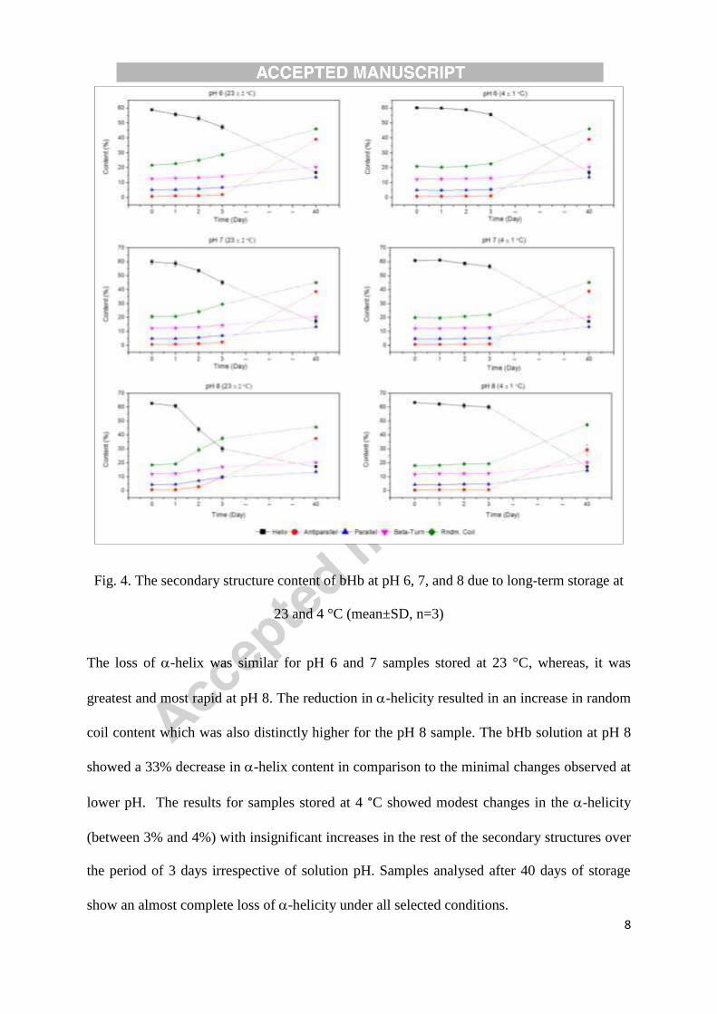
Fig. 4. The secondary structure content of bHb at pH 6, 7, and 8 due to long-term storage at
23 and 4 °C (mean±SD, n=3)
The loss of -helix was similar for pH 6 and 7 samples stored at 23 °C, whereas, it was
greatest and most rapid at pH 8. The reduction in -helicity resulted in an increase in random
coil content which was also distinctly higher for the pH 8 sample. The bHb solution at pH 8
showed a 33% decrease in -helix content in comparison to the minimal changes observed at
lower pH. The results for samples stored at 4 °C showed modest changes in the -helicity
(between 3% and 4%) with insignificant increases in the rest of the secondary structures over
the period of 3 days irrespective of solution pH. Samples analysed after 40 days of storage
show an almost complete loss of -helicity under all selected conditions.

9
3.2 Thermal stability of bHb
The effects of heating-cooling cycles (between 25 and 65 °C) on the UV-vis and CD spectra
of bHb in pH 6 PBS are presented in Fig. 5.
Fig. 5. UV-vis and CD spectra of bHb at pH 6. (A) UV-vis spectra upon heating between 25
and 65 °C; (B) UV-vis spectra upon cooling between 65 and 25 °C; (C) CD spectra upon
heating between 25 and 65 °C; (D) CD spectra upon cooling between 65 and 25 °C.
The changes in UV-vis absorbance behaviour of protein solutions at pH 6, 7 and 8 in PBS as
functions of a single heating-cooling cycle are shown in Fig. 6.

10
Fig. 6. The change in UV-vis absorbance of bHb at 274 and 405 nm at pH 6, 7, and 8 due to
heating-cooling cycle between 25 and 65 °C (mean±SD, n=3)
A constant decrease in absorbance at 405 nm at pH 8 during the heating cycle is related to the
loss of structural integrity of the heme moiety. The increase in absorbance at 274 nm was
seen only after 50 °C for pH 6 and 60 °C for pH 7 and 8 solutions. Limited reduction in
absorbance intensity at 274 nm for the pH 6 sample and absence of any significant changes at
pH 7 and 8 during the cooling cycle implies irreversible conformational changes to the bHb
structure.
The changes in the secondary structure content of bHb under the selected conditions are
presented in Fig. 7. The -helix content decreased steadily for all samples with the highest
decrease of 28% at pH 8 followed by 20% and 18% at pH 6 and 7, respectively. Interestingly,
a small fraction of this change was observed to be reversible during the cooling cycle. The
reversibility (i.e. recovery of -helix content) was found to be between 1% and 5 %
depending on the pH.

11
Fig. 7. The change in the secondary structure content of bHb at pH 6, 7, and 8 due to heating-
cooling cycle between 25 and 65 °C (mean±SD, n=3)
4. Discussion
Proteins are amphoteric molecules with a net positive or negative charge below or above their
isoelectric point (pI) [17]. The pI of bHb is 7.1 which implies electrostatic repulsion between
the molecules below and above this value [18]. Moreover, bHb is a large molecule with
intrinsically high charge density regions which also promote intra-molecular repulsions and
protein unfolding in unfavourable conditions [19]. The results presented in Fig. 3 show that
the rate of confirmation loss was highest at pH 8 suggesting a close arrangement of
negatively charged groups in the bHb molecule which experience strong repulsion.

12
Conversely, lower conformational changes at pH 6 indicate that positively charged groups are
more dispersed within the bHb molecule. It is known that the intra-molecular repulsive
interactions between charged groups can be enhanced at higher temperatures, which is
confirmed in this case by the observed accelerated loss in -helicity at 23 oC in comparison
with that at 4 oC.
According to CD data, the loss of -helicity can be attributed to the destabilisation of
hydrogen bonding between the amide hydrogen atom and carbonyl oxygen atom [20]. Results
presented in Fig. 4 confirm that the stabilising hydrogen bonds in bHb are influenced by the
storage temperature and pH of the solution. Furthermore, heme plays an important role in
retaining the native structure of the bHb molecule, hence, its loss is a significant contributory
factor in the decrease in -helicity [21]. This phenomenon holds true in the present study
where simultaneous reductions in absorbance at 405 nm (i.e. loss of heme) and -helicity
were observed at all pH and temperatures.
The destabilisation of the native structure of a protein due to thermal energy is a well known
phenomenon [20]. Hb denatures when exposed to heat and, since it consists of four sub-
units, the denaturation is known to be a cooperative transition. In a previous study,
differential scanning microcalorimetry showed that carboxyhemoglobin denatures at 82 °C,
oxyhemoglobin at 71 °C and methemoglobin at 67 °C, at pH 7.4 in PBS [22]. Whereas,
Michnik et al. [23] showed that human methemoglobin denatures at 62 °C in pH 6.5 buffer.
Artmann [24] used CD spectroscopy to evaluate structural changes in human haemoglobin
(HbA) and sickle cell haemoglobin (HbS) upon heating between 25 and 60 °C. He
demonstrated that thermal denaturation curves of HbA were non-linear and showed that
accelerated denaturation occurred between 35 and 39 °C along with reversible transitions
below 39 °C which were independent of solution pH in the range 6.8 to 7.8.

13
In the present study, the thermal stability of bHb was assessed under a broad pH range i.e. in
pH 6, 7 and 8 buffer solutions. The temperatures studied in this work (Fig. 3) provide
sufficient thermal energy to promote the disruption of essential stabilising forces leading to
the loss of the heme moiety and exposure of aromatic side chains to the surface. The native
structure of a protein has approximately 70% of its peptide groups and 81% of its non-polar
groups, 63% of its polar groups and 54% of its charged side chains buried in the interior of
the molecule [25]. However, protein unfolding exposes non-polar side chains to the solvent
when sufficient energy is applied to this system, as is apparent from the UV absorbance
spectra in Fig. 3. The hypochromic shift observed at 405 nm indicates the loss of the heme
moiety from the bHb molecule and the hyperchromic shift at 274 nm suggests exposure of
aromatic side chains of the protein to the solvent [19,26]. The observed, respective, decrease
and increase in these absorbance values were also pH dependent which suggests that the
stability of the iron-containing porphyrin ring is greater at pH 6 and 7 in comparison to pH 8.
The marginally higher stability under acidic and neutral conditions is likely to be attributed to
factors which include the overall charge on the protein molecule, ionic strength of the media
and inter-molecular protein-protein interactions.
The CD results presented in Fig. 7 showed a decrease in -helix content with an increase in
temperature which can be explained by the breakage of stabilising hydrogen bonds. The
majority of the bHb molecule (60 to 70%) exists in -helical form and the rest (20 to 30 %)
consists of -turns, parallel, antiparallel and random coils. Initial increases in temperature
result in the weakening of hydrogen bonds leading to a flexible protein molecule which
exposes its side chain groups to the solvent [19]. Most proteins readily refold to their native
configuration if heating is stopped at this early stage. However, continued heating breaks

14
these intra-molecular hydrogen bonds and gives rise to new hydrogen bonds between the
water, and amide nitrogen atoms and carbonyl oxygen atoms within the protein.
It is well known that proteins fold to attain a conformation that is essential for their biological
activity. The native conformation is more stable than unfolded biologically inactive
conformation under physiological conditions. Thermodynamically or in terms of free energy,
the native configuration is only 5-20 kcal/mole more stable than the unfolded confirmation
[20]. This small conformational stability of proteins is a result of large stabilizing and
destabilizing forces. The major stabilizing forces are hydrogen bonding and hydrophobic
effects whereas the major destabilizing force is the protein’s conformational entropy [25].
Therefore, as a result of this small conformational stability of the native state, modest
changes in external variables (i.e. temperature, pH, salt, concentration etc.) lead to
destabilization of protein’s structure and induce unfolding.
5. Conclusion
By analysing the UV-vis and CD spectroscopic data collected in this study, it can be
concluded that bHb can withstand temperatures up to 50 °C, but at higher temperatures major
irreversible changes take place in its conformation. The thermal stability was found to be
independent of pH of the solution. It was also confirmed that solutions of bHb were stable for
3 days if maintained under cold conditions (i.e. 4 °C); whereas, conformational changes
occurred after only 1 day when stored at room temperature. Unlike the thermal stability, these
conformational changes at room temperature were strongly influenced by the pH of the
solution where the rate of conformation change at pH 6 and 7 was markedly slower than that
at pH 8. The use of neural network-based software such as CDNN are beneficial in following
the conformational changes in a protein in comparison to raw CD data. They provide
quantitative data which are easier to interpret and offer better understanding when various
15
parameters and their effects are compared. These findings are of relevance to other
researchers who intend to use bHb as a model protein under varying experimental pH and
temperature regimes.
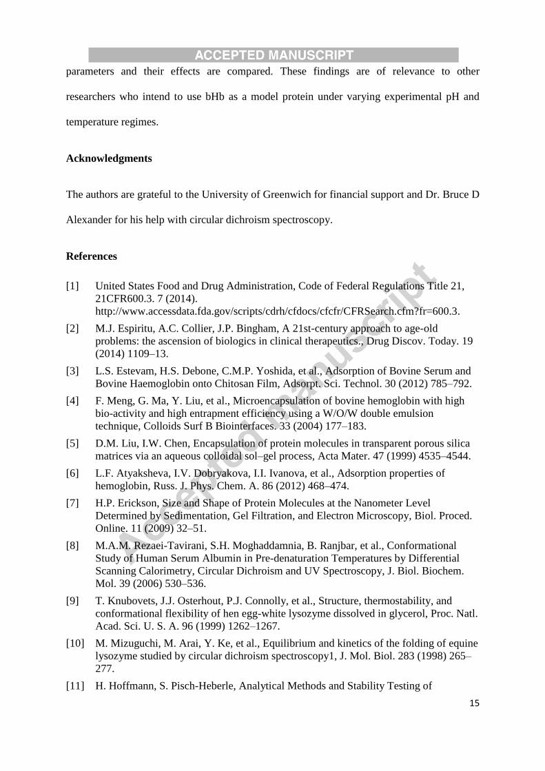
Acknowledgments
The authors are grateful to the University of Greenwich for financial support and Dr. Bruce D
Alexander for his help with circular dichroism spectroscopy.
References
[1] United States Food and Drug Administration, Code of Federal Regulations Title 21, 21CFR600.3. 7 (2014). http://www.accessdata.fda.gov/scripts/cdrh/cfdocs/cfcfr/CFRSearch.cfm?fr=600.3.
[2] M.J. Espiritu, A.C. Collier, J.P. Bingham, A 21st-century approach to age-old problems: the ascension of biologics in clinical therapeutics., Drug Discov. Today. 19 (2014) 1109–13.
[3] L.S. Estevam, H.S. Debone, C.M.P. Yoshida, et al., Adsorption of Bovine Serum and Bovine Haemoglobin onto Chitosan Film, Adsorpt. Sci. Technol. 30 (2012) 785–792.
[4] F. Meng, G. Ma, Y. Liu, et al., Microencapsulation of bovine hemoglobin with high bio-activity and high entrapment efficiency using a W/O/W double emulsion technique, Colloids Surf B Biointerfaces. 33 (2004) 177–183.
[5] D.M. Liu, I.W. Chen, Encapsulation of protein molecules in transparent porous silica matrices via an aqueous colloidal sol–gel process, Acta Mater. 47 (1999) 4535–4544.
[6] L.F. Atyaksheva, I.V. Dobryakova, I.I. Ivanova, et al., Adsorption properties of hemoglobin, Russ. J. Phys. Chem. A. 86 (2012) 468–474.
[7] H.P. Erickson, Size and Shape of Protein Molecules at the Nanometer Level Determined by Sedimentation, Gel Filtration, and Electron Microscopy, Biol. Proced. Online. 11 (2009) 32–51.
[8] M.A.M. Rezaei-Tavirani, S.H. Moghaddamnia, B. Ranjbar, et al., Conformational Study of Human Serum Albumin in Pre-denaturation Temperatures by Differential Scanning Calorimetry, Circular Dichroism and UV Spectroscopy, J. Biol. Biochem. Mol. 39 (2006) 530–536.
[9] T. Knubovets, J.J. Osterhout, P.J. Connolly, et al., Structure, thermostability, and conformational flexibility of hen egg-white lysozyme dissolved in glycerol, Proc. Natl. Acad. Sci. U. S. A. 96 (1999) 1262–1267.
[10] M. Mizuguchi, M. Arai, Y. Ke, et al., Equilibrium and kinetics of the folding of equine lysozyme studied by circular dichroism spectroscopy1, J. Mol. Biol. 283 (1998) 265–277.
[11] H. Hoffmann, S. Pisch-Heberle, Analytical Methods and Stability Testing of

16
Biopharmaceuticals, in: Protein Formulation and Delivery, 2nd ed., Informa Helthcare, New York, 2007, 73–108.
[12] S. Kelly, N. Price, The Use of Circular Dichroism in the Investigation of Protein Structure and Function, Curr. Protein Pept. Sci. 1 (2000) 349–384.
[13] S.M. Kelly, T.J. Jess, N.C. Price, How to study proteins by circular dichroism, Biochim. Biophys. Acta - Proteins Proteomics. 1751 (2005) 119–139.
[14] G. Böhm, R. Muhr, R. Jaenicke, Quantitative analysis of protein far UV circular dichroism spectra by neural networks., Protein Eng. 5 (1992) 191–195.
[15] J.E. Noble, M.J.A. Bailey, Quantitation of Protein, in: Guide to Protein Purification, 2nd ed., Academic Press, Philadelphia, 2009, 73–95.
[16] C. Rimington, Spectral-absorption coefficients of some porphyrins in the soret-band region, Biochem. J. 75 (1960) 620–623.
[17] T. Arakawa, S.J. Prestrelski, W.C. Kenney, et al., Factors affecting short-term and long-term stabilities of proteins., Adv. Drug Deliv. Rev. 46 (2001) 307–326.
[18] G. Zhan, C. Li, D. Luo, Electrochemical investigation of bovine hemoglobin at an acetylene black paste electrode in the presence of sodium dodecyl sulfate, Bull. Chem. Soc. 28 (2007) 1720–1724.
[19] M. Mangino, Protein Denaturation, Ohio State Univ. Food Sci. Lect. (2007). http://class.fst.ohio-state.edu/FST822/lectures/Denat.html.
[20] E.Y. Chi, S. Krishnan, T.W. Randolph, et al., Physical stability of proteins in aqueous solution: mechanism and driving forces in nonnative protein aggregation, Pharm. Res. 20 (2003) 1325–1336.
[21] M.S. Hargrove, J.S. Olson, The stability of holomyoglobin is determined by heme affinity, Biochemistry. 35 (1996) 11310–11318.
[22] E.A. Lapshina, I.B. Zavodnik, V.A. Ignatenko, et al., Thermal stability and functional properties of human hemoglobin in the presence of aliphatic alcohols., Mol. Biol. (Mosk). 26 (1992) 315–320.
[23] A. Michnik, Z. Drzazga, A. Kluczewska, et al., Differential scanning microcalorimetry study of the thermal denaturation of haemoglobin., Biophys. Chem. 118 (2005) 93–101.
[24] G.M. Artmann, L. Burns, J.M. Canaves, et al., Circular dichroism spectra of human hemoglobin reveal a reversible structural transition at body temperature., Eur. Biophys. J. 33 (2004) 490–496.
[25] C.N. Pace, B.A. Shirley, M. McNutt, et al., Forces contributing to the conformational stability of proteins., FASEB J. 10 (1996) 75–83.
[26] E.C. Liong, Y. Dou, E.E. Scott, et al., Waterproofing the heme pocket: role of proximal amino acid side chains in preventing hemin loss from myoglobin., J. Biol. Chem. 276 (2001) 9093–9100.


![Author’s Accepted Manuscriptsmeng.ucsd.edu/wp-content/uploads/Self-standing... · Author’s Accepted Manuscript ... cathode materials [24,25]. The difficulty lies in the fact that](https://static.fdocuments.net/doc/165x107/5e3fb950e0d622236f381b48/authoras-accepted-authoras-accepted-manuscript-cathode-materials-2425.jpg)