Author Manuscript NIH Public Access Jerome Mertz Kevin J...
21
Pyramidal cells accumulate chloride at seizure onset Kyle P Lillis a,b , Mark A Kramer c , Jerome Mertz d , Kevin J Staley a,b , and John A White e a Department of Neurology, Massachusetts General Hospital, 114 16 th St #2600, Charlestown, MA 02129, [email protected], [email protected] b Harvard Medical School, 25 Shattuck Street, Boston, MA 02115 c Department of Mathematics & Statistics, Boston University, 111 Cummington Street, Boston, MA 02215, [email protected] d Department of Biomedical Engineering, Boston University, 44 Cummington Street, Boston, MA 02215, [email protected] e Department of Bioengineering, University of Utah, 20 S. 2030 E., Salt Lake City, UT 84112, Phone: 801-587-1200, Fax: 801-585-5151, [email protected] Abstract Seizures are thought to originate from a failure of inhibition to quell hyperactive neural circuits, but the nature of this failure remains unknown. Here we combine high-speed two-photon imaging with electrophysiological recordings to directly evaluate the interaction between populations of interneurons and principal cells during the onset of seizure-like activity in mouse hippocampal slices. Both calcium imaging and dual patch clamp recordings reveal that in vitro seizure-like events (SLEs) are preceded by pre-ictal bursts of activity in which interneurons predominate. Corresponding changes in intracellular chloride concentration were observed in pyramidal cells using the chloride indicator Clomeleon. These changes were measurable at SLE onset and became very large during the SLE. Pharmacological manipulation of GABAergic transmission, either by blocking GABA A receptors or by hyperpolarizing the GABA A reversal potential, converted SLEs to short interictal-like bursts. Together, our results support a model in which pre-ictal GABA A receptor-mediated chloride influx shifts E GABA to produce a positive feedback loop that contributes to the initiation of seizure activity. Keywords epilepsy; ictogenesis; chloride accumulation; ion imaging; calcium; seizure; GABA; chloride transport; interneuron; targeted path scanning Introduction The failure of GABAergic inhibition has long been cited as a contributing factor to the generation of seizures in epilepsy. Pathological changes to inhibitory circuits have been argued to occur through the death of interneurons (de Lanerolle et al., 1989), change in the © 2012 Elsevier Inc. All rights reserved. Correspondence to: John A White. Publisher's Disclaimer: This is a PDF file of an unedited manuscript that has been accepted for publication. As a service to our customers we are providing this early version of the manuscript. The manuscript will undergo copyediting, typesetting, and review of the resulting proof before it is published in its final citable form. Please note that during the production process errors may be discovered which could affect the content, and all legal disclaimers that apply to the journal pertain. NIH Public Access Author Manuscript Neurobiol Dis. Author manuscript; available in PMC 2013 September 01. Published in final edited form as: Neurobiol Dis. 2012 September ; 47(3): 358–366. doi:10.1016/j.nbd.2012.05.016. NIH-PA Author Manuscript NIH-PA Author Manuscript NIH-PA Author Manuscript
Transcript of Author Manuscript NIH Public Access Jerome Mertz Kevin J...

Pyramidal cells accumulate chloride at seizure onset
Kyle P Lillisa,b, Mark A Kramerc, Jerome Mertzd, Kevin J Staleya,b, and John A Whitee
aDepartment of Neurology, Massachusetts General Hospital, 114 16th St #2600, Charlestown,MA 02129, [email protected], [email protected] Medical School, 25 Shattuck Street, Boston, MA 02115cDepartment of Mathematics & Statistics, Boston University, 111 Cummington Street, Boston, MA02215, [email protected] of Biomedical Engineering, Boston University, 44 Cummington Street, Boston, MA02215, [email protected] of Bioengineering, University of Utah, 20 S. 2030 E., Salt Lake City, UT 84112,Phone: 801-587-1200, Fax: 801-585-5151, [email protected]
AbstractSeizures are thought to originate from a failure of inhibition to quell hyperactive neural circuits,but the nature of this failure remains unknown. Here we combine high-speed two-photon imagingwith electrophysiological recordings to directly evaluate the interaction between populations ofinterneurons and principal cells during the onset of seizure-like activity in mouse hippocampalslices. Both calcium imaging and dual patch clamp recordings reveal that in vitro seizure-likeevents (SLEs) are preceded by pre-ictal bursts of activity in which interneurons predominate.Corresponding changes in intracellular chloride concentration were observed in pyramidal cellsusing the chloride indicator Clomeleon. These changes were measurable at SLE onset and becamevery large during the SLE. Pharmacological manipulation of GABAergic transmission, either byblocking GABAA receptors or by hyperpolarizing the GABAA reversal potential, converted SLEsto short interictal-like bursts. Together, our results support a model in which pre-ictal GABAAreceptor-mediated chloride influx shifts EGABA to produce a positive feedback loop thatcontributes to the initiation of seizure activity.
Keywordsepilepsy; ictogenesis; chloride accumulation; ion imaging; calcium; seizure; GABA; chloridetransport; interneuron; targeted path scanning
IntroductionThe failure of GABAergic inhibition has long been cited as a contributing factor to thegeneration of seizures in epilepsy. Pathological changes to inhibitory circuits have beenargued to occur through the death of interneurons (de Lanerolle et al., 1989), change in the
© 2012 Elsevier Inc. All rights reserved.
Correspondence to: John A White.
Publisher's Disclaimer: This is a PDF file of an unedited manuscript that has been accepted for publication. As a service to ourcustomers we are providing this early version of the manuscript. The manuscript will undergo copyediting, typesetting, and review ofthe resulting proof before it is published in its final citable form. Please note that during the production process errors may bediscovered which could affect the content, and all legal disclaimers that apply to the journal pertain.
NIH Public AccessAuthor ManuscriptNeurobiol Dis. Author manuscript; available in PMC 2013 September 01.
Published in final edited form as:Neurobiol Dis. 2012 September ; 47(3): 358–366. doi:10.1016/j.nbd.2012.05.016.
NIH
-PA Author Manuscript
NIH
-PA Author Manuscript
NIH
-PA Author Manuscript
organization of GABAergic synapses (Marchionni and Maccaferri, 2009; Thind et al.,2010), or reduction in interneuron excitability (Martin et al., 2010). While epilepsy is clearlyassociated with changes in the anatomical organization of GABAergic networks, thepathophysiological action of GABA in seizure generation remains unclear (Cossart et al.2005). In particular, it is difficult to determine whether GABA changes are adaptive orcausal solely by examining the anatomical changes that occur in animal models of epilepsy.
Some studies have begun to link dysfunction in GABAergic interneurons to onset ofepileptiform activity. For example, when firing at supraphysiological rates, interneurons canalso be transiently rendered ineffective at inhibiting postsynaptic targets either by enteringdepolarization block (Ziburkus et al., 2006) or by causing post-synaptic chloride toaccumulate to depolarizing concentrations, effectively making GABAA synapses excitatory(Staley et al., 1995; Taira et al., 1997; Köhling et al., 2000; Fujiwara-Tsukamoto et al.,2004; Ben-Ari and Holmes, 2005). The latter mechanism would have the effect oftransforming feedback inhibition into feedback excitation, producing an unstable, positive-feedback network. Large preictal alterations in the reversal potential of synaptic eventsassociated with epileptiform spikes have recently been reported. Although the responsibleneurotransmitter was proposed to be glutamate, some interneurons were found to fire priorto the preictal discharges (Huberfeld et al., 2011). Interestingly, reduced expression of theoutwardly-directed chloride transporter KCC2 have been found in both experimental (deGuzman et al., 2006) and human epilepsy (Aronica et al., 2007; Shimizu-Okabe et al., 2011;Huberfeld et al., 2011). Electrophysiological assays of KCC2 transport have demonstratedreduced KCC2 transport capacity in multiple experimental models (Jin et al., 2005; Pathaket al., 2007; Lee et al., 2011). These studies support the possibility that the chloride gradientmay be selectively labile in chronic epilepsy, and that chloride accumulation may be a pre-ictal mechanism of activity-dependent loss of inhibition.
Due to technical challenges in recording from large neural networks with cellular resolution,studying the physiology of this complex balance between excitation and inhibition in neuralcircuits has primarily been constrained to pharmacological manipulation and single-cell orpaired intracellular recordings (Köhling et al., 2000; Huberfeld et al., 2011). Here, wecombine these classic techniques with recently developed network imaging methods (Lilliset al., 2008) to measure the interactions between populations of inhibitory cells and principalcells of the hippocampus and entorhinal cortex. We find that the pre-ictal burst (also called“pre-ictal spike” or “sentinel spike”) is dominated by epileptiform activity in the populationof somatostatin-positive GABAergic interneurons. Chloride imaging reveals that thisGABAergic hyperactivity leads to a flux of chloride and leaves the population of post-synaptic pyramidal cells in a highly excitable state just before seizure onset. At seizureonset, there is a massive increase in intracellular chloride that is sufficient to make GABAcurrents excitatory.
Materials and MethodsAcute slice preparation
Acute slice protocols were approved by the Boston University Animal Care and UseCommittee. Transverse hippocampal brain slices (400 µm) were prepared as previouslydescribed (Netoff et al., 2005) from juvenile (P10-P20) mice expressing GFP insomatostatin-positive interneurons under the control of the Gad1 (GAD67) promoter (strainFVB-Tg(GadGFP)45704Swn/J, Jackson Laboratories, Bar Harbor, ME) or from Clomeleonmice. After a 1hr incubation period, they were transferred to the recording chamber wherethey were bathed is artificial cerebrospinal fluid (ACSF, in mM, 126 NaCl, 2.5 KCl, 1.25NaH2PO4, 2 MgCl2, 26 NaHCO3, 25 dextrose, 2mM CaCl2). 50µM 4-aminopyridine wasadded to induce epileptiform activity. In some experiments 10µM acetazolamide was added
Lillis et al. Page 2
Neurobiol Dis. Author manuscript; available in PMC 2013 September 01.
NIH
-PA Author Manuscript
NIH
-PA Author Manuscript
NIH
-PA Author Manuscript

inhibit carbonic anhydrase. To elicit seizures in Clomeleon acute slices, which did notspontaneously seize in 50 µM 4-AP, MgCl2 was omitted from the ACSF (in addition toadding 50 µM 4-AP). All chemicals were obtained from Sigma-Aldrich (St. Louis, MO).Slices were initially visualized using oblique illumination.
Organotypic slice culture preparationOrganotypic slice culture protocols were approved by the Massachusetts General HospitalSubcommittee on Research Animal Care. Roller tube type organotypic slice cultures wereprepared as described by (Gähwiler, 1981). Briefly, isolated hippocampi from P6–8 CLM-1mouse pups were cut into 350-µm slices on a McIlwain tissue chopper (Mickle LaboratoryEng. Co., Surrey, United Kingdom). Slices were mounted in clots of chicken plasma(Cocalico Biologicals, Reamstown, PA) and thrombin (Sigma-Aldrich, St. Louis, MO) onpoly-l-lysine-(Sigma-Aldrich) coated glass coverslips (Electron Microscopy Sciences,Hatfield, PA) and incubated in roller tubes (Nunc, Roskilde, Denmark) at 5% CO2, 36°C in750µL Neurobasal-A growth medium with 2% B27, 500uM Glutamax, and 0.03 mg/mLgentamycin added (all from Invitrogen, Carlsbad, CA). Growth medium was changed every7 days. Recordings were made in growth media, some with the addition of 10µM GABAzine(SR 95531).
Staining and imagingAreas of interest were stained with Indo-1 AM (Invitrogen, Carlsbad, CA) using an adaptedversion of multicell bolus loading (Stosiek et al., 2003; but c.f. Garaschuk et al., 2006), inwhich a Picospritzer II (Parker Hannifin, Pine Brook, NJ) is used to inject dye through aglass pipette directly into the brain tissue. In this modified version of MCBL, a larger-tippipette was used (~2MΩ when filled with KCl-based dye solution) to inject many sites for ashorter duration (~5s) than that previously described (1–2min). This resulted in the stainingof a large area, with relatively low background staining. The dye used for these experiments,Indo-1, is a ratiometric calcium dye, emitting at a shorter wavelength when bound tocalcium. However, the two-photon cross section of calcium-bound Indo-1 is so low that it isessentially invisible when using excitation light longer than 750nm (Xu et al., 1996). Wetake advantage of this by imaging at 820nm (a wavelength that conveniently excites bothGFP and calcium-unbound Indo-1) to get relative calcium measurements with highsensitivity. Because, in this configuration, fluorescence decreases when a cell is active, allIndo-1 fluorescence traces shown have been inverted for clarity.
All images of calcium dynamics were acquired using Targeted Path Scanning (Lillis et al.,2008). All images were acquired using a 20× 0.95 NA water immersion objective (Olympus,Tokyo, Japan). The calcium traces for each cell were then filtered using a 15 point medianfilter and a 10 point boxcar filter. To average calcium traces taken with different samplingrates (as is inherent to TPS, mean sampling rate for calcium data shown = 51.8Hz), traceswere upsampled using the Matlab (The Mathworks, Natick, MA) function resample, whichinterpolates using a polyphase filter. In 4-AP, SLEs occurred approximately once every 4minutes. To guarantee that a SLE would be captured, a four-minute scan was initiated 2–3minutes after the previous SLE. I/E ratios were calculated, for each recording, by dividingthe mean interneuron calcium trace by the mean principal neuron calcium trace.
Clomeleon images were acquired using custom-designed software and the scanhead from aRadiance 2000MP (BioRad, Hemel Hempstead, UK), equipped with a 20× 0.95 NA water-immersion objective (Olympus, Tokyo, Japan), and PMTs with appropriate filters for YFP(545/30) and CFP (450/80). A SpectraPhysics MaiTai laser (Newport, Irvine, CA), set to860nm, was used for two-photon excitation. Chloride concentrations were obtained byperforming a calibration as previously described (Glykys et al., 2009) using 10µM of the K+/
Lillis et al. Page 3
Neurobiol Dis. Author manuscript; available in PMC 2013 September 01.
NIH
-PA Author Manuscript
NIH
-PA Author Manuscript
NIH
-PA Author Manuscript

H+ ionophore nigericin, 100µM of OH−/Cl− antiporter tributyltin chloride, and knownconcentrations of extracellular chloride. YFP/CFP ratios for all selected cells were low-passfiltered at 0.5Hz and, because CFP and YFP bleach at different rates (Kuner and Augustine,2000), a linear trend was subtracted to remove the effect of differential bleaching. For thepre-ictal burst-triggered averaging shown in Figure 3, chloride traces were resampled in thesame manner described above for calcium traces. pH changes were measured by stainingorganotypic slice cultures with SNARF-1-AM by incubating them for >1hr in 10uM dye.Because of the spectral overlap between SNARF-1 and Clomeleon, wild type mice wereused for pH imaging experiments. Images were acquired using TPS with an excitationwavelength of 820nm and PMT emission filters centered at 585 and 640. The ratio of lightemitted at 585 and 640nm was recorded and linearly detrended (to correct for bleaching) andadjusted to baseline pH using a calibration strategy previously described (Sheldon et al.,2004).
ElectrophysiologyDuring imaging recordings, either a field potential recording (<1MΩ, filled with ACSF or1M KCl) or a patch-clamp recording (3–6MΩ, filled with 130 mM potassium gluconate,10mM KCl, 10 mM HEPES, 4 mM Mg-ATP, 0.4 mM Tris-GTP, and 50µM Indo-1pentapotassium salt) was acquired simultaneously from the region being imaged. For dual-patch clamp recordings, the patch electrodes were targeted to GFP+ and GFP− cells usingtwo-photon imaging. The seal was formed (and ruptured in the case of whole-cellrecordings), using oblique illumination for visualization. Whole-cell access was evaluatedby monitoring input resistance and imaging the presence of dye in the cell. Allelectrophysiological recordings were obtained with a Multiclamp 700B, AxoClamp 2B(Axon Instruments, Foster City, CA), or Cornerstone EX4-400 (Dagan, Minneapolis, MN).
Statistical AnalysesFor comparison of two populations (e.g. principal cells vs. interneurons), a Kruskal-Wallisanalysis was used to test for a significant difference between the two medians. Forcomparisons to zero (e.g. number of seizures in Figure 5B), a Wilcoxon signed rank test forzero median was performed. For tests of linear correlation, a p-value for Pearson’scorrelation was computed using Student’s t-distribution.
Spike-timing jitter was computed by analyzing the variability in principal neuron spikedelay following an interneuron spike for the 10 spikes preceding a pre-ictal burst and the 10spikes following the pre-ictal burst. Jitter was quantified as the coefficient of variation (CV)or standard deviation (SD) of interneuron->principal neuron spike delay.
ResultsTargeted path scanning of inhibition-excitation interplay at seizure onset
In acute slices of hippocampus/entorhinal cortex, 4-aminopyridine (4-AP) initiates seizuresthat originate in the entorhinal cortex (Avoli et al., 1996; Barbarosie and Avoli, 1997),where interneurons appear (using DIC microscopy) anatomically similar to principal cells.To distinguish interneurons from putative excitatory cells in this region, we prepared slicesfrom mice expressing GFP in 15–35% of somatostatin-positive GAD67-expressing cells(Oliva et al., 2000). Since >92% of neurons in the entorhinal cortex are principal cells(Kumar and Buckmaster, 2006) and 10–20% of all interneurons in these mice express GFP(Oliva et al., 2000), we refer to GFP-negative cells as principal neurons. Actual differencesbetween interneurons and principal neurons might be slightly larger than we measure due tothe presence of some interneurons in the GFP-negative population. We used two-photon,calcium imaging combined with targeted path scanning (TPS, Figure 1A, Lillis et al., 2008),
Lillis et al. Page 4
Neurobiol Dis. Author manuscript; available in PMC 2013 September 01.
NIH
-PA Author Manuscript
NIH
-PA Author Manuscript
NIH
-PA Author Manuscript

to image epileptiform activity, induced by conditions that leave inhibition intact, inpopulations of entorhinal cortical interneurons and principal cells.
Seizure-like events, induced by potassium channel blocker, 4-aminopyridine (4-AP) ordeveloping spontaneously in organotypic slice cultures of the hippocampus (Dyhrfjeld-Johnsen et al., 2010), consist of a stereotypical firing pattern (Figure 1B) that begins with apre-ictal burst, is followed a few seconds later by powerful ictal tonic firing (Figure 1C),then transitions to post-ictal clonic discharges before terminating completely. The datapresented here will focus on the preictal burst, which we define as a burst of activitypreceding the tonic phase of the seizure by <10s. Using TPS to scan both GFP+ and GFP−cells (i.e. interneurons vs. principal cells) produced calcium traces that could be grouped bycell type and provided sufficient spatial and temporal resolution to analyze the interactionbetween inhibition and excitation during ictogenesis (Figure 1D).
Interneurons fire at higher rates than principal cells at ictogenesisTPS recordings revealed that the ratio of interneuron to principal neuron calcium signal(mean interneuron / mean principal neuron, for each recording) significantly increasedduring pre-ictal bursts (t=0, Figure 1E, n=22 SLEs in slices from 7 animals, each slice withan average of 21 principal cells and 5 interneurons imaged, p<0.01). To verify that theproportionally larger calcium transients in interneurons are not a result of differences incalcium buffering capacity, we performed simultaneous patch clamp recordings frominterneurons and principal cells. We used both whole-cell patch clamp (Figure 2A) andloose-patch clamp (to avoid dialyzing the cell, Figure 2B) to compare instantaneous firingrates (calculated at each action potential as the inverse of interspike interval) in interneuronsand principal cells. We found that, indeed, pre-ictal bursts are dominated by interneuronfiring (Figure 2C, 4-AP, n=5 slices from 3 animals, p< 0.05). Prior to the pre-ictal burst,interneurons fire apparently randomly with respect to principal cells, with a large coefficientof variation and standard deviation of the intervals between spikes in the two cell types(CV=15.09±12.75, SD = 8.60±6.35 pre-burst). Interestingly, in recordings with actionpotentials in the seconds following the pre-ictal burst, just before ictogenesis, interneuronand principal cell firing are tightly coupled (CV=0.11±0.04, SD=0.30±0.12 post-burst), withthe interneuron leading the principal cell by 4.9±1.7ms (mean±SEM, n=5, 14 spike pairs,Figures 2A,B, insets). However, the sparsity of principal cell firing before the pre-ictal burstmake spike jitter difficult to interpret.
To quantify temporal relationships among imaged calcium signals, we performedwindowed, cross-correlation-based network analysis. By finding the time lag at which peakcorrelation occured for all pairs of calcium traces, we were able to determine if each imagedcell was, on average, leading or following activity in the network. We found that, at the timeof the pre-ictal burst, interneurons lead activity in the network (Supplemental Figure 1).
GABAergic depolarization contributes to SLE generationIf intense epochs of interneuron firing resulted in chloride influx sufficient to overwhelm thechloride transporter KCC2, then the resultant chloride accumulation should be evident bychloride imaging (Dallwig et al., 1999). We directly tested the hypothesis that chlorideaccumulates in neurons during SLEs using samples prepared from clomeleon mice(CLM-1), which express a chloride-sensitive ratiometric CFP-YFP construct in a subset ofprincipal neurons and interneurons under control of the Thy1 promoter (Kuner andAugustine, 2000; Berglund et al., 2006). CLM-1 mice are constructed on a C57 background,a strain that is resistant to 4-AP-induced seizures (Kosobud and Crabbe, 1990). To recordchloride transients associated with inhibition-intact epileptiform activity, we usedchronically epileptic CLM-1 hippocampal organotypic slice cultures, which spontaneously
Lillis et al. Page 5
Neurobiol Dis. Author manuscript; available in PMC 2013 September 01.
NIH
-PA Author Manuscript
NIH
-PA Author Manuscript
NIH
-PA Author Manuscript

(without the addition of drugs) produce tonic-clonic seizures similar to those observed in 4-AP-treated acute slices from GIN mice (McBain et al., 1989; Berdichevsky et al., 2009;Dyhrfjeld-Johnsen et al., 2010) and verified results using acute slices from CLM-1 micetreated with 4-AP and zero-Mg2+. Under control conditions, organotypic slice culturesexhibited a mixture of interictal-like and seizure-like activity (13/65 events were longer than20s, Figure 3A,4C), while in 10µM GABAzine, 184/185 events were less than 5s induration, with a maximum duration of 5.8s (n=4 slice cultures from two mice, Figure 3B,4D). Interestingly beginning at the pre-ictal burst, and dramatically accelerating at seizureonset, SLEs (events >20s) produced a sharp decrease in YFP/CFP ratio, corresponding to anincrease in chloride of 22.95±0.73mM (mean±SEM, n=4 slice cultures, 13 SLEs, Figure 3A,4A). For pre-ictal bursts preceding the SLE by >1s, it was possible to isolate a pre-ictalburst-induced chloride transient of 6.27±1.23mM (mean±SEM, Figure 3E, n=8 SLEs in 3slice cultures). Although the magnitude of this relative change in chloride is uncorrelated toSLE onset time (n=8, p=0.31), we hypothesize that the pre-ictal burst-evoked chloridetransients effectively decrease inhibition, leaving the network in a state of elevatedexcitability. We suspect that the timing of SLE onset may depend on the timing of activity,subsequent to the pre-ictal burst, in both principal neurons and interneurons.
Short duration events (<20s) in control conditions or in GABAzine produced chlorideincreases of 4.87±0.38 (mean±SEM, n=4 slice cultures, 52 events) and 2.97±0.07mM (mean±SEM, n=4 slice cultures, 185 events) respectively (Figure 4A,B). To verify that theseresults were not specific to chronically epileptic organotypic slice cultures, Clomeleonresults were confirmed in acute slices treated with both 4-AP and low-magnesium ACSF. Inthese recordings, ictal chloride transients recorded from the entorhinal cortex were10.22±2.33mM (mean±SEM, n=5 SLEs in 2 slices from 2 animals). As in organotypic slicecultures, SLEs were blocked with the addition of 10µM GABAzine (Figure 3C,D).
To test whether observed changes in Clomeleon ratio could be explained by SLE-inducedchanges in pH, we imaged pyramidal cells during seizure activity in SNARF-1-AM-stainedhippocampal slice cultures (Figure 5, largest recorded pH transient shown). During seizures,intracellular pH increased by an average of 0.080 ± 0.025 (mean±SEM, n=3 slices from twomice), corresponding to a ~4.8mM decrease in apparent chloride (Kuner and Augustine,2000). Thus, the intracellular ictal chloride transients might actually be around 20% largerthan those shown in Figures 3–4.
We hypothesized that the massive barrage of GABAA activation during the pre-ictal burstcauses chloride to overwhelm KCC2 chloride transport capacity and accumulate todepolarizing concentrations, leaving the network in a hyper-excitable state. Chlorideaccumulation requires a sustained driving force for chloride entry, and thus only occurswhen GABA currents cannot drive the membrane potential to the chloride equilibriumpotential. This can occur as a consequence of depolarizing bicarbonate efflux through theGABAA ionophore (Bormann et al., 1987; Staley and Proctor, 1999), or as a consequence ofconcurrent activation of strongly depolarizing ligand and voltage-gated cationicconductances (Figure 1D; Doyon et al., 2011). Although it is not possible to block cationicconductances without fundamentally altering ictogenesis, the bicarbonate efflux ismaintained by hydration of CO2, and so can be selectively diminished by inhibitingcarbonic anhydrase (Staley et al., 1995). We therefore tested the effect of blocking theintracellular production of bicarbonate ions with the carbonic anhydrase inhibitoracetazolamide (ACTZ). As predicted, ACTZ statistically eliminated SLEs in 4-AP (n=8slices from 5 animals, Figure 6). Thus, both enhancement of GABAergic inhibition via anon-chloride pathway (ACTZ) and blockade of GABAergic inhibition (GABAzine)eliminate seizures. Together these results support the hypothesis that activity-dependent
Lillis et al. Page 6
Neurobiol Dis. Author manuscript; available in PMC 2013 September 01.
NIH
-PA Author Manuscript
NIH
-PA Author Manuscript
NIH
-PA Author Manuscript

chloride accumulation and consequent shift in GABAA reversal potential contribute to apreictal decrease in inhibition.
DiscussionEpilepsy is often described in the context of an excitatory shift in the complex balance ofinhibition and excitation in the brain, which can be caused by excessive neuronal sprouting(Cavazos et al., 1991; Sutula and Dudek, 2007), interneuron death (Maglóczky and Freund,2005), ion channel mutations (Reid et al., 2009), pathological plasticity of synapses (Ben-Ari, 2008), or other causes. However, even an epileptic brain functions normally most of thetime. The mechanisms underlying the sudden massive increase in neural activity duringseizures remain elusive. In this paper, we provide evidence that ictal activity in GABAergicinterneurons cause sufficient neuronal chloride accumulation that subsequent GABAergicactivity will be depolarizing. In this mode, increased interneuron firing leads to increasedprincipal cell activity, which in turn leads to more interneuron firing. This effective switchof interneurons from negative to positive feedback circuit elements contributed to seizuregeneration in both acute (4-AP) and chronic (organotypic) models of ictogenesis. Thismechanism of transient positive feedback compliments previously described mechanismssuch as extracellular K+ accumulation (Fisher et al., 1976; Heinemann et al., 1977), and infact because extracellular K+ accumulation alters the transport capacity of KCC2 (Staleyand Proctor, 1999; Bihi et al., 2005) these two mechanisms of ictogenesis are likely to becomplementary.
The question remains as to what drives interneuron firing during the pre-ictal burst. Recentevidence suggests that somatostatin-positive interneurons fire more readily than regular-spiking pyramidal cells or fast-spiking interneurons in response to activating stimuli such asextracellular current, low-Mg2+/Ca2+ ACSF, mGluR agonists, and cholinergic agonists(Fanselow et al., 2008). Furthermore, somatostatin-positive interneurons have been shown tobe coupled extensively via gap-junctions (Gibson et al., 1999; Fanselow et al., 2008; Amitaiet al., 2002), which increases synchrony between coupled neurons (Fanselow et al., 2008).Our data are consistent with the hypothesis that interneurons such as the somatostatin-positive subtype activate most readily in response to the 4-AP-induced increase inexcitability (Lopantsev and Avoli, 1998; Barbarosie et al., 2002). This activation couldpropagate throughout the gap-junction coupled network of interneurons to generate asynchronous event across the population of interneurons, corresponding to the interneuron-dominated pre-ictal burst observed in Figures 1–2. Alternatively, entorhinal corticalinterneurons could be synchronized by long-range-projecting interneurons from thehippocampus (Melzer et al., 2012), a phenomenon recently shown to occur during pre-ictalactivity in hippocamposeptal cells (Quilichini et al., 2012). Either method ofsynchronization would lead to a coordinated activation of post-synaptic GABAA receptors,which could explain the pre-ictal chloride accumulation observed in Figure 3.
The presence of GABA-mediated depolarizing potentials has been shown previously in the4-AP model of seizure (Avoli et al., 1996). In those studies, depolarizing field potentialspersisted even when blocking the NMDA and AMPA glutamate receptors, but they wereprevented by GABAA antagonist bicuculline methiodide and µ-opiod agonist DAGO, whichhyperpolarizes interneurons, reducing GABA release (Lupica and Dunwiddie, 1991). TheGABA-mediated potentials were largest at the onset of ictal-like events, suggesting apotential role in ictogenesis. The large GABA conductance induces an activity-dependentneuronal chloride accumulation and a consequent depolarizing shift in the GABAA reversalpotential. The chloride accumulation is most pronounced in structures with large numbers ofreceptors per unit volume, e.g. dendrites, and less pronounced at the soma (Staley andProctor, 1999; Isomura et al., 2003; Doyon et al., 2011). An open question has been whether
Lillis et al. Page 7
Neurobiol Dis. Author manuscript; available in PMC 2013 September 01.
NIH
-PA Author Manuscript
NIH
-PA Author Manuscript
NIH
-PA Author Manuscript

somatic chloride accumulation is sufficient to induce GABA-mediated excitation, orwhether this effect would be limited to dendrites, and, perhaps due to ClC-2 chlorideregulation (Foldy et al., 2010), shunted at the soma by depolarizing but still inhibitoryGABAergic conductances. Using the Nernst equation, we calculated the somatic chlorideaccumulations observed here to correspond to 26±3mV (mean±SEM) shifts in EGABA (tolevels positive to action potential threshold). This shift in EGABA at the soma supports theidea that ictal activity causes GABA to become frankly excitatory.
We provide direct evidence of the interneuron-dominated pre-ictal burst, presumablyresponsible for GABA-mediated potentials. Our data suggest that GABA-mediated activityat ictogenesis floods postsynaptic targets with chloride, resulting in an ineffective inhibitorynetwork that contributes to the generation of a seizure. These data are consistent with recentfindings of preictal spikes that elicit synaptic activity with dramatically shifted reversalpotentials in pyramidal neurons in both intact mouse hippocampal preparations (Zhang etal., 2012) and resected human epileptic brain tissue (Huberfeld et al., 2007, 2011). Our datacontribute to the study of complex pre-ictal network phenomena by providing a largesimultaneous sampling of calcium or chloride concentrations in interneurons and principalcells. These data and the robust effect of carbonic anhydrase blockade favor GABAergicover glutamatergic activity as the mechanism underlying the sharp pre-ictal shift in thereversal potential of postsynaptic potentials in pyramidal cells. Future studies employingadditional interneuron labeling, voltage-dependent fluorophores, and additional models ofepilepsy will further clarify the critical mechanisms of preictal loss of inhibition.
Studying the role of GABAergic inhibition in epilepsy is complicated by both the widedistribution of intracellular chloride concentrations (Glykys et al., 2009) and the highlyvariable expression of KCC2 (Huberfeld et al., 2007). This variability makes it difficult topredict what the net effect of blocking GABA synapses will be. In organotypic slices cutfrom different planes (but otherwise similarly prepared), GABA blockade has been shown tohave opposite effects on network excitability (McBain et al., 1989, Figures 3–4). Inrecordings from human tissue, IPSP reversal potentials varied from hyperpolarizing todepolarizing depending on KCC2 expression (Huberfeld et al., 2007). Likely, the overallrole of GABA in the network depends on the distribution of intracellular chloride, chloridetransporter expression, and interneuron connectivity. Our data elucidate another degree offreedom for GABA in which the net effect of GABA may be inhibitory at rest, but becomestransiently excitatory during ictogenesis. We hypothesize that epilepsy can develop in partfrom pathologies that make chloride gradients at GABAA synapses labile such as enhancedfunctional inhibition. These include increased GABA synapse activity, (Buckmaster andDudek, 1997), reduced expression of KCC2 and consequent reduced chloride efflux, (Jin etal., 2005; de Guzman et al., 2006; Pathak et al., 2007), and increased GABAA synapticdensity (Bausch, 2005; Thind et al., 2010) with increased postsynaptic chloride influx.
Supplementary MaterialRefer to Web version on PubMed Central for supplementary material.
AcknowledgmentsThis work was supported by grants from the NIH, the Epilepsy Foundation and the Burroughs Wellcome Fund. Wethank G. Feng, T. Kuner, and G.J. Augustine for generously providing us with the Clomeleon mice.
Lillis et al. Page 8
Neurobiol Dis. Author manuscript; available in PMC 2013 September 01.
NIH
-PA Author Manuscript
NIH
-PA Author Manuscript
NIH
-PA Author Manuscript

ReferencesAmitai Y, Gibson JR, Beierlein M, Patrick SL, Ho AM, Connors BW, Golomb D. The Spatial
Dimensions of Electrically Coupled Networks of Interneurons in the Neocortex. J Neurosci. 2002;22:4142–4152. [PubMed: 12019332]
Aronica E, Boer K, Redeker S, Spliet WGM, van Rijen PC, Troost D, Gorter JA. Differentialexpression patterns of chloride transporters, Na+-K+-2Cl−-cotransporter and K+-Cl−-cotransporter,in epilepsy-associated malformations of cortical development. Neuroscience. 2007; 145:185–196.[PubMed: 17207578]
Avoli M, Barbarosie M, Lucke A, Nagao T, Lopantsev V, Kohling R. Synchronous GABA-MediatedPotentials and Epileptiform Discharges in the Rat Limbic System In Vitro. J Neurosci. 1996;16:3912–3924. [PubMed: 8656285]
Barbarosie M, Avoli M. CA3-Driven Hippocampal-Entorhinal Loop Controls Rather than Sustains InVitro Limbic Seizures. J Neurosci. 1997; 17:9308–9314. [PubMed: 9364076]
Barbarosie M, Louvel J, D’Antuono M, Kurcewicz I, Avoli M. Masking synchronous GABA-mediated potentials controls limbic seizures. Epilepsia. 2002; 43:1469–1479. [PubMed: 12460247]
Bausch SB. Axonal sprouting of GABAergic interneurons in temporal lobe epilepsy. Epilepsy Behav.2005; 7:390–400. [PubMed: 16198153]
Ben-Ari Y. Epilepsies and neuronal plasticity: for better or for worse? Dialogues Clin Neurosci. 2008;10:17–27. [PubMed: 18472481]
Ben-Ari Y, Holmes GL. The multiple facets of gamma-aminobutyric acid dysfunction in epilepsy.Curr Opin Neurol. 2005; 18:141–145. [PubMed: 15791144]
Berdichevsky Y, Sabolek H, Levine JB, Staley KJ, Yarmush ML. Microfluidics and multielectrodearray-compatible organotypic slice culture method. J Neurosci Methods. 2009; 178:59–64.[PubMed: 19100768]
Berglund K, Schleich W, Krieger P, Loo LS, Wang D, Cant NB, Feng G, Augustine GJ, Kuner T.Imaging synaptic inhibition in transgenic mice expressing the chloride indicator, Clomeleon. BrainCell Biol. 2006; 35:207–228. [PubMed: 18398684]
Bihi RI, Jefferys JGR, Vreugdenhil M. The role of extracellular potassium in the epileptogenictransformation of recurrent GABAergic inhibition. Epilepsia. 2005; 46(Suppl 5):64–71. [PubMed:15987256]
Bormann J, Hamill OP, Sakmann B. Mechanism of anion permeation through channels gated byglycine and gamma-aminobutyric acid in mouse cultured spinal neurones. J Physiol. 1987;385:243–286. [PubMed: 2443667]
Buckmaster PS, Dudek FE. Neuron loss, granule cell axon reorganization, and functional changes inthe dentate gyrus of epileptic kainate-treated rats. J Comp Neurol. 1997; 385:385–404. [PubMed:9300766]
Cavazos JE, Golarai G, Sutula TP. Mossy fiber synaptic reorganization induced by kindling: timecourse of development, progression, and permanence. J Neurosci. 1991; 11:2795–2803. [PubMed:1880549]
Dallwig R, Deitmer JW, Backus KH. On the mechanism of GABA-induced currents in cultured ratcortical neurons. Pflugers Arch. 1999; 437:289–297. [PubMed: 9929572]
Doyon N, Prescott S, Castonguay A, Godin A, De Koninck Y. Efficacy of synaptic inhibition dependson multiple, dynamically interacting mechanisms implicated in chloride homeostasis. PLoSComputational Biology. 2011 In press.
Dyhrfjeld-Johnsen J, Berdichevsky Y, Swiercz W, Sabolek H, Staley KJ. Interictal spikes precede ictaldischarges in an organotypic hippocampal slice culture model of epileptogenesis. J ClinNeurophysiol. 2010; 27:418–424. [PubMed: 21076333]
Fanselow EE, Richardson KA, Connors BW. Selective, State-Dependent Activation of Somatostatin-Expressing Inhibitory Interneurons in Mouse Neocortex. J Neurophysiol. 2008; 100:2640–2652.[PubMed: 18799598]
Fisher RS, Pedley TA, Moody WJ Jr, Prince DA. The role of extracellular potassium in hippocampalepilepsy. Arch Neurol. 1976; 33:76–83. [PubMed: 1252153]
Lillis et al. Page 9
Neurobiol Dis. Author manuscript; available in PMC 2013 September 01.
NIH
-PA Author Manuscript
NIH
-PA Author Manuscript
NIH
-PA Author Manuscript

Foldy C, Lee S-H, Morgan RJ, Soltesz I. Regulation of fast-spiking basket cell synapses by thechloride channel ClC-2. Nat Neurosci. 2010; 13:1047–1049. [PubMed: 20676104]
Fujiwara-Tsukamoto Y, Isomura Y, Kaneda K, Takada M. Synaptic interactions between pyramidalcells and interneurone subtypes during seizure-like activity in the rat hippocampus. J Physiol.2004; 557:961–979. [PubMed: 15107470]
Gähwiler BH. Organotypic monolayer cultures of nervous tissue. J Neurosci Methods. 1981; 4:329–342. [PubMed: 7033675]
Gibson JR, Beierlein M, Connors BW. Two networks of electrically coupled inhibitory neurons inneocortex. Nature. 1999; 402:75–79. [PubMed: 10573419]
Glykys J, Dzhala VI, Kuchibhotla KV, Feng G, Kuner T, Augustine G, Bacskai BJ, Staley KJ.Differences in Cortical versus Subcortical GABAergic Signaling: A Candidate Mechanism ofElectroclinical Uncoupling of Neonatal Seizures. Neuron. 2009; 63:657–672. [PubMed:19755108]
de Guzman P, Inaba Y, Biagini G, Baldelli E, Mollinari C, Merlo D, Avoli M. Subiculum networkexcitability is increased in a rodent model of temporal lobe epilepsy. Hippocampus. 2006; 16:843–860. [PubMed: 16897722]
Heinemann U, Lux HD, Gutnick MJ. Extracellular free calcium and potassium during paroxsmalactivity in the cerebral cortex of the cat. Exp Brain Res. 1977; 27:237–243. [PubMed: 880984]
Huberfeld G, Menendez de la Prida L, Pallud J, Cohen I, Le Van Quyen M, Adam C, Clemenceau S,Baulac M, Miles R. Glutamatergic pre-ictal discharges emerge at the transition to seizure in humanepilepsy. Nat Neurosci. 2011; 14:627–634. [PubMed: 21460834]
Huberfeld G, Wittner L, Clemenceau S, Baulac M, Kaila K, Miles R, Rivera C. Perturbed chloridehomeostasis and GABAergic signaling in human temporal lobe epilepsy. J Neurosci. 2007;27:9866–9873. [PubMed: 17855601]
Isomura Y, Sugimoto M, Fujiwara-Tsukamoto Y, Yamamoto-Muraki S, Yamada J, Fukuda A.Synaptically Activated Cl− Accumulation Responsible for Depolarizing GABAergic Responses inMature Hippocampal Neurons. Journal of Neurophysiology. 2003; 90:2752–2756. [PubMed:14534278]
Jin X, Huguenard JR, Prince DA. Impaired Cl− extrusion in layer V pyramidal neurons of chronicallyinjured epileptogenic neocortex. J Neurophysiol. 2005; 93:2117–2126. [PubMed: 15774713]
Köhling R, Vreugdenhil M, Bracci E, Jefferys JG. Ictal epileptiform activity is facilitated byhippocampal GABAA receptor-mediated oscillations. J Neurosci. 2000; 20:6820–6829. [PubMed:10995826]
Kosobud AE, Crabbe JC. Genetic correlations among inbred strain sensitivities to convulsions inducedby 9 convulsant drugs. Brain Research. 1990; 526:8–16. [PubMed: 2078820]
Kumar SS, Buckmaster PS. Hyperexcitability, Interneurons, and Loss of GABAergic Synapses inEntorhinal Cortex in a Model of Temporal Lobe Epilepsy. The Journal of Neuroscience. 2006;26:4613–4623. [PubMed: 16641241]
Kuner T, Augustine GJ. A Genetically Encoded Ratiometric Indicator for Chloride: CapturingChloride Transients in Cultured Hippocampal Neurons. Neuron. 2000; 27:447–459. [PubMed:11055428]
de Lanerolle NC, Kim JH, Robbins RJ, Spencer DD. Hippocampal interneuron loss and plasticity inhuman temporal lobe epilepsy. Brain Res. 1989; 495:387–395. [PubMed: 2569920]
Lee HHC, Deeb TZ, Walker JA, Davies PA, Moss SJ. NMDA receptor activity downregulates KCC2resulting in depolarizing GABAA receptor-mediated currents. Nat Neurosci. 2011; 14:736–743.[PubMed: 21532577]
Lillis KP, Eng A, White JA, Mertz J. Two-photon imaging of spatially extended neuronal networkdynamics with high temporal resolution. J Neurosci Methods. 2008; 172:178–184. [PubMed:18539336]
Lopantsev V, Avoli M. Participation of GABAA-mediated inhibition in ictallike discharges in the ratentorhinal cortex. J Neurophysiol. 1998; 79:352–360. [PubMed: 9425204]
Lupica CR, Dunwiddie TV. Differential effects of mu- and delta-receptor selective opioid agonists onfeedforward and feedback GABAergic inhibition in hippocampal brain slices. Synapse. 1991;8:237–248. [PubMed: 1656539]
Lillis et al. Page 10
Neurobiol Dis. Author manuscript; available in PMC 2013 September 01.
NIH
-PA Author Manuscript
NIH
-PA Author Manuscript
NIH
-PA Author Manuscript

Maglóczky Z, Freund TF. Impaired and repaired inhibitory circuits in the epileptic humanhippocampus. Trends in Neurosciences. 2005; 28:334–340. [PubMed: 15927690]
Marchionni I, Maccaferri G. Quantitative dynamics and spatial profile of perisomatic GABAergicinput during epileptiform synchronization in the CA1 hippocampus. J Physiol (Lond). 2009;587:5691–5708. [PubMed: 19840998]
Martin MS, Dutt K, Papale LA, Dubé CM, Dutton SB, de Haan G, Shankar A, Tufik S, Meisler MH,Baram TZ, Goldin AL, Escayg A. Altered function of the SCN1A voltage-gated sodium channelleads to gamma-aminobutyric acid-ergic (GABAergic) interneuron abnormalities. J Biol Chem.2010; 285:9823–9834. [PubMed: 20100831]
McBain CJ, Boden P, Hill RG. Rat hippocampal slices “in vitro” display spontaneous epileptiformactivity following long-term organotypic culture. J Neurosci Methods. 1989; 27:35–49. [PubMed:2563782]
Melzer S, Michael M, Caputi A, Eliava M, Fuchs EC, Whittington MA, Monyer H. Long-Range–Projecting GABAergic Neurons Modulate Inhibition in Hippocampus and Entorhinal Cortex.Science. 2012; 335:1506–1510. [PubMed: 22442486]
Netoff TI, Banks MI, Dorval AD, Acker CD, Haas JS, Kopell N, White JA. Synchronization in hybridneuronal networks of the hippocampal formation. J Neurophysiol. 2005; 93:1197–1208. [PubMed:15525802]
Oliva AA, Jiang M, Lam T, Smith KL, Swann JW. Novel hippocampal interneuronal subtypesidentified using transgenic mice that express green fluorescent protein in GABAergic interneurons.J Neurosci. 2000; 20:3354–3368. [PubMed: 10777798]
Pathak HR, Weissinger F, Terunuma M, Carlson GC, Hsu F-C, Moss SJ, Coulter DA. Disrupteddentate granule cell chloride regulation enhances synaptic excitability during development oftemporal lobe epilepsy. J Neurosci. 2007; 27:14012–14022. [PubMed: 18094240]
Quilichini PP, Le Van Quyen M, Ivanov A, Turner DA, Carabalona A, Gozlan H, Esclapez M,Bernard C. Hub GABA Neurons Mediate Gamma-Frequency Oscillations at Ictal-like Event Onsetin the Immature Hippocampus. Neuron. 2012; 74:57–64. [PubMed: 22500630]
Reid CA, Berkovic SF, Petrou S. Mechanisms of human inherited epilepsies. Prog Neurobiol. 2009;87:41–57. [PubMed: 18952142]
Sheldon C, Cheng YM, Church J. Concurrent measurements of the free cytosolic concentrations of H+and Na+ ions with fluorescent indicators. Pflugers Arch. 2004; 449:307–318. [PubMed:15452716]
Shimizu-Okabe C, Tanaka M, Matsuda K, Mihara T, Okabe A, Sato K, Inoue Y, Fujiwara T, Yagi K,Fukuda A. KCC2 was downregulated in small neurons localized in epileptogenic human focalcortical dysplasia. Epilepsy Research. 2011; 93:177–184. [PubMed: 21256718]
Staley KJ, Proctor WR. Modulation of mammalian dendritic GABA(A) receptor function by thekinetics of Cl− and HCO3− transport. J Physiol (Lond). 1999; 519(Pt 3):693–712. [PubMed:10457084]
Staley KJ, Soldo BL, Proctor WR. Ionic mechanisms of neuronal excitation by inhibitory GABAAreceptors. Science. 1995; 269:977–981. [PubMed: 7638623]
Sutula TP, Dudek FE. Unmasking recurrent excitation generated by mossy fiber sprouting in theepileptic dentate gyrus: an emergent property of a complex system. Prog Brain Res. 2007;163:541–563. [PubMed: 17765737]
Taira T, Lamsa K, Kaila K. Posttetanic excitation mediated by GABA(A) receptors in rat CA1pyramidal neurons. J Neurophysiol. 1997; 77:2213–2218. [PubMed: 9114269]
Thind KK, Yamawaki R, Phanwar I, Zhang G, Wen X, Buckmaster PS. Initial loss but later excess ofGABAergic synapses with dentate granule cells in a rat model of temporal lobe epilepsy. J CompNeurol. 2010; 518:647–667. [PubMed: 20034063]
Xu C, Zipfel W, Shear JB, Williams RM, Webb WW. Multiphoton fluorescence excitation: newspectral windows for biological nonlinear microscopy. Proc Natl Acad Sci USA. 1996; 93:10763–10768. [PubMed: 8855254]
Zhang ZJ, Koifman J, Shin DS, Ye H, Florez CM, Zhang L, Valiante TA, Carlen PL. Transition toSeizure: Ictal Discharge Is Preceded by Exhausted Presynaptic GABA Release in the HippocampalCA3 Region. J Neurosci. 2012; 32:2499–2512. [PubMed: 22396423]
Lillis et al. Page 11
Neurobiol Dis. Author manuscript; available in PMC 2013 September 01.
NIH
-PA Author Manuscript
NIH
-PA Author Manuscript
NIH
-PA Author Manuscript

Ziburkus J, Cressman JR, Barreto E, Schiff SJ. Interneuron and pyramidal cell interplay during in vitroseizure-like events. J Neurophysiol. 2006; 95:3948–3954. [PubMed: 16554499]
Lillis et al. Page 12
Neurobiol Dis. Author manuscript; available in PMC 2013 September 01.
NIH
-PA Author Manuscript
NIH
-PA Author Manuscript
NIH
-PA Author Manuscript

Highlights
- Seizures in vitro are preceded by an interneuron-dominated preictal burst.
- Interneuron-pyramidal cell firing is tightly coupled following the preictalburst.
- Chloride transiently increases during seizure, raising EGABA above spikethreshold.
- Blocking GABAA receptors results in short bursting, small chloridetransients.
Lillis et al. Page 13
Neurobiol Dis. Author manuscript; available in PMC 2013 September 01.
NIH
-PA Author Manuscript
NIH
-PA Author Manuscript
NIH
-PA Author Manuscript

Figure 1. Targeted Path Scanning of interneurons and principle cellsA) Using two-photon TPS, a laser path (yellow dotted line) is selected that includes cellsstained with Indo-1 only (principal cells, green) and cells that express GFP and are stainedwith Indo-1 (interneurons, pink). B) 50 µM 4-AP ACSF produces SLEs that have acharacteristic pattern (mean calcium: black, LFP: blue) beginning with a pre-ictal burst,followed by the tonic phase of the SLE, and finishing with clonic discharges. C) The delaybetween the pre-ictal burst and the onset of the tonic phase of the SLE varied within a rangeof 0–5s. D) A sample of 7 interneuron (red) and 7 principal cell (blue) calcium traces from asingle pre-ictal burst (at t=0) in a single slice suggests that calcium transients are larger ininterneurons. E) Indeed pooling calcium data across all recorded pre-ictal bursts (n=22
Lillis et al. Page 14
Neurobiol Dis. Author manuscript; available in PMC 2013 September 01.
NIH
-PA Author Manuscript
NIH
-PA Author Manuscript
NIH
-PA Author Manuscript

SLEs, 533 principal cells, 115 interneurons) reveals a significant shift in the balance ofinterneuron vs. principal cell calcium concentration (p<0.01, green asterisks indicate timepoints at which I-E ratio is significantly different from baseline).
Lillis et al. Page 15
Neurobiol Dis. Author manuscript; available in PMC 2013 September 01.
NIH
-PA Author Manuscript
NIH
-PA Author Manuscript
NIH
-PA Author Manuscript

Figure 2. Interneurons fire at higher rates than principal cells at ictogenesisUsing both whole-cell and loose-patch clamp recordings (A and B, respectively), weobserved (insets) uncorrelated interneuron (red) and principal cell (blue) firing before SLEonset and precise I-before-E firing immediately following the interneuron dominated pre-ictal burst (PB). C) Instantaneous spike rates calculated at the time of the pre-ictal burst (t=0is time of pre-ictal burst onset, shaded regions include mean ± SEM) further validate that theelevated interneuron calcium levels observed in Figure 1 are a result of an increase ininterneuron firing rate.
Lillis et al. Page 16
Neurobiol Dis. Author manuscript; available in PMC 2013 September 01.
NIH
-PA Author Manuscript
NIH
-PA Author Manuscript
NIH
-PA Author Manuscript

Figure 3. Intracellular chloride is elevated during ictogenesisA) At SLE onset there was a sharp decrease in clomeleon ratio (YFP/CFP), corresponding toan increase in intracellular chloride. B) Blocking GABAA with 10uM GABAzine eliminatedSLEs and the corresponding high-amplitude chloride transients, leaving interictal-likedischarges at a rate of ~0.1Hz. C,D) Acute slices treated with 4-AP and zero-Mg2+ ACSFproduced similar results to those observed in A,B, indicating that the effect is not specific tochronically epileptic tissue. E) Aligning chloride measurements at the time of the pre-ictalbursts reveals a pre-ictal burst-evoked increase in intracellular chloride. The mean chlorideconcentration at the time of SLE onset (indicated by * for each of the 8 SLEs) was
Lillis et al. Page 17
Neurobiol Dis. Author manuscript; available in PMC 2013 September 01.
NIH
-PA Author Manuscript
NIH
-PA Author Manuscript
NIH
-PA Author Manuscript

6.27±1.23mM above baseline. A,B, and insets share a scale bar. C and D also share ascalebar.
Lillis et al. Page 18
Neurobiol Dis. Author manuscript; available in PMC 2013 September 01.
NIH
-PA Author Manuscript
NIH
-PA Author Manuscript
NIH
-PA Author Manuscript
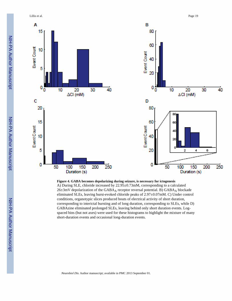
Figure 4. GABA becomes depolarizing during seizure, is necessary for ictogenesisA) During SLE, chloride increased by 22.95±0.73mM, corresponding to a calculated26±3mV depolarization of the GABAA receptor reversal potential. B) GABAA blockadeeliminated SLEs, leaving burst-evoked chloride peaks of 2.97±0.07mM. C) Under controlconditions, organotypic slices produced bouts of electrical activity of short duration,corresponding to interictal bursting and of long duration, corresponding to SLEs, while D)GABAzine eliminated prolonged SLEs, leaving behind only short duration events. Log-spaced bins (but not axes) were used for these histograms to highlight the mixture of manyshort-duration events and occasional long-duration events.
Lillis et al. Page 19
Neurobiol Dis. Author manuscript; available in PMC 2013 September 01.
NIH
-PA Author Manuscript
NIH
-PA Author Manuscript
NIH
-PA Author Manuscript

Figure 5. Ictal pH changesOrganotypic slices prepared from wild-type mice were stained with the ratiometric pHindicator SNARF-1 AM. Spontaneous SLEs were apparent (red bar) in a synchronouslyrecorded field potential (green). Both the polarity (alkali ictal transient) and low amplitude(0.080 ± 0.025, mean±SEM, n=3 slices) of observed pH changes (blue) were insufficient toaccount for changes observed in intracellular chloride using Clomeleon.
Lillis et al. Page 20
Neurobiol Dis. Author manuscript; available in PMC 2013 September 01.
NIH
-PA Author Manuscript
NIH
-PA Author Manuscript
NIH
-PA Author Manuscript

Figure 6. Bicarbonate current contributes to ictogenesisA) In 4-AP-treated acute slices from GIN mice, pharmacologically hyperpolarizing theGABAA reversal potential by blocking intracellular bicarbonate production withacetazolamide (ACTZ) eliminates SLEs, leaving only interictal bursts. Upon washout ofACTZ, SLEs return. B) ACTZ reversibly eliminates SLEs in 4-AP (n = 11, ** indicate thatthe number of SLEs is significantly different from zero, p<0.05).
Lillis et al. Page 21
Neurobiol Dis. Author manuscript; available in PMC 2013 September 01.
NIH
-PA Author Manuscript
NIH
-PA Author Manuscript
NIH
-PA Author Manuscript