Australian Labradoodle Dystrophinopathy
121
Australian Labradoodle Dystrophinopathy: A Novel Canine Model for the Study of Duchenne Muscular Dystrophy Cardiomyopathy by Stephanie M. Shrader, DVM, DACVP A dissertation submitted to the Graduate Faculty of Auburn University in partial fulfillment of the requirements for the Degree of Doctor of Philosophy Auburn, Alabama May 2, 2020 Key Words: Duchenne, Cardiomyopathy, Transcriptome, Dystrophin, Labradoodle Copyright 2020 by Stephanie M. Shrader Approved by Bruce F. Smith VMD, PhD, Chair, Professor of Pathobiology, Director of AURIC Russell C. Cattley, VMD, PhD, DACVP, FIATP, Head of Department of Pathobiology Richard C. Bird, PhD, Professor of Molecular Biology and Cancer Genetics
Transcript of Australian Labradoodle Dystrophinopathy

Australian Labradoodle Dystrophinopathy: A Novel Canine Model for the Study of Duchenne Muscular Dystrophy Cardiomyopathy
by
Stephanie M. Shrader, DVM, DACVP
A dissertation submitted to the Graduate Faculty of Auburn University
in partial fulfillment of the requirements for the Degree of
Doctor of Philosophy
Auburn, Alabama May 2, 2020
Key Words: Duchenne, Cardiomyopathy, Transcriptome, Dystrophin, Labradoodle
Copyright 2020 by Stephanie M. Shrader
Approved by
Bruce F. Smith VMD, PhD, Chair, Professor of Pathobiology, Director of AURIC Russell C. Cattley, VMD, PhD, DACVP, FIATP, Head of Department of Pathobiology
Richard C. Bird, PhD, Professor of Molecular Biology and Cancer Genetics

ii
Abstract
Duchenne muscular dystrophy (DMD) is an X-linked recessive disorder that results in
progressive damage to both skeletal and cardiac myocytes. As a result of improved ventilatory
care, mortality in affected patients is increasingly attributed to cardiomyopathy. We have
identified a novel dystrophin mutation in exon 21 in Australian Labradoodles. Affected dogs
have poor weight gain and weight loss with gait abnormalities, exercise intolerance, skeletal
muscle atrophy, macroglossa, ptyalism, dysphagia, kyphosis, and a plantigrade stance
developing by 6 months of age. Concurrent echocardiographic and electrocardiographic
abnormalities include hyperechoic foci in the left ventricular papillary muscles, septal
hypokinesis, decreased left ventricular systolic and diastolic volume and internal diameter, and
atrioventricular (AV) block. Skeletal muscle pathology in affected dogs is similar to what has
been described in people with DMD and includes myocyte degeneration, necrosis, and
regeneration, fibrofatty infiltration, lymphohistiocytic inflammation, and mineralization.
Histopathologic findings in the heart were observed in the dog with the AV block and consisted
of a focal area of mineralization adjacent to the sinoatrial node. Cardiac transcriptome
sequencing on left ventricular myocardial samples found 29,740 genes expressed and 1267
differentially expressed genes. Expression patterns from affected dogs were generally distinct
from controls and there was a high correlation between samples. The ten gene transcripts with

iii
the greatest up regulation (in descending order of fold change) included BDNF, MYL4, PENK,
BSPRY, PRR32, NPPA, LOC490471, LYZF2, CDH10, and FGF6. We also found that the most
down-regulated transcripts (in ascending order of fold change) included LOC612108, ST8SIA2,
FOXR1, P2RX6, LOC610380, DAO, CNR2, SDC1, LRRC55, and TMEM171). The majority of
the above-mentioned genes have known roles in cardiac compensatory changes secondary to
dystrophinopathic-associated damage, including survival, remodeling, contractility, conduction,
and immunoregulation. Differential expression of genes was also observed in pathways
associated with cardiac oxidative stress, apoptosis, and contractility. These findings are
significant because they support the use of the Australian Labradoodle as a novel animal model
for the study of DMD cardiomyopathy, they elucidate pathways and differential gene expression
involved in Labradoodle dystrophinopathic cardiomyopathy, and they may aid in the
development of therapeutic targets to treat dystrophinopathy-associated cardiac disease.

iv
Acknowledgments
The completion of this work was made possible by the assistance of many people whose
names may not all be enumerated. The contributions and support that I have received from the
faculty, staff, and students at the Auburn University College of Veterinary Medicine and the
Scott Ritchey Research Center have been sincerely appreciated and are gratefully acknowledged.
There are a few people, however, that deserve special mention:
My husband - for his constant support, understanding, and encouragement throughout veterinary
school, residency, and my doctoral research.
Mr. Stephen Waters – for his high standard of animal care, devotion to animal welfare, and his
willingness to assist with innumerable technical procedures.
The Labradoodles – for giving the ultimate sacrifice to aid in the understanding and treatment of
DMD.

v
Table of Contents
Abstract ........................................................................................................................................... ii
Acknowledgments ......................................................................................................................... iv
List of Tables ................................................................................................................................ vii
List of Figures .............................................................................................................................. viii
List of Abbreviations ..................................................................................................................... ix
Chapter 1: Literature Review .......................................................................................................... 1
DMD Epidemiology .................................................................................................................... 2
DMD Clinical Findings ............................................................................................................... 3
Therapeutic Strategies ............................................................................................................... 11
Dystrophin and the Dystrophin Associated Protein Complex .................................................. 13
DMD Animal Models ................................................................................................................ 21
Labradoodle History .................................................................................................................. 26
Transcriptomics ......................................................................................................................... 27
Chapter 2: Characterization of Australian Labradoodle Dystrophinopathy ................................. 31
Chapter 3: Cardiac transcriptome analysis identifies differentially expressed genes in Australian
Labradoodle dystrophinopathy ..................................................................................................... 43
Chapter 4: Conclusions ................................................................................................................. 82
References ..................................................................................................................................... 86

vi
Appendix I: Supportive Photographs and Figures ...................................................................... 107

vii
List of Tables
Table 1. Dystrophin Isoforms ....................................................................................................... 19
Table 2. Comparison of GRMD and DMD cardiac abnormalities ............................................... 24

viii
List of Figures
Figure 1. Photographs of dystrophin-deficient Labradoodles ..................................................... 107
Figure 2. Comparison of weight gains between dystrophin-deficient and control dogs ............ 108
Figure 3. 31P MR spectra ............................................................................................................. 109
Figure 4. Echocardiographic image of tricuspid valve regurgitation ......................................... 110
Figure 5. Gross photograph of the diaphragm from an affected dog .......................................... 111

ix
List of Abbreviations
ACE Angiotensin Converting Enzyme
ADHD Attention-Deficit/Hyperactivity Disorder
AKC American Kennel Club
ALT Alanine transaminase
AST Aspartate aminotransferase
AV Atrioventricular
ATP Adenosine triphosphate
BMD Becker Muscular Dystrophy
CBC Complete blood count
cGMP Cyclic guanosine monophosphate
Chem Chemistry
CK Creatine kinase
DAPC Dystrophin-Associated Protein Complex
DMD Duchenne Muscular Dystrophy
ECG Electrocardiogram
FS Fractional shortening
GRMD Golden Retriever Muscular Dystrophy
x
IQ Intelligence quotient
LGMD Limb-Girdle Muscular Dystrophy
LMD Labrador Retriever Muscular Dystrophy
LVEF Left Ventricular Ejection Fraction
MAPK Mitogen-Activated Protein Kinase
MD STARnet Muscular Dystrophy Surveillance Tracking and Research Network
MR Magnetic resonance
MRI Magnetic Resonance Imaging
MRS Magnetic Resonance Spectroscopy
NOS Nitric Oxide Synthase
ORF Open reading frame
RNA Ribonucleic acid
RT-PCR Reverse transcriptase polymerase chain reaction
SA Sinoatrial
SRRC Scott Ritchey Research Center
TTE Transthoracic Echocardiography
XLDCM X-Linked Dilated Cardiomyopathy
1
Chapter 1: Literature Review
Duchenne muscular dystrophy (DMD) is an X-linked recessive disorder that is caused by
mutations in the dystrophin gene. The result is a lack of functional dystrophin, or less commonly,
a truncated, partially functional N-terminal dystrophin fragment. It inevitably causes progressive,
degenerative changes in skeletal and cardiac muscle and also alters the functionality of other cell
types within the body, such as cerebral and cerebellar neurons, depending on the mutation
involved and isoform(s) affected (Table 1) (Hoffman et al., 1987). Boys with this disorder are
often diagnosed when young (less than five-years-old), and generally become non-ambulatory by
the age of ten. Wheelchair bound, these patients typically develop various medical problems
associated with skeletal and respiratory muscle weakness (such as difficulty rising from a seated
position, frequent falls, delayed motor-skill development, gait abnormalities, and respiratory
infections), in addition to cardiomyopathy. Death often occurs in the second or third decades of
life, historically attributed primarily to respiratory failure (Spurney et al., 2008). Complications
of respiratory muscle weakness resulting in failure include progressive restrictive ventilatory
defects, chronic hypoventilation, and various pulmonary infections (Wagner et al., 2007).
According to a recent longitudinal study evaluating the cause of death in DMD patients over the
last 50 years, mean age at death before initiation of mechanical ventilation (January 1977 - July
1984) was 18.9±4.1 years. Following the use of mechanical ventilation, the mean age of death
improved each decade to 31.1±5.4 years (January 2004 to December 2010). Prior to use of

2
mechanical ventilation, more than half of all deaths in DMD patients were due to respiratory
failure. Based on the cohort of patients included in this longitudinal analysis, no DMD patients
have died from respiratory failure since 2000 (Matsumara et al., 2011).
In the last few decades, treatment of skeletal muscle and respiratory complications has
improved, and as a result, mortality in DMD patients is increasingly attributed to cardiac-related
disease. Between 1985 and 1989, greater than 30% of reported DMD deaths in Japan resulted
from cardiac failure (Moriuchi et al., 1993). Similarly, a separate study conducted at the Naples
Centre of Cardiomyology and Medical Genetics reviewed the records from 835 DMD patients
who were examined between 1961 and 2006. Similar to the study published by Matsumara and
colleagues, their findings also indicated that the major cause of death until the 1980’s was due to
respiratory illness and that there has been a significant decade on decade improvement in the
survival rate (the mean age of death in the 1960’s was 14.4 years, while the mean age of death
for those ventilated since 1990 was 25.3 years). The same study found that cardiomyopathy
significantly decreased life expectancy from a mean age of 19 years to 16.9 years (Passamano et
al., 2012).
DMD Epidemiology
Duchenne muscular dystrophy is an X-linked recessive disorder caused by various types
of mutations in the dystrophin gene, all of which result in the lack of a functional dystrophin
protein product. As such, DMD is classified as a type of dystrophinopathy (literally meaning a
disorder of dystrophin). Although the disease phenotypes vary, other forms of muscular
dystrophies categorized as dystrophinopathies include: Becker muscular dystrophy (BMD),
isolated quadriceps myopathy, muscle cramps with myoglobinuria, and X-linked dilated
3
cardiomyopathy (XLDCM) (Beggs, 1997; Sunohara et al., 1990; Wicklund, 2013). With Becker
muscular dystrophy, partial functionality of dystrophin is maintained, and the disease phenotype
can vary widely. With progression, the clinical course can resemble DMD; however, the severity
of skeletal muscle weakness can vary substantially, with milder respiratory dysfunction and
variable cardiac involvement compared to patients with DMD (Wicklund, 2013).
According to the CDC, in 2007, 349 out of 2.37 million males between the ages of 5-24
had either DMD or BMD in the United States. This means that 15 out of every 100,000 males
aged 5-24 years were affected that survey year. Similar survey results were reported from
Northern England in 2009: 233 out of 1.49 million males were reported to have DMD or BMD,
equating to roughly 16 out of every 100,000 males (CDC, 2009; Norwood et al., 2009). In the
United States, the prevalence of DMD and BMD is currently estimated to be 1 in every 7,250
males aged 5-24 years with DMD being 3x more common than BMD (Romitti et al., 2015).
DMD Clinical Findings
Based on data compiled by the Muscular Dystrophy Surveillance, Tracking, and
Research Network (MD STARnet), a definitive diagnosis of DMD typically does not occur until
2.5 years after the parents first noticed physical developmental delays in their child (Ciafaloni et
al., 2009). Because of this delay, boys with DMD are typically presented to the pediatrician
between 3 and 5 years of age with difficulties rising from a seated position, frequent falls, and
delayed milestones in motor-skill development. Physical examination findings often include
(pseudo)hypertrophy of the calves (seen in about 2/3 of patients), variably severe lumbar
lordosis, waddling gait, macroglossa (seen in about 1/3 of patients) and a distinctive way of
rising from the floor (known as Gowers’ maneuver). Because of weakened leg muscles, boys

4
with DMD rise from the floor by first getting on hands and knees, then elevating the posterior,
followed by “walking” their hands up the legs to raise the upper body (Gozal, 2000; MDA,
2014). Muscle weakness becomes progressive over time, eventually resulting in wheelchair
dependence at an early age (usually by 10-years-old). By convention, pediatricians consider boys
with dystrophinopathies to have DMD if the ability to walk ceases before 12-years of age. Boys
who are able to walk beyond their 16th birthday are considered to have BMD (Wicklund, 2013).
Skeletal muscle biopsy samples from patients with DMD are characterized by necrotic
and/or degenerating myocytes with foci of mineralization, myofiber size variability, and
myofibers with centralized nuclei (evidence of regeneration from myoblasts). Necrotic
myocytes/myofibers are often surrounded and/or infiltrated by macrophages and CD4+ T-
lymphocytes. As the regenerative capacity of the affected muscle wanes, there is gradual
replacement of the tissue by fibrosis and infiltrating adipose tissue (Klinger et al., 2012).
Because of dystrophin’s role in myocyte structural integrity (to be discussed at length in the
section entitled “Dystrophin and the Dystrophin Associated Protein Complex”), it has been
proposed that the occurrence of muscular necrosis is resultant from recurrent contraction-
associated mechanical damage. Others have proposed that altered myocyte calcium homeostasis
is to blame for the presence of myocyte necrosis (Deconinck & Bernard, 2007). This second
hypothesis seems short-sighted and lacks experimental evidence to support it. Mechanical
cellular damage can and will cause altered sarcolemmal integrity, secondary calcium influx, and
resultant cell death. Because inherently altered calcium channels have not been documented in
dystrophin-deficient myofibers, calcium-associated muscular necrosis is likely secondary to
contraction-related myocyte damage. Vascular anomalies, aberrant glycosylation of dystrophin-
associated proteins, and gene regulatory anomalies have also been proposed as potential causes

5
for the observed muscular necrosis; however, they lack supportive experimental data (Deconinck
& Bernard, 2007).
Another interesting finding in >50% of patients with DMD is the presence of rare
“revertant fibers,” myofibers that have strong, seemingly counterintuitive, dystrophin
immunolabeling (Nicholson et al., 1989). Studies have shown that although these revertant fibers
produce a dystrophin protein that lacks the region encoded by the deleted exons, they do have a
normal C-terminus, consistent with a restored reading frame (Hoffman et al., 1990; Klein et al.,
1992). The prevalence of somatic mutations resulting in reading frame restoration is unknown. It
has been hypothesized that many somatic mosaic males for DMD exist, and indeed there are
numerous case reports describing somatic mosaicism in individual patients; however, most are
likely not detected clinically because of genetic normalization (Klein et al, 1992). By utilizing
exon-specific monoclonal antibodies against dystrophin, Le Thiet Thanh and colleagues
demonstrated that somatic mutations in revertant-fiber nuclei result in the removal of additional
exons from dystrophin mRNA, confirming the previous supposition that the reading frame was
restored (Thanh et al., 1995). The clinical implications of revertant fibers continue to be
explored. For example, in a recent phase I clinical trial (that sought to deliver a transgene that
encoded a miniaturized version of human dystrophin), dystrophin-specific T-cell responses were
unexpectedly detected in two patients prior to gene therapy delivery. Interestingly, the epitopes
were mapped to myofibers with revertant dystrophin expression. It has been postulated that
revertant dystrophin expression may participate in the poorly understood mechanisms behind
DMD-related myositis (Campbell et al., 2010).
Additional physical examination findings in DMD patients often include fatigue, joint
contractures, decreased cardiopulmonary functionality, and cognitive impairment. Joint

6
contractures typically develop in the hips and knees, owing to the time spent in a wheelchair as
the disease progresses. Abnormal seating postures (i.e. kyphosis, lordosis, and scoliosis) are
reported in 68-90% of ambulatory and non-ambulatory DMD patients. In non-ambulant patients,
there is a much higher risk for scoliosis development, which can be rapidly progressive without
aggressive orthotic measures and/or surgical interventions (Oda et al., 1993; Yamashita et al.,
2001). Respiratory complications are typically attributed to decreased thoracic compliance
resulting from diaphragmatic, intercostal, and accessory respiratory muscle dysfunction and
weakness. Ventilatory failure inevitably develops over time, necessitating the use of non-
invasive and invasive ventilation techniques (Boland et al., 1996; Gozal, 2000; Wicklund, 2013).
DMD patients should remain up-to-date on vaccines in order to prevent pneumonia and they may
require manual or mechanically assisted support to stimulate coughing when pulmonary
infections do occur (CDC, 2016). The ability to better manage DMD-associated respiratory
complications has resulted in prolongation of life, but it has also unmasked concurrent cardiac
dysfunction in these patients (Bach, 1994).
Currently, most if not all patients with DMD that survive into their thirties will be
diagnosed with cardiomyopathy at some point. Traditionally, recognition was often delayed due
to a general lack of physical activity; however, in more recent years, clinicians are using more
sophisticated screening techniques at earlier ages, regardless of outward symptomology
(McNally, 2007). A study published in 2013 found that cardiac problems typically begin by 14
years of age, and that for every year corticosteroids were taken by the patient (see “Therapeutic
Strategies” below for more information on corticosteroid usage in DMD), the chance of
developing heart problems decreased by 4% (Barber et al., 2013). Cardiac involvement in

7
patients with DMD can be associated with various cardiac alterations and therefore variable
phenotypes.
In 2008, the recommended cardiac screening protocol for boys with DMD included an
electrocardiogram (ECG) and transthoracic echocardiography (TTE) every two years until the
age of ten, and then once a year thereafter (Bouhouch et al., 2008). In 2010, the DMD Care
Considerations were published; these too addressed cardiac care recommendations based on
minimal surveillance standards. Similar to Bouhouch and colleagues, the working group
recommended that minimum assessment include an ECG at least once every two years until the
age of ten and then once a year thereafter. If ventricular functional abnormalities are noted,
cardiac functionality should be evaluated at least every six months, and pharmacological
interventions should be initiated immediately (Bushby et al., 2010). Most commonly, children
with the most severe manifestations of DMD will have persistent sinus tachycardia, an increased
R-S ratio in the right precordial leads, deep Q waves in the lateral leads, and conduction
abnormalities (i.e. alterations in the progression of electrical impulses through the heart). In
addition to persistent sinus tachycardia, other common arrhythmias in DMD patients include
sinus bradycardia, atrial premature beats (usually isolated), and ventricular premature beats (also
typically isolated). Often, patients with ventricular premature beats are in advanced stages of
disease (Yanagisawa et al., 1992). In one study, cardiac conduction abnormalities were reported
in 24 of the 50 patients examined (48%). Of the 24 patients, 23 had intra-atrial conduction
abnormalities, five had infranodal conduction abnormalities, and one patient had prolongation of
atrioventricular (AV) conduction. Of those with infranodal conduction defects, one patient had a
right bundle branch block and three patients had a left anterior fascicular block (Sanyal &

8
Johnson, 1982). First- and second-degree AV blocks have also been documented in DMD
patients (Perloff, 1984; Sanyal & Johnson, 1982; Yanagisawa et al., 1992).
Heart blocks occur when the electrical signal is slowed or disrupted as it moves through
the heart. In order of least to most severe, the three types of heart block are first-degree, second-
degree, and third-degree. With a first-degree block, the heart's electrical signals are slowed as
they progress from the atria to the ventricles. Electrocardiographically, this is represented by a
longer, flatter line between the P and the R waves. Second-degree heart blocks are characterized
by atrioventricular electrical signals that are slowed to a large degree; as a result, a QRS wave
doesn’t always follow each P wave as it should. Second-degree heart blocks can be further
categorized as either a Mobitz type I or Mobitz type II block. In a Mobitz type I block, electrical
conduction is progressively delayed with each heartbeat, until the heart skips a beat. When
viewed using an ECG, the delay is shown as a PR interval that gets longer with each heartbeat
until the QRS waves don't follow the subsequent P wave. Motbitz type II blocks are less
common than type I and are generally more severe. With this type of block, some of the
electrical signals fail to reach the ventricles, making the pattern less regular than that which is
seen with Mobitz type I. Electrocardiographically, the QRS wave follows the P wave at a normal
speed; however, when it is blocked, the QRS wave is missing. A third-degree block (also known
as a complete heart block or complete AV block) is characterized by complete failure of the
electrical signals to reach the ventricles. When this occurs, independent accessory pacemaker
signals will attempt to activate the ventricles (known as an escape rhythm). The resultant ECG
pattern is characterized by P waves that occur at a faster rate and aren’t coordinated with the
QRS waves. This type of conduction anomaly can result in bradycardia, hypotension, sudden
cardiac arrest, and death (NIH, 2012).

9
Histologically, myocardial changes consist of multifocal areas of fibrosis (most
prominent in the inferolateral wall), myocyte vacuolation (consistent with degeneration),
myocyte necrosis, myofiber size variation, and fatty infiltration. Specific areas of the conduction
system that have been reported to be affected include the sinoatrial (SA) and AV nodes, AV
bundle, left bundle branch, and right bundle branch. It has been postulated that the myocardial
lesions result from mechanical stresses imposed on a metabolically and structurally abnormal
heart; however, it remains to be determined why a segmental, rather than diffuse, pattern of
myocardial injury occurs. Although other factors are likely at play, these microscopic changes
presumably play a role in the development of the before mentioned conduction anomalies,
arrhythmias (both subclinical and fatal), and compensatory changes (such as dilated
cardiomyopathy (DCM)) (Sanyal & Johnson, 1982; Verhaert et al., 2011; Yanagisawa et al.,
1992).
Recently, cardiac magnetic resonance imaging (MRI) has also been proposed as a useful
(and perhaps better) diagnostic tool to identify early myocardial remodeling changes (Verhaert et
al., 2011). Both echocardiography and MRI are able to identify abnormal left ventricular
fractional shortening (FS), left ventricular hypokinesia, and left ventricular dilation; however,
research shows that the majority of echocardiographic studies have suboptimal scanning
windows and significantly over- or underestimated left-ventricular function compared to cardiac
MRI. As an example, Brunklaus and colleagues performed a retrospective analysis of case
records from 35 boys with DMD underwent cardiac evaluation for surgical procedures between
2010 and 2013. They reported ECG found a median left ventricular FS of 29/%. 37% of boys
(13/35) had abnormal FS <25%, 66% (23/35) had evidence of hypokinesia, and 26% (9/35) had
left ventricular dilatation. Cardiac MRI revealed a median left ventricular ejection fraction

10
(LVEF) of 52%. 57% of boys (20/35) had abnormal LVEF <55%, 71% (25/35) had left
ventricular hypokinesia, and 82% had late gadolinium enhancement. Data also showed that
extensive late gadolinium enhancement is associated with reduced LVEF (48% vs 58%),
consistent with more severe cardiac pathology (Brunklaus et al., 2015). In addition to cardiac
MRI, cardiac magnetic resonance spectroscopy (MRS) is also being utilized to better evaluate
cardiac functionality in patients with DMD and BMD. One of the benefits is that MRS can be
used to noninvasively analyze the cardiac phosphocreatine to adenosine triphosphate ratio
(PCr/ATP), a useful measure of cardiac energy metabolism. In a recent publication, 13 men with
BMD, 10 female carriers, and 23 control patients were studied using phosphorus-31 MRS and
conventional echocardiography. Results indicated that the PCr/ATP was significantly reduced in
BMD patients (1.55 ± 0.37) and carriers (1.37 ± 0.25) when compared to controls (2.44 ± 0.33; p
< 0.0001). Interestingly, the PCr/ATP did not correlate with LVEF or mass index. Because the
reduced PCr/ATP lacked correlation with indices of left ventricular functionality, it has been
posited that there may be a direct link between altered dystrophin expression and the
development of cardiomyopathy (Grilley et al., 2000).
Cognitive impairment is also a common manifestation in patients with Duchenne
muscular dystrophy, and although it was first noted by Duchenne in his original description of
the disease (Duchenne, 1868), it hasn’t been until much more recently that the underlying
mechanisms involved in this process have been elucidated. Recent data suggests that 45% of the
oldest males affected with DMD or BMD has at least one of the following mental health
concerns: behavior anomalies, depression, or attention-deficit/hyperactivity disorder (ADHD)
(Caspers et al., 2015). Behavioral studies have shown that roughly 30% of boys with DMD are
intellectually disabled with an average IQ around 85 (one standard deviation below the mean).
11
Verbal IQ is reportedly more affected than performance IQ and developmental language delays
are common. DMD patients also have an increased prevalence of obsessive compulsive disorder
(5%) and autism spectrum disorder (3% to 6%) (Anderson et al., 2002; Bresolin et al., 1994;
Hendriksen & Vles, 2008; Leibowitz & Dubowitz, 1981). Recently, in a sample of 80 boys with
DMD, it was found that regardless of IQ, the patients displayed a specific cognitive profile,
characterized by poor performance in digit span, story recall, and comprehension. The authors
found that the degree of cognitive impairment did vary between patients (similar to variances
seen in DMD-related skeletal and cardiac pathologies), which they surmised was due to
variations in dystrophin gene mutations in the sampled patient population (Hinton et al., 2000).
Therapeutic Strategies
Currently, there is no cure for Duchenne muscular dystrophy and treatment is generally
only palliative. One of the current mainstays of treatment is prolonged glucocorticosteroid use,
which improves quality of life by increasing the potential years of ambulation, and likely plays a
role in increased survival (Wagner et al., 2007). Clinical studies with glucocorticoids have
demonstrated a prolongation of ambulatory abilities by approximately 2 years (Manzur et al.,
2008). These clinical effects likely result from the ability of glucocorticoids to up-regulate anti-
inflammatory mediators and inhibit pro-inflammatory molecules, including various cytokines,
chemokines, arachidonic acid metabolites, and adhesion molecules (Velden, 1998). There are
numerous documented side effects associated with glucocorticosteroid administration, such as
weight gain, hirsutism, behavioral abnormalities, hyperglycemia, hypertension, shortened stature,
cataracts, Cushing’s syndrome (hyperadrenalcortisolism), and osteoporosis. Unfortunately, these
negative side effects are not infrequent; the most common reason for DMD patients to
12
discontinue corticosteroid use (specifically prednisone) is due to weight gain (Wagner et al.,
2007).
Other available therapies are mainly supportive, such as:
• Physiotherapy – Focusing on stretching the upper extremity musculature to decrease
contractures. This daily routine of stretching often focuses on forearm pronators, elbow
flexors, wrist flexors, and long finger flexors (Bushby et al., 2010; Collins & Morgan,
2003). Stretching of the distal extremities, even when non-ambulatory, is also beneficial.
• Implantable cardio-defibrillators - For those patients with an increased risk of fatal
ventricular arrhythmias.
• Cardiac medications – There are a wide range of clinical presentations associated with
cardiomyopathy and heart failure. Based on the individual, several different medications
may be prescribed, such as ACE inhibitors, spironolactone, loop diuretics, and digoxin
(Bushby et al., 2010).
• Wheelchair and other mechanical support (braces).
• Scoliosis surgery – Often surgery is performed when DMD patients are adolescents in
order to decrease respiratory compromise, pain and deformity.
• Assisted ventilation – This includes noninvasive positive pressure ventilators,
tracheostomy, and mechanical insufflation-exsufflation (Bushby et al., 2010; Passamano
et al., 2012).
• Treatment of respiratory infections.
Although beneficial, none of these therapeutic measures impact the fundamental cause of DMD,
the lack of dystrophin expression in skeletal and cardiac musculature.

13
Dystrophin and the Dystrophin Associated Protein Complex
The dystrophin gene, the largest gene currently known in nature, measures 2.4-megabases
at locus Xp21 (NCBI, 2020). It was isolated by Louis Kunkel and his colleagues in 1985 (Kunkel
et al., 1985), 117-years after Duchenne first described the phenotype associated with the disease
that now bears his name (Duchenne, 1868). This gene encodes the protein dystrophin, which is a
large, 427-kD sub-membrane protein that links cytoskeletal F-actin to the extracellular matrix
protein laminin via the transmembrane protein α/β-dystroglycan complex. Dystrophin is a
member of the β-spectrin/α-actinin protein family; together, it and its associated sarcolemmal
proteins form the dystrophin-associated protein complex (DAPC) (Fine et al., 2011; Holland et
al., 2013; Koenig & Kunkel, 1990; Spurney et al., 2008).
The overall role of the DAPC is to mediate signaling between the intracellular
cytoskeleton and the extracellular matrix. As it pertains to muscular function, the role of this
complex is to protect myocytes and myofibers from injury associated with contractile forces.
When membrane degradation occurs (such is the case with dystrophinopathies), the
histomorphologic changes include edema, inflammation, necrosis, and muscle fibrosis, resulting
in the well-known physical manifestations of the disease (Frankel & Rosser, 1976).
Dystrophin is composed of four, well-described functional domains. The N-terminal
domain (composed of two calponin homology [CH] molecules) binds F-actin within the
cytoplasm (Winder et al., 1995). The adjacent central rod domain is composed of greater than
2800 amino acids that build 24 spectrin-like triple helical repeats with four intervening non-
helical segments (known as “hinge” regions) that are thought to result in protein flexibility
(Ervasti & Campbell, 1993; Koenig & Kunkel, 1990; Sunada et al., 1994; Suzuki et al., 1994;

14
Sweeney & Barton, 2000; Yang, Jung, Rafael, et al., 1995). The third domain, the WW region, is
a β-sheet motif involved in intracellular signaling via recognition of proline-rich or
phosphorylated linear peptides. This domain recognizes a PPxY motif and plays a role in the β-
dystroglycan interaction.(Bork & Sudol, 1994) Adjacent to the WW domain is a cysteine-rich
region composed of two ER-hand motifs (Koenig et al., 1988) and two ZZ modules (Ponting et
al., 1996) that bind calmodulin. Studies later showed that this binding takes place in a calcium-
dependent manner (Anderson et al., 1996).
The COOH terminal domain is composed of two regions that form α-helical coiled coils,
which make up the binding site for dystrobrevin (Blake et al., 1995). This region also associates
with syntrophin (alpha and beta subunits) and neuronal nitric oxide synthase (nNOS) indirectly,
binding to dystroglycan within the transmembrane portion of the DAPC. Dystrophin, along with
the syntrophins and dystrobrevin, form the subsarcolemma subcomplex. The dystrophin
subcomplex is stabilized the sarcoglycan-sarcospan subcomplex, which is composed of alpha,
beta, gamma and delta subunits of sarcoglycan, and sarcospan (Kobayashi & Campbell, 2012).
Dystroglycan, a ubiquitously expressed protein, consists of an alpha and beta subunit
(Ibraghimov-Beskrovnaya et al., 1992). The alpha subunit consists of a mucin domain and a C-
terminal domain, which has been shown to bind to laminin-2 (also known as merosin) with high
affinity in a calcium-dependent manner (Ervasti & Campbell, 1991; Ibraghimov-Beskrovnaya et
al., 1992). In addition, α-dystroglycan serves as an agrin receptor in muscle, regulating agrin-
induced acetylcholine receptor clustering at the neuromuscular junction. Related to this function,
it has been shown that dystroglycan can cluster within the sarcolemma in response to rapsyn (a
receptor-associated synapse protein), thus playing a pivotal role in neuromuscular junction
formation and activity (Campanelli et al., 1994). Recently, it has been shown that neither
15
dystrophin nor utrophin are required for the expression of the DAPC in cardiac musculature;
however, α-dystroglycan is differentially glycosylated in mdx and dystrophin/utrophin double
knock-out mouse hearts with aberrant sarcolemmal localization. Of note is that these alterations
do not affect laminin binding, and the implications of these changes are as of yet, unknown
(Sharpe et al., 2013).
Beta-dystroglycan is composed of a single transmembrane domain that spans the
sarcolemma and an extracellular amino-terminal domain that binds the carboxy-terminal globular
domain of the alpha subunit (Boffi et al., 2001; Di et al., 1999). The cytoplasmic COOH
terminus has multiple proline residues necessary for dystrophin binding; it binds directly to the
WW modules and the cysteine-rich domain that have the EF and ZZ regions (Ishikawa-Sakurai
et al., 2004; Renschler et al., 1999; Suzuki et al., 1992). A separate study found that the WW-like
domain within caveolin-3 recognizes the C-terminal end of β-dystroglycan which has a PPXY
motif (Sotgia et al., 2000). Based on these findings, it was surmised that the interaction of
caveolin-3 with β-dystroglycan may be able to competitively regulate dystrophin recruitment to
the complex.
In addition to the role of anchoring dystrophin to the sarcolemma, β-dystroglycan has
also been shown to participate in MAPK (mitogen-activated protein kinase) signaling.
Interaction with laminin results in recruitment of a Grb2 (Growth factor receptor-bound protein
2) - Sos1 (Son of sevenless homolog 1) complex to dystroglycan (Oak et al., 2003). Within
dystroglycan, the proline-rich motif then interacts directly with Grb2, an adaptor protein
involved in signal transduction, cell communication and cytoskeletal organization (Yang, Jung,
Motto, et al., 1995). The dystroglycan-Grb2 interaction results in activation of Rac1 which in
turn activates JNK via the Cdc42-Race effector PAK1 (p21 activated kinase 1). The β-

16
dystroglycan interaction with MEK and ERK may indicate the dystroglycan acts as a signaling
scaffold (Spence et al., 2004).
Interestingly, and contradictory to earlier research, a recent study indicates that most of
the dystroglycan complexes at the sarcolemma do not in fact interact with dystrophin or utrophin
in wild-type muscles. Evidence also suggests that a subset of dystroglycan that does not directly
bind to dystrophin is also destabilized in the absence of dystrophin. Although much more work
needs to be done to further characterize the potential differences in dystroglycan, this research
seems to indicate that additional disease mechanisms may be involved in the variable disease
phenotypes associated with dystrophinopathies and dystroglycanopathies (Johnson et al., 2013).
As mentioned, the sarcoglycan complex is composed of α-, β-, γ- and δ-sarcoglycan
subunits and sarcospan, and like other portions of the DAPC, this complex and its components
can be secondarily disrupted and destabilized when dystrophin is mutated. The sarcoglycans are
single transmembrane glycoproteins with N-terminal domains oriented extracellularly (α-
sarcoglycan) and intracellularly (β-, γ- and δ-sarcoglycans) (Lim et al., 1995; Noguchi et al.,
1995; Roberds et al., 1993). Sarcospan is composed of four transmembrane-spanning portions,
homologous to the tetraspanin family (Crosbie et al., 1999). The sarcoglycan complex appears to
function by strengthening the interaction between β-dystroglycan, α-dystroglycan and dystrophin
(Ozawa et al., 2005).
Unlike dystroglycan, which has been shown to be expressed in various cell types, the
sarcoglycans have a much narrower tissue range and appear to only be expressed in skeletal and
cardiac myocytes (Roberds et al., 1993; Roberds et al., 1994). In addition to being disrupted by
dystrophin mutations, autosomal recessive mutations in any of the four main sarcoglycan
isoforms result in autosomal recessive limb-girdle muscular dystrophy (LGMD-2C-2 F). When

17
δ-sarcoglycan is lacking, as seen with LGMD-2 F, the remaining three sarcoglycan members are
unable to assemble and are thus degraded before transport from the Golgi (Draviam et al., 2006;
Shi et al., 2004).
Recently, additional sarcoglycans have been identified. Epsilon-sarcoglycan was
discovered and found to be highly similar to α-sarcoglycan. Like the other four sarcoglycans,
epsilon can also be found in cardiac and skeletal musculature; however, analysis has shown that
it is not part of the sarcoglycan complex (Durbeej et al., 2000). Epsilon-sarcoglycan has been
found to be highly expressed in the central and peripheral nervous systems and loss-of-function
mutations in its associated gene result in myoclonus-dystonia syndrome in people (Zimprich et
al., 2001). Zeta-sarcoglycan, which shows the most sequence similarity to γ- and δ-sarcoglycan
isoforms, has also been discovered (Wheeler et al., 2002).
Together with the sarcoglycan subunits, sarcospan helps form the link between the
subsarcolemmal cytoskeleton and the extracellular matrix. As seen with sarcoglycans, sarcospan
localization to the membrane is dependent on proper dystrophin expression; therefore, its
expression is markedly decreased in patients with DMD. Interestingly, the gene encoding
sarcospan maps to human chromosome 12p11.2, part of the locus for an autosomal dominant
muscular dystrophy known as congenital fibrosis of the extraocular muscle (Crosbie et al., 1997;
Crosbie et al., 2000).
Not only does the DAPC play a role in protecting the myocytes from contraction-
associated damage, it also serves in scaffolding signaling molecules like nNOS. Sarcolemmal
nNOS is lacking when dystrophin levels are low or absent due to deletions in critical regions of
the rod domain. This results in deficiency in the normal contraction-induced cGMP-dependent
attenuation of local vasculature leading to post-exercise vasoconstriction within muscles

18
(Kobayashi & Campbell, 2012; Torelli et al., 2004). The degree and extent of vasoconstriction
can lead to muscle ischemia, as reported in DMD patients (Sander et al., 2000), in addition to
exaggerated fatigue and intramuscular edema (Kobayashi & Campbell, 2012).
The dystrophin gene produces a variety of different transcripts encoding various
dystrophin isoforms (proteins of varying lengths that have different segments of the basic
dystrophin sequence). Transcription of this gene is controlled by three different promoters whose
names reflect the general tissue distribution of dystrophin expression: brain (B), skeletal and
cardiac muscle (M) and purkinje (P) (Blake et al., 2002). The dystrophin gene also has four
internal promoters that result in shorter transcripts encoding for several isoforms (Retinal [R],
Brain 3 [B3], Schwann cells [S], and General [G]) (Constantin, 2014). The different isoforms,
generated via splicing at a unique first exon, function in various tissue types and their functions
are summarized in Table 1 (below):

19
Table 1. Dystrophin Isoforms
Isoform Synonyms and Subtypes Tissue of Expression
Dp427
Dp427l (L-Dystrophin)
Lymphoblastoid (Nishio et al., 1994; Wheway & Roberts, 2003)
Dp427c (Brain or C-Dystrophin)
Brain (hypothalamus and cortex) (Bovolenta et al., 2012; Muntoni et al., 2003; Nudel et al., 1989; Tokarz et al., 1998)
Dp427m (M-Dystrophin)
Skeletal and cardiac muscle (Bovolenta et al., 2012; Holland et al., 2013; Muntoni et al., 2003; Tokarz et al., 1998)
Dp427p (P-Dystrophin)
Purkinje cerebellar neurons (Bovolenta et al., 2012; Gorecki et al., 1992; Muntoni et al., 2003)
Expression in other tissues: Fetal tissues (Feener et al., 1989; Muntoni et al., 2003), eye, intestine, kidney, liver, lung, spleen, stomach, testis, thymus, and uterus (Tokarz et al., 1998)
Dp260
Retina (D'Souza et al., 1995), brain, cardiac muscle, intestine, kidney, liver, lung, skeletal muscle, spleen, stomach, testis, thymus, and uterus (Tokarz et al., 1998)
Dp140
Dp140b Kidney (Tokarz et al., 1998) Dp140ab Cerebellum and kidney (Lidov et al., 1990;
Lidov et al., 1995) Dp140c Cerebellum (Lidov et al., 1990; Lidov et al.,
1995) Dp140bc Cerebellum and kidney (Lidov et al., 1990;
Lidov et al., 1995) Expression in other tissues:
Other portions of brain, cardiac muscle, eye, intestine, liver, lung, skeletal muscle, spleen, stomach, testis, thymus, and uterus (Tokarz et al., 1998)
Dp71 apo-dystrophin 1 (Liver or G-Dystrophin)
Expressed in all tissues (Tadayoni et al., 2012), including: kidney (Austin et al., 1995; Haenggi et al., 2005; Tokarz et al., 1998), CNS (numerous cell types) (Austin et al., 1995; Benabdesselam et al., 2012; Schofield et al., 1994; Tokarz et al., 1998), liver (deLeon et al., 2006; Tokarz et al., 1998), skeletal muscle, heart, lung, testis (Austin et al., 1995; Tokarz et al., 1998), eye, intestine,

20
spleen, stomach, thymus, and uterus (Tokarz et al., 1998)
Dp116
apo-dystrophin 2 (S-Dystrophin)
Typically described in Schwann cells (Byers et al., 1993) and brain (Schofield et al., 1994); however, RT-PCR has revealed expression in multiple other organs: cardiac muscle, eye, intestine, kidney, lung, skeletal muscle, spleen, stomach, testis, thymus, and uterus (Tokarz et al., 1998)
Dp40 Brain (synaptic vesicles in neurons) (Tinsley et al., 1993; Tozawa et al., 2012)
Various types of dystrophin mutations have been documented (in both people and animal
models). The majority of these mutations are deletions, accounting for nearly 43% -65% of all
DMD mutations, depending on cohort evaluated (Dent et al., 2005; Flanigan et al., 2009). Two-
thirds of DMD patients inherit the mutation from a carrier mother and the remaining one-third of
patients have de novo DMD mutations. These de novo mutations are believed to occur via the
following situations:
1. De novo mutations arise in meiosis either in the grand-parental generation or in the
mother
2. Mitotic de novo mutations arise during spermatogenesis of the grandfather, during
oogenesis of the grandmother, or during oogenesis of the mother (Grimm et al., 2012)
Several studies have reviewed the relative probabilities of de novo mutations per mutation type
and have found that large deletions arise predominantly during oogenesis while point mutations
and duplications more commonly result from errors during spermatogenesis (Grimm et al., 1994;
Kawamura et al., 1997). Spontaneous DMD mutations have been reported in dogs (see DMD

21
Animal Models below); however, the prevalence of de novo DMD mutations in canine
dystrophinopathies has not been reported.
Roughly two-thirds of DMD deletions are reportedly greater than one exon and are
typically found in the central rod domain around exons 44-53 and to a lesser extent, the 5’
terminus (Beggs et al., 1990; Dent et al., 2005; Johnson et al., 2012). In the mdx mouse, and in
people with DMD, loss of the dystrophin C-terminal domain results in loss of the entire DAPC
(Ohlendieck & Campbell, 1991). In addition to deletions, duplications and point mutations have
also been identified in DMD patients. For example, in a study by Flanigan and colleagues, point
mutations accounted for 46% of all mutations in the cohort evaluated. In that study, there were
no point mutation hotspots; the mutations were evenly distributed across the exons of the DMD
gene (Flanigan et al., 2009).
Additional cardiac-specific, dystrophin-associated proteins (Cypher, Ahnak1, Cavin-1
and CRYAB) have been identified in humans and mice (Johnson et al., 2012), and recently
decreased Claudin-5 (a cell junction protein) was demonstrated in the cardiac tissue of
dystrophin deficient mice (Delfin et al., 2012). It has been posited that variability in cardiac
disease progression and severity may be due to differences in expression of these dystrophin
associated proteins.
DMD Animal Models
Although the clinical course, disease severity, and microscopic lesions don’t completely
recapitulate DMD-associated skeletal and cardiac disease, rodent models are frequently utilized
to study DMD because they are convenient, reproduce quickly, have a short life-span, and are
relatively inexpensive to maintain (compared to other laboratory animals). The mdx mouse, first

22
described in 1977 in mice from a C57BL/10ScSn background, is one of the conventional animal
models of DMD. This model is genetically similar to DMD patients with a deletion in the
Xp21.1 locus; it results from a T to C substitution at position 3185. The substitution creates a
stop codon in exon 23 with the remainder of the protein being produced in frame (Sicinski et al.,
1989; Spurney et al., 2008). Although these spontaneous Dmdmdx mutant mice do not express
dystrophin, the disease phenotype is generally milder than what is seen in people with DMD.
Mechanical function and muscular changes are less severe, which result in an almost normal
lifespan for these mice (Collins & Morgan, 2003). In the young mdx mouse, the resultant
phenotype includes stunted growth and muscle atrophy. In aged mice, skeletal muscle atrophy,
weakness, and extensive compensatory hypertrophy are typically observed (Lefaucheur et al.,
1995). Although not a consistent finding, prominent cardiac dysfunction has been reported as
early as 9-10 months of age. Cardiac imaging in these mice shows decreased fractional
shortening (Spurney et al., 2008). Over time, fibrosis occurs (Quinlan et al., 2004) as it does in
humans, but it is much less severe. Histopathologic findings in mdx mice typically include
skeletal myofiber degeneration, myonecrosis, chronic-active inflammation, numerous centralized
nuclei, and increased numbers of satellite cells. With the notable exception of the diaphragm,
mdx mice do not typically develop fibro-fatty muscular replacement, a frequent finding in people
with DMD (Stedman et al., 1991). They do, however, develop “revertant fibers” within skeletal
muscles and the myocardium (Danko et al., 1992).
Another mouse model that initially held great promise for DMD therapeutic development
is the double knockout (dko) model. This mouse model is deficient in both dystrophin and a
dystrophin homologue known as utrophin. Utrophin is normally localized to the neuromuscular
junction; however, it is also capable of sarcolemmal redistribution. At the neuromuscular

23
junction, utrophin co-localizes with the acetylcholine receptors and is thought to play a role in
synaptic cytoskeletal stabilization. Because these two proteins have reciprocal roles in muscle
function and development, it was initially theorized that utrophin could compensate for the lack
of dystrophin. Although the dko mice have aided in the understanding of utrophin-dystrophin
interactions and premature musculoskeletal aging, they develop a disease phenotype that
resembles that which is seen in DMD, indicating that utrophin therapy is not the therapeutic
breakthrough for which researchers and patients were hoping (Blake et al., 1996; Deconinck et
al., 1997).
Dystrophin-deficiency has also been discovered in various dog breeds; several of these
have subsequently been studied as potential DMD animal models. Compared to other animal
models, such as the mdx mouse, the disease phenotype that is noted in dogs is much more
comparable to the disease manifestations characterized in humans. In Golden Retriever muscular
dystrophy (GRMD), the most well-documented of the canine dystrophinopathies, a point
mutation in intron 6 disrupts normal splicing, resulting in elimination of exon 7 and a premature
stop codon (Cooper et al., 1988; Sharp et al., 1991). The effect of this mutation is stunted
growth, altered gait, skeletal muscle atrophy, a plantigrade stance secondary to hyperextension of
the carpal joints and flexion of the tibiotarsal joints, marked ptyalism, and lumbar kyphosis that
can progress to lordosis. Paradoxical muscular hypertrophy, as well as cardiac failure associated
with cardiomyopathy, can also occur in GRMD dogs just as in humans (Kornegay et al., 2012).
Cardiac dysfunction is similar in presentation in both GRMD and DMD, but in both diseases,
signs/symptoms can vary widely in severity, and may include a combination of findings. Table 2
compares typical cardiac abnormalities in both GRMD and DMD (Kornegay et al., 2012; Mazur
et al., 2012; Verhaert et al., 2011):
24
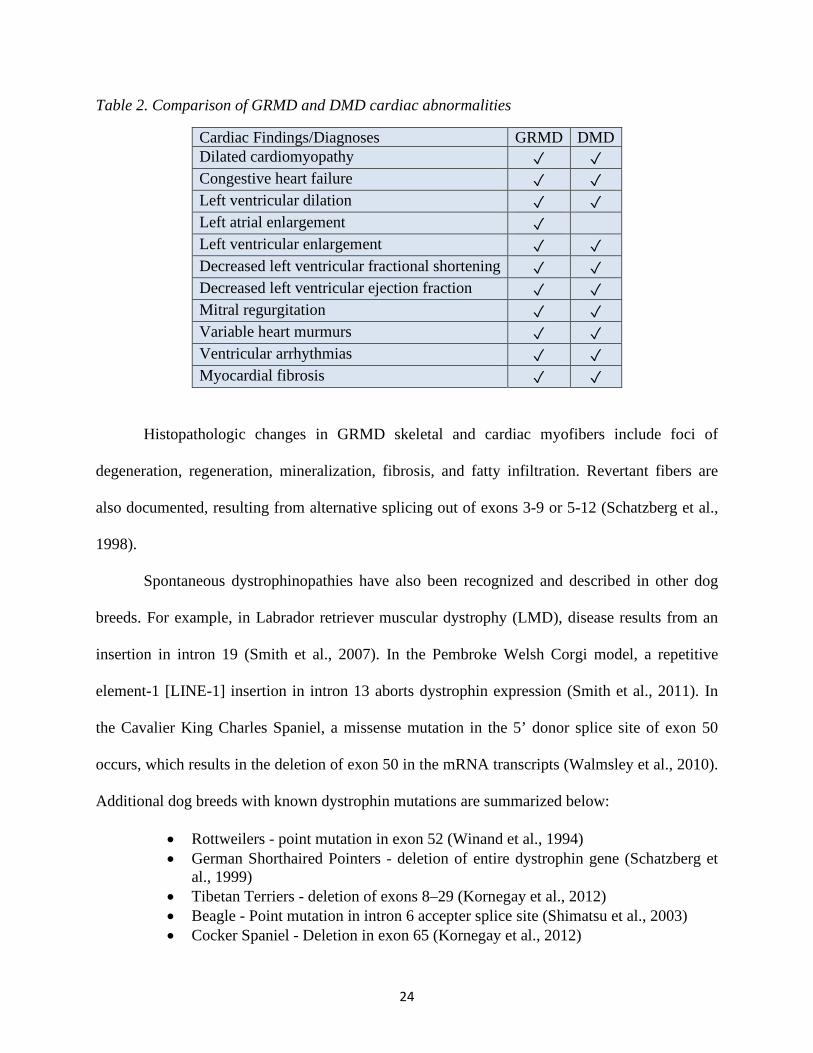
Table 2. Comparison of GRMD and DMD cardiac abnormalities
Cardiac Findings/Diagnoses GRMD DMD Dilated cardiomyopathy ✓ ✓ Congestive heart failure ✓ ✓ Left ventricular dilation ✓ ✓ Left atrial enlargement ✓ Left ventricular enlargement ✓ ✓ Decreased left ventricular fractional shortening ✓ ✓ Decreased left ventricular ejection fraction ✓ ✓ Mitral regurgitation ✓ ✓ Variable heart murmurs ✓ ✓ Ventricular arrhythmias ✓ ✓ Myocardial fibrosis ✓ ✓
Histopathologic changes in GRMD skeletal and cardiac myofibers include foci of
degeneration, regeneration, mineralization, fibrosis, and fatty infiltration. Revertant fibers are
also documented, resulting from alternative splicing out of exons 3-9 or 5-12 (Schatzberg et al.,
1998).
Spontaneous dystrophinopathies have also been recognized and described in other dog
breeds. For example, in Labrador retriever muscular dystrophy (LMD), disease results from an
insertion in intron 19 (Smith et al., 2007). In the Pembroke Welsh Corgi model, a repetitive
element-1 [LINE-1] insertion in intron 13 aborts dystrophin expression (Smith et al., 2011). In
the Cavalier King Charles Spaniel, a missense mutation in the 5’ donor splice site of exon 50
occurs, which results in the deletion of exon 50 in the mRNA transcripts (Walmsley et al., 2010).
Additional dog breeds with known dystrophin mutations are summarized below:
• Rottweilers - point mutation in exon 52 (Winand et al., 1994) • German Shorthaired Pointers - deletion of entire dystrophin gene (Schatzberg et
al., 1999) • Tibetan Terriers - deletion of exons 8–29 (Kornegay et al., 2012) • Beagle - Point mutation in intron 6 accepter splice site (Shimatsu et al., 2003) • Cocker Spaniel - Deletion in exon 65 (Kornegay et al., 2012)
25
• Japanese Spitz - Inversion disrupts dystrophin and RPGR genes (Jones et al., 2004)
Although individual mutations have not yet been characterized, based on clinicopathologic data,
dystrophinopathies are suspected in the Alaskan Malamute, Old English Sheepdog, Grand Basset
Griffon Vendeen, and Norfolk Terrier (Beltran et al., 2015; Ito et al., 2011; Klarenbeek et al.,
2007; Wieczorek et al., 2006).
Several other lesser-known DMD animal models have been studied. For example,
dystrophin deficiency in domestic short-haired cats results in hypertrophic feline muscular
dystrophy. Periods of degeneration and regeneration of muscle occur, but debilitating fibrosis,
which is characteristic of DMD and GRMD, does not develop. Post-mortem evaluation of
affected cats finds substantial glossal and diaphragmatic hypertrophy. Microscopic skeletal
muscle changes include myofiber hypertrophy (with resultant size variation), foci of
mineralization, myofiber splitting, and nuclear centralization. Endomysial fibrosis is typically
minimal (Blunden & Gower, 2011; Carpenter et al., 1989; Kohn et al., 1993). Although affected
cats lack outward evidence of cardiac disease, gross findings, histopathologic evaluation, and
imaging studies indicate that cats can develop myocardial hypertrophy (Gaschen et al., 1999).
Recently a large animal model of DMD was created via deletion of exon 52 in male pig
cells. The resultant pigs exhibited absence of dystrophin in skeletal muscles, increased serum
creatine kinase levels, progressive dysfunction of skeletal muscles, impaired mobility, muscular
weakness and a maximum life span of 3-months resultant from respiratory impairment. Unlike
human DMD patients, some dystrophin-deficient pigs in the study died shortly after birth
(Klymiuk et al., 2013). Non-mammalian models of DMD, such as the Zebrafish and the
nematode Caenorhabditis elegans, both express a dystrophin orthologue, resulting in their use in
DMD-related gene analyses and drug discovery studies (Collins & Morgan, 2003).

26
Labradoodle History
Labradoodles were first created in 1988 by Wally Conron (of The Royal Guide Dog
Association of Australia) by crossing a Labrador Retriever with a Standard Poodle. This
purposeful breeding occurred after Mr. Cochran received a request from a client to produce a
guide-dog with a low allergen coat for people with allergies. Today, the Australian Labradoodle
is the result of breeding a Labradoodle with a Cocker Spaniel (American or English), while the
true Labradoodle is only a cross between the Labrador Retriever and Standard Poodle. Although
the Labradoodle is popular among pet owners, the American Kennel Club (AKC) does not
currently recognize it, or the Australian Labradoodle, as one of the 175 pure breeds of dogs. In
order to be considered an official pure breed by the AKC, the breed must have at least 300
Labradoodles within the U.S. and be distributed among at least 20 states, the dogs must have a
national breed club demonstrating interest, and there must be at least a three generation pedigree.
In addition, each recognized breed must also have predictable characteristics and fulfill a specific
purpose (AKC, 2019; ALAA, 2018).
The dystrophin deficient Labradoodle colony housed at the Scott Ritchey Research Center
(SRRC), at Auburn University, was established via a carrier dam named “Scout” that was
donated to the research center in 2008. This dam resulted from breeding two Australian
Labradoodles. Subsequent breeding of “Scout” resulted in the following familial lines:
• “Scout” x “Kodiak” (normal Australian Labradoodle) Carrier female named “Bella”
o “Bella” x “Yaz” (normal Labrador retriever) 1 dystrophin deficient male
• “Scout” x “Tegan” (normal Australian Labradoodle) 1 dystrophin deficient male
• “Scout” x “Rody” (normal Australian Labradoodle) 4 dystrophin deficient males
27
Additional breeding of “Bella” and her carrier female offspring have resulted in continued
selection for this novel Dystrophin mutation.
Transcriptomics
RNA sequencing provides an excellent way to characterize transcriptomic variations by
measuring the expression of thousands of genes in a tissue type at the same time. This expanding
technology allows for long read lengths, which enables accurate de-novo transcriptome assembly
and high-confidence identification of transcripts and isoform variants, essential for interpreting
the functional elements of the genome and understanding disease pathogenesis. The goals of
transcriptome sequencing are to determine all transcript species (i.e. mRNAs, non-coding RNAs
and small RNAs), the transcriptional structure of genes, and to evaluate differential gene
expression (Wang et al., 2009).
The standard process of RNA sequencing can be broken down into a few main steps.
Once RNA has been isolated and purified from the desired tissue sample, the process of
sequencing begins by converting the RNA (total or fractionated) to a library of cDNA fragments.
Library preparation involves generating the cDNA fragments, adding adapters, and amplifying
the DNA for sequencing. The adapter sequences vary depending upon platform and contain
various functional elements necessary for sequencing; regardless of the platform, adaptors must
contain a terminal sequence (used for clonal amplification and attachment to the sequencing
support) and sequencing reaction priming elements. Once the adapters have been added,
generally via RT-PCR or ligation, the stranded cDNA libraries are prepared and checked for
quality prior to sequencing. The cDNAs are then sequenced using the sequencing platform that

28
best matches the researcher’s needs (multiple platforms are now available to choose from,
including Ion Torrent, PacBio, and Illumina) ("RNA-seqlopedia,").
Transcriptome sequencing has been performed on tissue samples from DMD patients in
order to detect mutations, understand aberrant splicing events and variants, evaluate allelic
imbalances, and to analyze differential gene expression. RNA sequencing on skeletal muscle
from patients with Duchenne muscular dystrophy has found that 30% of human genes were
expressed and detectable in skeletal muscle, of which, 3% showed differential expression in
dystrophic muscle compared to controls. 1,324 of the 1,882 dysregulated probe sets
corresponded to characterized genes/proteins. As part of the evaluation, members of the insulin-
like growth factor (IGF) pathway were preferentially investigated, as was the potential for
cardiac gene expression in skeletal muscle and sex-specific transcripts. Results indicated that
dystrophic skeletal muscle not only had up-regulation of IGF-I and IGF-II, but also upregulation
of inhibitory IGF-binding proteins and regulators such as IGFBP-2, -4, -6 and -7 and IGFBP-5
protease. Evaluation of six genes predominantly expressed in cardiac tissues, including Actin
alpha cardiac muscle 1 (ACTC1), Cardiac ankyrin repeat protein (CARP), Calsequestrin 2
(CASQ2), Troponin T2 cardiac-type (TNNT2), CUG triplet repeat RNA-binding protein 2
(CUGBP2), and Connexin 43 (CX43), found that CARP and CX43 were macrophage-associated
and TNNT2 activated-myoblast-associated. ACTC1 and CUGBP2 up-regulation were not
associated with muscle regeneration. They also found two Y-linked genes only expressed in male
muscle (Ribosomal protein S4 Y-linked 1 [RPS4Y] and DEAD-box helicase 3 Y-linked
[DDX3Y]) and two autosomal genes with increased expression in female muscle (C-X-C motif
chemokine ligand 2 [CXCL2; also known as GRO2] and Zinc finger protein 91 [ZNF91])
(Bakay et al., 2002). Although endomyocardial biopsies have been performed on DMD patients

29
with clinical and cardiac MRI-confirmed cardiovascular compromise, the biopsies were utilized
for DNA and RNA viral genome extraction rather than cardiac RNA sequencing (Mavrogeni et
al., 2010).
RNA sequencing has also been performed in canine and murine models of DMD. In
dogs, profiling in GRMD skeletal muscle has yielded differentially expressed genes associated
with myogenesis/muscle regeneration, metabolism, and inflammation. Researchers also found
up-regulation of Chitinase 3-like 1 (CHI3L1) in GRMD dogs with a more rapid clinical course,
suggesting an association with disease progression in GRMD and potentially
DMD.(Brinkmeyer-Langford et al., 2018) Though cardiac transcriptome sequencing has not
previously been performed in a canine model of DMD, it has been utilized to study the
consequences of obesity-related hypertension on cardiac gene regulation and end-stage heart
failure (Gao et al., 2006; Philip-Couderc et al., 2003). To date, cardiac muscle RNA sequencing
has only been performed in the Australian Labradoodle model. Transcriptome analysis of mdx
mouse skeletal muscle has found significant differential expression of 3844 genes, of which 2695
were upregulated and 1149 downregulated. Enrichment pathway analysis using Ingenuity
Pathway Analysis (IPA) software found enrichment genes associated with inflammation,
fibrosis, adhesion, apoptosis, muscle cell structure and metabolism. Additionally, there was
enrichment of canonical signaling pathways (e.g. NF-kB, Wnt, calcium signaling, etc.) (Yanay et
al., 2017).
Because mortality in DMD patients is increasingly associated with cardiomyopathy and
heart failure, it is imperative that we gain a better understanding of the possible pathways
involved in the development of DMD-associated cardiac disease. We have identified a novel
dystrophin mutation in Australian Labradoodles that develop early-onset cardiac disease in

30
addition to the typical clinical signs associated with skeletal myopathy. Based on these
preliminary findings, we sought to study the Australian Labradoodle as a potential model for
DMD cardiomyopathy.
The first research objective was to fully characterize the disease phenotype that occurs in
the Australian Labradoodle model. To that end, we performed monthly echocardiography and
trimonthly cardiac Magnetic Resonance Imaging (MRI) / Magnetic Resonance Spectroscopy
(MRS). The results of these cardiac analyses were then correlated to the observed clinical signs,
necropsy findings, and histopathology data to determine if dystrophin-deficient Australian
Labradoodles are an appropriate model for the study of DMD-associated cardiomyopathy.
The second research objective was to compare the RNA profiles of normal and
dystrophin-deficient Australian Labradoodle hearts via transcriptome sequencing. These
transcriptomic comparisons allowed us to study how changes in the normal level of gene activity
may reflect or contribute to the cardiac disease process. We hypothesized that cardiac
transcriptome sequencing in the Australian Labradoodle model of DMD would reveal key
genetic and epigenetic markers that may be associated with cardiomyopathy. Understanding the
cardiac manifestation of DMD at the level of gene expression would potentially allow for future
development of targeted therapeutics for both dystrophin-deficient dogs and Duchenne muscular
dystrophy patients.

31
Chapter 2: Characterization of Australian Labradoodle Dystrophinopathy
Available online at www.sciencedirect.com
Neuromuscular Disorders 28 (2018) 927–937 www.elsevier.com/locate/nmd
Characterization of Australian Labradoodle dystrophinopathy
Stephanie M. Shrader a , ∗, SeungWoo Jung
b , Thomas S. Denney
c , d , Bruce F. Smith
a , e
a Department of Pathobiology, Auburn University College of Veterinary Medicine, Auburn, AL 36849, USA
b Department of Clinical Sciences, Auburn University College of Veterinary Medicine, Auburn, AL 36849, USA
c Department of Electrical and Computer Engineering, Auburn University, Auburn, AL 36849, USA
d Auburn University MRI Research Center, Auburn, AL 36849, USA
e Scott-Ritchey Research Center, Auburn University College of Veterinary Medicine, Auburn, AL 36849, USA
Received 16 February 2018; received in revised form 5 August 2018; accepted 23 August 2018
Abstract
In humans, dystrophin mutations cause the X-linked recessive disorder known as Duchenne muscular dystrophy (DMD). These mutations result in skeletal and cardiac muscle damage with mortality increasingly associated with cardiomyopathy. We have identified a novel dystrophin mutation in exon 21 in a line of Australian Labradoodles; affected dogs develop progressive clinical signs including poor weight gain and weight loss, gait abnormalities, exercise intolerance, skeletal muscle atrophy, macroglossa, ptyalism, dysphagia, kyphosis, and a plantigrade stance. Echocardiographic abnormalities include hyperechoic foci in the left ventricular papillary muscles, septal hypokinesis, and decreased left ventricular systolic and diastolic volume and internal diameter. Holter recordings found a Mobitz type II second-degree atrioventricular (AV) block in one affected dog. Analysis of phosphocreatine-to-ATP ratios (PCr/ATP) (obtained via cardiac magnetic resonance imaging and spectroscopy evaluation), found no statistically significant difference in the mean PCr/ATP between groups. Histopathologic skeletal muscle changes included fibrofatty infiltration, myocyte degeneration, necrosis, and regeneration, lymphohistiocytic inflammation, and mineralization; cardiac changes were limited to a focal area of mineralization adjacent to the sinoatrial node in the dog with a second-degree AV block. Due to rapidly progressive clinical signs, a severe phenotype, and potential for cardiac involvement, Australian Labradoodle dystrophinopathy may be a useful model to further study DMD pathogenesis. © 2018 Elsevier B.V. All rights reserved.
Keywords: Labradoodle; Dystrophinopathy; Duchenne; Myopathy; Cardiomyopathy; MRI.
1. Introduction
Duchenne muscular dystrophy is an X-linked recessivedisorder in humans caused by mutations in the largest known gene in nature, dystrophin. Dystrophin measures 2.4-megabases at locus Xp21 and encodes for a 427-kD
sub-membrane protein that anchors cytoskeletal F-actin to
the extracellular matrix protein laminin [1] . Mutations in the dystrophin gene result in a partially functional or completely
non-functional protein product, causing affected individuals to develop progressive skeletal and cardiac myofiber damage with an early loss of ambulatory ability [2] . Historically, mortality in DMD patients has been attributed primarily to
respiratory failure, most often associated with ventilatory
∗ Corresponding author. E-mail address: [email protected] (S.M. Shrader).
defects, chronic hypoventilation, and respiratory infections [3,4] . Over the past two decades, the treatment of skeletal muscle complications and respiratory disease has improved; however, these advances have unmasked DMD-associated
cardiac disease in many patients. Recent retrospective studies indicate that death attributable to cardiac disease occurs in
approximately 20–30% of DMD patients [5–7] . Early clinical findings in DMD patients include difficulties
rising from a seated position, frequent falls, delayed motor- skill development, and learning disabilities. Physical examina- tion findings often include (pseudo)hypertrophy of the calves, lumbar lordosis, waddling gait, macroglossa, and a distinctive way of rising from the floor (known as Gowers’ maneuver). Progressive muscle weakness eventually results in wheel chair dependence, generally by ten years of age [8,9] . Histologi- cally, skeletal muscle changes in DMD patients are charac- terized by necrotic and/or degenerating myocytes with foci of
https://doi.org/10.1016/j.nmd.2018.08.008 0960-8966/© 2018 Elsevier B.V. All rights reserved.
32

928 S.M. Shrader et al. / Neuromuscular Disorders 28 (2018) 927–937
mineralization, myofiber regeneration (characterized by my- ofiber basophilia, enlarged centralized nuclei with prominent nucleoli, and/or nuclear rowing), and resultant variation in
myofiber cross-sectional diameter. Necrotic myofibers are of- ten surrounded and/or infiltrated by macrophages and lympho- cytes. As the regenerative capacity of the affected muscle de- clines over time, there is gradual replacement of the tissue by
mature fibrous connective tissue and infiltrating adipose tissue (i.e. fibrofatty replacement). Individuals with cardiomyopathy
may also have foci of degeneration, necrosis, inflammation, and fibrofatty replacement in the myocardium [10] .
Cardiac pathology varies among DMD patients; however, findings can include dilated cardiomyopathy (DCM), conges- tive heart failure, left ventricular eccentric hypertrophy, de- creased left ventricular systolic function assessed by fractional shortening (FS) and ejection fraction (EF), mitral regurgita- tion, tricuspid regurgitation, and myocardial fibrosis [11,12] . Affected individuals may also show persistent electrophysio- logic abnormalities on ECG; sinus tachycardia, an increased
R-S ratio in the right precordial leads, deep Q waves inthe frontal leads, and conduction abnormalities. In additionto persistent sinus tachycardia, other common arrhythmiasin DMD patients include sinus bradycardia, atrial prematurebeats, and ventricular premature beats. Conduction defects,including right bundle branch blocks, left anterior fascicularblocks, and first- and second-degree AV blocks have also beenobserved [13–15] .
Recently, cardiac magnetic resonance imaging (MRI) has been proposed as a beneficial diagnostic tool to identify early
myocardial remodeling changes [11] . Both echocardiography
and MRI are able to identify abnormal left ventricular FS, left ventricular hypokinesia, and left ventricular dilation; however, research shows that the majority of echocardiographic studies have suboptimal scanning windows and significantly over- or under-estimate left-ventricular systolic function compared to
cardiac MRI [16] . In addition to cardiac MRI, cardiac mag- netic resonance spectroscopy (MRS) is also being utilized
to accurately evaluate cardiac functionality in patients with
DMD. The metabolic demands of the heart necessitate the need for high-energy phosphate being transferred from ATP
(via oxidative phosphorylation in the mitochondria) to creati- nine, generating phosphocreatine (PCr) and ADP. Phosphorus- 31 is utilized for the detection of PCr, ATP, intracellular pH, and flux through the creatine kinase reaction. Analyzing these spectra via MRS is therefore beneficial because the PCr-to- ATP ratio (PCr/ATP) provides a useful measure of cardiac energy metabolism and functionality [17] . PCr/ATP has been
shown to be significantly reduced in Becker Muscular Dystro- phy (BMD) patients and carriers when compared to controls; this change did not, however, correlate with left ventricular EF or mass index, indicating that a direct association be- tween altered dystrophin expression and the development of cardiomyopathy may be possible, and that MRS may be a use- ful diagnostic tool to detect early stage cardiomyopathy prior to echocardiographic identification of left ventricular systolic dysfunction [18] .
Various animal models have been utilized in the study of DMD. The traditional model is the mdx mouse, which has a deletion in the Xp21.1 locus that results in a stop codon in
exon 23 [19,20] . Compared to people with DMD, the disease phenotype in mdx mice is generally milder with an almost normal lifespan, stunted growth, skeletal muscle atrophy, and
eventual compensatory hypertrophy [21,22] . Cardiac dysfunc- tion is not a consistent finding in mice, but has been reported
as early as 9–10 months of age; decreased FS values have been demonstrated via echocardiographic analyses [19,23,24] . Spontaneous dystrophin mutations have been reported in mul- tiple dog breeds, though Golden Retriever muscular dystrophy
(GRMD) continues to be the best documented of the canine dystrophinopathies. It results from a point mutation in intron
6 that disrupts normal splicing and creates a premature stop
codon [25–27] . Cardiac dysfunction has been documented in
GRMD; however, clinical signs can vary widely in incidence and severity. Reported clinical, echocardiographic, and elec- trocardiographic findings in the GRMD model include DCM, congestive heart failure, left atrial enlargement, left ventricu- lar enlargement, decreased left ventricular FS, decreased left ventricular EF, mitral regurgitation, heart murmurs, ventricu- lar arrhythmias, and myocardial fibrosis [28,29] .
We have identified a novel dystrophin mutation in exon
21 in a line of Australian Labradoodles. The propositus of this pedigree died suddenly at six months of age with clinical signs suggestive of cardiac disease. Subsequent males in this line have consistently developed early clinical signs of skele- tal muscle pathology similar to what has been reported in
other dog breeds. We therefore hypothesized that dystrophin- deficient Labradoodles would show echocardiographic, elec- trocardiographic, and cardiac MRI/MRS abnormalities indica- tive of cardiomyopathy and that cardiac MRI/MRS may be more sensitive than the other two diagnostic modalities for detecting early cardiac dysfunction. In addition to identify- ing early manifestations of cardiac disease in this model, we aimed to correlate these findings with clinical signs and
necropsy and histopathology data, and to determine whether Labradoodle dystrophinopathy is an appropriate model for studying DMD-associated cardiomyopathy.
2. Materials and methods
All animal experiments were approved by the Animal Careand Use Committee of Auburn University. A 3-month old
male Australian Labradoodle presented to the Auburn Univer- sity College of Veterinary Medicine for a suspected muscular dystrophy and genetic testing for the Labrador Retriever dys- trophin mutation (intron 19 insertion) after showing signs of muscle atrophy and exercise intolerance. PCR amplification
of intron 19 from whole blood showed no insertion. mRNA
was obtained from skeletal muscle and sequenced, demon- strating a C to T transition at position 2668 of the dystrophin
open reading frame. This nonsense mutation results in a CGA
codon (arginine) becoming a stop codon (TGA), causing a
33

S.M. Shrader et al. / Neuromuscular Disorders 28 (2018) 927–937 929
truncated protein. This dog was used to establish a breeding
colony of carrier females that continues to be maintained at the Scott Ritchey Research Center (SRRC) at Auburn Univer- sity; male offspring of the colony were used for the present study.
Experimental dogs were produced using the semen from
a normal Labrador retriever, a normal Australian Labradoo- dle, or a dystrophin-deficient Australian Labradoodle (male donor selection based on availability) to vaginally inseminate a carrier Australian Labradoodle (several related females were utilized). Multiple breeding cycles produced seven dystrophin- deficient and five control (normal) male littermates.
For each dog in every litter produced during the course of the study, the dystrophin genotype was determined via DNA sequencing, performed on whole blood submitted to
Eurofins Genomics (12701 Plantside Drive, Louisville, KY
40299, USA). Male offspring with a correct dystrophin gene sequence (and lacking clinical signs of a muscular dystrophy) were used as controls. Female offspring, regardless of their dystrophin status, were not included in this study.
The founder of this line died unexpectedly at 6-mos of age from a possible terminal arrhythmia or cardiac conduc- tion anomaly; therefore, all study dogs had monthly echocar- diographic and electrocardiographic evaluations, in addition
to cardiac magnetic resonance imaging (MRI) and magnetic resonance spectroscopy (MRS) at three and six months of age. Echocardiographic examinations were performed by a board-certified veterinary cardiologist (SWJ) at monthly in- tervals starting at 1-month of age. Phased-array transducers between 5.0–8.5 MHz (Vivid E9, GE, Milwaukee, WI, USA) were used with a probe selection based on image optimiza- tion, gain, and filter settings that were required to reduce background noise and provide clear images of blood flow
and tissue signals. Electrocardiogram (ECG) was simultane- ously recorded during echocardiographic evaluation. Ventric- ular end-diastole was defined as the frame before mitral valve closure, and ventricular end-systole was defined as the frame before mitral valve opening. Echocardiographic analyses in- cluded evaluation of left ventricular internal diameter during
diastole (LVIDd), left ventricular internal diameter during sys- tole (LVIDs), left ventricular end diastolic volume (LVEDV), left ventricular end systolic volume (LVESV), ejection frac- tion (EF), epicardial velocity, endocardial velocity, left ven- tricular free wall thickness during diastole (LVFWd), and in- terventricular septum thickness during diastole (IVSd). FS%
was calculated as [(LVIDd-LVIDs)/LVIDd] ∗100. Myocardialvelocity gradient was calculated as the difference between en- docardial velocity and epicardial velocity [31] .
A 24 h ambulatory Holter recording was performed (Tril- lium 5000, Forest Medical Holter Analysis System, Syracuse, NY, USA) when ECG abnormalities were observed during
echocardiographic evaluations. Manual adjustments and accu- racy verification of the arrhythmias were performed by SWJ.
MRI/MRS evaluation was performed on eight of the study
dogs (3 control, 5 affected) using a MAGNETOM 7 Tesla scanner (Siemens Healthineers, Malvern, PA) and a dual- tuned 1H- 31 P loop coil (RAPID Biomedical GmbH, Rimpar,
Germany) at the Auburn University MRI Research Center. Dogs were scanned using a 3-dimensional ultra-short echo
time chemical shift imaging sequence [32] with 16 ×16 ×8matrix, 11.25 mm × 11.25 mm × 22.5 mm voxel size, 2.3 msecho time, 1640 ms repetition time, 12 averages, 6 kHz band- width, and 2048 vector size. 31 P MR spectra were ana- lyzed using a custom implementation of AMARES [33] to
obtain amplitudes of the PCr and gamma ATP resonances. PCr/gamma ATP amplitude ratios were computed from three voxels in the midventricular anterior and anteroseptal my- ocardium and averaged. For MRI/MRS evaluation, each study
dog was premedicated with 0.2–1.0 mg/kg butorphanol (sub- cutaneous administration), and induced with 3–5 mg/kg of propofol (intravenous administration). General anesthesia was maintained with isoflurane and the dogs were placed in left lateral recumbency for imaging acquisition.
All dogs were euthanized at 6 months of age (or earlier if a humane endpoint was reached). This terminal endpoint was established based on the founding dog’s sudden death at 6- mos of age and progressive clinical decline in other affected
males from this line beginning at 4-mos of age (including di- minished/absent ambulation, weakness, and dysphagia requir- ing a gruel-based diet and supplemental fluid administration).
At the time of death, each dog had a post-mortem exam- ination performed by a board certified veterinary pathologist (SMS), which included collection of blood for serum bio- chemistry analyses (performed by the Auburn University Clin- ical Pathology Laboratory), organ weight evaluation (heart, liver, kidneys, lung, spleen, brain), and measurement of car- diac parameters (left ventricular free wall thickness, right ven- tricular free wall thickness, septal thickness, aortic circum- ference, base-apex length, heart circumference [at the widest point], and pulmonic valve circumference). A complete set of tissues was collected for histopathologic evaluation (to include at a minimum: entire heart, entire diaphragm, tongue, spleen, liver, kidneys, pancreas, stomach, esophagus, duodenum, je- junum, ileum, colon, urinary bladder, adrenal glands, lung, bi- ceps femoris, gluteus medius, and triceps brachii). Additional tissue sites were collected if gross lesions were observed.
All tissues were fixed in 10% neutral buffered formalin, processed routinely, and embedded in paraffin wax. Sections were cut to 5- μm thickness and stained with hematoxylin and
eosin (H&E). Histopathologic evaluation of H&E stained tis- sue sections was performed by a board certified veterinary
pathologist (SMS). For each dog, sections of heart that were histologically evaluated included the aorta, right ventricular free wall (including papillary muscle), interventricular sep- tum, and left ventricular free wall (including papillary mus- cle). Additional cardiac anatomic sites were evaluated based
on individual animal clinical signs and cardiac imaging find- ings.
Serum biochemistry values, gross findings, organ weights, cardiac measurements, and histopathologic findings were en- tered into Microsoft ® Excel. Group means, standard devia- tions, and statistical significance were calculated with a Stu- dent’s t -test (SAS® version 9.4) for independent samples. The level of significance was set at p < 0.05.
34

930 S.M. Shrader et al. / Neuromuscular Disorders 28 (2018) 927–937
Fig. 1. Comparison of DNA sequences from exon 21 of the dystrophin gene from a normal male and affected male Labradoodle. Base pair 43 is indicated for each animal by an arrow; note the C to T transition in the affected dog.
3. Results
Seven dystrophin-deficient and five littermate control maleLabradoodles were utilized for this study. Each affected
Labradoodle had a C to T transition at base pair 43 in exon
21 of the dystrophin gene ( Fig. 1 ). To the authors’ knowledge, this is a novel spontaneous dystrophin mutation that has not been previously reported in dogs. Clinically, the dystrophin- deficient dogs had stunted growth, exercise intolerance, poor weight gains, and eventual weight loss (typically beginning
at 4-months of age). There was a statistically significant de- crease in the mean terminal body weight in the affected group
(7.5-kg) compared to the control group (21.21-kg) ( p < 0.0001). By 3-months of age, affected dogs devel- oped a stiff gait that progressed to shuffling with slight carpal valgus and a pronounced plantigrade stance. At the time of study termination, all affected dogs had moderate to
severe appendicular and axial skeletal muscle atrophy (most pronounced in the proximal limb musculature), temporalis at- rophy, moderate to severe macroglossa, ptyalism, dysphagia, and kyphosis.
The dogs had monthly echocardiographic evaluations which included qualitative cardiac assessment and measure- ment of various parameters for cardiac functionality. In one of the affected dogs (14.3%), multifocal to coalescing, mild, hyperechoic foci in the left ventricular anterior and posterior papillary muscles were noted ( Fig. 2 ). These changes became apparent at 2-months of age and remained static until study
termination at 6-months of age. Trivial mitral valve regurgita- tion was observed in 1/7 affected dogs (14.3%) and 1/5 con- trol dogs (20%), both at the 6-month time point. Mild tricus- pid regurgitation was consistently present in 3/7 affected dogs (42.9%) throughout the study. Trivial to mild tricuspid regur- gitation was also seen in 1/5 (20%) of control dogs throughout the study. Non-progressive, decreased dynamic septal motion, consistent with septal hypokinesis, was present in 3/7 (42.9%) of affected dogs as early as one month of age ( Fig. 3 ). Two
of the five control littermates, from related female carriers but different sires, also had a hypokinetic septal motion. Sep- tal hypokinesis was non-progressive, minimal, and apparent at one-month of age in one of the control dogs; in the other
Fig. 2. Conventional echocardiographic image (right parasternal short axis view at the level of the papillary muscles) of one of the affected dogs show- ing multifocal hyperechoic foci in the left ventricular anterior and posterior papillary muscles (arrows).
dog, it was first noted at 4-months of age and progressed from
mild to severe by study termination. Quantitative echocardiographic parameters were evaluated
at monthly intervals; a percent increase (%) of LVIDd, LVIDs, LVEDV, and LVESV from 3 month to 6 month was compared
between control and affected dogs. The mean percent increase of LVIDs was significantly lower in affected dogs (7.6% ±5.2,p = 0.0045) as compared to control dogs (29.8% ±2.1)( Fig. 4 ). There was a trend that the mean percent increase of LVIDd was lower in affected dogs (9.5% ±6.6, p = 0.08)as compared to control dogs (25.3% ±4.3). The mean per- cent increase of LVESV was markedly diminished in af- fected dogs (21.3% ±12.3) in comparison to control dogs(98.1% ±6.1). A trend was noted in the mean percent in- crease of LVEDV that affected dogs had smaller diastolic filling volume (26.3% ±15.8, p = 0.075) than control dogs(69.2% ±13.7). These findings are consistent with a signifi- cant developmental growth defect of cardiac size and volume in dystrophin-deficient Labradoodle dogs.
35

S.M. Shrader et al. / Neuromuscular Disorders 28 (2018) 927–937 931
Fig. 3. Echocardiographic evaluation of the left ventricle on M-mode from a control dog (A) and an affected dog (B). (A) Normal dynamic motion of the left ventricular septum and free wall is seen in a control dog. (B) A marked hypokinetic motion of the interventricular septum is apparent in an affected dog. S: interventricular septum. W: left ventricular free wall.
Fig. 4. Comparison of a percent increase (%) between control and affected dogs of echocardiographic dimensional parameters (LVIDd, LVIDs, LVEDV, and LVESV) from 3 month to 6 month.
At the 3-month time point, affected dogs had a statisti- cally significant decrease in mean LVIDd (24.29 mm ±2.69;p = 0.0287), LVIDs (16.14 mm ±1.46; p = 0.0195), LVEDV(21.43 ml ±6.13; p = 0.0281 ) , and LVESV (7.43 ml ±1.81;p = 0.0090) when compared to the control dogs. Atthe 6-month time point, affected dogs had a statisti- cally significant decrease in mean LVIDd (27.20 mm ±3.56;p = 0.0054), LVIDs (17.80 mm ±1.92; p = 0.0007), LVEDV(28.40 ml ±9.34; p = 0.0028), and LVESV (9.60 ml ±2.70;p = 0.0011) when compared to the control dogs. There wereno statistically significant differences in the remainder of the examined parameters (FS%, EF%, epicardial velocity, endo- cardial velocity, MVG, LVFWd, or IVSd). While perform- ing the 3-month echocardiographic evaluations, one of the af- fected dogs was noted to have marked arrhythmias. The dog
was subjected to a 24 h Holter monitor. Holter recordings revealed the presence of low amplitude R-waves and pro- found S-waves and also showed intermittent, non-conducted
P-waves (without progressive prolongation of the PR inter- val) that typically occurred at a frequency of 1–2 events/min.The PR intervals of the conducted beats consistently remainedwithin normal limits (less than 130 ms). Findings were con- sistent with a Mobitz type II 2nd degree atrioventricular (AV)block ( Fig. 5 ).
MRI/MRS evaluation was performed on eight of the study
dogs (3 control, 5 affected). Only two of the dogs were evalu- ated at both the 3- and 6-month time points due to scheduling
limitations and early euthanasia of two of the affected dogs following the 3-month MRI/MRS evaluation (at 100 days and 113 days of age). Both dogs were euthanized due to
36
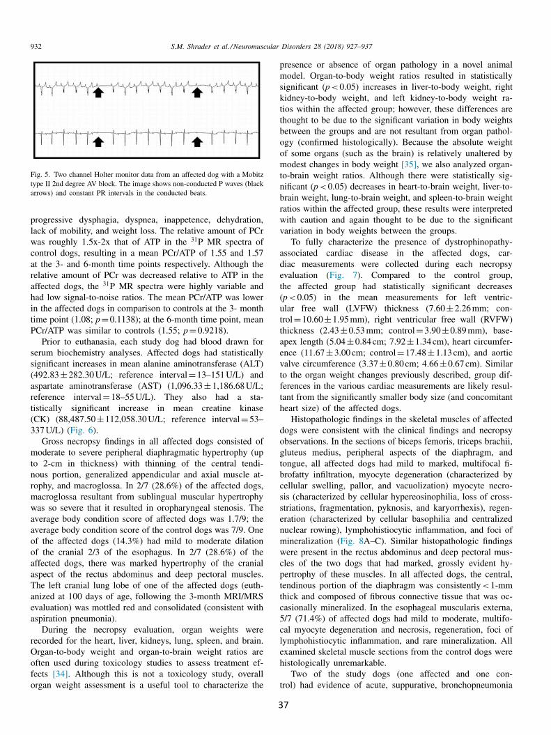
932 S.M. Shrader et al. / Neuromuscular Disorders 28 (2018) 927–937
Fig. 5. Two channel Holter monitor data from an affected dog with a Mobitz type II 2nd degree AV block. The image shows non-conducted P waves (black arrows) and constant PR intervals in the conducted beats.
progressive dysphagia, dyspnea, inappetence, dehydration, lack of mobility, and weight loss. The relative amount of PCr was roughly 1.5x-2x that of ATP in the 31 P MR spectra ofcontrol dogs, resulting in a mean PCr/ATP of 1.55 and 1.57
at the 3- and 6-month time points respectively. Although the relative amount of PCr was decreased relative to ATP in the affected dogs, the 31 P MR spectra were highly variable andhad low signal-to-noise ratios. The mean PCr/ATP was lower in the affected dogs in comparison to controls at the 3- month
time point (1.08; p = 0.1138); at the 6-month time point, meanPCr/ATP was similar to controls (1.55; p = 0.9218).
Prior to euthanasia, each study dog had blood drawn for serum biochemistry analyses. Affected dogs had statistically
significant increases in mean alanine aminotransferase (ALT) (492.83 ±282.30 U/L; reference interval = 13–151 U/L) andaspartate aminotransferase (AST) (1,096.33 ±1,186.68 U/L;reference interval = 18–55 U/L). They also had a sta- tistically significant increase in mean creatine kinase (CK) (88,487.50 ±112,058.30 U/L; reference interval = 53–337 U/L) ( Fig. 6 ).
Gross necropsy findings in all affected dogs consisted of moderate to severe peripheral diaphragmatic hypertrophy (up
to 2-cm in thickness) with thinning of the central tendi- nous portion, generalized appendicular and axial muscle at- rophy, and macroglossa. In 2/7 (28.6%) of the affected dogs, macroglossa resultant from sublingual muscular hypertrophy
was so severe that it resulted in oropharyngeal stenosis. The average body condition score of affected dogs was 1.7/9; the average body condition score of the control dogs was 7/9. One of the affected dogs (14.3%) had mild to moderate dilation
of the cranial 2/3 of the esophagus. In 2/7 (28.6%) of the affected dogs, there was marked hypertrophy of the cranial aspect of the rectus abdominus and deep pectoral muscles. The left cranial lung lobe of one of the affected dogs (euth- anized at 100 days of age, following the 3-month MRI/MRS
evaluation) was mottled red and consolidated (consistent with
aspiration pneumonia). During the necropsy evaluation, organ weights were
recorded for the heart, liver, kidneys, lung, spleen, and brain. Organ-to-body weight and organ-to-brain weight ratios are often used during toxicology studies to assess treatment ef- fects [34] . Although this is not a toxicology study, overall organ weight assessment is a useful tool to characterize the
presence or absence of organ pathology in a novel animal model. Organ-to-body weight ratios resulted in statistically
significant ( p < 0.05) increases in liver-to-body weight, right kidney-to-body weight, and left kidney-to-body weight ra- tios within the affected group; however, these differences are thought to be due to the significant variation in body weights between the groups and are not resultant from organ pathol- ogy (confirmed histologically). Because the absolute weight of some organs (such as the brain) is relatively unaltered by
modest changes in body weight [35] , we also analyzed organ- to-brain weight ratios. Although there were statistically sig- nificant ( p < 0.05) decreases in heart-to-brain weight, liver-to- brain weight, lung-to-brain weight, and spleen-to-brain weight ratios within the affected group, these results were interpreted
with caution and again thought to be due to the significant variation in body weights between the groups.
To fully characterize the presence of dystrophinopathy- associated cardiac disease in the affected dogs, car- diac measurements were collected during each necropsy
evaluation ( Fig. 7 ). Compared to the control group, the affected group had statistically significant decreases ( p < 0.05) in the mean measurements for left ventric- ular free wall (LVFW) thickness (7.60 ±2.26 mm; con- trol = 10.60 ±1.95 mm), right ventricular free wall (RVFW)thickness (2.43 ±0.53 mm; control = 3.90 ±0.89 mm), base- apex length (5.04 ±0.84 cm; 7.92 ±1.34 cm), heart circumfer- ence (11.67 ±3.00 cm; control = 17.48 ±1.13 cm), and aorticvalve circumference (3.37 ±0.80 cm; 4.66 ±0.67 cm). Similarto the organ weight changes previously described, group dif- ferences in the various cardiac measurements are likely resul- tant from the significantly smaller body size (and concomitant heart size) of the affected dogs.
Histopathologic findings in the skeletal muscles of affected
dogs were consistent with the clinical findings and necropsy
observations. In the sections of biceps femoris, triceps brachii, gluteus medius, peripheral aspects of the diaphragm, and
tongue, all affected dogs had mild to marked, multifocal fi- brofatty infiltration, myocyte degeneration (characterized by
cellular swelling, pallor, and vacuolization) myocyte necro- sis (characterized by cellular hypereosinophilia, loss of cross- striations, fragmentation, pyknosis, and karyorrhexis), regen- eration (characterized by cellular basophilia and centralized
nuclear rowing), lymphohistiocytic inflammation, and foci of mineralization ( Fig. 8 A–C). Similar histopathologic findings were present in the rectus abdominus and deep pectoral mus- cles of the two dogs that had marked, grossly evident hy- pertrophy of these muscles. In all affected dogs, the central, tendinous portion of the diaphragm was consistently < 1-mm
thick and composed of fibrous connective tissue that was oc- casionally mineralized. In the esophageal muscularis externa, 5/7 (71.4%) of affected dogs had mild to moderate, multifo- cal myocyte degeneration and necrosis, regeneration, foci of lymphohistiocytic inflammation, and rare mineralization. All examined skeletal muscle sections from the control dogs were histologically unremarkable.
Two of the study dogs (one affected and one con- trol) had evidence of acute, suppurative, bronchopneumonia
37
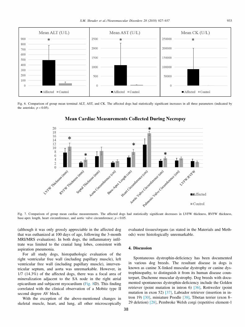
S.M. Shrader et al. / Neuromuscular Disorders 28 (2018) 927–937 933
Fig. 6. Comparison of group mean terminal ALT, AST, and CK. The affected dogs had statistically significant increases in all three parameters (indicated by the asterisks; p < 0.05).
Fig. 7. Comparison of group mean cardiac measurements. The affected dogs had statistically significant decreases in LVFW thickness, RVFW thickness, base-apex length, heart circumference, and aortic valve circumference; p < 0.05.
(although it was only grossly appreciable in the affected dog
that was euthanized at 100 days of age, following the 3-month
MRI/MRS evaluation). In both dogs, the inflammatory infil- trate was limited to the cranial lung lobes, consistent with
aspiration pneumonia. For all study dogs, histopathologic evaluation of the
right ventricular free wall (including papillary muscle), left ventricular free wall (including papillary muscle), interven- tricular septum, and aorta was unremarkable. However, in
1/7 (14.3%) of the affected dogs, there was a focal area of mineralization adjacent to the SA node in the right atrial epicardium and subjacent myocardium ( Fig. 8 D). This finding
correlated with the clinical observation of a Mobitz type II second degree AV block.
With the exception of the above-mentioned changes in
skeletal muscle, heart, and lung, all other microscopically
evaluated tissues/organs (as stated in the Materials and Meth- ods) were histologically unremarkable.
4. Discussion
Spontaneous dystrophin-deficiency has been documentedin various dog breeds. The resultant disease in dogs is known as canine X-linked muscular dystrophy or canine dys- trophinopathy, to distinguish it from its human disease coun- terpart, Duchenne muscular dystrophy. Dog breeds with docu- mented spontaneous dystrophin-deficiency include the Golden
retriever (point mutation in intron 6) [36] , Rottweiler (point mutation in exon 52) [37] , Labrador retriever (insertion in in- tron 19) [30] , miniature Poodle [38] , Tibetan terrier (exon 8–29 deletion) [28] , Pembroke Welsh corgi (repetitive element-1
38

934 S.M. Shrader et al. / Neuromuscular Disorders 28 (2018) 927–937
Fig. 8. A. Photomicrograph of a peripheral section of the diaphragm from an affected dog showing variation in myofiber size, myofiber regeneration (basophilia and nuclear rowing; arrowhead), lymphohistiocytic inflammation (asterisk), and necrosis (arrow). H&E. Bar = 100 μm. B. Photomicrograph of the biceps femoris from an affected dog showing variation in myofiber size and myofiber degeneration (hypereosinophilia and/or vacuolation). H&E. Bar = 20 μm. C. Photomicrograph of the triceps brachii from an affected dog showing myofiber degeneration (swelling and hypereosinophilia) and necrosis (arrow). H&E. Bar = 100 μm. D. Photomicrograph showing focal right atrial epicardial and myocardial mineralization (arrows) adjacent to the SA node in the affected dog with a Mobitz type II second degree AV block. H&E. Bar = 100 μm.
[LINE-1] insertion in intron 13) [39] , and the Cocker spaniel (deletion in exon 65) [28] .
Similar to what has been found in people, many of the reported canine dystrophin mutations are point mutations. In
a recent study of DMD patients, point mutations accounted
for 46% of all mutations in the studied cohort. The study
also found that point mutations appeared to be randomly dis- tributed across the dystrophin exons and were not found in
hotspots [40] . Although mutations in exon 21 have not been previously
reported in canine models of DMD, they have been noted in
people with DMD. It is also interesting to note that in DMD
patients, G: C > A: T transitions have been shown to be the most common stop mutation class, with the majority of these mutations being due to C > T transitions [40] , similar to what we found in the Labradoodle model.
Dystrophin-deficiency in this line of Australian Labradoo- dles results in clinical signs that mimic those reported in
other dystrophin-deficient dogs, including the well-studied
Golden retriever model [28] . Like other dogs with dystrophin- deficiency, Labradoodles develop clinical signs shortly af- ter birth, generally requiring extra care and nutritional man- agement. In some instances, affected dogs die in the early
post-natal period; often these that succumb extremely early
have already experienced stunted growth and are substantially
smaller than their littermates. For all affected dogs used dur- ing the course of this study, stunted growth and weakness were evident by 4-weeks of age. Unlike what has been previ- ously reported in other dystrophin-deficient dogs [28,41,42] , the clinical course in the Labradoodle model is consistently
very rapid, with weight loss, extensive skeletal muscular at- rophy, difficulty ambulating, and moderate to severe dyspha- gia with ptyalism beginning by 4-months of age, necessitat- ing study termination at 6-months of age. As mentioned, two
of the affected dogs had such severe macroglossa that it re- sulted in oropharyngeal stenosis, making intubation during the MRI/MRS procedures extremely difficult.
Similar to what is seen in patients with DMD and other animal models of DMD, cardiac findings in the dystrophin- deficient Labradoodle vary between littermates and litters. Multifocal to coalescing, hyperechoic foci were observed in
the left ventricular anterior and posterior papillary muscles in 1/7 affected dogs (14.3%). Differential diagnoses included
areas of mineralization, fibrosis, and/or ischemia. There were no gross or histopathologic lesions correlating to these foci. Although hyperechoic foci in the left ventricular wall have been previously reported at later ages in both the GRMD
and CXMD J (Canine X-linked muscular dystrophy in Japan)
39
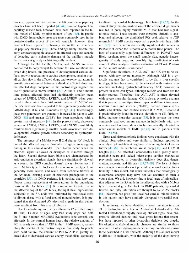
S.M. Shrader et al. / Neuromuscular Disorders 28 (2018) 927–937 935
models, hyperechoic foci within the left ventricular papillary
muscles have not been reported [43,44] . Similar hyperechoic papillary muscle foci, however, have been reported in the fe- line model of DMD by nine months of age [45] . In people with DMD, hyperechoic areas are most commonly seen in the posterior-basilar aspect of the left ventricle; however, they
have not been reported exclusively within the left ventricu- lar papillary muscles [46] . These findings likely indicate that early echocardiographic analyses in affected dogs are capable of detecting early ischemic damage of the papillary muscle that is not yet grossly or histologically evident.
Although LVIDd, LVIDs, LVEDV, and LVESV are often
normalized to body weight to account for size variations be- tween dog breeds, the presence of developmental cardiac de- fects, growth retardation in cardiac development, smaller over- all cardiac size in the affected dogs, and extreme variations in
growth rates observed between individual affected dogs and
the affected dogs compared to the control dogs negated the use of quantitative normalization [29] . At the 3- and 6-month
time points, affected dogs had significantly smaller mean
values of LVIDd, LVIDs, LVEDV, and LVESV when com- pared to the control dogs. Volumetric indices of LVEDV and
LVESV have also been reported to be significantly reduced in
GRMD dogs at 6- and 12-months of age [47] . Interestingly, these two parameters are generally increased in patients with
DMD [48] and greater LVESV has been associated with a greater risk of mortality [49] . In the present study, decreased
values of LVIDd, LVIDs, LVEDV, and LVESV most likely
resulted from significantly smaller hearts associated with de- velopmental cardiac growth defects secondary to dystrophin- deficiency.
The presence of a Mobitz type II 2nd degree AV block in
one of the affected dogs at 3-months of age is an intriguing
finding in this animal model. Heart blocks occur when the electrical signal is slowed or disrupted as it moves through
the heart. Second-degree heart blocks are characterized by
atrioventricular electrical signals that are significantly slowed; as a result, the QRS complex doesn’t always follow each P
wave. Mobitz type II blocks are less common than type I, are generally more severe, and result from ischemic fibrosis in
the AV node, causing a loss of electrical propagation to the ventricles [50] . In DMD patients, it is posited that fatty and
fibrous tissue replacement of myocardium is the underlying
cause of the AV block [51] . It is important to note that in
the affected dog of the AV block, the right atrial myocardium
subjacent to the SA node was infiltrated by multifocal areas of fibrous tissue with a focal area of mineralization. It is pre- sumed that the disrupted AV electrical signals in this patient were resultant from this area of fibrosis.
Due to scheduling and early euthanasia (2 affected dogs; 100 and 113 days of age), only two study dogs had both
the 3- and 6-month MRI/MRS evaluations (one control, one affected). In the normal human heart, the relative amount of PCr is nearly twice that of ATP in the 31 P MR spectra, resem- bling the spectra of the control dogs in this study. In people with heart failure, the amount of PCr to ATP is greatly re- duced due to decreased cardiac energy metabolism secondary
to altered myocardial high-energy phosphates [17,52] . In the current study, the diminished size of the affected dogs’ hearts resulted in poor, highly variable MRS that had low signal- to-noise ratios. These spectra were therefore difficult to ana- lyze, and although the diminished PCr peak relative to ATP
resembled
31 P MR spectra expected in people with heart fail- ure [52] , there were no statistically significant differences in
PCr/ATP at either the 3-month or 6-month time points. The lack of statistically significant differences in PCr/ATP was likely resultant from the small sample size, genetic hetero- geneity of study dogs, and possibly high coefficient of vari- ation of MRS analysis. Further evaluation of PCr/ATP ratios in this animal model is warranted.
Significant elevations in ALT, AST, and CK are to be ex- pected with any severe myopathy. Although ALT is a cy- tosolic enzyme that is considered to be fairly liver-specific in dogs, it has been noted to be elevated with various my- opathies, including dystrophin-deficiency. AST, however, is present in most cell types, although muscle and liver are the major sources. Therefore, like ALT, AST is often elevated
with severe muscular damage. CK is also a cytosolic enzyme that is present in multiple tissue types as different isozymes: nervous tissue and viscera (CK-BB), cardiac muscle (CK- MB), and skeletal and cardiac muscle (CK-MM). Because it is predominantly found in muscle, elevations in CK fairly re- liably indicate muscular damage [53] . It is perhaps the most commonly analyzed serum enzyme in individuals with my- opathies and has been shown to be dramatically elevated in
other canine models of DMD [42,47] and in patients with
DMD [54,55] . Gross and histopathologic findings were consistent with the
clinical observations and were similar to findings reported in
other dystrophin-deficient dog breeds including the Golden re- triever [41,56] , the Pembroke Welsh corgi [39] , and CXMDJ beagles [42] . All affected Labradoodles had a grossly unre- markable heart and lacked microscopic cardiac changes as previously reported in dystrophin-deficient dogs (i.e. degen- eration, necrosis, and fibrosis) [39,57–59] . The lack of these microscopic lesions does not preclude abnormal cardiac func- tionality in this model, but rather indicates that histologically
discernable changes may have not yet occurred in such a young dog. We did, however, find a focal area of mineraliza- tion adjacent to the SA node in the affected dog with a Mobitz type II second degree AV block. In DMD patients, myocardial fibrosis and fatty infiltration are thought to cause AV blocks [51] ; however, we posit that localized cardiac mineralizationin this patient may have similarly disrupted myocardial con- duction.
In summary, we have identified a novel mutation in exon
21 of dystrophin in a line of Australian Labradoodles. Af- fected Labradoodles rapidly develop clinical signs, have pro- gressive clinical decline, and have gross lesions that resem- ble those reported in other dystrophin-deficient dog breeds. Histologically, skeletal muscle changes are similar to those observed in other dystrophin-deficient dog breeds and mirror those described in DMD patients. Although this animal model is difficult to maintain, with the majority of the dogs having
40

936 S.M. Shrader et al. / Neuromuscular Disorders 28 (2018) 927–937
progressive clinical deterioration by 6-months of age, echocar- diographic and electrocardiographic findings suggest that fea- tures of Labradoodle dystrophinopathy-related cardiomyopa- thy resemble DMD-related cardiomyopathy and warrant ad- ditional study.
Declarations of interest
None.
Acknowledgments
The authors would like to acknowledge Stephen Waters, Richard Rathbun, Ronald Beyers, Xiaoxia Zhang, and Atoska Gentry for their assistance throughout the course of the study.
Supplementary materials
Supplementary material associated with this article can be found, in the online version, at doi: 10.1016/j.nmd.2018.08. 008 .
References
[1] Holland A , Carberry S , Ohlendieck K . Proteomics of the dys- trophin-glycoprotein complex and dystropinopathy. Curr Protein PeptSci 2013;14(8):680–97 .
[2] Hoffman E , Brown R , Kunkel L . Dystrophin: the protein product of theDuchenne muscular dystrophy locus. Cell 1987;51(6):919–28 .
[3] Simonds A . Respiratory complications of the muscular dystrophies.Semin Respir Crit Care Med 2002;23(3):321–8 .
[4] Wagner KR , Lechtzin N , Judge DP . Current treatment ofadult Duchenne muscular dystrophy. Biochim Biophys Acta2007;1772:229–37 .
[5] Eagle M , Baudouin S , Chandler C , Giddings D , Bullock R , Bushby K .Survival in Duchenne muscular dystrophy: improvements in life ex- pectancy since 1967 and the impact of home nocturnal ventilation. Neu- romuscul Disord 2002;12(10):926–9 .
[6] Moriuchi T , Kagawa N , Mukoyama M , Hizawa K . Autopsy analyses ofthe muscular dystrophies. Tokushima J Exp Med 1993;40(1–2):83–93 .
[7] Passamano L , Taglia A , Palladino A , Viggiano E , D’Ambrosio P , Scutif- ero M , et al. Improvement of survival in Duchenne muscular dystrophy:retrospective analysis of 835 patients. Acta Myol 2012;XXXI:121–5 .
[8] Ciafaloni E , Fox D , Pandya S , Westfield C , Puzhankara S , Romitti P ,et al. Delayed diagnosis in Duchenne muscular dystrophy: data from themuscular dystrophy surveillance, tracking, and research network (MDSTAR net ). J Pediatr 2009;155:380–5 .
[9] MDA Duchenne Muscular Dystrophy (DMD): muscular dystro- phy association. Available from:, http:// mda.org/ disease/ duchenne- muscular- dystrophy/signs- and- symptoms ; 2014 .
[10] Deconinck N , Bernard D . Pathophysiology of Duchenne muscular dys- trophy: current hypotheses. Pediatr Neurol 2007;36:1–7 .
[11] Verhaert D , Richards K , Rafael-Fortney J , Raman S . Cardiac involve- ment in patients with muscular dystrophies: magnetic resonance imag- ing phenotype and genotypic considerations. Circ Cardiovasc Imaging2011;4(1):67–76 .
[12] Connuck D , Sleeper L , Colan S , Cox G , Jowbin J , Lowe A . Charac- teristics and outcomes of cardiomyopathy in children with Duchenneor becker muscular dystrophy: a comparative study from the pediatriccardiomyopathy registry. Am Heart J 2008;155:998–1005 .
[13] Sanyal SK , Johnson WW . Cardiac conduction abnormalities in childrenwith Duchenne’s progressive muscular dystrophy: electrocardiographicfeatures and morphologic correlates. Circulation 1982;66:853–63 .
[14] Yanagisawa A , Miyagawa M , Yotsukura M , Tsuya T . The prevalenceand prognostic significance of arrhythmias in Duchenne type musculardystrophy. Am Heart J 1992;124:1244–50 .
[15] Perloff J . Cardiac rhythm and conduction in Duchenne’s musculardystrophy: a prospective study of 20 patients. J Am Coll Cardiol1984;3:1263–8 .
[16] Brunklaus E , Parish E , Muntoni F , Scuplak S . The value of cardiac MRIversus echocardiography in the pre-operative assessment of patients withDuchenne muscular dystrophy. Eur J Paediatr Neurol 2015;19:395–401 .
[17] Hove MT , Neubauer S . Evaluating metabolic changes in heart diseaseby magnetic resonance spectroscopy. Heart Metab 2006;32:18–21 .
[18] Crilley J , Boehm E , Rajagopalan B , Blamire A , Styles P , Muntoni F .Magnetic resonance spectroscopy evidence of abnormal cardiac energet- ics in Xp21 muscular dystrophy. J Am Coll Cardiol 2000;36:1953–8 .
[19] Spurney C , Knoblach S , Pistilli E , Nagaraju K , Martin G , Hoffman E .Dystrophin-deficient cardiomyopathy in mouse: expression of Nox4 andLox are associated with fibrosis and altered functional parameter in theheart. Neuromuscul Disord 2008;18(5):371–81 .
[20] Sicinski P , Geng Y , Ryder-Cook A , Barnard E , Darlison M , Barnard P .The molecular basis of muscular dystrophy in the mdx mouse: a pointmutation. Science 1989;244(4912):1578–80 .
[21] Collins C , Morgan J . Duchenne’s muscular dystrophy: animal modelsused to investigate pathogenesis and develop therapeutic strategies. IntJ Exp Pathol 2003;84(4):165–72 .
[22] Lefaucheur J , Pastoret C , Sebille A . Phenotype of dystrophinopathy inold mdx mice. Anat Rec 1995;242(1):70–6 .
[23] Bostick B , Yue Y , Long C , Duan D . Prevention of Dystrophin-defi- cient cardiomyopathy in twenty-one-month-old carrier mice by mosaicdystrophin expression or complementary dystrophin/utrophin expression.Circ Res 2008;102(1):121–30 .
[24] Bostick B , Yue Y , Duan D . Gender influences cardiac functionin the mdx model of Duchenne cardiomyopathy. Muscle Nerve2010;42(4):600–3 .
[25] Cooper B , Winand N , Stedman H , Valentine B , Hoffman E , Kunkel L ,et al. The homologue of the Duchenne locus is defective in X-linkedmuscular dystrophy of dogs. Nature 1988;334(6178):154–6 .
[26] Sharp N , Kornegay J , Camp SV . An error in dystrophin mRNA pro- cessing in golden retriever muscular dystrophy, an animal homologueof Duchenne muscular dystrophy. Genomics 1991;13:115–21 .
[27] Schatzberg S , Anderson L , Wilton S , Kornegay J , Mann C , Solomon G ,et al. Alternative dystrophin gene transcripts in golden retriever musculardystrophy. Muscle Nerve 1998;21(8):991–8 .
[28] Kornegay J , Bogan J , Bogan D , Childers M , Li J , Nghiem P , et al. Ca- nine models of Duchenne muscular dystrophy and their use in thera- peutic strategies. Mamm Genome 2012;23(1–2):85–108 .
[29] Mazur W , Hor KN , Germann JT , Fleck RJ , Al-Khalidi HR , Wansa- pura JP , et al. Patterns of left ventricular remodeling in patients withDuchenne muscular dystrophy: a cardiac MRI study of ventricular ge- ometry, global function, and strain. Int J Card Imaging 2012;28:99–107 .
[30] Smith B , Kornegay J , Duan D . Independent canine models of Duchennemuscular dystrophy due to intronic insertions of repetitive DNA. MolTher 2007;15(Suppl 1):S51 .
[31] Boon J . Veterinary echocardiography. 2nd ed. West Sussex, UK: JohnWiley & Sons; 2011 .
[32] Rodgers C , Clarke W , Snyder C , Vaughan J , Neubauer S , Robson M .Human cardiac 31P magnetic resonance spectroscopy at 7 Tesla. MagnReson Med 2014;72:304–15 .
[33] Vanhamme L , Huffel SV , Hecke PV , Dv Ormondt . Time-domain quan- tification of series of biomedical manetic resonance spectroscopy sig- nals. J Magn Reson 1999;140(120-130) .
[34] Piao Y , Liu Y , Xie X . Change trends of organ weight backgrounddata in Sprague Dawley rats at different ages. J Toxicol Pathol2013;26(1):29–34 .
[35] Frame S , Mann P . Principles of pathology for toxicology studies. In:Hayes A, editor. Principles and methods of toxicology. 5th ed. BocaRaton: CRC Press; 2008. p. 604 .
[36] Sharp N , Kornegay J , Camp SV , Herbstreith M , Secore S , Kettle S ,et al. An error in dystrophin mRNA processing in golden retriever mus-
41
S.M. Shrader et al. / Neuromuscular Disorders 28 (2018) 927–937 937
cular dystrophy, an animal homologue of Duchenne muscular dystrophy. Genomics 1992;13(1):115–21 .
[37] Winand N , Pradham D , Cooper B . Molecular characterization of severeDuchenne-type muscular dystrophy in a family of Rottweiler dogs. In:Proceedings of the muscular dystrophin association: molecular mecha- nisms of neuromuscular disease; 1994 .
[38] Sanchez L , Beltran E , deStefani A , Guo L , Shea A , Shelton G ,et al. Clinical and genetic characterisation of dystrophin-deficient mus- cular dystrophy in a family of miniature poodle dogs. Plos One2018;13(2):e0193372 .
[39] Smith B , Yue Y , Woods P , Kornegay J , Shin J , Williams R , et al. Anintronic LINE-1 element insertion in the dystrophin gene aborts dys- trophin expression and results in Duchenne-like muscular dystrophy inthe Corgi breed. Lab Investig 2011;91(2):216–31 .
[40] Flanigan K , Dunn D , von Niederhausern A , Soltanzadeh P , Gapp- maier E , Howard M , et al. Mutational spectrum of DMD mutations indystrophinopathy patients: application of modern diagnostic techniquesto a large cohort. Hum Mutat 2009;30(12):1657–66 .
[41] Valentine B , Cooper B , Lahunta Ad , O’Quinn R , Blue J . CanineX-linked muscular dystrophy. An animal model of Duchenne muscu- lar dystrophy: clinical studies. J Neurol Sci 1988;88(1-3):69–81 .
[42] Shimatsu Y , Katagiri K , Furuta T , Nakura M , Tanioka Y , Yuasa K ,et al. Canine X-linked muscular dystrophy in Japan (CXMDJ). ExpAnim 2003;52(2):93–7 .
[43] Yugeta N , Urasawa N , Fujii Y , Yoshimura M , Yuasa K , Wada M . Car- diac involvement in Beagle-based canine X-linked muscular dystrophyin Japan (CXMDJ): electrocardiographic, echocardiographic, and mor- phologic studies. BMC Cardiovasc Disord 2006;6:47 .
[44] Moise N , Valentine B , Brown C , Erb H , Beck K , Cooper B . Duchenne’scardiomyopathy in a canine model: electrocardiographic and echocardio- graphic studies. J Am Coll Cardiol 1991;17:812–20 .
[45] Gaschen L , Lang J , Lin S , Ade-Damilano M , Busato A , Lombard C ,et al. Cardiomyopathy in dystrophin-deficient hypertrophic feline mus- cular dystrophy. J Vet Intern Med 1999;13(4):346–56 .
[46] Caplan L , Hurst J , Chimowitz M . Cardiac and cardiovascular findings inpatients with nervous system diseases. Clinical neurocardiology, funda- mentals and clinical cardiology. 1st ed. Boca Raton: CRC Press; 1999.p. 372 .
[47] Fine D , Shin J , Yue Y , Volkmann D , Leach S , Smith B , et al. Age–matched comparison reveals early electrocardiography and echocar- diography changes in dystrophin-deficient dogs. Neuromuscul Disord2011;21(7):453–61 .
[48] Mertens L , Ganame J , Claus P , Goemans N , Thijs D , Eyskens B . Earlyregional myocardial dysfunction in young patients with Duchenne mus- cular dystrophy. J Am Soc Echocardiogr 2008;21:1049–54 .
[49] Menon S , Ehteridge S , Liesemer K , Williams R , Bardsley T , Hey- wood M . Predictive value of myocardial delayed enhancement inDuchenne muscular dystrohpy. Pediatr Cardiol 2014;35:1279–85 .
[50] Health NIo Types of heart block2012 February 12. Available from:,https:// www.nhlbi.nih.gov/ health/ health-topics/ topics/ hb/ types ; 2017 .
[51] Bies R , Friedman D , Roberts P , Perryman M , Caskey C . Expressionand localization of the dystrophin in human cardiac Purkinje fibers.Circulation 1992;86:147–53 .
[52] Ingwall J , Weiss R . Is the failing heart energy starved? Circ Res2004;95:135–45 .
[53] Hall R , Bender H . Muscle. In: Latimer K, editor. Duncan & Prasse’sveterinary laboratory medicine: clinical pathology. 5th ed. Ames: Wi- ley-Blackwell; 2011. p. 283–7 .
[54] Burch PM , Pogoryelova O , Goldstein R , Bennett D , Guglieri M ,Straub V , et al. Muscle-derived proteins as serum biomarkers for moni- toring disease progression in three forms of muscular dystrophy. J Neu- romuscul Dis 2015;2(3):241–55 .
[55] Sun S , Peng Y , He J . Changes of serum creatine kinase levels in childrenwith Duchenne muscular dystrophy. Zhongguo Dang Dai Er Ke Za Zhi2008;10(1):35–7 .
[56] Kornegay JN . The golden retriever model of Duchenne muscular dys- trophy. Skelet Muscle 2017;7(9):1–22 .
[57] Valentine B , Cooper B , Cummings J , Ad Lahunta . Canine X-linkedmuscular dystrophy: morphologic lesions. J Neurol Sci 1990;97(1):1–23 .
[58] Valentine B , Cooper B , Cummings J , Lahunta Ad . A progressive muscu- lar dystrophy in a golden retriever dog: light microscope and ultrastruc- tural features at 4 and 8 months. Acta Neuropathol 1986;71:301–10 .
[59] Valentine B , Cummings J , Cooper B . Development of Duchenne-typecardiomyopathy. Morphologic studies in a canine model. Am J Pathol1989;135:671–8 .
42

43
Chapter 3: Cardiac transcriptome analysis identifies differentially expressed genes in Australian
Labradoodle dystrophinopathy

44
Cardiac Transcriptome Analysis Identifies Differentially Expressed Genes in Australian
Labradoodle Dystrophinopathy
Submitted to Journal of Neuromuscular Diseases
Stephanie M. Shrader a, Nripesh Prasad b, Bruce F. Smith a,c
a Department of Pathobiology, Auburn University College of Veterinary Medicine, Auburn, AL
36849, USA.
b Genomic Services Laboratory, Hudson Alpha Institute for Biotechnology, Huntsville, AL
35806, USA.
c Scott-Ritchey Research Center, Auburn University College of Veterinary Medicine, Auburn,
AL 36849, USA.
Correspondence: Correspondence to Dr. Stephanie Shrader: Department of Pathobiology,
Auburn University College of Veterinary Medicine, Auburn, AL 36849, USA. Phone: 334-844-
4539. E-mail: [email protected].
Running Title: Dystrophinopathic Labradoodle Transcriptome

45
Abstract
Background: In people, Duchenne muscular dystrophy (DMD) is an X-linked recessive disorder
caused by mutations in the DMD gene. The resultant disease phenotype is associated with
progressive skeletal and cardiac muscle pathology and cardiomyopathy-associated mortality. We
previously identified the dystrophin-deficient Australian Labradoodle as a novel animal model
for the study of DMD-related cardiomyopathy. Objective: To evaluate the cardiac transcriptome
in affected dogs in order to elucidate potential regulators of cardiomyopathy. Methods: Left
ventricular myocardial samples from seven dystrophin-deficient and five control (normal) male
littermates were included in the analyses, which were performed at the HudsonAlpha Genomic
Services Laboratory. Results: Sequencing found 29,740 gene transcripts expressed in the
ventricular myocardium samples; of these, there was a statistically significant difference (p <
0.05) and a log2-fold change higher than 1.5 between affected and control dogs in 1267
transcripts. Differential expression patterns from affected dogs were generally distinct from
controls, similar across affected vs. control dogs with a high correlation between samples, and
suggestive of possible roles in processes such as cardiac oxidative stress, apoptosis, and
contractility. Conclusions: These findings are significant because they could represent future
therapeutic targets to aid in the treatment of Duchenne muscular dystrophy-associated cardiac
disease.
Keywords: Dystrophin, Cardiomyopathies, Dogs, Transcriptome

46
1. Introduction
Duchenne muscular dystrophy, an X-linked recessive disorder, is recognized as the most
common muscular dystrophy to arise in childhood with a birth prevalence ranging from 15.9 to
19.5 per 100,000 live births [1]. It is caused by mutations in the DMD gene and results in a well-
documented spectrum of muscle pathology, clinical signs, and symptoms. Often, the first
symptoms (e.g., difficulty rising from a seated position, delays in motor skill development,
difficulty climbing stairs) are noted by 5 years of age with loss of ambulation and wheel chair
dependence occurring by 12 years of age [2]. Death in affected individuals has traditionally been
attributed to respiratory failure; however, improvements in ventilatory care over the last two
decades has resulted in an increasingly high burden of cardiomyopathy in DMD patients. DMD-
associated cardiomyopathy is characterized by ventricular dysfunction, attributable to
cardiomyocyte hypertrophy, atrophy, and fibrosis; these changes ultimately result in chronic
heart failure, arrhythmias, and conduction anomalies. Life expectancy now ranges from 30-40
years of age with intensive ventilatory support, heart medications, and advancements in cardiac
monitoring [2, 3].
A recent retrospective study of 43 DMD patients found that all patients within the cohort
developed a cardiomyopathy and most were being treated with angiotensin-converting enzyme
(ACE) inhibitors and steroids. The study also found that poor prognostic factors for survival
included lower body mass index (BMI), lower alanine aminotransferase (ALT) levels, decreased
maximum inspiratory pressures, and elevated cardiac biomarkers (e.g., N-terminal pro-brain
natriuretic peptide [NT-pro-BNP]) [4]. Because cardiac-related deaths are reported to occur in
approximately 20-30% of DMD patients [5, 6], DMD-related cardiomyopathy is an active area

47
of research that includes evaluation and development of various animal models, therapeutic
interventions, and cardiac diagnostic technologies.
We recently identified a novel dystrophinopathy in a line of Australian Labradoodles that
results in skeletal and cardiac muscle changes similar to those observed in people with DMD [7].
The founding male of this line died suddenly at 6-months of age from presumptive cardiac
disease and subsequent males in this lineage have consistently developed a rapidly progressive
clinical course with weight loss, skeletal muscle atrophy, ambulatory difficulties, and dysphagia
by 6-months of age. Echocardiographic abnormalities in affected dogs have included
hyperechoic foci in the left ventricular papillary muscles, septal hypokinesis, and decreased left
ventricular systolic and diastolic volume and internal diameter. One of the affected dogs also had
a Mobitz type II second-degree atrioventricular (AV) block and mineralization associated with
the sinoatrial (SA) node [7]. Because dystrophin-deficient Labradoodles have cardiac-related
findings that resemble those observed in DMD-related cardiomyopathy, the purpose of this
current study was to evaluate the cardiac transcriptome in affected dogs and their unaffected
(normal) control male littermates in order to better understand the development of
dystrophinopathy-associated cardiomyopathy. Over the last decade, cardiac transcriptome
analysis has gained favor in human medicine and animal model research because of its potential
to identify biomarkers of cardiac disease and to gain insights into the mechanisms and
pathophysiology of heart diseases and heart failure [8-10].
We hypothesized that in addition to altered dystrophin expression, other cardiac genes
would have altered expression profiles and that these may contribute to the cardiac disease
phenotype in the Australian Labradoodle model of DMD. Cardiac transcriptome analysis found
29,740 gene transcripts expressed in the ventricular myocardial samples; of these, there was a

48
statistically significant difference (p < 0.05) and a log2-fold change higher than 1.5 between
affected and control dogs in 1267 transcripts. Many of the top ten most up-regulated and down-
regulated genes from this cohort have important roles in cardiac stress response, tissue
remodeling, and inflammation. Knowledge of such gene expression variations gives researchers
greater insight into disease pathogenesis and progression and could help guide development of
therapeutic interventions for DMD patients with cardiomyopathy.
2. Materials and Methods
All animal experiments were approved by the Institutional Animal Care and Use Committee of
Auburn University. A 3-month old male Australian Labradoodle was referred to the Auburn
University College of Veterinary Medicine due to skeletal muscle atrophy and exercise
intolerance, consistent with a potential muscular dystrophy. Skeletal muscle mRNA sequencing
found a novel C to T transition at position 2668 of the dystrophin open reading frame (nonsense
mutation resulting in a TGA stop codon) [7]. This dog was then used to establish a breeding
colony of carrier females at the Scott Ritchey Research Center (SRRC) at Auburn University.
Several related carrier females were utilized during the course of the study; selected females
were vaginally inseminated with semen from a normal Australian Labradoodle, a normal
Labrador retriever, or a dystrophin-deficient Australian Labradoodle (selection of males was
dependent upon availability). The resultant progeny included seven dystrophin-deficient and five
control (normal) male littermates (female offspring were not included in study-related
procedures/analyses). The dystrophin genotype was determined for each dog via DNA
sequencing, performed on whole blood submitted to Eurofins Genomics (12701 Plantside Drive,
Louisville, KY 40299, USA). All male offspring included in the study were humanely

49
euthanized at 6 months of age (or earlier if a humane endpoint was reached). The terminal
endpoint was decided upon due to sudden death of the founding male at 6-mos of age (suspected
to have been cardiac-related) and rapid clinical decline in other males of his lineage beginning at
4-mos of age (e.g., weakness, dysphagia, difficulty or inability to rise and/or ambulate). Clinical
signs in the dystrophin-deficient cohort used for this study included poor weight gain and weight
loss, gait abnormalities, exercise intolerance, skeletal muscle atrophy, macroglossa, ptyalism,
dysphagia, kyphosis, and a plantigrade stance.
Due to the presence of echocardiographic, electrocardiographic, and histopathological
cardiac abnormalities in the affected dogs (previously reported) [7], transcriptome sequencing
was performed on left ventricular myocardium from each dog (sampled and snap frozen using
liquid nitrogen during post-mortem examination). For each cardiac sample, total RNA was
isolated by using TRIzol™ reagent (Invitrogen, Carlsbad, CA) following the manufacturer’s
protocol and submitted to the HudsonAlpha Genomic Services Laboratory (GSL; HudsonAlpha
Institute for Biotechnology, Huntsville, AL) for library preparation, RNA sequencing, and
descriptive analyses. The concentration and integrity of the extracted total RNA were estimated
using the Qubit® 2.0 Fluorometer (Invitrogen) and Agilent 2100 Bioanalyzer (Applied
Biosystems, Carlsbad, CA, USA), respectively. Five hundred nanograms of total RNA was
required for downstream RNA-seq applications. Polyadenylated RNAs were isolated using
NEBNext Magnetic Oligo d(T)25 Beads. The NEBNext mRNA Library Prep Reagent Set for
Illumina (New England BioLabs Inc., Ipswich, MA, USA) was then used to prepare individually
bar-coded next-generation sequencing expression libraries as per manufacturer's recommended
protocol. Library quality was assessed using the Qubit 2.0 Fluorometer, and the library
concentration was estimated by utilizing a DNA HS Chip on a Caliper Gx. Accurate

50
quantification for sequencing applications was determined using the qPCR-based KAPA
Biosystems Library Quantification Kit (Kapa Biosystems, Inc., Woburn, MA, USA). Each
library was diluted to a final concentration of 12.5 nM and pooled in an equimolar ratio prior to
clustering. Paired-end sequencing (75bp, paired-end reads) was performed using an Illumina
NextSeq 550 sequencer (Illumina, Inc., San Diego, CA, USA).
Post-processing of the sequencing reads from RNA-seq experiments for each sample was
performed using HudsonAlpha’s unique in-house RNA-seq data analysis pipeline. Briefly,
quality control checks on raw sequence data for each sample were performed using FastQC
(Babraham Bioinformatics, Cambridge, UK). Raw reads imported to the commercial data
analysis platform AvadisNGS (Strand Scientifics, CA, USA) and were mapped to the reference
canFam3. After quality inspection, the aligned reads were filtered on the basis of read quality
metrics; reads with a base quality score of less than 30, alignment score of less than 95, and
mapping quality of less than 40 were removed. Remaining reads were then filtered on the basis
of their read statistics; missing mates, translocated, unaligned, and flipped reads were removed.
The reads list was then filtered to remove duplicates. Samples were grouped and transcript
abundance was quantified for this final read list using Trimmed Means of M-values (TMM) as
the normalization method. Output data utilized for all subsequent comparisons were summarized
as normalized signal values generated by AvadisNGS. Differential expression of genes was
calculated on the basis of log2-fold change (FC; using the default cut-off ≥ ±1.5) observed in
comparisons between defined conditions, and the p-value of the differentially expressed gene list
was estimated by ANOVA using Benjamini Hochberg corrections of 0.05 for false-discovery
rate.

51
3. Results
RNA sequencing found 29,740 genes expressed in the ventricular myocardial samples. Cardiac
gene expression patterns from dystrophin-deficient male Australian Labradoodles were generally
distinct from those seen in littermate controls, and similar across affected vs. control dogs, as
observed in the principal component analysis (PCA; Figure 1) and Spearman correlation analysis
(Figure 2). The correlation between all samples was very high (>0.95).
When using p < 0.05 and a log2-fold change higher than 1.5 as cut-off, there were 1267
differentially expressed genes (DEGs; Figure 3). There was statistically significant down-
regulation of the DMD gene transcript in the dystrophin-deficient dogs (-4.89 FC; p-value <
0.0001) but no statistically significant (p < 0.05) or remarkable (FC > 1.5) differences in
expression of the other members of the dystrophin-associated glycoprotein complex; however,
compared to controls, there was down-regulation in AGRN (-1.3 FC), DAG1 (-1.1 FC), and
SGCA (-1.5 FC). Due to their importance in cardiovascular disease, differential expression of
genes associated with oxidative stress, apoptosis, and contractility were also evaluated (Figure
4). The top 10 up- and down-regulated gene transcripts are shown in Table 1 and discussed
below.
4. Discussion
Top 10 up-regulated gene transcripts
The gene transcript with the greatest up-regulation was BDNF (Brain-derived
neurotrophic factor) with 14.67 FC in expression in affected dogs compared to controls. Similar
upregulation has been found in cardiac tissue from dogs with GRMD (Golden retriever muscular
dystrophy). Additionally, circulating levels of BDNF in people with DMD has been shown to

52
inversely correlate with ventricular function and fibrosis [11]. BDNF has well-described roles in
nervous system growth, survival, and regenerative responses, but more recent research has also
shown that it has important roles in cardiovascular system development, angiogenesis, and
cardiac contractility [12, 13]. Both BDNF and its receptor, TrkB, are expressed in coronary
artery endothelial cells and are associated with capillary and cardiac endothelium development
during late gestation [13, 14]. Studies of ischemic myocardial injury have shown that BDNF acts
as a myocardial protectant, increasing factors that promote cardiomyocyte survival and
stimulating angiogenesis to reduce cardiac remodeling [15]. These cardio-protective mechanisms
involve survival signaling pathways that include vascular endothelial growth factor (VEGF) [15],
protein kinase B (Akt) [16], transient receptor potential canonical (TRPC)3/6 channels, and
macrophage activation [17]. In this study, elevated cardiac expression levels of BDNF in the
affected dogs is considered secondary to dystrophinopathy-related cardiac damage as a
compensatory response to stimulate cardiomyocyte viability and decrease fibrosis.
MYL4 (Myosin Light Chain 4; also known as Atrial Light Chain 1 [ALC1]) had the 2nd-
highest level of transcript up-regulation at 14.23 FC above controls. MYL4 encodes a myosin
light chain that is usually expressed in the atria following birth; however, it can also be re-
expressed in the ventricular myocardium in response to ventricular pathology [18]. This protein
functions to regulate cross-bridge cycling kinetics which in turn regulates cardiac contractility
[19, 20]. Because expression of MYL4 improves contractility, its re-expression is thought to be
part of the adaptive response that occurs during cardiac hypertrophy. It’s re-expression in the
affected dogs in this study is consistent with early subclinical dystrophinopathy-associated
cardiac damage.

53
PENK (Proenkephalin) had the 3rd-highest level of transcript up-regulation at 10.72 FC
above controls. PENK encodes a preproprotein that is proteolytically processed to generate
multiple protein products known as enkephalins. Enkephalins are opioid peptides that are found
in various tissues, with the highest levels of expression in tissues of the nervous and endocrine
systems. These proteins activate G protein-coupled receptors (GPCRs) in the central and
autonomic nervous system as a means to modulate thermoregulation, pain perception, respiratory
rate, heart rate, and blood pressure. In the heart, myocardial expression of PENK results in a
polypeptide precursor, which can undergo post-translational modifications to yield various
protein products, including the pentapeptides Met- and Leu-enkephalin, the heptapeptide MEAP,
the octapeptide Met-enkephalin-Arg6-Gly7-Leu8, the analogs amidorphin and metorphamide, and
other larger peptides [21, 22]. Research has found that PENK expression levels are greater in the
left vs. right ventricle and elevated PENK mRNA levels occur in the hearts of hamsters with
hypertrophic cardiomyopathy [23-25]. Data from studies in various animal models suggest that
enkephalin production and release from the heart is associated with cardioprotection and results
from periods of ischemia and hemodynamic loads associated with cardiac hypertrophy [24, 26].
Elevated PENK expression levels in the hearts of dystrophin-deficient Labradoodles likely
represent compensatory cardioprotection secondary to dystrophinopathy-induced cardiomyocyte
injury.
BSPRY (B-Box and SPRY Domain Containing) had the 4th-highest level of transcript up-
regulation at 10.05 FC above controls. The BSPRY protein is known to interact with TRPV5
(Transient receptor potential cation channel subfamily V member 5) and TRPV6 and is therefore
suspected to play a role in epithelial calcium transport in the kidney and intestine [27]. It has also
been found to have possible roles in immunoregulation. For example, BSPRY is up-regulated in

54
human T helper cell differentiation [28] and murine CD8+ tumor infiltrating lymphocytes in a
transplantable colon carcinoma model [29]. In the normal human heart, there is little to no
BSPRY RNA or protein expression [13, 30] and its role in cardiac disease has not been
described. Based on these roles, upregulation in dystrophin-deficient Labradoodles may be
resultant from immunoregulatory functions secondary to myocardial damage.
PRR32 (Proline Rich 32) had the 5th-highest level of transcript up-regulation at 10.00 FC
above controls. The PRR32 gene is known to be highly conserved across various species
(including dogs, cows, mice, rats, chimpanzees, and Rhesus macaques); however, the function of
the resultant protein product is not well understood. RNA sequencing of human tissues has found
that the greatest expression of PRR32 occurs in the heart, brain (cerebellum), and skeletal muscle
(in order of descending expression levels) [26]. PRR32 is one of 50 genes known to be
overexpressed in patients with Amyotrophic Lateral Sclerosis (ALS; a progressive
neurodegenerative disease). Though PRR32’s exact role in ALS disease progression is unknown,
it is interesting to note that many ALS patients have subclinical cardiac deficits and cardiac-
related death in later stages of disease. Affected patients have structural myocardial defects
(discernible via cardiac magnetic resonance imaging) which may be attributable to sympathetic
dysfunction [31]. PRR32’s potential role in the manifestation and progression of cardiac disease
in dystrophin-deficient Labradoodles remains undetermined.
NPPA (Natriuretic Peptide A) had the 6th-highest level of transcript up-regulation at 9.93
FC above controls. NPAA encodes for an atrial natriuretic peptide (ANP), an endogenous peptide
hormone. It is predominantly expressed by cardiomyocytes during embryo-fetal development
and has greatly reduced expression in the ventricles following birth. However, following cardiac
stress (i.e. ischemia and hypertrophy), NPPA is upregulated in the ventricular myocardium [32]

55
and plays a role in the maintenance of blood pressure homeostasis.[33] NPAA may be playing a
similar role in dystrophin-deficient hearts.
LOC490471 (homolog of human SLC41A2; Solute Carrier Family 41 Member 2) had the
7th-highest level of transcript up-regulation at 9.75 FC above controls. Studies have indicated that
in people, SLC41A2 acts as a Mg2+ transporter involved in magnesium homeostasis in renal
epithelial cells [34]. Analysis has also found low levels of SLC41A2 RNA expression in the
human heart [35]; however, it’s functional role in this tissue has not been evaluated. Therefore,
its role in Australian Labradoodle dystrophinopathy-associated cardiomyopathy remains
undetermined.
LYZF2 (Lysozyme F2) had the 8th-highest level of transcript up-regulation at 8.88 FC
above controls. Lysozyme is a conserved antimicrobial enzyme that plays a critical role in host
defenses against Gram-positive and Gram-negative bacteria [36]. In mammals, lysozyme is
found on mucosal surfaces, in the blood and liver, in various secretions (predominantly tears,
urine, saliva, and milk), and in phagocytic cells (macrophages, neutrophils, and dendritic cells)
[37]. Though lysozyme has been identified as a potential biomarker in patients with
atheromatous cardiovascular disease (related to the presence of macrophages within
atheromatous plaques) [38], its function in other, non-septic, cardiovascular diseases has not
been established; therefore, its role in dystrophinopathy-associated cardiomyopathy is
undetermined.
CDH10 (Cadherin 10) had the 9th-highest level of transcript up-regulation at 7.61 FC
above controls. This gene encodes a type II classical cadherin. Alternative splicing results in
multiple transcript variants that are integral membrane proteins with roles in calcium-dependent
cell-cell adhesion, intracellular signaling, neural tube regionalization, neuronal migration, gray
56
matter differentiation, neural circuit formation, spine morphology, synapse formation, and
synapse remodeling [30, 39, 40]. Diseases associated with alterations in CDH10 expression
include autism spectrum disorders, schizophrenia, bipolar disorder, major depressive disorder,
and fetal ventricular septal defect associated with a familial 5p14.3-p14.1 deletion [40].
CDH10’s potential role in dystrophinopathy-associated cardiomyopathy is undetermined.
FGF6 (Fibroblast Growth Factor 6) had the 10th-highest level of transcript up-regulation
at 6.72 FC above controls. Phylogenetic analyses has found that FGF6 belongs to the FGF4
subfamily, which is composed of secreted proteins with cleavable N-terminal signal peptides that
regulate biological responses by binding to and activating FGF receptors (FGFR) [41, 42]. FGF6
is known to play an integral role in skeletal muscle development and regeneration [43, 44]. FGF6
knockout results in defective skeletal muscle regeneration [43] and a combined loss of FGF2,
FGF6, and MDX results in severe dystrophic changes and reduced myotubule formation in
regenerating skeletal muscle [44]. Overexpression of FGF6 has been documented in human
prostate cancer [45]. Its roles in cardiac disease in general, and dystrophin-deficiency-associated
cardiomyopathy specifically, are unknown.
Top 10 down-regulated gene transcripts
LOC612108 had the highest level of transcript down-regulation at -23.83 FC compared to
controls. This is an uncharacterized protein coding gene on the X-chromosome with base pair
coordinates starting at 111,233,874 and stopping at 111,250,204. It has no known interactions
with the DMD gene, which spans from base pair 26,290,903 to 28,444,635 [46]. LOC612108’s
potential role in Labradoodle dystrophinopathic cardiomyopathy remains undetermined.
57
ST8SIA2 (ST8 alpha-N-acetyl-neuraminide alpha-2,8-sialyltransferase 2) had the 2nd-
highest level of transcript down-regulation at -15.94 FC compared to controls. The encoded
protein product is a type II membrane protein that is believed to catalyze the transfer of sialic
acid from CMP-sialic acid to N-linked oligosaccharides and glycoproteins [39]. A recent study in
mice found that this gene is only expressed in neonatal atria. In that study, ST8sia2(−/−) mice
had consistently altered atrial myocyte action potential waveforms and gating of less sialylated
voltage-gated Na+ channels. The same study found that ST8sia2 expression did not affect
ventricular myocyte excitability, indicating that regulated and aberrant expression is sufficient to
modulate cardiomyocyte excitability [47]. Because sodium ion flux plays a critical role in
cardiac electrical activity, altered ST8SIA2 expression in dystrophin-deficient hearts may
represent altered cardiomyocyte excitability.
FOXR1 (Forkhead Box R1) had the 3rd-highest level of transcript down-regulation at -
15.04 FC compared to controls. FOX transcription factors often play roles in determining early
developmental cell fates in various tissues. Transcriptome analysis of 35 human fetal samples,
collected between 10 and 20 weeks gestational time, found that the highest expression levels of
FOXR1 were in the heart at 10 weeks [48]. In adult humans, FOXR1 expression has been shown
to be limited to the testis [49]. To the authors’ knowledge, altered FOXR1 expression has no
known association with cardiac disease in people and differential developmental and disease-
associated expression patterns have not been evaluated in dogs.
P2RX6 (Purinergic Receptor P2X 6) had the 4th-highest level of transcript down-
regulation at -14.61 FC compared to controls. Based on human tissue analyses, this gene is
predominantly expressed in skeletal muscle and is regulated by p53. The encoded protein
belongs to the family of P2X receptors (ATP-gated ion channels) which regulate cation

58
permeability [30, 50]. Copy number variants of P2RX6 have been noted in cases of congenital
heart disease in people (including atrial septal defect, ventricular septal defect, subaortic stenosis,
double outlet right ventricle, and tetralogy of Fallot). However, the etiologic role of P2RX6 in
these developmental cardiac malformations remains undetermined. P2RX6 has also been found
to be up-regulated in people with chronic heart failure undergoing heart transplantation [51];
however, the relationship between receptor downregulation and cardiac disease etiopathogenesis
remains undetermined.
LOC610380 (CRHR1; Corticotropin-Releasing Factor Receptor 1) had the 5th-highest
level of transcript down-regulation at -14.52 FC compared to controls. Corticotropin-releasing
hormone (CRH) is a neuropeptide that plays an important neuroendocrinological role in stress-
associated responses involving the hypothalamic-pituitary-adrenal (HPA) axis and extra-
hypothalamic circuits [52]. In addition to being expressed in the central nervous system, CRH is
also expressed in peripheral tissues, including the heart and vasculature. CRH acts through two
receptors, GPCRs, CRHR1 and CRHR2. There are several known splice variants of CRHR1;
however, a cardiac-specific role remains undetermined [53]. Glucocorticoid-mediated down-
regulation of CRHR2 is known to occur in rat hearts and aortic smooth muscle as part of a
neuroendocrine stress response [54].
DAO (D-Amino Acid Oxidase) had the 6th-highest level of transcript down-regulation at
-12.70 FC compared to controls. DAO is a flavoprotein that functions to degrade D-amino acids
through oxidative deamination [55]. High levels of DAO expression and enzyme activity have
been found in the mammalian liver, kidney, and brain; however, in human cardiac and skeletal
muscles, DAO mRNA expression is limited and protein expression is lacking [55-57]. Research
suggests that in people, DAO may play a minor or redundant role in D-serine regulation in the
59
brain and peripheral organs [58]. DAO does not yet have a defined role in normal or abnormal
cardiac functionality; however, it is important to note that D-serine is a coagonist at the N-methyl
D-aspartate receptor (NMDAR), and receptor activation is dependent on D-serine binding.
NMDARs are expressed in the heart and are thought to play a role in autonomic heart rate
regulation and cardiac conduction functionality [59].
CNR2 (Cannabinoid [CB] Receptor 2) had the 7th-highest level of transcript down-
regulation at -11.48 FC compared to controls. Recent research indicates that the CNR2–
endocannabinoid axis plays an important role in cardioprotection by preventing cardiomyocyte
apoptosis and modulating the expression of antioxidative enzymes and contractile elements. This
receptor also plays a regulatory role in the cardiac inflammatory response, macrophage activity
following myocardial ischemia, and reparative responses via modulation of myofibroblasts and
collagen production [60]. Based on these known functions, decreased expression of CNR2 in
dystrophin-deficient hearts likely plays a role in the perpetuation of cardiac damage via
dysregulation of myocardial inflammation and fibrosis.
SDC1 (Syndecan 1) had the 8th-highest level of transcript down-regulation at -10.73 FC
compared to controls. Syndecan-1 is the main component of the endothelial glycocalyx (eGC), a
proteoglycan complex covering endothelial cells. The glycocalyx functions as a protective
barrier to help maintain the endothelial surface, prevent unwanted extravasation of various
molecules (water, salts, proteins), and inhibit unintended binding of cytokines and proteins on
the endothelial surface [50]. Preclinical studies in mice have yielded varied results regarding the
function of SDC1 in cardiac disease. One study found that loss of expression results in attenuated
angiotensin II-induced cardiac dysfunction and fibrosis [61]. Others have found that loss of
expression results in greater endothelial adhesion and extravasation of inflammatory cells,

60
enhanced adverse collagen matrix remodeling [62], and the development of greater neointimal
hyperplasia and proliferation of vascular smooth muscle cells following vascular injury [63].
Based on these findings, down-regulation of SDC1 in the present study likely reflects its role in
cardiac injury and tissue remodeling.
LRRC55 (Leucine Rich Repeat Containing 55) had the 9th-highest level of transcript
down-regulation at -10.45 FC compared to controls. LRRC55, and other LRRC proteins
(including LRRC38, LRRC52 and LRRC26) have been identified as auxiliary proteins of BK
(Big conductance of K+) channels. BK channels function to shift the conductance voltage curve
of Slo1 channels, which in the heart, results in a mitochondrial BK channel cardioprotective role
[64, 65]. Slo1 channels are Ca2+ activated potassium channels that are known to play a role in
vascular fibrosis and remodeling [66]. Minimal LRRC55 RNA expression has been noted in
human and mouse hearts; however, the exact role in normal and diseased cardiac tissue remains
to be studied [30].
TMEM171 (Transmembrane Protein 171) had the 10th-highest level of transcript down-
regulation at -10.11 FC compared to controls. RNA sequencing of normal adult and fetal human
hearts has found minimal expression of TMEM171 [48, 49]. Its function in normal cardiac tissue
and in cardiac disease is undetermined.
Though not all of the cardiac genes with altered expression profiles have known
functions in normal or diseased cardiac tissue, many of the most up- or down-regulated genes
have roles consistent with adaptive and/or compensatory cellular responses secondary to early
dystrophinopathy-related cardiomyocyte damage. These findings are significant because they
could represent future therapeutic targets to aid in the treatment of dystrophinopathy-associated
cardiac disease.

61
Differential expression of genes associated with cardiac oxidative stress, apoptosis, and
contractility
Oxidative Stress
Cardiac hypertrophy and decreased functionality occur in response to various stresses,
including myocardial infarction, hypertension, coronary vascular disease, dilated hypertrophy,
fibrosis, and contractile anomalies [67]. In patients with DMD, progressive weakening of the
sarcolemma, resultant from a lack of functional dystrophin, results in degeneration of cardiac
myocytes with eventual myocardial impairment that involves decreased left ventricular ejection
fraction, left ventricular dilatation, fibrosis, increased cardiac workload, and altered heart rate
and rhythm [68]. As part of this degenerative process, the affected cells have a build-up of
intracellular calcium with subsequent leakage, activation of various proteases, and alteration of
pathways involved in the production of reactive oxygen species (ROS) and nitrogen oxide (NO)
[69].
The most up-regulated gene transcripts associated with oxidative stress were Glutathione
Peroxidase 3 (GPX3; 2.18 FC), Carboxypeptidase A2 (CPA2; 2.01 FC), Carboxypeptidase A5
(CPA5; 2.04 FC), and Thioredoxin Interacting Protein (TXNIP; 2.06 FC). The initial defensive
response to ROS-associated cardiac injury is mediated by several antioxidant enzymes, including
various peroxidases. There are a number of glutathione peroxidase isozymes, each varying in
tissue-specific functionality and substrate usage. These isozymes can be found in both the
cytosol and in the mitochondrial matrix and have ability to utilize both hydrogen peroxide and
lipid peroxides [70]. GPX3 is found abundantly in plasma and is known to be expressed in the
kidney, lung, heart, liver, eyes, and white adipose tissue [71]. In people with heart failure and in
62
patients with DMD, GPX expression is known to be decreased; however, in dogs with congestive
heart failure secondary to dilated cardiomyopathy (DCM), GPX expression has been found to be
increased [57, 72, 73]. It is important to note that GPX measurements in these studies were
obtained on plasma samples rather than tissue samples and GPX expression was not delineated
based on isozymes involved. Though the exact role of GPX in the heart of dystrophin-deficient
Labradoodles is unknown, results indicate that functionality of these antioxidant enzymes may
be altered in affected dogs and that this altered enzyme activity may differ from what occurs in
people with DMD.
Compared to controls, the dystrophin-deficient Labradoodles had up-regulation of CPA2
and CPA5. Carboxypeptidase A isoforms 1 and 2 (CPA1 and CPA2) have traditionally been
referred to as pancreatic carboxypeptidases; however, gene transcripts for CPA1 and CPA2 have
been detected in mesenteric vasculature and other extra-pancreatic tissues (suggestive of roles
beyond digestion) [74]. In people, the highest levels of CPA5 expression are known to occur in
the testes; however, low levels of expression are found in a variety of tissues, including the heart
[75]. Recent research in rats indicates that CPA1 and CPA2 play a role in the renin-angiotensin
system (RAS) by assisting in the reactions that convert Angiotensin I to Angiotensin II [76].
Though typically associated with kidney pathophysiology, a cardiac RAS has been described,
with the principle enzymatic components being found in the atria, ventricles, valves, conduction
system, vessels, fibroblasts, and cardiac myocytes [77]. Altered cardiac CPA2 expression has not
been described in patients with DMD or other animal models for the disease; however,
upregulation of CPA2 in the affected dogs in this study likely represents a compensatory
alteration in cardiac RAS regulation secondary to myocardial damage. CPA5’s role in normal

63
cardiac physiology and disease is uncertain; therefore, its role in dystrophinopathy-associated
cardiomyopathy is undetermined.
The TXNIP gene encodes a thioredoxin-binding protein, which in addition to binding to
and negatively regulating thioredoxin, also regulates cellular metabolism and endoplasmic
reticulum stress. Thioredoxin is an oxidoreductase that participates in cellular protection from
oxidative stress by regulating redox signaling in the heart, kidney, and vasculature [78-80]. In
mice with cardiac TXNIP knock-out, researchers have found increased uptake in myocardial
glucose, indicating that in the mouse heart, TXNIP plays a role in myocardial energy
homeostasis rather than redox regulation [78]. TXNIP-KO mouse hearts have also been shown to
have more robust cardiac recovery following ischemia-reperfusion injury as a result of enhanced
anaerobic metabolism [81]. Though TXNIP’s role in dystrophinopathy-associated
cardiomyopathy has not been evaluated, blockade of myostatins/activins in mdx mouse skeletal
muscle has been reported to result in increased protein expression levels of TXNIP in
conjunction with up-regulation of unfolded protein response (UPR)/ER-pathway proteins [82].
These findings indicate that up-regulation in the dystrophin-deficient dogs in the current study
may be associated with cardiomyocyte ER stress.
In affected dogs, the most down-regulated gene transcript associated with oxidative stress
was NADPH oxidase 4 (NOX4; -2.61 FC). NOX4 expression is considered to be a source of
oxidative stress in the failing heart, with up-regulation in aging cardiomyocytes and in response
to hypertrophic stimuli and pressure overload [83]. Conversely, knock out of myocardial NOX4
in mice results in decreased levels of cardiac ROS, decreased mitochondrial swelling, decreased
release of cytochromc c, and diminished cardiac hypertrophy and fibrosis following pressure
overload [84]. In the mdx mouse heart, mRNA profiling has found up-regulation of NOX4,

64
considered to be associated with myocardial fibrosis [85]. Cardiac NOX4 expression has not
been previously described in DMD patients or canine models of DMD. Based on what has been
reported in mdx mice, down-regulation of the NOX4 transcript in the hearts of affected dogs in
the current study is consistent with early cardiomyopathy and a lack of myocardial fibrosis in all
but one animal. Decreased levels of NOX4 may also be indicative of a compensatory response
attempting to limit generation of cardiac ROS.
Apoptosis
Oxidative stress and apoptotic cell death are interconnected processes. As discussed
previously, cellular damage (in this instance mechanical damage resultant from diminished levels
of dystrophin) causes increased intracellular calcium with a subsequent increase in ROS
production, activation of Ca2+ dependent proteases, and activation of the caspase cascade. If left
unchecked, these processes culminate in cell death, predominantly via apoptosis or necrosis [69,
86].
The most up-regulated gene transcript involved in the apoptotic signaling pathway was
BCL2 Interacting Killer (BIK; 2.29 FC). BIK is a pro-apoptotic member of the BCL-2 family
that has increased expression in response to oxidative stress, resulting in mitochondrial cristae
reorganization and early release of Ca2+ from the ER [87]. It is expressed in various tissues
including the liver, lung, heart, and kidneys and in hematopoietic cells including granulocytes,
macrophages, and lymphocytes [88]. To the authors’ knowledge, BIK expression has not been
previously evaluated in cardiac tissue from DMD patients. It’s up-regulation in the affected dogs
in this study, in association with differential expression of other transcripts involved in cardiac

65
oxidative stress, suggest that apoptosis-mediated cell death pathways are involved in the
pathogenesis of cardiomyopathy in dystrophin-deficient Labradoodles.
The most down-regulated apoptosis-associated gene transcripts were Apoptosis Inducing
Factor 1 (AIF1; -2.58 FC), Caspase 10 (CASP10; -2.01 FC) and Caspase 12 (CASP12; -2.17
FC). AIF1 is a pro-apoptotic NADH-dependent oxidoreductase that is found in the mitochondrial
intermembrane space. AIF1 plays an important role in the maintenance of mitochondrial
morphology, energy metabolism, and oxidative phosphorylation and may also aid in the
regulation of the respiratory chain [89]. Caspase 10 is an initiator caspase, involved in the
initiation of apoptosis as the name suggests. Caspase 12 is an inflammatory caspase, traditionally
believed to be involved in the innate immune response rather than apoptosis; however, recent
research indicates that it can activate caspase 9 independent of Apaf-1, resulting in the activation
of caspase 3 [90]. Compared to controls, the affected dogs in this study also had variable up- or
down-regulation of other initiator caspases (2, 4, 8, and 9) and executioner caspases (3, 6, and 7).
Because AIF1, CASP10, and CASP12 are the most down-regulated gene transcripts associated
with apoptosis and because they are all involved in the initiation of apoptotic cell death, it is
reasonable to propose that at least during the early stages of Labradoodle dystrophinopathic
cardiomyopathy, cardiomyocyte apoptosis may be attenuated by decreased expression of pro-
apoptotic genes such as AIF1, CASP10, and CASP12.
Cardiac contractility
In the dystrophin-deficient dogs, the most up-regulated gene transcripts associated with
cardiac contractility (and indeed the most up-regulated of all transcripts evaluated) were BDNF
(14.67 FC) and MYL4 (14.23 FC). As previously discussed, increased cardiac BDNF is likely

66
associated with a compensatory response to stimulate cardiomyocyte viability and decrease
fibrosis while MYL4 improves cardiac contractility as part of an adaptive response to myocardial
injury.
The greatest limitation of this study is the small sample size; this is expected given the
clinical difficulties of maintaining this particular animal model and the general limitations of
maintaining large animal models in research colonies. Additional analyses, such as a comparison
to DMD patient ventricular myocardial transcriptional variation patterns and ingenuity pathway
analysis, would provide further useful information to characterize the similarities and differences
between cardiac changes seen in DMD patients and the dystrophin-deficient Australian
Labradoodle. Regardless, the findings presented herein are significant because they elucidate
potential pathways involved in Australian Labradoodle dystrophinopathic cardiomyopathy, shed
light on how differential expression of genes in these pathways may vary between dystrophin-
deficient humans and Australian Labradoodles, and represent potential future therapeutic targets
to aid in the treatment of dystrophinopathy-associated cardiac disease.
Acknowledgements: The authors would like to thank Stephen Waters and Richard Rathbun for
their assistance throughout the course of the study. This research received no specific grant from
any funding agency in the public, commercial or not-for-profit sectors.
Conflict of Interest: The authors have no conflict of interest to report.

67
References
1. Ryder S, Leadley R, Armstron N, Westwood M, Kock Wd, Butt T, et al. The burden, epidemiology,
costs and treatment for Duchenne muscular dystrophy: an evidence review. Orphanet Journal of Rare
Diseases. 2017;12:2017.
2. Eagle M, Baudouin S, Chandler C, Giddings D, Bullock R, Bushby K. Survival in Duchenne
muscular dystrophy: improvements in life expectancy since 1967 and the impact of home nocturnal
ventilation. Neuromuscul Disord. 2002;12(10):926-9.
3. Bushby K, Finkel R, Birnkrant D, Case L. Diagnosis and management of Duchenne muscular
dystrophy, part 2: implementation of multidisciplinary care. Lancet Neurol. 2010;9:177-89.
4. Cheeran D, Khan S, Khera R, Bhatt A, Garg S, Grodin J, et al. Predictors of Death in Adults With
Duchenne Muscular Dystrophy-Associated Cardiomyopathy. J Am Heart Assoc. 2017;6(10):1-12.
5. Passamano L, Taglia A, Palladino A, Viggiano E, D'Ambrosio P, Scutifero M, et al. Improvement of
survival in Duchenne Muscular Dystrophy: retrospective analysis of 835 patients. Acta Myologica.
2012;XXXI:121-5.
6. Connuck D, Sleeper L, Colan S, Cox G, JTJowbin J, Lowe A. Characteristics and Outcomes of
Cardiomyopathy in Children with Duchenne or Becker Muscular Dystrophy: A Comparative Study from
The Pediatric Cardiomyopathy Registry. Am Heart J. 2008;155:998-1005.
7. Shrader SM, Jung S, Denney TS, Smith BF. Characterization of Australian Labradoodle
dystrophinopathy. Neuromuscul Disord. 2018;28(11):927-37.
8. Matkovich S. Transcriptome analysis in heart failure. Curr Opin Cardiol. 2016;31(3):242-8.
9. Ounzain S, Micheletti R, Beckmann T, Schroen B, Alexanian M, Pezzuto I, et al. Genome-wide
profiling of the cardiac transcriptome after myocardial infarction identifies novel heart-specific long non-
coding RNAs. Eur Heart J. 2015;36(6):353–68.

68
10. Isono T, Matsumoto T, Wada A, Suzaki M, Chano T. A Global Transcriptome Analysis of a Dog
Model of Congestive Heart Failure With the Human Genome as a Reference. J Card Fail.
2012;18(11):872-8.
11. Galindo CL, Soslow JH, Brinkmeyer-Langford CL, Gupte M, Smith HM, Sensayadeth S, et al.
Translating golden retriever muscular dystrophy microarray findings to novel biomarkers for
cardiac/skeletal muscle function in Duchenne Muscular Dystrophy. Pediatr Res. 2016;79(4):629-36.
12. Fulgenzi G, Tomassoni-Ardori F, Babini L, Becker J, Barrick C, Puverel S, et al. BDNF modulates
heart contraction force and long-term homeostasis through truncated TrkB.T1 receptor activation. J Cell
Biol. 2015;210(6):1003-12.
13. Pius-Sadowska E, Machalinski B. BDNF - A key player in cardiovascular system. J Mol Cell Cardiol.
2017;110:54-60.
14. Kermani P, Hempstead B. Brain-derived neurotrophic factor: a newly described mediator of
angiogenesis. Trends Cardiovasc Med. 2007;17(4):140-3.
15. Halade G, Ma Y, Ramirez T, Zhang J, Dai Q, Hensler J. Reduced BDNF attenuates inflammation
and angiogenesis to improve survival and cardiac function following myocardial infarction in mice. Am J
Physiol Heart Circ Physiol. 2013;305(12):H1830-H42.
16. Katare R, kakinuma Y, Arkawa M, Yamasaki F, Sato T. Chronic intermittent fasting improves the
survival following large myocardial ischemia by activation of BDNF/VEGF/PI3K signaling pathway. J mol
Cell Cardiol. 2009;46(3):405-12.
17. Zhao R, Wang X, Wang H, Yu T, Wang Q, Yan X. Inhibition of long noncoding RNA BDNF-AS
rescues cell death and apoptosis in hypoxia/reoxygenation damaged murine cardiomyocyte. Biochimie.
2017;138:43-9.
18. Hernandez O, Guzman J, Szczesna-Cordary D. Myosin essential light chain in health and disease.
Am J Physiol Heart Circ Physiol. 2007;292:H1643-H54.
69
19. Morano I, Ritter O, Bonz A, Timek T, Vahl C, Michel G. Myosin light chain-actin interaction
regulates cardiac contractility. Circ Res. 1995;76(5):720-5.
20. Morano M, Zacharzowki U, Maier M. Regulation of human heart contractility by essential
myosin light chain isoforms. J Clin Invest. 1996;98(2):467-73.
21. Udenfriend S, Kilpatrick D. Biochemistry of the enkephalins and enkephalin-containing peptides.
Arch Biochem Biophys. 1983;221:309-23.
22. Younes A, Pepe S, Barron B, Spurgeon H, Lakatta E, Caffrey J. Cardiac synthesis, processing, and
coronary release of enkephalin-related peptides. Am J Physiol Heart Circ Physiol. 2000;279:H1989-98.
23. Oullette M, Brakia-Gingras L. Increase in the relative abundance of preprokephalin A messanger
RNA in the ventricles of cardiomyopathic hamsters. Biochem Biophys Res Commun. 1988;155(1):449-54.
24. van den Brink O, Delbridge L, Rosenfeldt F, Penny D, Esmore DS, Quick D, et al. Endogenous
Cardiac Opioids: Enkephalins in Adaptation and Protection of the Heart. Heart, Lung and Circulation.
2003;12(3):178-87.
25. Weil J, Eschenhagen T, Fleige G, Mittmann C, Orthey E, Scholz H. Localization of
preproencephalin mRNA in rat heart: selective gene expression in left ventricular myocardium. Am J
Physiol. 1998;275(2 Pt 2):H378-84.
26. Denning G, Ackermann L, Barna T, Armstrong J, Stoll L, Weintraub N, et al. Proenkephalin
expression and enkephalin release are widely observed in non-neuronal tissues. Peptides.
2008;29(1):83-92.
27. van de Graff S, van der Kemp A, van den Berg D, van Oorschot M, Hoenderop J, Bindels R.
Identification of BSPRY as a Novel Auxiliary Protein Inhibiting TRPV5 Activity. J Am Soc Nephrol
2006;17(1):26-30.
28. Aijo T, Edelman S, Lonnber T, Larjo A, Kallionpaa H, Tuomela S, et al. An integrative
computational systems biology approach identifies differentially regulated dynamic transcriptome

70
signatures which drive the initiation of human T helper cell differentiation. BMC Genomics. 2012;13:1-
20.
29. Waugh K, Leach S, Moore B, Bruno T, Buhrman J, Slansky J. Molecular Profile of Tumor-Specific
CD8+ T Cell Hypofunction in a Transplantable Murine Cancer Model. J Immunol. 2016:1-13.
30. Uhlen M, Fagerberg L, Hallstrom B, Lindskog C, Oksvold P, Mardinoglu A, et al. Tissue-based map
of the human proteome. Science. 2015;347(6220).
31. Rosenbohm A, Schmid B, Buckert D, Rottbauer W, Kassubek J, Ludolph A, et al. Cardiac Findings
in Amyotrophic Lateral Sclerosis: A Magnetic Resonance Imaging Study. Front Neurol. 2017;8:1-8.
32. Man J, Barnett P, Christoffels V. Structure and function of the Nppa–Nppb cluster locus during
heart development and disease. Cell Mol Life Sci. 2018;75(8):1435-44.
33. Annilo T, Kepp K, Laan M. Natural antisense transcript of natriuretic peptide precursor A (NPPA):
structural organization and modulation of NPPA expression. BMC Mol Biol. 2009;10:1-13.
34. Goytain A, Quamme GA. Functional characterization of the human solute carrier, SLC41A2.
Biochem Biophys Res Commun. 2005;330(3):701-5.
35. Atlas THP. SLC41A2 [December 10, 2018]. Available from:
https://www.proteinatlas.org/ENSG00000136052-SLC41A2/tissue.
36. UniProtKB - A0A077S9R2 (A0A077S9R2_CANLF) [Internet]. UniProt. 2014 [cited January 7, 2019].
Available from: https://www.uniprot.org/uniprot/A0A077S9R2.
37. Callewaert L, Michiels C. Lysozymes in the animal kingdom. J Biosci. 2010;35(1):127-60.
38. Abdul-Salam V, Ramrakha P, Krishnan U, Owen D, Shalhoub J, Davies A, et al. Identification and
Assessment of Plasma Lysozyme as a Putative Biomarker of Atherosclerosis. Arterioscler Thromb Vasc
Biol. 2010;30(5):1027-33.
39. ST8SIA2 ST8 alpha-N-acetyl-neuraminide alpha-2,8-sialyltransferase 2 [ Homo sapiens (human) ]
[Internet]. [cited Nov. 30, 2018]. Available from: https://www.ncbi.nlm.nih.gov/gene/8128.
71
40. Chen C, Chang S, Lin C, Chern S, Wu P, Chen S, et al. Prenatal diagnosis of a familial 5p14.3-p14.1
deletion encompassing CDH18, CDH12, PMCHL1, PRDM9 and CDH10 in a fetus with congenital heart
disease on prenatal ultrasound. Taiwan J Obstet Gynecol. 2018;57(5):734-8.
41. Itoh N, Ornitz D. Evolution of the Fgf and Fgfr gene families. Trends Genet. 2004;20(11):563-9.
42. Itoh N, Ornitz D. Fibroblast growth factors: from molecular evolution to roles in development,
metabolism and disease. J Biochem. 2011;149(2):121-30.
43. Floss T, Arnold H, Braun T. A role for Fgf-6 in skeletal muscle regeneration. Genes Dev.
1997;11:2040-51.
44. Neuhaus P, Oustanina S, Loch T, Kruger M, Bober E, Dono R, et al. Reduced mobility of Fibroblast
Growth Factor (FGF)-deficient myoblasts might contribute to dystrophic changes in the musculature of
FGF2/FGF6/mdx triple-mutant mice. 2003;Mol Cell Biol(23).
45. Ropiquet F, Giri D, Kwabi-Addo B, Mansukhani A, Ittmann M. Increased expression of Fibroblast
Growth Factor 6 in human prostatic intraepithelial neoplasia and prostate cancer. Cancer Res.
2000;60:4245-50.
46. The Rat Genome Database 2015: genomic, phenotypic and environmental variations and
disease. [Internet]. 2015 [cited December 10, 2018]. Available from:
https://rgd.mcw.edu/rgdweb/search/genes.html?term=ENSRNOG00000033816&chr=X&start=&stop=&
map=360&imapped=1&speciesType=6&obj=gene&sort=7.
47. Montpetit ML, Stocker PJ, Schwetz TA, Harper JM, Norring SA, Schaffer L, et al. Regulated and
aberrant glycosylation modulate cardiac electrical signaling. PNAS. 2009;106(38):16517-22.
48. Szabo L, Morey R, Palpant N, Wang P, Afari N, Jiang C, et al. Statistically based splicing detection
reveals neural enrichment and tissue-specific induction of circular RNA during human fetal
development. Genome Biol. 2015;16(126):1-26.

72
49. Fagerberg L, Hallstrom B, Oksvold P, Kampf C, Djureinovic D, Odeberg J, et al. Analysis of the
human tissue-specific expression by genome-wide integration of transcriptomics and antibody-based
proteomics. Mol Cell Proteomics. 2014;13(2):397-406.
50. Kim Y, Nijst P, Kiefer K, Wilson Tang W. Endothelial Glycocalyx as Biomarker for Cardiovascular
Diseases: Mechanistic and Clinical Implications. Curr Heart Fail Rep. 2017;14(2):117-26.
51. Banfi C, Ferrario S, De Vincenti O, Ceruti S, Fumagalli M, Mazzola A, et al. P2 receptors in human
heart: upregulation of P2X6 in patients undergoing heart transplantation, interaction with TNFalpha and
potential role in myocardial cell death. J Mol Cell Cardiol. 2005;39(6):929-39.
52. Inda C, Armando N, dos Santos Claro P, Silberstein S. Endocrinology and the brain: corticotropin-
releasing hormone signaling. Endocr Connect. 2017;6(6):R99-120.
53. Hauger R, Risbrough V, Brauns O, Dautzenberg F. Corticotropin releasing factor (CRF) receptor
signaling in the central nervous system: new molecular targets. CNS Neurol Disord Drug Targets.
2006;5(4):453-79.
54. Tillinger A, Nostramo R, Kvetnansky R, Serova L, Sabban E. Stress-induced changes in gene
expression of urocortin 2 and other CRH peptides in rat adrenal medulla: involvement of glucocorticoids.
J Neurochem. 2013;125(2):185-92.
55. Smith S, Uslaner J, Hutson P. The therapeutic potential of D-Amino Acid Oxidase (DAAO)
inhibitors. Open Med Chem J. 2010;4:3-9.
56. Tishkov V, Khoronenkova S. D-Amino acid oxidase: structure, catalytic mechanism, and practical
application. Biochemistry (Mosc). 2005;70(1):40-54.
57. Petrillo S, Pelosi L, Piemonte F, Travaglinni L, Forcina L, Catteruccia M, et al. Oxidative stress in
Duchenne muscular dystrophy: focus on the NRF2 redox pathway. Hum Mol Genet. 2017;26(14):2781-
90.
73
58. Ohnuma T, Shibata N, Maeshima H, Baba H, Hatano T, Hanzawa R, et al. Association analysis of
glycine- and serine-related genes in a Japanese population of patients with schizophrenia. Prog
Neuropsychopharmacol Biol Psychiatry. 2009;33(3):511-8.
59. Makhro A, Tian Q, Kaestner L, Kosenkov D, Faggian G, Gassmann M, et al. Cardiac N-methyl D-
aspartate Receptors as a Pharmacological Target. J Cardiovasc Pharmacol. 2016;68(5):356-73.
60. Dewald O, Duerr GD. The Role for the Endocannabinoid System in Cardioprotection and
Myocardial Adaptation. In: Meccariello R, Chianese R, editors. Cannabinoids in Health and Disease2016.
61. Schellings M, Vanhoutte D, van Almen G. Syndecan-1 amplifies angiotensin II-induced cardiac
fibrosis. Hypertension. 2010;55(2):249-56.
62. Vanhoutte D, Schellings M, Gotte M. Increased expression of syndecan-1 protects against
cardiac dilatation and dysfunction after myocardial infarction. Circulation. 2007;115(4):475-82.
63. Fukai N, Kenagy R, Chen L, Gao L, Daum G, Clowes A. Syndecan-1: an inhibitor of arterial smooth
muscle cell growth and intimal hyperplasia. Arterioscler Thromb Vasc Biol. 2009;29(9):1356-62.
64. Yan J, Aldrich R. BK potassium channel modulation by leucine-rich repeat-containing proteins.
Proc Natl Acad Sci USA. 2012;109:7917-22.
65. Singh H, Lu R, Bopassa J, Meredith A, Stefani E, Toro L. MitoBK(Ca) is encoded by the Kcnma1
gene, and a splicing sequence defines its mitochondrial location. Proc Natl Acad Sci USA.
2013;110:10836-41.
66. Xu H, Garver H, Fernandes R, Phelps J, Harkema J. BK channel beta1-subunit deficiency
exacerbates vascular fibrosis and remodelling but does not promote hypertension in high-fat fed obesity
in mice. J Hypertens. 2015;33:1611-23.
67. Rababa'h A, Guillory A, Mustafa R, Hijjawi T. Oxidative Stress and Cardiac Remodeling: An
Updated Edge. Curr Cardiol Rev. 2018;14(1):53-9.

74
68. Fayssoil A, Abasse S, Silverston K. Cardiac involvement classification and therapeutic
management in patients with Duchenne muscular dystrophy. J Neuromuscul Dis. 2017;4(1):17-23.
69. Verhaert D, Richards K, Rafael-Fortney J, Raman S. Cardiac involvement in patients with
muscular dystrophies: magnetic resonance imaging phenotype and genotypic considerations. Circ
Cardiovasc Imaging. 2011;4(1):67-76.
70. Brigelius-Flohe R. Tissue-specific functions of individual glutathione peroxidases. Free Radic Biol
Med. 1999;27:951-65.
71. Chu F, Esworthy R, Doroshow J, Doan K, Liu X. Expression of plasma glutathione peroxidase in
human liver in addition to kidney, heart, lung, and breast in humans and rodents. Blood. 1992;79:3233-
8.
72. Keith M, Geranmayegan A, Sole M, Kurian R, Robinson A, Omran A, et al. Increased oxidative
stress in patients with congestive heart failure. J Am Coll Cardiol. 1998;31(6):1352-6.
73. Freeman L, Rush J, Milbury P, Blumberg J. Antioxidant Status and Biomarkers of Oxidative Stress
in Dogs with Congestive Heart Failure. J Vet Intern Med. 2008;19(4):537-41.
74. Normant E, Gros C, Schwartz J. Carboxypeptidase A isoforms produced by distinct genes or
alternative splicing in brain and other extrapancreatic tissues. J Biol Chem. 1995;270(35):20543-49.
75. Fricker L. Carboxypeptidase A5. 3 ed. Rawlings N, Salvesen G, editors. San Diego, CA: Academic
Press; 2013.
76. Pereira H, Souza L, Costa-Neto C, Salgado M, Oliveira E. Carboxypeptidases A1 and A2 from the
perfusate of rat mesenteric arterial bed differentially process angiotensin peptides. Pepties.
2012;33(1):67-76.
77. Paul M, Poyan M, Kreutz R. Physiology of local renin–angiotensin system. Physiol Rev.
2006;86(3):747-803.

75
78. Yoshioka J, Imahashi K, Gabel S, Chutkow W, Burds A, Gannon J, et al. Targeted Deletion of
Thioredoxin-Interacting Protein Regulates Cardiac Dysfunction in Response to Pressure Overload. Circ
Res. 2007;101:1328-38.
79. Forred B, Neuharth S, Kim D, Amolins M, Motamedchaboki K, Roux K, et al. Identification of
Redox and Glucose-Dependent Txnip Protein Interactions. Oxid Med Cell Longev. 2016;2016:1-10.
80. Yamawaki H, Berk B. Thioredoxin: a multifunctional antioxidant enzyme in kidney, heart and
vessels. Curr Opin Nephrol Hypertens. 2005;14(2):149-53.
81. Yoshioka J, Chutkow W, Lee W, Kim J, Yan J, Tian R, et al. Deletion of thioredoxin-interacting
protein in mice impairs mitochondrial function but protects the myocardium from ischemia-reperfusion
injury. J Clin Invest. 2012;122(1):267-79.
82. Hulmi J, Hentila J, DeRuisseau K, Oliveira B, Papaionnou K, Autio R, et al. Effects of muscular
dystrophy, exercise and blocking activin receptor IIB ligands on the unfolded protein response and
oxidative stress. Free Radical Bio Med. 2016;99:308-22.
83. Ago T, Kuroda J, Pain J, Fu C, Li H, Sadoshima J. Upregulation of Nox4 by hypertrophic stimuli
promotes apoptosis and mitochondrial dysfunction in cardiac myocytes. Circ Res. 2010;106(7):1253-64.
84. Kuroda J, Ago T, Matsushima S, Zhai P, Scheider M, Sadoshima J. NADPH oxidase 4 (Nox4) is a
major source of oxidative stress in the failing heart. Proc Natl Acad Sci USA. 2010;107(35):15565-70.
85. Spurney C, Knoblach S, Pistilli E, Nagaraju K, Martin G, Hoffman E. Dystrophin-deficient
cardiomyopathy in mouse: Expression of Nox4 and Lox are associated with fibrosis and altered
functional parameter in the heart. Neuromuscul Disord. 2008;18(5):371-81.
86. Shirokova N, Niggli E. Cardiac Phenotype of Duchenne Muscular Dystrophy: Insights from
Cellular Studies. J Mol Cell Cardiol. 2013;58:217-24.

76
87. Dalal S, Zha Q, Singh M, Singh K. Osteopontin-stimulated apoptosis in cardiac myocytes involves
oxidative stress and mitochondrial death pathway: role of a pro-apoptotic protein BIK. Mol Cell
Biochem. 2016;418(1-2):1-11.
88. Coultas L, Bouillet P, Stanley E, Brodnicki T, Adams J, Strasser A. Proapoptotic BH3-only Bcl-2
family member Bik/Blk/Nbk is expressed in hemopoietic and endothelial cells but is redundant for their
programmed death. Mol Cell Biol. 2004;24:1570-81.
89. Seviroukova I. Apoptosis-Inducing Factor: Structure, Function, and Redox Regulation. Antioxid
Redox Signal. 2011;14(12):2545-79.
90. Shiraishi H, Okamoto H, Yoshimura A, Yoshida H. ER stress-induced apoptosis and caspase-12
activation occurs downstream of mitochondrial apoptosis involving Apaf-1. J Cell Sci. 2006;119(Pt
19):3958-66.
91. Gunning P, O'Neill G, Hardeman E. Tropomyosin-based regulation of the actin cytoskeleton in
time and space. Physiol Rev. 2008;88:1-35.
92. Jiang H, Qiu G, Li-Ling J, Xin N, Sun K. Reduced ACTC1 Expression Might Play a Role in the Onset
of Congenital Heart Disease by Inducing Cardiomyocyte Apoptosis. Circ J. 2010;74(2410-8).
93. Rittoo D, Jones A, Lecky B, Neithercut D. Elevation of cardiac troponin T, but not cardiac
troponin I, in patients with neuromuscular diseases: implications for the diagnosis of myocardial
infarction. J Am Coll Cardiol. 2014;63:2411-20.

77
Tables
Up-regulated Down-regulated
Gene
BDNF
MYL4
PENK
BSPRY
PRR32
NPPA
LOC490471
LYZF2
CDH10
FGF6
FC
14.67
14.23
10.72
10.05
10.00
9.93
9.75
8.88
7.61
6.72
p-value
<0.0001
<0.0001
<0.0001
<0.0001
<0.0001
<0.0001
<0.0001
<0.0001
<0.0001
<0.0001
Gene
LOC612108
ST8SIA2
FOXR1
P2RX6
LOC610380
DAO
CNR2
SDC1
LRRC55
TMEM171
FC
-23.83
-15.94
-15.04
-14.61
-14.52
-12.70
-11.48
-10.73
-10.45
-10.11
p-value
<0.0001
<0.0001
<0.0001
<0.0001
<0.0001
<0.0001
<0.0001
<0.0001
<0.0001
<0.0001
Table 1. Top 10 up-regulated and down-regulated gene transcripts from dystrophin-deficient
Australian Labradoodle ventricular myocardium.

78
Figures
Figure 1. Principal component analysis of RNA sequencing read counts for individual cardiac
transcriptomes. Axis percentages indicate variance contribution. Red dots represent dystrophin-
deficient animals and blue dots represent littermate controls.

79
Figure 2. Heat map of Spearman’s correlation of the normalized counts as expression levels
from all individual samples compared against each other, represented by a colored field ranging
from blue (0.95) to red (1). Dystrophin-deficiency is indicated by yellow highlighting of the
animal number.

80
Figure 3. Volcano plot showing DEGs between dystrophin-deficient and littermate control male
Australian labradoodles. The x-axis corresponds to the log2 fold-change value and the y-axis
shows the log10 corrected p-value. Red and dark blue dots represent significant (p < 0.05) and
remarkable (FC > 1.5) DEGs.

81
Figure 4. Differential expression of genes associated with cardiac oxidative stress, apoptosis,
and contractility. The colorimetric scale represents the fold change in the transcripts of
dystrophin deficient animals compared to controls.
82
Chapter 4: Conclusions
Despite early detection and improvements in diagnostic capabilities and treatment
modalities, Duchenne muscular dystrophy remains one of the most serious genetic diseases in
children worldwide. Fatality is now commonly associated with the development of
cardiomyopathy, stemming from early left ventricular strain and myocardial fibrosis. Current
DMD research focuses on gene therapy techniques, inhibitors of skeletal muscle hypertrophy,
skeletal muscle repair, preventing fibrosis, improving cardiac function, and limiting
cardiomyopathy progression. In order to develop cardiac therapeutics, an appropriate animal
model is required, and as is typical for animal model research, there is no perfect animal model
to evaluate all aspects of the disease. Besides the dystrophin-deficient Australian Labradoodle,
numerous other animal models for DMD exist; however, they fail to consistently recapitulate the
echocardiographic, electrocardiographic, and cardiac histopathologic changes that occur in
DMD-related cardiomyopathy. In order to establish the dystrophin-deficient Australian
Labradoodle as a novel model for the study of DMD-associated cardiac changes, we undertook a
multi-part study to fully characterize this model.
In the first part, we identified a novel Dystrophin mutation in Australian Labradoodles.
We found a C to T transition in exon 21 (at position 2668 of the dystrophin ORF) resulting in a
CGA codon (arginine) becoming a stop codon (TGA). Mutations in exon 21 have not been

83
reported in any other DMD canine model; however, they have been noted in DMD patients.
Additionally, G:C>A:T transitions, particularly those due to C>T transitions, are the most
common stop mutation class in people with DMD.
In the second part, we examined the clinical and histopathological manifestations of
Australian Labradoodle dystrophin deficiency via evaluation of clinical symptomology, clinical
pathology parameters (CBC and standard chemistries), echocardiographic and
electrocardiographic analyses, cardiac MRI/MRS, and postmortem examination (macroscopic
and microscopic). Similar to what is reported in other dogs with dystrophin-deficiency,
Labradoodles develop clinical signs shortly after birth; however, unlike other DMD canine
models, affected Labradoodles have a very rapid clinical course with weight loss, extensive
skeletal muscular atrophy, difficulty ambulating, and moderate to severe dysphagia with
ptyalism beginning by 4-months of age, necessitating study termination at 6-months of age.
Though occurring at a low incidence, echocardiographic and electrocardiographic findings were
similar to what has been reported in people with DMD and included hyperechoic foci in the left
ventricular papillary muscles, septal hypokinesis, decreased left ventricular systolic and diastolic
volume and internal diameter, and AV block. MRI/MRS analyses failed to reveal statistically
significant differences in the mean PCr/ATP between groups; however, due to scheduling and
early euthanasias, only two of the study dogs had both the 3- and 6-month MRI/MRS evaluations
(one control, one affected). Clinicopathologic abnormalities in affected dogs included significant
elevations in ALT, AST, and CK, common findings with myopathies secondary to muscular
damage. All affected Labradoodles lacked macroscopic cardiac changes and microscopic
changes were limited to a focal area of mineralization adjacent to the SA node in the affected
dog with an AV block.
84
Together, these findings indicate that affected dogs have a rapidly progressive clinical
course with echocardiographic and electrocardiographic findings that may be suggestive of early
cardiac dysfunction that is similar to what occurs in people with DMD. It is also important to
note that these findings are not typical for other DMD animal models and/or occur at a much
later age than in the Labradoodle. However, with the exception of SA node mineralization in one
affected dog, correlating microscopic cardiac alterations, which are known to occur in DMD
patients and other canine models for DMD, were not observed.
In the third part, we performed transcriptome sequencing and analyses on left ventricular
myocardial samples. Sequencing found 29,740 gene transcripts expressed in the heart samples
with statistically significant differential expression of 1267 transcripts. Expression patterns from
affected dogs were generally distinct from controls and similar across affected vs. control dogs
with a high correlation between samples. The majority of the genes with the greatest up- and
down-regulation had functions associated with cardiac survival, remodeling, contractility,
conduction, and immunoregulation. As part of this evaluation, pathway analyses were also
sought; however, the HudsonAlpha canine database was not yet robust enough to perform such
analyses. Therefore, based on published literature relating to pathways previously evaluated in
human cardiac disease differential expression of genes involved in pathways associated with
cardiac oxidative stress, apoptosis, and contractility was also evaluated. These findings are
significant because similar cardiac transcriptome sequencing has not previously been reported in
DMD patients or canine DMD models. Moreover, these findings support the use of the
dystrophin-deficient Australian Labradoodle as an appropriate animal model for the study of
DMD cardiomyopathy and the results provide insights into pathways and differential gene
expression in dystrophinopathic cardiomyopathy. Such findings are important because they aid
85
researchers in the understanding of potential gene expression patterns involved in the
development of DMD cardiomyopathy. Elucidation of differential cardiac gene expression
patterns in DMD patients has yet to be completed (primarily because tissue collection would
occur postmortem); however, such data would be invaluable for understanding DMD
cardiomyopathy and for development of potential targeted gene therapeutics, for which the
Australian Labradoodle model could be of benefit, provided similar expression patterns are
observed in the human genes and pathways of interest.
As with other DMD animal models, the dystrophin-deficient Australian Labradoodle
does not fully recapitulate all aspects of the human disease process, at least not by 6-months of
age. Although microscopic cardiac findings may develop in dystrophin-deficient Australian
Labradoodles over time, the affected dogs used for this study had difficulty surviving to the 6-
month endpoint without supplemental nutrition and fluid therapy, precluding any future attempts
at a longer-term study. While the initial echocardiographic and electrocardiographic results are
promising, and potentially suggestive of early cardiac changes that are not yet histologically
discernible, the incidence of these cardiac findings is low and additional analyses, such as
cardiac MRI/MRS, myocardial protein expression, and in-depth pathway analyses, would be
necessary to further characterize this animal model and determine its utility for studying DMD
cardiomyopathy. In light of the difficulty and expense maintaining an animal model with such a
severe skeletal muscle phenotype and early mortality, other means of exploring DMD-related
cardiomyopathy may also be warranted, such as developing a novel large animal model using
conditional DMD gene modification.

86
References
AKC. (2019). Dog Breeds. Retrieved from https://www.akc.org/
ALAA. (2018). About Labradoodles. Retrieved from https://alaa-labradoodles.com/
Anderson, J., Head, S., Rae, C., & Morley, J. (2002). Brain function in Duchenne muscular
dystrophy. Brain, 125, 4-13.
Anderson, J., Rogers, R., & Jarrett, H. (1996). Ca2+ -calmodulin binds to the carboxyl-terminal
domain of dystrophin. J Biol Chem, 271, 6605.
Austin, R., Howard, P., D'Souza, V., Klamut, H., & Ray, P. (1995). Cloning and characterization
of alternatively spliced isoforms of Dp71. Hum Mol Genet, 4(9), 1475-1483.
Bach, J. (1994). Update and perspective on noninvasive respiratory muscle aids, part 2: the
expiratory aids. Chest, 105, 1538-1544.
Bakay, M., Zhao, P., Chen, J., & Hoffman, E. P. (2002). A web-accessible complete
transcriptome of normal human and DMD muscle. Neuromuscular Disorders, 12, S125-
S141.
Barber, B., Andrews, J., Lu, Z., West, N., & Meaney, F. (2013). Oral corticosteroids and onset of
cardiomyopathy in Duchenne muscular dystrophy. Journal of Pediatrics, 163, 1080-
1084.e1081.

87
Beggs, A. (1997). Dystrophinopathy, The Expanding Phenotype Dystrophin Abnormalities in X-
Linked Dilated Cardiomyopathy. Circulation, 95, 2344-2347.
Beggs, A., Koenig, M., Boyce, F., & Kunkel, L. (1990). Detection of 98% of DMD/BMD gene
deletions by polymerase chain reaction. Human Genetics, 86(1), 45-48.
Beltran, E., Shelton, G., & Guo, L. (2015). Dystrophin-deficient muscular dystrophy in a Norfolk
terrier. Journal of Small Animal Practice, 56, 351-354.
Benabdesselam, R., Dorbani-Mamine, L., Benmassaoud-Mesbah, O., Rendon, A., Mhaouty-
Kodja, S., & Hardin-Pouzet, H. (2012). Dp71 gene disruption alters the composition of
the dystrophin-associated protein complex and neuronal nitric oxide synthase expression
in the hypothalamic supraoptic and paraventricular nuclei. J Endocrinol, 213(3), 239-249.
Blake, D., Tinsley, J., & Davies, K. (1996). Utrophin: a structural and functional comparison to
dystrophin. Brain Pathology, 6, 37-47.
Blake, D., Tinsley, J., Davies, K., Knight, A., Winder, S., & Kendrick-Jones, J. (1995). Coiled-
coil regions in the carboxy-terminal domains of dystrophin and related proteins:
potentials for protein-protein interactions. Trends Biochem. Sci., 20, 133.
Blake, D., Weir, A., Newey, W., & Davies, K. (2002). Function and genetics of dystrophin and
dystrophin-related proteins in muscle. Physiol Rev, 82, 291.
Blunden, A., & Gower, S. (2011). Hypertrophic feline muscular dystrophy: diagnostic overview
and a novel immunohistochemical diagnostic method using formalin-fixed tissue.
Veterinary Record, 168, 510-515.
Boffi, A., Bozzi, M., Sciandra, F., Woellner, C., Bigotti, M., Ilari, A., & Brancaccio, A. (2001).
Plasticity of secondary structure in the N-terminal region of beta dystroglycan. Biochim.
Biophys. Acta, 1546, 114.

88
Boland, B., Silbert, P., Groover, R., Wollan, P., & Silverstein, M. (1996). Skeletal, Cardiac, and
Smooth Muscle Failure in Duchenne Muscular Dystrophy. Pediatric Neurology, 14, 7-12.
Bork, P., & Sudol, M. (1994). The WW domain: a signalling site in dystrophin? Trends Biochem.
Sci., 19, 531.
Bouhouch, R., Elhouari, T., Oukerraj, L., Fellat, I., Zarzur, J., Bennani, R., & Arharbi, M.
(2008). Management of Cardiac Involvement in NeuroMuscular Diseases: Review. The
Open Cardiovascular Medicine Journal, 2, 93-96.
Bovolenta, M., Erriquez, D., Valli, E., Brioschi, S., Scotton, C., & Neri, M. (2012). The DMD
locus harbours multiple long non-coding RNAs which orchestrate and control
transcription of muscle dystrophin mRNA isoforms. Plos One, 7(9), e45328.
Bresolin, N., Castelli, E., Comi, G., Felisari, G., Bardoni, A., & Perani, D. (1994). Cognitive
impairment in Duchenne muscular dystrophy. Neuromuscular Disorders, 4, 359-369.
Brinkmeyer-Langford, C., Chu, C., Balog-Alvarez, C., Yu, X., Cai, J., Nabity, M., & Kornegay,
J. (2018). Expression profiling of disease progression in canine model of Duchenne
muscular dystrophy. Plos One, 13(3), e0194485.
Brunklaus, E., Parish, E., Muntoni, F., & Scuplak, S. (2015). The value of cardiac MRI versus
echocardiography in the pre-operative assessment of patients with Duchenne muscular
dystrophy. European Journal of Paediatric Neurology, 19, 395-401.
Bushby, K., Finkel, R., Birnkrant, D., Case, L. E., Clemens, P. R., Cripe, L., Kaul, A., Kinnett,
K., McDonald, C., Pandya, S., Poysky, J., Shapiro, F., Tomezsko, J., & Constatin, C.
(2010). Diagnosis and management of Duchenne muscular dystrophy, part 2:
implementation of multidisciplinary care. Lancet: Neurology, 9, 177-189.

89
Byers, T., Lidov, H., & Kunkel, L. (1993). An alternative dystrophin transcript specific to
peripheral nerve. Nat Genet, 4(1), 77-81.
Campanelli, J., Roberds, S., Campbell, K., & Scheller, R. (1994). A role for dystrophin-
associated glycoproteins and utrophin in agrin-induced AChR clustering. Cell, 77(5),
663-674.
Campbell, K., Rodino-Klapac, L., Sahenk, Z., Shilling, C., & Lewis, S. (2010). Revertant muscle
fibers expressing dystrophin do not tolerize the immune system in Duchenne muscular
dystrophy: lessons learned from a Phase I clinical trial (939). Journal of Immunology,
184(Supplement 96.9).
Carpenter, J., Hoffman, E., & Romanul, F. (1989). Feline Muscular Dystrophy with Dystrophin
Deficiency. American Journal of Pathology, 135, 909-919.
Caspers, C., Mathews, K., Paramsothy, P., & Oleszek, J. (2015). Neurobehavioral concerns
among males with dystrophinopathy using population-based surveillance data from the
Muscular Dystrophy Surveillance, Tracking, and Research Network. Journal of
Developmental Behavioral Pediatrics, 36, 455-463.
CDC. (2009). Prevalence of Duchenne/Becker muscular dystrophy among males aged 5-24 years
- four states, 2007. MMWR Morb Mortal Wkly Rep, 58(40), 1119-1122.
CDC. (2016). Muscular Dystrophy. Retrieved from
https://www.cdc.gov/ncbddd/musculardystrophy/. Retrieved Jan 1, 2017, from National
Center on Birth Defects and Developmental Disabilities, Centers for Disease Control and
Prevention https://www.cdc.gov/ncbddd/musculardystrophy/
Ciafaloni, E., Fox, D., Pandya, S., Westfield, C., Puzhankara, S., Romitti, P., & Matthews, K.
(2009). Delayed diagnosis in Duchenne muscular dystrophy: data from the Muscular
90
Dystrophy Surveillance, Tracking, and Research Network (MD STARnet). J Pediatr,
155, 380-385.
Collins, C., & Morgan, J. (2003). Duchenne's muscular dystrophy: animal models used to
investigate pathogenesis and develop therapeutic strategies. International Journal of
Experimental Pathology, 84(4), 165-172.
Constantin, B. (2014). Dystrophin complex functions as a scaffold for signalling proteins.
Biochimica et Biophysica Acta, 1838, 635-642.
Cooper, B., Winand, N., Stedman, H., Valentine, B., Hoffman, E., Kunkel, L., Scott, M.,
Fischbeck, K., Kornegay, J., & Avery, R. (1988). The homologue of the Duchenne locus
is defective in X-linked muscular dystrophy of dogs. Nature, 334(6178), 154-156.
Crosbie, R., Heighway, J., Venzke, D., Lee, J., & Campbell, K. (1997). Sarcospan, the 25-kDa
transmembrane component of the dystrophin-glycoprotein complex. J Biol Chem,
272(50), 31221-31224.
Crosbie, R., Lebakken, C., Holt, K., Venzke, D., straub, V., Lee, J., Grady, R., & Chamberlain, J.
(1999). Membrane targeting and stabilization of sarcospan is mediated by the sarcoglycan
subcomplex. J Cell Biol, 145, 153.
Crosbie, R., Lim, L., Moore, S., Hirano, M., & Hays, A. (2000). Molecular and genetic
characterization of sarcospan: insights into sarcoglycan-sarcospan interactions. Hum Mol
Genet, 9(13), 2019-2027.
D'Souza, V., Nguyen, T., Morris, G., Karges, W., Pillers, D., & Ray, P. (1995). A novel
dystrophin isoform is required for normal retinal electrophysiology. Hum Mol Genet,
4(5), 837-842.
91
Danko, I., Chapman, V., & Wolff, J. (1992). The frequency of revertants in the mdx mouse
genetic models for Duchenne muscular dystrophy. Pediatric Research, 32, 128-131.
Deconinck, A., Rafael, J., & Skinner, J. (1997). Utrophin-Dystrophin-Deficient Mice as a Model
for Duchenne Muscular Dystrophy. Cell, 90, 717-727.
Deconinck, N., & Bernard, D. (2007). Pathophysiology of Duchenne Muscular Dystrophy:
Current Hypotheses. Pediatr Neurol, 36, 1-7.
deLeon, M. B., Gomez, P., Elizondo, G., Zatarain-Palacios, R., Garcia-Sierra, F., & Cisneros, B.
(2006). Beta-naphthoflavone represses dystrophin Dp71 expression in hepatic cells.
Biochim Biophys Acta, 1759(3-4), 152-158.
Delfin, D. A., Xu, Y., Schill, K. E., Mays, T. A., Canan, B. D., Zang, K. E., Barnum, J. A.,
Janssen, P. M., & Rafael-Fortney, J. A. (2012). Sustaining cardiac Claudin-5 levels
prevents functional hallmarks of cardiomyopathy in a muscular dystrophy mouse model.
Molecular Therapy, 20(7), 1378-1383.
Dent, K., Dunn, D., Niederhausern, A. v., Aoyagi, A., Kerr, L., Bromberg, M., Hart, K., Tuohy,
T., White, S., Dunnen, J. d., Weiss, R., & Flanigan, K. (2005). Improved molecular
diagnosis of dystrophinopathies in an unselected clinical cohort. American Journal of
Medical Genetics. Part A., 134(3), 295-298.
Di, S., Sciandra, F., Maras, B., Di, R., Petrucci, T., Giardina, B., & Brancaccio, A. (1999).
Structural and functional analysis of the N-terminal extracellular region of beta-
dystroglycan. Biochem. Biophys. Res. Commun., 266, 274.
Draviam, R., Shand, S., & Watkins, S. (2006). The beta-delta-core of sarcoglycan is essential for
deposition at the plasma membrane. Muscle and Nerve, 34, 691.
92
Duchenne, G. (1868). Recherches sur la paralysie musculaire psuedohypertrophique, ou
paralysie myo-slcerosique. Arch Gen Med, 11.
Durbeej, M., Cohn, R., Hrstka, R., & Moore, S. (2000). Disruption of the beta-sarcoglycan gene
reveals pathogenetic complexity of limb-girdle muscular dystrophy type 2E. Molecular
Cell, 5, 141-151.
Ervasti, J., & Campbell, K. (1991). Membrane organization of the dystrophin-glycoprotein
complex. Cell, 66, 1121-1131.
Ervasti, J., & Campbell, K. (1993). A role for the dystrophin-glycoprotein complex as a
transmembrane linker between laminin and actin. Journal of Cell Biology, 122(4), 809-
823.
Feener, C., Koenig, M., & Kunkel, L. (1989). Alternative splicing of human dystrophin mRNa
generates isoforms at the carboxy terminus. Nature, 338(6215), 509-511.
Fine, D. M., Shin, J.-H., Yue, Y., Volkmann, D., Leach, S. B., Smith, B. F., McIntosh, M., &
Duan, D. (2011). Age-matched comparison reveals early electrocardiography and
echocardiography changes in dystrophin-deficient dogs. Neuromuscular Disorders, 21,
453-461.
Flanigan, K., Dunn, D., Niederhausern, A. v., Soltanzadeh, P., Gappmaier, E., Howard, M.,
Sampson, J., Mendell, J., Wall, C., King, W., Pestronk, A., Florence, J., Connolly, A.,
Mathews, K., Stephan, C., Laubenthal, K., Wong, B., Morehart, P., Meyer, A., Finkel, R.,
Bonnemann, C., Medne, L., Day, J., Dalton, J., Margolis, M., Hinton, V., Consortium, U.
D. P., & Weiss, R. (2009). Mutational spectrum of DMD mutations in dystrophinopathy
patients: application of modern diagnostic techniques to a large cohort. Human Mutation,
30(12), 1657-1666.

93
Frankel, K., & Rosser, R. (1976). The pathology of the heart in progressive muscular dystrophy:
epimyocardial fibrosis. Human Pathology, 7(4), 375-386.
Gao, Z., Xu, H., DiSilvestre, D., Halperin, V. L., Tunin, R., Tian, Y., Yu, W., Winslow, R. L., &
Tomaselli, G. F. (2006). Transcriptomic profiling of the canine tachycardia-induced heart
failure model: global comparison to human and murine heart failure. Journal of
Molecular and Cellular Cardiology, 40(1), 76-86.
Gaschen, L., Lang, J., & Lin, S. (1999). Cardiomyopathy in dystrophin-deficient hypertrophic
feline muscular dystrophy. Journal of Veterinary Internal Medicine, 13, 346-356.
Gorecki, D., Monaco, A., Derry, J., Walker, A., Barnard, E., & Barnard, P. (1992). Expression of
four alterantive dystrophin transcripts in brain regions regulated by different promoters.
Hum Mol Genet, 7(7), 505-510.
Gozal, D. (2000). Pulmonary Manifestations of Neuromuscular Disease With Special Reference
to Duchenne Muscular Dystrophy and Spinal Muscular Atrophy. Pediatric Pulmonology,
29, 141-150.
Grilley, J., Boehm, E., Rajagopalan, B., & Blamire, A. (2000). Magnetic Resonance
Spectroscopy Evidence of Abnormal Cardiac energetics in Xp21 Muscular Dystrophy.
Journal of the American College of Cardiology, 36, 1953-1958.
Grimm, T., Kress, W., Meng, G., & Muller, C. R. (2012). Risk assessment and genetic
counseling in families with Duchenne muscular dystrophy. Acta Myol, XXXI, 179-183.
Grimm, T., Meng, G., Liechti-Gallati, S., Bettecken, T., Muller, C., & Muller, B. (1994). On the
origin of deletions and point mutations in Duchenne muscular dystrophy: most deletions
arise in oogenesis and most point mutations result from events in spermatogenesis. J Med
Genet, 31(3), 183-186.
94
Haenggi, T., Schaub, M., & Fritschy, J. (2005). Molecular heterogeneity of the dystrophin-
associated protein complex in the mouse kidney nephron: differential alterations in the
absnece of utrophin and dystrophin. Cell Tissue Res, 319(2), 299-313.
Hendriksen, J., & Vles, J. (2008). Neuropsychiatric disorders in males with duchenne muscular
dystrophy: frequency rate of attention-deficit hyperactivity disorder (ADHD), autism
spectrum disorder, and obsessive-compulsive disorder. J Child Neurol, 23(5), 477-481.
Hinton, V., Vivo, D. D., Nereo, N., Goldstein, E., & Stern, Y. (2000). Poor verbal working
memory across intellectual level in boys with Duchenne dystrophy. Neurology, 54, 2127-
2132.
Hoffman, E., Brown, R., & Kunkel, L. (1987). Dystrophin: the protein product of the Duchenne
muscular dystrophy locus. Cell, 51(6), 919-928.
Hoffman, E., Morgan, J., Watkins, S., & Partridge, T. (1990). Somatic reversion/suppression of
the mouse mdx phenotype in vivo. Journal of Neurological Science, 99, 9-25.
Holland, A., Carberry, S., & Ohlendieck, K. (2013). Proteomics of the Dystrophin-glycoprotein
Complex and Dystropinopathy. Curr Protein Pept Sci, 14(8), 680-697.
Ibraghimov-Beskrovnaya, O., Ervasti, J., Leveille, C., Slaughter, C., Sernett, S., & Campbell, K.
(1992). Primary structure of the 43 K and 156 K dystrophin-associated glycoproteins
linking dystrophin to the extracellular matrix. Nature, 355, 696-702.
Ishikawa-Sakurai, M., Yoshida, M., Imamura, M., Davies, K., & Ozawa, E. (2004). ZZ domain
is essentially required for the physiological binding of dystrophin and utrophin to beta-
dystroglycan. Hum Mol Genet, 13, 693.
Ito, D., Kitagawa, M., & Jeffrey, N. (2011). Dystrophin-deficient muscular dystrophy in an
Alaskan malamute. Veterinary Record, 169, 127.
95
Johnson, E. K., Li, B., Yoon, J. H., Flanigan, K. M., Martin, P. T., Ervasti, J., & Montanaro, F.
(2013). Identification of New Dystroglycan Complexes in Skeletal Muscle. Plos One,
8(8), 1-17.
Johnson, E. K., Zhang, L., Adams, M. E., Phillips, A., Freitas, M. A., Froehner, S. C., Green-
Church, K. B., & Montanaro, F. (2012). Proteomic analysis reveals new cardiac-specific
dystrophin-associated proteins. PLoS One, 7(8), e43515.
doi:10.1371/journal.pone.0043515
Jones, B., Brennan, S., & Mooney, C. (2004). Muscular dystrophy with truncated dystrophin in a
family of Japanese Spitz dogs. Journal of Neurological Science, 217, 413-149.
Kawamura, J., Kato, S., Ishihara, T., Hiraishi, Y., & Kawashiro, T. (1997). Difference of new
mutation rates in dystrophin gene between deletion and duplication mutation in
Duchenne and Becker muscular dystrophy. Rinsho Shinkeigaku, 37, 212-217.
Klarenbeek, S., Gerritzen-Bruning, M., & Rozenmuller, A. (2007). Canine X-linked muscular
dystrophy in a family of Grand Basset Griffon Vendeen dogs. Journal of Comparative
Pathology, 137, 249-252.
Klein, C., Coovert, D., Bulman, D., Ray, P., Mendell, J., & Burghes, A. (1992). Somatic
reversion/suppression in Duchenne muscular dystrophy (DMD): evidence supporting a
frame-restoring mechanism in rare dystrophin-positive fibers. American Journal of
Human Genetics, 50, 950-959.
Klinger, W., Jurkat-Rott, K., Lehmann-Horn, F., & Schleip, R. (2012). The role of fibrosis in
Duchenne muscular dystrophy. Acta Myol, 31(3), 184-195.
Klymiuk, N., Blutke, A., Graf, A., Krause, S., Burkhardt, K., Weunsch, A., Krebs, S., Kessler,
B., Zakhartchenko, V., Kurome, M., Kemter, E., Nagashima, H., Schoser, B., Herbach,

96
N., Blum, H., Wanke, R., Aatsma-Ru, A., Thirion, C., Lochmuller, H., Walter, M., &
Wolf, E. (2013). Dystrophin-deficient pigs provide new insights into the hierarchy of
physiological derangements of dystrophic muscle. Hum Mol Genet, 22(21), 4368-4382.
Kobayashi, Y., & Campbell, K. (2012). Skeletal Muscle Dystrophin-Glycoprotein Complex and
Muscular Dystrophy. In J. A. Hill & E. N. Olson (Eds.), Muscle, Fundamental Biology
and Mechanisms of Disease (pp. 935-941): Academic Press.
Koenig, M., & Kunkel, L. (1990). Detailed analysis of the repeat domain of dystrophin reveals
four potential hinge segments that may confer flexibility. J Biol Chem, 265, 4560.
Koenig, M., Monaco, A., & Kunkel, L. (1988). The complete sequence of dystrophin predicts a
rod-shaped cytoskeletal protein. Cell, 53, 219.
Kohn, B., Guscetti, F., & Waxenberger, M. (1993). Muscular dystrophy in a cat. Tierarztl Prax,
21, 451-457.
Kornegay, J. N., Bogan, J. R., Bogan, D. J., Childers, M. K., Li, J., Nghiem, P., Detwiler, D. A.,
Larsen, C. A., Grange, R. W., Bhavaraju-Sanka, R. K., Tou, S., Keene, B. P., Jr., J. F. H.,
Wang, J., Fan, Q., Schatzberg, S. J., Styner, M. A., Flanigan, K. M., Xiao, X., &
Hoffman, E. P. (2012). Canine models of Duchenne muscular dystrophy and their use in
therapeutic strategies. Mammalian Genome, 23, 85-108.
Kunkel, L., Monaco, A., Middlesworth, W., Ochs, N., & Latt, S. (1985). Specific cloning of
DNA fragments absent from teh DNA of a male patient with an X-chromosome deletion.
PNAS, 82, 4778-4782.
Lefaucheur, J., Pastoret, C., & Sebille, A. (1995). Phenotype of dystrophinopathy in old mdx
mice. The Anatomical Record, 242(1), 70-76.

97
Leibowitz, D., & Dubowitz, V. (1981). Intellect and behaviour in Duchenne muscular dystrophy.
Dev Med Child Neurol, 23, 577-590.
Lidov, H., Byers, T., Watkins, S., & Kunkel, L. (1990). Localization of dystrophin to
postsynaptic regions of central nervous system cortical neurons. Nature, 348(6303), 725-
728.
Lidov, H., Selig, S., & Kunkel, L. (1995). Dp140: a novel 140 kDa CNs transcript from the
dystrophin locus. Hum Mol Genet, 4(3), 329-335.
Lim, L., Duclos, F., Broux, O., Bourg, N., Sunada, Y., allamand, V., Meyer, J., Richard, I.,
Moormaw, C., & Slaughter, C. (1995). Beta-sarcoglycan: characterization and role in
limb-girdle muscular dystrophy linked to 4q12. Nat Genet, 11, 257.
Manzur, A., Kuntzer, T., Pike, M., & Swan, A. (2008). Glucocorticoid corticosteroids for
Duchenne muscular dystrophy. Cochrane Database of Systematic Reviews,
1(CD003725).
Matsumara, T., Saito, T., Fujimura, H., Shinno, S., & Sakoda, S. (2011). A longitudinal cause-
of-death analysis of patients with Duchenne muscular dystrophy. Rinsho Shinkeigaku,
51(10), 743-750.
Mavrogeni, S., Papvasiliou, A., Spargias, K., Constandoulakis, P., Papadopoulos, G., Karanasios,
E., Georgakopoulos, D., Kolovou, G., Papandopoulou, E., Markussis, V., & Cokkinos, D.
(2010). Myocardial inflammation in Duchenne Muscular Dystrophy as a precipitating
factor for heart failure: a prospective study. BMC Neurol, 10(33), 1-7.
Mazur, W., Hor, K. N., Germann, J. T., Fleck, R. J., Al-Khalidi, H. R., Wansapura, J. P., Chung,
E. S., Taylor, M. D., Jefferies, J. L., Benson, D. W., & Gottliebson, W. M. (2012).
Patterns of left ventricular remodeling in patients with Duchenne Muscular Dystrophy: a

98
cardiac MRI study of ventricular geometry, global function, and strain. International
Journal of Cardiovascular Imaging, 28, 99-107.
McNally, E. (2007). New approaches in the therapy of cardiomyoapthy in muscular dystrophy.
Annu Rev Med, 58, 75-88.
MDA. (2014). Duchenne Muscular Dystrophy (DMD). Signs and Symptoms. Retrieved from
http://mda.org/disease/duchenne-muscular-dystrophy/signs-and-symptoms
Moriuchi, T., Kagawa, N., Mukoyama, M., & Hizawa, K. (1993). Autopsy analyses of the
muscular dystrophies. Tokushima J Exp Med, 40(1-2), 83-93.
Muntoni, F., Torelli, S., & Ferlini, A. (2003). Dystrophin an dmutations: one gene, several
proteins, multiple phenotypes. Lancet Neurol, 2, 731-740.
NCBI. (2020). DMD dystrophin [Homo sapiens (human)] (Webpage). Retrieved from
http://www.ncbi.nlm.nih.gov/gene/1756. Retrieved Feb 26, 2020, from National Center
for Biotechnology Information, U.S. National Library of Medicine
http://www.ncbi.nlm.nih.gov/gene/1756
Nicholson, L., Davison, K., Johnson, M., Slater, C., & Young, C. (1989). Dystrophin in skeletal
muscle. II. Immunoreactivity in patients with Xp21 muscular dystrophy. Journal of
Neurological Science, 94, 137-146.
NIH. (2012, February 12, 2017). Types of Heart Block. Retrieved from
https://www.nhlbi.nih.gov/health/health-topics/topics/hb/types
Nishio, H., Takeshima, Y., Narita, N., Yanagawa, H., Suzuki, Y., Ishikawa, Y., Minami, R.,
Nakamura, H., & Matsuo, M. (1994). Identification of a novel first exon in the human
dystrophin gene and of a new promoter located more than 500 kb upstream of the nearest
known promoter. J Clin Invest, 94(3), 1037-1042.

99
Noguchi, S., McNally, E., Ben, O., Hagiwara, Y., Mizuno, Y., Yoshia, M., Yamamoto, H.,
Bonnemann, C., Gussoni, E., & Denton, P. (1995). Mutations in the dystrophin-
associated protein gamma-sarcoglycan in chromosome 13 muscular dystrophy. Science,
270, 819.
Norwood, R., Harling, C., Chinnery, P., Eagle, M., Bushby, K., & Straub, V. (2009). Prevalence
of genetic muscle disease in Northern England: in-depth analysis of muscle clinic
population. Brain, 132(Pt 11), 3175-3186.
Nudel, U., Zuk, D., Einat, P., Zeelon, E., Levy, Z., Neuman, S., & Yaffe, D. (1989). Duchenne
muscular dystrophy gene product is not identical in muscle and brain. Nature, 337(6202),
76-78.
Oak, S., Zhou, Y., & Jarrett, H. (2003). Skeletal muscle signaling pathway through the
dystrophin glycoprotein complex and Rac1. J Biol Chem, 278, 39287.
Oda, T., Shimizu, N., Yonenobu, K., & Nabeshima, T. (1993). Longitudinal study of spinal
deformity in Duchenne muscular dystrophy. Journal of Pediatric Orthopedics, 13, 478-
488.
Ohlendieck, K., & Campbell, K. (1991). Dystrophin-associated proteins are greatly reduced in
skeletal muscle from mdx mice. Journal of Cell Biology, 115(6), 1685-1694.
Ozawa, E., Mizuno, Y., Hagiwara, Y., & Sasaoka, T. (2005). Molecular and cell biology of the
sarcoglycan complex. Muscle and Nerve, 32, 563.
Passamano, L., Taglia, A., Palladino, A., Viggiano, E., D'Ambrosio, P., Scutifero, M., Cecio, M.
R., Torre, V., Luca, F. D., Picillo, E., Paciello, O., Piluso, G., Nigro, G., & Politano, L.
(2012). Improvement of survival in Duchenne Muscular Dystrophy: retrospective
analysis of 835 patients. Acta Myologica, XXXI, 121-125.

100
Perloff, J. (1984). Cardiac rhythm and conduction in Duchenne's muscular dystrophy: a
prospective study of 20 patients. Journal of the American College of Cardiology, 3, 1263-
1268.
Philip-Couderc, P., Smih, F., Pelat, M., Vidal, C., Verwaerde, P., Pathak, A., Buys, S., Galinier,
M., Senard, J.-M., & Rouet, P. (2003). Cardiac transcriptome analysis in obesity-related
hypertension. Hypertension, 41, 414-421.
Ponting, C., Blake, D., Davies, K., Kendrick-Jones, J., & Winder, S. (1996). ZZ and TAZ: new
putative zinc fingers in dystrophin and other proteins. Trends Biochem. Sci., 21, 11.
Quinlan, J. G., Han, J. S., Wong, B. L., Lorenz, J. N., Wenisch, A. S., & Levin, L. S. (2004).
Evolution of the mdx mouse cardiomyopathy: physiological and morphological findings.
Neuromuscular Disorders, 14(8-9), 491-496.
Renschler, S., Linn, H., Deininger, K., Bedford, M., Espanel, X., & Sudol, M. (1999). The WW
domain of dystrophin requires Ef-hands region to interact with beta-dystroglycan. Biol
Chem, 280, 431.
RNA-seqlopedia. Retrieved from https://rnaseq.uoregon.edu/#analysis-overview. Retrieved May
15, 2019, from Cresko Laboratory of the University of Oregon
https://rnaseq.uoregon.edu/#analysis-overview
Roberds, S., Anderson, R., Ibraghimov-Beskravnaya, O., & Campbell, K. (1993). Primary
structure and muscle-specific expression of the 50-kDa dystrophin-associated
glycoprotein (adhalin). Journal of Biological Chemistry, 268, 23739-23742.
Roberds, S., Leturcq, F., Allamand, V., Piccolo, F., Jeanpierre, M., & Anderson, R. (1994).
Missense mutations in the adhalin gene linked to autosomal recessive muscular
dystrophy. Cell, 78(4), 625-633.

101
Romitti, P., Zhu, Y., Puzhankara, S., James, K., Nabukera, S., & Zamba, G. (2015). Prevalence
of Duchenne and Becker muscular dystrophies in the United States. Pediatrics, 135(3),
513-521.
Sander, M., Chavoshan, B., Harris, S., Iannaccone, S., Stull, J., Thomas, G., & Victor, R. (2000).
Functional muscle ischemia in neuronal nitric oxide synthase-deficient skeletal muscle of
children with Duchenne muscular dystrophy. PNAS, 97(2513818-13823).
Sanyal, S. K., & Johnson, W. W. (1982). Cardiac Conduction Abnormalities in Children with
Duchenne's Progressive Muscular Dystrophy: Electrocardiographic Features and
Morphologic Correlates. Circulation, 66, 853-863.
Schatzberg, S., Anderson, L., & Wilton, S. (1998). Alternative dystrophin gene transcripts in
golden retriever muscular dystrophy. Muscle and Nerve, 21, 991-998.
Schatzberg, S. J., Olby, N. J., Breen, M., Anderson, L. V. B., Langford, C. F., Dickens, H. F.,
Wilton, S. D., Zeiss, C. J., Binns, M. M., Kornegay, J. N., Morris, G. E., & Sharp, N. J.
H. (1999). Molecular analysis of a spontaneous dystrophin 'knockout' dog.
Neuromuscular Disorders, 9(5), 289-295.
Schofield, J., Blake, D., Simmons, C., Morris, G., Tinsley, J., Davies, K., & Edwards, Y. (1994).
Apo-dystrophin-1 and apo-dystrophin-2, products of the Duchenne muscular dystrophy
locus: expression during mouse embryogenesis and in cultured cell lines. Hum Mol
Genet, 3(8), 1309-1316.
Sharp, N., Kornegay, J., & Camp, S. V. (1991). An error in dystrophin mRNA processing in
golden retriever muscular dystrophy, an animal homologue of Duchenne muscular
dystrophy. Genomics, 13, 115-121.
102
Sharpe, K. M., Premsukh, M. D., & Townsend, D. (2013). Alterations of dystrophin-associated
glycoproteins in the heart lacking dystrophin or dystrophin and utrophin. J Muscle Res
Cell Motil, 34, 395-405.
Shi, W., Chen, Z., Schottenfeld, J., Stahl, R., Kunkel, L., & Chan, Y. (2004). Specific assembly
pathway of sarcoglycans is dependent on beta- and delta-sarcoglycan. Muscle and Nerve,
29, 409.
Shimatsu, Y., Katagiri, K., Furuta, T., Nakura, M., Tanioka, Y., Yuasa, K., Tomohiro, M.,
Kornegay, J. N., Nonaka, I., & Takeda, S. i. (2003). Canine X-linked muscular dystrophy
in Japan (CXMDJ). Experimental Animals, 52(2), 93-97.
Sicinski, P., Geng, Y., & Ryder-Cook, A. (1989). The molecular basis of muscular dystrophy in
the mdx mouse: a point mutation. Science, 244, 1578-1580.
Smith, B., Kornegay, J., & Duan, D. (2007). Independent canine models of Duchenne muscular
dystrophy due to intronic insertions of repetitive DNA. Mol Ther, 15(Suppl 1), S51.
Smith, B. F., Yue, Y., Woods, P. R., Kornegay, J. N., Shin, J.-H., Williams, R. R., & Duan, D.
(2011). An intronic LINE-1 element insertion in the dystrophin gene aborts dystrophin
expression and results in Duchenne-like muscular dystrophy in the corgi breed.
Laboratory Investigation, 91(2), 216-231.
Sotgia, F., Lee, J., Das, K., Bedford, M., Petrucci, T., Macioce, P., Sargiacomo, M., Bricarelli,
F., Minetti, C., Sudol, M., & Lisanti, M. (2000). Caveolin-3 directly interacts with the C-
terminal tail fo beta-dystroglycan. Identification of a central WW-like domain within
caveolin family members. J Biol Chem, 275, 38048.
Spence, H., Dhillon, A., James, M., & Winder, S. (2004). Dystroglycan, a scaffold for the ERK-
MAP kinase cascade. EMBO Rep, 5, 484.

103
Spurney, C. F., Knoblach, S., Pistilli, E. E., Nagaraju, K., Martin, G. R., & Hoffman, E. P.
(2008). Dystrophin-deficient cardiomyopathy in mouse: Expression of Nox4 and Lox are
associated with fibrosis and altered functional parameter in the heart. Neuromuscular
Disorders, 18, 371-381.
Stedman, H., Sweeney, H., & Shrager, J. (1991). The mdx mouse diaphragm reproduces the
degenerative changes of Duchenne muscular dystrophy. Nature, 352, 536-539.
Sunada, Y., Bernier, S., Kozak, C., Yamada, Y., & Campbell, K. (1994). Deficiency of merosin
in dystrophic dy mice and genetic linkage of laminin M chain gene to dy locus. Journal
of Biological Chemistry, 269(19), 13729-13732.
Sunohara, N., Arahata, K., & Hoffman, E. (1990). Quadriceps myopathy: forme frusta of Becker
muscular dystrophy. Ann Neuol, 28(5), 634-639.
Suzuki, A., Yoshida, M., Hayashi, K., Mizuno, Y., Hagiwara, Y., & Ozawa, E. (1994).
Molecular organization at the glycoprotein-complex-binding site of dystrophin - Three
dystrophin-associated proteins bind directly to teh carboxy-terminal portion of
dystrophin. European Journal of Biochemistry, 220(2), 283-292.
Suzuki, A., Yoshida, M., Yamamoto, H., & Ozawa, E. (1992). Glycoprotein-binding site of
dystrophin is confined to the cysteine-rich domain and the first half of the carboxy-
terminal domain. FEBS Lett, 308, 154.
Sweeney, H. L., & Barton, E. R. (2000). The dystrophin-associated glycoprotein complex: What
parts can you do without? PNAS, 97(25), 13464-13466.
Tadayoni, R., Rendon, A., Soria-Jasso, L., & Cisneros, B. (2012). Dystrophin dp71: the smallest
but multifunctional product of the Duchenne muscular dystrophy gene. Mol Neurobiol,
45(1), 43-60.

104
Thanh, L., Man, N., Helliwell, T., & Morris, G. (1995). Characterization of Revertant Muscle
Fibers in Duchenne Muscular Dystrophy, Using Exon-Specific Monoclonal Antibodies
against Dystrophin. American Journal of Human Genetics, 56, 725-731.
Tinsley, J., Blake, D., & Davies, K. (1993). Apo-dystrophin-3: a 2.2 kb transcript from the DMD
locus encoding the dystrophin glycoprotein binding site. Hum Mol Genet, 2(5), 521-524.
Tokarz, S., Duncan, N., Rash, S., Sadeghi, A., Dewan, A., & Pillers, D. (1998). Redefinition of
dystrophin isoform distribution in mouse tissue by RT-PCR implies role in nonmuscle
manifestations of duchenne muscular dystrophy. Mol Genet Metab, 65(4), 272-281.
Torelli, S., Brown, S., Jimenez-Mallebrera, C., Feng, L., Muntoni, F., & Sewry, C. (2004).
Absence of neuronal nitric oxide synthase (nNOS) as a pathological marker for the
diagnosis of Becker muscular dystrophy with rod domain deletions. Neuropathol Appl
Neurobiol, 30(5), 540-545.
Tozawa, T., Itoh, K., Yaoi, T., Tando, S., Umekage, M., Dai, H., Hosoi, H., & Fushiki, S.
(2012). The shortest isoform of dystorphin (Dp40) interacts with a group fo prosynaptic
proteins to form a presumptive novel complex in mouse brain. Mol Neurobiol, 45(2),
287-297.
Velden, V. v. d. (1998). Glucocorticoids: mechanisms of action and anti-inflammatory potential
in asthma. Mediators of Inflammation, 7(4), 229–237.
Verhaert, D., Richards, K., Rafael-Fortney, J. A., & Raman, S. V. (2011). Cardiac involvement
in patients with muscular dystrophies: magnetic resonance imaging phenotype and
genotypic considerations. Circulation Cardiovascular Imaging, 4, 67-76.
Wagner, K. R., Lechtzin, N., & Judge, D. P. (2007). Current treatment of adult Duchenne
muscular dystrophy. Biochemica et Biophysica Acta, 1772, 229-237.

105
Walmsley, G. L., Arechavala-Gomeza, V., Fernandez-Fuente, M., Burke, M. M., Nagel, N.,
Holder, A., Stanley, R., Chandler, K., Marks, S. L., Muntoni, F., Shelton, G. D., &
Piercy, r. J. (2010). A Duchenne muscular dystrophy gene hot spot mutation in
dystrophin-deficient Cavalier Kin Charles spaniels is amenable to Exon 51 skipping. Plos
One, 5(1), 1-9.
Wang, Z., Gerstein, M., & Snyder, M. (2009). RNA-Seq: a revolutionary tool for
transcriptomics. Nat Rev Genet, 10(1), 57-63.
Wheeler, M., Zarnegar, S., & McNally, E. (2002). Zeta-sarcoglycan, a novel component of the
sarcoglycan complex, is reduced in muscular dystrophy. Hum Mol Genet, 11, 2147.
Wheway, J., & Roberts, R. (2003). The dystrophin lymphocyte promoter revisited: 4.5-megabase
intron, or artifact? Neuromuscular Disorders, 13(1), 17-20.
Wicklund, M. (2013). The Muscular Dystrophies. Continuum, 19(6), 1535-1570.
Wieczorek, L., Garosi, L., & Shelton, G. (2006). Dystrophin-deficient muscular dystrophy in an
old English sheepdog. Veterinary Record, 158, 270-273.
Winand, N., Pradham, D., & Cooper, B. (1994). Molecular characterization of severe Duchenne-
type muscular dystrophy in a family of Rottweiler dogs. Proceedings of the Muscular
Dystrophin Association: Molecular Mechanisms of Neuromuscular Disease.
Winder, S., Gibson, T., & Kendrick-Jones, J. (1995). Dystrophin and utrophin: the missing links!
FEBS Lett, 169, 27.
Yamashita, T., Kanaya, K., Kawaguchi, S., & Murakami, T. (2001). Prediction of progression of
spinal deformity in Duchenne muscular dystrophy: A preliminary report. Spine, 26,
E223-226.

106
Yanagisawa, A., Miyagawa, M., Yotsukura, M., & Tsuya, T. (1992). The prevalence and
prognostic significance of arrhythmias in Duchenne type muscular dystrophy. American
Heart Journal, 124, 1244-1250.
Yanay, N., Elbaz, M., Konikov, J., Elgavish, S., Rabie, M., Mitrani-Rosenbaum, S., & Nevo, Y.
(2017). Characterization of molecular pathophysiology in muscular dystrophy by next
generation RNA sequencing using DMD and CMD mouse models. Neuromuscul Disord,
27(Supplement 2), S169.
Yang, B., Jung, D., Motto, D., Meyer, J., Koretzky, G., & Campbell, K. (1995). SH3 domain-
mediated interaction of dystroglycan and Grb2. J Biol Chem, 270(20), 11711-11714.
Yang, B., Jung, D., Rafael, J., Chamberlain, J., & Campbell, K. (1995). Identification of alpha-
syntropin binding to syntrophin triplet, dystrophin and utrophin. Journal of Biological
Chemistry, 270(10).
Zimprich, A., Grabowski, M., & Asmus, F. (2001). Mutations in the gene encoding epsilon-
sarcoglycan cause myoclonus-dystonia syndrome. Nat Genet, 29, 66-69.

107
Appendix I: Supportive Photographs and Figures
Figure 1. Photographs of dystrophin-deficient Labradoodles
(A) Affected dog at 2-months of age with a mild plantigrade stance and minimal lumbar
kyphosis. (B) Affected dog at 6-months of age with severe skeletal muscle atrophy, plantigrade
stance, and lumbar kyphosis.

108
Figure 2. Comparison of weight gains between dystrophin-deficient and control dogs
The dystrophin-deficient dogs had poor weight gains, and in several instances weight losses,
compared to their normal (control) littermates. The horizontal axis represents time in months; the
initial weights were collected at 1-month of age and the final weights were collected at the time
of euthanasia.
0.00
10.00
20.00
30.00
40.00
50.00
60.00
70.00W
eigh
t (L
BS)
Normal BlitzenNormal TaftNormal FrodoNormal CarlinNormal PistachioAffected PeanutAffected AlmondAffected CupidAffected DasherAffected ChampAffected HersheyAffected York

109
Figure 3. 31P MR spectra
(A) Control dog vs (B) affected dog. The peaks, from left to right, are inorganic phosphate,
creatine phosphate, and the [γ-P], [α-P], and [β-P] of ATP. There were no significant differences
in PCr/ATP between affected and control dogs.

110
Figure 4. Echocardiographic image of tricuspid valve regurgitation
Mild tricuspid valve regurgitation was consistently present in 3/7 affected dogs (42.9%)
throughout the course of the study. The yellow arrow indicates the backward flow of blood into
the right atrium (i.e. regurgitant jet).

111
Figure 5. Gross photograph of the diaphragm from an affected dog
Compared to controls, the dystrophin-deficient dogs had severe skeletal muscle hypertrophy and
fibrosis involving the peripheral portions of the diaphragm (resulting in increased thickness and
decreased compliance) with severe thinning centrally.