Australasian guidelines for the management of...
122
Australasian guidelines for the management of pancreatic exocrine insufficiency OCTOBER 2015
Transcript of Australasian guidelines for the management of...
PERT Guidelines 2015 – Chapter 1 – Background and Methods 1-1
Australasian guidelines for the management of pancreatic exocrine insufficiency OCTOBER 2015
APC PERT Guidelines 2015 – front matter i
Published by the Australasian Pancreatic Club October 2015
Pancreas.org.au/contact
All rights reserved. No part of this document may be reproduced without the written consent of the Australasian Pancreatic Club.
The Australasian Pancreatic Club does not accept responsibility for the accuracy of statements made by the authors.

APC PERT Guidelines 2015 – front matter ii
Contents
Introduction
Glossary and abbreviations
Chapter 1 – Methods
Chapter 2 – Pancreatic exocrine insufficiency and its diagnosis
Chapter 3 – Pancreatic enzyme replacement therapy
Chapter 4 – Dietary management of PEI
Chapter 5 – Acute pancreatitis and the use of PERT
Chapter 6 – Chronic pancreatitis and the use of PERT
Chapter 7 – Pancreatic exocrine insufficiency in cystic fibrosis
Chapter 8 – Use of PERT after bowel resection
Chapter 9 – Use of PERT after gastric surgery
Chapter 10 – Use of PERT after pancreatectomy
Chapter 11 – Use of PERT in unresectable pancreatic cancer
Chapter 12 – Diabetes mellitus and PERT
Chapter 13 – Coeliac disease and PERT
Chapter 14 – The irritable bowel syndrome and PERT
Chapter 15 – The ageing pancreas and PERT
Conclusion
List of Contributors.
APC PERT Guidelines 2015 – front matter iii
Introduction
In March 2014 a working party of committed clinical pancreatologists from The Australasian Pancreatic Club came together to review the literature regarding pancreatic enzyme replacement therapy (PERT) in an impartial way. We believed there were very good reasons why we should devote our time to this project. Firstly we were aware of differing views in our community, some of which encouraged the use of this treatment when the evidence for benefit was not strong. Secondly, we were aware that some patients were not being prescribed enzymes when their use had clear clinical benefit. It was generally considered that further evidence had been published since the previous Guidelines from our group and we wished to evaluate this literature. Further, It is important to utilise an evidence based approach for prescribing medication, particularly when patients have a lifetime requirement such as for the use of PERT. A strong view was expressed that there was a need for a complete rewriting of the guidelines rather than a simple update. I was very glad that a group of expert physicians, surgeons, dietitians and paediatricians became involved in this project, all of whom were highly regarded in our community with most having an international reputation of excellence in the field. They all gave their time for no monetary reward and were allocated chapters to appraise the literature and formulate guidelines. Members recognised that there was likely to be a range of diagnostic difficulties but that it was important to review this now and determine the strength of indication for treatment. A new classification was needed for patients who might need PERT. I believe this is a worthwhile document presenting many new concepts which I trust will be useful to clinicians who are guiding treatment for patients with pancreatic exocrine failure. Ross C. Smith Chairman, Australasian Pancreatic Club PERT Guidelines Working Party September 2015.
APC PERT Guidelines 2015 – front matter iv
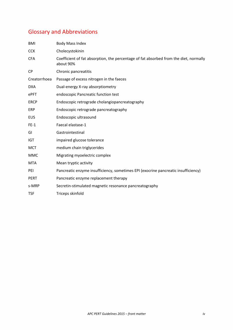
Glossary and Abbreviations BMI Body Mass Index
CCK Cholecystokinin
CFA Coefficient of fat absorption, the percentage of fat absorbed from the diet, normally about 90%
CP Chronic pancreatitis
Creatorrhoea Passage of excess nitrogen in the faeces
DXA Dual-energy X-ray absorptiometry
ePFT endoscopic Pancreatic function test
ERCP Endoscopic retrograde cholangiopancreatography
ERP Endoscopic retrograde pancreatography
EUS Endoscopic ultrasound
FE-1 Faecal elastase-1
GI Gastrointestinal
IGT impaired glucose tolerance
MCT medium chain triglycerides
MMC Migrating myoelectric complex
MTA Mean tryptic activity
PEI Pancreatic enzyme insufficiency, sometimes EPI (exocrine pancreatic insufficiency)
PERT Pancreatic enzyme replacement therapy
s-MRP Secretin-stimulated magnetic resonance pancreatography
TSF Triceps skinfold
APC PERT Guidelines 2015 – Chapter 1 – Background and Methods 1-1
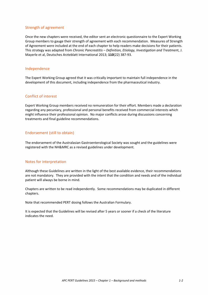
Chapter 1 Methods Intended audience The Guidelines were intended to be read by prescribers, dietitians and GPs and published online via a link on the APC home page. Some hard copes would be made available. An abridged version of the Guidelines would be developed for submission for publication in an international journal. Coordinator Dr Sarah Smith, a medical scientist with previous experience in guidelines development, was engaged and paid by the APC to edit and co-ordinate the Guidelines. Chapters It was decided that where possible, chapters would be re-written rather than simply updated. A chapter leader and secondary reviewer was assigned to each topic in line with members’ areas of expertise. New literature searches over the last seven years were performed in Medline, PubMed, Google Scholar, Embase and the Cochrane library. Literature and draft chapters were made available on Dropbox so that authors could collaborate freely. A summary and recommendations with the highest level of evidence were added to each chapter according to the Oxford Centre for Evidence-Based Medicine (www.cebm.net), with the inclusion of a category 3c: Critical review of the literature. Because such a review is not a systematic review, it may be subject to bias, but this category was included because some references cited in the Guidelines have evaluated multiple studies including RCTs and cohort studies and it was considered that they warrant a higher level of evidence than 4 (case series) and 5 (expert opinion without explicit critical appraisal, bench research or "first principles") (Table 1.1). Table 1.1 Levels of Evidence 1a Systematic reviews (with homogeneity) of randomized controlled trials
1b Individual randomized controlled trials (with narrow confidence interval)
1c All or none randomized controlled trials
2a Systematic reviews (with homogeneity) of cohort studies
2b Individual cohort study or low quality randomized controlled trials (e.g. <80% follow-up)
2c "Outcomes" Research; ecological studies
3a Systematic review (with homogeneity) of case-control studies
3b Individual case-control study
3c Critical review of the literature, including multiple experimental and observational studies
4 Case-series (and poor quality cohort and case-control studies)
5 Expert opinion without explicit critical appraisal, or based on physiology, bench research or "first principles"
APC PERT Guidelines 2015 – Chapter 1 – Background and methods 1-2
Strength of agreement Once the new chapters were received, the editor sent an electronic questionnaire to the Expert Working Group members to gauge their strength of agreement with each recommendation. Measures of Strength of Agreement were included at the end of each chapter to help readers make decisions for their patients. This strategy was adapted from Chronic Pancreatitis – Definition, Etiology, Investigation and Treatment, J. Mayerle et al, Deutsches Arzteblatt International 2013; 110(22) 387-93. Independence The Expert Working Group agreed that it was critically important to maintain full independence in the development of this document, including independence from the pharmaceutical industry. Conflict of interest Expert Working Group members received no remuneration for their effort. Members made a declaration regarding any pecuniary, professional and personal benefits received from commercial interests which might influence their professional opinion. No major conflicts arose during discussions concerning treatments and final guideline recommendations. Endorsement (still to obtain) The endorsement of the Australasian Gastroenterological Society was sought and the guidelines were registered with the NH&MRC as a revised guidelines under development. Notes for interpretation Although these Guidelines are written in the light of the best available evidence, their recommendations are not mandatory. They are provided with the intent that the condition and needs and of the individual patient will always be borne in mind. Chapters are written to be read independently. Some recommendations may be duplicated in different chapters. Note that recommended PERT dosing follows the Australian Formulary. It is expected that the Guidelines will be revised after 5 years or sooner if a check of the literature indicates the need.

APC PERT Guidelines 2015 – Chapter 2 – Pancreatic enzyme insufficiency 2-1
Chapter 2
Pancreatic exocrine insufficiency and its diagnosis Pancreatic exocrine insufficiency (PEI) is a condition resulting in maldigestion, with the potential for malnutrition.
The pancreas is a glandular organ located in the upper abdomen behind the stomach (Figure 2.1). It is the major solid digestive organ of the body, secreting digestive enzymes and bicarbonate into the duodenum via a ductal system (Figure 2.2). In addition, it is an endocrine organ producing insulin and glucagon to regulate blood sugar levels. Figure 2.1: Location of the pancreas
Reproduced from the National Cancer Institute: http://www.cancer.gov/cancertopics/wyntk/pancreas/page2
Figure 2.2: The relationship of the pancreas to nearby organs.
Reproduced from the National Cancer Institute: http://www.cancer.gov/cancertopics/wyntk/pancreas/page2
Pancreatic exocrine insufficiency (PEI) can result from various conditions which damage the pancreas, resulting in either gross alteration of structure or more diffuse functional change. PEI causes a cluster of symptoms which vary with its severity and include abdominal pain, diarrhoea, weight loss and nutritional deficiencies.

APC PERT Guidelines 2015 – Chapter 2 – Pancreatic enzyme insufficiency 2-2
Patients with suspected clinically significant PEI can be graded into three subgroups (Table 2.1). In those with gross changes such as total pancreatectomy or with radiologic investigations showing severe calcific pancreatitis or neoplasm in the head of the pancreas (PEI definite), the presence of severe steatorrhoea and weight loss enables the diagnosis of PEI to be made on clinical grounds alone. In the case of moderate structural alteration of the pancreas (PEI possible), the diagnosis may be hinted at when the patient presents with a degree of nutritional impairment and diarrhoea, but there may be other reasons for these conditions. In the third group of patients (PEI unlikely), a clinical diagnosis such as irritable bowel syndrome may only occasionally be caused by PEI. Here, tests of lower sensitivity and specificity may result in under- or over-diagnosis. Careful evaluation may be necessary before committing these people to long term pancreatic enzyme replacement therapy (PERT). Table 2.1: Proposed subgroups of Pancreatic Exocrine Insufficiency.
Category of PEI PEI definite (d) PEI possible (p) PEI unlikely (u) Examples of aetiology Total pancreatectomy Gastric surgery with
postcibal asynchrony Irritable Bowel Syndrome
Severe chronic pancreatitis with calcific changes, steatorrhoea, weight loss
Mild & moderate chronic pancreatitis
Coeliac disease
Pancreatic-insufficient Cystic Fibrosis
Pancreatic-sufficient Cystic Fibrosis
Inflammatory Bowel Disease
Tumour destroying head of pancreas
Post severe pancreatitis Weight loss in the elderly
Acute pancreatitis destroying head of pancreas
Vitamin A, E, D, K deficiency
Diabetes type II
Post Whipple procedure (partial pancreatectomy)
Bowel resection?
Bowel resection? Symptoms +++ +/- <10% cases Probability of +ve objective test (diagnosis) and likelihood of response to PERT*
100% 30-70% <10% cases
*see relevant chapters
Pancreatic exocrine secretion Pancreatic enzymes, especially lipase, amylase, trypsin and chymotrypsin, are vital for macronutrient digestion.
Traditionally, pancreatic secretion resulting from a meal has been viewed as taking place in a number of phases. The cephalic phase resulting from the sight and smell of food is mediated by cholinergic vagal pathways which stimulate gastric acid secretion and secretion of enzymes from pancreatic acinar cells; it accounts for 20-25% of secretion.
The gastric phase resulting from gastric distension activating vago-vagal reflexes, accounts for approximately 10% of secretion.
The intestinal phase accounts for most of the pancreatic secretory response (60-70%) and is a consequence acidic chyme entering the duodenum. Acid provokes release of secretin from the duodenal mucosa, which in turn stimulates bicarbonate secretion from Brunner’s glands in the duodenum and from pancreatic ductal cells. This bicarbonate rapidly neutralises acid from the stomach, protecting pancreatic enzymes from destruction. Cholecystokinin (CCK) is released from the intestinal mucosa by the presence of fat and protein in the chyme and acts as the major secretagogue for enzyme secretion from pancreatic acinar cells. Other elements of the intestinal phase include enteroenteric reflexes involving a variety of neurotransmitters, predominantly acetylcholine.

APC PERT Guidelines 2015 – Chapter 2 – Pancreatic enzyme insufficiency 2-3
It is debatable whether human pancreatic acinar cells manifest CCK receptors. There is evidence that CCK may act via intrapancreatic neurones and stellate cells, which in turn release acetylcholine as the “final common pathway”.
The pancreas also secretes between meals. Interdigestive secretion occurs in association with the migrating myoelectric complex (MMC) every 60 to 120 min. This secretion is mediated cholinergically. The MMC and interdigestive pancreatic secretion serve a “housekeeper” function, clearing the stomach and intestine of debris and bacteria between meals.
Although the schema described above seems relatively straightforward, it only outlines the known major elements involved in the regulation of pancreatic secretion. This regulation is quite complex, involving other hormones such as gastrin, insulin, pancreatic polypeptide, serotonin and other neurotransmitters such as vasoactive intestinal peptide and gastrin releasing peptide.1-4 Pancreatic enzyme secretion into the duodenum Following a regular meal, enzyme delivery into the duodenum increases rapidly and reaches maximal values within 30 -60 minutes (Figure 2.3). Following peak output, enzyme secretion decreases to almost stable secretory rates at lower levels until about 3–4 hours postprandially, depending on the size of the meal. The interdigestive range is reached again at the end of the digestive period.7–13
Figure 2.3: Digestive pancreatic enzyme response to a regular meal5
Interdigestive and postprandial duodenal enzyme outputs for lipase, amylase and trypsin are listed in Table 2.2 below. At the end of the digestive period, enzyme secretion returns to baseline levels, usually 3 to 4 hours postprandially. The extent and duration of pancreatic enzyme secretion depend on the caloric content, nutritional composition and physical properties of the meal.
Table 2.2: Duodenal enzyme outputs5
Interdigestive Early/maximal postprandial Late/mean postprandial Lipase (U/min) 1000 3000-6000 2000-4000 Amylase (U/min) 50-250 500-1000 500 Trypsin (U/min) 50-100 200-1000 150-500 Data are derived from studies using test meals with 300-600 kcal.
APC PERT Guidelines 2015 – Chapter 2 – Pancreatic enzyme insufficiency 2-4
Pancreatic exocrine insufficiency Pancreatic exocrine insufficiency occurs when the amounts of enzymes secreted into the duodenum in response to ingestion of a meal are not sufficient to maintain normal digestive processes. There are three chief reasons why sufficient pancreatic enzymes may not be available5: 1. Insufficient capacity of the pancreas to synthesise enzymes due to loss of or injury to the pancreatic
parenchyma.
2. Reduced stimulation of enzyme production due to postcibal asynchrony. This can be anatomical, e.g. following gastroenterostomy, or physiological (poor timing of pancreatic juice release after eating).
3. Impaired delivery of enzymes to the duodenum due to obstruction of the pancreatic duct.
Because of the high reserve capacity of the pancreas and compensatory mechanisms that partly replace the loss of pancreatic enzymes, clinical symptoms of PEI do not usually manifest until duodenal lipase levels fall below 5-10% of normal postprandial levels5;6. The most severe clinical outcome of PEI is fat maldigestion and malabsorption, causing steatorrhoea and weight loss. Steatorrhoea is characterised by stools which are frothy, foul smelling and buoyant due to their high fat content. Other symptoms may also include abdominal pain, flatulence and weight loss in adults or lack of weight gain in children. If left untreated, fat maldigestion may result in low circulating levels of micronutrients, fat-soluble vitamins7;8 and lipoproteins9, which have been associated with high morbidity due to increased risk of malnutrition-related complications such as osteopenia and fracture7;10-16. However, fat maldigestion may not always be obvious, so that levels of micronutrients, fat-soluble vitamins and lipoproteins can be low even though steatorrhoea is not clinically apparent17. Note that the visual appearance of the stool does not necessarily indicate its fat content18, and that steatorrhoea can be present without diarrhoea. Patients may consciously or unconsciously reduce their fat intake in an attempt to ease their symptoms and this can also result in malnutrition without overt steatorrhoea and/or weight loss. Nonetheless, the presence of steatorrhoea, either proven or implied, is the foundation for initiating pancreatic enzyme replacement therapy (PERT). A decrease in pancreatic enzyme secretion detected by a sensitive direct measurement of pancreatic secretion does not by itself mandate the initiation of PERT. STEATORRHOEA MUST BE PRESENT. If the presence of steatorrhoea cannot be established, for whatever reason, by direct measurement of faecal fat, its presence may be inferred by the clinical diagnosis, imaging and patient characteristics, including suggestive changes in stool habit, weight loss, measured deficiencies in fat-soluble vitamins and osteoporosis. Diagnosis of pancreatic exocrine insufficiency Pancreatic exocrine function is difficult to assess because the organ and its secretions are relatively inaccessible. However, it is important to be able to differentiate malabsorption/maldigestion due to pancreatic causes from other causes and to assess the efficacy of treatment19. For example, pancreatic disease may explain the symptoms of some patients with nonulcer dyspepsia20. In clinical practice, the diagnosis of PEI begins with an assessment of the patient’s clinical state, a self-report of bowel movements and weight loss in adults or failure to thrive in children, followed by morphological and functional assessments. Assessment of pancreatic structure
Initial assessment of patients presenting with the above symptoms will include a CT scan to assess pancreatic structure. � Strengths: CT is widely available and can identify pancreatic calcification, dilated pancreatic ducts,
pancreatic masses (inflammatory and neoplastic), peri-pancreatic inflammation and fluid collections.

APC PERT Guidelines 2015 – Chapter 2 – Pancreatic enzyme insufficiency 2-5
CT should clearly identify patients with gross changes and frequently, if a close cut study with sufficient contrast is undertaken, diagnose those with lesser degrees of structural change.
� Limitations: In many cases these changes are not specific and require further investigation with magnetic resonance cholangiopancreatography (MRCP) or endoscopic ultrasonography (EUS).
Subsequent assessments can include – Magnetic resonance imaging (MRI) and magnetic resonance cholangiopancreatography (MRCP):
� Strengths: MRCP is non-invasive and does not involve radiation. It is valuable in assessing duct morphology such as dilatation, stricture and side branch ectasia.
The secretin-MRCP stimulation test (s-MRCP) involves the infusion of secretin after initial MRCP imaging. Changes in the volume of fluid in the duodenum and pancreatic duct are measured in the T2 phase. The change in duodenal volume in response to secretin21 has been shown to correlate with the changes in volume and bicarbonate secretion after secretin stimulation using the standard duodenal tube test (see below). � Limitation: As yet, the s-MRCP test has not been standardised for general inter-centre applicability.
Endoscopic ultrasonography (EUS), a high resolution but operator-dependent modality for imaging the pancreas, is now regarded as the most sensitive test for identification of early chronic pancreatitis (CP)22, supplanting ERCP. Diagnosis is based on the number of parenchymal and ductal criteria (determined by expert consensus) present23;24. The greater the number of criteria present, the more likely the diagnosis of CP.
� Strengths: Useful for diagnosis of early CP; ≥ 4 EUS features are highly sensitive and specific for diagnosis of CP and correlate well with histopathologic criteria25.
� Limitation: Inter-observer agreement remains a problem for the use of EUS in the diagnosis of CP.
Endoscopic retrograde cholangiopancreatography (ERCP) was once a ‘gold standard’ for the diagnosis of CP but is now rarely required, unless endotherapy is undertaken.
� Strength: higher sensitivity for ductal abnormalities over CT and MRCP. � Limitations are the inability of ERCP to visualise the pancreatic parenchyma and its invasive nature,
which carries a significant risk of pancreatitis17;26;27;28. Although these methods can further define structural changes in the pancreas, they may give inconclusive results in mild and moderate disease. They require more validation for reliable recommendation 29;30. Direct assessment of pancreatic function
Direct tests involve collection of pancreatic secretions via duodenal intubation while the pancreas is stimulated with exogenous hormones (secretin with or without CCK) or intestinal nutrients given as a standardised test meal (the Lundh test). Although direct tests are the most sensitive and specific methods to assess pancreatic exocrine function, their cost and invasive nature has limited their routine use in clinical practice. However, the endoscopic pancreatic function test (ePFT, see below) is becoming increasingly adopted as a direct function test. Secretin-CCK stimulation test
A double lumen naso-duodenal (Dreiling) tube is positioned in the second and third parts of the duodenum under radiological control and all gastric and duodenal fluid continuously aspirated for the test duration with a nonabsorbable marker to ensure all aspirations are analysed31;32. The duodenal fluid is collected on ice. Secretin 1U/kg/h and caerulein (similar in action to CCK) 100ng/kg/h are infused continuously over 90 minutes. Duodenal fluid is sampled at 10-minute intervals for measurement of volume, bicarbonate, lipase and protease output. A peak bicarbonate secretion of 30-75% of the normal value for the testing laboratory indicates moderate dysfunction and <30% of the lower limit of normal indicates severe dysfunction33. Some units also
APC PERT Guidelines 2015 – Chapter 2 – Pancreatic enzyme insufficiency 2-6
measure lipase over 80 minutes, when values of <780,000 units/L are considered abnormal. A number of minor modifications in technique are sometimes used in different institutions.
� Strength: This is the gold standard direct test of pancreatic exocrine function. It has high sensitivity and specificity22;34;35.
� Limitation: invasive, requires anaesthesia and endoscopy; expensive. The Lundh test
This also utilises a tube placed in the second and third parts of the duodenum. All gastric and duodenal juice is aspirated. A liquid meal equivalent to 250ml of Ensure® is consumed and half-hourly aspirates from the duodenum stored on ice for the subsequent measurement of mean tryptic activity. Lipase output can be substituted for mean tryptic activity (MTA) because it can more easily be measured in the routine biochemistry laboratory20;36. Severe pancreatic insufficiency with steatorrhoea is associated with MTA of <7U or a peak lipase excretion of <780,000 U/ml.
� Strength: this is the most physiologic test of pancreatic exocrine function and the best test to demonstrate postcibal asynchrony35
� Limitation: time-consuming, cumbersome; no longer routinely carried out in clinical practice. Endoscopic pancreatic function test (ePFT)
Procedural details of this new test are described in Law et al24: Patients are ‘placed in leftward supine and reverse Trendelenberg position to maximize pooling of fluid in the dependent portion of the duodenum. Conscious sedation using pethidine and midazolam is administered’ and monitored by an anaesthetist, and has been shown not to limit electrolyte secretion37. Hormones are ‘administered intravenously starting at time 0 minutes. First, CCK (Kinevac®, Bracco Diagnostics Inc, Princeton, NJ, USA) is infused at 40 ng/kg/h. Next, a 0.2g test dose of synthetic human secretin is administered (ChiRhoStim®, ChiRhoClin Inc, Burtonsville, Md, USA). After 5 minutes, the standard dose of synthetic human secretin (0.2g/kg) is administered as an intravenous bolus. After 15 minutes, ’a standard upper endoscope is passed into the stomach. All gastric fluid must be thoroughly aspirated and discarded. The endoscope is then passed through the pylorus and positioned in the second portion of the duodenum. Residual duodenal fluid must be aspirated and discarded. Timed duodenal fluid samples are collected’ from continuous suction through the endoscope into a fluid trap at times 25 to 29, 30 to 34, 35 to 39, 40 to 45 and 46 to 50 minutes. The times of collection are based on previous studies showing peak concentrations of bicarbonate and pancreatic enzymes at 30 to 50 minutes after hormonal stimulation. Each sample is placed on ice and analysed within 3 hours for the highest concentrations of lipase, amylase and bicarbonate, which are considered the peak concentrations. The authors found that peak bicarbonate concentration provided the best discrimination between mild and more severe CP as determined by the presence of five or more abnormalities on EUS. A similar prospective study was undertaken by Gardner et al 38.
� Strengths: found to correlate well with the standard Dreiling tube pancreatic function test39, standardised against the Lundh test40; 86% sensitivity, 67% specificity for CP against histological standard 41.
� Limitations: requires sedation for 1 hour, cumbersome, expensive and is difficult to be sure all secretions are collected32.
Indirect tests of pancreatic exocrine function These are cheaper and easier to administer, but are less sensitive and less specific because they are designed to detect abnormalities secondary to loss of pancreatic exocrine function. They rely on the health of other organ systems for their precision. Indirect tests can be divided into four categories: faecal tests, breath tests, urinary tests and blood tests. Some indirect tests have high sensitivity and specificity for severe CP but not for mild and moderate cases of CP.
APC PERT Guidelines 2015 – Chapter 2 – Pancreatic enzyme insufficiency 2-7
Faecal tests
3-day faecal fat test
The 3-day faecal fat test is considered the gold standard for diagnosing and quantifying steatorrhoea. The method most commonly used is the Van de Kamer method42. Adults consume a diet containing 100g of fat for 3-5 days. Children must meticulously weigh their food and maintain careful dietary records in order to calculate the mean daily fat intake. Stools are collected over 72 hours and pooled for analysis. The coefficient of fat absorption (CFA, % fat in the normal diet) is measured. Near infrared reflectance analysis (NIRA) has simplified the quantification of fat in stool. Steatorrhoea is present if more than 7% of ingested fat is excreted in patients over 6 months of age or more than 15% in patients under 6 months of age43;44.
� Strengths: traditionally considered the gold standard for diagnosis of steatorrhoea; 92% sensitivity for PEI (but 42% specificity for PEI)45;
� Limitations: inconvenient for patients and potential for poor patient compliance; unpleasant for laboratory technicians; not widely available; does not distinguish between pancreatic and non-pancreatic causes.
Steatocrit
A steatocrit determines the proportion of fat in a single stool. Here, homogenised faeces are centrifuged at 15,000 rpm for 15 minutes causing the lipid and aqueous phases to separate from each other and from the stool residue46. A lipid phase representing less than 10% of volume is considered normal in patients older than 6 months of age. Perchloric acid can be added to the faecal homogenate to improve sensitivity of the test47;48.
� Strengths: Easily done on a single stool sample; useful as a screening test; the acid steatocrit method correlates with the 3-day faecal fat test49;50 in patient cohorts but results of these tests were not found to be interchangeable on an individual patient basis.
� Limitation: The determination of steatocrit is not in general use. Microscopic examination of stools for fat droplets
Microscopic examination of stool for fat globules may be used as a crude screening test for malabsorption. A simple qualitative technique utilises the Sudan III stain in which neutral fat globules are visualised under the microscope. If fat globules are present, then it may be prudent to perform additional tests.
� Strength: easily performed, cheap. � Limitation: use as a crude screening test only.
Faecal chymotrypsin and elastase-1
Measurements of faecal chymotrypsin and elastase-1 have both been employed as measures of pancreatic exocrine secretion. FE-1 is a pancreas-specific protease. It is not degraded by intestinal passage. FE-1 is stable in faeces stored at room temperature for up to 3 days 51. In addition, it is concentrated five to six times higher in faeces compared with the concentration originally found in pancreatic secretion 51; 52. A low FE-1 concentration of <200 μg/g suggests PEI, whereas <100 μg/g suggests severe PEI. The reliability of the FE-1 test in determining the presence of PEI varies with the severity of PEI present as determined by direct tube testing of pancreatic function. Sensitivities for the test vary from 0–63% in mild-to-moderate cases of PEI in CP to 77–100% in moderate-to-severe PEI in CP, while specificities range from 80–95% in mild-to-moderate cases to 76–100% in moderate-to-severe cases 53-57. Likewise, it is not reliable for predicting CP in patients with equivocal imaging findings58. Similarly, FE-1 concentrations are unable to differentiate patients with and without imaging evidence of CP with respect to calcification or abnormalities on ERCP 57. In children, an FE-1 concentration of 100 μg/g stool provided excellent diagnostic performance to distinguish steatorrhoea caused by PI: 96% sensitivity and 93.6% specificity, 87.7% positive predictive value and 99% negative predictive value59. FE-1 showed excellent sensitivity (98%) but a lower specificity of 80% as a
APC PERT Guidelines 2015 – Chapter 2 – Pancreatic enzyme insufficiency 2-8
determinant of the underlying cause of steatorrhoea (pancreatic vs. intestinal). This low specificity was attributed to children with short gut syndrome who had FE-1 concentrations <100 μg/g stool.
� Strengths: FE-1 levels have been shown to have good correlation with duodenal pancreatic juice concentrations after secretin-cholecystokinin stimulation 51. The advantages of the FE-1 test over the 3-day faecal fat collection include: - it can be measured on a ‘spot’ stool specimen; - it is not affected by the quantity of dietary fat intake; - it is not affected by oral pancreatic enzyme replacement therapy, because it is specific to human
elastase, while oral pancreatic enzyme replacement extracts are either bovine or porcine in origin. This is also an advantage of FE-1 over faecal chymotrypsin measurement.
� Limitations: Falsely low results are a major pitfall; false positive results have been shown to occur in
up to 7% of healthy control individuals, and up to 38% of patients with non-pancreatic diarrhoea due to dilution60; Diabetes mellitus, both types 1 and 2, have been associated with a high prevalence of low FE-1 levels 61;62, though it has been shown at least in type 1 diabetes mellitus that FE-1 is not reliable for diagnosing steatorrhoea or PEI 63; FE-1 is not sensitive for detecting PEI in gastrectomy patients due to asynchrony. FE-1 cannot be considered an adequate substitute for the 3-day faecal fat test for the diagnosis of PEI.
Faecal chymotrypsin is no longer employed as the faecal elastase-1 (FE-1) test has been deemed to be superior in terms of sensitivity and specificity 56.
Breath tests
Radiolabelled carbon breath tests are emerging as an indirect PFT in Europe. They use the fact that ingested lipids are mainly hydrolysed by pancreatic lipases in the small intestine, absorbed as free fatty acids and monoglycerides, and transported to the liver, where oxidative metabolism liberates carbon dioxide. Breath samples are taken before and after a test meal containing 13C-labelled substrate. The most widely used has been the 13C-labelled mixed triglyceride breath test. This involves the ingestion of 250 mg of 2-octanoyl (1-13C)-1,3 distearoyl glycerol (Eurisotop, Saint Aubin, France) with 16 g of fat 64. This substrate is digested by lipase, releasing 13C-labeled octanoic acid, which is then absorbed and metabolized to form 13CO2, and is ultimately released in expired breath. Breath samples are collected at 15 minute intervals for 6 hours. Abnormal results tend to correlate with other markers of advanced CP, such as the number of EUS features 65. The clinical use of radiolabelled breath tests for the diagnosis of PEI is still limited in Australia.
� Limitations: expensive substrates which are currently unavailable from Australian manufacturers; need to take samples over long test periods; poor sensitivity for diagnosing CP in patients without steatorrhoea 66 ; potential confounding effects of non-pancreatic organ dysfunction (biliary, intestinal, liver and lung) 67; inability to differentiate between pancreatic and non-pancreatic causes of fat maldigestion.
Urine tests
Urine tests use non-absorbable substrates which are specifically cleaved by pancreatic enzymes. This results in the release of a rapidly absorbable marker which is conjugated in the liver and excreted in urine. Two substrates have been used – bentiromide and fluorescein dilaurate68-70. Following substrate ingestion, urine is collected over a specified time period.
� Limitation: superseded by the FE-1 and direct tests with better specificity and sensitivity. Blood tests
Trypsin is exclusively synthesised by the pancreas and small amounts are released into the blood as the proenzyme trypsinogen. Measurement of serum immunoreactive trypsinogen is a sensitive and relatively non-
APC PERT Guidelines 2015 – Chapter 2 – Pancreatic enzyme insufficiency 2-9
invasive method of screening for pancreatic insufficiency in older children. It has been validated in children with cystic fibrosis and PEI due to other causes71;72. Serum trypsinogen levels below 20 ng/mL are reasonably specific for PEI in patients over seven years of age. Elevated serum immunoreactive trypsinogen level is also used as part of the screening test for cystic fibrosis in newborns73. Individuals with cystic fibrosis have elevated serum immunoreactive trypsinogen levels during the first years of life. The levels fall to subnormal values by 6 years of age in those cystic fibrosis patients who are pancreatic-insufficient.
� Strengths: sensitive and relatively non-invasive screening test. � Limitation: fluctuating pattern in the first decade of life, hence serial measurements are needed then.
The importance of nutritional markers is being increasingly recognised.
Steatorrhoea is associated with a poorer nutritional status19 but fat-soluble vitamins can be deficient in patients with PEI who do not have overt steatorrhoea7. There is good pathophysiological rationale for fat-soluble vitamin and lipoprotein deficiencies occurring as a consequence of PEI. Lindkvist et al8 examined the nutritional markers haemoglobin, mean corpuscular volume, lymphocyte count, prothrombin time, and serum levels of total protein, albumin, prealbumin, retinol binding protein, cholesterol, triglycerides, amylase, folic acid, vitamin B12, HbA1C, transferrin, ferritin, magnesium and zinc in a cohort of 114 people, where 38 had PEI. Patients with PEI had abnormal levels of magnesium (below 2.05 mg/dL), haemoglobin, albumin, prealbumin and retinol binding protein and HbA1C above the upper limit of normal on univariate analysis. Magnesium below 2.05 mg/dL detected PEI with sensitivity, specificity and positive and negative predictive values of 0.88 (95% confidence interval, 0.66-0.97), 0.66 (0.48-0.80), 0.58 (0.39-0.75) and 0.91 (0.73-0.98), respectively. Although they concluded that serum nutritional markers can be used to predict the probability of PEI in CP, there is insufficient evidence at present to recommend use of these markers as a screening test for an individual patient and the ability of PERT to reverse these anomalies has not been reported. Patients with CP often have multiple factors affecting their nutritional status and to isolate PEI from these can be difficult. Nonetheless they are at risk of nutritional compromise and the presence of fat-soluble vitamin and nutritional marker anomalies can alert clinicians to the possibility of PEI. Evaluation of the clinical use of tests
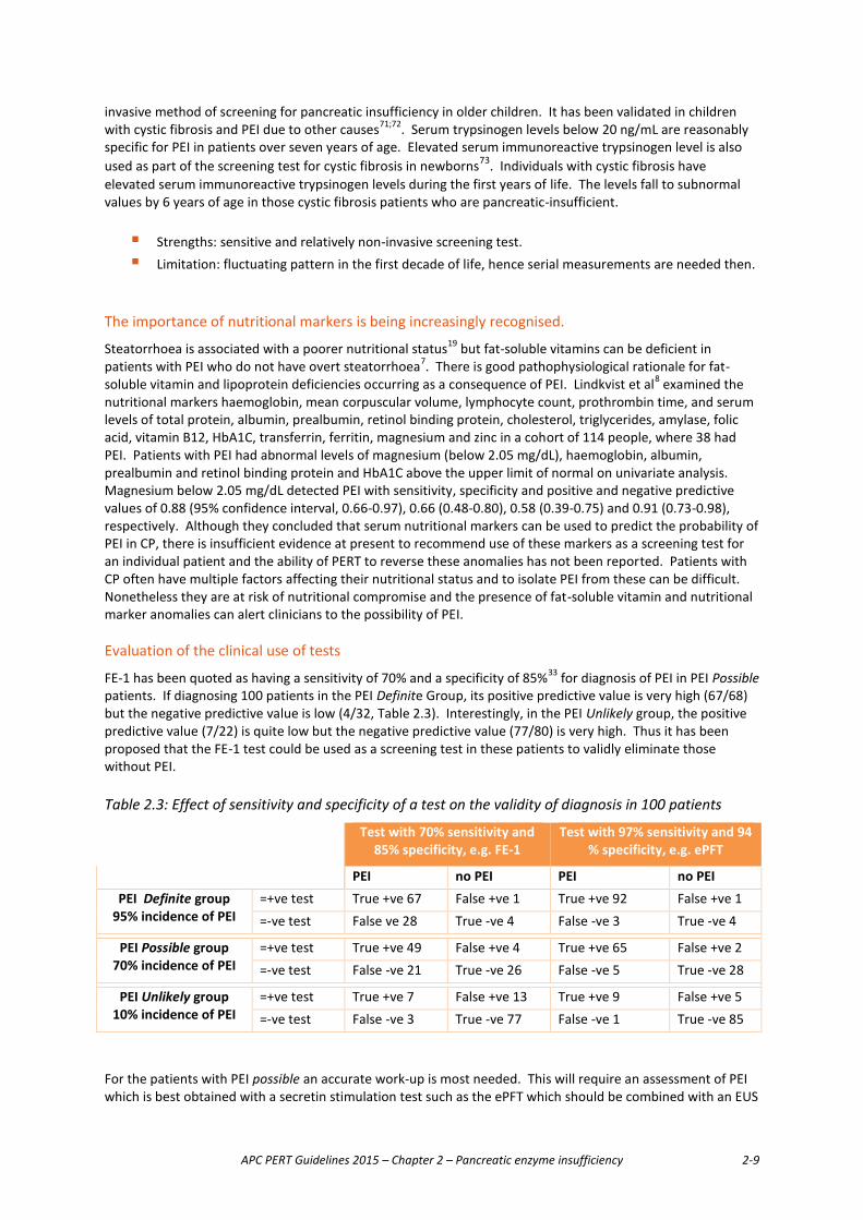
FE-1 has been quoted as having a sensitivity of 70% and a specificity of 85%33 for diagnosis of PEI in PEI Possible patients. If diagnosing 100 patients in the PEI Definite Group, its positive predictive value is very high (67/68) but the negative predictive value is low (4/32, Table 2.3). Interestingly, in the PEI Unlikely group, the positive predictive value (7/22) is quite low but the negative predictive value (77/80) is very high. Thus it has been proposed that the FE-1 test could be used as a screening test in these patients to validly eliminate those without PEI. Table 2.3: Effect of sensitivity and specificity of a test on the validity of diagnosis in 100 patients
Test with 70% sensitivity and 85% specificity, e.g. FE-1
Test with 97% sensitivity and 94 % specificity, e.g. ePFT
PEI no PEI PEI no PEI PEI Definite group
95% incidence of PEI =+ve test True +ve 67 False +ve 1 True +ve 92 False +ve 1 =-ve test False ve 28 True -ve 4 False -ve 3 True -ve 4
PEI Possible group 70% incidence of PEI
=+ve test True +ve 49 False +ve 4 True +ve 65 False +ve 2 =-ve test False -ve 21 True -ve 26 False -ve 5 True -ve 28
PEI Unlikely group 10% incidence of PEI
=+ve test True +ve 7 False +ve 13 True +ve 9 False +ve 5 =-ve test False -ve 3 True -ve 77 False -ve 1 True -ve 85
For the patients with PEI possible an accurate work-up is most needed. This will require an assessment of PEI which is best obtained with a secretin stimulation test such as the ePFT which should be combined with an EUS
APC PERT Guidelines 2015 – Chapter 2 – Pancreatic enzyme insufficiency 2-10
to provide structural and functional information 74. However this will require a longer sedation unless a tube is left in the duodenum at the completion of the EUS and aspirates retrieved in the recovery room. Because of the shortcomings of all these tests, there is as yet no single perfect test which can accurately diagnose PEI, particularly in the PEI Possible and PEI Unlikely patient groups. For PEI Definite, no diagnostic test is necessary. For patients where PEI is possible, imaging (CT or EUS), then a functional test (ePFT, Dreiling or 13C breath test if available) should be utilised. In the PEI Unlikely group, imaging followed by an FE-1 test should provide helpful information. If this indicates a low enzyme level, follow up with an ePFT or a Secretin-CCK stimulation test, depending on availability. In all patient groups, screening for nutritional markers including magnesium, fat-soluble vitamins and lipoproteins will unmask nutritional deficiencies. Ultimately PERT can be trialled, and symptom improvement would support a diagnosis of PEI, but a placebo effect needs to be taken into consideration in this clinical scenario.
Summary and Recommendations Symptoms of PEI
� Include abdominal pain, diarrhoea, weight loss and malnutrition.
� Do not manifest until duodenal lipase levels fall below 5-10% of normal postprandial levels5;6.
Clinical consequences of PEI
� The most common is fat maldigestion
� Maldigestion can result in steatorrhoea and weight loss (or failure to thrive in children), which are risk factors for high morbidity 9;14.
� Maldigestion is not always obvious; levels of micronutrients, fat-soluble vitamins and lipoproteins may be pathologically low 7 but not clinically apparent 7;11;17.
� The possibility of non-pancreatic causes of steatorrhoea should be considered, e.g. Crohn’s disease of the ileum, in order to give appropriate treatment.
�Testing for PEI. Patients with suspected PEI can be grouped into
� Definite (total pancreatectomy, severe calcific pancreatitis or neoplasm in the pancreatic head). No diagnostic test for PEI is needed.
� Possible (moderate structural alteration of the pancreas).
All tests for an accurate diagnosis of PEI have significant drawbacks but consider:
o Structural imaging. First step: CT with contrast - often good results, readily available. In some cases, CT may be followed with MRI, EUS and/or secretin-MRCP 29;30.
Endoscopic retrograde cholangiopancreatography (ERCP) may occasionally be required. It carries a significant measure of risk 26-28.
o Direct pancreatic function tests are the most specific and sensitive, but are too expensive, cumbersome and invasive for routine clinical use.
- The ‘gold standard’ direct test for PEI has been the secretin-CCK stimulation test with duodenal tube 33.
APC PERT Guidelines 2015 – Chapter 2 – Pancreatic enzyme insufficiency 2-11
- This is likely to be replaced by the ePFT which can provide morphological as well as functional information 74. Results of these tests were shown in a small well-conducted cross-over RCT to correlate reasonably well, but sensitivity was only 83% and specificity 59% compared with the Dreiling tube test in this study. 39 (Level I).
o Indirect pancreatic function tests.
- The three-day faecal fat is the ‘gold standard’ for diagnosing steatorrhoea but is unpopular with patients and lab technicians. It is being superseded by the FE-1 test but without sufficient basis.
- The FE-1 test is easy and conveniently done on a single stool sample but is appropriate only as a screening test for excluding PEI.
- Blood tests for magnesium, nutritional markers, bone mineral density and in particular the fat-soluble vitamins A, D, E and K are important in the diagnostic workup as they may hint at the presence of PEI and should be part of the follow-up of all patients with suspected or proven PEI.
� Unlikely (e.g. rare cases of IBS, IBD, coeliac disease, diabetes type II).
o Tests for PEI are the same as for Possible cases (above), with even more chance of being inconclusive.
o A trial of PERT can be considered but in the absence of an objective marker of PEI, the danger of a placebo response is considerable.
References (1) Chey WY, Chang TM. Secretin: historical perspective and current status. Pancreas 2014; 43(2):162-
182.
(2) Chandra R, Liddle RA. Recent advances in pancreatic endocrine and exocrine secretion. Curr Opin Gastroenterol 2011; 27(5):439-443.
(3) Dockray GJ. Cholecystokinin. Curr Opin Endocrinol Diabetes Obes 2012; 19(1):8-12.
(4) Dockray GJ. Gastrointestinal hormones and the dialogue between gut and brain. J Physiol 2014.
(5) Keller J, Layer P. Human pancreatic exocrine response to nutrients in health and disease. Gut 2005; 54 Suppl 6:vi1-28.
(6) DiMagno EP, Go VLW, Summerskill WHJ. Relations between pancreatic enzyme outputs and malabsorption in severe pancreatic insufficiency. N E J M 1973; 288(16):813-815.
(7) Sikkens EC, Cahen DL, Koch AD, Braat H, Poley JW, Kuipers EJ et al. The prevalence of fat-soluble vitamin deficiencies and a decreased bone mass in patients with chronic pancreatitis. Pancreatology 2013; 13(3):238-242.
(8) Lindkvist B, Dominguez-Munoz JE, Luaces-Regueira M, Castineiras-Alvarino M, Nieto-Garcia L, Iglesias-Garcia J. Serum nutritional markers for prediction of pancreatic exocrine insufficiency in chronic pancreatitis. Pancreatology 2012; 12(4):305-310.

APC PERT Guidelines 2015 – Chapter 2 – Pancreatic enzyme insufficiency 2-12
(9) Montalto G, Soresi M, Carroccio A, Scafidi E, Barbagallo CM, Ippolito S et al. Lipoproteins and chronic pancreatitis. Pancreas 1994; 9(1):137-138.
(10) Dominguez-Munoz JE. Pancreatic enzyme therapy for pancreatic exocrine insufficiency. Curr Gastroenterol Rep 2007; 9(2):116-122.
(11) Duggan SN, Smyth ND, O'Sullivan M, Feehan S, Ridgway PF, Conlon KC. The prevalence of malnutrition and fat-soluble vitamin deficiencies in chronic pancreatitis. Nutr Clin Pract 2014; 29(3):348-354.
(12) Duggan SN, Smyth ND, Murphy A, Macnaughton D, O'Keefe SJ, Conlon KC. High prevalence of osteoporosis in patients with chronic pancreatitis: a systematic review and meta-analysis. Clin Gastroenterol Hepatol 2014; 12(2):219-228.
(13) Dujsikova H, Dite P, Tomandl J, Sevcikova A, Precechtelova M. Occurrence of metabolic osteopathy in patients with chronic pancreatitis. Pancreatology 2008; 8(6):583-586.
(14) Nojgaard C, Bendtsen F, Becker U, Andersen JR, Holst C, Matzen P. Danish patients with chronic pancreatitis have a four-fold higher mortality rate than the Danish population. Clin Gastroenterol Hepatol 2010; 8(4):384-390.
(15) Sudeep K, Chacko A, Thomas N, Selvakumar R, George B, Paul TV et al. Predictors of osteodystrophy in patients with chronic nonalcoholic pancreatitis with or without diabetes. Endocr Pract 2011; 17(6):897-905.
(16) Tignor AS, Wu BU, Whitlock TL, Lopez R, Repas K, Banks PA et al. High prevalence of low-trauma fracture in chronic pancreatitis. Am J Gastroenterol 2010; 105(12):2680-2686.
(17) Dumasy V, Delhaye M, Cotton F, Deviere J. Fat malabsorption screening in chronic pancreatitis. Am J Gastroenterol 2004; 99(7):1350-1354.
(18) Lankisch PG, Droge M, Hofses S, Konig H, Lembcke B. Steatorrhoea: you cannot trust your eyes when it comes to diagnosis. Lancet 1996; 347(9015):1620-1621.
(19) Dominguez-Munoz JE, Iglesias-Garcia J. Oral pancreatic enzyme substitution therapy in chronic pancreatitis: is clinical response an appropriate marker for evaluation of therapeutic efficacy? JOP 2010; 11(2):158-162.
(20) Smith RC, Talley NJ, Dent OF, Jones M, Waller SL. Exocrine pancreatic function and chronic unexplained dyspepsia. A case-control study. Int J Pancreatol 1991; 8(3):253-262.
(21) Wathle GK, Tjora E, Ersland L, Dimcevski G, Salvesen OO, Molven A et al. Assessment of exocrine pancreatic function by secretin-stimulated magnetic resonance cholangiopancreaticography and diffusion-weighted imaging in healthy controls. J Magn Reson Imaging 2014; 39(2):448-454.
(22) Afghani E, Sinha A, Singh VK. An Overview of the Diagnosis and Management of Nutrition in Chronic Pancreatitis. Nutrition in Clinical Practice 2014.
(23) Wiersema MJ, Hawes RH, Lehman GA, Kochman ML, Sherman S, Kopecky KK. Prospective evaluation of endoscopic ultrasonography and endoscopic retrograde cholangiopancreatography in patients with chronic abdominal pain of suspected pancreatic origin. Endoscopy 1993; 25(9):555-564.
(24) Law R, Lopez R, Costanzo A, Parsi MA, Stevens T. Endoscopic pancreatic function test using combined secretin and cholecystokinin stimulation for the evaluation of chronic pancreatitis. Gastrointest Endosc 2012; 75(4):764-768.

APC PERT Guidelines 2015 – Chapter 2 – Pancreatic enzyme insufficiency 2-13
(25) Sze KC, Pirola RC, Apte MV, Wilson JS. Current options for the diagnosis of chronic pancreatitis. Expert Rev Mol Diagn 2014; 14(2):199-215.
(26) Iorgulescu A, Sandu I, Turcu F, Iordache N. Post-ERCP acute pancreatitis and its risk factors. J Med Life 2013; 6(1):109-113.
(27) Elmunzer BJ, Scheiman JM, Lehman GA, Chak A, Mosler P, Higgins PD et al. A randomized trial of rectal indomethacin to prevent post-ERCP pancreatitis. N Engl J Med 2012; 366(15):1414-1422.
(28) Zhou W, Li Y, Zhang Q, Li X, Meng W, Zhang L et al. Risk factors for postendoscopic retrograde cholangiopancreatography pancreatitis: a retrospective analysis of 7,168 cases. Pancreatology 2011; 11(4):399-405.
(29) Sai JK, Suyama M, Kubokawa Y, Watanabe S. Diagnosis of mild chronic pancreatitis (Cambridge classification): comparative study using secretin injection-magnetic resonance cholangiopancreatography and endoscopic retrograde pancreatography. World J Gastroenterol 2008; 14(8):1218-1221.
(30) Schneider AR, Hammerstingl R, Heller M, Povse N, Murzynski L, Vogl TJ et al. Does secretin-stimulated MRCP predict exocrine pancreatic insufficiency?: A comparison with noninvasive exocrine pancreatic function tests. J Clin Gastroenterol 2006; 40(9):851-855.
(31) Gaskin KJ, Durie PR, Hill RE, Lee LM, Forstner GG. Colipase and maximally activated pancreatic lipase in normal subjects and patients with steatorrhea. J Clin Invest 1982; 69(2):427-434.
(32) Schibli S, Corey M, Gaskin KJ, Ellis L, Durie PR. Towards the ideal quantitative pancreatic function test: analysis of test variables that influence validity. Clin Gastroenterol Hepatol 2006; 4(1):90-97.
(33) Dominguez-Munoz JE. Pancreatic function tests for diagnosis and staging of chronic pancreatitis, cystic fibrosis, and exocrine pancreatic insufficiency of other etiologies:which tests are necessary and how should they be performed in clinical routine? In: Dominguez-Munoz JE, editor. Clinical pancreatology for practising gastroenterologists and surgeons. Blackwell Publishing Ltd; 2005. 259-266.
(34) Dominguez Munoz JE. Diagnosis of chronic pancreatitis: Functional testing. Best Pract Res Clin Gastroenterol 2010; 24(3):233-241.
(35) Dreiling DA. Pancreatic secretory testing in 1974. Gut 1975; 16(8):653-657.
(36) Waller SL, Smithies A, Kapp F, Loyaza M, Noguera EC, Philippakos D et al. The Lundh test: a review of its clinical use in the diagnosis of pancreatic disease. Aust N Z J Med 1981; 11(5):488-493.
(37) Conwell DL, Zuccaro G, Purich E, Fein S, Vanlente F, Vargo J et al. The effect of moderate sedation on exocrine pancreas function in normal healthy subjects: a prospective, randomized, cross-over trial using the synthetic porcine secretin stimulated Endoscopic Pancreatic Function Test (ePFT). Am J Gastroenterol 2005; 100(5):1161-1166.
(38) Gardner TB, Purich ED, Gordon SR. Pancreatic duct compliance after secretin stimulation: a novel endoscopic ultrasound diagnostic tool for chronic pancreatitis. Pancreas 2012; 41(2):290-294.
(39) Stevens T, Conwell DL, Zuccaro G, Jr., Van LF, Lopez R, Purich E et al. A prospective crossover study comparing secretin-stimulated endoscopic and Dreiling tube pancreatic function testing in patients evaluated for chronic pancreatitis. Gastrointest Endosc 2008; 67(3):458-466.
(40) Jensen NM, Larsen S. A rapid, endoscopic exocrine pancreatic function test and the Lundh test: a comparative study. Pancreatology 2008; 8(6):617-624.
APC PERT Guidelines 2015 – Chapter 2 – Pancreatic enzyme insufficiency 2-14
(41) Albashir S, Bronner MP, Parsi MA, Walsh RM, Stevens T. Endoscopic ultrasound, secretin endoscopic pancreatic function test, and histology: correlation in chronic pancreatitis. Am J Gastroenterol 2010; 105(11):2498-2503.
(42) Van de Kamer JH, Ten Bokkel Huinink H, Weyers HA. Rapid method for the determination of fat in feces. J Biol Chem 1949; 177(1):347-355.
(43) Thompson JB, Su CK, Ringrose RE, Welsh JD. Fecal triglycerides. II. Digestive versus absorptive steatorrhea. J Lab Clin Med 1969; 73(3):521-530.
(44) Fomon SJ, Ziegler EE, Thomas LN, Jensen RL, Filer LJ, Jr. Excretion of fat by normal full-term infants fed various milks and formulas. Am J Clin Nutr 1970; 23(10):1299-1313.
(45) Roberts IM, Poturich C, Wald A. Utility of fecal fat concentrations as screening test in pancreatic insufficiency. Dig Dis Sci 1986; 31(10):1021-1024.
(46) Colombo C, Maiavacca R, Ronchi M, Consalvo E, Amoretti M, Giunta A. The steatocrit: a simple method for monitoring fat malabsorption in patients with cystic fibrosis. J Pediatr Gastroenterol Nutr 1987; 6(6):926-930.
(47) Van den Neucker AM, Kerkvliet EM, Theunissen PM, Forget PP. Acid steatocrit: a reliable screening tool for steatorrhoea. Acta Paediatr 2001; 90(8):873-875.
(48) Wagner MH, Bowser EK, Sherman JM, Francisco MP, Theriaque D, Novak DA. Comparison of steatocrit and fat absorption in persons with cystic fibrosis. J Pediatr Gastroenterol Nutr 2002; 35(2):202-205.
(49) Neucker AV, Bijleveld CM, Wolthers BG, Swaaneburg JC, Kester AD, Kreel B et al. Comparison of near infrared reflectance analysis of fecal fat, nitrogen and water with conventional methods, and fecal energy content. Clin Biochem 2002; 35(1):29-33.
(50) Amann ST, Josephson SA, Toskes PP. Acid steatocrit: a simple, rapid gravimetric method to determine steatorrhea. Am J Gastroenterol 1997; 92(12):2280-2284.
(51) Stein J, Jung M, Sziegoleit A, Zeuzem S, Caspary WF, Lembcke B. Immunoreactive elastase I: clinical evaluation of a new noninvasive test of pancreatic function. Clin Chem 1996; 42(2):222-226.
(52) Sziegoleit A, Knapler H, Peters B. ELISA for human pancreatic elastase 1. Clin Biochem 1989; 22(2):79-83.
(53) Dominguez-Munoz JE, Hieronymus C, Sauerbruch T, Malfertheiner P. Fecal elastase test: evaluation of a new noninvasive pancreatic function test. Am J Gastroenterol 1995; 90(10):1834-1837.
(54) Gullo L, Graziano L, Babbini S, Battistini A, Lazzari R, Pezzilli R. Faecal elastase 1 in children with cystic fibrosis. Eur J Pediatr 1997; 156(10):770-772.
(55) Hahn JU, Bochnig S, Kerner W, Koenig H, Sporleder B, Lankisch PG et al. A new fecal elastase 1 test using polyclonal antibodies for the detection of exocrine pancreatic insufficiency. Pancreas 2005; 30(2):189-191.
(56) Keim V, Teich N, Moessner J. Clinical value of a new fecal elastase test for detection of chronic pancreatitis. Clin Lab 2003; 49(5-6):209-215.
(57) Lankisch PG, Schmidt I, Konig H, Lehnick D, Knollmann R, Lohr M et al. Faecal elastase 1: not helpful in diagnosing chronic pancreatitis associated with mild to moderate exocrine pancreatic insufficiency. Gut 1998; 42(4):551-554.
APC PERT Guidelines 2015 – Chapter 2 – Pancreatic enzyme insufficiency 2-15
(58) Gredal C, Madsen LG, Larsen S. The Lundh test and faecal elastase 1 determination in chronic pancreatitis: a comparative study. Pancreatology 2003; 3(5):389-394.
(59) Beharry S, Ellis L, Corey M, Marcon M, Durie P. How useful is fecal pancreatic elastase 1 as a marker of exocrine pancreatic disease? J Pediatr 2002; 141(1):84-90.
(60) Glasbrenner B, Schon A, Klatt S, Beckh K, Adler G. Clinical evaluation of the faecal elastase test in the diagnosis and staging of chronic pancreatitis. Eur J Gastroenterol Hepatol 1996; 8(11):1117-1120.
(61) Hardt PD, Hauenschild A, Nalop J, Marzeion AM, Jaeger C, Teichmann J et al. High prevalence of exocrine pancreatic insufficiency in diabetes mellitus. A multicenter study screening fecal elastase 1 concentrations in 1,021 diabetic patients. Pancreatology 2003; 3(5):395-402.
(62) Larger E, Philippe MF, Barbot-Trystram L, Radu A, Rotariu M, Nobecourt E et al. Pancreatic exocrine function in patients with diabetes. Diabet Med 2012; 29(8):1047-1054.
(63) Hahn JU, Kerner W, Maisonneuve P, Lowenfels AB, Lankisch PG. Low fecal elastase 1 levels do not indicate exocrine pancreatic insufficiency in type-1 diabetes mellitus. Pancreas 2008; 36(3):274-278.
(64) Dominguez-Munoz JE, Iglesias-Garcia J, Vilarino-Insua M, Iglesias-Rey M. 13C-mixed triglyceride breath test to assess oral enzyme substitution therapy in patients with chronic pancreatitis. Clin Gastroenterol Hepatol 2007; 5(4):484-488.
(65) Dominguez-Munoz JE, Alvarez-Castro A, Larino-Noia J, Nieto L, Iglesias-Garcia J. Endoscopic ultrasonography of the pancreas as an indirect method to predict pancreatic exocrine insufficiency in patients with chronic pancreatitis. Pancreas 2012; 41(5):724-728.
(66) Loser C, Brauer C, Aygen S, Hennemann O, Folsch UR. Comparative clinical evaluation of the 13C-mixed triglyceride breath test as an indirect pancreatic function test. Scand J Gastroenterol 1998; 33(3):327-334.
(67) Keller J. Diagnosis of fat malabsorption by breath tests: just a breeze? Digestion 2009; 80(2):95-97.
(68) Scharpe S, Iliano L. Two indirect tests of exocrine pancreatic function evaluated. Clin Chem 1987; 33(1):5-12.
(69) Mitchell CJ, Humphrey CS, Bullen AW, Kelleher J, Losowsky MS. Improved diagnostic accuracy of a modified oral pancreatic function test. Scand J Gastroenterol 1979; 14(6):737-741.
(70) Toskes PP. Bentiromide as a test of exocrine pancreatic function in adult patients with pancreatic exocrine insufficiency. Determination of appropriate dose and urinary collection interval. Gastroenterology 1983; 85(3):565-569.
(71) Durie PR, Forstner GG, Gaskin KJ, Moore DJ, Cleghorn GJ, Wong SS et al. Age-related alterations of immunoreactive pancreatic cationic trypsinogen in sera from cystic fibrosis patients with and without pancreatic insufficiency. Pediatr Res 1986; 20(3):209-213.
(72) Moore DJ, Forstner GG, Largman C, Cleghorn GJ, Wong SS, Durie PR. Serum immunoreactive cationic trypsinogen: a useful indicator of severe exocrine dysfunction in the paediatric patient without cystic fibrosis. Gut 1986; 27(11):1362-1368.
(73) Couper RT, Corey M, Durie PR, Forstner GG, Moore DJ. Longitudinal evaluation of serum trypsinogen measurement in pancreatic-insufficient and pancreatic-sufficient patients with cystic fibrosis. J Pediatr 1995; 127(3):408-413.
APC PERT Guidelines 2015 – Chapter 2 – Pancreatic enzyme insufficiency 2-16
(74) Stevens T, Dumot JA, Parsi MA, Zuccaro G, Vargo JJ. Combined endoscopic ultrasound and secretin endoscopic pancreatic function test in patients evaluated for chronic pancreatitis. Dig Dis Sci 2010; 55(9):2681-2687.
APC PERT Guidelines 2015 – Chapter 3 – Pancreatic enzyme replacement therapy 3-1
Chapter 3 Pancreatic enzyme replacement therapy Introduction The primary treatment goal for pancreatic exocrine insufficiency (PEI) is to eliminate maldigestion/malabsorption and maintain adequate nutrition. Ideally, treatment would perfectly mimic the exocrine secretory response of a healthy pancreas in terms of the quantity, composition and timing of luminal enzymatic activity. Pancreatic enzyme replacement therapy (PERT) is the mainstay of treatment for PEI. The objective is to deliver sufficient enzymatic activity into the duodenal lumen as simultaneously as possible with the meal in order to restore nutrient digestion and aid absorption. Two pancreatic enzyme replacement agents are available in Australia – Creon® and Panzytrat®. Porcine pancreatic enzyme extracts are known in other parts of the world as ‘pancreatin’ and ‘pancrelipase’.
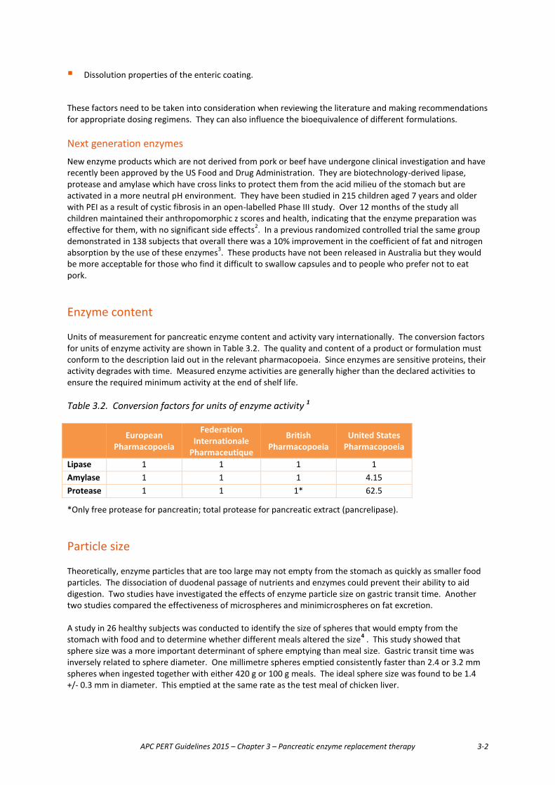
Pancreatic enzyme formulations Creon® is a porcine pancreatic enzyme extract encapsulated in minimicrospheres with a pH-sensitive coating. The minimicrospheres are similar in size to food particles to enable them to mix homogenously with the chyme (0.7-1.6 mm diameter). As of October 2014, Creon is available in capsules of three different strengths (Table 3.1). The 5,000u dose of Creon capsules is no longer available, but Creon enteric-coated granules are available as 5,000 BP units lipase in each 100mg scoop (called Creon Micro). Table 3.1: Minimum enzyme activities in each Creon product1 Creon 10,000
per capsule Creon 25,000 per capsule
Creon 40,000 per capsule
Creon Micro granules per 20g scoop
Lipase (BP units) 10,000 25,000 40,000 5,000
Amylase (BP units) 8,000 18,000 25,000 3,600
Protease (Ph Eur Units) 600 1,000 1,600 200
Panzytrat® 25000 is a porcine pancreatic enzyme preparation of encapsulated enteric-coated microtablets. The microtablets are uniform in size and shaped for maximum contact surface area (2 mm diameter convex spheres of thickness 1.90-2.10 mm). Each capsule contains no less than lipase 25,000 BP units, amylase 22,500 BP units and protease 1,250 Ph Eur Units. Both preparations contain a pH-sensitive coating to allow the enzymes to mix with the chyme while being protected from inactivation by gastric acid. The intact enzymes then pass into the alkaline pH of the duodenum where the enteric coating rapidly dissolves and the enzymes are released. Several factors influence the effectiveness of pancreatic enzyme replacement therapy: � Variations in enzyme content
� Size of the enzyme particles
APC PERT Guidelines 2015 – Chapter 3 – Pancreatic enzyme replacement therapy 3-2
� Dissolution properties of the enteric coating.
These factors need to be taken into consideration when reviewing the literature and making recommendations for appropriate dosing regimens. They can also influence the bioequivalence of different formulations. Next generation enzymes New enzyme products which are not derived from pork or beef have undergone clinical investigation and have recently been approved by the US Food and Drug Administration. They are biotechnology-derived lipase, protease and amylase which have cross links to protect them from the acid milieu of the stomach but are activated in a more neutral pH environment. They have been studied in 215 children aged 7 years and older with PEI as a result of cystic fibrosis in an open-labelled Phase III study. Over 12 months of the study all children maintained their anthropomorphic z scores and health, indicating that the enzyme preparation was effective for them, with no significant side effects2. In a previous randomized controlled trial the same group demonstrated in 138 subjects that overall there was a 10% improvement in the coefficient of fat and nitrogen absorption by the use of these enzymes3. These products have not been released in Australia but they would be more acceptable for those who find it difficult to swallow capsules and to people who prefer not to eat pork.
Enzyme content Units of measurement for pancreatic enzyme content and activity vary internationally. The conversion factors for units of enzyme activity are shown in Table 3.2. The quality and content of a product or formulation must conform to the description laid out in the relevant pharmacopoeia. Since enzymes are sensitive proteins, their activity degrades with time. Measured enzyme activities are generally higher than the declared activities to ensure the required minimum activity at the end of shelf life. Table 3.2. Conversion factors for units of enzyme activity 1
European Pharmacopoeia
Federation Internationale
Pharmaceutique
British Pharmacopoeia
United States Pharmacopoeia
Lipase 1 1 1 1 Amylase 1 1 1 4.15 Protease 1 1 1* 62.5
*Only free protease for pancreatin; total protease for pancreatic extract (pancrelipase). Particle size Theoretically, enzyme particles that are too large may not empty from the stomach as quickly as smaller food particles. The dissociation of duodenal passage of nutrients and enzymes could prevent their ability to aid digestion. Two studies have investigated the effects of enzyme particle size on gastric transit time. Another two studies compared the effectiveness of microspheres and minimicrospheres on fat excretion. A study in 26 healthy subjects was conducted to identify the size of spheres that would empty from the stomach with food and to determine whether different meals altered the size4 . This study showed that sphere size was a more important determinant of sphere emptying than meal size. Gastric transit time was inversely related to sphere diameter. One millimetre spheres emptied consistently faster than 2.4 or 3.2 mm spheres when ingested together with either 420 g or 100 g meals. The ideal sphere size was found to be 1.4 +/- 0.3 mm in diameter. This emptied at the same rate as the test meal of chicken liver.
APC PERT Guidelines 2015 – Chapter 3 – Pancreatic enzyme replacement therapy 3-3
Another study compared the gastric transit time of 2 mm microspheres with 1.2 mm minimicrospheres in pancreatic-insufficient subjects with cystic fibrosis5 . Patients consumed 20 g of free oil in spaghetti meals or 20 g of oil emulsified in a milk meal. This study did not show a difference in gastric transit time between the two preparations. The effect of microspheres (1-2 mm diameter) and smaller minimicrospheres (0.7-1.25 mm diameter) on fat excretion and fat intake was evaluated in a double-blind, randomised, multicentre, crossover study6 . Twenty-three patients with chronic pancreatitis and faecal fat excretion of greater than 7.5 g/day during a placebo period were randomly assigned to receive the two treatments in random order. The results showed that the minimicrospheres were equally effective as microspheres in improving the coefficient of fat absorption. Similar results were obtained in a study of 24 cystic fibrosis patients.7 Patients took microspheres for 14 days and were then randomised to 28 days of microspheres followed by 28 days of minimicrospheres or vice versa. Stool fat (g/day) and coefficient of fat absorption were measured at the end of each treatment period, and both products were found to be therapeutically equivalent. Taken together, these results suggest that spheres with a diameter of 2 mm or less are mixed intragastrically with the meal and emptied intact into the duodenum within the chyme. However, a further decrease in sphere size may not be associated with greater clinical benefit.
Dissolution properties of the enteric coating All digestive enzymes are susceptible to acid degradation, especially lipase. Modern preparations protect enzymes from denaturation with a pH-sensitive enteric coating. The polymer coatings of these preparations are designed to release the enzymes when exposed to the higher pH environment of the duodenum. If enzyme release takes too long after exposure to the intestinal milieu, then the digestive action may be delayed. Therefore the physicochemical properties of the enteric coating are crucial for the efficacy of enzyme therapy. Duodenal pH is normally between 6 and 7, but after a meal, it drops to around 5.5. In vitro studies show that the coating of most preparations dissolves over a variable period of time at a pH 5.0-6.08-10. Most preparations show more than 90% dissolution within 30 minutes at pH greater than 6.0. These results suggest that even if enzyme preparations have equivalent enzyme content, they may not be equivalent with respect to their release of enzymatic activity.
Adjunct therapy for acid suppression The pH of the duodenum in patients with pancreatic disease may be even lower than normal due to bicarbonate deficiency.11 It has been shown that duodenal pH declines to less than 4 after 100 minutes postprandially in some patients with PEI due to chronic pancreatitis.12 This lower pH may impair the release of enteric coated pancreatic enzymes and reduce their effectiveness. In theory, acid suppression may improve fat absorption by providing a duodenal environment more conducive to efficient enzyme function. Several different classes of agents have been used to evaluate the role of acid suppression in the treatment of PEI (Table 3.3). In general, the results have been mixed, and there is limited evidence that these agents improve fat absorption in patients with PEI on enzyme therapy. However, they may be useful in patients who continue to experience symptoms of PEI, particularly steatorrhoea, despite enzyme therapy.
APC PERT Guidelines 2015 – Chapter 3 – Pancreatic enzyme replacement therapy 3-4
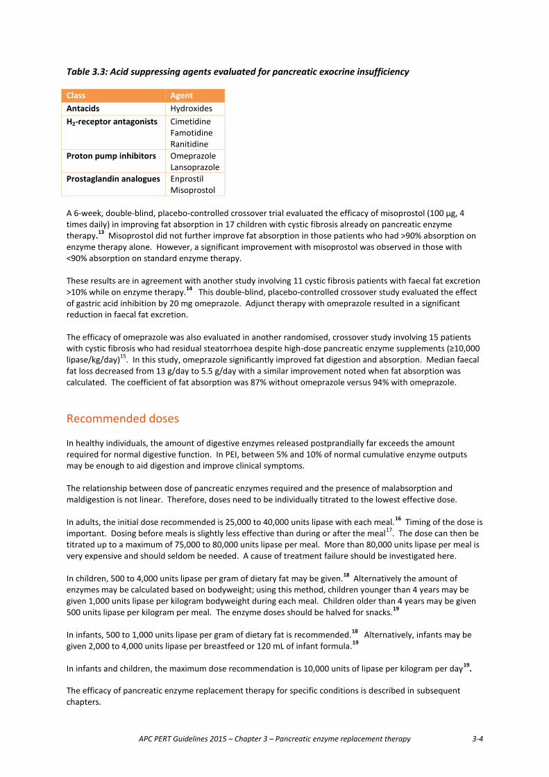
Table 3.3: Acid suppressing agents evaluated for pancreatic exocrine insufficiency Class Agent Antacids Hydroxides H2-receptor antagonists Cimetidine
Famotidine Ranitidine
Proton pump inhibitors Omeprazole Lansoprazole
Prostaglandin analogues Enprostil Misoprostol
A 6-week, double-blind, placebo-controlled crossover trial evaluated the efficacy of misoprostol (100 μg, 4 times daily) in improving fat absorption in 17 children with cystic fibrosis already on pancreatic enzyme therapy.13 Misoprostol did not further improve fat absorption in those patients who had >90% absorption on enzyme therapy alone. However, a significant improvement with misoprostol was observed in those with <90% absorption on standard enzyme therapy. These results are in agreement with another study involving 11 cystic fibrosis patients with faecal fat excretion >10% while on enzyme therapy.14 This double-blind, placebo-controlled crossover study evaluated the effect of gastric acid inhibition by 20 mg omeprazole. Adjunct therapy with omeprazole resulted in a significant reduction in faecal fat excretion. The efficacy of omeprazole was also evaluated in another randomised, crossover study involving 15 patients with cystic fibrosis who had residual steatorrhoea despite high-dose pancreatic enzyme supplements (≥10,000 lipase/kg/day)15. In this study, omeprazole significantly improved fat digestion and absorption. Median faecal fat loss decreased from 13 g/day to 5.5 g/day with a similar improvement noted when fat absorption was calculated. The coefficient of fat absorption was 87% without omeprazole versus 94% with omeprazole.
Recommended doses In healthy individuals, the amount of digestive enzymes released postprandially far exceeds the amount required for normal digestive function. In PEI, between 5% and 10% of normal cumulative enzyme outputs may be enough to aid digestion and improve clinical symptoms. The relationship between dose of pancreatic enzymes required and the presence of malabsorption and maldigestion is not linear. Therefore, doses need to be individually titrated to the lowest effective dose. In adults, the initial dose recommended is 25,000 to 40,000 units lipase with each meal.16 Timing of the dose is important. Dosing before meals is slightly less effective than during or after the meal17. The dose can then be titrated up to a maximum of 75,000 to 80,000 units lipase per meal. More than 80,000 units lipase per meal is very expensive and should seldom be needed. A cause of treatment failure should be investigated here. In children, 500 to 4,000 units lipase per gram of dietary fat may be given.18 Alternatively the amount of enzymes may be calculated based on bodyweight; using this method, children younger than 4 years may be given 1,000 units lipase per kilogram bodyweight during each meal. Children older than 4 years may be given 500 units lipase per kilogram per meal. The enzyme doses should be halved for snacks.19 In infants, 500 to 1,000 units lipase per gram of dietary fat is recommended.18 Alternatively, infants may be given 2,000 to 4,000 units lipase per breastfeed or 120 mL of infant formula.19 In infants and children, the maximum dose recommendation is 10,000 units of lipase per kilogram per day19. The efficacy of pancreatic enzyme replacement therapy for specific conditions is described in subsequent chapters.

APC PERT Guidelines 2015 – Chapter 3 – Pancreatic enzyme replacement therapy 3-5
Determination of response to treatment This is a difficult problem because it is not practical to undertake frequent 3-day faecal fat tests to determine the coefficients of fat absorption and nitrogen absorption (CFA and CAN), which are the gold standard outcome measures. Stool FE-1 measures are not appropriate to measure treatment outcome. Clinical evaluation can be subjective. Some use the 13C breath tests but they are not freely available. Some practitioners recommend increasing the dose if patients do not respond to treatment, which has some rationale because the total enzyme dose is relatively small compared to the normal enzyme output of a healthy pancreas. However other reasons for a poor response should be considered, such as timing of dose so that the enzymes mix with the food, and bacterial overgrowth. Complications of PERT � Fibrosing colonopathy has been reported in patients with cystic fibrosis taking high dose pancreatic
enzyme replacement therapy (REF). High dosing in children was considered a cause of fibrosing colonopathy but this is not thought to be a problem with modern medication.
� Use caution when prescribing PERT to patients with gout, renal impairment, or hyperuricaemia, and in patients with pork allergies.
� There is a theoretical risk of viral transmission with pancreatic enzyme products.
� PERT should always be taken with food and water. PERT should not be crushed or chewed. Concomitant taking of antacids may result in disruption of the coating of the granules and if there is an acidic area of the distant stomach it may destroy the enzymes.
� Some patients complain of abdominal pain and others complain of diarrhoea but the reason is not defined in the literature.
Summary � Pancreatic enzyme replacement therapy is the main pharmacological treatment for PEI. Modern
preparations contain pancreatic extract encapsulated in microtablets or (mini)microspheres with pH-sensitive enteric coating.
� The enzymes mix intragastrically with the chyme while being protected from acid degradation by the enteric coating. The enzymes are then emptied from the stomach simultaneously with the chyme. The higher pH in the duodenum dissolves the enteric coating, releasing the enzymes at the appropriate site for digestion and absorption.
� Not all pancreatic enzyme replacement agents are equivalent, although the therapeutic implications of the differences are not yet clear.

APC PERT Guidelines 2015 – Chapter 3 – Pancreatic enzyme replacement therapy 3-6
Recommendations
No. Recommendation Level of evidence Strength of agreement
3.1. Patients with PEI should be commenced on the lowest recommended dose of PERT (25,000-40,000 units lipase per meal), with a view to then titrating upwards according to clinical response.
3c (critical review)
3.2 The dose of PERT should be increased if necessary and titrated against the presence of malabsorption to the lowest effective dose.
3c (critical review)
3.3 In adults, the maximum recommended dose of PERT is 75,000 to 80,000 units lipase with each meal.
3c (critical review)
3.4 Enzymes are most effective when given with the meal,
rather than before or after it. 2b
3.5 In infants and children, the maximum recommended
dose is 10,000 units lipase per kilogram per day, taken during meals.
5 (clinical guidelines)
3.6 Trial acid-suppressing agents in those patients who continue to experience symptoms of PEI despite high-dose PERT.
1b
References (1) Layer P, Keller J, Lankisch PG. Pancreatic enzyme replacement therapy. Curr Gastroenterol Rep 2001;
3(2):101-108.
(2) Borowitz D, Stevens C, Brettman LR, Campion M, Wilschanski M, Thompson H. Liprotamase long-term safety and support of nutritional status in pancreatic-insufficient cystic fibrosis. J Pediatr Gastroenterol Nutr 2012; 54(2):248-257.
(3) Borowitz D, Stevens C, Brettman LR, Campion M, Chatfield B, Cipolli M. International phase III trial of liprotamase efficacy and safety in pancreatic-insufficient cystic fibrosis patients. J Cyst Fibros 2011; 10(6):443-452.
(4) Meyer JH, Elashoff J, Porter-Fink V, Dressman J, Amidon GL. Human postprandial gastric emptying of 1-3-millimeter spheres. Gastroenterology 1988; 94(6):1315-1325.
(5) Meyer JH, Lake R. Mismatch of duodenal deliveries of dietary fat and pancreatin from enterically coated microspheres. Pancreas 1997; 15(3):226-235.
(6) Halm U, Loser C, Lohr M, Katschinski M, Mossner J. A double-blind, randomized, multicentre, crossover study to prove equivalence of pancreatin minimicrospheres versus microspheres in exocrine pancreatic insufficiency. Aliment Pharmacol Ther 1999; 13(7):951-957.
APC PERT Guidelines 2015 – Chapter 3 – Pancreatic enzyme replacement therapy 3-7
(7) Patchell CJ, Desai M, Weller PH, Macdonald A, Smyth RL, Bush A et al. Creon 10,000 Minimicrospheres vs. Creon 8,000 microspheres--an open randomised crossover preference study. J Cyst Fibros 2002; 1(4):287-291.
(8) Gan KH, Geus WP, Bakker W, Lamers CB, Heijerman HG. In vitro dissolution profiles of enteric-coated microsphere/microtablet pancreatin preparations at different pH values. Aliment Pharmacol Ther 1996; 10(5):771-775.
(9) Case CL, Henniges F, Barkin JS. Enzyme content and acid stability of enteric-coated pancreatic enzyme products in vitro. Pancreas 2005; 30(2):180-183.
(10) Littlewood JM, Kelleher J, Walters MP, Johnson AW. In vivo and in vitro studies of microsphere pancreatic supplements. J Pediatr Gastroenterol Nutr 1988; 7 Suppl 1:S22-S29.
(11) Keller J, Layer P. Human pancreatic exocrine response to nutrients in health and disease. Gut 2005; 54 Suppl 6:vi1-28.
(12) DiMagno EP, Malagelada JR, Go VL, Moertel CG. Fate of orally ingested enzymes in pancreatic insufficiency. Comparison of two dosage schedules. N Engl J Med 1977; 296(23):1318-1322.
(13) Robinson P, Sly PD. Placebo-controlled trial of misoprostol in cystic fibrosis. J Pediatr Gastroenterol Nutr 1990; 11(1):37-40.
(14) Heijerman HG, Lamers CB, Bakker W, Dijkman JH. Improvement of fecal fat excretion after addition of omeprazole to pancrease in cystic fibrosis is related to residual exocrine function of the pancreas. Dig Dis Sci 1993; 38(1):1-6.
(15) Proesmans M, DeBoeck K. Omeprazole, a proton pump inhibitor, improves residual steatorrhoea in cystic fibrosis patients treated with high dose pancreatic enzymes. Eur J Pediatr 2003; 162(11):760-763.
(16) Dominguez-Munoz JE. Pancreatic enzyme therapy for pancreatic exocrine insufficiency. Curr Gastroenterol Rep 2007; 9(2):116-122.
(17) Dominguez-Munoz JE, Iglesias-Garcia J, Iglesias-Rey M, Figueiras A, Vilarino-Insua M. Effect of the administration schedule on the therapeutic efficacy of oral pancreatic enzyme supplements in patients with exocrine pancreatic insufficiency: a randomized, three-way crossover study. Aliment Pharmacol Ther 2005; 21(8):993-1000.
(18) Anthony H, Collins CE, Davidson G, Mews C, Robinson P, Shepherd R et al. Pancreatic enzyme replacement therapy in cystic fibrosis: Australian guidelines. Pediatric Gastroenterological Society and the Dietitians Association of Australia. J Paediatr Child Health 1999; 35(2):125-129.
(19) Baker SS. Delayed release pancrelipase for the treatment of pancreatic exocrine insufficiency associated with cystic fibrosis. Ther Clin Risk Manag 2008; 4(5):1079-1084.
APC PERT Guidelines 2015 – Chapter 4 – Dietary management of PEI 4-1
Chapter 4 Dietary management of PEI Introduction The management of patients with pancreatic exocrine insufficiency (PEI) is a complex issue. A thorough nutrition assessment is essential due to the weight loss and malnutrition risk in adults or lack of weight gain in children associated with malabsorption, and the potential for nutritional deficiency. The involvement of a dietitian to oversee dietary management is essential. The dietitian’s role is to make an initial assessment of the nutritional status of each patient, their weight status, macronutrient intake as well as any micronutrient deficiencies. Dietary advice can then be specifically tailored to improve each patient’s nutritional issue. The dietitian can also play a significant role in ongoing patient management by monitoring dietary and PERT compliance, nutritional deficiencies, weight status and steatorrhoea. Nutrition Assessment 1. Aetiology of PEI It is important to understand the underlying diagnosis and cause of PEI so nutritional management can address both the PEI and any specific needs associated with the disease state. Nutritional management differs according to different diagnoses, particularly in recommendations of meal size, frequency, and the potential need for nutritional supplementation. 2. Diet history A detailed diet history is essential to establish baseline diet. This enables an estimation of the patient’s total energy, fat and protein intake which can then answer some important questions regarding the dietary adequacy of each individual. From this assessment the fat content of the diet can be established and adjusted upwards if not consuming enough fat or has been following a low fat diet. The patient’s protein intake and total energy intake will determine whether sufficient calories are being consumed for weight gain and protein for muscle mass preservation. How the patient eats is important – do they eat three meals/day or smaller, more frequent meals which in general are better tolerated? Previous history of diets and/or food modification is important as it may explain why the patient is eating in their current pattern. An assessment of the patient’s understanding of PEI and why they are taking digestive enzymes enables a better understanding of where to direct nutritional recommendations and educate accordingly. It is also important to determine the patient’s independence regarding physical and cognitive function which can influence compliance with PERT. As alcohol inhibits gastric lipase secretion and therefore contributes to fat malabsorption1, an alcohol assessment is advised and zero alcohol intake is recommended. Patients may often tell their treating specialist they are alcohol-free but disclose it to the dietitian, which is crucial feedback to the specialist. A history should be taken of their current medication use, both prescribed medication and any vitamins or minerals that are consumed. 3. Malnutrition assessment Weight loss in adults or lack of weight gain in children is common in PEI due to malabsorption and the patient’s fear of eating. Maldigestion can occur before steatorrhoea is apparent and is associated with future weight loss2. Weight loss is usually gradual in chronic pancreatitis and develops in the early stages of the disease before steatorrhoea develops. In contrast, weight loss is rapid in pancreatic cancer. Up to 90% of patients with pancreatic cancer present with weight loss and malabsorption at the time of diagnosis2.
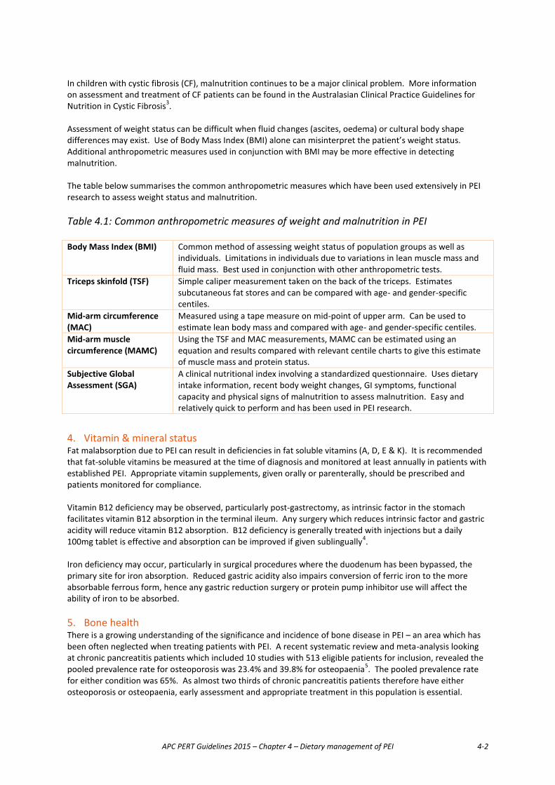
APC PERT Guidelines 2015 – Chapter 4 – Dietary management of PEI 4-2
In children with cystic fibrosis (CF), malnutrition continues to be a major clinical problem. More information on assessment and treatment of CF patients can be found in the Australasian Clinical Practice Guidelines for Nutrition in Cystic Fibrosis3. Assessment of weight status can be difficult when fluid changes (ascites, oedema) or cultural body shape differences may exist. Use of Body Mass Index (BMI) alone can misinterpret the patient’s weight status. Additional anthropometric measures used in conjunction with BMI may be more effective in detecting malnutrition. The table below summarises the common anthropometric measures which have been used extensively in PEI research to assess weight status and malnutrition. Table 4.1: Common anthropometric measures of weight and malnutrition in PEI Body Mass Index (BMI) Common method of assessing weight status of population groups as well as
individuals. Limitations in individuals due to variations in lean muscle mass and fluid mass. Best used in conjunction with other anthropometric tests.
Triceps skinfold (TSF) Simple caliper measurement taken on the back of the triceps. Estimates subcutaneous fat stores and can be compared with age- and gender-specific centiles.
Mid-arm circumference (MAC)
Measured using a tape measure on mid-point of upper arm. Can be used to estimate lean body mass and compared with age- and gender-specific centiles.
Mid-arm muscle circumference (MAMC)
Using the TSF and MAC measurements, MAMC can be estimated using an equation and results compared with relevant centile charts to give this estimate of muscle mass and protein status.
Subjective Global Assessment (SGA)
A clinical nutritional index involving a standardized questionnaire. Uses dietary intake information, recent body weight changes, GI symptoms, functional capacity and physical signs of malnutrition to assess malnutrition. Easy and relatively quick to perform and has been used in PEI research.
4. Vitamin & mineral status Fat malabsorption due to PEI can result in deficiencies in fat soluble vitamins (A, D, E & K). It is recommended that fat-soluble vitamins be measured at the time of diagnosis and monitored at least annually in patients with established PEI. Appropriate vitamin supplements, given orally or parenterally, should be prescribed and patients monitored for compliance. Vitamin B12 deficiency may be observed, particularly post-gastrectomy, as intrinsic factor in the stomach facilitates vitamin B12 absorption in the terminal ileum. Any surgery which reduces intrinsic factor and gastric acidity will reduce vitamin B12 absorption. B12 deficiency is generally treated with injections but a daily 100mg tablet is effective and absorption can be improved if given sublingually4. Iron deficiency may occur, particularly in surgical procedures where the duodenum has been bypassed, the primary site for iron absorption. Reduced gastric acidity also impairs conversion of ferric iron to the more absorbable ferrous form, hence any gastric reduction surgery or protein pump inhibitor use will affect the ability of iron to be absorbed. 5. Bone health There is a growing understanding of the significance and incidence of bone disease in PEI – an area which has been often neglected when treating patients with PEI. A recent systematic review and meta-analysis looking at chronic pancreatitis patients which included 10 studies with 513 eligible patients for inclusion, revealed the pooled prevalence rate for osteoporosis was 23.4% and 39.8% for osteopaenia5. The pooled prevalence rate for either condition was 65%. As almost two thirds of chronic pancreatitis patients therefore have either osteoporosis or osteopaenia, early assessment and appropriate treatment in this population is essential.

APC PERT Guidelines 2015 – Chapter 4 – Dietary management of PEI 4-3
The pathogenesis of low bone mineral density is likely to be multifactorial. Factors suggested include low vitamin D levels, poor dietary intake, reduced absorption, excess vitamin D faecal loss, smoking and low sunlight exposure5-7. Dual-energy X-ray absorptiometry (DXA) is the gold standard for measuring bone mineral density. The American Gastroenterological Association in its recommendations on osteoporosis in GI disease (IBD, coeliac disease and post-gastrectomy) recommended that patients with one or more known osteoporosis risk factors should undergo screening with DXA8. Whilst chronic pancreatitis and PEI was not included in these GI disease conditions, patients with these conditions should undergo the same screening protocol, given the incidence of bone disease in this group. A DXA scan should be performed once PEI is diagnosed, particularly in patients with another bone density risk factor such as post-menopausal status in women, those with previous low trauma fractures and men over 50 years old5. The scan should be repeated after two years5;9 and appropriate vitamin D and calcium supplementation initiated with referral to a bone specialist where appropriate. Nutrition intervention & PEI 1. Carbohydrate Requirement Carbohydrate digestion is not overly compromised in PEI due to ongoing digestion from salivary amylase and brush border enzymes which are independent of a compromised pancreas. The loss of endocrine function can lead to Impaired Glucose Tolerance (IGT) and eventually diabetes which require dietary modification to treat. IGT has been reported to occur in 40-90% of chronic pancreatitis patients, while 20-30% of cases require insulin therapy10. 2. Protein Requirement Like carbohydrate, protein digestion is successfully maintained in PEI. This is due to ongoing brush border peptide release. Severe cases of PEI which result in malnutrition may see a reduction in dietary protein intake which is more a supply issue rather than a digestion issue. Nutritional supplementation and or enteral feeding are sometimes required and are discussed separately, below. It is recommended that dietary protein intake meets 1.0-1.5g/kg body weight/day for adequacy11. 3. Fat requirement Fat digestion in the small intestine requires pancreatic lipase in combination with cofactors such as bile salts and colipase. Given there are no triglyceride digestive enzymes released from the small intestinal brush border, fat digestion is totally dependent on pancreatic enzymes. Fat digestion therefore remains the main nutritional consideration and issue in patients with PEI. Significant malabsorption of fat usually occurs when pancreatic lipase secretion falls below 10% of normal12 and steatorrhoea results. Historically dietary fat intake has been restricted in patients with PEI to minimise fat malabsorption and reduce steatorrhoea. Diets as low as 20g/day of fat have been prescribed1, however the concern has been that such diets are lower in total energy and fat soluble vitamins. More recently, studies have successfully used higher fat diets with adequate PERT with good results. A randomised, double blind, placebo-controlled trial showed that lipase-deficient chronic pancreatitis patients do not require a fat-restricted diet when adequate enzyme therapy is prescribed13. In this study patients consumed at least 100g of dietary fat/day. Low fat or reduced fat diets are not therefore recommended. A target of 30% total energy from dietary fat is now considered appropriate11 with an adequate enzyme prescription. A higher fat diet may be recommended by the dietitian for some patients who are having difficulty gaining weight or maintaining weight. Close attention to adverse symptoms such as steatorrhoea is recommended, particularly when using higher fat diets (i.e. >30% total energy). The use of medium chain triglycerides (MCT) has been recommended in the past as MCT do not require pancreatic enzymes or bile for absorption. However MCT have not shown any consistent benefits over long chain triglycerides when PERT is used to manage PEI. A randomised controlled trial showed that MCT-enriched

APC PERT Guidelines 2015 – Chapter 4 – Dietary management of PEI 4-4
commercial supplements offered no advantage over a homemade balanced diet for improving nutritional status of patients with chronic pancreatitis14. MCTs have also shown to have poor palatability and higher cost10.
4. Energy requirements Individual energy requirements can vary enormously but 30 kcal/kg body mass has been suggested in the Spanish Guidelines11. This appears a logical target but close attention to weight/BMI and nutritional progress may see this target increased in some cases. 5. Nutrition Support Tips for increasing energy intake
� Use full fat dairy products rather than reduced fat products � Use spreadable fats (e.g. butter, margarine, peanut butter, cream cheese, mayonnaise) wherever possible
on crackers, on vegetables, in mashed potato, in sandwiches. Mono- or polyunsaturated fats are preferred due to their cardiovascular benefits over saturated fats
� Fry meat, chicken, fish and vegetables in plenty of mono- or polyunsaturated oil or margarine � Enjoy high fat snacks such as nuts, seeds, cheese & crackers, dips, chips, cake and biscuits � Have high fat desserts after meals (e.g. cheesecake, puddings, ice cream, custard) � Enjoy nutritious low fat foods (fruit, dried fruit, vegetables, bread) but try to consume with additional fats
such as margarine, nuts, cream or butter � Make sure main meals include a generous portion of protein (e.g. meat, fish, chicken, eggs, tofu/vegetable
protein) � Make up fortified milk (add skim milk powder to fresh milk) to increase protein and energy, and use on
cereal, in tea and coffee, to make custards and desserts etc. Oral nutritional supplements In patients who are struggling to achieve their energy or protein requirements orally with their regular diet, oral polymeric high energy/protein nutritional supplements can successfully be used. Most commercial formulations contain fat so an adequate PERT dose is necessary, however some formulations are simply carbohydrate and protein-based so would not require PERT. In the chronic pancreatitis population, 10-15% of patients have been reported to require oral nutritional supplements10.
Enteral feeding When a patient requires enteral feeding either to supplement inadequate oral intake or for their sole source of nutrition and that patient cannot consume pancreatic enzymes orally, PERT needs to be administered via a feeding tube. In the chronic pancreatitis population, enteral nutrition has been reported to be necessary in 5% of cases10. In the absence of evidence-based guidelines, various practical strategies have been suggested for administering PERT via the enteral feeding tube15;16. Whatever the strategy chosen, best practice guidelines17;18 should also be adhered to in order to minimise tube occlusion, which is a common complication associated with enteral feeding19. The following are recommendations based on the available, yet limited evidence for situations in which PEI has been suspected or confirmed and enteral feeding considered appropriate: Formula selection
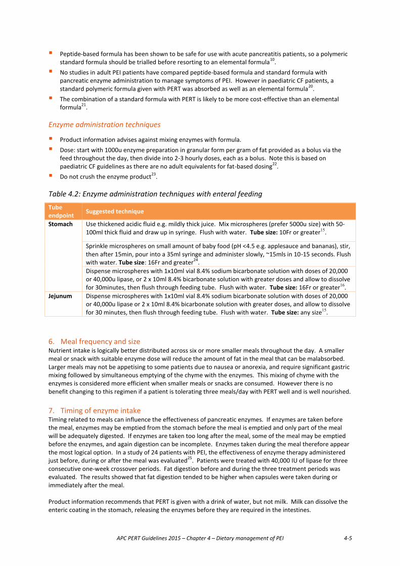
APC PERT Guidelines 2015 – Chapter 4 – Dietary management of PEI 4-5
� Peptide-based formula has been shown to be safe for use with acute pancreatitis patients, so a polymeric standard formula should be trialled before resorting to an elemental formula10.
� No studies in adult PEI patients have compared peptide-based formula and standard formula with pancreatic enzyme administration to manage symptoms of PEI. However in paediatric CF patients, a standard polymeric formula given with PERT was absorbed as well as an elemental formula20.
� The combination of a standard formula with PERT is likely to be more cost-effective than an elemental formula21.
Enzyme administration techniques
� Product information advises against mixing enzymes with formula. � Dose: start with 1000u enzyme preparation in granular form per gram of fat provided as a bolus via the
feed throughout the day, then divide into 2-3 hourly doses, each as a bolus. Note this is based on paediatric CF guidelines as there are no adult equivalents for fat-based dosing22.
� Do not crush the enzyme product23.
Table 4.2: Enzyme administration techniques with enteral feeding
Tube endpoint Suggested technique
Stomach Use thickened acidic fluid e.g. mildly thick juice. Mix microspheres (prefer 5000u size) with 50-100ml thick fluid and draw up in syringe. Flush with water. Tube size: 10Fr or greater15.
Sprinkle microspheres on small amount of baby food (pH <4.5 e.g. applesauce and bananas), stir, then after 15min, pour into a 35ml syringe and administer slowly, ~15mls in 10-15 seconds. Flush with water. Tube size: 16Fr and greater24. Dispense microspheres with 1x10ml vial 8.4% sodium bicarbonate solution with doses of 20,000 or 40,000u lipase, or 2 x 10ml 8.4% bicarbonate solution with greater doses and allow to dissolve for 30minutes, then flush through feeding tube. Flush with water. Tube size: 16Fr or greater16.
Jejunum Dispense microspheres with 1x10ml vial 8.4% sodium bicarbonate solution with doses of 20,000 or 40,000u lipase or 2 x 10ml 8.4% bicarbonate solution with greater doses, and allow to dissolve for 30 minutes, then flush through feeding tube. Flush with water. Tube size: any size15.
6. Meal frequency and size Nutrient intake is logically better distributed across six or more smaller meals throughout the day. A smaller meal or snack with suitable enzyme dose will reduce the amount of fat in the meal that can be malabsorbed. Larger meals may not be appetising to some patients due to nausea or anorexia, and require significant gastric mixing followed by simultaneous emptying of the chyme with the enzymes. This mixing of chyme with the enzymes is considered more efficient when smaller meals or snacks are consumed. However there is no benefit changing to this regimen if a patient is tolerating three meals/day with PERT well and is well nourished. 7. Timing of enzyme intake Timing related to meals can influence the effectiveness of pancreatic enzymes. If enzymes are taken before the meal, enzymes may be emptied from the stomach before the meal is emptied and only part of the meal will be adequately digested. If enzymes are taken too long after the meal, some of the meal may be emptied before the enzymes, and again digestion can be incomplete. Enzymes taken during the meal therefore appear the most logical option. In a study of 24 patients with PEI, the effectiveness of enzyme therapy administered just before, during or after the meal was evaluated25. Patients were treated with 40,000 IU of lipase for three consecutive one-week crossover periods. Fat digestion before and during the three treatment periods was evaluated. The results showed that fat digestion tended to be higher when capsules were taken during or immediately after the meal. Product information recommends that PERT is given with a drink of water, but not milk. Milk can dissolve the enteric coating in the stomach, releasing the enzymes before they are required in the intestines.
APC PERT Guidelines 2015 – Chapter 4 – Dietary management of PEI 4-6
After a total gastrectomy, administration of enzymes may need to be different given lack of gastric acid to help dissolve the capsule. Consider dispensing enteric coated microspheres in acidic liquid (e.g. a small amount of fruit juice) and consume on commencement of oral intake in order to allow enzymes to remain deactivated until reaching bile in the small bowel. 8. Nutrition monitoring & evaluation Nutrition monitoring and evaluation aims -
� To achieve normal nutritional status via adequate use of PERT � To equip the patient to be independent with appropriate use of PERT with diet.
The following are suggested-
� Regular patient reviews until stable � Checking for resolution of maldigestion and malabsorption (i.e. weight, bowel habits and consistency,
post-prandial symptoms) � Checking for compliance with medication � If limited progress, consider increasing the dose of PERT +/- Proton Pump Inhibitors and report back to
GP/Specialist prescribing PERT.
Some key points Statement Level of evidence Nearly two thirds of patients with CP have osteoporosis or osteopaenia 3a Routine nutritional assessment of patients with PEI is essential due to the potential impact of malabsorption on nutritional status and quality of life
5
MCT-enriched commercial supplements offer no advantage over a homemade balanced diet for improving nutritional status of patients with chronic pancreatitis14
1b
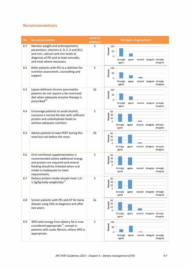
APC PERT Guidelines 2015 – Chapter 4 – Dietary management of PEI 4-7
Recommendations
No. Recommendation Level of evidence Strength of agreement
4.1 Monitor weight and anthropometric parameters, vitamins A, D, E, K and B12, and iron, calcium and zinc levels at diagnosis of PEI and at least annually, and treat where necessary.
5
4.2 Refer patients with PEI to a dietitian for
nutrition assessment, counselling and support.
5
4.3 Lipase-deficient chronic pancreatitis
patients do not require a fat-restricted diet when adequate enzyme therapy is prescribed13
1b
4.4 Encourage patients to avoid alcohol,
consume a normal fat diet with sufficient protein and carbohydrate foods to achieve adequate nutrition.
5
4.5 Advise patients to take PERT during the
meal but not before the meal. 2b
4.6 Oral nutritional supplementation is
recommended where additional energy and protein are required and enteral feeding should be initiated when oral intake is inadequate to meet requirements.
5
4.7 Dietary protein intake should meet 1.0 -
1.5g/kg body weight/day11. 5
4.8 Screen patients with PEI and CP for bone
disease using DXA at diagnosis and after two years.
2a
4.9 30% total energy from dietary fat is now
considered appropriate11, except in patients with cystic fibrosis, where 40% is appropriate.
5
APC PERT Guidelines 2015 – Chapter 4 – Dietary management of PEI 4-8
References
(1) Dominguez-Munoz JE. Pancreatic enzyme therapy for pancreatic exocrine insufficiency. Curr
Gastroenterol Rep 2007; 9(2):116-122.
(2) Dumasy V, Delhaye M, Cotton F, Deviere J. Fat malabsorption screening in chronic pancreatitis. Am J Gastroenterol 2004; 99(7):1350-1354.
(3) Dietitians Association of Australia. Australasian Clinical Practice Guidelines for Nutrition in Cystic Fibrosis. 2006.
(4) Stabler SP. Clinical practice. Vitamin B12 deficiency. N Engl J Med 2013; 368(2):149-160.
(5) Duggan SN, Smyth ND, Murphy A, Macnaughton D, O'Keefe SJ, Conlon KC. High prevalence of osteoporosis in patients with chronic pancreatitis: a systematic review and meta-analysis. Clin Gastroenterol Hepatol 2014; 12(2):219-228.
(6) Haaber AB, Rosenfalck AM, Hansen B, Hilsted J, Larsen S. Bone mineral metabolism, bone mineral density, and body composition in patients with chronic pancreatitis and pancreatic exocrine insufficiency. Int J Pancreatol 2000; 27(1):21-27.
(7) Duggan SN, Conlon KC. Bone health guidelines for patients with chronic pancreatitis. Gastroenterology 2013; 145(4):911.
(8) American Gastroenterological Association medical position statement: guidelines on osteoporosis in gastrointestinal diseases. Gastroenterology 2003; 124(3):791-794.
(9) Sikkens EC, Cahen DL, Koch AD, Braat H, Poley JW, Kuipers EJ et al. The prevalence of fat-soluble vitamin deficiencies and a decreased bone mass in patients with chronic pancreatitis. Pancreatology 2013; 13(3):238-242.
(10) Meier R, Ockenga J, Pertkiewicz M, Pap A, Milinic N, Macfie J et al. ESPEN Guidelines on Enteral Nutrition: Pancreas. Clin Nutr 2006; 25(2):275-284.
(11) de-Madaria E, Abad-Gonzalez A, Aparicio JR, Aparisi L, Boadas J, Boix E et al. The Spanish Pancreatic Club's recommendations for the diagnosis and treatment of chronic pancreatitis: part 2 (treatment). Pancreatology 2013; 13(1):18-28.
(12) Lindkvist B, Dominguez-Munoz JE, Luaces-Regueira M, Castineiras-Alvarino M, Nieto-Garcia L, Iglesias-Garcia J. Serum nutritional markers for prediction of pancreatic exocrine insufficiency in chronic pancreatitis. Pancreatology 2012; 12(4):305-310.
(13) Safdi M, Bekal PK, Martin S, Saeed ZA, Burton F, Toskes PP. The effects of oral pancreatic enzymes (Creon 10 capsule) on steatorrhea: a multicenter, placebo-controlled, parallel group trial in subjects with chronic pancreatitis. Pancreas 2006; 33(2):156-162.
(14) Singh S, Midha S, Singh N, Joshi YK, Garg PK. Dietary counseling versus dietary supplements for malnutrition in chronic pancreatitis: a randomized controlled trial. Clin Gastroenterol Hepatol 2008; 6(3):353-359.
(15) Ferrie S, Graham C, Hoyle M. Pancreatic enzyme supplementation for patients receiving enteral feeds. Nutr Clin Pract 2011; 26(3):349-351.
(16) Nicolo M, Stratton KW, Rooney W, Boullata J. Pancreatic enzyme replacement therapy for enterally fed patients with cystic fibrosis. Nutr Clin Pract 2013; 28(4):485-489.

APC PERT Guidelines 2015 – Chapter 4 – Dietary management of PEI 4-9
(17) Enteral Nutrition Manual for Adults in Health Care Facilities. 2011. Dietitians Association of Australia. 5-4-2014.
(18) Bankhead R, Boullata J, Brantley S, Corkins M, Guenter P, Krenitsky J et al. Enteral nutrition practice recommendations. JPEN J Parenter Enteral Nutr 2009; 33(2):122-167.
(19) Boullata JI. Drug administration through an enteral feeding tube. Am J Nurs 2009; 109(10):34-42.
(20) Erskine JM, Lingard CD, Sontag MK, Accurso FJ. Enteral nutrition for patients with cystic fibrosis: comparison of a semi-elemental and nonelemental formula. J Pediatr 1998; 132(2):265-269.
(21) Makola D, Krenitsky J, Parrish CR. Enteral feeding in acute and chronic pancreatitis. Gastrointest Endosc Clin N Am 2007; 17(4):747-764.
(22) Anthony H, Collins CE, Davidson G, Mews C, Robinson P, Shepherd R et al. Pancreatic enzyme replacement therapy in cystic fibrosis: Australian guidelines. Pediatric Gastroenterological Society and the Dietitians Association of Australia. J Paediatr Child Health 1999; 35(2):125-129.
(23) Mitchell JF, Pawlicki KS. Oral solid dosage forms that should not be crushed: 1994 revision. Hosp Pharm 1994; 29(7):666-5.
(24) Shlieout G, Koerner A, Maffert M, Forssmann K, Caras S. Administration of CREON(R) pancrelipase pellets via gastrostomy tube is feasible with no loss of gastric resistance or lipase activity: an in vitro study. Clin Drug Investig 2011; 31(7):e1-e7.
(25) Dominguez-Munoz JE, Iglesias-Garcia J, Iglesias-Rey M, Figueiras A, Vilarino-Insua M. Effect of the administration schedule on the therapeutic efficacy of oral pancreatic enzyme supplements in patients with exocrine pancreatic insufficiency: a randomized, three-way crossover study. Aliment Pharmacol Ther 2005; 21(8):993-1000.

APC PERT Guidelines 2015 – Chapter 5 – Acute pancreatitis and PERT 5-1
Chapter 5 Acute pancreatitis and the use of PERT
Introduction Acute pancreatitis is a common inflammatory condition triggered by the unregulated activation of trypsin within pancreatic acinar cells. Many aetiologies for acute pancreatitis exist, the most common being gallstones and alcohol abuse.1;2 In about 10% of cases, no cause is identifiable3. The incidence in Australia from a study from North Queensland was estimated to be 77 per 100,0004, which is consistent with incidence from Europe. Recently the classification of acute pancreatitis has evolved to Mild, Moderate and Severe, where Moderate represents cases with transient organ failure, local complications or exacerbation of co-morbid disease and Severe pancreatitis results in multi-organ failure5. Most episodes of acute pancreatitis are mild (without complication) and these patients are generally discharged from hospital within three days. Approximately 15-20% of patients require more than three days’ hospitalisation and about one third of these have very severe pancreatitis resulting in pancreatic and peri-pancreatic collections which are associated with systemic complications. Overall, the mortality in severe acute pancreatitis is between 5 and 15%6;7, but is higher in elderly patients with infected pancreatic necrosis and multisystem organ failure.8 Effect of acute pancreatitis on pancreatic exocrine function When the head of the pancreas is disrupted by pancreatic necrosis in severe pancreatitis, enzymes cannot flow into the duodenum. Such patients will have little effective enzymic digestion and are therefore clear candidates for long term enzyme replacement therapy. However in many, the pancreatic necrosis heals in a way where the pancreatic duct is preserved sufficiently to allow for recovery of enzyme production and flow. This process can be prolonged, but at three-year follow-up in one series, FE-1 testing showed that enzyme secretion had usually returned to normal9. Severe acute pancreatitis has a significant impact on a patient’s nutritional reserves which would be expected to be more slowly repaired if pancreatic function were impaired by interference with pancreatic secretion. Indeed, studies have demonstrated a slow recovery of physical function taking up to three years.10 Pancreatic function impairment after acute pancreatitis has mostly been studied using FE-1 testing because of convenience. In 2012 Xu et al showed that low FE-1 levels were more frequently seen after severe pancreatitis than mild pancreatitis (in 60.5 v. 39.5% of cases, respectively).11 In this study, patients requiring surgery had lower FE-1 levels but a background of chronic pancreatitis did not appear to be important. On the other hand, low levels of FE-1 have been used to distinguish patients with chronic pancreatitis from those with acute pancreatitis, with a sensitivity of 79.5% and a specificity of 98%.12 The aetiology of acute pancreatitis may be an important factor influencing pancreatic insufficiency but studies investigating this question are difficult to conduct and have been measured at different periods. One study demonstrated more severe pancreatic dysfunction resulting from an alcohol insult than from biliary pancreatitis13. This is explained by the greater likelihood of alcohol causing recurrent insults to the pancreas and a degree of pancreatic scarring that is not always evident when the patient presents. Similarly, alcoholic pancreatitis is more likely to recur some years after the acute event.14 When taken together, the data support the fact that some patients have pancreatic exocrine dysfunction for a period of time after an episode of acute pancreatitis. Pancreatic exocrine insufficiency occurs more frequently in patients with alcohol as the aetiological factor, in those recovering from severe episodes versus mild episodes and in those who developed necrosis or pseudocyst.15;16 It would appear reasonable to offer the malnourished group enzyme replacement therapy and to use the FE-1 test as a means of determining which patients have normal enzyme production and therefore could be excluded from treatment, bearing in mind

APC PERT Guidelines 2015 – Chapter 5 – Acute pancreatitis and PERT 5-2
the false positive rate of this test (see Chapter 2). It should be noted that some studies undertaken before 2004 demonstrated the onset of steatorrhoea but in no study where patients were randomised to receive enzyme treatment were nutritional outcomes measured, nor was it noted if steatorrhoea was reversed. The level of PEI may be insufficient to cause malabsorption. Early phase acute pancreatitis Limited data are available on exocrine pancreatic function in the early phase of acute pancreatitis. In a small case-control study, interdigestive pancreatic secretion was compared between eight patients with acute pancreatitis within 72 hours of symptom onset and 26 normal subjects using a duodenal intubation perfusion technique.17 The results showed that exocrine pancreatic secretion in the early phase of mild-to moderate acute pancreatitis remained within the normal range. In a more recent study of 75 patients experiencing their first episode of acute pancreatitis, FE-1 was determined on the day of re-feeding, which was on average 11.2 days after the attack.18 Abnormal FE-1 values were found in 9 of the 75 patients. The results were not significantly related to severity.
The convalescent phase of acute pancreatitis Impaired exocrine pancreatic function as determined by the para-aminobenzoic acid test (no longer used) has been demonstrated in the convalescent period after an episode of acute pancreatitis. Two to six months after the attack, four out of six patients tested still had abnormal results and improved such that only three of 15 patients had abnormal results after one year19. In a study of 54 patients with alcoholic acute pancreatitis who were evaluated by secretin-stimulated magnetic resonance pancreatography, baseline measures demonstrated exocrine insufficiency in 34% and this reduced to 9% by two years.20 In another study, 18 patients surviving at least one month after necrosectomy for acute necrotising pancreatitis, were monitored for steatorrhoea.21 At the time of discharge from hospital, 13 patients had PEI as determined by faecal fat excretion of greater than 7g/d. After six weeks, nine of these patients retained exocrine insufficiency that continued for six months. By 18 months, seven of these nine patients had resolution of symptoms, but steatorrhoea persisted in two patients. The pancreatic exocrine function of 75 patients who had a single attack of acute pancreatitis was studied by secretin/caerulein tube test or by an amino acid consumption test.16 Among the 36 patients with alcoholic pancreatitis, 29 had impaired pancreatic function between four and 18 months after the episode. Of the 39 patients with biliary pancreatitis, only nine had pancreatic insufficiency. When the tests were repeated one year later, 18 out of 23 patients with alcoholic pancreatitis continued to have pancreatic insufficiency, whereas only four out of 26 patients with biliary pancreatitis showed insufficiency. A prospective cohort study in 39 patients determined fat malabsorption following severe acute biliary pancreatitis and, further, evaluated the influence of necrosectomy on pancreatic exocrine function after 12 months.22 Most of the patients with necrosectomy had abnormal exocrine pancreatic function, with steatorrhoea in 25%. By comparison, exocrine function was abnormal in only 13.3% of patients who did not require surgery, with no cases of steatorrhoea. This may reflect that the patients with more severe pancreatitis required surgery. Another prospective study assessed pancreatic exocrine function using the FE-1 test in 23 patients recovering from a first attack of acute pancreatitis and evaluated its relationship to severity of attack and extent of pancreatic necrosis.15 Pancreatic exocrine insufficiency occurred in 6 out of 7 patients recovering from severe attacks compared with two of 16 patients recovering from mild attacks. All five patients who developed pancreatic necrosis or pseudocyst developed PEI whereas only three out of 18 patients who did not develop necrosis or pseudocyst had exocrine insufficiency. The development of PEI was strongly correlated with the extent of pancreatic necrosis. In contrast to the above studies, an examination of 63 patients with acute
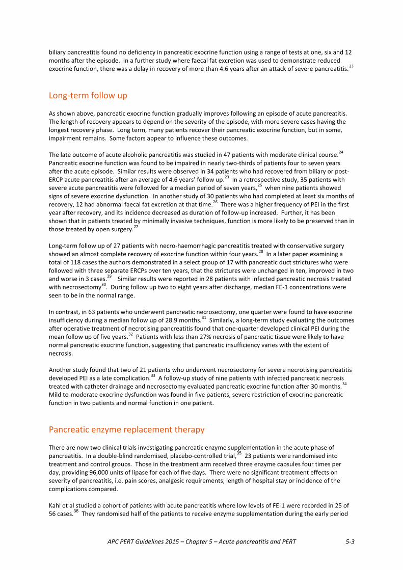
APC PERT Guidelines 2015 – Chapter 5 – Acute pancreatitis and PERT 5-3
biliary pancreatitis found no deficiency in pancreatic exocrine function using a range of tests at one, six and 12 months after the episode. In a further study where faecal fat excretion was used to demonstrate reduced exocrine function, there was a delay in recovery of more than 4.6 years after an attack of severe pancreatitis.23
Long-term follow up As shown above, pancreatic exocrine function gradually improves following an episode of acute pancreatitis. The length of recovery appears to depend on the severity of the episode, with more severe cases having the longest recovery phase. Long term, many patients recover their pancreatic exocrine function, but in some, impairment remains. Some factors appear to influence these outcomes. The late outcome of acute alcoholic pancreatitis was studied in 47 patients with moderate clinical course.24 Pancreatic exocrine function was found to be impaired in nearly two-thirds of patients four to seven years after the acute episode. Similar results were observed in 34 patients who had recovered from biliary or post-ERCP acute pancreatitis after an average of 4.6 years’ follow up.23 In a retrospective study, 35 patients with severe acute pancreatitis were followed for a median period of seven years,25 when nine patients showed signs of severe exocrine dysfunction. In another study of 30 patients who had completed at least six months of recovery, 12 had abnormal faecal fat excretion at that time.26 There was a higher frequency of PEI in the first year after recovery, and its incidence decreased as duration of follow-up increased. Further, it has been shown that in patients treated by minimally invasive techniques, function is more likely to be preserved than in those treated by open surgery.27 Long-term follow up of 27 patients with necro-haemorrhagic pancreatitis treated with conservative surgery showed an almost complete recovery of exocrine function within four years.28 In a later paper examining a total of 118 cases the authors demonstrated in a select group of 17 with pancreatic duct strictures who were followed with three separate ERCPs over ten years, that the strictures were unchanged in ten, improved in two and worse in 3 cases.29 Similar results were reported in 28 patients with infected pancreatic necrosis treated with necrosectomy30. During follow up two to eight years after discharge, median FE-1 concentrations were seen to be in the normal range. In contrast, in 63 patients who underwent pancreatic necrosectomy, one quarter were found to have exocrine insufficiency during a median follow up of 28.9 months.31 Similarly, a long-term study evaluating the outcomes after operative treatment of necrotising pancreatitis found that one-quarter developed clinical PEI during the mean follow up of five years.32 Patients with less than 27% necrosis of pancreatic tissue were likely to have normal pancreatic exocrine function, suggesting that pancreatic insufficiency varies with the extent of necrosis. Another study found that two of 21 patients who underwent necrosectomy for severe necrotising pancreatitis developed PEI as a late complication.33 A follow-up study of nine patients with infected pancreatic necrosis treated with catheter drainage and necrosectomy evaluated pancreatic exocrine function after 30 months.34 Mild to-moderate exocrine dysfunction was found in five patients, severe restriction of exocrine pancreatic function in two patients and normal function in one patient. Pancreatic enzyme replacement therapy There are now two clinical trials investigating pancreatic enzyme supplementation in the acute phase of pancreatitis. In a double-blind randomised, placebo-controlled trial,35 23 patients were randomised into treatment and control groups. Those in the treatment arm received three enzyme capsules four times per day, providing 96,000 units of lipase for each of five days. There were no significant treatment effects on severity of pancreatitis, i.e. pain scores, analgesic requirements, length of hospital stay or incidence of the complications compared. Kahl et al studied a cohort of patients with acute pancreatitis where low levels of FE-1 were recorded in 25 of 56 cases.36 They randomised half of the patients to receive enzyme supplementation during the early period
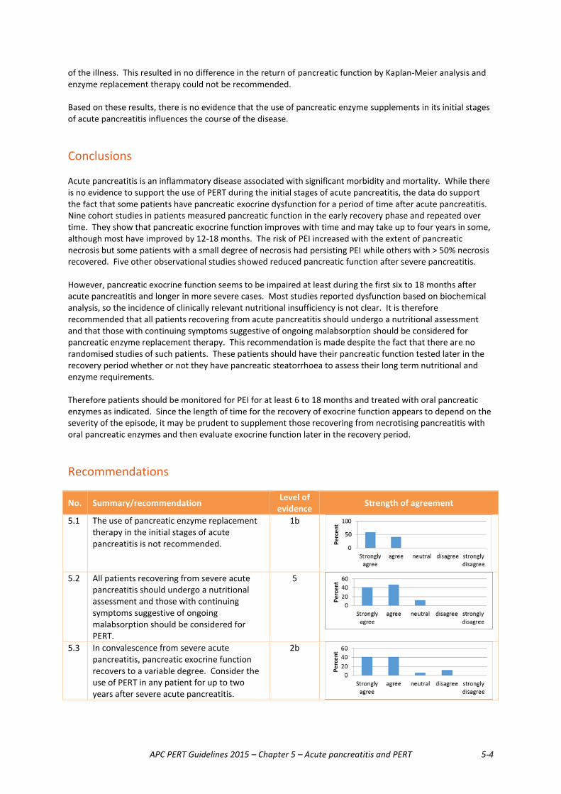
APC PERT Guidelines 2015 – Chapter 5 – Acute pancreatitis and PERT 5-4
of the illness. This resulted in no difference in the return of pancreatic function by Kaplan-Meier analysis and enzyme replacement therapy could not be recommended. Based on these results, there is no evidence that the use of pancreatic enzyme supplements in its initial stages of acute pancreatitis influences the course of the disease. Conclusions Acute pancreatitis is an inflammatory disease associated with significant morbidity and mortality. While there is no evidence to support the use of PERT during the initial stages of acute pancreatitis, the data do support the fact that some patients have pancreatic exocrine dysfunction for a period of time after acute pancreatitis. Nine cohort studies in patients measured pancreatic function in the early recovery phase and repeated over time. They show that pancreatic exocrine function improves with time and may take up to four years in some, although most have improved by 12-18 months. The risk of PEI increased with the extent of pancreatic necrosis but some patients with a small degree of necrosis had persisting PEI while others with > 50% necrosis recovered. Five other observational studies showed reduced pancreatic function after severe pancreatitis. However, pancreatic exocrine function seems to be impaired at least during the first six to 18 months after acute pancreatitis and longer in more severe cases. Most studies reported dysfunction based on biochemical analysis, so the incidence of clinically relevant nutritional insufficiency is not clear. It is therefore recommended that all patients recovering from acute pancreatitis should undergo a nutritional assessment and that those with continuing symptoms suggestive of ongoing malabsorption should be considered for pancreatic enzyme replacement therapy. This recommendation is made despite the fact that there are no randomised studies of such patients. These patients should have their pancreatic function tested later in the recovery period whether or not they have pancreatic steatorrhoea to assess their long term nutritional and enzyme requirements. Therefore patients should be monitored for PEI for at least 6 to 18 months and treated with oral pancreatic enzymes as indicated. Since the length of time for the recovery of exocrine function appears to depend on the severity of the episode, it may be prudent to supplement those recovering from necrotising pancreatitis with oral pancreatic enzymes and then evaluate exocrine function later in the recovery period. Recommendations
No. Summary/recommendation Level of evidence Strength of agreement
5.1 The use of pancreatic enzyme replacement therapy in the initial stages of acute pancreatitis is not recommended.
1b
5.2 All patients recovering from severe acute
pancreatitis should undergo a nutritional assessment and those with continuing symptoms suggestive of ongoing malabsorption should be considered for PERT.
5
5.3 In convalescence from severe acute pancreatitis, pancreatic exocrine function recovers to a variable degree. Consider the use of PERT in any patient for up to two years after severe acute pancreatitis.
2b
APC PERT Guidelines 2015 – Chapter 5 – Acute pancreatitis and PERT 5-5
No. Summary/recommendation Level of evidence Strength of agreement
5.4 Long-term follow-up of patients after severe acute pancreatitis should include monitoring for PEI for up to six years.
2b
Review process 2,002 papers referring to acute pancreatitis published between 2005 and 2014 were reviewed for reference to the influence of acute pancreatitis on exocrine function or the use of pancreatic enzyme therapy. These were combined with the publications in the previous guidelines document.
References
(1) Vidarsdottir H, Moller PH, Vidarsdottir H, Thorarinsdottir H, Bjornsson ES. Acute pancreatitis: a prospective study on incidence, etiology, and outcome. Eur J Gastroenterol Hepatol 2013; 25(9):1068-1075.
(2) Yadav D, Lowenfels AB. The epidemiology of pancreatitis and pancreatic cancer. Gastroenterology 2013; 144(6):1252-1261.
(3) Maes B, Hastier P, Buckley MJ, Peten EP, Paolini O, Staccini P et al. Extensive aetiological investigations in acute pancreatitis: results of a 1-year prospective study. Eur J Gastroenterol Hepatol 1999; 11(8):891-896.
(4) Turner RC, D'Onise K, Wang Y. What is the 'real' admission rate of acute pancreatitis in a regional Australian population? Aust Health Rev 2013; 37(2):205-209.
(5) Banks PA, Bollen TL, Dervenis C, Gooszen HG, Johnson CD, Sarr MG et al. Classification of acute pancreatitis--2012: revision of the Atlanta classification and definitions by international consensus. Gut 2013; 62(1):102-111.
(6) Haghshenasskashani A, Laurence JM, Kwan V, Johnston E, Hollands MJ, Richardson AJ et al. Endoscopic necrosectomy of pancreatic necrosis: a systematic review. Surg Endosc 2011; 25(12):3724-3730.
(7) Davies AR, Morrison SS, Ridley EJ, Bailey M, Banks MD, Cooper DJ et al. Nutritional therapy in patients with acute pancreatitis requiring critical care unit management: a prospective observational study in Australia and New Zealand. Crit Care Med 2011; 39(3):462-468.
(8) Wu BU, Johannes RS, Sun X, Tabak Y, Conwell DL, Banks PA. The early prediction of mortality in acute pancreatitis: a large population-based study. Gut 2008; 57(12):1698-1703.
(9) Uomo G, Gallucci F, Madrid E, Miraglia S, Manes G, Rabitti PG. Pancreatic functional impairment following acute necrotizing pancreatitis: long-term outcome of a non-surgically treated series. Dig Liver Dis 2010; 42(2):149-152.
(10) Wright SE, Lochan R, Imrie K, Baker C, Nesbitt ID, Kilner AJ et al. Quality of life and functional outcome at 3, 6 and 12 months after acute necrotising pancreatitis. Intensive Care Med 2009; 35(11):1974-1978.
APC PERT Guidelines 2015 – Chapter 5 – Acute pancreatitis and PERT 5-6
(11) Xu Y, Wu D, Zeng Y, Wang X. Pancreatic exocrine function and morphology following an episode of acute pancreatitis. Pancreas 2012; 41(6):922-927.
(12) Turner RC, McDermott R. Using faecal elastase-1 to screen for chronic pancreatitis in patients admitted with acute pancreatitis. HPB (Oxford) 2006; 8(3):223-226.
(13) Migliori M, Pezzilli R, Tomassetti P, Gullo L. Exocrine pancreatic function after alcoholic or biliary acute pancreatitis. Pancreas 2004; 28(4):359-363.
(14) Lankisch PG, Breuer N, Bruns A, Weber-Dany B, Lowenfels AB, Maisonneuve P. Natural history of acute pancreatitis: a long-term population-based study. Am J Gastroenterol 2009; 104(11):2797-2805.
(15) Boreham B, Ammori BJ. A prospective evaluation of pancreatic exocrine function in patients with acute pancreatitis: correlation with extent of necrosis and pancreatic endocrine insufficiency. Pancreatology 2003; 3(4):303-308.
(16) Migliori M, Pezzilli R, Tomassetti P, Gullo L. Exocrine pancreatic function after alcoholic or biliary acute pancreatitis. Pancreas 2004; 28(4):359-363.
(17) Dominguez-Munoz JE, Pieramico O, Buchler M, Malfertheiner P. Exocrine pancreatic function in the early phase of human acute pancreatitis. Scand J Gastroenterol 1995; 30(2):186-191.
(18) Pezzilli R, Simoni P, Casadei R, Morselli-Labate AM. Exocrine pancreatic function during the early recovery phase of acute pancreatitis. Hepatobiliary Pancreat Dis Int 2009; 8(3):316-319.
(19) Mitchell CJ, Playforth MJ, Kelleher J, McMahon MJ. Functional recovery of the exocrine pancreas after acute pancreatitis. Scand J Gastroenterol 1983; 18(1):5-8.
(20) Pelli H, Lappalainen-Lehto R, Piironen A, Jarvinen S, Sand J, Nordback I. Pancreatic damage after the first episode of acute alcoholic pancreatitis and its association with the later recurrence rate. Pancreatology 2009; 9(3):245-251.
(21) Bavare C, Prabhu R, Supe A. Early morphological and functional changes in pancreas following necrosectomy for acute severe necrotizing pancreatitis. Indian J Gastroenterol 2004; 23(6):203-205.
(22) Sabater L, Pareja E, Aparisi L, Calvete J, Camps B, Sastre J et al. Pancreatic function after severe acute biliary pancreatitis: the role of necrosectomy. Pancreas 2004; 28(1):65-68.
(23) Symersky T, van HB, Masclee AA. The outcome of a long-term follow-up of pancreatic function after recovery from acute pancreatitis. JOP 2006; 7(5):447-453.
(24) Malecka-Panas E, Juszynski A, Wilamski E. Acute alcoholic pancreatitis does not lead to complete recovery. Mater Med Pol 1996; 28(2):64-68.
(25) Appelros S, Lindgren S, Borgstrom A. Short and long term outcome of severe acute pancreatitis. Eur J Surg 2001; 167(4):281-286.
(26) Gupta R, Wig JD, Bhasin DK, Singh P, Suri S, Kang M et al. Severe acute pancreatitis: the life after. J Gastrointest Surg 2009; 13(7):1328-1336.
(27) Rana SS, Bhasin DK, Rao C, Sharma R, Gupta R. Comparative evaluation of structural and functional changes in pancreas after endoscopic and surgical management of pancreatic necrosis. Ann Gastroenterol 2014; 27(2):162-166.
(28) Angelini G, Pederzoli P, Caliari S, Fratton S, Brocco G, Marzoli G et al. Long-term outcome of acute necrohemorrhagic pancreatitis. A 4-year follow-up. Digestion 1984; 30(3):131-137.
APC PERT Guidelines 2015 – Chapter 5 – Acute pancreatitis and PERT 5-7
(29) Angelini G, Cavallini G, Pederzoli P, Bovo P, Bassi C, Di F, V et al. Long-term outcome of acute pancreatitis: a prospective study with 118 patients. Digestion 1993; 54(3):143-147.
(30) Reszetow J, Hac S, Dobrowolski S, Stefaniak T, Wajda Z, Gruca Z et al. Biliary versus alcohol-related infected pancreatic necrosis: similarities and differences in the follow-up. Pancreas 2007; 35(3):267-272.
(31) Connor S, Raraty MG, Howes N, Evans J, Ghaneh P, Sutton R et al. Surgery in the treatment of acute pancreatitis--minimal access pancreatic necrosectomy. Scand J Surg 2005; 94(2):135-142.
(32) Tsiotos GG, Luque-De LE, Sarr MG. Long-term outcome of necrotizing pancreatitis treated by necrosectomy. Br J Surg 1998; 85(12):1650-1653.
(33) Tzovaras G, Parks RW, Diamond T, Rowlands BJ. Early and long-term results of surgery for severe necrotising pancreatitis. Dig Surg 2004; 21(1):41-46.
(34) Endlicher E, Volk M, Feuerbach S, Scholmerich J, Schaffler A, Messmann H. Long-term follow-up of patients with necrotizing pancreatitis treated by percutaneous necrosectomy. Hepatogastroenterology 2003; 50(54):2225-2228.
(35) Patankar RV, Chand R, Johnson CD. Pancreatic enzyme supplementation in acute pancreatitis. HPB Surg 1995; 8(3):159-162.
(36) Kahl S, Schutte K, Glasbrenner B, Mayerle J, Simon P, Henniges F et al. The effect of oral pancreatic enzyme supplementation on the course and outcome of acute pancreatitis: a randomized, double-blind parallel-group study. JOP 2014; 15(2):165-174.
APC PERT Guidelines 2015 – Chapter 6 – Chronic pancreatitis and PERT 6-1
Chapter 6 Chronic pancreatitis and the use of PERT Introduction Chronic pancreatitis is characterised by the persistent loss of pancreatic structure and function due to various aetiologies. It is distinguished by progressive and irreversible damage to both the exocrine and endocrine components of the gland1. Its reported incidence in industrialised countries ranges from 3.5 to 10 per 100,000 people2;3 . Aetiology Alcohol is considered the primary cause of chronic pancreatitis, and accounts for 60-70% of all cases2;4. However, its aetiology varies with geographic location. There is a body of evidence suggesting that alcoholic pancreatitis may begin as acute pancreatitis, with recurrent episodes of acute necroinflammation leading to irreversible loss of pancreatic structure and function (the “necrosis-fibrosis hypothesis”)2. The types and causes of chronic pancreatitis are listed in Table 6.1. Table 6.1: Classification of chronic pancreatitis
� Alcoholic
� Autoimmune - Idiopathic
� Due to trauma
� Inherited factors
� Congenital
� Due to hyperparathyroidism
� Hyperlipidaemic
� Due to cystic fibrosis
� Due to protein-energy malnutrition
In about 10-30% of chronic pancreatitis cases, no identifiable cause can be found and a diagnosis of idiopathic chronic pancreatitis is made3;4. However, ongoing (particularly genetic) research indicates that a significant percentage of these patients may have an identifiable cause for their condition, as described below.
Pathology The pathology of chronic pancreatitis is characterised by fibrosis, acinar loss, eventual loss of pancreatic islets, chronic inflammation and ductular changes (distortion and intraductular protein plugs and calculi). Superimposed on these chronic changes may be evidence of acute inflammation and fat necrosis, as well as pancreatic pseudocysts.
Inherited factors in pancreatitis A major breakthrough in our understanding of the pathogenesis of chronic pancreatitis occurred in 1996 when Whitcomb and colleagues reported a gain of function mutation in the cationic trypsinogen gene in a kindred with hereditary pancreatitis5. Since then, some other mutations in the trypsinogen gene have been described.
APC PERT Guidelines 2015 – Chapter 6 – Chronic pancreatitis and PERT 6-2
Additionally, a mutation in the serine protease inhibitor (SPINK 1) gene (possibly causing an increased propensity for auto-digestion) has been associated with idiopathic, tropical and autoimmune pancreatitis. This mutation is not thought to be primarily pathogenic but to have a disease modifier role6. A variety of CFTR (Cystic Fibrosis Transmembrane Regulator) mutations has been described in patients with idiopathic pancreatitis. Affected individuals do not have classical cystic fibrosis (CF) and should not be regarded as having CF. They usually possess two copies of “mild” CFTR mutations or one “severe” copy and one “mild” copy7. Autoimmune pancreatitis This relatively recently described and rare entity typically presents as segmental or total pancreatic enlargement causing obstructive jaundice in an elderly male. The disease can present as acute pancreatitis but this is much less common. The main differential diagnosis is of pancreatic cancer. Histologically, there is diffuse periductal lymphoplasmacytic infiltration and fibrosis of the pancreas. Serum IgG4 levels are raised in 70-80% of cases. Other organs and tissues can be affected with lymphoplasmacytic infiltration and fibrosis including salivary glands, bile ducts, kidney and retroperitoneum. A second type of autoimmune pancreatitis has been described with a distinct histology and less involvement of other organs, although inflammatory bowel disease can be associated. In this type, serum IgG4 levels are not elevated8.
Symptoms The three cardinal clinical features of chronic pancreatitis are pain, maldigestion and diabetes3.
Abdominal pain is usually, but not always, the initial manifestation of chronic pancreatitis, typically presenting as acute discrete episodes initially and then becoming more chronic with time. Typically, the pain is epigastric or periumbilical in location and may radiate to the back and into the chest or flanks. Although recurrent or continuous pain is considered an important symptom of chronic pancreatitis, a subgroup of patients may have no pain at all, presenting instead with other symptoms including steatorrhoea (maldigestion). Maldigestion is a relatively late manifestation of chronic pancreatitis. It does not become clinically evident until digestive enzyme output is reduced to less than 10% of normal secretion9. In general, maldigestion of fat occurs earlier than that of carbohydrate or proteins because secretion of lipase decreases more rapidly than that of amylase or proteases10. As such, steatorrhoea is the predominant manifestation of maldigestion, with overt cases occurring in about 30% of patients with chronic pancreatitis11. Steatorrhoea may be associated with weight loss, with deficiencies of fat soluble vitamins (A, D, E and K) and osteoporosis.
Diagnosis The diagnosis of chronic pancreatitis relies on relevant symptoms, imaging of pancreatic structure and assessment of pancreatic function. In general, advanced stages of chronic pancreatitis may be easily diagnosed by imaging procedures whereas diagnosis of early disease presents a considerable challenge. Ideally, histology i s the gold standard for the diagnosis of chronic pancreatitis, but it is difficult to justify biopsies to obtain pancreatic tissue in clinical practice. Diagnosis based on imaging The next best diagnostic methods to demonstrate changes consistent with chronic pancreatitis are computed axial tomography (CT), magnetic resonance cholangiopancreatography (MRCP), endoscopic retrograde

APC PERT Guidelines 2015 – Chapter 6 – Chronic pancreatitis and PERT 6-3
cholangiopancreatography (ERCP) and endoscopic ultrasonography (EUS). Twenty to thirty years ago, ERCP was the definitive method for delineating pancreatic anatomy (particularly ductal anatomy) but it is an invasive test carrying a risk of post-ERCP pancreatitis. Times have changed with advances in CT and magnetic resonance technology. The modern helical CT scan now is the preferred method for imaging the size and shape of the pancreas, fluid collections, pseudocysts, pancreatic calcification and perhaps some ductal anatomy. MRCP is the preferred non-invasive method for delineating pancreatic ductal structure. ERCP is now reserved for those pancreatic imaging procedures where intervention such as stenting may be required. EUS is the emerging entity for pancreatic imaging. It has the potential for biopsy and the assessment of stiffness (elastography). However it is an invasive test susceptible to inter-observer variability, particularly in the diagnosis of chronic pancreatitis. Diagnosis based on pancreatic function Traditionally, the gold standard for the assessment of pancreatic function was a tube test whereby a tube was placed via the nose under fluoroscopic control so that its perforations lay in the second part of the duodenum. Pancreatic secretagogues (secretin, cholecystokinin) were administered intravenously and pancreatic secretions (enzymes, fluid, bicarbonate) were collected and measured. Obviously, this was/is a time- and resource-consuming test, dependent on the worldwide availability of secretagogues. As such, it is essentially confined to a few centres worldwide and none in Australia, and is no longer used routinely. Use of the tube test has been modified recently to collect pancreatic juice at the time of endoscopy. The performance of this endoscopic pancreatic function test (ePFT) correlates well with the direct tube test. The ePFT has a high negative predictive value, whereby a negative result indicates a high unlikelihood of a patient having or developing chronic pancreatitis. Given the cumbersome nature of direct assessment of pancreatic function, a number of indirect tests have been devised. The pancreolauryl and bentiromide tests rely on the use of pancreatic substrates and the measurement of metabolites in urine and/or serum. They are no longer in use. A number of centres have used ingested radiolabelled triglycerides with measurement of CO2 in breath; this test needs to be validated further in order to be applicable worldwide. Of increasing vogue are the measurements of pancreatic enzymes chymotrypsin and elastase [faecal elastase 1 (FE-1)] in faeces. FE-1 has been proven to be superior but the test’s value is limited by false positive results. A recent comprehensive review of the diagnosis of chronic pancreatitis has been published12. The biochemical, structural and functional parameters used to assist in the diagnosis of chronic pancreatitis are outlined in Table 6.2.
APC PERT Guidelines 2015 – Chapter 6 – Chronic pancreatitis and PERT 6-4
Table 6.2: Patient assessment for chronic pancreatitis Parameter Interpretation
Surgical biopsy The diagnostic gold standard of early stage disease, but rarely advisable in view of the risk of pancreatic damage.
Serum levels of lipase or amylase
Used to identify an acute episode of the disease in patients with pain Levels may be low in pancreatic insufficiency and may not be raised with acute flares of the disease Cannot be used in isolation to diagnose chronic pancreatitis due to low sensitivity.
Analysis of fat in stools Used for the diagnosis of steatorrhoea Alleviation of steatorrhoea with pancreatic enzymes suggests a pancreatic cause Most useful parameter for determining efficacy of treatment of maldigestion.
Abdominal x-ray Calcification on abdominal x-ray and associated steatorrhoea makes chronic pancreatitis likely.
Transabdominal ultrasound First procedure usually performed in patients with suspected chronic pancreatitis Operator-dependent More sensitive than abdominal x-ray for calcification Clarity of images often confounded by bowel gas.
Abdominal computed tomography
Widely used Useful for outlining pancreatic and surrounding anatomy Sensitive in the detection of pancreatic calcification Can detect ductular abnormalities although ERCP and MRCP are more sensitive Useful for detecting/excluding pancreatic neoplasms and cystic lesions.
Endoscopic retrograde cholangiopancreatography (ERCP)
Used for identification of ductular structural abnormalities. Most common imaging modality for planning intervention (endoscopic or surgical).
Magnetic resonance cholangiopancreatography (MRCP)
Is superseding ERCP as a non-invasive alternative. Useful in patients at high risk of developing post-ERCP pancreatitis or where the pancreatic duct is inaccessible as a result of surgery. Can outline ductal and parenchymal changes Can be combined with secretagogues to provide functional as well as structural information.
Endoscopic ultrasonography (EUS)
Possibly most sensitive procedure to detect chronic pancreatitis although analysis of accuracy is ongoing Unclear diagnostic role in early stage disease. Needle biopsy under EUS control may distinguish CP from pancreatic cancer.
All patients with painful established chronic pancreatitis should undergo an upper gastrointestinal endoscopy, abdominal CT scan and ERCP/MRCP in order to detect a potentially reversible cause of pain, such as peptic ulcer, pseudocyst or common bile duct stricture. Stages of chronic pancreatitis Chronic pancreatitis may be separated into four different stages based on disease presentation (Table 6.3). It is important to note that patients may not experience every stage of illness.

APC PERT Guidelines 2015 – Chapter 6 – Chronic pancreatitis and PERT 6-5
Table 6.3: Stages of chronic pancreatitis3
Stage Symptoms
I A pre-clinical stage with absent or uncharacteristic symptoms
II Recurrent acute episodes of pancreatitis without definite signs of chronic pancreatitis on imaging.
III Further recurrent episodes with intermittent or constant pain in between and signs of chronic pancreatitis on imaging.
IV Final stage mostly without acute flares and absence or decreased frequency of pain, possibly associated with evidence of endocrine and exocrine insufficiency.
Associated morbidity and mortality Up to 70% of patients with calcific pancreatitis develop diabetes2. Diabetes develops late in the disease course of chronic pancreatitis and therefore denotes advanced disease. There are exceptions to this dictum in patients with Type 2 diabetes associated with the metabolic syndrome. Chronic pancreatitis patients have an increased incidence of pancreatic cancer. The risk of developing pancreatic cancer is 16.5-fold higher in chronic pancreatitis patients than age- matched healthy controls13. Those with hereditary pancreatitis have an even higher risk of pancreatic cancer. The estimated cumulative risk of pancreatic cancer to age 70 years in patients with hereditary pancreatitis is as high as 40%14. There is also a five-fold increase in relative risk of developing pancreatic cancer in those with tropical pancreatitis15. Mortality in chronic pancreatitis, particularly alcoholic pancreatitis, is reported to be approximately 30% higher than that in the age- and sex-matched general population16. One fifth of this excess mortality can be directly attributed to pancreatitis itself. Most of the excess mortality is secondary to the effects of alcohol and/or smoking on the liver, the respiratory and digestive systems.
Pancreatic exocrine insufficiency Pancreatic exocrine insufficiency (PEI) due to chronic pancreatitis is a consequence of various factors which regulate digestion and absorption of nutrients17. Although pancreatic function has been extensively studied, some aspects of secretion and gastrointestinal adaptation are not well understood. However, it is known that a progressive loss of pancreatic acinar cell function in chronic pancreatitis does lead to deficiencies in the secretion of digestive enzymes from the pancreas. This results in maldigestion of nutrients. PEI after disease onset depends on the type of pancreatitis3. PEI develops earlier in alcoholic, tropical and late-onset idiopathic pancreatitis than early-onset idiopathic pancreatitis. Those with alcoholic pancreatitis generally develop PEI within 5-6 years of disease onset. Pancreatic exocrine and endocrine insufficiency is usually present at the time of presentation in 70% of patients with tropical pancreatitis. The main clinical manifestations of PEI are fat malabsorption (steatorrhoea), weight loss, abdominal discomfort and distention. Overt steatorrhoea occurs in about one-third of patients with chronic pancreatitis11. Fat malabsorption also results in a deficit of fat-soluble vitamins (A, D, E and K) with consequent clinical manifestations. Evidence clearly shows that malabsorption does occur in patients with chronic pancreatitis. Further, natural history studies indicate that the clinical diagnoses of steatorrhoea and exocrine insufficiency are usually not
APC PERT Guidelines 2015 – Chapter 6 – Chronic pancreatitis and PERT 6-6
evident until relatively late in the course of the disease18. Untreated malabsorption is related to many long-term harmful effects including weight loss4, malnutrition and osteoporosis18.
Management of PEI in chronic pancreatitis The general principles for the management of chronic pancreatitis are mainly to control symptoms, improve nutrition and treat complications3. These principles should be kept in mind when managing associated PEI.
Pancreatic enzyme replacement therapy (PERT) Although treatment of PEI with PERT is logical and is recommended, the evidence in the literature for the efficacy of PERT is not great. Shafiq et al19 performed a Cochrane review/meta-analysis of randomised controlled trials of the use of PERT in chronic pancreatitis. Ten trials involving 361 participants satisfied the inclusion criteria. Although some individual studies reported a beneficial effect of pancreatic enzyme over placebo in improving pain, incidence of steatorrhoea and analgesic consumption, the authors concluded that the role of pancreatic enzymes for abdominal pain, weight loss, steatorrhoea, analgesic use and quality of life in patients with chronic pancreatitis remains equivocal and that good quality, adequately powered (non-pharmaceutical) studies are needed. Since that review two multicentre double blind controlled trials have been published, one from North America20 and another from India21. These study groups had different demographics but they carefully determined the degree of pancreatic insufficiency before the randomisation by the use of measurement of the coefficient of fat absorption (CFA). They both demonstrated improvement in CFA but while flatulence and stool consistency improved with treatment pain was not improved. PERT in painful chronic pancreatitis The mechanisms for pain in chronic pancreatitis are poorly understood. They may include duct obstruction, tissue interstitial hypertension, pancreatic neuritis, pseudocyst formation, biliary obstruction and the presence of steatorrhoea with abdominal cramping. Although the evidence is complex and species-dependent, it appears that exocrine pancreatic secretion is subject to negative feedback regulation. A number of CCK-releasing factors have been identified in the intestine and as part of pancreatic secretion (such as monitor peptide). These releasing factors can be degraded by proteases thus inhibiting CCK release and pancreatic secretion and theoretically ameliorating the pain of chronic pancreatitis22. The discovery of this negative feedback loop led to trials of pancreatic proteases in the treatment of pain in chronic pancreatitis; unfortunately, a meta-analysis of these trials has failed to demonstrate benefit23. Since then, two prospective, multicentre, follow-up studies of patients with chronic pancreatitis have been conducted to assess quality of life before and after PERT24;25. PERT therapy reduced the extent of steatorrhoea and pain and was associated with a significant improvement in quality of life. The role of PERT in reducing pain in chronic pancreatitis remains unclear26. The American Gastroenterological Association recommends a trial of high-dose pancreatic enzymes coupled with acid suppression therapy before proceeding with continuous use of narcotics or invasive treatment27. Dosage and administration The relationship between the dose of pancreatic enzymes required and symptoms of maldigestion is not linear. For efficient digestion, it is essential that the concentration of enzymes delivered exogenously to the gut represents at least 5% of normal digestive enzyme output1. In general, the dose of lipase required with each meal is of the order of 25,000 to 50,000 units28. Dosing should be with, or immediately following each meal29.
In case of an inadequate response to therapy, compliance could be checked by measurement of faecal chymotrypsin, although this is not a standardised (nor generally practised) procedure30. In the compliant patient, it has been recommended that doses may be doubled or tripled, but other reasons for malabsorption
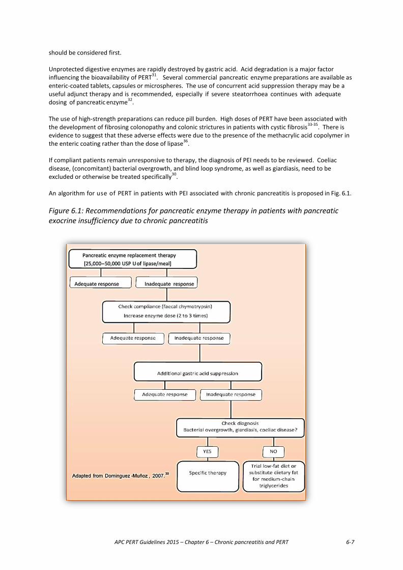
APC PERT Guidelines 2015 – Chapter 6 – Chronic pancreatitis and PERT 6-7
should be considered first. Unprotected digestive enzymes are rapidly destroyed by gastric acid. Acid degradation is a major factor influencing the bioavailability of PERT31. Several commercial pancreatic enzyme preparations are available as enteric-coated tablets, capsules or microspheres. The use of concurrent acid suppression therapy may be a useful adjunct therapy and is recommended, especially if severe steatorrhoea continues with adequate dosing of pancreatic enzyme32. The use of high-strength preparations can reduce pill burden. High doses of PERT have been associated with the development of fibrosing colonopathy and colonic strictures in patients with cystic fibrosis33-35. There is evidence to suggest that these adverse effects were due to the presence of the methacrylic acid copolymer in the enteric coating rather than the dose of lipase36.
If compliant patients remain unresponsive to therapy, the diagnosis of PEI needs to be reviewed. Coeliac disease, (concomitant) bacterial overgrowth, and blind loop syndrome, as well as giardiasis, need to be excluded or otherwise be treated specifically30. An algorithm for use of PERT in patients with PEI associated with chronic pancreatitis is proposed in Fig. 6.1. Figure 6.1: Recommendations for pancreatic enzyme therapy in patients with pancreatic exocrine insufficiency due to chronic pancreatitis

APC PERT Guidelines 2015 – Chapter 6 – Chronic pancreatitis and PERT 6-8
Summary � One of the most common causes of PEI is chronic pancreatitis. Alcohol is considered the primary
cause. The progressive loss of pancreatic parenchyma leads to impaired exocrine function. Maldigestion occurs in the majority of patients with chronic pancreatitis. However, the clinical diagnoses of steatorrhoea and exocrine insufficiency are usually not evident until relatively late in the course of the disease.
� It must be remembered that the presence of steatorrhoea, either proven or implied, is the foundation for initiating pancreatic enzyme replacement therapy (PERT). A decrease in pancreatic enzyme secretion detected by a sensitive direct measurement of pancreatic secretion does not by itself mandate the initiation of PERT. Steatorrhoea must be present. If the presence of steatorrhoea cannot be established, for whatever reason, by direct measurement of faecal fat, its presence may be inferred by the clinical diagnosis, imaging and patient characteristics, including suggestive changes in stool habit, weight loss, measured deficiencies in fat-soluble vitamins and osteoporosis.
� The efficacy of PERT may be influenced by a number of factors including type of preparation, enzyme concentration, dosage schedule and the use of adjuvant therapy to improve bioavailability of enzymes. For the treatment of PEI, 25,000 to 50,000 units of lipase are required with each meal. PERT should be taken preferably throughout or immediately following meals.
� In cases of an inadequate response to therapy, doses could be increased two- to three-fold. If severe steatorrhoea continues with adequate dosing of pancreatic enzyme, adjunctive acid suppression therapy is recommended. If patients remain unresponsive to therapy, other possible causes (e.g. bacterial overgrowth) should be considered.
� General dietary and nutritional considerations for patients with PEI due to other causes can be applicable to patients with chronic pancreatitis.
� Abstinence from alcohol cannot be overemphasised for patients with chronic pancreatitis. � Dietary counselling, coupled with PERT, in patients with chronic pancreatitis not only reduced the
extent of steatorrhoea and pain, but also significantly improved patients’ quality of life.
Recommendations for pancreatic enzyme replacement therapy in chronic pancreatitis
No. Recommendation Level of evidence Strength of agreement
6.1 PERT can improve the symptoms of PEI in patients with chronic pancreatitis 24;25
1b
6.2 PERT can improve quality of life in patients with chronic pancreatitis.24;25
1b
6.3 The required amount of lipase with each meal is generally 25,000 to 50,000 units of lipase BP.37
5
6.4 For severe, persisting steatorrhoea, consider the use
of adjunct acid suppressant therapy.27 4-5
6.5 Patients with chronic pancreatitis should abstain from
alcohol.3 3a
APC PERT Guidelines 2015 – Chapter 6 – Chronic pancreatitis and PERT 6-9
References
(1) DiMagno EP, et al. Chronic pancreatitis. The pancreas: biology, pathobiology and disease. New York:
Plenum Press; 1993. 665-706.
(2) Tandon RK, Sato N, Garg PK. Chronic pancreatitis: Asia-Pacific consensus report. J Gastroenterol Hepatol 2002; 17(4):508-518.
(3) Witt H, Apte MV, Keim V, Wilson JS. Chronic pancreatitis: challenges and advances in pathogenesis, genetics, diagnosis, and therapy. Gastroenterology 2007; 132(4):1557-1573.
(4) Dumasy V, Delhaye M, Cotton F, Deviere J. Fat malabsorption screening in chronic pancreatitis. Am J Gastroenterol 2004; 99(7):1350-1354.
(5) Whitcomb DC, Gorry MC, Preston RA, Furey W, Sossenheimer MJ, Ulrich CD et al. Hereditary pancreatitis is caused by a mutation in the cationic trypsinogen gene. Nat Genet 1996; 14(2):141-145.
(6) Threadgold J, Greenhalf W, Ellis I, Howes N, Lerch MM, Simon P et al. The N34S mutation of SPINK1 (PSTI) is associated with a familial pattern of idiopathic chronic pancreatitis but does not cause the disease. Gut 2002; 50(5):675-681.
(7) Cohn JA, Bornstein JD, Jowell PS. Cystic fibrosis mutations and genetic predisposition to idiopathic chronic pancreatitis. Med Clin North Am 2000; 84(3):621-31, ix.
(8) Kamisawa T, Chari ST, Lerch MM, Kim MH, Gress TM, Shimosegawa T. Recent advances in autoimmune pancreatitis: type 1 and type 2. Gut 2013; 62(9):1373-1380.
(9) DiMagno EP, Go VL, Summerskill WH. Relations between pancreatic enzyme ouputs and malabsorption in severe pancreatic insufficiency. N Engl J Med 1973; 288(16):813-815.
(10) DiMagno EP, Malagelada JR, Go VL. Relationship between alcoholism and pancreatic insufficiency. Ann N Y Acad Sci 1975; 252:200-207.
(11) Lankisch PG, Droge M, Hofses S, Konig H, Lembcke B. Steatorrhoea: you cannot trust your eyes when it comes to diagnosis. Lancet 1996; 347(9015):1620-1621.
(12) Sze KC, Pirola RC, Apte MV, Wilson JS. Current options for the diagnosis of chronic pancreatitis. Expert Rev Mol Diagn 2014; 14(2):199-215.
(13) Lowenfels AB, Maisonneuve P, Cavallini G, Ammann RW, Lankisch PG, Andersen JR et al. Pancreatitis and the risk of pancreatic cancer. International Pancreatitis Study Group. N Engl J Med 1993; 328(20):1433-1437.
(14) Lowenfels AB, Maisonneuve P, DiMagno EP, Elitsur Y, Gates LK, Jr., Perrault J et al. Hereditary pancreatitis and the risk of pancreatic cancer. International Hereditary Pancreatitis Study Group. J Natl Cancer Inst 1997; 89(6):442-446.
(15) Chari ST, Mohan V, Pitchumoni CS, Viswanathan M, Madanagopalan N, Lowenfels AB. Risk of pancreatic carcinoma in tropical calcifying pancreatitis: an epidemiologic study. Pancreas 1994; 9(1):62-66.
(16) Levy P, Milan C, Pignon JP, Baetz A, Bernades P. Mortality factors associated with chronic pancreatitis. Unidimensional and multidimensional analysis of a medical-surgical series of 240 patients. Gastroenterology 1989; 96(4):1165-1172.
(17) Pezzilli R. Chronic pancreatitis: maldigestion, intestinal ecology and intestinal inflammation. World J

APC PERT Guidelines 2015 – Chapter 6 – Chronic pancreatitis and PERT 6-10
Gastroenterol 2009; 15(14):1673-1676.
(18) Forsmark CE. Chronic pancreatitis and malabsorption. Am J Gastroenterol 2004; 99(7):1355-1357.
(19) Shafiq N, Rana S, Bhasin D, Pandhi P, Srivastava P, Sehmby SS et al. Pancreatic enzymes for chronic pancreatitis. Cochrane Database Syst Rev 2009;(4):CD006302.
(20) Whitcomb DC, Lehman GA, Vasileva G, Malecka-Panas E, Gubergrits N, Shen Y et al. Pancrelipase delayed-release capsules (CREON) for exocrine pancreatic insufficiency due to chronic pancreatitis or pancreatic surgery: A double-blind randomized trial. Am J Gastroenterol 2010; 105(10):2276-2286.
(21) Thorat V, Reddy N, Bhatia S, Bapaye A, Rajkumar JS, Kini DD et al. Randomised clinical trial: the efficacy and safety of pancreatin enteric-coated minimicrospheres (Creon 40000 MMS) in patients with pancreatic exocrine insufficiency due to chronic pancreatitis--a double-blind, placebo-controlled study. Aliment Pharmacol Ther 2012; 36(5):426-436.
(22) Liddle RA. Regulation of pancreatic secretion. In: Johnson LR, editor. Physiology of the Digestive Tract. London: Academic Press; 2012. 1425.
(23) Brown A, Hughes M, Tenner S, Banks PA. Does pancreatic enzyme supplementation reduce pain in patients with chronic pancreatitis: a meta-analysis. Am J Gastroenterol 1997; 92(11):2032-2035.
(24) Czako L, Takacs T, Hegyi P, Pronai L, Tulassay Z, Lakner L et al. Quality of life assessment after pancreatic enzyme replacement therapy in chronic pancreatitis. Can J Gastroenterol 2003; 17(10):597-603.
(25) D'Haese JG, Ceyhan GO, Demir IE, Layer P, Uhl W, Lohr M et al. Pancreatic enzyme replacement therapy in patients with exocrine pancreatic insufficiency due to chronic pancreatitis: a 1-year disease management study on symptom control and quality of life. Pancreas 2014; 43(6):834-841.
(26) Warshaw AL, Banks PA, Fernandez-Del CC. AGA technical review: treatment of pain in chronic pancreatitis. Gastroenterology 1998; 115(3):765-776.
(27) American Gastroenterological Association Medical Position Statement: treatment of pain in chronic pancreatitis. Gastroenterology 1998; 115(3):763-764.
(28) Ferrone M, Raimondo M, Scolapio JS. Pancreatic enzyme pharmacotherapy. Pharmacotherapy 2007; 27(6):910-920.
(29) Dominguez-Munoz JE, Iglesias-Garcia J, Iglesias-Rey M, Figueiras A, Vilarino-Insua M. Effect of the administration schedule on the therapeutic efficacy of oral pancreatic enzyme supplements in patients with exocrine pancreatic insufficiency: a randomized, three-way crossover study. Aliment Pharmacol Ther 2005; 21(8):993-1000.
(30) Keller J, Layer P. Pancreatic Enzyme Supplementation Therapy. Curr Treat Options Gastroenterol 2003; 6(5):369-374.
(31) Dominguez-Munoz JE. Pancreatic enzyme therapy for pancreatic exocrine insufficiency. Curr Gastroenterol Rep 2007; 9(2):116-122.
(32) Marotta F, O'Keefe SJ, Marks IN, Girdwood A, Young G. Pancreatic enzyme replacement therapy. Importance of gastric acid secretion, H2-antagonists, and enteric coating. Dig Dis Sci 1989; 34(3):456-461.
(33) Pawel BR, de Chadarevian JP, Franco ME. The pathology of fibrosing colonopathy of cystic fibrosis: a study of 12 cases and review of the literature. Hum Pathol 1997; 28(4):395-399.
APC PERT Guidelines 2015 – Chapter 6 – Chronic pancreatitis and PERT 6-11
(34) Smyth RL, Van VD, Smyth AR, Lloyd DA, Heaf DP. Strictures of ascending colon in cystic fibrosis and high-strength pancreatic enzymes. Lancet 1994; 343(8889):85-86.
(35) Stevens JC, Maguiness KM, Hollingsworth J, Heilman DK, Chong SK. Pancreatic enzyme supplementation in cystic fibrosis patients before and after fibrosing colonopathy. J Pediatr Gastroenterol Nutr 1998; 26(1):80-84.
(36) Prescott P, Bakowski MT. Pathogenesis of fibrosing colonopathy: the role of methacrylic acid copolymer. Pharmacoepidemiol Drug Saf 1999; 8(6):377-384.
(37) Ferrone M, Raimondo M, Scolapio JS. Pancreatic enzyme pharmacotherapy. Pharmacotherapy 2007; 27(6):910-920.
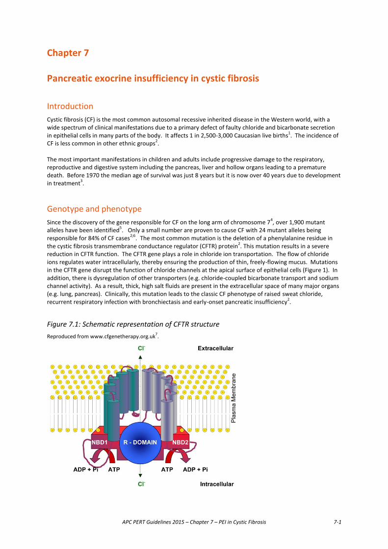
APC PERT Guidelines 2015 – Chapter 7 – PEI in Cystic Fibrosis 7-1
Chapter 7 Pancreatic exocrine insufficiency in cystic fibrosis Introduction Cystic fibrosis (CF) is the most common autosomal recessive inherited disease in the Western world, with a wide spectrum of clinical manifestations due to a primary defect of faulty chloride and bicarbonate secretion in epithelial cells in many parts of the body. It affects 1 in 2,500-3,000 Caucasian live births1. The incidence of CF is less common in other ethnic groups2. The most important manifestations in children and adults include progressive damage to the respiratory, reproductive and digestive system including the pancreas, liver and hollow organs leading to a premature death. Before 1970 the median age of survival was just 8 years but it is now over 40 years due to development in treatment3. Genotype and phenotype Since the discovery of the gene responsible for CF on the long arm of chromosome 74, over 1,900 mutant alleles have been identified5. Only a small number are proven to cause CF with 24 mutant alleles being responsible for 84% of CF cases2;6. The most common mutation is the deletion of a phenylalanine residue in the cystic fibrosis transmembrane conductance regulator (CFTR) protein2. This mutation results in a severe reduction in CFTR function. The CFTR gene plays a role in chloride ion transportation. The flow of chloride ions regulates water intracellularly, thereby ensuring the production of thin, freely-flowing mucus. Mutations in the CFTR gene disrupt the function of chloride channels at the apical surface of epithelial cells (Figure 1). In addition, there is dysregulation of other transporters (e.g. chloride-coupled bicarbonate transport and sodium channel activity). As a result, thick, high salt fluids are present in the extracellular space of many major organs (e.g. lung, pancreas). Clinically, this mutation leads to the classic CF phenotype of raised sweat chloride, recurrent respiratory infection with bronchiectasis and early-onset pancreatic insufficiency2.
Figure 7.1: Schematic representation of CFTR structure Reproduced from www.cfgenetherapy.org.uk7.
APC PERT Guidelines 2015 – Chapter 7 – PEI in Cystic Fibrosis 7-2
The clinical manifestations of CF can vary greatly between affected individuals8, and largely depend on the amount of normal CFTR function. A classification system based on the functional effect that genotype has on production of CFTR could indicate a biological mechanism by which genotype could affect mortality (Table 7.1). Patients with CFTR genotypes associated with severely reduced CFTR production (Class I to III) have a very similar severe phenotype and are associated with higher mortality rates than patients with a CFTR genotype associated with some residual CFTR function (Class IV and V)5. Table 7.1: Classification system of CFTR mutations based on their functional effects
Risk
stratification Class of
mutation
Functional effect of mutation
High risk of CFTR
genotype
I Defective protein production with premature termination of CFTR protein production. Produces few or no functioning CFTR chloride channels
II Defective trafficking of CFTR protein so that it does not reach the apical surface membrane where it is intended to function
III Defective regulation of CFTR protein even though it is able to reach the apical cell surface
Low risk of CFTR
genotype
IV CFTR protein reaches the apical surface but conduction through the channel is defective
V Associated with reduced synthesis of functional CFTR protein
Adapted from McKone et al. 20032. Diagnosis and screening About 80-100 new diagnoses of CF are made annually in Australasia9. In Australia and New Zealand, all newborn babies are screened for CF using the immunoreactive trypsinogen test (IRT). A blood sample is taken three days after birth and analysed for a specific protein called trypsinogen. If this is positive (showing raised serum trypsinogen), reflex genetic testing of the CFTR gene is done (the type of CFTR mutations tested varies with different newborn screening programs). In the presence of 1 or 2 CFTR mutations, a diagnostic sweat test is recommended to measure the sweat chloride concentration. About 95% of babies with CF are identified by newborn screening10. While newborn screening for CF is universal in Australia and New Zealand, this has not been the case in other parts of the world. In unscreened populations, diagnosis of CF is based on a clinical phenotype (or first-degree family history) accompanied by elevated sweat chloride and/or identification of two CF-causing mutations. Prognosis There are over 3,000 people living with CF in Australia9. Of these, two-thirds are children and adolescents. The long-term survival for children with CF has improved significantly in recent years, with a predicted mean life expectancy of about 40 years in many parts of the world including Australia3;11;12. The factors contributing to improved survival include improved management of respiratory infections and in nutritional status12;13.
Pancreatic function in CF patients The CFTR gene is expressed in many tissues including the pancreas. The pancreas is vulnerable to luminal concentration defects due to the high protein content in the ducts14. The viscous secretions can cause luminal obstruction of ducts leading to acinar cell destruction, fibrosis and pancreatic exocrine insufficiency (PEI)15. Ductal obstruction is also associated with the development of symptomatic acute pancreatitis in a small subset of patients with CF16.
APC PERT Guidelines 2015 – Chapter 7 – PEI in Cystic Fibrosis 7-3
Pancreatic function in CF patients is classified into two distinct phenotypes: 1. Pancreatic sufficient (PS): patients have adequate endogenous pancreatic exocrine function to provide
normal absorption 2. Pancreatic insufficient (PI): patients require exogenous pancreatic exocrine enzymes to maintain adequate
absorption of nutrients. Most CF patients are pancreatic insufficient either at the time of diagnosis or as the disease progresses. Only 5-15% of affected individuals retain some level of pancreatic function16. There is a clear correlation between genotype and pancreatic function14. Most patients with CF who carry Class I-III mutations on both alleles have a PI phenotype. Patients who carry a mild mutation (Class IV-V) on at least one allele, which confers some residual ion channel function, usually are PS. Thus, mutations that confer the PS phenotype do so in a dominant fashion16. A study of 78 CF patients identified in a newborn screening program showed that 37% were PS at the time of diagnosis. These children had growth that was close to normal and comparable to growth in children with PEI who received oral enzyme therapy. PEI subsequently developed in one in five of these patients at 3 to 36 months of age17. These data indicate that a proportion of PS infants can lose their residual pancreatic function over time, spontaneously. The development of symptomatic acute pancreatitis in PS CF patients is also a risk factor for the progression to PI status16. CF patients identified soon after birth should be continually monitored. Growth measurements and indirect tests for pancreatic exocrine function should be routinely conducted, with the latter particularly for those patients with PS CF.
Consequences of PEI in CF Although chronic pulmonary disease is the major cause of mortality in CF, significant morbidity is attributed to gastrointestinal dysfunction, particularly PEI. PEI results in the inability to properly digest food due to the lack of pancreatic digestive enzymes. Maldigestion, and hence malabsorption of nutrients and poor nutrition, due to PEI occurs in approximately 85% of CF patients18-20. Maldigestion in CF affects energy and protein availability. Without treatment, gastrointestinal losses of fat and nitrogen are severe. This causes energy and protein deficits leading to growth failure and protein catabolism20. Fat malabsorption is the most important digestive malfunction in PEI. This is because fat is the most energy-dense macronutrient and fat-soluble vitamins require normal or near normal processes of fat digestion for their absorption. Prolonged, untreated PEI is associated with a poorer prognosis long term. A study of 72 CF patients with normal fat absorption found that pancreatic sufficient patients generally have significantly better lung function, milder clinical symptoms, lower sweat chloride and fewer gut and liver complications than those with symptoms of PEI21. Benefits of good nutritional management Evidence in the literature demonstrates the importance of improving the nutritional status of CF patients. This was highlighted in a comparison of nutritional management policies in Canada and the United States22;23. Corey et al. conducted a study involving over 1,000 patients from two CF clinic populations in Boston and Toronto. Demographic data were not significantly different between the two populations. The Toronto clinic had abandoned the traditional low fat, high energy diet in favour of more liberal use of both fat and pancreatic enzyme replacement. Data showed that patients in Toronto tended to be taller, heavier and had a higher median age than those at the Boston clinic. Although progressive pulmonary disease is the major cause of mortality in cystic fibrosis, the differences in growth and survival in these two patient groups, with very similar age-specific pulmonary function, suggest the potential benefits of aggressive nutritional management with a high energy, high-fat diet and pancreatic enzyme replacement therapy (PERT)22. Following the dissemination of the results of this study, CF clinics in the United States adopted the Canadian approach to nutritional management in CF patients. The effects of this intervention were evaluated about 10 years later23.

APC PERT Guidelines 2015 – Chapter 7 – PEI in Cystic Fibrosis 7-4
In the subsequent trial the growth status of the CF populations in the United States and Canada were evaluated. This study involved analyses of CF Patient Registries of both countries. Results show that children with CF in Canada had significantly higher mean height and weight than those in the United States. Although mean height was similar for adults with CF in both populations, weight and percentage of ideal weight was significantly better for Canadian adult CF patients than their American counterparts. Nevertheless, the authors observed substantially smaller differences in the growth indices of CF patients between the United States and Canada compared with results from the 1980s23. These findings demonstrated significant improvements in growth and survival of CF patients following the adoption of good nutritional management strategies. Growth failure and chronic malnutrition, once considered acceptable and inevitable consequences of CF, should now be regarded as preventable with appropriate nutritional management.
Diagnosis of PEI in CF The specific details of different diagnostic tests for PEI are discussed in Chapter 2 of these guidelines. This section focuses on tests used clinically to diagnose PEI in CF patients. Exocrine pancreatic function is notoriously difficult to assess. Practically, the pancreas (and its secretions) is relatively inaccessible and direct assessment requires duodenal intubation to collect pancreatic secretions. There are three categories of exocrine pancreatic function tests: 1. Direct tests – assess the secretory capacity of the exocrine pancreas
2. Indirect tests – detect abnormalities secondary to loss of pancreatic function
3. Blood tests – rely on the small but significant amounts of enzymes and enteroendocrine hormones synthesised by the pancreas in systemic circulation.
Direct tests are the most sensitive and specific measurements of exocrine pancreatic function. However, these tests are invasive and expensive. Indirect and blood tests are most frequently used because these tend to be inexpensive and easy to administer. However, they are less sensitive and not as specific as direct tests. Ideally, a 3-5 day faecal fat balance study (an indirect test) should be used because there is evidence of a good correlation with pancreatic stimulation tests (direct tests)24. The 3-5 day faecal fat balance study involves meticulous weighing of food and careful dietary records to calculate mean daily fat intake. Stools collected over 72-120 hours are pooled and refrigerated. Steatorrhoea is considered to be present if more than 7% of ingested fat is excreted. As infants below 6 months of age have immature pancreatic and biliary secretions, test results are only considered abnormal if these infants excrete in excess of 15% of ingested fat. Faecal fat balance studies should ideally be conducted at diagnosis. In children consuming medium chain triglyceride (MCT)-rich formula, the faecal fat measure will be inaccurate and the Jeejeebhoy modification should be used by the laboratory25. Due to the odious nature of faecal fat balance studies for both patients and laboratory staff, it has fallen into disfavour with some clinicians. Faecal elastase-1 (FE-1) and microscopic assessments are practical alternatives to faecal fat balance studies. Faecal elastase-1 is highly sensitive for severe PEI as is seen in CF, especially if levels are <100. It is a simple test which could be used to predict response to pancreatic enzyme supplementation in patients with chronic, unexplained diarrhoea with a clinical suspicion of PEI26. Microscopic examination of stools may reveal meat fibres, neutral fat droplets or free fatty acid crystals, suggesting partial fat hydrolysis. Steatorrhoea can also be quantified by counting the number and size of fat globules27. The recommended frequency of monitoring is based on the natural history of the disease in the screened population. Weight and height should be measured every 2-3 months in children and more frequently in infants. Every 4-6 months, an indirect test for PEI should be performed. If CF patients do not appear to be gaining adequate weight or develop symptoms consistent with PEI (e.g. steatorrhoea), the frequency of these tests should be increased and other conditions considered (Table 7.2).

APC PERT Guidelines 2015 – Chapter 7 – PEI in Cystic Fibrosis 7-5
Table 7.2: Suggested minimum frequencies for recording anthropometric measurements in CF patients Measurement Infants (0-2 yrs.) Children (2-18 yrs.) Adults (>18 yrs.)
Height (supine/length) 1-2 weekly until thriving, then monthly
- -
Height (standing) - 3 monthly Annually†
Weight 1-2 weekly until thriving, then monthly
Every clinic visit* Every clinic visit*
Head circumference 1-2 weekly until thriving, then monthly
- -
Plot on appropriate growth chart 9 9 -
% IBW 9 9 -
BMI - 9 9
Plot on BMI centile chart - 9 -
Adapted from Stapleton et al. 20061. * Fortnightly if clinic visits are more frequent than this. † If growth has ceased; otherwise 3 monthly until cessation of growth is demonstrated (consider that growth may continue up to 20 years in males with CF). % IBW: percentage ideal body weight, BMI: body mass index.
Pancreatic enzyme replacement therapy in CF CF patients with gastrointestinal problems related to inadequately controlled intestinal absorption secondary to PEI may experience symptoms including distal intestinal obstruction syndrome and constipation and rectal prolapse. If the intestinal malabsorption is well controlled with an effective pancreatic enzyme preparation, distal intestinal obstruction syndrome, constipation and rectal prolapse are less frequently observed28. PERT is indicated in those with documented fat malabsorption. The objectives of PERT replacement are to:
� Correct macro- and micronutrient maldigestion � Eliminate abdominal symptoms directly attributable to maldigestion � Establish normal stools and bowel habits � Sustain normal growth and nutritional status29
. Evidence-based practice recommendations for nutritional-related management of CF patients with PEI advocate the use of non-generic (i.e. branded) proprietary pancreatic enzyme preparations to ensure efficacy in the treatment of CF-related PEI30.
Dosing The severity of PEI can vary enormously from patient to patient. Therefore, PERT doses should be individualised29. There are two primary approaches to PERT dosing and in neither case are there any dose-response data: 1. Based on bodyweight
2. According to fat intake.
Patients should be commenced on the minimum dose. Doses can then be titrated based on weight gain and bowel signs to ascertain the lowest effective dose29. Signs and symptoms of malabsorption such as pain,
APC PERT Guidelines 2015 – Chapter 7 – PEI in Cystic Fibrosis 7-6
excessive gas, frequency and stool consistency are not indications for dose adjustments. PEI is not the main cause of abdominal pain and increasing the dose of PERT does not alleviate these symptoms15. Similarly, increasing doses of PERT does not improve patients’ subjective sense of “gassiness”. Therefore, this is also not an appropriate marker for dosing15. Number of stools per day is the same between pancreatic sufficient and insufficient patients and PERT dosing does not affect frequency of stools15. Constipation is a common complaint of PEI patients. In one study, no correlation was observed between constipation and PERT dose, suggesting that constipation cannot be used as a marker for inappropriate PERT dosing. The commonly held belief that constipation is a consequence of high enzyme doses is also not supported by evidence15. PERT dosing for infants, based on fat intake, is 500-1,000 units lipase per gram of dietary fat29. Alternatively, infants may be given 2,000-4,000 units lipase per breastfeed or 120 mL of infant formula15. The infant’s mouth should be swept after administration of PERT to prevent ulceration in the alkaline salivary environment. In children, 500-4,000 units lipase per gram of dietary fat may be given29. For dosing based on bodyweight, those under the age of 4 years may be given 1,000 units lipase per kilogram bodyweight per meal, whereas those older than 4 years may be given 500 units lipase per kilogram per meal. If patients are having a snack rather than a full meal, these doses could be halved15. In addition to PERT, prophylactic supplementation of fat-soluble vitamins A, D, E and K is recommended. Routine assessment of levels should be done annually and a 3-monthly check should occur after a change in dose. In the early days, PERT was thought to be free of major complications and side effects. Increasingly, there is recognition of fibrosing colonopathy and its association with very high doses of PERT31;32. In view of this, the maximum dose recommendation is 10,000 units of lipase per kilogram per day15.
Administration of PERT - issues Pancrelipase capsules should not be crushed or chewed. Patients who are unable to swallow capsules may be prescribed enteric coated microspheres. These could be sprinkled on soft food and acidic foods that do not require chewing (e.g. pureed fruits). They should not be sprinkled on foods with a pH above 7.3 (such as milk, custard or ice cream) as the protective enteric coating can dissolve at high pH15. Enteral feeding is indicated if ongoing comprehensive assessments of CF patients indicate that nutritional status is deteriorating or failing to improve1. The appropriate dosing regimen of PERT with enteral tube feeding has yet to be determined. Possible options for PERT dose calculations are suggested in Table 7.333. Enzyme microspheres should not be crushed prior to delivery via the feeding tube. Table 7.3: Proposed PERT dosing regimens for CF patients using enteral tube feeding. Adapted from Stapleton et al. 199933.
Plan A Plan B
Step 1 Estimate the amount of fat in the total volume of feed to be delivered
Estimate the amount of fat to be delivered every three hours
Step2 Divide the total amount of fat by the individual’s recommended PERT dose to determine the number of capsules required for the whole feed
Divide the total amount of fat by the individual’s recommended PERT dose to find the number of capsules required every 3 hours
Step 3 Possible dosing options:
Single: take one dose, as determined in step 2, prior to commencing the feed34. Double: take 50% of the dose determined in step 2 prior to commencing the feed and before sleep, if more than one hour has lapsed, or whenever voluntarily awake through the night34. Multiple: if feeding for longer than six hours, take additional 50% of doses if voluntarily awake at any time after this.
Single: take one dose, as determined in step 2, prior to commencing the feed Double: as per the single option plus take the same dose again prior to going to sleep, if more than one hour has lapsed, or whenever voluntarily awake through the night Multiple: if feeding for longer than six hours, take additional doses as determined in step 2, if voluntarily awake at any time after this.
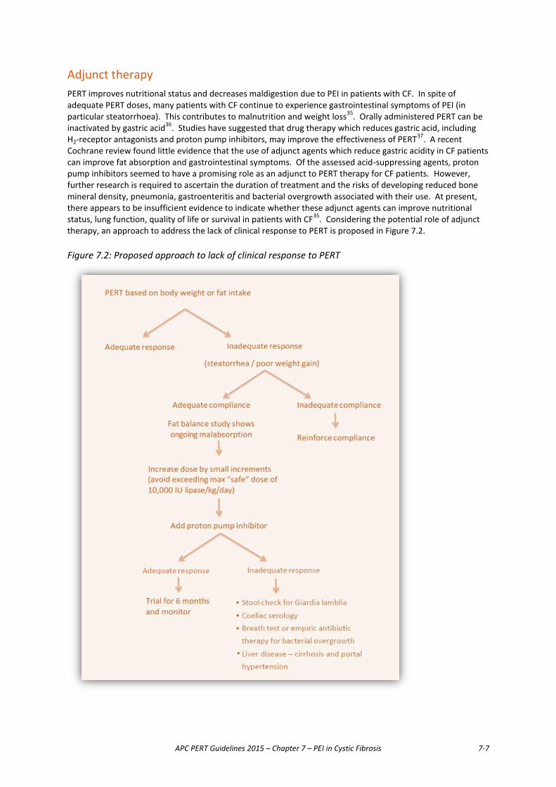
APC PERT Guidelines 2015 – Chapter 7 – PEI in Cystic Fibrosis 7-7
Adjunct therapy PERT improves nutritional status and decreases maldigestion due to PEI in patients with CF. In spite of adequate PERT doses, many patients with CF continue to experience gastrointestinal symptoms of PEI (in particular steatorrhoea). This contributes to malnutrition and weight loss35. Orally administered PERT can be inactivated by gastric acid36. Studies have suggested that drug therapy which reduces gastric acid, including H2-receptor antagonists and proton pump inhibitors, may improve the effectiveness of PERT37. A recent Cochrane review found little evidence that the use of adjunct agents which reduce gastric acidity in CF patients can improve fat absorption and gastrointestinal symptoms. Of the assessed acid-suppressing agents, proton pump inhibitors seemed to have a promising role as an adjunct to PERT therapy for CF patients. However, further research is required to ascertain the duration of treatment and the risks of developing reduced bone mineral density, pneumonia, gastroenteritis and bacterial overgrowth associated with their use. At present, there appears to be insufficient evidence to indicate whether these adjunct agents can improve nutritional status, lung function, quality of life or survival in patients with CF35. Considering the potential role of adjunct therapy, an approach to address the lack of clinical response to PERT is proposed in Figure 7.2. Figure 7.2: Proposed approach to lack of clinical response to PERT
APC PERT Guidelines 2015 – Chapter 7 – PEI in Cystic Fibrosis 7-8
Long-term management and monitoring for PEI Periodic nutritional assessments should include a collation of anthropometric measures (height, weight and head circumference), dietary intake, biochemical assessments and a review of bowel habit and function. Deterioration in parameters of nutritional status should be detected early, before growth and lung function are compromised. Anthropometric measurements should be regularly assessed (Table 2). It is important to note that adults may lose height over time due to ageing, osteoporosis or kyphosis1. A thorough assessment of dietary intake should be conducted at least once a year. In very young children, dietary intake assessments should be more frequently assessed. Table 4 highlights the key areas of dietary assessment in CF patients1. Table 4: Key areas of dietary intake assessment in CF patients
Key areas of assessment Purpose
Energy intake Energy balance and effect on weight
Fat intake Energy density, PERT adequacy
Food preferences and variety - Meal pattern and behaviours
- Knowledge and attitudes about nutrition including body image
Adequacy of micronutrient intake Fibre intake Target areas for change
PERT: dosage, adherence and gastrointestinal symptoms Adequacy of PERT regimen
Supplements: vitamins and minerals, oral and enteral nutritional supplements
Adequacy of micronutrient intake Contribution to energy and nutrient intakes
Sodium and fluid intake Hydration and sodium status
Nutrition-related complementary or alternative therapies Adequacy of micro and macronutrient intake
Adapted from Stapleton et al. 20061. Biochemical tests provide important information regarding the nutritional status of those with CF. All patients should be assessed at diagnosis and subsequently at annual review. If there is a risk of deterioration in nutritional status, or if there is a change in treatment, then the frequency of biochemical assessments should be increased1. Other nutritional assessment in CF patients should also include a review of bowel habits and function. Information regarding relevant lifestyle factors including exercise and physical activity should also be collected1. The Australasian Clinical Practice Guidelines for Nutrition in Cystic Fibrosis discusses these management and monitoring strategies in detail.
Summary � CF is a common lethal genetic disorder caused by mutations in the gene that encodes the CFTR protein.
CFTR mutations disrupt the function of water and chloride ion transportation at a cellular level, leading to classic CF phenotypes such as raised sweat chloride, recurrent respiratory infection with bronchiectasis and early-onset pancreatic insufficiency.
� The majority of babies with CF are diagnosed by 4-6 weeks after birth through newborn screening. � The long-term survival for children with CF has improved significantly. With early diagnosis, appropriate
management of respiratory infections and improved nutrition, most CF patients live well into adulthood. � About 85% of CF patients are pancreatic-insufficient by early childhood. There is evidence that a
proportion of infants found to be pancreatic-sufficient at birth will lose their residual pancreatic function over time.
� Without treatment, gastrointestinal losses of fat and nitrogen can be severe. These cause growth failure and protein catabolism.

APC PERT Guidelines 2015 – Chapter 7 – PEI in Cystic Fibrosis 7-9
� Fat malabsorption, the most important digestive malfunction in PEI, affects macro- and micronutrient absorption. Prolonged, untreated PEI is associated with a poorer prognosis long term. This highlights the importance of aggressive nutritional management focusing on a high energy, high fat diet and PERT. Good nutritional management of CF patients can prevent growth failure and chronic malnutrition.
� PERT is indicated in those with documented fat malabsorption or PEI as established by a pancreatic function test.
� PERT dosing should be individualised. CF patients with PEI should be started on the lowest recommended dose and up-titrated based on weight gain and gastrointestinal symptoms to ascertain the lowest effective dose.
� Excessive doses of PERT are associated with fibrosing colonopathy. Therefore doses should not exceed 10,000 units of lipase per kilogram bodyweight per day or 2,500 units per kilogram bodyweight per meal.
� The role of acid-suppressing agents in improving fat absorption and gastrointestinal symptoms of CF patients is still under debate. Proton pump inhibitors appear to have a promising role as an adjunct to PERT therapy for CF patients. However, further research is required to ascertain the duration of treatment and the risks of pneumonia, gastroenteritis and bacterial overgrowth associated with their use. In addition to this there is the development of new agents such as Liprotamase, a novel non-porcine PERT which contains a proprietary biotechnology-derived formulation allowing purity, precise dosing and acid resistance which could result in a more scientific approach to dose response and negate the need for adjunct therapy38.
� It should be noted that the principles of PERT that have been discussed in this chapter can be applied to other paediatric conditions involving PEI, such as Shwachman–Diamond and Pearson syndromes.
Recommendations for pancreatic enzyme replacement therapy in cystic fibrosis
No. Recommendation Level of evidence Strength of agreement
7.1 CF patients identified at birth should be continually monitored for PEI
4
7.2 Aggressive nutritional management with a high-
energy, high fat diet and PERT is recommended for CF patients with documented fat malabsorption or PEI found on pancreatic function testing.
1b, 2b
7.3 PERT doses should be individualised based on
bodyweight or fat intake, and titrated based on weight gain and bowel signs and symptoms.
5
7.4 Recommended doses
For infants: 500-1,000 U lipase / g of dietary fat OR 2,000- 4,000 U lipase / breastfeed or 120 mL of infant formula. For children: 500-4,000 U lipase / g of dietary fat OR 1,000 U lipase / kg bodyweight / meal (<4 years old); 500 U lipase / kg bodyweight / meal (>4 years old). Doses could be halved for a snack instead of a full meal. Maximum dose: 10,000 U / kg bodyweight / day or 2,500 U / kg bodyweight / meal.
5

APC PERT Guidelines 2015 – Chapter 7 – PEI in Cystic Fibrosis 7-10
No. Recommendation Level of evidence Strength of agreement
7.5 Branded PERT preparations should be used to ensure efficacy.
2a
7.6 Capsules should not be crushed, chewed or
sprinkled on foods with a high pH. 5
7.7 Enteral feeding is indicated if ongoing
comprehensive assessments of CF patients indicate deterioration of nutritional status or failure to improve on oral PERT.
5
7.8 Proton Pump Inhibitors may have a role in
decreasing gastric acidity and improving fat absorption and gastrointestinal symptoms in CF patients on PERT (and see Chapter 3).
1b
7.9 Patients with CF and PEI should be referred to a
multidisciplinary clinic for management. 5
7.10 Long-term management should include ongoing
periodic nutritional assessments, growth measurements and monitoring for PEI with indirect tests for pancreatic exocrine function, particularly for patients with PS CF.
3
References
(1) Stapleton D. Australasian clinical practice guidelines for nutrition in cystic fibrosis. http://daa.asn.au/wp-content/uploads/2012/09/Guidelines_CF-Final.pdf . 2006. Dietitians Association of Australia.
(2) McKone EF, Emerson SS, Edwards KL, Aitken ML. Effect of genotype on phenotype and mortality in cystic fibrosis: a retrospective cohort study. Lancet 2003; 361(9370):1671-1676.
(3) Reid DW, Blizzard CL, Shugg DM, Flowers C, Cash C, Greville HM. Changes in cystic fibrosis mortality in Australia, 1979-2005. Med J Aust 2011; 195(7):392-395.
(4) Riordan JR, Rommens JM, Kerem B, Alon N, Rozmahel R, Grzelczak Z et al. Identification of the cystic fibrosis gene: cloning and characterization of complementary DNA. Science 1989; 245(4922):1066-1073.
(5) McKone EF, Goss CH, Aitken ML. CFTR genotype as a predictor of prognosis in cystic fibrosis. Chest 2006; 130(5):1441-1447.
(6) Mickle JE, Cutting GR. Genotype-phenotype relationships in cystic fibrosis. Med Clin North Am 2000; 84(3):597-607.
(7) UK cystic fibrosis gene therapy consortium. CFTR protein structure. 2015. 7-12-2009.
(8) Kerem E, Kerem B. Genotype-phenotype correlations in cystic fibrosis. Pediatr Pulmonol 1996; 22(6):387-395.

APC PERT Guidelines 2015 – Chapter 7 – PEI in Cystic Fibrosis 7-11
(9) Cystic Fibrosis Australia. Cystic fibrosis in Australia and New Zealand 2002. Annual report from the Australasian Cystic Fibrosis Data Registry. http://www cysticfibrosis org au/all/data-registry [ 2004 Available from: URL:http://www.cysticfibrosis.org.au/all/data-registry
(10) Cystic Fibrosis Australia. Cystic Fibrosis National Website. www cysticfibrosis org au [ 2015 [cited 2009 June 4]; Available from: URL:www.cysticfibrosis.org.au
(11) Elborn JS, Prescott RJ, Stack BH, Goodchild MC, Bates J, Pantin C et al. Elective versus symptomatic antibiotic treatment in cystic fibrosis patients with chronic Pseudomonas infection of the lungs. Thorax 2000; 55(5):355-358.
(12) Thomas CL, O'Rourke PK, Wainwright CE. Clinical outcomes of Queensland children with cystic fibrosis: a comparison between tertiary centre and outreach services. Med J Aust 2008; 188(3):135-139.
(13) Merelle ME, Schouten JP, Gerritsen J, Dankert-Roelse JE. Influence of neonatal screening and centralized treatment on long-term clinical outcome and survival of CF patients. Eur Respir J 2001; 18(2):306-315.
(14) Tizzano EF, Buchwald M. CFTR expression and organ damage in cystic fibrosis. Ann Intern Med 1995; 123(4):305-308.
(15) Baker SS. Delayed release pancrelipase for the treatment of pancreatic exocrine insufficiency associated with cystic fibrosis. Ther Clin Risk Manag 2008; 4(5):1079-1084.
(16) Ooi CY, Dorfman R, Cipolli M, Gonska T, Castellani C, Keenan K et al. Type of CFTR mutation determines risk of pancreatitis in patients with cystic fibrosis. Gastroenterology 2011; 140(1):153-161.
(17) Waters DL, Dorney SF, Gaskin KJ, Gruca MA, O'Halloran M, Wilcken B. Pancreatic function in infants identified as having cystic fibrosis in a neonatal screening program. N Engl J Med 1990; 322(5):303-308.
(18) Kalivianakis M, Verkade HJ. The mechanisms of fat malabsorption in cystic fibrosis patients. Nutrition 1999; 15(2):167-169.
(19) Littlewood JM. Cystic fibrosis: gastrointestinal complications. Br Med Bull 1992; 48(4):847-859.
(20) Pencharz PB, Durie PR. Pathogenesis of malnutrition in cystic fibrosis, and its treatment. Clin Nutr 2000; 19(6):387-394.
(21) Gaskin K, Gurwitz D, Durie P, Corey M, Levison H, Forstner G. Improved respiratory prognosis in patients with cystic fibrosis with normal fat absorption. J Pediatr 1982; 100(6):857-862.
(22) Corey M, McLaughlin FJ, Williams M, Levison H. A comparison of survival, growth, and pulmonary function in patients with cystic fibrosis in Boston and Toronto. J Clin Epidemiol 1988; 41(6):583-591.
(23) Lai HC, Corey M, FitzSimmons S, Kosorok MR, Farrell PM. Comparison of growth status of patients with cystic fibrosis between the United States and Canada. Am J Clin Nutr 1999; 69(3):531-538.
(24) Gaskin KJ, Durie PR, Lee L, Hill R, Forstner GG. Colipase and lipase secretion in childhood-onset pancreatic insufficiency. Delineation of patients with steatorrhea secondary to relative colipase deficiency. Gastroenterology 1984; 86(1):1-7.
(25) Caliari S, Vantini I, Sembenini C, Gregori B, Carnielli V, Benini L. Fecal fat measurement in the presence of long- and medium-chain triglycerides and fatty acids. Comparison of three methods. Scand J Gastroenterol 1996; 31(9):863-867.
APC PERT Guidelines 2015 – Chapter 7 – PEI in Cystic Fibrosis 7-12
(26) Beharry S, Ellis L, Corey M, Marcon M, Durie P. How useful is fecal pancreatic elastase 1 as a marker of exocrine pancreatic disease? J Pediatr 2002; 141(1):84-90.
(27) DRUMMEY GD, BENSON JA, Jr., JONES CM. Microscopical examination of the stool for steatorrhea. N Engl J Med 1961; 264:85-87.
(28) Littlewood JM, Wolfe SP, Conway SP. Diagnosis and treatment of intestinal malabsorption in cystic fibrosis. Pediatr Pulmonol 2006; 41(1):35-49.
(29) Anthony H, Collins CE, Davidson G, Mews C, Robinson P, Shepherd R et al. Pancreatic enzyme replacement therapy in cystic fibrosis: Australian guidelines. Pediatric Gastroenterological Society and the Dietitians Association of Australia. J Paediatr Child Health 1999; 35(2):125-129.
(30) Stallings VA, Stark LJ, Robinson KA, Feranchak AP, Quinton H. Evidence-based practice recommendations for nutrition-related management of children and adults with cystic fibrosis and pancreatic insufficiency: results of a systematic review. J Am Diet Assoc 2008; 108(5):832-839.
(31) Smyth RL, van VD, Smyth AR, Lloyd DA, Heaf DP. Strictures of ascending colon in cystic fibrosis and high-strength pancreatic enzymes. Lancet 1994; 343(8889):85-86.
(32) Pawel BR, de Chadarevian JP, Franco ME. The pathology of fibrosing colonopathy of cystic fibrosis: a study of 12 cases and review of the literature. Hum Pathol 1997; 28(4):395-399.
(33) Stapleton D, Anthony H, et al. Implementing the Australian pancreatic enzyme replacement therapy guidelines for cystic fibrosis. Australian Journal of Nutrition and Dietetics 1999; 56(2):91-96.
(34) Patchell CJ, MacDonald A, Weller PW. Pancreatic enzymes with enteral feeds: how should we give them? [Abstract]. 22nd European Cystic Fibrosis Conference. 104. 1998.
(35) Ng SM, Jones AP. Drug therapies for reducing gastric acidity in people with cystic fibrosis. Cochrane Database Syst Rev 2003;(2):CD003424.
(36) Zentler-Munro PL, Fine DR, Batten JC, Northfield TC. Effect of cimetidine on enzyme inactivation, bile acid precipitation, and lipid solubilisation in pancreatic steatorrhoea due to cystic fibrosis. Gut 1985; 26(9):892-901.
(37) DiMagno EP. Gastric acid suppression and treatment of severe exocrine pancreatic insufficiency. Best Pract Res Clin Gastroenterol 2001; 15(3):477-486.
(38) Borowitz D, Stevens C, Brettman LR, Campion M, Wilschanski M, Thompson H. Liprotamase long-term safety and support of nutritional status in pancreatic-insufficient cystic fibrosis. J Pediatr Gastroenterol Nutr 2012; 54(2):248-257.

APC PERT Guidelines 2015 – Chapter 8 – PERT after bowel resection 8-1
Chapter 8 Use of PERT after bowel resection Introduction The intestines are vital for the absorption of external nutrients. Food is mixed with digestive enzymes and bile in the duodenum, further mixed and broken down in the jejunum, and absorbed largely in the ileum. The colon also plays a significant role in the absorption of water, electrolytes, short-chain fatty acids, and certain fat-soluble vitamins.1 Various disease processes may necessitate resection of variable lengths of small and large intestine, by open or laparoscopic approaches. Small bowel resections are performed to treat conditions such as Crohn’s disease, tumours, ischaemia, and trauma. Additional conditions necessitating colonic resection include neoplasia (cancer and polyposis), ulcerative colitis, and toxic megacolon. Biliopancreatic diversion for morbid obesity also functionally removes a large part of small bowel by bypassing it from the passage of nutrients. The physiological consequences of bowel resection or bypass depend on the site and amount of absorptive bowel removed. So-called short bowel syndrome (SBS) is a malabsorptive state following massive resection of small intestine2, usually when there is less than 200 cm of bowel remaining3. Factors attenuating the impact of SBS include remnant ileum, an intact ileocaecal valve, absence of residual mucosal disease, and an intact stomach, pancreas and liver4. It is known that numerous changes occur in the remaining alimentary tract to help adaptation to the shortened intestine after extensive small bowel resections. There is mucosal hyperplasia and an accelerated rate of epithelial cell renewal5-7. In addition there is increased proliferation of parietal cells in the gastric glands8. This is caused by a rise in gastrin in the plasma9;10. Exogenous gastrin and pentagastrin have been recognised to exert a trophic action upon the exocrine pancreas11;12 but do not affect the small bowel distal to the duodenum13. Thus hypergastrinaemia, induced by intestinal resection, may well alter the size and composition of the exocrine pancreas. The process of intestinal adaptation occurs from 6 months to 3 years after extensive bowel resection1;14.
Medical management Although corrective surgical procedures are sometimes possible, the mainstay of treatment of symptoms following extensive bowel resection is a combination of dietary and pharmaceutical therapies. The goal of treatment is to optimise digestion and absorption of nutrient molecules, as well as to promote intestinal adaptation if possible. As part of any management protocol, regular monitoring of nutritional status is important to guide ongoing therapy. Severe nutrient and fluid malabsorption occurs following extensive small bowel resections. Patients with less than 100 cm of residual jejunum generally have a net secretory response to food, and as such, may lose more fluid than they ingest. Bowel resections can lead to malabsorption of fluid, electrolytes, minerals and other essential nutrients, resulting in malnutrition and dehydration. Individualised and tailored nutritional management helps to optimise intestinal absorption, leading to nutritional independence such that a patient can resume as normal a lifestyle as possible. In extreme cases, total parenteral nutrition (TPN) may be necessary, with the aim of reintroducing enteral feeding as adaptation occurs. Adjustment of the various nutritional components is complex and should be individualised based on gastrointestinal anatomy, underlying disease and lifestyle15. Referral to a dietician is recommended.

APC PERT Guidelines 2015 – Chapter 8 – PERT after bowel resection 8-2
Adjunctive pharmaceutical management may consist of:
� Antimotility agents to slow intestinal transit. These include loperamide, diphenoxylate and codeine1;14. Octreotide may also be used in cases of high output rapid transit, but because it inhibits the adaptation process, it is usually only seen as a temporising measure15;16.
� Bile-acid resins (e.g. cholestyramine) in cases of steatorrhoea secondary to bile acid malabsorption after ileal resection1;4.
� Proton pump inhibitors or H2 antagonists to suppress gastric acid hypersecretion. This serves to prevent peptic ulceration, reduce diarrhoea, and maximise activation of endogenous pancreatic enzymes4;14.
� Antibiotics (eg. metronidazole, cephalosporins) and/or probiotics to suppress intestinal bacterial overgrowth14;17.
� Recombinant human growth hormone to enhance intestinal adaptation and thus improve absorption18. Teduglutide, a long-acting analogue of glucagon-like peptide-2, has also shown promising results in a multicentre randomised controlled trial19.
Pancreatic exocrine insufficiency and the role of pancreatic enzyme replacement therapy Animal models show that massive small bowel resection provokes hyperplasia, not only in the stomach and the remaining intestine, but also in the exocrine pancreas20. However after bowel resections, a decrease in digestive enzymes of the pancreatic tissue, namely amylase, lipase, and chymotrypsinogen have also been detected in animal models. The complex interaction between secretions of the gut, stomach and pancreas are likely to be interrupted by the total, or partial, removal of the small bowel21. Therefore, it is reasonable to expect patients who have had extensive small bowel resections to experience a degree of PEI. PEI symptoms after bowel resection are nonspecific. Typically, patients can experience abdominal pain, diarrhoea, steatorrhoea, weight loss, fatigue and malnutrition. There are several physiological mechanisms by which bowel resection may result in absolute or relative PEI, thus contributing to the malabsorptive state.
� Decreased intestinal transit time leads to inadequate mixing of nutrient macromolecules with endogenous pancreatic enzymes, thereby reducing the efficacy of digestion4;22.
� The presence of enteric contents causes the small bowel to secrete hormones that stimulate output of pancreatic enzymes and/or juice22, or elicit a stimulatory enteropancreatic cholinergic reflex23. This is particularly the case for duodenum and proximal jejunum, which is pivotal in the so-called intestinal phase of digestion. Removal of this part of the bowel may therefore render pancreatic exocrine output insufficient to fully digest nutrient intake.
� Gastric hyperacidity occurs due to compensatory hypersecretion of gastrin as well as loss of one or more inhibitors of gastric acid secretion that would otherwise be produced by proximal small bowel (e.g. cholecystokinin and secretin). The reduced pH impairs the activation of pancreatic enzymes in the normally alkaline milieu of the duodenum22;24;25. This may not be entirely redressed by the gastrin-induced hypertrophy of the exocrine pancreas.
APC PERT Guidelines 2015 – Chapter 8 – PERT after bowel resection 8-3
� Bacterial overgrowth may occur in small bowel following extensive resection, particularly where this has included the ileocaecal valve. While being a direct cause of malabsorption, small bowel bacterial overgrowth may also lead to premature enzyme degradation in the gastrointestinal tract17.
� If employed as a temporising measure to slow gastrointestinal transit, somatostatin analogues such as octreotide also reduce the output of pancreatic juice1. This needs to be taken into account if non-elemental enteral feeding has been instituted.
A number of reviews and case series have recommended the use of pancreatic enzyme replacement therapy (PERT) as part of the multimodal pharmaceutical and nutritional management of SBS14;15;22;26-29. However there remains a paucity of randomised controlled trials demonstrating a specific positive effect of PERT in this population, and it is unclear that PERT is of benefit in the absence of a history of PEI15;30. Theoretically, the use of pancreatic enzymes together with gastric acid suppression therapy should enable gastric digestion of nutrients with increased delivery of digestive products in the small bowel. Because of the superior stimulatory capacity of peptides, amino acids and free fatty acids compared with macronutrients, conditions for nutrient absorption should be improved by increased endogenous pancreatic enzyme secretion, delayed gastric emptying and small intestinal transit as well as mucosal hypertrophy31. An uncoated form of pancreatic enzyme replacement is recommended because it will dissolve rapidly enough to have maximum efficacy4. If PERT is prescribed, it should be administered with meals to allow for adequate mixing of enzymes and meal nutrients32. Standard lipase doses of 25,000 to 40,000 IU with regular meals could be prescribed33. For smaller meals or snacks, 10,000 IU of lipase should be adequate32. If standard doses do not achieve adequate reduction of symptoms, the dosage can be increased two to three times. Because of potential side effects, dosages of more than 75,000 IU of lipase per meal are not recommended34.
Summary Pancreatic hyperplasia and a decrease in pancreatic digestive enzymes are consequences of bowel surgery, (level of evidence 2a). However the digestive consequences of bowel resection are subject to considerable individual variation, largely due to the site and extent of resection. There is limited information in recent published literature around PEI in patients after bowel surgery, although the consensus is that it does occur, particularly affecting those patients who have undergone extensive small bowel resections. Where maldigestion and/or malabsorption are clinically evident, a tailored multimodal therapeutic approach is required. PERT may be prescribed for confirmed PEI in this patient population. However, it is important to note that there is a lack of robust clinical trials to support this practice. Theoretically, PERT could be prescribed with gastric acid suppression therapy to enable gastric digestion of nutrients with increased delivery of digestive products to the small bowel.
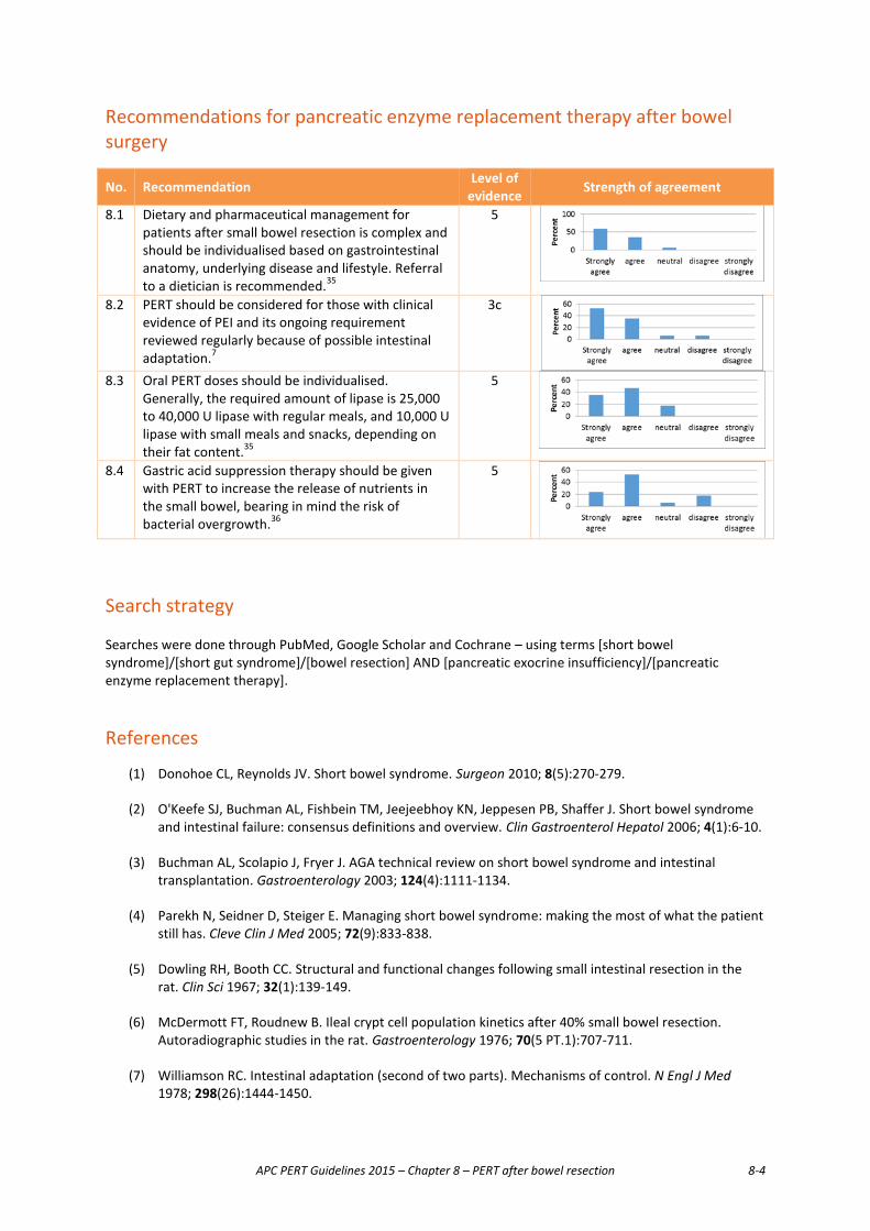
APC PERT Guidelines 2015 – Chapter 8 – PERT after bowel resection 8-4
Recommendations for pancreatic enzyme replacement therapy after bowel surgery
No. Recommendation Level of evidence Strength of agreement
8.1 Dietary and pharmaceutical management for patients after small bowel resection is complex and should be individualised based on gastrointestinal anatomy, underlying disease and lifestyle. Referral to a dietician is recommended.35
5
8.2 PERT should be considered for those with clinical evidence of PEI and its ongoing requirement reviewed regularly because of possible intestinal adaptation.7
3c
8.3 Oral PERT doses should be individualised.
Generally, the required amount of lipase is 25,000 to 40,000 U lipase with regular meals, and 10,000 U lipase with small meals and snacks, depending on their fat content.35
5
8.4 Gastric acid suppression therapy should be given with PERT to increase the release of nutrients in the small bowel, bearing in mind the risk of bacterial overgrowth.36
5
Search strategy Searches were done through PubMed, Google Scholar and Cochrane – using terms [short bowel syndrome]/[short gut syndrome]/[bowel resection] AND [pancreatic exocrine insufficiency]/[pancreatic enzyme replacement therapy].
References
(1) Donohoe CL, Reynolds JV. Short bowel syndrome. Surgeon 2010; 8(5):270-279.
(2) O'Keefe SJ, Buchman AL, Fishbein TM, Jeejeebhoy KN, Jeppesen PB, Shaffer J. Short bowel syndrome and intestinal failure: consensus definitions and overview. Clin Gastroenterol Hepatol 2006; 4(1):6-10.
(3) Buchman AL, Scolapio J, Fryer J. AGA technical review on short bowel syndrome and intestinal transplantation. Gastroenterology 2003; 124(4):1111-1134.
(4) Parekh N, Seidner D, Steiger E. Managing short bowel syndrome: making the most of what the patient still has. Cleve Clin J Med 2005; 72(9):833-838.
(5) Dowling RH, Booth CC. Structural and functional changes following small intestinal resection in the rat. Clin Sci 1967; 32(1):139-149.
(6) McDermott FT, Roudnew B. Ileal crypt cell population kinetics after 40% small bowel resection. Autoradiographic studies in the rat. Gastroenterology 1976; 70(5 PT.1):707-711.
(7) Williamson RC. Intestinal adaptation (second of two parts). Mechanisms of control. N Engl J Med 1978; 298(26):1444-1450.
APC PERT Guidelines 2015 – Chapter 8 – PERT after bowel resection 8-5
(8) Winborn WB, Seelig LL, Jr., Nakayama H, Weser E. Hyperplasia of the gastric glands after small bowel resection in the rat. Gastroenterology 1974; 66(3):384-395.
(9) Buxton B. Small bowel resection and gastric acid hypersecretion. Gut 1974; 15(3):229-238.
(10) D'Sa AA, Buchanan KD. Role of gastrointestinal hormones in the response to massive resection of the small bowel. Gut 1977; 18(11):877-881.
(11) Johnson LR. The trophic action of gastrointestinal hormones. Gastroenterology 1976; 70(2):278-288.
(12) Reber HA, Johnson F, Deveney K, Montgomery C, Way LW. Trophic effects of gastrin on the exocrine pancreas in rats. J Surg Res 1977; 22(5):554-560.
(13) Oscarson JE, Veen HF, Williamson RC, Ross JS, Malt RA. Compensatory postresectional hyperplasia and starvation atrophy in small bowel: dissociation from endogenous gastrin levels. Gastroenterology 1977; 72(5 Pt 1):890-895.
(14) Kumpf VJ. Pharmacologic management of diarrhea in patients with short bowel syndrome. JPEN J Parenter Enteral Nutr 2014; 38(1 Suppl):38S-44S.
(15) Matarese LE, Steiger E. Dietary and medical management of short bowel syndrome in adult patients. J Clin Gastroenterol 2006; 40 Suppl 2:S85-S93.
(16) O'Keefe SJ, Haymond MW, Bennet WM, Oswald B, Nelson DK, Shorter RG. Long-acting somatostatin analogue therapy and protein metabolism in patients with jejunostomies. Gastroenterology 1994; 107(2):379-388.
(17) Bures J, Cyrany J, Kohoutova D, Forstl M, Rejchrt S, Kvetina J et al. Small intestinal bacterial overgrowth syndrome. World J Gastroenterol 2010; 16(24):2978-2990.
(18) Steiger E. Guidelines for pharmacotherapy, nutritional management, and weaning parenteral nutrition in adult patients with short bowel syndrome: introduction. J Clin Gastroenterol 2006; 40 Suppl 2:S73-S74.
(19) O'Keefe SJ, Jeppesen PB, Gilroy R, Pertkiewicz M, Allard JP, Messing B. Safety and efficacy of teduglutide after 52 weeks of treatment in patients with short bowel intestinal failure. Clin Gastroenterol Hepatol 2013; 11(7):815-823.
(20) Haegel P, Stock C, Marescaux J, Petit B, Grenier JF. Hyperplasia of the exocrine pancreas after small bowel resection in the rat. Gut 1981; 22(3):207-212.
(21) Gelinas MD, Morin CL, Morisset J. Exocrine pancreatic function following proximal small bowel resection in rats. J Physiol 1982; 322:71-82.
(22) Layer P, Melle U. Indication for Pancreatic Enzyme Substitution following Small Intestinal Resection (Short Bowel Syndrome). Pancreatology 2001; 1(Suppl 1):49-54.
(23) Vu MK, van der Veek PP, Frolich M, Souverijn JH, Biemond I, Lamers CB et al. Does jejunal feeding activate exocrine pancreatic secretion? Eur J Clin Invest 1999; 29(12):1053-1059.
(24) Schubert ML. Hormonal regulation of gastric acid secretion. Curr Gastroenterol Rep 2008; 10(6):523-527.
(25) Thompson JS, Rochling FA, Weseman RA, Mercer DF. Current management of short bowel syndrome. Curr Probl Surg 2012; 49(2):52-115.

APC PERT Guidelines 2015 – Chapter 8 – PERT after bowel resection 8-6
(26) Leyva-Martinez S, Fernandez-Lloret S, Martin-Ruiz JL. [Massive intestinal resection. Nutritional adaptation process]. Nutr Hosp 2007; 22(5):616-620.
(27) Nakajima K, Oshida H, Muneyuki T, Kakei M. Pancrelipase: an evidence-based review of its use for treating pancreatic exocrine insufficiency. Core Evid 2012; 7:77-91.
(28) Pezzilli R, Andriulli A, Bassi C, Balzano G, Cantore M, Delle FG et al. Exocrine pancreatic insufficiency in adults: a shared position statement of the Italian Association for the Study of the Pancreas. World J Gastroenterol 2013; 19(44):7930-7946.
(29) Slaff J, Jacobson D, Tillman CR, Curington C, Toskes P. Protease-specific suppression of pancreatic exocrine secretion. Gastroenterology 1984; 87(1):44-52.
(30) Seidner DI, Matarese LE. Short bowel syndrome: etiology, pathophysiology and management. 2003. Selected topics in gastrotherapy. Ref Type: Serial (Book,Monograph)
(31) Keller J, Layer P. Human pancreatic exocrine response to nutrients in health and disease. Gut 2005; 54
Suppl 6:vi1-28.
(32) Keller J, Layer P. Pancreatic Enzyme Supplementation Therapy. Curr Treat Options Gastroenterol 2003; 6(5):369-374.
(33) Layer P, Yamamoto H, Kalthoff L, Clain JE, Bakken LJ, DiMagno EP. The different courses of early- and late-onset idiopathic and alcoholic chronic pancreatitis. Gastroenterology 1994; 107(5):1481-1487.
(34) Layer P, Keller J, Lankisch PG. Pancreatic enzyme replacement therapy. Curr Gastroenterol Rep 2001; 3(2):101-108.
(35) Matarese LE, Steiger E. Dietary and medical management of short bowel syndrome in adult patients. J Clin Gastroenterol 2006; 40 Suppl 2:S85-S93.
(36) Keller J, Layer P. Pancreatic Enzyme Supplementation Therapy. Curr Treat Options Gastroenterol 2003; 6(5):369-374.
APC PERT Guidelines 2015 – Chapter 9 – PERT post gastric surgery 9-1
Chapter 9 Use of PERT after gastric surgery Anatomical and nutritional consequences of gastric surgery Gastric surgery is frequently associated with diarrhoea and weight loss as well as various micronutrient deficiencies. The physiological functions of the stomach are complex, and its interactions with other organs are poorly understood1. Nevertheless, through its mechanical and chemical processes, the stomach has a unique role in food processing and bioavailability2. Gastric surgery has become an uncommon procedure in developed countries over the last two decades with a greatly reduced need to undertake surgery for peptic ulcer disease and a reduced incidence of gastric cancer to 5 to 9 per 100,0003, although the incidence of gastric cancer rises to 60 per 100,000 in some regions of Japan and Chile, and world-wide, gastric cancer remains an important condition. There has also been a great increase in the use of gastric surgery for the treatment of obesity. In the 1960s and 1970s gastric surgery could lead to the conversion of a patient into a ‘gastric cripple’ who experienced weight loss, diarrhoea, and various micronutrient deficiencies. The cause of this was unclear, although in a number of gastric operations, inadequate or asynchronous pancreatic enzyme secretion can lead to poor mixing of enzymes with intestinal chyme.
Gastric surgery procedures
� Gastric surgery is still occasionally required to treat severe peptic ulcer disease or its complications. While the vast majority of peptic ulcers are now managed with medication, partial gastrectomy is sometimes indicated for patients who do not respond satisfactorily to medical therapy, those who develop a bleeding or perforated ulcer and those who develop pyloric obstruction.
� Truncal vagotomy was popular until the introduction of proton pump inhibitors (PPIs). It required a drainage procedure such as gastroenterostomy, pyloroplasty or antrectomy, because of denervation of the antrum. Sometimes the severe, persistent abdominal pain, vomiting, dumping or diarrhoea seen several years after truncal vagotomy were improved by conversion of the gastroenterostomy to a pyloroplasty.4 Truncal vagotomy also denervates the pancreas and the drainage procedure (e.g. gastroenterostomy) bypasses the duodenum, so these lesser procedures can also lead to asynchrony between pancreatic secretion and the passage of intestinal contents, and dumping can also occur2;5. Highly selective vagotomy, a technically difficult procedure, was developed to prevent these problems, but before the advent of PPIs, was associated with a higher rate of ulcer recurrence. Thankfully, the development of PPIs has greatly reduced the need for such surgery and its consequent problems.
� Total gastrectomy results in anastomosis of the oesophagus to a Roux-en-Y loop of jejunum, bypassing the duodenum. The nutritional status and symptoms in a cohort of 19 patients, at 30 months after a total gastrectomy, have been reviewed. One patient was house-bound because of post-gastrectomy symptoms, three had reduced their daily activities and the remainder experienced minimal disturbance of daily activity. All patients underwent a three-day faecal fat determination. Although no patient had symptomatic steatorrhoea, the mean excretion of fat was 8.1g/d with five patients losing between 10 and 19g fat/d. These patients frequently took nutritional supplements (particularly vitamin B12) so it is difficult to attribute micronutrient deficiencies to asynchrony of bile and pancreatic secretions with the presence of a food bolus, but low folate and iron levels along with osteoporosis were the most frequent nutritional problems. Patients who were not bothered by digestive symptoms tended to increase their overall food intake6. It would have been interesting to
APC PERT Guidelines 2015 – Chapter 9 – PERT post gastric surgery 9-2
give a trial of PERT although it is not certain whether this is beneficial7. Others have confirmed fat malabsorption to be the main nutritional consequence of total gastrectomy8.
� Pancreatico-biliary bypass, a procedure used for severe obesity, results in an extreme example of asynchrony because bile and pancreatic secretions arrive in the distal ileum via a short common channel. One paper reports that the above complications were reduced when this common channel was increased from 50 to 60cm9. It is not surprising, therefore, that following a pancreatico-biliary bypass considerable weight loss occurs, associated with significant vitamin and trace element deficiencies and that these patients require supplementation with multiple micro-nutrients10.
Causes of maldigestion after gastric surgery Clinically relevant postoperative maldigestion is associated with PEI and occurs irrespective of the type of gastrectomy procedure1. In addition, malnutrition and consequent body weight loss may be related to the loss of gastric reservoir. Coupled with this, centrally mediated hypothalamic factors may also cause appetite suppression11. Increased peristalsis and bacterial overgrowth may cause diarrhoea, while rapid small bowel transit and intestinal malabsorption play a part in causing steatorrhoea12. The causes of malabsorption following gastric surgery are most likely multifactorial and although they include a degree of pancreatic enzyme insufficiency (PEI) it has been shown that maldigestion is not completely reversed by PERT.7
Managing nutritional deficiencies after gastric surgery The majority of patients undergoing a gastrectomy do not have symptomatic PEI and do not require long term management with PERT, but do require monitoring of nutritional status including weight, body muscle and fat mass, iron, folate and vitamin B12 status as well as bone mineral density. Because intrinsic factor in the stomach facilitates vitamin B12 absorption in the terminal ileum, after partial gastrectomy intrinsic factor is reduced and after total gastrectomy, abolished, causing vitamin B12 deficiency. In addition, where the duodenum has been bypassed, iron deficiency results. Hence, all total gastrectomy and many partial gastrectomy patients need to have vitamin B12 and iron replacement therapy. As well, calcium absorption and vitamin D deficiency can be associated with the steatorrhoea which can follow gastric surgery, with implications for bone mineral density. Patients who are symptomatic after gastric surgery require evaluation by a gastroenterologist for dumping syndrome, intestinal transit time and bacterial overgrowth, including small bowel biopsy. Dumping syndrome can be managed by encouraging the intake of whole grain food without sugars and limiting liquids with meals. These patients should also sit at the table for half an hour after eating. Culture of intestinal fluid and small bowel biopsy may provide guidance on specific antibiotic management if bacterial overgrowth is detected.
Decision about the use of PERT in symptomatic patients requires consideration of specific issues, including:
1. In some cases the gastric remnant is so small that food passes more rapidly than normal into the small bowel. In these cases the PERT capsule may pass into the small bowel and avoid being mixed with the food13.
2. In other cases, gastric emptying may be so slow that the enzymes are exposed to gastric acid and inactivated.
3. Polya (gastrojejunal) or Roux-en-Y reconstructions result in bypass of the duodenum so that pancreatic secretions might not be synchronised with the passage of food. This is aggravated by vagotomy.
4. Bacterial overgrowth is a result of a lack of acid and influences absorption by the intestine, resulting in steatorrhoea.12 It could be improved with appropriate use of antibiotics14.
5. Assessment of pancreatic function is challenging because of the difficulty of accessing the duodenum for endoscopic or tube-based tests15. Further, the FE-1 test is not reliable because asynchrony results in the passage of enzymes to the stool even though they may not have been effective. The 13C triglyceride

APC PERT Guidelines 2015 – Chapter 9 – PERT post gastric surgery 9-3
breath or other indirect test may be the best way to study PEI here. Others have considered the Lundh test as valuable in this situation.
Managing PEI after gastric surgery PERT has been shown to improve, but not completely reverse symptoms which can follow gastric surgery. It appears to be warranted for patients with severe steatorroea16. Although PEI can occur after any type of gastric surgery, its aetiology needs to be assessed to exclude reversible causes. Studies on the assessment of pancreatic exocrine insufficiency after gastric surgery It has long been established that patients develop a degree of PEI following gastrectomy17. In one study, the secretion of bicarbonate, lipase and chymotrypsin into the duodenum was investigated in 12 patients at an average of 21 months after total gastrectomy. In this study, 14 healthy individuals with no history of digestive disease acted as control subjects. The results showed a significant reduction in bicarbonate and lipase secretion in the gastrectomy patients compared with controls. Two-thirds of gastrectomy patients also had steatorrhoea. The authors concluded that the majority of patients who had undergone gastrectomy had impaired exocrine function17. In a study by Friess et al, patients had secretin stimulation studies before and after gastrectomy for cancer. All measures were reduced by 72 to 84 percent of their preoperative values.11 However as pointed out by Keller15 this is due to a number of factors such as denervation, lack of duodenal stimulation and possibly a degree of pancreatitis. In addition, gastric emptying after a meal is not synchronous with the discharge of bile and pancreatic enzymes into the small intestine15 and the ensuing small bowel motor dysfunction exacerbates the situation, with –
� Decreased endogenous stimulation of the pancreas, leading to reduced intraluminal pancreatic enzymes,
� Insufficient mixing of pancreatic juice with the nutrients in chyme and
� Reduced contact time for digestion of nutrients.
Studies on the effectiveness of PERT after gastric surgery A double-blind, crossover study involving 15 patients evaluated the effects of 300 mg of an enteric-coated pancreatin (10,000 IU lipase; 10,000 IU amylase; 650 IU protease) on abdominal symptoms, bowel habits, faecal fat excretion and oro-caecal transit time in patients after total gastrectomy. Patients were treated with either pancreatin or placebo in two test periods each of seven days. During treatment with pancreatin, stool consistency was significantly more solid. However, the numbers of bowel movements and abdominal symptoms were not affected. Only patients with considerable steatorrhoea experienced a significant reduction in faecal fat excretion. The authors concluded that the use of PERT post-gastrectomy can improve stool consistency and decrease faecal fat excretion in patients experiencing considerable steatorrhoea14. A randomised, double-blind, prospective study of 52 patients was conducted to assess the effect of PERT on symptoms, energy intake, bowel habits and fat malassimilation post gastrectomy. Surprisingly, no differences were found between placebo and PERT patient groups. The authors concluded that PERT confers only marginal improvements in symptoms and steatorrhoea after gastrectomy18. However, data from this study did show that patients on PERT had significant improvement in dyspepsia, significant decrease in symptoms of early satiety and an overall improvement in general wellbeing18. Nevertheless, as PEI is a well-established complication following gastrectomy, postoperative management should include consideration of the use of PERT for these patients1. Adequate enzyme substitution prevents maldigestion and improves postoperative nutritional status, as well as other non-specific gastrointestinal symptoms19.
APC PERT Guidelines 2015 – Chapter 9 – PERT post gastric surgery 9-4
Dosing of PERT As there are no strict dosing regimens for gastrectomy patients, the required doses of PERT should be individually adjusted.20 Clinicians should keep in mind, however, that there is no linear relationship between the dose of pancreatic enzyme and the symptoms of maldigestion18. Normally, 25,000 to 40,000 IU of lipase taken with regular meals are considered standard doses14 while 10,000 IU of lipase should supplement small meals or snacks.21 The exact dosages of other pancreatic enzymes are less important for therapeutic efficacy22. PERT should be administered together with the meal; otherwise, enzymes and meal nutrients cannot mix adequately and nutrient digestion and absorption will be impaired23. If standard doses do not achieve adequate reduction of steatorrhoea, dosage may be increased two to three times, but should be monitored for its effect on fat absorption. Because of potential side effects, dosages of more than 75,000 IU of lipase per meal are not recommended.22
Patients with accelerated gastric emptying due to gastric resection or gastroenterostomy should be treated with pancreatic enzyme granule preparations23. Patients with rapid gastrojejunal transit, including those associated with gastrectomy have a significant decrease in gastric acid secretion. This population is often best treated with granular enzyme preparations.22 Granular enzyme preparations can be rapidly and safely dispersed in the hypoacidic stomach and act quickly. The disintegration delay conferred by an enteric coating is unnecessary and may in fact be detrimental when small-bowel transit is accelerated22. When the granules are not available and the capsules are not providing sufficient symptom relief, then consideration should be given to having the patients open the capsules and mix the contents with soft food. Speeding up the release of the mini-granules may be obtained by piercing the capsule with a needle or by opening the capsule and sprinkling it over the food. However there is no specific principle that fits all patients and the alteration of their physiology as discussed in Chapter 2 and above needs to be considered for each patient. Adjunct therapy with PERT The most important clinical goal of the use of PERT is to achieve effective lipase activity in the intestine.24 Acid-unstable lipase is predominantly inactivated by gastric acid and proteases, and although acid inactivation is not a problem for patients who have undergone total gastrectomy, those who have had partial gastrectomy still secrete a variable amount of gastric acid. Accordingly, acid suppressants (e.g. H2 antagonists, PPIs) should be considered as adjunct therapy for these patients.24
Compliance with PERT Patients on PERT should be carefully and regularly evaluated. If steatorrhoea does not improve after a trial of PERT as described above, faecal chymotrypsin measurements, if available, could be conducted to determine if patients are compliant with medications. Low activities suggest an insufficient intake of enzymes. Note that results may not be indicative of non-compliance as there are no standardised normal values for enzyme-treated patients. If symptoms and subjective measures such as dyspepsia, early satiety and general wellbeing do not improve with PERT, then discontinuation of treatment should be considered.24
Summary � A majority of patients develop maldigestion after gastric surgery. This can be due to multiple and
complex factors which need to be investigated before resorting to PERT (Level of evidence 3b).
� The majority of patients develop a degree of PEI after gastric surgery but for most this does not affect their wellbeing (level of evidence 2a). Patients after gastric surgery whose wellbeing is not severely affected do not require long term PERT.
� All post gastrectomy patients require regular monitoring of nutritional status including weight, body muscle and fat mass, iron, folate, B12 vitamin levels and bone mineral density.
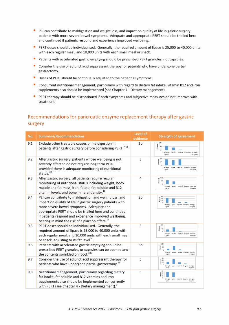
APC PERT Guidelines 2015 – Chapter 9 – PERT post gastric surgery 9-5
� PEI can contribute to maldigestion and weight loss, and impact on quality of life in gastric surgery patients with more severe bowel symptoms. Adequate and appropriate PERT should be trialled here and continued if patients respond and experience improved wellbeing.
� PERT doses should be individualised. Generally, the required amount of lipase is 25,000 to 40,000 units with each regular meal, and 10,000 units with each small meal or snack.
� Patients with accelerated gastric emptying should be prescribed PERT granules, not capsules.
� Consider the use of adjunct acid suppressant therapy for patients who have undergone partial gastrectomy.
� Doses of PERT should be continually adjusted to the patient’s symptoms.
� Concurrent nutritional management, particularly with regard to dietary fat intake, vitamin B12 and iron supplements also should be implemented (see Chapter 4 - Dietary management).
� PERT therapy should be discontinued if both symptoms and subjective measures do not improve with treatment.
Recommendations for pancreatic enzyme replacement therapy after gastric surgery
No. Summary/Recommendation Level of evidence Strength of agreement
9.1 Exclude other treatable causes of maldigestion in patients after gastric surgery before considering PERT.7;11
3b
9.2 After gastric surgery, patients whose wellbeing is not
severely affected do not require long term PERT, provided there is adequate monitoring of nutritional status.18
5
9.3 After gastric surgery, all patients require regular monitoring of nutritional status including weight, body muscle and fat mass, iron, folate, fat-soluble and B12 vitamin levels, and bone mineral density.18
4
9.4 PEI can contribute to maldigestion and weight loss, and impact on quality of life in gastric surgery patients with more severe bowel symptoms. Adequate and appropriate PERT should be trialled here and continued if patients respond and experience improved wellbeing, bearing in mind the risk of a placebo effect.11
3b
9.5 PERT doses should be individualised. Generally, the required amount of lipase is 25,000 to 40,000 units with each regular meal, and 10,000 units with each small meal or snack, adjusting to its fat level14.
5
9.6 Patients with accelerated gastric emptying should be prescribed PERT granules, or capsules can be opened and the contents sprinkled on food.7;13
3b
9.7 Consider the use of adjunct acid suppressant therapy for
patients who have undergone partial gastrectomy.25 5
9.8 Nutritional management, particularly regarding dietary
fat intake, fat-soluble and B12 vitamins and iron supplements also should be implemented concurrently with PERT (see Chapter 4 - Dietary management).1
5
APC PERT Guidelines 2015 – Chapter 9 – PERT post gastric surgery 9-6
No. Summary/Recommendation Level of evidence Strength of agreement
9.9 Bacterial overgrowth is a common cause of malabsorption after gastric surgery and does not respond to PERT.26
2b
References
(1) Friess H, Michalski CW. Diagnosing exocrine pancreatic insufficiency after surgery: when and which patients to treat. HPB (Oxford) 2009; 11 Suppl 3:7-10.
(2) Papini-Berto SJ, Burini RC. [Causes of malnutrition in post-gastrectomy patient]. Arq Gastroenterol 2001; 38(4):272-275.
(3) Cancer Incidences in Five Continents, Vol.IX. IARC Scientific Publications No. 160 ed. 2007.
(4) McMahon MJ, Johnston D, Hill GL, Goligher JC. Treatment of severe side effects after vagotomy and gastroenterostomy by closure of gastroenterostomy without pyloroplasty. Br Med J 1978; 1(6104):7-8.
(5) Papini-Berto SJ, Maio R, Modolo AK, dos Santos MD, Dichi I, Burini RC. [Protein-energy malnutrition in the gastrectomized patient]. Arq Gastroenterol 2002; 39(1):3-10.
(6) Smith RC, Hollinshead J, Eade YE, Holloway R, Gillett DJ. Digestive and Nutritional Status after Total Gastrectomy with a Long Roux-en-Y Reconstruction. J Gastroenterol Hepatol 1988; 3:253-279.
(7) Griffiths A, Taylor RH. Postgastrectomy pancreatic malabsorption: is there a case for intervention? Eur J Gastroenterol Hepatol 1999; 11(3):219-221.
(8) Bae JM, Park JW, Yang HK, Kim JP. Nutritional status of gastric cancer patients after total gastrectomy. World J Surg 1998; 22(3):254-260.
(9) Curro G, Centorrino T, Cogliandolo A, Dattola A, Pagano G, Barbera A et al. A clinical and nutritional comparison of biliopancreatic diversion performed with different common and alimentary channel lengths. Obes Surg 2015; 25(1):45-49.
(10) Topart P, Becouarn G, Salle A, Ritz P. Biliopancreatic diversion requires multiple vitamin and micronutrient adjustments within 2 years of surgery. Surg Obes Relat Dis 2014.
(11) Friess H, Bohm J, Muller MW, Glasbrenner B, Riepl RL, Malfertheiner P et al. Maldigestion after total gastrectomy is associated with pancreatic insufficiency. Am J Gastroenterol 1996; 91(2):341-347.
(12) Iivonen MK, Ahola TO, Matikainen MJ. Bacterial overgrowth, intestinal transit, and nutrition after total gastrectomy. Comparison of a jejunal pouch with Roux-en-Y reconstruction in a prospective random study. Scand J Gastroenterol 1998; 33(1):63-70.
(13) Rieu PN, Jansen JB, Joosten HJ, Lamers CB. Effect of gastrectomy with either Roux-en-Y or Billroth II anastomosis on small-intestinal function. Scand J Gastroenterol 1990; 25(2):185-192.
(14) Armbrecht U, Lundell L, Stockbrugger RW. The benefit of pancreatic enzyme substitution after total gastrectomy. Aliment Pharmacol Ther 1988; 2(6):493-500.

APC PERT Guidelines 2015 – Chapter 9 – PERT post gastric surgery 9-7
(15) Keller J, Layer P. Human pancreatic exocrine response to nutrients in health and disease. Gut 2005; 54 Suppl 6:vi1-28.
(16) Armbrecht U, Lundell L, Stockbruegger RW. Nutrient malassimilation after total gastrectomy and possible intervention. Digestion 1987; 37 Suppl 1:56-60.
(17) Gullo L, Costa PL, Ventrucci M, Mattioli S, Viti G, Labo G. Exocrine pancreatic function after total gastrectomy. Scand J Gastroenterol 1979; 14(4):401-407.
(18) Bragelmann R, Armbrecht U, Rosemeyer D, Schneider B, Zilly W, Stockbrugger RW. The effect of pancreatic enzyme supplementation in patients with steatorrhoea after total gastrectomy. Eur J Gastroenterol Hepatol 1999; 11(3):231-237.
(19) Dominguez-Munoz JE. Pancreatic enzyme replacement therapy: exocrine pancreatic insufficiency after gastrointestinal surgery. HPB (Oxford) 2009; 11 Suppl 3:3-6.
(20) Friess H, Bohm J, Ebert M, Buchler M. Enzyme treatment after gastrointestinal surgery. Digestion 1993; 54 Suppl 2:48-53.
(21) Keller J, Layer P. Pancreatic Enzyme Supplementation Therapy. Curr Treat Options Gastroenterol 2003; 6(5):369-374.
(22) Layer P, Keller J, Lankisch PG. Pancreatic enzyme replacement therapy. Curr Gastroenterol Rep 2001; 3(2):101-108.
(23) Keller J, Layer P. Pancreatic Enzyme Supplementation Therapy. Curr Treat Options Gastroenterol 2003; 6(5):369-374.
(24) Friess H, Tempia-Caliers AA, Gammerer G, Buchler M. Indication for Pancreatic Enzyme Substitution following Gastric Resection. Pancreatology 1[1 (Sup.)], 41-48. 2001.
Ref Type: Abstract
(25) Layer P, Keller J, Lankisch PG. Pancreatic enzyme replacement therapy. Curr Gastroenterol Rep 2001; 3(2):101-108.
(26) Iivonen MK, Ahola TO, Matikainen MJ. Bacterial overgrowth, intestinal transit, and nutrition after total gastrectomy. Comparison of a jejunal pouch with Roux-en-Y reconstruction in a prospective random study. Scand J Gastroenterol 1998; 33(1):63-70.

APC PERT Guidelines 2015 – Chapter 10 – Use of PERT after pancreatectomy 10-1
Chapter 10 Use of PERT after pancreatectomy Introduction Pancreatic resection for benign or malignant disease is a major cause of PEI1-3. Due to large residual exocrine secretion capacity, only significant (90%) loss of normal parenchyma will result in clinical PEI4;5. Various factors influence the development of PEI post parenchymal reduction. These include pre-existing parenchymal disease3;6, the pathological processes affecting the pancreatic exocrine gland and the presence of PEI before parenchymal resection. The extent of parenchymal resection as well as the type of surgical resection also influence post-surgical pancreatic exocrine function. Most patients with PEI prior to parenchymal resection are likely to have persisting or worse PEI after resection, with a few exceptions2. Figure 10.1 shows the relationship of post-resection PEI with pre-existing parenchymal damage and the extent of pancreatic resection. In patients with borderline PEI, a smaller parenchymal resection is likely to tip them over to clinical PEI. Patients with normal parenchyma will tolerate a great degree of parenchymal reduction before developing clinically significant PEI. In general, patients with normal parenchyma could tolerate a 50% parenchymal resection without developing clinically significant PEI1;2. Figure 10.1. The relationship of post-resection PEI with pre-existing parenchymal damage and the extent of pancreatic resection
The pancreatic parenchyma pre-resection In patients with normal parenchyma, resection of 30-50% of the gland is unlikely to cause PEI unless there is a post-resection complication such as ductal anastomotic stricture or remnant ischaemia.
Exte
nt o
f pre
-exi
stin
g pa
renc
hym
al d
iseas
e
Extent of parenchymal reduction
APC PERT Guidelines 2015 – Chapter 10 – Use of PERT after pancreatectomy 10-2
In patients with pre-existing PEI, it is unlikely to be reversed following parenchymal resection. However, in patients with early pancreatic duct obstruction causing PEI, a lesser resection can result in resolution of the PEI due to relief of pancreatic duct (PD) obstruction before significant parenchymal atrophy develops.
Pancreatic pathology prior to parenchymal resection
1. Pancreatic neoplasia. Pancreatic head malignancies causing PD obstruction can result in PEI prior to surgery. In patients with significant pancreatic atrophy, relief of obstruction following pancreaticoduodenectomy (Whipple’s procedure) is unlikely to improve the PEI. However in patients with benign or malignant tumours (e.g. neuroendocrine tumours) in the head without significant obstruction, pancreatic exocrine function may be preserved, especially when the parenchymal reduction is less than 50%. The patient with a neoplasm in the pancreatic tail without atrophy in the head or body is unlikely to develop PEI after distal pancreatectomy.
2. Inflammatory processes.
i) Chronic pancreatitis. The majority of patients with chronic pancreatitis have clinical or subclinical PEI (see Chapter 6). Resectional surgery will exacerbate PEI. Non-resectional surgery such as side-to-side pancreaticojejunostomy (Puestow-type procedures) may relieve symptoms of pain but are unlikely to improve pancreatic exocrine status unless surgery was performed prior to the development of pancreatic atrophy7-9.
ii) Acute pancreatitis. The development of PEI is dependent on the degree of pancreatic destruction through inflammation, ischaemic necrosis and/or subsequent necrosectomy10;11. PEI can develop after an episode of pancreatitis (see Chapter 5)10.
Pancreatic resection As indicated in Figure 10.1, the likelihood of developing PEI correlates with the extent of parenchymal resection and increases with the extent of existing parenchymal disease.
1. Extent of resection. Studies indicate that a 30-50% resection of normal parenchyma in otherwise normal pancreas is unlikely to cause clinically significant PEI. However, in the diseased pancreas with atrophy, inflammation or ductal obstruction, only a minor reduction in parenchyma, in the setting of borderline exocrine function, is likely to result in PEI.
2. Type of resection.
i) Pancreaticoduodenectomy. The most common pancreatic resection is pancreaticoduodenectomy (Whipple’s procedure or its variations). This includes resection of the pancreatic head, gallbladder, common bile duct and duodenum en-bloc as illustrated in Figure 10.2. The pancreas, bile duct and stomach are reconnected to the small intestine. This restores digestive function by re-establishing the flow of pancreatic juice from the pancreas, bile from the bile duct and food from the stomach. The site of transection of the pancreas varies depending on the extent of disease process in the head. In general, the resection line is anterior to the superior mesenteric vein (SMV). However, this may be extended to the left of the SMV into the body of the pancreas depending on the extent of disease. Most Whipple’s procedures are performed for tumours causing PD obstruction with varying degrees of PEI as well as endocrine insufficiency (diabetes). Hence, published series of Whipple’s resections report a higher rate of PEI compared with those of distal pancreatectomy2. Note that -
a. Creation of a long jejunal loop to the pancreaticojejunostomy can result in asynchrony where enzymes do not mix with the chyme,
b. A pylorus-preserving procedure with a Billroth I type gastrojejunal anastomosis may achieve a more anatomical connection of the pancreaticojejunostomy, thereby reducing the risk of postoperative PEI,

APC PERT Guidelines 2015 – Chapter 10 – Use of PERT after pancreatectomy 10-3
c. Pancreaticogastrostomy results in acid destruction of enzymes released into the stomach and therefore increases the risk of PEI,
d. Comparison of Cattell (mucosa to mucosa) and mattress anastomotic techniques in a randomised controlled study of 113 patients12 showed no difference in enzyme requirements.
Figure 10.2 Pancreaticoduodenectomy (Whipple’s resection) for pancreatic head tumour.
Made available with permission from The Elkins Pancreas Center at Baylor College of Medicine, Houston, Texas, USA. ii) Distal pancreatectomy. The majority of these patients have normal PD in the head and body of the
pancreas and hence a relatively normal parenchyma (see Figure 10.3). Distal pancreatectomy is therefore less likely to be associated with PEI post resection in the absence of diffuse parenchymal disease13.
Figure 10.3 Distal pancreatectomy with preservation of the pancreatic body and head.
Made available with permission from The Elkins Pancreas Center at Baylor College of Medicine, Houston, Texas, USA.

APC PERT Guidelines 2015 – Chapter 10 – Use of PERT after pancreatectomy 10-4
iii) Central pancreatectomy. This is a less common type of resection. Patients with central pancreatectomy may develop PEI depending on the extent of resection as well as adequate drainage of the tail of the pancreas. Central resection is often performed for more benign tumours to preserve pancreatic exocrine and endocrine function. Recent reports indicate that central parenchymal resection preserves pancreatic exocrine function14-16.
iv) Total or subtotal pancreatectomy. Subtotal pancreatectomy is sometimes performed for the adult form of nesidioblastosis17-19, and involves resection of 90-95% of the parenchyma, leaving a rim of pancreatic head next to the duodenum. Invariably, all these patients develop PEI, similar to those undergoing total pancreatectomy.
Assessment of PEI after resection
1. Pre-resectional PEI. The patient in this category is likely to have worse PEI postoperatively, with a few exceptions where the pancreatic duct obstruction is relieved at resection.
2. Normal parenchyma. Patients with <50% resection, especially of the pancreatic tail, may not need to undergo formal PEI testing in the absence of clinical PEI symptoms.
3. Parenchymal atrophy or inflammation with pancreatic resection. Patients with no clinical PEI will need to undergo PEI investigation as outlined in Chapter 2.
4. Late PEI post resection. A significant number of patients will develop pancreatic duct anastomotic stricture after Whipple’s procedure or central pancreatectomy. Patients with dilated ducts may develop PEI which will require pancreatic exocrine replacement.
5. The types of pancreatic resection affect PEI evaluation as well as the efficacy of PERT. Surgical resection, including classical pancreaticoduodenectomy and its variants, can affect intestinal anatomy. With jejunal reconstruction, pancreatic secretions are delivered to food further downstream, which affects the type of test which can be used for valid assessment of PEI. The administration of PERT is also affected, with distal enzyme release leading to uneven mixing with chyme. This renders the use of FE-1 to detect PEI inaccurate20. Patients with preservation of the duodenum after distal pancreatectomy are less likely to suffer from such asynchrony.
Pancreatic enzyme replacement therapy Most patients who undergo major pancreatic resection have a degree of exocrine insufficiency. Pancreatic enzyme replacement therapy (PERT) may be an important part of the long-term management of some of these patients21;22. The key clinical issue is to identify those who require enzyme replacement. Symptoms of pancreatic insufficiency may not be obvious, and sequelae of malnutrition such as osteoporosis due to deficiency of the fat-soluble vitamins, particularly Vitamin D, may not become apparent for many years. In patients where PEI is identified or suspected, adequate PERT reduces malnutrition, normalises biochemical indices of malnutrition, assists a patient to recover much of their original body weight and improves their overall quality of life23;24. There is limited information concerning the routine use of PERT, and there are no data that define a threshold for the requirement of replacement therapy. Furthermore, objective measures of PEI have not proven to be clinically useful at this point in time.
Individualised treatment is required Patients can have different degrees of PEI. The dosage of pancreatic enzymes should therefore be titrated to the symptoms of the individual patient23;25;26. All patients should be monitored and screened for symptoms and signs of inadequate enzyme replacement. These include weight loss, diarrhoea, steatorrhoea and stool fat excretion in excess of 15 g/day23.
APC PERT Guidelines 2015 – Chapter 10 – Use of PERT after pancreatectomy 10-5
Efficacy, dose and administration To date, there are only a few robust randomised clinical trials concerning the use of PERT post-pancreatectomy. Moreover, existing studies utilise only small sample sizes.
A study evaluating the efficacy of PERT post-pancreatectomy involved 11 patients. After surgery, all patients received four weeks of daily individualised doses of pancreatin. Patients were then randomised to receive a further four weeks of pancreatin or placebo. Intestinal absorption and nutritional status were measured at baseline, four weeks and eight weeks. Results showed that pancreatin supplementation significantly improved total energy absorption with increased absorption of dietary fat. The study also indicated advantages of continuing therapy1.
Jang et al. studied the metabolic effects of anastomotic site after a standard pancreatoduodenectomy27 in two cohorts of patients, comparing pancreatojejunostomy with pancreatogastrostomy. While quality of life was similar for patients after either anastomosis, the degree of steatorrhoea was greater after pancreatogastrostomy. This group has also demonstrated that 3 months after a standard pancreatoduodenectomy or a pylorus-preserving pancreatoduodenectomy, about 13 percent develop steatorrhoea28. In a randomised study involving 39 patients who had undergone total or partial pancreatectomy, high-dose and standard-dose pancreatin were equally effective for the treatment of PEI. However, the authors noted that improved approaches are required for managing fat malabsorption in this group of patients29.
A more recent randomised double-blinded 1-week placebo-controlled study of 58 post-pancreatectomy patients using high dose lipase (75,000 units per meal and 25,000 units per snack) for PEI after pancreatic surgery with a one-year open label extension, confirmed the efficacy of PERT with an increased co-efficient of fat absorption (CFA) in the treated group with compared with a decrease in placebo group30. This study also found that where surgery resulted in asynchrony, only 27% achieved CFA >85% when treated with PERT. In both groups at the end of the open label extension (all treated with high dose lipase after 1 week of randomisation), CFA values were all improved from baseline. The authors also noted that although the high dose PERT was effective in increasing the CFA, it was not sufficient to normalise fat absorption. The promising result at the end of one year’s treatment was a significant increase in body weight and BMI from baseline and a significant reduction of daily stool frequency and stool weight.
The relationship between the dose of pancreatic enzymes required and symptoms of maldigestion is not linear. In general, the required amount of lipase to be delivered to the small intestine with each meal is in the order of 25,000 to 50,000 units23;31;32. However, one study indicated that a higher lipase level of 75,000 units or greater may be required30. Even at this level, CFA can be improved but not normalised. Acid suppression therapy may be a useful adjunct therapy and is recommended, especially if severe steatorrhoea continues despite normal dosing of pancreatic enzymes23;31;33.
Summary Major pancreatic resections impair not only pancreatic function but also the function of the entire upper gastrointestinal tract. This can adversely affect the nutritional status and overall quality of life of these individuals.
Current literature suggests that with advances in surgical techniques and post-operative care (including optimal pancreatic enzyme replacement), the majority of patients will have good outcomes with minimal gastrointestinal symptoms after pancreatic resection, will be able to maintain adequate weight and achieve a high quality of life.
Total pancreatectomy and radical pancreatectomy are always associated with PEI and therefore require PERT.
Pancreaticoduodenectomy frequently results in PEI and this increases in incidence over time (level of evidence 2b).
Tumours in the pancreatic head are associated with a high rate of PEI at diagnosis. This increases following pancreatic head resection (level 4).
Central pancreatic resection is associated with a lower rate of PEI (level 4).
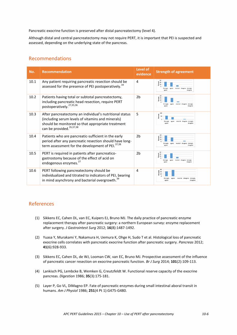
APC PERT Guidelines 2015 – Chapter 10 – Use of PERT after pancreatectomy 10-6
Pancreatic exocrine function is preserved after distal pancreatectomy (level 4).
Although distal and central pancreatectomy may not require PERT, it is important that PEI is suspected and assessed, depending on the underlying state of the pancreas.
Recommendations
No. Recommendation Level of evidence Strength of agreement
10.1 Any patient requiring pancreatic resection should be assessed for the presence of PEI postoperatively.34
4
10.2 Patients having total or subtotal pancreatectomy, including pancreatic head resection, require PERT postoperatively.27;35;36
2b
10.3 After pancreatectomy an individual’s nutritional status (including serum levels of vitamins and minerals) should be monitored so that appropriate treatment can be provided.35;37;38
5
10.4 Patients who are pancreatic-sufficient in the early period after any pancreatic resection should have long-term assessment for the development of PEI.37;38
2b
10.5 PERT is required in patients after pancreatico-gastrostomy because of the effect of acid on endogenous enzymes.27
2b
10.6 PERT following pancreatectomy should be individualized and titrated to indicators of PEI, bearing in mind asynchrony and bacterial overgrowth.39
4
References
(1) Sikkens EC, Cahen DL, van EC, Kuipers EJ, Bruno MJ. The daily practice of pancreatic enzyme replacement therapy after pancreatic surgery: a northern European survey: enzyme replacement after surgery. J Gastrointest Surg 2012; 16(8):1487-1492.
(2) Yuasa Y, Murakami Y, Nakamura H, Uemura K, Ohge H, Sudo T et al. Histological loss of pancreatic exocrine cells correlates with pancreatic exocrine function after pancreatic surgery. Pancreas 2012; 41(6):928-933.
(3) Sikkens EC, Cahen DL, de WJ, Looman CW, van EC, Bruno MJ. Prospective assessment of the influence of pancreatic cancer resection on exocrine pancreatic function. Br J Surg 2014; 101(2):109-113.
(4) Lankisch PG, Lembcke B, Wemken G, Creutzfeldt W. Functional reserve capacity of the exocrine pancreas. Digestion 1986; 35(3):175-181.
(5) Layer P, Go VL, DiMagno EP. Fate of pancreatic enzymes during small intestinal aboral transit in humans. Am J Physiol 1986; 251(4 Pt 1):G475-G480.
APC PERT Guidelines 2015 – Chapter 10 – Use of PERT after pancreatectomy 10-7
(6) Nakamura H, Murakami Y, Uemura K, Hayashidani Y, Sudo T, Ohge H et al. Reduced pancreatic parenchymal thickness indicates exocrine pancreatic insufficiency after pancreatoduodenectomy. J Surg Res 2011; 171(2):473-478.
(7) Bachmann K, Tomkoetter L, Erbes J, Hofmann B, Reeh M, Perez D et al. Beger and Frey procedures for treatment of chronic pancreatitis: comparison of outcomes at 16-year follow-up. J Am Coll Surg 2014; 219(2):208-216.
(8) Sudo T, Murakami Y, Uemura K, Hashimoto Y, Kondo N, Nakagawa N et al. Short- and long-term results of lateral pancreaticojejunostomy for chronic pancreatitis: a retrospective Japanese single-center study. J Hepatobiliary Pancreat Sci 2014; 21(6):426-432.
(9) Zheng Z, Xiang G, Tan C, Zhang H, Liu B, Gong J et al. Pancreaticoduodenectomy versus duodenum-preserving pancreatic head resection for the treatment of chronic pancreatitis. Pancreas 2012; 41(1):147-152.
(10) Xu Y, Wu D, Zeng Y, Wang X. Pancreatic exocrine function and morphology following an episode of acute pancreatitis. Pancreas 2012; 41(6):922-927.
(11) Rana SS, Bhasin DK, Rao C, Sharma R, Gupta R. Comparative evaluation of structural and functional changes in pancreas after endoscopic and surgical management of pancreatic necrosis. Ann Gastroenterol 2014; 27(2):162-166.
(12) Langrehr JM, Bahra M, Jacob D, Glanemann M, Neuhaus P. Prospective randomized comparison between a new mattress technique and Cattell (duct-to-mucosa) pancreaticojejunostomy for pancreatic resection. World J Surg 2005; 29(9):1111-9, discussion.
(13) Speicher JE, Traverso LW. Pancreatic exocrine function is preserved after distal pancreatectomy. J Gastrointest Surg 2010; 14(6):1006-1011.
(14) Zhang R, Xu X, Yan J, Wu D, Ajoodhea H, Mou Y. Laparoscopic central pancreatectomy with pancreaticojejunostomy: preliminary experience with 8 cases. J Laparoendosc Adv Surg Tech A 2013; 23(11):912-918.
(15) Xu SB, Zhu YP, Zhou W, Xie K, Mou YP. Patients get more long-term benefit from central pancreatectomy than distal resection: a meta-analysis. Eur J Surg Oncol 2013; 39(6):567-574.
(16) Sudo T, Murakami Y, Uemura K, Hayashidani Y, Hashimoto Y, Ohge H et al. Middle pancreatectomy with pancreaticogastrostomy: a technique, operative outcomes, and long-term pancreatic function. J Surg Oncol 2010; 101(1):61-65.
(17) Witteles RM, Straus II FH, Sugg SL, Koka MR, Costa EA, Kaplan EL. Adult-onset nesidioblastosis causing hypoglycemia: an important clinical entity and continuing treatment dilemma. Arch Surg 2001; 136(6):656-663.
(18) Thompson GB, Service FJ, Andrews JC, Lloyd RV, Natt N, van Heerden JA et al. Noninsulinoma pancreatogenous hypoglycemia syndrome: an update in 10 surgically treated patients. Surgery 2000; 128(6):937-944.
(19) Bjerke HS, Kelly RE, Jr., Geffner ME, Fonkalsrud EW. Surgical management of islet cell dysmaturation syndrome in young children. Surg Gynecol Obstet 1990; 171(4):321-325.
(20) Benini L, Amodio A, Campagnola P, Agugiaro F, Cristofori C, Micciolo R et al. Fecal elastase-1 is useful in the detection of steatorrhea in patients with pancreatic diseases but not after pancreatic resection. Pancreatology 2013; 13(1):38-42.
APC PERT Guidelines 2015 – Chapter 10 – Use of PERT after pancreatectomy 10-8
(21) Thorat V, Reddy N, Bhatia S, Bapaye A, Rajkumar JS, Kini DD et al. Randomised clinical trial: the efficacy and safety of pancreatin enteric-coated minimicrospheres (Creon 40000 MMS) in patients with pancreatic exocrine insufficiency due to chronic pancreatitis--a double-blind, placebo-controlled study. Aliment Pharmacol Ther 2012; 36(5):426-436.
(22) Taylor JR, Gardner TB, Waljee AK, Dimagno MJ, Schoenfeld PS. Systematic review: efficacy and safety of pancreatic enzyme supplements for exocrine pancreatic insufficiency. Aliment Pharmacol Ther 2010; 31(1):57-72.
(23) Braga M, Cristallo M, De FR, Mangiagalli A, Zerbi A, Agape D et al. Pancreatic enzyme replacement therapy in post-pancreatectomy patients. Int J Pancreatol 1989; 5 Suppl:37-44.
(24) Czako L, Takacs T, Hegyi P, Pronai L, Tulassay Z, Lakner L et al. Quality of life assessment after pancreatic enzyme replacement therapy in chronic pancreatitis. Can J Gastroenterol 2003; 17(10):597-603.
(25) Layer P, Keller J, Lankisch PG. Pancreatic enzyme replacement therapy. Curr Gastroenterol Rep 2001; 3(2):101-108.
(26) Morrow JD. Pancreatic enzyme replacement therapy. Am J Med Sci 1989; 298(5):357-359.
(27) Jang JY, Kim SW, Park SJ, Park YH. Comparison of the functional outcome after pylorus-preserving pancreatoduodenectomy: pancreatogastrostomy and pancreatojejunostomy. World J Surg 2002; 26(3):366-371.
(28) Han SS, Kim SW, Jang JY, Park YH. A comparison of the long-term functional outcomes of standard pancreatoduodenectomy and pylorus-preserving pancreatoduodenectomy. Hepatogastroenterology 2007; 54(78):1831-1835.
(29) Neoptolemos JP, Ghaneh P, Andren-Sandberg A, Bramhall S, Patankar R, Kleibeuker JH et al. Treatment of pancreatic exocrine insufficiency after pancreatic resection. Results of a randomized, double-blind, placebo-controlled, crossover study of high vs standard dose pancreatin. Int J Pancreatol 1999; 25(3):171-180.
(30) Seiler CM, Izbicki J, Varga-Szabo L, Czako L, Fiok J, Sperti C et al. Randomised clinical trial: a 1-week, double-blind, placebo-controlled study of pancreatin 25 000 Ph. Eur. minimicrospheres (Creon 25000 MMS) for pancreatic exocrine insufficiency after pancreatic surgery, with a 1-year open-label extension. Aliment Pharmacol Ther 2013; 37(7):691-702.
(31) Ferrone M, Raimondo M, Scolapio JS. Pancreatic enzyme pharmacotherapy. Pharmacotherapy 2007; 27(6):910-920.
(32) Keller J, Layer P. Pancreatic Enzyme Supplementation Therapy. Curr Treat Options Gastroenterol 2003; 6(5):369-374.
(33) Marotta F, O'Keefe SJ, Marks IN, Girdwood A, Young G. Pancreatic enzyme replacement therapy. Importance of gastric acid secretion, H2-antagonists, and enteric coating. Dig Dis Sci 1989; 34(3):456-461.
(34) Sikkens EC, Cahen DL, de WJ, Looman CW, van EC, Bruno MJ. Prospective assessment of the influence of pancreatic cancer resection on exocrine pancreatic function. Br J Surg 2014; 101(2):109-113.
(35) Neoptolemos JP, Ghaneh P, Andren-Sandberg A, Bramhall S, Patankar R, Kleibeuker JH et al. Treatment of pancreatic exocrine insufficiency after pancreatic resection. Results of a randomized, double-blind, placebo-controlled, crossover study of high vs standard dose pancreatin. Int J Pancreatol 1999; 25(3):171-180.
APC PERT Guidelines 2015 – Chapter 10 – Use of PERT after pancreatectomy 10-9
(36) Han SS, Kim SW, Jang JY, Park YH. A comparison of the long-term functional outcomes of standard pancreatoduodenectomy and pylorus-preserving pancreatoduodenectomy. Hepatogastroenterology 2007; 54(78):1831-1835.
(37) Czako L, Takacs T, Hegyi P, Pronai L, Tulassay Z, Lakner L et al. Quality of life assessment after pancreatic enzyme replacement therapy in chronic pancreatitis. Can J Gastroenterol 2003; 17(10):597-603.
(38) Braga M, Cristallo M, De FR, Mangiagalli A, Zerbi A, Agape D et al. Pancreatic enzyme replacement therapy in post-pancreatectomy patients. Int J Pancreatol 1989; 5 Suppl:37-44.
(39) Seiler CM, Izbicki J, Varga-Szabo L, Czako L, Fiok J, Sperti C et al. Randomised clinical trial: a 1-week, double-blind, placebo-controlled study of pancreatin 25 000 Ph. Eur. minimicrospheres (Creon 25000 MMS) for pancreatic exocrine insufficiency after pancreatic surgery, with a 1-year open-label extension. Aliment Pharmacol Ther 2013; 37(7):691-702.

APC PERT Guidelines 2015 – Chapter 11 – PERT in unresectable pancreatic cancer 11-1
Chapter 11
Use of PERT in unresectable pancreatic cancer Introduction Pancreatic ductal adenocarcinoma is the most common cancer of the exocrine pancreas and is the fourth most common cause of cancer-related mortality in Australia1. Nationwide, 2,780 new cases of pancreatic cancer were diagnosed in 2010 with 2,434 deaths occurring during the same time-period1. In 2012 there were an estimated 330,000 deaths attributed to pancreatic cancer globally, with an age-standardised annual incidence rate of 7.2 cases per 100,000 in developed countries2. Pancreatic cancer has one of the worst overall survival rates of any cancer with less than 5% of affected patients alive five years after initial diagnosis3. Only 10-20% of all patients with pancreatic cancer are eligible for potentially curative resection. Consequently, for the vast majority of patients, only palliative treatment options remain3.
Palliative treatment of patients with unresectable pancreatic cancer is important to achieve optimal quality of life for the duration of illness4. Palliative treatment mainly aims to prevent or treat obstructive jaundice, duodenal obstruction and pain. Many clinicians fail to give adequate attention to the cachexia, weight loss and malabsorption seen with patients during the course of their illness. The majority of patients with pancreatic carcinomas report weight loss at the time of diagnosis, with pancreatic exocrine insufficiency (PEI) appearing to be an important contributing factor5;6. This not only contributes to ongoing weight loss, but the actual extent of PEI as measured by faecal elastase-1 (FE-1) testing appears to be an independent prognostic factor7. Partelli et al. reported on 194 patients with advanced pancreatic cancer and found that the median survival was 7.3 months for 25% of patients with extremely low FE-1 level (≤ 20 µg/g) compared to 11 months in the remainder7.
Pancreatic exocrine insufficiency Patients with pancreatic cancer experience some degree of PEI, ranging from 46% to 92%, during the course of their disease6-11. In one large study of 215 patients with locally advanced and metastatic pancreatic cancer, 46% had a diagnosis of steatorrhoea at presentation6. A recent study consisting of 24 patients with locally advanced unresectable pancreatic cancer and 7 with resected pancreatic cancer identified issues relating to symptoms of malabsorption and PEI to be the major cause of impaired quality of life12. It also highlighted deficiencies in provision of adequate dietary consultation for patients with pancreatic cancer. Weight loss and cancer cachexia appears to be a universal feature of pancreatic cancer. About 90% of patients with pancreatic cancer experience weight loss at the time of diagnosis13.
Weight loss has been reported to be
an independent predictor of survival in patients with locally advanced or metastatic pancreatic cancer6. Tumour-derived factors cause metabolic abnormalities that result in increased glucose production, increased whole body protein breakdown and increased lipolysis, with an overall depletion of body protein and fat stores14. This is compounded by the release of pro-inflammatory cytokines in the tumour microenvironment, acting through peripheral pathways to augment lipolysis, proteolysis and insulin resistance and centrally mediated pathways that cause anorexia. These effects can be augmented by secondary mechanical factors that can occur such as intestinal obstruction, pain and nausea and vomiting. PEI predominantly occurs as a result of blockage of the main pancreatic duct and is compounded by pancreatic parenchymal tumour replacement15.
The likelihood of PEI is greatest in patients with tumours involving the pancreatic head, that
cause pancreatic and bile duct obstruction. In one recent study the prevalence of PEI was 66% in 32 patients with a pancreatic head tumours and increased to 92% at 2 months’ follow-up10. Palliative surgery when performed to bypass the bile duct and stomach adds to the problem of PEI when it results in an asynchrony between gastric emptying and discharge of pancreatic enzymes and bile into the
APC PERT Guidelines 2015 – Chapter 11 – PERT in unresectable pancreatic cancer 11-2
intestine16. The overall effects of PEI are faecal losses of energy through steatorrhoea and creatorrhoea5. PEI also directly impacts absorption of fat-soluble vitamins and omega-3 fatty acids, which appear particularly important for weight-gain and body mass composition17. Management Palliative care, including prevention of further weight loss, is important for these patients and their families. The provision of individualised treatment plans for patients with distressing nausea, weight loss and pain is a specialised service. The involvement of palliative care teams provides support and expertise which is greatly appreciated by patients and their families. They should be introduced to these teams early in the cancer journey so they are prepared for and confident about their symptom control as the tumour progresses. Pancreatic enzyme replacement therapy (PERT) can be important for patients with weight loss associated with pancreatic cancer18. The main principle of PERT is simulation of the normal physiologic state of exocrine pancreatic products by exogenous administration during meals19. PERT can often relieve many of the symptoms associated with PEI and can allow patients to increase food intake and improve their nutritional status20. A dietitian assessment, with evaluation of the patient’s caloric and nutrient intake, is an important component of the management approach. A prospective, randomised, placebo-controlled study of 21 patients with unresectable cancer of the pancreatic head region showed that weight loss in these patients can be reduced by high dose PERT in combination with appropriate dietary counselling5. However, PERT improved only moderate-to-severe fat and protein malabsorption. Mild fat or protein malabsorption in pancreatic cancer patients did not seem to improve with PERT15.
A separate randomised, double-blind, placebo-controlled trial also found that PERT improved
steatorrhoea and stool consistency21. In a retrospective study, 21 patients with unresectable pancreatic cancer were assessed for PEI and treated appropriately. They had significantly longer survival than 45 patients during the same period who were not assessed for PEI and did not receive PERT22. Dosage Patients with unresectable pancreatic cancer should be offered PERT and guidance by a dietitian early in their management, with a view of enabling best quality of life (see Chapter 4). A typical starting dose to consider is 25,000-50,000 IU of pancreatic lipase taken with main meals and 10,000-25,000 IU with snacks. Dosage increases may be required with increased fat intake. Other associated nutritional deficiencies Dietary omega-3 fatty acids supplementation should be considered in pancreatic cancer patients. Studies show potential benefits of omega-3 fatty acids in in suppressing the inflammatory pathways involved in cancer cachexia syndrome23. Due to potential deficiencies in fat-soluble vitamins such as A, D, E and K that can accompany PEI, multivitamin supplementation should also be considered.

APC PERT Guidelines 2015 – Chapter 11 – PERT in unresectable pancreatic cancer 11-3
Summary � Pancreatic cancer is associated with a poor prognosis. Many patients present at an advanced stage, when
curative surgery is not an option.
� About 90% of patients with pancreatic cancer have weight loss at the time of diagnosis. Weight loss may be exacerbated by malabsorption, as a result of pancreatic duct obstruction and destroyed pancreatic tissue, reducing the availability of pancreatic enzymes. This results in PEI with associated steatorrhoea.
� Even if patients do not have PEI at the time of diagnosis, the majority will develop it during the course of their disease.
� Palliative care should include the control of symptoms associated with PEI as this can have a significant impact on quality of life.
� PERT can often relieve symptoms associated with PEI and can allow patients to increase food intake and improve their nutritional status.
� Clinical studies have shown that PERT is effective and important in the nutritional management of patients with unresectable pancreatic cancer.
Recommendations for pancreatic enzyme replacement therapy in unresectable pancreatic cancer
No. Recommendation Level of evidence Strength of agreement
11.1 PERT and dietary guidance by a dietitian should be considered for treatment of PEI in patients with unresectable pancreatic cancers from the time of diagnosis in order to maintain weight and improve quality of life. 5, 21
2a
11.2 Palliative care should continue the control of symptoms associated with PEI as this can have a significant impact on quality of life of patients with unresectable pancreatic cancer. 5;6
5
Search strategy PubMed, Google Scholar and Cochrane – using terms [pancreatic cancer]/[pancreatic adenocarcinoma] AND [pancreatic exocrine insufficiency]/[pancreatic enzyme replacement therapy] from January 1974 – May 2014. Abstracts published from International Pancreas Meetings in 2013 were reviewed for relevant articles applying the same search terms to the titles. References
(1) Australian Institute of Health and Welfare & Australasian Association of Cancer Registries. Cancer in Australia: An overview 2012. Cancer Series No. 74. 2012. Canberra,ACT, Australian Institute of Health and Welfare. Ref Type: Report
APC PERT Guidelines 2015 – Chapter 11 – PERT in unresectable pancreatic cancer 11-4
(2) Ferlay J, Soerjomataram I, Ervik M, Eser S, Mathers C, Rebolo M et al. GLOBOCAN 2012 v1.0, Cancer Incidence and Mortality Worldwide. IARC CancerBase No. 11 [Internet]. 2013. Lyon, France, International Agency for Research on Cancer. Ref Type: Report
(3) Warshaw AL, Fernandez-del CC. Pancreatic carcinoma. N Engl J Med 1992; 326(7):455-465.
(4) Freelove R, Walling AD. Pancreatic cancer: diagnosis and management. Am Fam Physician 2006; 73(3):485-492.
(5) Bruno MJ, Haverkort EB, Tijssen GP, Tytgat GN, van Leeuwen DJ. Placebo controlled trial of enteric coated pancreatin microsphere treatment in patients with unresectable cancer of the pancreatic head region. Gut 1998; 42(1):92-96.
(6) Papadoniou N, Kosmas C, Gennatas K, Polyzos A, Mouratidou D, Skopelitis E et al. Prognostic factors in patients with locally advanced (unresectable) or metastatic pancreatic adenocarcinoma: a retrospective analysis. Anticancer Res 2008; 28(1B):543-549.
(7) Partelli S, Frulloni L, Minniti C, Bassi C, Barugola G, D'Onofrio M et al. Faecal elastase-1 is an independent predictor of survival in advanced pancreatic cancer. Dig Liver Dis 2012; 44(11):945-951.
(8) Matsumoto J, Traverso LW. Exocrine function following the whipple operation as assessed by stool elastase. J Gastrointest Surg 2006; 10(9):1225-1229.
(9) Perez MM, Newcomer AD, Moertel CG, Go VL, Dimagno EP. Assessment of weight loss, food intake, fat metabolism, malabsorption, and treatment of pancreatic insufficiency in pancreatic cancer. Cancer 1983; 52(2):346-352.
(10) Sikkens EC, Cahen DL, de WJ, Looman CW, van EC, Bruno MJ. A prospective assessment of the natural course of the exocrine pancreatic function in patients with a pancreatic head tumor. J Clin Gastroenterol 2014; 48(5):e43-e46.
(11) Watson L. Exocrine insufficiency and pancreatic enzyme replacement therapy in pancreatic cancer. Clin Oncol (R Coll Radiol ) 2010; 22(5):391.
(12) Gooden HM, White KJ. Pancreatic cancer and supportive care--pancreatic exocrine insufficiency negatively impacts on quality of life. Support Care Cancer 2013; 21(7):1835-1841.
(13) Smith RC, Talley NJ, Dent OF, Jones M, Waller SL. Exocrine pancreatic function and chronic unexplained dyspepsia. A case-control study. Int J Pancreatol 1991; 8(3):253-262.
(14) Heber D, Tchekmedyian NS. Pathophysiology of cancer: hormonal and metabolic abnormalities. Oncology 1992; 49 Suppl 2:28-31.
(15) Dimagno EP, Malagelada JR, Go VL. The relationships between pancreatic ductal obstruction and pancreatic secretion in man. Mayo Clin Proc 1979; 54(3):157-162.
(16) Bruno MJ, Haverkort EB, Tytgat GN, van Leeuwen DJ. Maldigestion associated with exocrine pancreatic insufficiency: implications of gastrointestinal physiology and properties of enzyme preparations for a cause-related and patient-tailored treatment. Am J Gastroenterol 1995; 90(9):1383-1393.
(17) Tan CR, Yaffee PM, Jamil LH, Lo SK, Nissen N, Pandol SJ et al. Pancreatic cancer cachexia: a review of mechanisms and therapeutics. Front Physiol 2014; 5:88.
(18) Keller J, Layer P. Human pancreatic exocrine response to nutrients in health and disease. Gut 2005; 54 Suppl 6:vi1-28.
APC PERT Guidelines 2015 – Chapter 11 – PERT in unresectable pancreatic cancer 11-5
(19) Damerla V, Gotlieb V, Larson H, Saif MW. Pancreatic enzyme supplementation in pancreatic cancer. J Support Oncol 2008; 6(8):393-396.
(20) Guidelines for the management of patients with pancreatic cancer periampullary and ampullary carcinomas. Gut 2005; 54 Suppl 5:v1-16.
(21) Safdi M, Bekal PK, Martin S, Saeed ZA, Burton F, Toskes PP. The effects of oral pancreatic enzymes (Creon 10 capsule) on steatorrhea: a multicenter, placebo-controlled, parallel group trial in subjects with chronic pancreatitis. Pancreas 2006; 33(2):156-162.
(22) Dominguez-Munoz JE, Nieto-Garcia L, Iglesias-Garcia J. Impact of diagnosis and treatment of pancreatic exocrine insufficiency (PEI) on survival of patients with unresectable pancreatic cancer (PC). Pancreatology 13[3], S80. 2013. Ref Type: Abstract
(23) Boutros C, Somasundar P, Razzak A, Helton S, Espat NJ. Omega-3 fatty acids: investigations from cytokine regulation to pancreatic cancer gene suppression. Arch Surg 2010; 145(6):515-520.

APC PERT Guidelines 2015 – Chapter 12 – Diabetes and PERT 12-1
Chapter 12
Diabetes Mellitus Introduction Diabetes is a well-recognised complication of diseases of the pancreas; this form of diabetes has pathophysiology and management considerations that are distinct from those for type 1 or 2 diabetes, and pancreatic enzyme replacement therapy has a potential role in improving glycaemic control in such patients. Conversely, both PEI and steatorrhoea can be documented in a substantial proportion of patients with type 1 and 2 diabetes. However, direct tests of pancreatic exocrine function suggest that PEI in this setting is usually only mild to moderate, and many cases of steatorrhoea appear unrelated to pancreatic exocrine dysfunction. Therefore, the value of investigating pancreatic exocrine function in patients with diabetes remains controversial.
Diabetes as a consequence of pancreatic exocrine disease (type 3c) Incidence and criteria for diagnosis The American Diabetes Association Classification of Diabetes recognises pancreatogenic diabetes as a distinct form of the disease, designated type 3c1. Specific underlying pancreatic diseases listed in the classification include pancreatitis, trauma or pancreatectomy, neoplasia, cystic fibrosis, haemochromatosis and fibrocalculous pancreatopathy. Estimates of the proportion of diabetes that is pancreatogenic vary widely, from 1-2% in western nations to 15-20% in India and Southeast Asia, with the high prevalence in the latter regions attributable to fibrocalculous disease2. A review of almost 2000 patients with diabetes attending a German tertiary hospital suggested that pancreatogenic diabetes is under-recognised, and that 8% could be classified as type 3c on the basis of imaging, demonstration of PEI, and absence of autoantibodies3. Although there are no agreed guidelines, Ewald et al proposed that all these three criteria must be present to categorise diabetes as type 3c. Additional features of impaired beta cell function (as determined, for example, by HOMA-B (homeostasis model assessment of beta cell function), lack of insulin resistance (e.g. by HOMA-IR), impaired incretin secretion, and low concentrations of fat-soluble vitamins are deemed suggestive, but not essential to the diagnosis4. The risk of diabetes in chronic pancreatitis increases over time; in a French cohort, over 80% developed diabetes when followed for up to 25 years, and about half ultimately required insulin therapy5. Pathophysiology of type 3c diabetes The pathophysiology of type 3c diabetes is distinct from types 1 and 22, and predisposes patients to postprandial hyperglycaemia in particular. In addition to deficient insulin secretion, there is a deficiency of pancreatic polypeptide (mainly secreted from the head of the pancreas), which may result in failure to suppress postprandial hepatic glucose production, and impaired secretion of the incretin hormones, which play an important role in postprandial blood glucose regulation. Moreover, a deficiency of glucagon secretion (in contrast to types 1 and 2 diabetes), together with malabsorption of nutrients, and intact or enhanced peripheral insulin sensitivity, predispose these patients to episodes of hypoglycaemia. Treatment of type 3c diabetes There is limited evidence to guide treatment of type 3c diabetes2. Glycaemic control is important, given that these patients are at similar risk of microvascular complications as patients with type 1 and 2 diabetes6. In the latter, prevention of macrovascular complications appears to rely more on good
APC PERT Guidelines 2015 – Chapter 12 – Diabetes and PERT 12-2
control of lipids and blood pressure than on tight glycaemic control7, but there is no evidence specifically relating to type 3c diabetes. Patients should be screened for deficiencies of fat-soluble vitamins. Metformin is generally recommended for control of glycaemia, and may reduce the risk of pancreatic cancer8. Many patients require insulin or insulin secretagogues, although there is evidence that these may increase cancer risk8. Thiazolidinediones, which are peripheral insulin sensitizers, should be avoided. Incretin-based therapies (glucagon-like peptide-1 (GLP-1) receptor agonists and dipeptidyl peptidase 4 inhibitors) have in themselves been linked to acute pancreatitis – though this is controversial9 – and for this reason they have not been generally recommended in patients with chronic pancreatitis. The incretin hormones, GLP-1 and glucose-dependent insulinotropic polypeptide (GIP) are released from the small and large intestine in response to nutrient exposure, with fat being a potent stimulus for their secretion. Both hormones stimulate insulin secretion in a glucose-dependent fashion, while GLP-1 also slows gastric emptying and suppresses glucagon, all of which limit postprandial glycaemic excursions. Patients with chronic pancreatitis and fat malabsorption (e.g. due to alcohol use or cystic fibrosis) have impaired secretion of GLP-1 and GIP together with rapid emptying of high fat/carbohydrate meals from the stomach and prominent early postprandial hyperglycaemia, all of which are ameliorated acutely when pancreatic enzyme replacement therapy (PERT) is provided 10-13. This should translate to improved glycaemic control with use of long term PERT, but remains to be firmly established in a clinical trial. Patients with diabetes in the setting of tropical calculous pancreatitis had improvements in postprandial blood glucose and glycated haemoglobin (HbA1c) when treated with PERT for 6 months 14, but this study was open-label and uncontrolled. Two placebo-controlled studies failed to show improvements in overall glycaemic control after 4 or 14 days’ PERT in patients with chronic pancreatitis, although the variation in blood glucose values was decreased during PERT in one study 15, and glycaemic control became more unstable when changing from placebo to PERT, or vice versa, in the other, with more hypoglycaemic episodes seen in the placebo group16. New-onset diabetes and diagnosis of pancreatic cancer Identifying patients with early pancreatic cancer is difficult but important.17 Recent-onset diabetes is more common in patients with a new diagnosis of pancreatic cancer18 and a clue to the diagnosis may be greater weight loss in diabetic than in non-diabetic cancer patients. CA 19-9 has been found to be elevated up to 24 months before pancreatic cancer becomes overt and may be a useful indicator.19 Diabetic patients with pancreatic cancer were found to be more likely to have perineural invasion and were less likely to have pain.20 Pancreatic exocrine insufficiency as a complication of diabetes types 1 and 2 There are several plausible reasons why diabetes could be complicated by pancreatic exocrine dysfunction4. Any process affecting the islets – such as autoimmune destruction – could also affect the exocrine component of the gland. Moreover, there may be a loss of the trophic effect of insulin on the pancreas, or a suppressive effect from high levels of glucagon, vascular damage resulting from small vessel disease, or impaired entero-pancreatic reflexes due to autonomic neuropathy. Finally, acute hyperglycaemia can reversibly impair exocrine pancreatic secretion21. Direct pancreatic function tests have been relatively consistent over several decades in showing reduced enzyme and/or bicarbonate secretion in response to hormonal stimulation in about 40-80% of patients with type 1 or 2 diabetes22-26. However, in almost all cases the impairment in exocrine function was mild to moderate, and not to the degree that would be associated with overt fat malabsorption. Furthermore, most studies showed no association – or only a weak relationship – between pancreatic exocrine dysfunction and duration of type 1 diabetes, and when such patients
APC PERT Guidelines 2015 – Chapter 12 – Diabetes and PERT 12-3
were followed up over a decade after their initial evaluation, any exocrine insufficiency tended not to progress27. Value of indirect pancreatic function tests in diabetes Over the last 10-15 years there has been considerable interest in the use of indirect pancreatic function tests, particularly faecal elastase-1 (FE-1) assays, to explore the prevalence of PEI in patients with diabetes28-32. Most studies indicate that in type 1 diabetes, 35-50% of patients have FE-1 concentrations < 200 µg/g, of whom 20-30% have concentrations < 100 µg/g (“severe” deficiency), with corresponding values of 20-35% and 10-20% in type 2 diabetes, compared to healthy controls where about 5% have FE-1 < 200 µg/g. Most of these studies specifically examined whether duration of diabetes was related to FE-1 concentration, and could find no association. Few studies have determined whether low FE-1 concentrations are associated with symptoms; Hardt et al could not show such a relationship, although their patients with diabetes overall had more gastrointestinal symptoms than non-diabetic controls28. A fundamental concern is whether FE-1 is a sufficiently accurate measure of pancreatic exocrine function to be useful in clinical practice. Although it has good sensitivity for detecting patients who have severe PEI on direct testing, both its sensitivity and specificity for detecting mild to moderate PEI are limited33. In patients with diabetes, there have been few attempts to compare FE-1 concentrations with direct tests of pancreatic exocrine function. Hahn et al studied 33 patients with longstanding type 1 diabetes using the secretin-caerulein test, and reported that 11 patients had PEI, all of whom were only mildly to moderately deficient34. The positive predictive value of an abnormal FE-1 concentration for PEI was about 40%, the negative predictive value about 70%, and sensitivity and specificity about 55% and 60% respectively. FE-1 testing has been reported in general to have good reproducibility35, although this issue has not been specifically examined in patients with diabetes. Few studies have related abnormal FE-1 to steatorrhoea in patients with type 1 or 2 diabetes. Two groups evaluated faecal fat in patients with FE-1 deficiency in the “severe” range (< 100 µg/g), and found that 35-40% had steatorrhoea31;36, but in neither case was there a control group of patients with normal FE-1. In the study of Hahn et al, 8 of 33 patients with type 1 diabetes had steatorrhoea, but only half of these had any degree of PEI on direct function testing, and none had <10% of normal pancreatic exocrine function, which would be the usual threshold for inducing pancreatogenic steatorrhoea34. The implication is that steatorrhoea is relatively common in diabetes, but is often of non-pancreatogenic origin, e.g. due to small intestinal bacterial overgrowth. One study specifically examined the impact of PERT in patients with diabetes who were found to have FE-1 concentrations in the “severely deficient” range (< 100 µg/g)37. Eighty “insulin-treated” patients were randomized to 16 weeks of PERT or placebo. Although PERT had no apparent adverse effects, there were no differences in fasting or 2-hour blood glucose concentrations on an oral glucose tolerance test, between the PERT and placebo groups, and no differences in HbA1c, insulin doses, or gastrointestinal symptoms. However, this study has the limitations that it would likely have been underpowered to show a change in HbA1c, and it would be more relevant to study blood glucose concentrations after a mixed meal containing fat, than after a glucose drink. Better-designed and adequately powered trials are needed to determine the benefits of PERT in this setting in terms of postprandial glycaemic control, gastrointestinal symptoms, and absorption of fat-soluble vitamins.

APC PERT Guidelines 2015 – Chapter 12 – Diabetes and PERT 12-4
Summary The proportion of diabetes that is pancreatogenic (type 3c) is likely to be underestimated, but may be as high as 5-10% in western nations. There are specific management considerations in type 3c diabetes, but more evidence is needed to determine the benefits of stringent attempts to exclude a pancreatic cause in patients thought otherwise to have type 1 or 2 diabetes (or other types). Pancreatic exocrine deficiency is prevalent in type 1 or 2 diabetes when patients are evaluated with direct tests of exocrine function. However, this form of PEI is usually mild to moderate and appears not to progress substantially over time. It is unclear whether this has any implications for management. FE-1 results appear to correlate poorly with results of direct tests of exocrine function in diabetes type I and 2 patients, but more data are needed. Limited randomised controlled trial data do not support treating patients with PERT simply on the basis of very low faecal elastase levels (< 100 µg/g), but further studies with carefully chosen endpoints are needed to be more definitive.
Recommendations
No. Recommendation Level of evidence Strength of agreement
12.1 Rarely is there a need to use PERT in patients with diabetes. Limited randomised controlled trial data do not support treating patients with PERT simply on the basis of very low faecal elastase-1 levels (< 100 µg/g). 22-26
2b
12.2 New onset diabetes in an at-risk patient, especially if they have lost weight, should prompt consideration of tests for pancreatic exocrine disease and for cancer; e.g. faecal fat, other pancreatic function tests, Ca19-9.19
3b
12.3 Patients with type 3c diabetes should be screened for deficiencies of fat-soluble vitamins.
5
Search strategy This chapter evaluated published data regarding pancreatic exocrine insufficiency and the use of pancreatic enzyme replacement therapy in diabetes mellitus. Abstracts were sourced using the MEDLINE database from 1966 to September 2014 and full papers were obtained for all relevant studies. The search was performed by combining the terms ‘diabetes’, ‘type 3c diabetes’, ‘type 3 diabetes’, ‘blood glucose’, or ‘glycaemia’ with ‘pancreatogenic’, ‘pancreatic exocrine insufficiency’, ‘chronic pancreatitis’, ‘faecal elastase’, ‘pancreatic enzyme replacement’, ‘exocrine function’, ‘secretin-pancreozymin’, ‘cerulein’, or ‘steatorrhoea’. Reference lists from relevant papers were also
APC PERT Guidelines 2015 – Chapter 12 – Diabetes and PERT 12-5
searched for additional publications not identified in the original search. A total of 37 original articles and reviews were identified as being relevant to this chapter.
References (1) American Diabetes Association. Diagnosis and classification of diabetes mellitus. Diabetes
Care 2010; 33(Suppl 1):S62-S69.
(2) Cui Y, Andersen DK. Pancreatogenic diabetes: special considerations for management. Pancreatology 2011; 11:279-294.
(3) Hardt PD, Brendel MD, Kloer HU, Bretzel RG. Is pancreatic diabetes (type 3c diabetes) underdiagnosed and misdiagnosed? Diabetes Care 2008; 31(Suppl 2):S165-S169.
(4) Ewald N, Bretzel RG. Diabetes mellitus secondary to pancreatic diseases (Type 3c)--are we neglecting an important disease? Eur J Intern Med 2013; 24:203-206.
(5) Malka D, Hammel P, Sauvanet A, Rufat P, O'Toole D, Bardet P et al. Risk factors for diabetes mellitus in chronic pancreatitis. Gastroenterology 2000; 119:1324-1332.
(6) Couet C, Genton P, Pointel JP, Louis J, Gross P, Saudax E et al. The prevalence of retinopathy is similar in diabetes mellitus secondary to chronic pancreatitis with or without pancreatectomy and in idiopathic diabetes mellitus. Diabetes Care 1985; 8:323-328.
(7) Joseph JJ, Golden SH. Type 2 diabetes and cardiovascular disease: what next? Curr Opin Endocrinol Diabetes Obes 2014; 21:109-120.
(8) Li D, Yeung SC, Hassan MM, Konopleva M, Abbruzzese JL. Antidiabetic therapies affect risk of pancreatic cancer. Gastroenterology 2009; 137:482-488.
(9) Meier JJ, Nauck MA. Risk of pancreatitis in patients treated with incretin-based therapies. Diabetologia 2014; 57:1320-1324.
(10) Ebert R, Creutzfeldt W. Reversal of impaired GIP and insulin secretion in patients with pancreatogenic steatorrhea following enzyme substitution. Diabetologia 1980; 19:198-204.
(11) Knop FK, Vilsboll T, Larsen S, Hojberg PV, Volund A, Madsbad S et al. Increased postprandial responses of GLP-1 and GIP in patients with chronic pancreatitis and steatorrhea following pancreatic enzyme substitution. 292:E324-330. Am J Physiol Endocrinol Metab 2007; 292:E324-E330.
(12) Kuo P, Stevens JE, Russo A, Maddox A, Wishart JM, Jones KL et al. Gastric emptying, incretin hormone secretion, and postprandial glycemia in cystic fibrosis--effects of pancreatic enzyme supplementation.96:E851-855. J Clin Endocrinol Metab 2011; 96:E851-E855.
(13) Perano SJ, Couper JJ, Horowitz M, Martin AJ, Kritas S, Sullivan T et al. Pancreatic enzyme supplementation improves the incretin hormone response and attenuates postprandial glycemia in adolescents with cystic fibrosis: a randomized crossover trial. J Clin Endocrinol Metab 2014; 99:2486-2493.
(14) Mohan V, Poongothai S, Pitchumoni CS. Oral pancreatic enzyme therapy in the control of diabetes mellitus in tropical calculous pancreatitis. Int J Pancreatol 1998; 24:19-22.
(15) Glasbrenner B, Malfertheiner P, Kerner W, Scherbaum WA, Ditschuneit H. [Effect of pancreatin on diabetes mellitus in chronic pancreatitis]. Z Gastroenterol 1990; 28:275-279.
APC PERT Guidelines 2015 – Chapter 12 – Diabetes and PERT 12-6
(16) O'Keefe SJ, Cariem AK, Levy M. The exacerbation of pancreatic endocrine dysfunction by potent pancreatic exocrine supplements in patients with chronic pancreatitis. J Clin Gastroenterol 2001; 32:319-323.
(17) Mizuno S, Nakai Y, Isayama H, Yanai A, Takahara N, Miyabayashi K et al. Risk factors and early signs of pancreatic cancer in diabetes: screening strategy based on diabetes onset age. J Gastroenterol 2013; 48(2):238-246.
(18) Tantau A, Negrean V, Alexescu T, Para I, Tarmure S, Casoinic F et al. Two different types of diabetes mellitus in pancreatic cancer population. Comparative study between new onset and long standing diabetes mellitus on 76 patients with pancreatic cancer. Rom J Intern Med 2014; 52(1):18-23.
(19) O'Brien DP, Sandanayake NS, Jenkinson C, Gentry-Maharaj A, Apostolidou S, Fourkala EO et al. Serum CA19-9 Is Significantly Upregulated up to 2 Years before Diagnosis with Pancreatic Cancer: Implications for Early Disease Detection. Clin Cancer Res 2015; 21(3):622-631.
(20) Sahin IH, Shama MA, Tanaka M, Abbruzzese JL, Curley SA, Hassan M et al. Association of diabetes and perineural invasion in pancreatic cancer. Cancer Med 2012; 1(3):357-362.
(21) Lam WF, Masclee AA, Souverijn JH, Lamers CB. Effect of acute hyperglycemia on basal, secretin and secretin + cholecystokinin stimulated exocrine pancreatic secretion in humans. Life Sci 1999; 64:617-626.
(22) Chey WY, Shay H, Shuman CR. External pancreatic secretion in diabetes mellitus. Ann Intern Med 1963; 59:812-821.
(23) Frier BM, Saunders JH, Wormsley KG, Bouchier IA. Exocrine pancreatic function in juvenile-onset diabetes mellitus. Gut 1976; 17:685-691.
(24) Lankisch PG, Manthey G, Otto J, Koop H, Talaulicar M, Willms B et al. Exocrine pancreatic function in insulin-dependent diabetes mellitus. Digestion 1982; 25:211-216.
(25) Pollard HM, Miller L, Brewer WA. The external secretion of the pancreas and diabetes mellitus. Am J Dig Dis 1943; 10:20-23.
(26) Vacca JB, Henke WJ, Knight WAJr. The exocrine pancreas in diabetes mellitus. Ann Intern Med 1964; 61:242-247.
(27) Creutzfeldt W, Gleichmann D, Otto J, Stockmann F, Maisonneuve P, Lankisch PG. Follow-up of exocrine pancreatic function in type-1 diabetes mellitus. Digestion 2005; 72:71-75.
(28) Hardt PD, Krauss A, Bretz L, Porsch-Ozcurumez M, Schnell-Kretschmer H, Maser E et al. Pancreatic exocrine function in patients with type 1 and type 2 diabetes mellitus. Acta Diabetol 2000; 37:105-110.
(29) Hardt PD, Hauenschild A, Nalop J, Marzeion AM, Jaeger C, Teichmann J et al. High prevalence of exocrine pancreatic insufficiency in diabetes mellitus. A multicenter study screening fecal elastase 1 concentrations in 1,021 diabetic patients. Pancreatology 2003; 3:395-402.
(30) Larger E, Philippe MF, Barbot-Trystram L, Radu A, Rotariu M, Nobecourt E et al. Pancreatic exocrine function in patients with diabetes. Diabet Med 2012; 29:1047-1054.
(31) Nunes AC, Pontes JM, Rosa A, Gomes L, Carvalheiro M, Freitas D. Screening for pancreatic exocrine insufficiency in patients with diabetes mellitus. 2003;98:2672-2675. Am J Gastroenterol 2003; 98:2672-2675.

APC PERT Guidelines 2015 – Chapter 12 – Diabetes and PERT 12-7
(32) Rathmann W, Haastert B, Icks A, Giani G, Hennings S, Mitchell J et al. Low faecal elastase 1 concentrations in type 2 diabetes mellitus. Scand J Gastroenterol 2001; 36:1056-1061.
(33) Lankisch PG, Schmidt I, Konig H, Lehnick D, Knollmann R, Lohr M et al. Faecal elastase 1: not helpful in diagnosing chronic pancreatitis associated with mild to moderate exocrine pancreatic insufficiency. Gut 1998; 42:551-554.
(34) Hahn JU, Kerner W, Maisonneuve P, Lowenfels AB, Lankisch PG. Low fecal elastase 1 levels do not indicate exocrine pancreatic insufficiency in type-1 diabetes mellitus. Pancreas 2008; 36:274-278.
(35) Boeck WG, Adler G, Gress TM. Pancreatic function tests: when to choose, what to use. Curr Gastroenterol Rep 2001; 3:95-100.
(36) Hardt PD, Hauenschild A, Jaeger C, Teichmann J, Bretzel RG, Kloer HU. High prevalence of steatorrhea in 101 diabetic patients likely to suffer from exocrine pancreatic insufficiency according to low fecal elastase 1 concentrations: a prospective multicenter study. 2003;48:1688-1692. Dig Dis Sci 2003; 48:1688-1692.
(37) Ewald N, Bretzel RG, Fantus IG, Hollenhorst M, Kloer HU, Hardt PD. Pancreatin therapy in patients with insulin-treated diabetes mellitus and exocrine pancreatic insufficiency according to low fecal elastase 1 concentrations. Results of a prospective multi-centre trial. Diabetes Metab Res Rev 2007; 23:386-391.
APC PERT Guidelines 2015 – Chapter 13 – Coeliac disease and PERT 13-1
Chapter 13 Coeliac disease and pancreatic enzyme replacement therapy Introduction Coeliac disease (CD, also known as nontropical sprue or gluten-sensitive enteropathy) is an autoimmune disorder targeting the small intestine that is morphologically characterised by blunting of small intestinal villi, crypt hyperplasia, and lymphocyte infiltration of crypts. This damage impairs the absorptive function of the gut, resulting in diarrhoea, malabsorption and related systemic changes1. The manifestation of coeliac disease is the result of an interaction between genetic, environmental, and immunologic factors. On the background of a genetic predisposition (the presence of the HLADQ2/DQ8 serotype) and environmental triggers (consumption of gluten), an immune dysregulation occurs resulting in structural damage to the intestinal mucosa2. Gluten is the trigger for the immune process. Gluten is a storage protein that can be found in wheat. This storage protein provides elasticity to dough. While initially only used for wheat protein, gluten is now used as the term to describe all similar proteins from other grains including rye or barley, that are able to induce an immune response since the proteins are recognised as antigens. Oats contain a gluten-type protein (avenin) that share some peptide sequences with wheat gluten. However, concentrations are substantially lower. Prolamin proteins from other grains mostly lack the peptide sequences that trigger the immune response. CD is triggered by the ingestion of gluten. The gluten must breach the protective barrier of the small intestine and be presented to the B and T cells of the mucosal immune system by major histocompatibility complex molecules (MHCs), and present on antigen-presenting cells (APCs) such as dendritic cells. This presentation of gluten to APCs appears to be facilitated by a defective regulation of tight junction proteins3-5. Australian data suggest that the prevalence of anti-tTG antibodies in the population is 1.56%, and the prevalence of CD is at least 0.56% based upon histology or positive results of anti-tTG assays and an HLA haplotype consistent with CD6. Effects of impaired intestinal function on pancreatic function Pancreatic exocrine secretion in humans occurs during the interdigestive and postprandial periods of the digestive cycle7 and is controlled via afferent messaging originating from the upper small intestine. The physiological processes involved include well described feedback mechanisms related to cholecystokinin (CCK)- and secretin-releasing factors of pancreatic and duodenal origin, along with the active pancreatic proteases present in the upper gut8. In addition there are peptides – mostly originating from the intestine – that are involved in the regulatory process of pancreatic secretion. Regulatory peptides include somatostatin, peptide YY, pancreatic polypeptide, glucagon, ghrelin, and leptin8;9. Finally, luminal bile and bile salts modulate pancreatic enzyme secretion10. The well-established role of enteropancreatic pathways for the regulation of pancreatic function suggests that the inflammatory process that characterises CD is likely to have an effect on pancreatic secretion. Exocrine pancreatic function in coeliac disease Studies examining pancreatic exocrine function in CD are summarised in Table 13.1. In the presence of mucosal atrophy, pancreatic function (response to nutrients) is abnormal in CD. The impairment is related to the severity of mucosal damage11. Diminished exocrine pancreatic function in individuals having villous
APC PERT Guidelines 2015 – Chapter 13 – Coeliac disease and PERT 13-2
atrophy appears to be linked to decreased CCK secretion11. However, CCK as well as pancreatic secretion are normalised after healing of the CD-related mucosal inflammation11.
On the other hand, Otte et al. observed normal HCO3 but reduced enzyme secretion in 22 patients by using secretin-pancreozymin stimulation before the start of a gluten-free diet12. It was noted that even after injection of CCK-pancreozymin, there was reduced contraction of the gall bladder. While the enzyme secretion was significantly reduced as compared to healthy controls, the reduction was much smaller as compared to reductions seen in patients with clinically relevant pancreatic enzyme insufficiency (PEI)13.
While overall the coexistence of CD and advanced PEI appears to be rare, Regan and DiMagno from the Mayo Clinic identified in a series of 31 patients with CD, 3 patients who had severe pancreatic insufficiency14. CCK-stimulated duodenal tryptic activity or lipolytic activity (or both) was reduced in 13 (42%), but severely reduced duodenal enzyme activities were only found in three cases (10%). At the same time, the morphology of the small bowel appeared normal in 21 patients. The authors concluded that mild-to-moderate PEI is a frequent finding in untreated CD. It needs to be noted that the significant, but nevertheless small reduction of enzyme secretion was unlikely to explain steatorrhoea in the majority of patients. In another study by Carroccio et al15, paediatric CD patients were included. One group of patients had total or subtotal atrophy of the intestinal mucosa, another group had complete remission of histologic changes during a gluten free diet and in addition, control subjects of similar age with normal jejunal histology were included. Exocrine pancreatic function was determined utilising the secretin-caerulein test. During the test, bicarbonate concentration and lipase, phospholipase, and chymotrypsin activity were measured after intravenous stimulation with 1 clinical unit of secretin and 75 ng/kg body weight of ceruletide. Ten out of 44 untreated coeliac patients showed reduced tryptic or lipolytic activity. In addition, the mean value of the faecal chymotrypsin concentration was significantly lower in untreated than in treated coeliac patients or in control subjects. In another study, pancreatic enzyme secretion was stimulated with intravenous arginine in 18 adult patients with treated CD. In three patients, the indirect para-aminobenzoic acid (PABA) test suggested PEI16. However, it needs to be noted that the results of this indirect oral pancreatic function test might be affected by impaired absorptive function of the small bowel17. The largest observational study is authored by Leeds and co-workers18. This study included newly diagnosed CD patients, patients in remission and CD patients with ongoing GI symptoms. While 11% of the newly diagnosed patients had lowered pancreatic elastase levels in the stool, this proportion was 30% in patients with ongoing GI symptoms (diarrhoea) in spite of a gluten free diet. Interestingly, it is reported that in 18 out of 20 patients, enzyme replacement therapy improved symptoms18. In summary, the available data suggest that in patients with active CD, there is a reduction of pancreatic enzyme secretion in response to endogenous (meal) stimulation. In addition, there appears to be a reduced response to humoral stimulation. While the reduction in pancreatic enzyme secretion is statistically significant, it is not necessarily sufficient to explain on its own clinical malabsorption or diarrhoea since there is general consensus that, for example, lipolytic activity must be reduced by 80 to 90% before clinical signs of pancreatic insufficiency occurs13. Nevertheless, it might be speculated that in the presence of a significant mucosal inflammation that also impairs the absorptive function, clinical signs of PEI may occur much earlier. Pancreatic disease in patients with CD The available data suggest that at least in a subgroup of patients with CD, there is impaired pancreatic function (see above) and cases of calcifying pancreatitis in CD have been reported19. However, little is known about the prevalence of pancreatic disease in patients with CD. A recent study from Sweden20 analysed data of 29,000 patients with biopsy-proven CD by matching histology data with hospital records. The absolute risk of any pancreatitis among patients with CD was 126/100,000 person-years, with an excess risk of 81/100,000 person-years. The risk for gallstone-related acute pancreatitis was 1.59 (95% CI, 1.06-2.40), for non-gallstone-related acute pancreatitis 1.86 (95% CI, 1.52-2.26), for chronic pancreatitis was 3.33 (95% CI, 2.33-4.76). In addition, patients with CD were 5 times more likely to receive pancreatic enzyme replacement therapy (PERT). Thus,
APC PERT Guidelines 2015 – Chapter 13 – Coeliac disease and PERT 13-3
based upon these data, patients with CD have an almost 3-fold increase in risk of developing pancreatitis compared with the general population. It is unknown if the autoimmune process or other factors contribute to this. Thus, the data suggest that CD is a risk factor for the development of pancreatic disease. Based upon this, appropriate diagnostic work-up is required in patients with CD who may have – based upon persisting symptoms of malabsorption – true pancreatic comorbidities.
Role of pancreatic insufficiency in non-responsive CD In a case series from the Mayo clinic, 55 patients non-responsive to a gluten free diet were studied. More than 50% had gluten contamination while one patient was later diagnosed pancreatic cancer who had pancreatic insufficiency21. Thus, pancreatic insufficiency might be considered as a potential cause for non-response to gluten-free diet. However, while the reduction of enzyme secretion in patients with CD and the prevalence of severe pancreatic insufficiency (> 80-90% reduction of enzyme secretion) is low, there might be pancreatic insufficiency as an independent disease. Indeed, in one small study two out of 11 patients with CD not responding to a gluten free diet were diagnosed with pancreatic insufficiency22. Thus, testing for pancreatic insufficiency might be warranted in selected cases. Enzyme therapy in patients with CD A significant proportion of patients with CD do not respond to a gluten free diet. This might be due to the failure to comply with the dietary requirements, but even in patients with histologic remission, symptoms may persist. One group reported that in 20 out of 66 adult coeliac patients with current or persistent diarrhoea, the underlying cause was PEI21. Interestingly, 19 out of these 20 patients improved on pancreatic supplementation. When followed-up for up to 4 years, 11 out of these 19 patients continued to take the PERT (mean dose of 45,000 units of lipase per day). One patient had not experienced any improvement, and in total 8 out of 19 patients had discontinued supplementation. Overall repeat testing revealed that faecal elastase had substantially increased over time and the authors concluded that pancreatic enzyme supplementation could be discontinued in a substantial proportion of patients as symptoms improve21. PERT in patients with CD could target a concomitant pancreatic enzyme deficiency or could also be considered as an approach to deliver enzymes capable of proteolysing gluten (i.e., glutenases)23. A study conducted more than ten years ago assessed the effect of an enzyme preparation based on an animal intestinal extract. The concept is that these enzymes digest the gluten before it can initiate the immune process24. Patients received a modest gluten challenge consisting of three cracker biscuits (3.5 g each) daily for 14 days, making a total of 13 g of gluten for each day in randomised fashion with or without PERT. During the gluten challenge, epithelial stunting and lamina propria lymphocytic infiltration were the measures which improved most in association with PERT versus control. While this trial tested intestinal enzymes from an animal preparation, it is uncertain if commercially available pancreatic enzyme preparations will have similar effects. Overall, properly randomised and controlled trials assessing the value of PERT in CD patients are lacking. Summary The secretion of digestive enzymes into the duodenum is controlled by humoral and neural factors. For obvious reasons these enteropancreatic pathway25 factors are closely interrelated with the functional integrity of the small intestinal mucosa (e.g. by the release of regulatory peptides). While available data are limited (e.g. small sample sizes and many studies used indirect tests that might be affected by the impaired intestinal absorptive capacity), there is sufficient evidence to assume that in untreated CD patients, there is a reduced pancreatic enzyme response to luminal nutrients.
The data also suggest that even the pancreatic enzyme response to stimulation with secretin and ceruletide is diminished12. This may suggest that structural changes occur during the course of the disease.
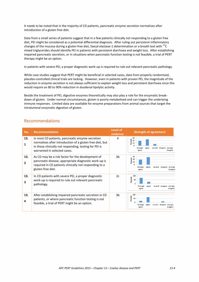
APC PERT Guidelines 2015 – Chapter 13 – Coeliac disease and PERT 13-4
It needs to be noted that in the majority of CD patients, pancreatic enzyme secretion normalises after introduction of a gluten free diet. Data from a small series of patients suggest that in a few patients clinically not responding to a gluten free diet, PEI might be considered as a potential differential diagnosis. After ruling out persistent inflammatory changes of the mucosa during a gluten free diet, faecal elastase-1 determination or a breath test with 13C mixed triglycerides should identify PEI in patients with persistent diarrhoea and weight loss. After establishing impaired pancreatic secretion, or in situations when pancreatic function testing is not feasible, a trial of PERT therapy might be an option. In patients with severe PEI, a proper diagnostic work-up is required to rule out relevant pancreatic pathology. While case studies suggest that PERT might be beneficial in selected cases, data from properly randomised, placebo-controlled clinical trials are lacking. However, even in patients with proven PEI, the magnitude of the reduction in enzyme secretion is not always sufficient to explain weight loss and persistent diarrhoea since this would require an 80 to 90% reduction in duodenal lipolytic activity. Beside the treatment of PEI, digestive enzymes theoretically may also play a role for the enzymatic break-down of gluten. Under normal circumstances, gluten is poorly metabolised and can trigger the underlying immune responses. Limited data are available for enzyme preparations from animal sources that target the intraluminal enzymatic digestion of gluten.
Recommendations
No. Recommendation Level of evidence Strength of agreement
13.
1
In most CD patients, pancreatic enzyme secretion normalises after introduction of a gluten free diet, but in those clinically not responding, testing for PEI is warranted in selected cases.
4
13.
2
As CD may be a risk factor for the development of pancreatic disease, appropriate diagnostic work-up is required in CD patients clinically not responding to a gluten-free diet.
3b
13.
3
In CD patients with severe PEI, a proper diagnostic work-up is required to rule out relevant pancreatic pathology.
2c
13.
4
After establishing impaired pancreatic secretion in CD patients, or where pancreatic function testing is not feasible, a trial of PERT might be an option.
3b
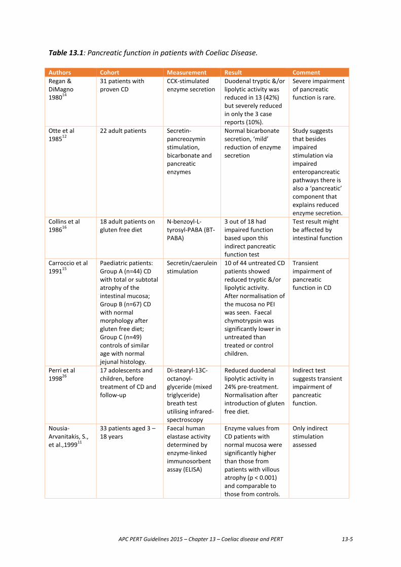
APC PERT Guidelines 2015 – Chapter 13 – Coeliac disease and PERT 13-5
Table 13.1: Pancreatic function in patients with Coeliac Disease.
Authors Cohort Measurement Result Comment Regan & DiMagno 198014
31 patients with proven CD
CCK-stimulated enzyme secretion
Duodenal tryptic &/or lipolytic activity was reduced in 13 (42%) but severely reduced in only the 3 case reports (10%).
Severe impairment of pancreatic function is rare.
Otte et al 198512
22 adult patients Secretin-pancreozymin stimulation, bicarbonate and pancreatic enzymes
Normal bicarbonate secretion, ‘mild’ reduction of enzyme secretion
Study suggests that besides impaired stimulation via impaired enteropancreatic pathways there is also a ‘pancreatic’ component that explains reduced enzyme secretion.
Collins et al 198616
18 adult patients on gluten free diet
N-benzoyl-L-tyrosyl-PABA (BT-PABA)
3 out of 18 had impaired function based upon this indirect pancreatic function test
Test result might be affected by intestinal function
Carroccio et al 199115
Paediatric patients: Group A (n=44) CD with total or subtotal atrophy of the intestinal mucosa; Group B (n=67) CD with normal morphology after gluten free diet; Group C (n=49) controls of similar age with normal jejunal histology.
Secretin/caerulein stimulation
10 of 44 untreated CD patients showed reduced tryptic &/or lipolytic activity. After normalisation of the mucosa no PEI was seen. Faecal chymotrypsin was significantly lower in untreated than treated or control children.
Transient impairment of pancreatic function in CD
Perri et al 199826
17 adolescents and children, before treatment of CD and follow-up
Di-stearyl-13C-octanoyl-glyceride (mixed triglyceride) breath test utilising infrared-spectroscopy
Reduced duodenal lipolytic activity in 24% pre-treatment. Normalisation after introduction of gluten free diet.
Indirect test suggests transient impairment of pancreatic function.
Nousia-Arvanitakis, S., et al.,199911
33 patients aged 3 – 18 years
Faecal human elastase activity determined by enzyme-linked immunosorbent assay (ELISA)
Enzyme values from CD patients with normal mucosa were significantly higher than those from patients with villous atrophy (p < 0.001) and comparable to those from controls.
Only indirect stimulation assessed
APC PERT Guidelines 2015 – Chapter 13 – Coeliac disease and PERT 13-6
Authors Cohort Measurement Result Comment Leeds et al 200718
259 patients: n=57 newly diagnosed CD, n=86 symptom free on gluten free diet, n=66 on gluten free diet with diarrhoea, n-50 chronic diarrhoea without CD
Faecal elastase Low faecal elastase is common in patients with CD before initiation of therapy or with ongoing symptoms.
In an open fashion, some patients received PERT and it is reported that symptoms improved.
References
(1) Kupfer SS, Jabri B. Pathophysiology of celiac disease. Gastrointest Endosc Clin N Am 2012; 22(4):639-660.
(2) Barker JM, Liu E. Celiac disease: pathophysiology, clinical manifestations, and associated autoimmune conditions. Adv Pediatr 2008; 55:349-365.
(3) Harris KM, Fasano A, Mann DL. Monocytes differentiated with IL-15 support Th17 and Th1 responses to wheat gliadin: implications for celiac disease. Clin Immunol 2010; 135(3):430-439.
(4) Palova-Jelinkova L, Rozkova D, Pecharova B, Bartova J, Sediva A, Tlaskalova-Hogenova H et al. Gliadin fragments induce phenotypic and functional maturation of human dendritic cells. J Immunol 2005; 175(10):7038-7045.
(5) Qiao SW, Bergseng E, Molberg O, Xia J, Fleckenstein B, Khosla C et al. Antigen presentation to celiac lesion-derived T cells of a 33-mer gliadin peptide naturally formed by gastrointestinal digestion. J Immunol 2004; 173(3):1757-1762.
(6) Chin MW, Mallon DF, Cullen DJ, Olynyk JK, Mollison LC, Pearce CB. Screening for coeliac disease using anti-tissue transglutaminase antibody assays, and prevalence of the disease in an Australian community. Med J Aust 2009; 190(8):429-432.
(7) Holtmann G, Kelly DG, DiMagno EP. Nutrients and cyclical interdigestive pancreatic enzyme secretion in humans. Gut 1996; 38(6):920-924.
(8) Garces MC, Gomez-Cerezo J, Codoceo R, Grande C, Barbado J, Vazquez JJ. Postprandial cholecystokinin response in patients with chronic pancreatitis in treatment with oral substitutive pancreatic enzymes. Dig Dis Sci 1998; 43(3):562-566.
(9) Dirksen C, Jorgensen NB, Bojsen-Moller KN, Kielgast U, Jacobsen SH, Clausen TR et al. Gut hormones, early dumping and resting energy expenditure in patients with good and poor weight loss response after Roux-en-Y gastric bypass. Int J Obes (Lond) 2013; 37(11):1452-1459.
(10) Morisset J. Negative control of human pancreatic secretion: physiological mechanisms and factors. Pancreas 2008; 37(1):1-12.
(11) Nousia-Arvanitakis S, Karagiozoglou-Lamboudes T, Aggouridaki C, Malaka-Lambrellis E, Galli-Tsinopoulou A, Xefteri M. Influence of jejunal morphology changes on exocrine pancreatic function in celiac disease. J Pediatr Gastroenterol Nutr 1999; 29(1):81-85.
(12) Otte M, Thurmayr GR, Dageforde J, Thurmayr R, Forell MM. [Pancreatic secretion in domestic sprue]. Dtsch Med Wochenschr 1985; 110(7):259-264.
APC PERT Guidelines 2015 – Chapter 13 – Coeliac disease and PERT 13-7
(13) DiMagno EP, Go VL, Summerskill WH. Relations between pancreatic enzyme ouputs and malabsorption in severe pancreatic insufficiency. N Engl J Med 1973; 288(16):813-815.
(14) Regan PT, DiMagno EP. Exocrine pancreatic insufficiency in celiac sprue: a cause of treatment failure. Gastroenterology 1980; 78(3):484-487.
(15) Carroccio A, Iacono G, Montalto G, Cavataio F, Di MC, Balsamo V et al. Exocrine pancreatic function in children with coeliac disease before and after a gluten free diet. Gut 1991; 32(7):796-799.
(16) Collins BJ, Bell PM, Boyd S, Kerr J, Buchanan KD, Love AH. Endocrine and exocrine pancreatic function in treated coeliac disease. Pancreas 1986; 1(2):143-147.
(17) Keller J, Aghdassi AA, Lerch MM, Mayerle JV, Layer P. Tests of pancreatic exocrine function - clinical significance in pancreatic and non-pancreatic disorders. Best Pract Res Clin Gastroenterol 2009; 23(3):425-439.
(18) Leeds JS, Hopper AD, Hurlstone DP, Edwards SJ, McAlindon ME, Lobo AJ et al. Is exocrine pancreatic insufficiency in adult coeliac disease a cause of persisting symptoms? Aliment Pharmacol Ther 2007; 25(3):265-271.
(19) Arya S, Rana SS, Sinha SK, Nagi B, Bhasin DK. Celiac disease and chronic calcific pancreatitis with pancreas divisum. Gastrointest Endosc 2006; 63(7):1080-1081.
(20) Sadr-Azodi O, Sanders DS, Murray JA, Ludvigsson JF. Patients with celiac disease have an increased risk for pancreatitis. Clin Gastroenterol Hepatol 2012; 10(10):1136-1142.
(21) Abdulkarim AS, Burgart LJ, See J, Murray JA. Etiology of nonresponsive celiac disease: results of a systematic approach. Am J Gastroenterol 2002; 97(8):2016-2021.
(22) Fine KD, Meyer RL, Lee EL. The prevalence and causes of chronic diarrhea in patients with celiac sprue treated with a gluten-free diet. Gastroenterology 1997; 112(6):1830-1838.
(23) Bethune MT, Khosla C. Oral enzyme therapy for celiac sprue. Methods Enzymol 2012; 502:241-271.
(24) Cornell HJ, Macrae FA, Melny J, Pizzey CJ, Cook F, Mason S et al. Enzyme therapy for management of coeliac disease. Scand J Gastroenterol 2005; 40(11):1304-1312.
(25) Niebergall-Roth E, Singer MV. Enteropancreatic reflexes mediating the pancreatic enzyme response to nutrients. Auton Neurosci 2006; 125(1-2):62-69.
(26) Perri F, Pastore M, Festa V, Clemente R, Quitadamo M, D'Altilia MR et al. Intraduodenal lipase activity in celiac disease assessed by means of 13C mixed-triglyceride breath test. J Pediatr Gastroenterol Nutr 1998; 27(4):407-410.
APC PERT Guidelines 2015 – Chapter 14 – PERT and IBS 14-1
Chapter 14 PERT and the irritable bowel syndrome
Introduction The irritable bowel syndrome (IBS) is a clinical condition characterized by abdominal pain or discomfort, bloating and disturbed defaecation1;2. Typically patients have intermittent and erratic symptoms but a subset have more chronic complaints. IBS afflicts approximately 10% to 15% of Australians1. Subtypes of IBS according to the Rome III criteria include diarrhoea-predominant, constipation-predominant and mixed depending on stool consistency (form) but an unclassifiable group is also recognized. IBS also overlaps with unexplained chronic (functional) diarrhoea and chronic (functional) constipation as well as functional dyspepsia and other disorders2. The pathogenesis of IBS is unclear but disturbances of motor and sensory gut function, and brain-gut disturbances have been described. IBS can follow an episode of infectious gastroenteritis2. More recently, subsets with IBS-like symptoms have been recognised to have undiagnosed coeliac disease, microscopic colitis and bile salt malabsorption causing diarrhoea3;4. There is preliminary evidence that pancreatic exocrine insufficiency (PEI) may also contribute to diarrhoea-predominant irritable bowel syndrome but this remains to be confirmed in large prospective studies5-8.
Pancreatic exocrine function The first suggestion that PEI may be present in some patients with IBS came from a case-control study conducted in 19868. In this small study, 14CO2 breath sampling was used to measure the absorption of 14C-triolein from a standard fat meal. Results from 66 patients with gastrointestinal disorders were compared with 60 controls. Twenty percent of those with IBS had subnormal values in the 14C-triolein breath test. However, this test has a low sensitivity and specificity for PEI8. A better-designed study investigated the prevalence of PEI which was assessed in 314 patients with diarrhoea-predominant IBS5. Nineteen patients (6.1%, 95% CI 3.7 - 9.3%) had faecal elastase-1 (FE-1) levels less than 100 µg/g stool, suggesting severe PEI, compared with no patients with chronic diarrhoea of other cause or healthy controls; this finding was unlikely to be due to chance (P<0.001). While the prevalence of low FE-1 levels was significantly higher in IBS with diarrhoea, selection or measurement bias cannot be ruled out as explanations for the findings (see Chapter 2). Pancreatic enzyme replacement therapy The efficacy of pancreatic enzyme replacement therapy (PERT) in diarrhoea-predominant IBS was also assessed in the above case-control study5. Nineteen patients with FE-1 levels below 100 µg/g stool and fifteen patients with normal FE-1 levels (>200 µg/g stool) were treated with 30,000 units of lipase three times per day for 12 weeks. Eighteen patients (94.7%) with low FE-1 levels and one patient with normal FE-1 (6.7%) had a clinically significant response to therapy. Supplementation with pancreatic enzymes led to significant improvements in stool frequency, stool consistency and abdominal pain in patients with low FE-1 levels compared with controls. However, objective evidence of PEI in all these cases was not obtained. Other data suggest that, irrespective of pancreatic function status, pancreatic enzymes may provide a benefit in IBS patients with diarrhoea. A case report of a patient with clinical features of diarrhoea-predominant IBS reported successful treatment with pancreatic enzymes6. She judged the enzyme supplementation to be more effective than placebo at reducing postprandial symptoms and continued to be successfully treated with enzymes for an additional four years.
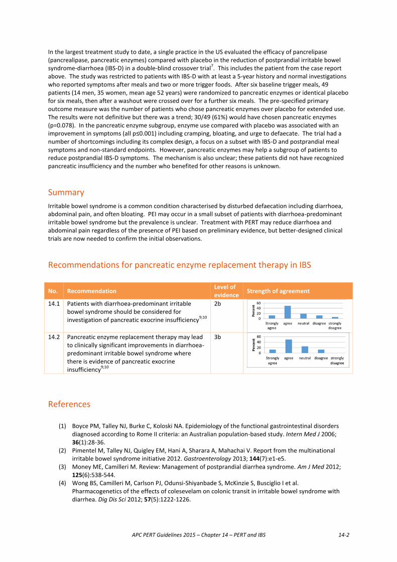
APC PERT Guidelines 2015 – Chapter 14 – PERT and IBS 14-2
In the largest treatment study to date, a single practice in the US evaluated the efficacy of pancrelipase (pancrealipase, pancreatic enzymes) compared with placebo in the reduction of postprandial irritable bowel syndrome-diarrhoea (IBS-D) in a double-blind crossover trial7. This includes the patient from the case report above. The study was restricted to patients with IBS-D with at least a 5-year history and normal investigations who reported symptoms after meals and two or more trigger foods. After six baseline trigger meals, 49 patients (14 men, 35 women, mean age 52 years) were randomized to pancreatic enzymes or identical placebo for six meals, then after a washout were crossed over for a further six meals. The pre-specified primary outcome measure was the number of patients who chose pancreatic enzymes over placebo for extended use. The results were not definitive but there was a trend; 30/49 (61%) would have chosen pancreatic enzymes (p=0.078). In the pancreatic enzyme subgroup, enzyme use compared with placebo was associated with an improvement in symptoms (all p≤0.001) including cramping, bloating, and urge to defaecate. The trial had a number of shortcomings including its complex design, a focus on a subset with IBS-D and postprandial meal symptoms and non-standard endpoints. However, pancreatic enzymes may help a subgroup of patients to reduce postprandial IBS-D symptoms. The mechanism is also unclear; these patients did not have recognized pancreatic insufficiency and the number who benefited for other reasons is unknown.
Summary Irritable bowel syndrome is a common condition characterised by disturbed defaecation including diarrhoea, abdominal pain, and often bloating. PEI may occur in a small subset of patients with diarrhoea-predominant irritable bowel syndrome but the prevalence is unclear. Treatment with PERT may reduce diarrhoea and abdominal pain regardless of the presence of PEI based on preliminary evidence, but better-designed clinical trials are now needed to confirm the initial observations.
Recommendations for pancreatic enzyme replacement therapy in IBS
No. Recommendation Level of evidence Strength of agreement
14.1 Patients with diarrhoea-predominant irritable bowel syndrome should be considered for investigation of pancreatic exocrine insufficiency9;10
2b
14.2 Pancreatic enzyme replacement therapy may lead
to clinically significant improvements in diarrhoea-predominant irritable bowel syndrome where there is evidence of pancreatic exocrine insufficiency9;10
3b
References
(1) Boyce PM, Talley NJ, Burke C, Koloski NA. Epidemiology of the functional gastrointestinal disorders diagnosed according to Rome II criteria: an Australian population-based study. Intern Med J 2006; 36(1):28-36.
(2) Pimentel M, Talley NJ, Quigley EM, Hani A, Sharara A, Mahachai V. Report from the multinational irritable bowel syndrome initiative 2012. Gastroenterology 2013; 144(7):e1-e5.
(3) Money ME, Camilleri M. Review: Management of postprandial diarrhea syndrome. Am J Med 2012; 125(6):538-544.
(4) Wong BS, Camilleri M, Carlson PJ, Odunsi-Shiyanbade S, McKinzie S, Busciglio I et al. Pharmacogenetics of the effects of colesevelam on colonic transit in irritable bowel syndrome with diarrhea. Dig Dis Sci 2012; 57(5):1222-1226.
APC PERT Guidelines 2015 – Chapter 14 – PERT and IBS 14-3
(5) Leeds JS, Hopper AD, Sidhu R, Simmonette A, Azadbakht N, Hoggard N et al. Some patients with irritable bowel syndrome may have exocrine pancreatic insufficiency. Clin Gastroenterol Hepatol 2010; 8(5):433-438.
(6) Money ME, Hofmann AF, Hagey LR, Walkowiak J, Talley NJ. Treatment of irritable bowel syndrome-diarrhea with pancrealipase or colesevelam and association with steatorrhea. Pancreas 2009; 38(2):232-233.
(7) Money ME, Walkowiak J, Virgilio C, Talley NJ. Pilot study: a randomised, double blind, placebo controlled trial of pancrealipase for the treatment of postprandial irritable bowel syndrome-diarrhoea. Frontline Gastroenterol 2011; 2(1):48-56.
(8) Mylvaganam K, Hudson PR, Ross A, Williams CP. 14C triolein breath test: a routine test in the gastroenterology clinic? Gut 1986; 27(11):1347-1352.
(9) Leeds JS, Hopper AD, Sidhu R, Simmonette A, Azadbakht N, Hoggard N et al. Some patients with irritable bowel syndrome may have exocrine pancreatic insufficiency. Clin Gastroenterol Hepatol 2010; 8(5):433-438.
(10) Money ME, Hofmann AF, Hagey LR, Walkowiak J, Talley NJ. Treatment of irritable bowel syndrome-diarrhea with pancrealipase or colesevelam and association with steatorrhea. Pancreas 2009; 38(2):232-233.
APC PERT Guidelines 2015 – Chapter 15 - The ageing pancreas 15-1
Chapter 15 The Ageing pancreas The number and proportion of elderly people in the Australian and New Zealand populations is rising because of declining fertility and increased longevity resulting from improvements in medical technologies. Elderly people may have accumulated structural changes in the pancreas which have not caused pain, resulting in silent pancreatic enzyme insufficiency (PEI). Many studies have demonstrated diminishing pancreatic enzyme secretion with ageing (See Table 15.1). Such patients should not be denied the possibility of a cure for their progressive weight loss. However, there are several difficulties with this concept:
� The reduced pancreatic function may be just a component of general functional decline of the gastrointestinal tract where the pancreas is not necessarily the only ageing organ.
� The tests used in these studies have a recognised false positive rate which is important when screening otherwise normal individuals, and therefore these results need to consider the high potential for over-diagnosis. Although over-prescribing pancreatic enzyme replacement therapy (PERT) is not considered to carry a risk of complications, it is inconvenient and expensive for society.
Recent authors have described the importance of PEI as a cause of weight loss and deterioration in elderly people1. Confirming which of these patients needs PERT requires measurement of PEI with minimal error. In the above study, 20% of people without gastrointestinal disease had low levels of FE-1 and 9% had levels less than 100 U1. Does this mean that 20% of elderly people require PERT? Given the growing number of elderly people in this situation, a test with greater accuracy than FE-1 is necessary because of the risk of false positive results in about 20% of subjects (See Table 2.2, Chapter2). However, undertaking invasive studies in this group is also impractical. In a study from 1992 where PABA excretion was used to measure PEI in 21 elderly normal subjects compared with 29 healthy young subjects, 19 percent of the elderly had significantly reduced pancreatic function and the remaining subjects had normal function2. Given that these subjects were clinically normal it is difficult to interpret this result as an indication for the prescription of PERT but this result does question the value of such studies in this population. The 14C triolein breath test also demonstrates progressive deterioration in pancreatic function with age so that age-dependent normal ranges have been proposed: 65 years or less (201 to 460% cumulative value of administered dose), 66 to 75 years (182 to 405%) and over 75 years (141 to 336%). However factors such as bacterial overgrowth, reduced intestinal absorption and a slower metabolic rate cannot be separated as a cause from PEI using this test. Even so, the authors found the test to be a simple and non-invasive means of studying this population,3 but it may be difficult to access in Australasia. However this test does require the collection of exhaled breath over a number of hours which can be inconvenient for elderly people. The nutritional status of any patient with PEI is always at risk. Malnutrition is a known problem for the ageing population, particularly within the acute setting where rates have shown to be as high as 61%18. Common contributing factors are diarrhoea and malabsorption, resulting in weight loss. While having a similar presentation, PEI is considered an uncommon cause of these symptoms 12-15. However it has been recommended that PEI is considered in older patients who present with weight loss of unknown aetiology 16-17.
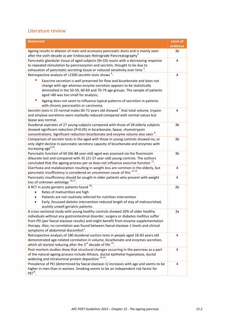
APC PERT Guidelines 2015 – Chapter 15 - The ageing pancreas 15-2
Literature review Statement Level of
evidence Ageing results in dilation of main and accessory pancreatic ducts and is mainly seen after the sixth decade as per Endoscopic Retrograde Pancreatography4
3b
Pancreatic glandular tissue of aged subjects (N=10) reacts with a decreasing response to repeated stimulation by pancreozymin and secretin, thought to be due to exhaustion of pancreatic secreting tissue or reduced sensitivity over time 5.
4
Retrospective analysis of >2300 secretin tests shows 6: � Exocrine secretion is well preserved for flow and bicarbonate and does not
change with age whereas enzyme secretion appears to be statistically diminished in the 50-59, 60-69 and 70-79 age groups. The sample of patients aged >80 was too small for analysis;
� Ageing does not seem to influence typical patterns of secretion in patients with chronic pancreatitis or carcinoma.
4
Secretin tests in 23 normal males 60-72 years old showed 7 that total volume, trypsin and amylase secretions were markedly reduced compared with normal values but lipase was normal.
4
Duodenal aspirates of 27 young subjects compared with those of 28 elderly subjects showed significant reduction (P<0.05) in bicarbonate, lipase, chymotrypsin concentrations. Significant reduction bicarbonate and enzyme volume also seen 8.
3b
Comparison of secretin tests in the aged with those in young controls showed no, or only slight decline in pancreatic secretory capacity of bicarbonate and enzymes with increasing age9;10.
3b
Pancreatic function of 60 (66-88 year-old) aged was assessed via the fluorescein dilaurate test and compared with 35 (21-57 year-old) young controls. The authors concluded that the ageing process per se does not influence exocrine function 11.
3b
Diarrhoea and malabsorption resulting in weight loss are common in the elderly, but pancreatic insufficiency is considered an uncommon cause of this 12-15.
4
Pancreatic insufficiency should be sought in older patients who present with weight loss of unknown aetiology 16;17.
4
A RCT in acute geriatric patients found 18: x Rates of malnutrition are high x Patients are not routinely referred for nutrition intervention x Early, focussed dietetic intervention reduced length of stay of malnourished,
acutely unwell geriatric patients.
2b
A cross-sectional study with young healthy controls showed 20% of older healthy individuals without any gastrointestinal disorder, surgery or diabetes mellitus suffer from PEI (per faecal elastase results) and might benefit from enzyme supplementation therapy. Also, no correlation was found between faecal elastase-1 levels and clinical symptoms of abdominal discomfort1.
3a
Retrospective analysis of 180 duodenal suction tests in people aged 18-83 years old demonstrated age-related correlation in volume, bicarbonate and enzymes secretion, which all started reducing after the 3rd decade of life 19.
4
Post-mortem studies show that structural changes occurring in the pancreas as a part of the natural ageing process include lithiasis, ductal epithelial hyperplasia, ductal widening and intraluminal protein deposition 20-23.
4
Prevalence of PEI (determined by faecal elastase-1) increases with age and seems to be higher in men than in women. Smoking seems to be an independent risk factor for PEI24.
4
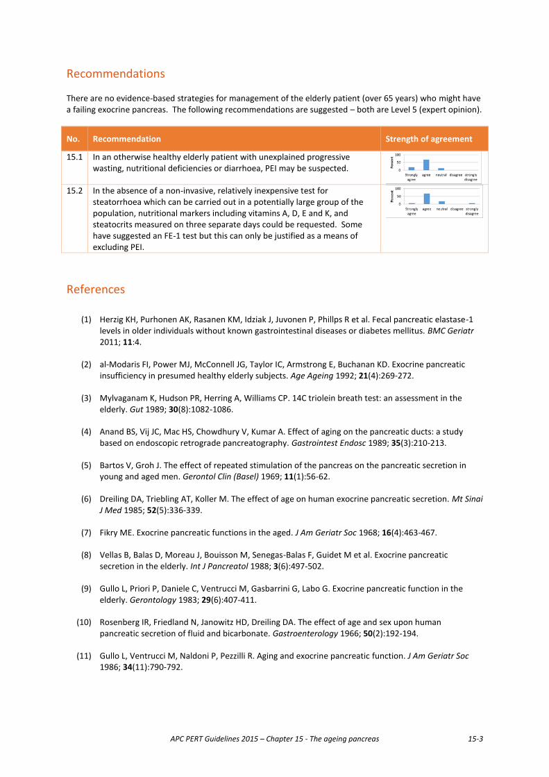
APC PERT Guidelines 2015 – Chapter 15 - The ageing pancreas 15-3
Recommendations There are no evidence-based strategies for management of the elderly patient (over 65 years) who might have a failing exocrine pancreas. The following recommendations are suggested – both are Level 5 (expert opinion).
No. Recommendation Strength of agreement
15.1 In an otherwise healthy elderly patient with unexplained progressive wasting, nutritional deficiencies or diarrhoea, PEI may be suspected.
15.2 In the absence of a non-invasive, relatively inexpensive test for
steatorrhoea which can be carried out in a potentially large group of the population, nutritional markers including vitamins A, D, E and K, and steatocrits measured on three separate days could be requested. Some have suggested an FE-1 test but this can only be justified as a means of excluding PEI.
References
(1) Herzig KH, Purhonen AK, Rasanen KM, Idziak J, Juvonen P, Phillps R et al. Fecal pancreatic elastase-1
levels in older individuals without known gastrointestinal diseases or diabetes mellitus. BMC Geriatr 2011; 11:4.
(2) al-Modaris FI, Power MJ, McConnell JG, Taylor IC, Armstrong E, Buchanan KD. Exocrine pancreatic insufficiency in presumed healthy elderly subjects. Age Ageing 1992; 21(4):269-272.
(3) Mylvaganam K, Hudson PR, Herring A, Williams CP. 14C triolein breath test: an assessment in the elderly. Gut 1989; 30(8):1082-1086.
(4) Anand BS, Vij JC, Mac HS, Chowdhury V, Kumar A. Effect of aging on the pancreatic ducts: a study based on endoscopic retrograde pancreatography. Gastrointest Endosc 1989; 35(3):210-213.
(5) Bartos V, Groh J. The effect of repeated stimulation of the pancreas on the pancreatic secretion in young and aged men. Gerontol Clin (Basel) 1969; 11(1):56-62.
(6) Dreiling DA, Triebling AT, Koller M. The effect of age on human exocrine pancreatic secretion. Mt Sinai J Med 1985; 52(5):336-339.
(7) Fikry ME. Exocrine pancreatic functions in the aged. J Am Geriatr Soc 1968; 16(4):463-467.
(8) Vellas B, Balas D, Moreau J, Bouisson M, Senegas-Balas F, Guidet M et al. Exocrine pancreatic secretion in the elderly. Int J Pancreatol 1988; 3(6):497-502.
(9) Gullo L, Priori P, Daniele C, Ventrucci M, Gasbarrini G, Labo G. Exocrine pancreatic function in the elderly. Gerontology 1983; 29(6):407-411.
(10) Rosenberg IR, Friedland N, Janowitz HD, Dreiling DA. The effect of age and sex upon human pancreatic secretion of fluid and bicarbonate. Gastroenterology 1966; 50(2):192-194.
(11) Gullo L, Ventrucci M, Naldoni P, Pezzilli R. Aging and exocrine pancreatic function. J Am Geriatr Soc 1986; 34(11):790-792.

APC PERT Guidelines 2015 – Chapter 15 - The ageing pancreas 15-4
(12) Gangarosa RE, Glass RI, Lew JF, Boring JR. Hospitalizations involving gastroenteritis in the United States, 1985: the special burden of the disease among the elderly. Am J Epidemiol 1992; 135(3):281-290.
(13) Holt PR. Diarrhea and malabsorption in the elderly. Gastroenterol Clin North Am 2001; 30(2):427-444.
(14) Montgomery RD, Haboubi NY, Mike NH, Chesner IM, Asquith P. Causes of malabsorption in the elderly. Age Ageing 1986; 15(4):235-240.
(15) Schiller LR. Diarrhea and malabsorption in the elderly. Gastroenterol Clin North Am 2009; 38(3):481-502.
(16) Coulson JM, Jones RD, Hubbard RE, Woodhouse KW, O'Mahony MS, Wheatley H. Pancreatic insufficiency and weight loss in older patients. QJM 2004; 97(6):377-378.
(17) Thomas PD, Forbes A, Green J, Howdle P, Long R, Playford R et al. Guidelines for the investigation of chronic diarrhoea, 2nd edition. Gut 2003; 52 Suppl 5:v1-15.
(18) Holyday M, Daniells S, Bare M, Caplan GA, Petocz P, Bolin T. Malnutrition screening and early nutrition intervention in hospitalised patients in acute aged care: a randomised controlled trial. J Nutr Health Aging 2012; 16(6):562-568.
(19) Laugier R, Bernard JP, Berthezene P, Dupuy P. Changes in pancreatic exocrine secretion with age: pancreatic exocrine secretion does decrease in the elderly. Digestion 1991; 50(3-4):202-211.
(20) Detlefsen S, Sipos B, Feyerabend B, Kloppel G. Pancreatic fibrosis associated with age and ductal papillary hyperplasia. Virchows Arch 2005; 447(5):800-805.
(21) Nagai H, Ohtsubo K. Pancreatic lithiasis in the aged. Its clinicopathology and pathogenesis. Gastroenterology 1984; 86(2):331-338.
(22) Pitchumoni CS, Glasser M, Saran RM, Panchacharam P, Thelmo W. Pancreatic fibrosis in chronic alcoholics and nonalcoholics without clinical pancreatitis. Am J Gastroenterol 1984; 79(5):382-388.
(23) Stamm BH. Incidence and diagnostic significance of minor pathologic changes in the adult pancreas at autopsy: a systematic study of 112 autopsies in patients without known pancreatic disease. Hum Pathol 1984; 15(7):677-683.
(24) Rothenbacher D, Low M, Hardt PD, Klor HU, Ziegler H, Brenner H. Prevalence and determinants of exocrine pancreatic insufficiency among older adults: results of a population-based study. Scand J Gastroenterol 2005; 40(6):697-704.
APC PERT Guidelines 2015 - Contributors 1
Conclusion This document summarises the current literature regarding the treatment of PEI (pancreatic enzyme insufficiency) for many different conditions. In Chapter 2 we have classified these conditions as being Definite, Likely and Unlikely to result in PEI because this consideration changes one’s approach to confirming the diagnosis and to treatment. This classification is a great help in considering patient management. There was a high level of agreement within the expert group that the starting dose of pancreatic enzyme replacement therapy (PERT) should be between 25,000 and 40,000 units lipase / meal but that if symptoms persist, a protein pump inhibitor should be considered and the dose of PERT titrated up towards 80,000 units lipase /meal. Other reasons for failure should be considered if this still fails. Because untreated PEI results in a malnourished state with low body fat and protein stores in association with osteopaenia, nutritional markers may be used to judge a satisfactory response to treatment. There is no benefit in restricting fat in the diet but rather the enzyme intake should be sufficient to allow normal fat absorption. Although there does not appear to be evidence for the value of PERT during the early phase of acute pancreatitis, it is common for PEI to be present in the recovery phase, which may last for two years. Patients should be assessed for this occurrence. PEI may develop a number of years after acute pancreatitis but patients with extensive pancreatic necrosis can recover sufficiently to not require replacement therapy. Chronic pancreatitis sufferers who secrete less than a tenth of the normal enzyme amount have improved symptoms after the introduction of PERT. PEI needs to be confirmed in these patients before treatment. Cystic fibrosis is a complex condition which should be managed by a multidisciplinary team to ensure adequate nutritional intake with appropriate PERT protocols. The paediatric literature contains some of the strongest evidence demonstrating the benefit of PERT in the presence of PEI. These patients frequently need proton pump inhibition and require ongoing nutritional assessment. PEI may be a consequence of many and different forms of gastrointestinal surgery. Because this can result in asynchrony, the FE-1 stool test can give false results in many of these patients and in many countries the 13C breath test is indicated for adequate diagnosis. However this is a specialised test which is not widely available in Australia. Bacterial overgrowth and acid suppression needs to be considered in these patients before committing them to long term PERT. It is important to emphasise that we found high agreement with the use of PERT in patients with unresectable pancreatic adenocarcinoma. Three conditions where PEI is considered to be unlikely (diabetes, coeliac disease and irritable bowel syndrome) were covered. There was good evidence that in a subset of such patients PEI should be considered but in these patients FE-1 testing is likely to result in over-diagnosis and more direct testing should be considered before committing them to long term PERT. Similarly, the literature suggests that PEI may be an important silent cause of weight loss and malnutrition in elderly patients. The FE-1 test may be useful in ruling out PEI in these patients but a low value will result in a large number of false positive results. However formal testing would be impractical. A clinical trial may be useful but it will be difficult to judge the effectiveness of treatment on clinical grounds.
APC PERT Guidelines 2015 - Contributors 2
There is still very little level 1 evidence examining the use of PERT in the literature and the group sends out a plea to correct this deficit through the development of multi-centre studies and for the adequate funding of pancreatic disease investigation centres so that these patients can be better advised. Ross C. Smith Chairman, Australasian Pancreatic Club PERT Guidelines Working Group September 2015
APC PERT Guidelines 2015 - Contributors 3
List of contributors Authors
Chapter 1 – Introduction and Methods
Emeritus Professor Ross C. Smith, Department of Surgery, University of Sydney; Immediate Past President, Australasian Pancreatic Club
Dr Sarah F. Smith, Medical Editor
Chapter 2 – Pancreatic exocrine insufficiency
Professor Jeremy Wilson, Director of Medicine, Liverpool Hospital, University of NSW.
Professor Ross C. Smith
Chapter 3 – Pancreatic enzyme replacement therapy
Assoc. Professor Callum Pearce, Institute for Immunology and Infectious Diseases, Murdoch University, WA; Consultant Gastroenterologist, Fremantle Hospital, WA.
Currently: Dr Callum Pearce, Department of Gastroenterology, Fiona Stanley Hospital, WA.
Dr Pearce has received clinical and research funding assistance from Ferring Pharmaceuticals, Janssen, Abbvie, Shire, Fresenius-Kabi and Baxter in the past.
Chapter 4 – Dietary Management of PEI
Nick Wray, Nutrition & Dietetics, School of Health Sciences, Flinders University, Adelaide, South Australia.
Ms Ruth Vo, B. Nutr&Diet (Hons), MHthSc (Edu), APD, AN, CNSC; Senior Gastro Dietitian, Dietetics Department, Liverpool Hospital, NSW.
Chapter 5 – Acute pancreatitis and the use of PERT
Professor Ross C. Smith
Professor John Chen, Director, SA Liver Transplant Unit & Senior Surgical Consultant, HPB Unit, Royal Adelaide Hospital & Flinders Medical Centre, South Australia.
Chapter 6 – Chronic pancreatitis and the use of PERT
Professor Jeremy Wilson
Professor Ross C. Smith
Chapter 7 – PEI in cystic fibrosis
Associate Professor Mark Oliver, Deputy Director and Head of Clinical Services, Department of Gastroenterology, Royal Children's Hospital, Melbourne, Victoria; Associate Professor, University of Melbourne; Honorary Fellow, Murdoch Children’s Research Institute.
Dr (Keith) Chee Y. Ooi, Paediatric Gastroenterologist, School of Women’s and Children’s Health, Faculty of Medicine, University of NSW; Department of Gastroenterology, Sydney Children’s Hospital, Randwick, NSW
Advisory board member of Vertex Pharmaceuticals.
Ms Tamarah Katz, Paediatric Dietitian, Sydney Children’s Hospital, Randwick, NSW.
APC PERT Guidelines 2015 - Contributors 4
Chapter 8 – Use of PERT after bowel resection
Professor Richard Turner, Associate Head, Hobart Clinical School, and Dept. Surgery, University of Tasmania.
Professor Ross C. Smith
Chapter 9 – Use of PERT after gastric surgery
Professor Ross C. Smith
Professor Richard Turner
Chapter 10 – Use of PERT after pancreatectomy
Professor John Chen
Dr Mehrdad Nikfarjam, MD, PhD, FRACS, Liver, Pancreas and Biliary Surgeon; Senior Lecturer, University of Melbourne, Department of Surgery, Melbourne, Victoria; President, Australasian Pancreatic Club.
Chapter 11 – Use of PERT in unresectable pancreatic cancer
Dr Mehrdad Nikfarjam
Professor Richard Turner
Chapter 12 – Diabetes mellitus and PERT
Professor Christopher K. Rayner, School of Medicine, University of Adelaide; Department of Gastroenterology and Hepatology, Royal Adelaide Hospital, South Australia.
Professor Rayner has received research funding from AstraZeneca, Merck Sharp & Dohme and Novartis.
Professor Michael Horowitz, Director, Endocrine and Metabolic Unit, University of Adelaide and Royal Adelaide Hospital, SA.
Chapter 13 – Coeliac disease and PERT
Professor Gerald Holtmann, Faculty of Medicine and Biomedical Sciences, University of Queensland; Translational Research Institute, Department of Gastroenterology & Hepatology, Princess Alexandra Hospital, Queensland.
Chapter 14 – Irritable bowel syndrome and PERT
Professor Nick Talley, Pro-Vice Chancellor, Faculty of Health and Medicine, University of Newcastle, NSW; President, Royal Australasian College of Physicians.
Chapter 15 – The ageing pancreas and PERT
Ms Ruth Vo
Nick Wray.
Reviewers
Professor John A. Windsor, Department of Surgery, Faculty of Medical and Health Sciences, University of Auckland, NZ.

APC PERT Guidelines 2015 - Contributors 5
Abbott Australasia Pty Ltd provided some funding to his Department’s Foundation, which was used to support research into acute pancreatitis.
Professor Ron Pirola, Faculty of Medicine, SW Sydney Clinical School, University of NSW.
Acknowledgement
Associate Professor Rachel Neale, Senior Research Fellow, Cancer Aetiology Laboratory, Queensland Institute of Medical Research, Berghofer Medical Research Institute, Queensland, for help with survey design.