Aus der Orthopädischen Universitätsklinik mit Poliklinik ...
79
Aus der Orthopädischen Universitätsklinik mit Poliklinik Tübingen Biomechanical assessment of osteoarthritic articular cartilage and jaw periosteal cells-based bone constructs Thesis submitted as requirement to fulfill the degree „Doctor of Philosophy “(Ph.D) at the Faculty of Medicine Eberhard Karls University Tbingen by Danalache Marina 2020
Transcript of Aus der Orthopädischen Universitätsklinik mit Poliklinik ...

Aus der
Orthopädischen Universitätsklinik mit Poliklinik Tübingen
Biomechanical assessment of osteoarthritic articular cartilage
and jaw periosteal cells-based bone constructs
Thesis submitted as requirement to fulfill the degree
„Doctor of Philosophy “(Ph.D)
at the
Faculty of Medicine
Eberhard Karls University
Tubingen
by
Danalache Marina
2020

Dean: Professor Dr. B. Pichler
First reviewer: Privatdozent Dr. U. K. Hofmann
Second reviewer: Professor Dr. S. Huber
Date of oral examination: 12.05.2020

To my mom and my husband & to the memory of my dear father
~ “to him who had given me dreams to look forward to” ~

“The oak fought the wind and was broken,
the willow bent when it must and survived.“
Robert Jordan
Adapted from The Oak and the Reeds fable
by Aesop

Table of Contents
1. Introduction .................................................................................................................. 1
Biomechanical properties of cartilage and their role in osteoarthritis ................ 6
Role of biomechanical properties in bone tissue engineering .......................... 10
Aim of the thesis............................................................................................... 13
2. Results……………………………………………………………………………….16
Biomechanical cues of pericellular matrix degradation in osteoarthritic
cartilage ...................................................................................................................... 16
Biomechanical and biochemical cues for customizing jaw periosteal cells for
bone constructs .......................................................................................................... 26
3. Discussion .................................................................................................................. 43
Biomechanical cues in early osteoarthritis ....................................................... 44
Biomechanical cues for customizing jaw periosteal cells for bone constructs 48
Study limitations............................................................................................... 52
Conclusions ...................................................................................................... 53
4. Summary .................................................................................................................... 54
5. Zusammenfassung ..................................................................................................... 56
6. Bibliography .............................................................................................................. 58
7. Declaration of contribution ........................................................................................ 72
Acknowledgments .......................................................................................................... 73

List of abbreviations and acronyms
Abbreviation Definition
AFM Atomic force microscopy
BMP2 Bone morphogenic protein 2
BTE Bone tissue engineering
ECM Extracellular matrix
EGF Epidermal growth factor
ELISA Enzyme-linked Immunosorbent Assay
EM Elastic modulus
FCS Fetal calf serum
FWHM Full-width-half-maximum
GAGs Glycosaminoglycans
GMP Good manufacturing practice
hPL Human plasma lysate
HS Heparansulfate
IF Impact factor
ITM Interterritorial matrix
JPCs Jaw periosteum derived progenitor cells
MMP Matrix metalloproteinase enzymes
MSCA-1 Mesenchymal stromal cell antigen-1
MSC Mesenchymal stem cells
OA Osteoarthritis
PCM Pericellular matrix
PDGF Platelet derived growth factors
sGAGs Sulfated glycosaminoglycans
TGF Transforming growth factor
TM Territorial matrix

1 | Introduction
Marina Danalache Pg. 1
1. Introduction
Tissue performance and its physical properties are closely intertwined processes.
They depend on the cells present in the tissue, and on the loading forces to which the
respective tissue is exposed. As such, in the context of load bearing tissues, like articu-
lar cartilage and bone, cells are capable of generating or resisting mechanical forces that
can be many times the weight of these tissues. These tissues and their specific cells
therefore need to possess particular biomechanical characteristics with respect to elastic-
ity and rigidity in order to withstand continuous stretch or strain mechanisms, and to
maintain their form and function. In this context, analyses of the biomechanical proper-
ties of cells have already provided much information about the physiological cellular
characteristics, and in many cases information on how to differentiate a healthy cell
from a cell in a pathological condition. In fact, micromechanical characteristics have
now been proposed as a potential biomarker for allowing differentiation between differ-
ent physiological and pathological cellular or tissue states (Rianna and Radmacher,
2016). In view of the ongoing demand for cell based and regenerative treatment ap-
proaches, biomechanical properties of living cells as well as their response to external
stimuli are considered key components which have received much research attention in
the past few years (Butler et al., 2009, Guilak et al., 2014b, Tay et al., 2013).
In all complex biological structures, different types of highly specialized cells are
embedded in a three-dimensional heterogeneously network termed the extracellular ma-
trix (ECM). The ECM plays a physiological role as an active component of living tissue
and is responsible for a variety of functions (Frantz et al., 2010), such as providing sup-
port, segregating tissues, and regulating intercellular communication (Gao et al., 2014).
Its main components are various types of collagens, fibrous proteins, glycosaminogly-
cans (GAGs) and proteoglycans. These ECM components link together to form a struc-
turally stable composite, providing the mechanical properties to the tissue, and enabling
the communication between tissue and its residing cells (Pizzo et al., 2005). The ECM’s
biomechanical properties are mainly determined by the relative composition and net-
work structure of its collagens, proteoglycans and elastin (Suki et al., 2011). Interesting-
ly, the cells-as an independent entity-are capable of sensing and responding to mechani-
cal forces of varying magnitude, direction, and frequency (Discher et al., 2005, Cho et
al., 2017) through integrin-mediated interactions with the matrix (Plotnikov et al.,
1 | Introduction
Pg. 2 Marina Danalache
2012). In such, the mechanical features of a matrix influence important cellular process-
es such as mobility (Lange and Fabry, 2013, Lin et al., 2019), proliferation (Hadjipanayi
et al., 2009), differentiation (Smith et al., 2018) and apoptosis (Walker et al., 2018).
Moreover, alterations of biomechanical characteristics of the ECM have been associated
with severe pathologies such as chronic obstructive pulmonary disease and cancer (Liu
et al., 2010, Bidan et al., 2015, Gkretsi and Stylianopoulos, 2018, Broders-Bondon et
al., 2018). It is assumed that if a cell cannot sense its mechanical environment, it simply
cannot survive (Yusko and Asbury, 2014). The biochemical and biomechanical signa-
ture of the ECM as well as cell-microenvironment crosstalk are crucial to understanding
the structure-function-effect relationship of healthy as well as pathologic tissues.
To directly measure biomechanical characteristics on a micro- or even nanoscale
level, atomic force microscopy (AFM) has emerged as a novel technique. It allows the
precise determination of local biomechanical properties of biological components or
interfaces, such as ECM-cell interactions. It works at a nanometer resolution, and meas-
urements can be performed without damaging the tested samples (Dufrene and Pelling,
2013). Several studies have shown that AFM is a suitable and reliable technique with a
large repertoire of applications. Established applications of its use are for the measure-
ment of mechanical properties, behavior, and function of living cells (Moeendarbary
and Harris, 2014), as well as for determinating elastic properties of various tissues un-
der normal physiological and pathological conditions (Haase and Pelling, 2015, Dufrêne
et al., 2017, Marrese et al., 2017, Deng et al., 2018, Kwon et al., 2019). Moreover, AFM
can operate in a wide variety of conditions. It can, for example, be performed in situ, as
well as in a fluid environment, thus allowing for live monitoring in real-time. Addition-
ally, AFM probing can be combined with various microscopy techniques such as fluo-
resce staining and imaging, to specifically probe a region where the protein/-s of interest
are located (Wilusz et al., 2012b, El-Kirat-Chatel and Dufrene, 2012). AFM operates on
the principle of surface sensing using an extremely sharp tip (usually 5-10 nm), also
known as an AFM probe, that interacts with the sample. The interaction between the
AFM probe and the sample then provides information on the sample’s biomechanical
features.
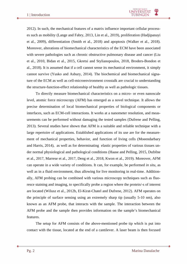
The setup for AFM consists of the above-mentioned probe tip which is put into
contact with the tissue, located at the end of a cantilever. A laser beam is then focused
1 | Introduction
Marina Danalache Pg. 3
onto the very end of the cantilever, which it is reflected onto a position-sensitive photo-
diode. The diode is designed to measure the horizontal and vertical deflection of the
laser on the cantilever. An x-y-z piezoelectric scanner allows for the movement of the
sample or the cantilever in all three directions (Figure 1)(Gavara, 2017). As the tip scans
over the surface, the interactions between the AFM tip and the features on the surface
cause displacement or bending of the cantilever. This bending is indicative of the tip-
sample interaction force. The up/down and side to side motion of the AFM tip as it
scans along the surface is measured by means of a laser beam which is projected on and
then reflected off the cantilever. The reflected laser beam is then tracked by the sensi-
tive photo-detector. The deflection sensitivity of these detectors must be calibrated be-
forehand. The calibration defines a correlation between how many nanometers of mo-
tion correspond to a unit of voltage.
Figure 1. Schematical representation of the AFM setup.
Biological samples can be investigated using an AFM cantilever or probe which is
controlled by a piezoelectric scanner. A phase inverted optical microscope at-
tached to the AFM system allows the user to specifically analyze single living cells
or specific select areas within the tissue. The laser tracking system for the reflected
beam is represented by a photodiode.
Abbreviation: AFM- atomic force microscopy

1 | Introduction
Pg. 4 Marina Danalache
Depending on the nature of the interaction between the tip and sample surface, the
AFM can be operated in different modes, such as the AFM-contact mode (e.g. force-
distance curve analysis) or the tapping mode (semi-contact). While in contact mode, the
AFM tip is brought into physical contact with the sample, and the cantilever deflection
is measured. In tapping mode, the tip is periodically retracted from the sample, and the
cantilever height is modulated at the cantilever’s resonance frequency. The latter is
largely used for imaging biomolecules such as DNA, proteins, and lipids (Viji Babu and
Radmacher, 2019). The most basic AFM technique operated in contact mode in terms of
quantitative studies of biomechanical characteristics of cells and tissues is represented
by a force-distance curve analysis (Figure 2). The applications of these experiments
range from nano-mechanical investigation of elastic properties to protein unfolding and
investigation of single chemical bonds (Janshoff et al., 2000).
In a force-distance curve, the interaction forces between the AFM probe and
sample are measured while the AFM probe is approached to and afterwards retracted
from the sample. In doing so, both the approach and the retraction curve offer cues with
respect to the viscoelastic properties of the sample, as well as possible adhesion proper-
ties between the tip and sample. By fitting the data obtained from the force curve with
an appropriate geometric model (e.g. Hertz fit model) of the AFM tip, the sample’s
Young's modulus can be calculated. The Young's modulus, also known as the elastic
modulus (EM), characterizes the resistance of a material/tissue when it is subjected to
compression and elongation. This describes the stiffness of the sample. A stiffer materi-
al will therefore exhibit a higher elastic modulus and vice versa (Vinckier and Semenza,
1998).

1 | Introduction
Marina Danalache Pg. 5
Figure 2. Schematical representation of a force-distance curve in the AFM con-
tact mode.
The force curve can be divided into two different segments: 1-(green) refers to mo-
tion where the AFM probe approaches the surface, and 2-(red) when the probe re-
tracts from the surface. As the probe initially approaches the surface, the forces
are too small to give a measurable deflection of the tip, thus leaving the tip in its
undisturbed position. The further the cantilever approaches the sample, the more
the force increases. In the retracting regime, the probe and sample are already in
contact with one another. Thus, in contrast to the approaching curve where bend-
ing forces are solely due to the increasing proximity of the cantilever with the
probe, additional adhesion forces are present in the retracting regime while the
cantilever is retracted in the z-axis from the sample. As the AFM probe is pulled
out of contact with the sample, it first gets “stuck” before it is able to loosen from
the adhesion at the interface, which even leads to a short negative deflection of the
cantilever. The AFM probe and its subsequent bending up or down and the unper-
turbed position of the AFM probe are also displayed in blue.
Abbreviation: AFM-atomic force microscopy
The focus of the thesis was to apply a customized AFM approach on load-bearing
connective tissue/cells, particularly articular cartilage and bone cells. This was done in
order to determine and differentiate between possible structural-functional changes in
the biomechanical properties (Young’s moduli) of the encompassing matrix/-es of the
targeted tissue/cells. In the first research part, the changes that occur due to alterations
in the biochemical signatures of the tissues due to ongoing pathological processes such
as osteoarthritis (OA) were investigated. The second research analyses, the changes in
the culture settings of the jaw periosteum derived progenitor cells (JPCs), such as media
supplementation (human platelet lysate (hPL) versus fetal calf serum (FCS)) were ex-
amined.

1 | Introduction
Pg. 6 Marina Danalache
Biomechanical properties of cartilage and their role in
osteoarthritis
Osteoarthritis (OA) is a degenerative joint disease that affects large parts of the
elderly population. Since there is thus far no defined cut-off threshold for the diagnosis
of OA, the condition is difficult to quantify. The incidence of symptomatic knee OA is
estimated to be around 1% per year, with a radiographic incidence of about 2% per year
(Felson et al., 1995, Oliveria et al., 1995). This results in an overall prevalence of the
disease in nearly 50% of adults aged 75 and above (Jordan et al., 2007). OA leads to
reduced flexibility and mobility of the joint, and to load-dependent joint pain that can
severely disable the patient, which also results in a high socio-economic burden. Risk
factors for OA are trauma, systemic inflammatory diseases, bacterial infections, meta-
bolic factors such as obesity, and mechanical factors (Felson, 2009). To date, conserva-
tive joint preserving therapies such as pain management, physiotherapy, or local infiltra-
tions cannot restore the original joint architecture. They only serve to alleviate the pain,
to help patients cope with the emerging symptoms, and to postpone the disease progres-
sion (Huey et al., 2012, Djouad et al., 2009).
Even though OA comprises the capsula and ligaments of the joint, as well as the
subchondral bone, the primary tissue destroyed throughout the degenerative process of
OA is the articular cartilage. The only cells present within the mature articular cartilage
are the chondrocytes, which form specific, unique, and easily distinguishable spatial
cellular patterns. These patterns are specific for each joint, as well as the joint’s loading
mechanism (Rolauffs et al., 2008, Schumacher et al., 2002). In healthy tissue areas,
chondrocytes are arranged as single strings in the femoral condyle, and with the onset
and progression of degenerative changes during OA, cellular rearrangement occurs: first
double strings arise (Rolauffs et al., 2010), followed by clustering of the cells in small
and then finally in big clusters (Lotz et al., 2010) (Figure 3).

1 | Introduction
Marina Danalache Pg. 7
Figure 3. Light microscopy pictures of chondrocyte rearrangement throughout
the course of OA.
While in healthy cartilage, single strings are present (A/D), OA initiation and pro-
gression triggers the occurrence of double strings (B/E), small clusters (F), and ul-
timately big clusters (C/G).
(A-C) Scale bar (white) measuring 100µm; (D-G) Scale bar (white) of 20µm.
Figure partially adapted from (Danalache et al., 2019).
These remodelling cellular processes have previously been suggested to have po-
tential as an “image-based biomarker” for OA-promoted degenerative processes occur-
ring at a cellular level (Aicher and Rolauffs, 2014). Like in any other connective tissue,
cartilage is comprised of a vast and dense ECM. The ECM-arciform collagen fibers not
only take their origin but also terminate in the subchondral bone. These fibers-possess
an enormous tensile strength and keep a large amount of proteoglycans entrapped,
which in turn can bind water molecules; in such providing the necessary compressive
resilience and elasticity to the tissue.
Within the articular cartilage, there are three different major zones: the superficial,
the middle, and the deep zone. Each can be distinguished based on differences in cellu-
lar morphology (Kuettner et al., 1991), composition of collagen (Nieminen et al., 2001),
content of glycosaminoglycans (Venn and Maroudas, 1977), and water content
(Maroudas and Venn, 1977). Within all the layers, a pericellular, a territorial, and an
interterritorial matrix (all ramifications of the ECM) are present. The pericellular matrix
(PCM) encompasses the chondrocytes and is both biochemically and biomechanically
distinct from the ECM.

1 | Introduction
Pg. 8 Marina Danalache
While the ECM is mainly comprised of collagen type II, the PCM is primarily de-
fined by the presence of collagen type VI (Guilak et al., 2006) and proteoglycans. The
PCM, together with the enclosed cell(s), have previously been termed the "chondron"-
also known as the metabolic unit of the tissue (Poole, 1997). The PCM is believed to act
as a mechano-sensitive cell-matrix interface (Guilak et al., 2006), protecting the chon-
drocyte (Peters et al., 2011), and modulating its biosynthetic response (Larson et al.,
2002). Additionally, the PCM plays a functional role in initiating signal transduction
within cartilage under load bearing (Eggli et al., 1985). The territorial matrix (TM) sur-
rounds the PCM, and is composed mostly of fine collagen fibrils, creating a basketlike
network around the cells (Muir, 1995, Sophia Fox et al., 2009). It has been suggested
that the TM protects the chondrocytes against mechanical stresses, and that it plays a
role in the resiliency of articular cartilage (Sophia Fox et al., 2009). In addition to the
TM, the interterritorial matrix (ITM) is comprised of randomly oriented bundles of large
collagen fibrils that run parallel to the surface of the superficial zone, obliquely in the
middle zone, and perpendicular to the joint surface in the deep zone of the tissue
(Sophia Fox et al., 2009). The ITM has a high content of proteoglycans, and contributes
to the biomechanical properties of the tissue (Mow and Guo, 2002).
OA onset is characterized by a biosynthetic phase, during which the chondrocytes
attempt to repair the damaged ECM, followed by a later degradative phase, in which
matrix synthesis inhibition and digestion by catabolic enzymes leads to collagen net-
work damage and subsequent matrix fissuring, erosion, and ultimately tissue loss
(Sandell and Aigner, 2001, Mankin et al., 1971, Brama et al., 2000, Howell, 1986,
Hamerman, 1989). Additionally, it must be borne in mind that articular cartilage is a
load bearing tissue. The biomechanical properties are thus of crucial importance for
normal functioning of the tissue, especially for the smooth gliding of its surfaces against
one another under load. This function is greatly impaired after initial disruption of the
tissue architecture, thus leading to increased friction, which in turn propagates tissue
destruction in a vicious circle. Intact biomechanical properties of articular cartilage are,
naturally, derived from its intact matrixes. OA progression is hence characterized by
extensive proteolysis of the type II collagen network and the proteoglycans of the ECM,
leading to a loss of mechanical properties (Plaas et al., 2007, Hollander et al., 1995).
1 | Introduction
Marina Danalache Pg. 9
Interestingly, OA also triggers early changes in the presence and amount of col-
lagen type VI of the PCM (Felka et al., 2016b). Moreover, proteoglycan distribution
within the chondron changes, which is in turn is followed by chondrocyte proliferation
coupled with expansion and structural alterations of the pericellular microenvironment,
and ultimately chondrocyte clustering (Poole et al., 1997). These changes affect the
biomechanical signals perceived by the chondrocytes (Guilak et al., 2006), explaining
how PCM damage potentially contributes to the ongoing OA-pathology. Recently, OA
has even been characterized as being a disease of the PCM (Guilak et al., 2018). The
mechanical properties of the cartilage (stiffness, hardness, dynamic Young’s modulus,
etc.) therefore play an important role in OA initiation and progression. The mechanical
properties of the cartilage have been investigated at all length scales (from macro to
nano-scale), and all the results obtained notably and consistently illustrated that carti-
lage properties with respect to tension, compression, and shear forces decline with OA
progression (Wilusz et al., 2014).
Several studies have indicated that the stiffness, and implicitly Young’s modulus-
of cartilage is a strong indicator of its material-bearing properties, crucial for joint lubri-
cation and function (Kiviranta et al., 2008, Korhonen et al., 2002). Young's modulus is
thus an attribute of cartilage elasticity, indicating its stiffness. A decrease in qualitative
stiffness of cartilage in the course of OA was highlighted in various studies by analyz-
ing the mechanical properties of both native and osteoarthritic cartilage in humans or
animal models (Kleemann et al., 2005, Desrochers et al., 2010, Vinckier and Semenza,
1998). It must be noted, however, that all of the reported findings until now have been
based on macroscopic classification systems for grading the osteoarthritic changes of
the cartilage (Wilusz et al., 2012b, McLeod et al., 2013, Kleemann et al., 2005, Kiviran-
ta et al., 2008). As such, cartilage surface parameters were used to grade the tissue, thus,
only allowing for the assessment of the micromechanical picture at an average level.
The AFM-elasticity measurements guided by more specific OA markers on a cellular
level-such as the cellular patterns can, therefore, provide new insights into the actual
biomechanical changes occurring during OA. This may refine and improve our current
understanding of OA physiopathology.
1 | Introduction
Pg. 10 Marina Danalache
Role of biomechanical properties in bone tissue
engineering
In the field of oral and maxillofacial surgery, bone defects in the oral cavity as a
result of congenital anomaly, trauma, periodontal disease, chronic infection, or surgical
resection following tumor treatment require extensive clinical attention. The current
medical practice still faces several significant challenges (Smith et al., 2015), especially
when attempting to regenerate the affected tissue and restore its initial function and aes-
thetics.
Artificial bone tissue constructs represent an essential part of bone tissue engi-
neering (BTE). The basic principle of such artificial constructs is to create a porous
scaffold functionalized with a cellular component. The biological functional unit of such
constructs is therefore the selected cells, which adhere to and grow on the surface of the
implanted scaffold. The goal of the scaffolds is to provide a tissue-like environment,
which helps the cells generate the normal biological structural components of their tis-
sue-specific ECM. In both articular cartilage and bone, the ECM provides essential
structural support for its cellular constituents, but it also modulates the mechanical
properties and the overall performance and efficacy of the tissue. For the design of func-
tional scaffolds for osseous engineered tissues, cell-ECM mechanical properties, struc-
tural, and biological characteristics must be taken into consideration. In a native state,
the ECM provides the structural support for cell attachment and subsequent tissue de-
velopment. The main challenge in engineering the constructs is to mimic these ECM
properties. To date, the use of engineered scaffolds made of artificial or native materials
often fails to allow for the formation of intact cell-cell junctions due to the unnatural
cellular surface properties (Kim et al., 2016, Yang and Temenoff, 2009). To overcome
this striking limitation, polymeric scaffolds can be replaced by an ECM, which is actu-
ally produced by the seeded cells as in native tissue. The final construct could thus
closely mimic the ECM architecture of target tissues with respect to both structure and
function. An extensive repertoire of scaffold materials ranging from metallic, synthetic,
polymeric, bio-polymeric, and smart hydrogel in various forms and compositions have
been investigated in the context of bone regeneration. The incorporation of cells and the
application of growth factor delivery strategies can significantly influence the regenera-
tive outcome. In terms of cellular components, current strategies include different cell
1 | Introduction
Marina Danalache Pg. 11
types, such as osteoblasts, periosteal cells, bone-marrow stromal cells, and stem cell
varieties, including mesenchymal stem cells (MSCs). MSCs represent a leading cell
type for the majority of regenerative medical purposes due to their self-renewal and
their potential for differentiation into different lineages (Fitzsimmons et al., 2018).
When it comes to bone scaffolds, the current state of the art is, however, the use of
periosteum progenitor cells (Chang and Knothe Tate, 2012). Particularly in the field of
maxillofacial surgery and orthopaedics, a strong emphasis is placed on jaw periosteum
derived progenitor cells (JPCs). These JPCs are suitable due to their easy availability
and their multipotent capacity at a single cell level (De Bari et al., 2006), coupled with a
high proliferation rate (Bruder et al., 1997). Clinically applicable cell-based therapies,
particularly tissue constructs intended for maxillofacial reconstruction are focused on
the use of patient derived JPCs. Especially in view of the requirements for good manu-
facturing practice (GMP) to ensure safety and efficacy (Fekete et al., 2012), a detailed
characterization of the cells, their expression patterns, and formed matrix compositional
signature, coupled with optimized culture conditions for differentiation are essential. In
this context, the use of fetal calf serum (FCS) media supplementation is discouraged by
regulatory authorities to limit the risk of immune reactions in the transplanted host, and
also due to animal welfare concerns. Consequently, substantial efforts have been made
in the last 15 years to identify a substitute for FCS supplementation. Recently, human
platelets lysate (hPL) has been suggested to be a suitable and effective alternative, out-
performing the former FCS supplementation (Tylek et al., 2019). It has been suggested
that hPL superiority is due to various growth factors, including platelet-derived growth
factor (PDGF), transforming growth factor (TGF), and epidermal growth factor (EGF)
(Antoninus et al., 2015), which enhance the proliferation rate of the MSCs while main-
taining their multilineage differentiation potential under culture (Hemeda et al.,
2014). However, despite aforementioned significant progress in cell-based therapies,
critical challenges remain in transitioning the in vitro use of JPCs into clinically-
applicable approaches. Our present understanding is still limited with respect to the pre-
cise biomechanical properties, the biological composition, and the functional perfor-
mance of the proposed procedures. Specifically, the repair or regeneration processes of
tissues are not yet fully understood at a macroenvironment level (scaffold collapse and

1 | Introduction
Pg. 12 Marina Danalache
on-the-shelf availability) as well as at a microenvironment level (e.g. cell migration, and
qualitative ECM formation) (Yuksel et al., 2005).
Although the biomechanical characteristics of the ECM in bone constructs (in-
cluding mechanobiology and mechanotransduction) have become an active research
field, relatively few published studies have investigated the biomechanical properties of
JPCs (Horimizu et al., 2013, Att et al., 2009). Especially the analysis of elastic adapta-
bility characteristics, as well as the corresponding biochemical signature (e.g. Raman
spectroscopy) of JPCs cultured under hPL media supplementation comparing the results
with those obtained with the standard FCS conditions, has not yet been investigated.
Both AFM and Raman spectroscopy are methods used to obtain data about the
surface properties of a sample. A combination of these methods could provide crucial
correlated morphological and chemical information. While the AFM is based on tip-
sample interactions at discrete locations in a sequential manner, in RAMAN spectrosco-
py, monochromatic light is focused on the sample, and the inelastic scattered light (the
Raman effect) is detected. The latter generates a molecular fingerprint of the investigat-
ed sample based on the detection of specific wavelength shifts caused by chemical bond
vibrations (Kunstar et al., 2013). Raman spectral studies have already been performed
for the identification of cell phenotype (Puppels et al., 1990), for assessment of malig-
nancy (Ramos et al., 2015), and for structural analysis of ECM components, such as
collagen (Frushour and Koenig, 1975), sulfated glycosaminoglycans (sGAGs), and pro-
teoglycans (Ellis et al., 2009). In mineralized tissue, the technique can detect the vibra-
tions obtained from the collagenous matrix. Even more so, it can give valuable insights
with respect to the biochemical structure of the bone, and more importantly the contri-
bution of various matrix proteins on the bone material properties (Bonewald et al.,
2003a). Using the combination of Raman microscopy with AFM, the high spatial and
topographical resolution obtained with an AFM can be directly linked to the molecular
information provided by the Raman spectroscopy. Such an experimental design com-
prised of AFM coupled with Raman spectroscopy-which may be employed for JPCs
investigations, for example is shown in Figure 4.

1 | Introduction
Marina Danalache Pg. 13
Figure 4. Experimental setup for biomechanical and biochemical characteriza-
tion of JPCs
Biomechanical properties are assessed via AFM-contact mode technique (A/B/C).
A polymeric bead (25µm) is attached to an AFM cantilever (A) in order to protect
the analyzed cells (B) from contact damage, and to increase the surface area to be
measured. By fitting the data generated from the resulting force-distance curve
(C), the elastic modulus (EM) can be calculated using the Hertz fit model. Raman
spectroscopy can be used for non-invasive sensing of the biochemical composition
of the cells and the ECM. It generates a Raman spectrum, which features peaks-
each peak corresponds to a specific molecular bond vibration (D).
Abbreviations: AFM-atomic force microscopy, ECM-extracellular matrix, EM-
elastic modulus, JPCs-jaw periosteal cells.
It can therefore be summarized that a better biochemical and biomechanical char-
acterization of the JPCs-ECM is still needed to improve the use of these cells for regen-
erative scaffolds.
Aim of the thesis
The biomechanical characteristics of tissues and of regenerative tissue scaffolds are
of crucial importance to their normal in vivo performance. Of note, their biological and
mechanobiological characteristics are not just of a passive nature, but they are also criti-
cal for regulating cell behavior. Alterations in the mechanical characteristics can occur

1 | Introduction
Pg. 14 Marina Danalache
as a result of aging, trauma, or disease. These changes directly translate into tissue dys-
function. For this reason, it is crucial to understand the role of the mechanical cues (e.g.
elasticity) and their relationship with the structural composition of the tissue. Especially
as the role and involvement of the different biomechanical properties in the successful
outcome of regenerative processes is not yet fully understood (Guilak et al., 2014a).
Manipulating biomechanical features may help to better understand, and in the long run
to also produce improved regenerated tissues with high strength and endurance. In this
setting, AFM has been emerging as a highly versatile, non-invasive, and interdiscipli-
nary technique that allows for a quantitative and qualitative biomechanical analysis of
living cells, tissues, and engineered constructs at a very high resolution. In this study, a
customized AFM approach was employed to investigate and quantitatively assess the
biomechanical characteristics of:
(1) pericellular matrix (PCM) degradation in early OA;
(2) jaw periosteal cells (JPCs)-ECM quality under different media supplementation;
The hypothesis of the first study (1) was that tissue changes in the cellular organi-
zational patterns during OA (strings, double strings, and small respectively big clusters)
are associated with structural changes of the PCM and with a loss of elastic properties.
Such an association between OA related spatial cellular organization and changes in the
local EM accompanied by structural changes of the PCM would also underline the func-
tional role of spatial organization as an image-based biomarker for OA. A better insight
into early OA mechanisms, particularly structural and mechanical changes of the PCM,
may facilitate our understanding of the patho-mechanism of the disease, and would thus,
help the development of better targeted therapies and diagnostic strategies.
The hypothesis of the second study (2) was that hPL substitution leads to a better
ECM quality in terms of biochemical composition and biomechanical characteristics in
JPCs when compared to FCS. In order to evaluate the suitability of JPCs cultivated un-
der hPL supplementation for in vitro culturing and osteogenic differentiation, quantita-
tive and qualitative analysis of the ECM produced by the JPCs under hPL versus FCS
supplementation was performed both biochemical (RAMAN spectroscopy) and biome-
chanical (AFM-elasticity measurements).

1 | Introduction
Marina Danalache Pg. 15
As the focus of this study was placed on bone tissue constructs, the ECM formed
by JPCs should mimic a “bone-like” structure and show similar biochemical and biome-
chanical functions.
The research projects presented in this thesis have already been published and will
be presented in the following order:
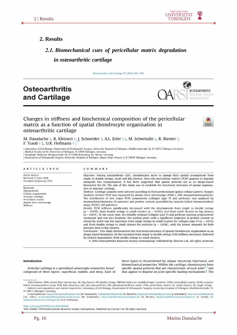
1. Danalache, M., Kleinert, R., Schneider, J., Erler, A.L., Schwitalle, M., Ries-
ter, R., Traub, F., Hofmann, U.K., 2019. Changes in stiffness and biochemical
composition of the pericellular matrix as a function of spatial chondrocyte or-
ganisation in osteoarthritic cartilage. Osteoarthritis and cartilage 27, 823-832.
2018 impact factor (IF): 4.9.
2. Danalache, M., Kliesch, S.M., Munz, M., Naros, A., Reinert, S., Alexander,
D., 2019a. Quality Analysis of Minerals Formed by Jaw Periosteal Cells under
Different Culture Conditions. International journal of molecular sciences 20.
2018 IF: 4.2.
2 | Results
Pg. 16 Marina Danalache
2. Results
Biomechanical cues of pericellular matrix degradation
in osteoarthritic cartilage

2 | Results
Marina Danalache Pg. 17

2 | Results
Pg. 18 Marina Danalache
2 | Results
Marina Danalache Pg. 19

2 | Results
Pg. 20 Marina Danalache

2 | Results
Marina Danalache Pg. 21

2 | Results
Pg. 22 Marina Danalache

2 | Results
Marina Danalache Pg. 23
2 | Results
Pg. 24 Marina Danalache

2 | Results
Marina Danalache Pg. 25

2 | Results
Pg. 26 Marina Danalache
Biomechanical and biochemical cues for customizing
jaw periosteal cells for bone constructs
2 | Results
Marina Danalache Pg. 27

2 | Results
Pg. 28 Marina Danalache

2 | Results
Marina Danalache Pg. 29

2 | Results
Pg. 30 Marina Danalache

2 | Results
Marina Danalache Pg. 31

2 | Results
Pg. 32 Marina Danalache

2 | Results
Marina Danalache Pg. 33

2 | Results
Pg. 34 Marina Danalache

2 | Results
Marina Danalache Pg. 35

2 | Results
Pg. 36 Marina Danalache

2 | Results
Marina Danalache Pg. 37

2 | Results
Pg. 38 Marina Danalache

2 | Results
Marina Danalache Pg. 39

2 | Results
Pg. 40 Marina Danalache

2 | Results
Marina Danalache Pg. 41

2 | Results
Pg. 42 Marina Danalache

3 | Discussion
Marina Danalache Pg. 43
3. Discussion
Cells are capable of adjusting their shape and function by altering their mechani-
cal properties through structural rearrangements in their biochemical composition at a
nanometer scale. The mechanical properties of a tissue are the sum of the comprised
cells and the vast matrix (i.e. ECM). There is a continuous interaction between the cel-
lular components and the surrounding ECM. On one side, these interactions actively
dictate the physiological remodeling of the tissue, thus its regeneration-and on the other
side, the catabolic degeneration of the tissue. Insights into the interaction between the
cellular components and the ECM, and into actual ECM functional biomechanical
transduction might hold a key for both the development of high-quality engineered tis-
sues, and clinical tools for early diagnostics.
In its physiological state, but even more in pathological conditions, the ECM is
constantly subjected to either enzymatic or non-enzymatic remodeling processes, and its
molecular components are subjected to a myriad of post-translational modifications
(Frantz et al., 2010). It is these ongoing ECM remodeling characteristics that dictate the
biochemical and mechanical properties of each organ, such as its tensile and compres-
sive strength and elasticity. So far, the ECM and its biomechanical properties have been
the root of useful pieces of information with respect to the presence of specific protein
biomarkers in a tissue e.g.: de novo collagen and proteoglycan synthesis. Moreover, the
non-invasive determination of biomechanical attributes can be used as a biomarker ca-
pable differentiating between the physiological and pathological state (Rianna and
Radmacher, 2016, Janmey and Miller, 2011). Correlating the biomechanical properties
of tissue with its function is an emerging area of research with a potential impact on
diagnostics and therapeutics, as well as on prognostics of pathological conditions. A
common parameter evaluated with the aim of differentiating between normal and dis-
eased tissue is tissue elasticity (respectively stiffness), which is generally expressed as
the EM. Malignant tumors, for example, are characteristically much stiffer than the
normal surroundings. The reason for this increase in stiffness lies in the excess ECM
deposition and the resulting increased rigidity (Cox and Erler, 2011). Measuring forces
at micro- or even nanoscale levels is a critical approach for defining our understanding
of cell-matrix interactions, and for understanding how the ECM regulates cellular func-
tion and how it contributes to the biomechanical functions of the tissue.

3 | Discussion
Pg. 44 Marina Danalache
Among the available techniques for measuring the mechanical properties of bio-
logical tissues, AFM has emerged as a non-invasive and highly versatile technique. It is
capable of quantifying and spatially mapping tissue and cellular mechanics, and physi-
cal properties, at a nanometer resolution. Information obtained from the AFM includes
surface morphology, frictional force on the nanoscale, hardness of surfaces, stiffness
and elasticity, load distribution, magnetization, yield stress, and elastic-plastic defor-
mation dynamics (Polini and Yang, 2017). In particular, the AFM-elasticity determina-
tion technique, sometimes referred to as force-distance analysis, is used to measure the
EM of a sample at distinct and user-defined points as the probe indentations are done
across a specified region.
In the present thesis, the biomechanical elasticity of load bearing tissue and cells
(cartilage and bone derived cells) were quantitatively assessed by means of AFM. The
results obtained from the two analyses are further discussed individually and in detail.
Biomechanical cues in early osteoarthritis
OA is a set of complex and difficult-to-identify processes leading to progressive
and irreversible degeneration of articular cartilage. It has been suggested that OA trig-
gers cellular remodelling processes which have been previously suggested to act as an
image-based biomarker for early OA promoted degenerative processes occurring at a
cellular level (Aicher and Rolauffs, 2014). The chondrocytes are fully surrounded by the
PCM. This microenvironment is believed to play a critical role in the transduction of
ECM compression into local physicochemical signals for the cell (Wilusz et al., 2014).
Any direct interactions between cell surface receptors such as integrins and the tissue
matrix are thus likely to occur first at the level of the PCM. In fact, evidence suggests
that the PCM acts as a transducer for both biomechanical as well as biochemical signals
for the cells it surrounds (Guilak et al., 2006, Chen et al., 2013, Vincent et al., 2007).
The PCM has been primarily defined by the presence of collagen type VI (Poole et al.,
1992) and perlecan, a large heparansulfate (HS) proteoglycan (Melrose et al., 2005). It
is known that during OA, changes in the collagen type VI synthesis of the PCM and
proteoglycan distribution within the chondron are followed by chondrocyte prolifera-
tion, expansion of the pericellular microenvironment, structural disruption, and chon-
drocyte clustering (Poole, 1997, Poole et al., 1996).
3 | Discussion
Marina Danalache Pg. 45
Changes in the PCM thus herald the upcoming degenerative processes and are as-
sociated with a significant loss of mechanical properties. Several technical challenges
arise when aiming to investigate and quantify the biochemical properties of the PCM,
such as the micrometer scale of the PCM (Youn et al., 2006), embedment of the PCM
into the ECM of the cartilage, and the low cell density of articular cartilage (Stockwell,
1971). Recent reports have, however, indicated that AFM-based microindentation al-
lows for direct quantification of PCM properties in situ with minimal disruption of the
native matrix integration between the PCM and ECM (Darling et al., 2010, Wilusz et
al., 2013, McLeod et al., 2013).
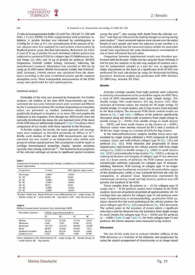
In this research study, the aim to evaluate whether the elasticity-EM (as measured
via AFM) of the PCM decreases as a function of OA onset and progression. OA tissue
categorization was done by employing the spatial arrangement of chondrocytes as an
image-based biomarker. EM was assessed by means of AFM on tissue selected accord-
ing to the locally predominant spatial chondrocyte pattern. Additionally, the pattern
related changes of two major PCM components (collagen type VI and perlecan) were
analyzed by ELISA (Enzyme-linked Immunosorbent Assay). The results obtained indi-
cate that there is a significant and stepwise EM decrease alongside each of the cellular
pattern rearrangements. The Young’s modulus of the PCM of healthy cartilage (single
strings) was about ~50 kPa (range 6-174 kPa). These results are comparable with those
obtained from isolated chondrons by micropipette aspiration technique (~40-70 kPa)
and in situ AFM measurements of the PCM from native human cartilage (range: 27-205
kPa) (Darling et al., 2010, Wilusz et al., 2013). Similar observations were made when
comparing the measured results of small clusters (early OA) with those obtained from
macroscopically assigned early OA samples measured by Wilusz et al. (Wilusz et al.,
2013). Overall, our study shows that a decrease of approximately 40% was observed
between healthy tissue and osteoarthritic cartilage, results which are again in line with
those of measured chondrons extracted by the micropipette aspiration technique
(Alexopoulos et al., 2005, Alexopoulos et al., 2003). Additionally, various computa-
tional models have also indicated similar values for EM reduction in OA, emphasizing
that the PCM in cartilage is approximately 1 order of magnitude less stiff than that of
the ECM. Also, while the hydraulic permeability of the PCM is 1-2 orders of magnitude
lower than that of the ECM, it seems to double with OA (Alexopoulos et al., 2003,
Alexopoulos et al., 2005).

3 | Discussion
Pg. 46 Marina Danalache
It needs to be pointed out, however, that cartilage slices subjected to AFM inden-
tations in the present study were obtained in the horizontal plane with respect to the
cartilage surface. In still intact cartilage areas where mostly strings are present, the main
collagen fiber orientation is also horizontal. Thus, these fibers remained largely intact
within the histologic sections. This was not the case in areas with already macroscopic
cartilage damage. When obtaining tissue from these areas, the section was rather prefer-
ably obtained from the middle or deep zone of the cartilage. Especially in the deep zone
of the cartilage, the collagen arcades are in their ascending/descending orientation
(Mow and Guo, 2002), and thus they were cut perpendicularly when sectioning. Since it
is in these areas with advanced local tissue degeneration where more pathologic cellular
patterns are observed, such as small and big clusters, it is possible that the measured
values bear a negative bias and appear worse than they would truly be without section-
ing. At the same time, it is known that osteoarthritis is a disease affecting the whole
joint (Loeser et al., 2012), with the inflammatory processes also changing cartilage are-
as that still appear macroscopically intact (own data not shown). It can thus be assumed
that in still intact and healthy cartilage, the values for the single strings would have still
been higher than the ones measured in the present study.
In terms of the structural composition of the PCM with OA onset and progression,
two of its main components (collagen type VI and perlecan) became progressively
structurally impaired. This structural impairment was coupled with a reduction in the
total protein content of these components as measured by ELISA. The progressive im-
pairment of the PCM, particularly collagen type VI, with increasingly pathological spa-
tial organization had been described previously (Felka et al., 2016a). Changes in the
perlecan expression in OA have also been documented before (Tesche and Miosge,
2004), pinpointing its possible involvement in the pathogenesis of the disease. Both
proteins have been suggested to dictate the overall biomechanical properties of the PCM
(Wilusz et al., 2012b, Wilusz et al., 2012a). Collagen type VI, which was initially well
defined pericellularly in strings and in double strings, showed a much more dispersed
and unorganized signal in big clusters, where its intracellular signal was completely
lost. In terms of perlecan staining, in strings it had a signal extra-and intracellularly,
followed by the same diffuse signal loss with cellular rearrangement. It could also be
noted that in advanced degenerative cartilage (as represented by big clusters), both
components were destroyed. As OA is characterized by an upregulation of protein pro-

3 | Discussion
Marina Danalache Pg. 47
duction of proteolytic enzymes (e.g. matrix metalloproteinase enzymes (MMPs))
(Martel-Pelletier et al., 1994), the loss of structural proteins (collagen type VI and per-
lecan) might represent the synergetic result of the catabolic activity of these enzymes.
Another possible cause for inconsistent PCM destruction of observed cell clusters could
be the consequence of nonhomogeneous matrix degradation, as already suggested by
Söder et al (Soder et al., 2002). In terms of collagen type VI, no relevant changes in
protein content and distribution could be observed between strings and double strings.
Clear and significant differences were visible, however, between small and big clusters,
as well as at the transition site from double strings to small clusters; this is the earliest
time point where relevant OA-triggered changes take place in the PCM. It could also be
noted that the earliest functional impairment with respect to the EM of the PCM was
detected at the transition from strings to double strings. Importantly, this is still at a
stage where the cartilage appears macroscopically to be intact. The EM decrease was
observed simultaneously with the alteration and destruction of the PCM. This suggests
that the two phenomena are linked to one another.
Up until now, most studies investigating the OA-related degeneration of cartilage
biochemically and/or biomechanically have based their grading of the severity of the
disease on macroscopic assessment grading systems such as the International Cartilage
Repair Society (ICRS) system (Kleemann et al., 2005), or the Collins grading system
(Wilusz et al., 2013). These grading systems have, however, limited utility, reproduci-
bility, and validity, particularly for early OA stages (Pritzker et al., 2006). To the
knowledge of the author, this is the first study to describe PCM structural and EM
changes based on tissue categorization by spatial arrangement of chondrocytes. The fact
that the EM decreases alongside the cellular rearrangement does not prove any causali-
ty. In a recently accepted manuscript from the author, the observed changes of the PCM
could also be observed in the ECM (Danalache et al., 2019). This leads to the specula-
tion that the cellular rearrangement is not solely and strictly involved in the PCM
changes and its microbiomechanical characteristics, rather that the processes of cellular
rearrangement and tissue destruction either go hand in hand, or tissue destruction is a
prerequisite for cellular rearrangement. Interestingly, changes in spatial organization are
not just correlated with the biomechanical features in the pericellular region, but also
with those of the ECM. It is indeed true that the decrease in EM of the PCM and ECM
goes simultaneously, unidirectionally, and at a comparable speed, suggesting that the

3 | Discussion
Pg. 48 Marina Danalache
underlying mechanisms are of the same nature (Danalache et al., 2019). This finding is
further supported by previous theoretical models of cell-matrix interactions in cartilage,
which suggest that the ratio of the two matrixes (ECM and PCM) with respect to their
mechanical properties may significantly influence the mechanical environment of the
chondrocyte (Alexopoulos et al., 2003, Guilak and Mow, 2000).
Overall, the present data shows that the first OA-PCM related degenerative
changes are already measurable at the cellular transition from single to double strings,
where cartilage still appears macroscopically “intact”. This further strengthens the idea
that the cellular spatial patterns play a measurable and functional role throughout the
course of the disease, instead of just being the passive result of OA-related remodeling
and rearrangement processes. As such there is potential to use an optical based bi-
omarker in the clinical setting for diagnosis of early stages of OA.
Biomechanical cues for customizing jaw periosteal cells
for bone constructs
The justification for the ongoing research efforts in BTE is founded on the fact
that bone autografting is still the golden standard in treating bone defects, despite the
invasiveness and traumatic nature of the procedure. In this respect, the ongoing demand
for personalized therapeutic approaches has enhanced the scientific interest and research
focus towards cell-based therapies and cell-enriched constructs for regenerative purpos-
es. In terms of a cell source, human jaw periosteum tissue contains osteoprogenitor cells
(JPCs) that have potential for tissue engineering applications in oral and maxillofacial
surgery. However, in order to bring such cell-based approaches into the clinical setting,
defined animal free culture conditions coupled with a precise characterization of the
cells and their formed ECM are a must. Hitherto, the benchmark in terms of media sup-
plementation for in vitro settings has been FCS. hPL has however been recently sug-
gested to be a suitable and “clinically acceptable” alternative.
In a former study, Wanner et al. 2017 showed that JPCs (to be precise: mesen-
chymal stromal cell antigen-1 (MSCA-1+) cells)) cultivated under hPL supplementation
were characterized by a higher proliferation and mineralization potential than JPCs cul-
tivated under FCS settings (Wanner et al., 2017). In the present study, the aim was to
further quantitatively analyze the biochemical composition as well as the biomechanical
properties (particularly elasticity) of the ECM formed by JPCs under both hPL and FCS

3 | Discussion
Marina Danalache Pg. 49
supplementation. RAMAN spectroscopy was used to investigate the biochemical com-
position and the biomechanical elasticity ere assessed by AFM. In the present study, we
observed higher proliferation rates of JPCs expanded under hPL conditions, results
which are in accordance with the ones previously obtained by Wanner et al (Wanner et
al., 2017). hPL is obtained via freeze-thaw cycles of platelets and subsequent centrifugal
separation of the debris from all the bioactive platelet factors (Schallmoser and Strunk,
2013). These platelets include various growth factors, such as PDGF, TGF-β1, VEGF,
EGF, attachment factors, and enzymes (Antoninus et al., 2015). It is conceivable that
such a potpourri of growth factors might indeed actually be the reason for the JPCs fast
expansion and differentiation.
The structural composition of the ECM determines the biomechanical properties.
These properties depend on the abundance of collagen and other proteins (non-
mineralized components), as well as on matrix mineralized components (phosphates).
Human bone minerals are constituted of a poorly crystallized apatite, comprised mainly
of calcium-deficient apatite, hydrogen phosphate, calcium-carbonate, and other ions.
The hydroxyapatite crystal structure exhibits a “hydrated layer”, where reactive ions
form the non-apatitic domains, which surround the stable apatite domains of the bone
crystals (Farlay et al., 2010). With bone maturation, the size and number of crystals
increase. Bone mineral maturity is expressed as the ratio of apatitic and non-apatitic
domains. The mineral crystallinity depends on both the size and amount, as well as on
the symmetry of the apatite crystalline domains and their size/strain (Farlay et al.,
2010). Even tough staining approaches such as Alizarin Red S and von Kossa are com-
monly used (Bonewald et al., 2003b, Gregory et al., 2004), when analyzing the mineral-
ization degree and bone quality formation, they fail to assess the quality of the mineral-
ized species. In this respect, RAMAN spectroscopy has already been successfully used
to identify changes in bone composition in metabolic bone diseases such as osteoporosis
and osteogenesis imperfecta (Roschger et al., 2008).
The RAMAN spectra results of the present study showed a higher phosphate to
protein ratio in the hPL group, indicative of higher phosphate ECM deposition. Moreo-
ver, the carbonate to phosphate ratio was higher in FCS containing culture conditions.
Similar observations were made by Brauchle et al. in a study where in vitro formation
of bone-specific matrixes by JPCSs was analyzed under serum free and FCS containing
media culture conditions (Brauchle et al., 2017). The authors of that study actually indi-

3 | Discussion
Pg. 50 Marina Danalache
cated that a higher carbonate to phosphate ratio combined with a low mineral to matrix
ratio might be indicative of poor bone quality (Brauchle et al., 2017). An inverse pro-
portionality relationship was observed in the case of carbonate and crystal content;
while carbonates exhibited a significant decrease in content, the crystal content in-
creased. This is indicative of a higher quality of the formed crystals. A lower carbonate
content coupled with a higher mineralization potential is usually the result of higher-
quality bone. Also, in the here presented study, hPL conditions led to overall lower car-
bonate levels than those obtained under FCS settings. As already suggested by Young et
al., FCS contains active components such as complement components 3 and 4A, fetuin-
A, and apolipoproteins A1 and B100, which are capable of accelerating crystallization
of free phosphates (Young et al., 2009). The mineral crystallinity can also be estimated
from the RAMAN spectra by using the width of the primary phosphate band, which is
mathematically described as the reciprocal of the full-width-half-maximum (FWHM)
(Freeman et al., 2001). According to the RAMAN data presented here, the mineral crys-
tallinity was significantly higher under hPL settings when compared to the one under
FCS supplementation. A lower mineral crystallinity, as observed under FCS settings,
might translate into altered bone mechanics by the occurrence of micro-strains within
and around the crystal lattice. In fact, in human cortical bone, Yerramshetty and Akkus
reported that the tissue-level strength and stiffness both increase with increasing crystal-
linity while ductility decreases (Yerramshetty and Akkus, 2008). In terms of collagen
maturity and quality, the RAMAN data shows that collagen cross-linking was signifi-
cantly higher under hPL than under FCS conditions; hPL supplementation might thus
induce a higher tensile strength of the matrix collagen. In support of this hypothesis, the
proline to hydroxyproline (amino acids precursors) ratio was also significantly higher in
hPL-JPCs than in FCS ones; hPL enriched media therefore seems to support mineraliza-
tion via maturation of collagens in JPCs.
The next question to address was whether or not the aforementioned observed bi-
ochemical composition ECM variations under FCS and hPL supplementation actually
translate into physical mechanical properties which reflect the JPCs’ mineralization ca-
pability. Until now, ample evidence suggested that biomechanical properties of the
ECM, particularly its stiffness, are capable of regulating and dictating MSC differentia-
tion characteristics (Lv et al., 2017). Even more so, according to a 2018 study by Sun et
al., the stiffness of the ECM actually seems to regulate MSC osteogenic differentiation

3 | Discussion
Marina Danalache Pg. 51
through mechanotransduction events mediated by integrin α5 (Sun et al., 2018). The
AFM results of our study indicated an EM increase of the ECM for the control as well
as the osteogenically induced JPCs under FCS settings as compared to the hPL setting.
The opposite effect was noted for the JPCs formed precipitates, where a significant EM
decrease was observed under FCS supplementation. Similar observations were made for
tissue engineered cartilage, where hPL media enrichment led to a significantly higher
EM (~ 45% increase), and a tissue with implicitly greater compressive mechanical
properties overall as when compared to FCS conditions (Petrera et al., 2013). The ma-
trix remodeling capacities of tissues cultured in hPL might therefore be higher than
those of tissues cultured in FCS.
In a study conducted by van Geemen where the formation and mechanical proper-
ties of heart valve engineered constructs was analyzed, MMP levels were shown to be
higher in the hPL group than in standard FCS culturing (van Geemen et al., 2011).
MMPs are key players in both inflammatory and remodeling phases of wound healing.
Their presence and upregulation might be indicative of an active remodeling process,
similar to that occurring in wound healing and formation of scar tissue. As suggested by
van Geerme, during wound healing, the presence of the scar, which can be recognized
by a disorganized collagen network and high remodeling properties, might result in an
initially weaker tissue (van Geemen et al., 2011). It is therefore conceivable that with
time, tissue cultured in hPL could undergo a remodeling process which might ultimately
lead to stronger and more organized tissue. This is in accordance with the AFM data
from the present study on JPCs-ECM, as lower EM were observed in early stages (con-
trol and osteogenic monolayers), followed by a significant increase in the JPCs formed
precipitates. In addition, even though FCS enhances early osteogenic differentiation, the
presence of osteo-inductive agents such as all trans-retinoic acid, bone morphogenic
protein 2 (BMP2), and dexamethasone are required for the expression of transcription
factors governing osteogenesis and hence differentiation towards a mature osteoblast
cell population (Roberts et al., 2011). Plasma platelets are known to be a rich source of
growth factors (Weibrich et al., 2002), and are therefore more efficient in terms of cell
differentiation capability and proliferation rates (Doucet et al., 2005) than using exoge-
nous recombinant growth factors and standard serum enriched media. The superiority of
hPL supplementation is thus further supported.

3 | Discussion
Pg. 52 Marina Danalache
Biomechanical characteristics are attributed also, to the orientation of the colla-
gen fibrils, as well as to differences in collagen content and its spatial distribution (Silva
et al., 2006). As observed in the here presented results and in accordance with previous
studies, changes in medium supplementation determine changes in the collagen expres-
sion (Brauchle et al., 2017). As already suggested by Brauchle et al., structural changes
in the orientation of collagen fibers might affect crystallization of phosphate along the
collagen fiber, ultimately controlling the mineralization process (Brauchle et al., 2017).
To the knowledge of the author, the here presented study is the first to investigate and
quantify cell mineralization quality, both biochemically and biomechanically, under
standard FCS and hPL settings.
All in all, the study illustrates that JPCs cultured under hPL supplementation form
an ECM of superior quality in terms of biochemical composition and elasticity.
Study limitations
It has to be borne in mind that AFM analysis is generally restricted to analysis of
the outer surface of cell membranes. As AFM is not capable of scanning the inside of a
cell membrane, this implicitly means that it is not able to directly investigate intracellu-
lar structures. One solution for overcoming this limitation is suggested by Usukura et
al., who employed a so called ”unroofing” method consisting of the breakage of cellular
membrane and the removal of cytoplasmic-soluble components (Usukura et al., 2016).
The focus of the both of studies presented here, however, was to investigate average
matrix-EM changes rather than probing specifies cellular/intracellular components. Al-
so, values obtained from AFM measurements from both studies indicate consistent and
individual ranges between groups; this might be explained by local variations in the
biomechanical properties of the PCM and ECM. Due to the limited availability of hu-
man tissue samples, the trade-off of using such a procedure is the mixing of statistically
dependent and independent data, which formally is not indicated. Even though the ob-
tained p-values thus need to be interpreted with the necessary caution, the measured
tendency should still not be affected. Experimental AFM parameters used for mechani-
cal testing, such as indentation velocity and depth, indenter shape and size, microsphere
size (25 µm), and accuracy of tip geometry in model fitting (Costa and Yin, 1999) might
also impact the absolute values of the measured mechanical properties (Stolz et al.,

3 | Discussion
Marina Danalache Pg. 53
2004, Park et al., 2009). These parameters should, however, not affect the results pre-
sented in both of the studies.
Conclusions
It remains a persistent challenge to understand how complex molecules biosynthe-
sized by cells assemble and function within the context of living cells, tissues, and or-
gans. Cellular functioning is governed by a multitude of processes. Quantifying cell and
tissue dynamics, particularly biomechanical features at multiple scales, is essential for
comprehensive studies of cellular function, as well as for unveiling the structure-
function relationship. Tissue and cell stiffness as measured by AFM is a widely used
parameter for fine cellular biomechanics, and it has been used to differentiate between
healthy and diseased states, as well as to assess structural changes. In both studies in the
present thesis, AFM was successfully used to asses the changes in elasticity, and to in-
vestigate whether structural changes translate into physical mechanical properties.
In the first study, it was concluded that OA related changes occurring in the struc-
tural composition of PCM and EM are associated with changes in cellular organization
as a biomarker for local tissue destruction. Early OA cellular changes represented by
transition from single to double strings are characterized by a significant PCM-EM de-
crease. At this stage the articular cartilage still appears macroscopically intact. The EM
decrease is accompanied by structural alterations of the PCM in its collagen type VI and
perlecan content, suggesting that the degeneration of these components and functional
disruption of the PCM are closely intertwined events in OA physiopathology.
The second study demonstrated that hPL media supplementation of JPCs enhanc-
es the formation of an organic material of superior quality in terms of its biochemical
composition and also of its biomechanical characteristic, as indicated by its elasticity.
The combination of biochemical characterization by means of RAMAN spectroscopy
coupled with AFM based-biomechanical elasticity assessment provided structural and
functional information that might help classify and differentiate the formation of poor
quality of bone tissue.
4 | Summary
Pg. 54 Marina Danalache
4. Summary
Mechanical features influence nearly every aspect of cell biology and function.
However, the underlying mechanisms of the role and how mechanical properties and
biochemical signals are interconnected is not clearly understood. The advent of atomic
force microscopy (AFM) provides a powerful tool for quantifying mechanical proper-
ties of living cell, typically the elastic modulus (EM). In the present study, a customized
AFM approach was applied on load connective tissues to address and investigate two
aspects of the musculoskeletal system. The first study investigated the EM changes that
occur in the pericellular matrix (PCM) as a function of the cellular pattern reorganiza-
tion throughout the course of osteoarthritis (OA). The second study investigated the
effect of human platelet lysate (hPL) supplementation on EM and biochemical composi-
tion of jaw periosteum derived progenitor cells (JPCs).
During osteoarthritis (OA) triggered cartilage degeneration, the chondrocytes in
the tissue spatially rearrange from single to double strings, and then to small and finally
big clusters. The spatial patterns act as an image-based biomarker for tissue degenera-
tion during OA. In a physiological state, chondrocytes are surrounded by a specialized
form of extracellular matrix (ECM) termed the PCM. The PCM, which also dictates the
biomechanical properties of the tissue, is also being progressively degraded throughout
the course of OA. The hypothesis of this study was that OA related changes in the cellu-
lar organizational patterns (strings, double strings, and clusters) are associated with
structural changes of the PCM and with a loss of elastic properties. The biomechanical
properties were measured by AFM on specific pattern selected tissue. Biochemical
changes of the main components of the PCM (collagen type VI and perlecan) were in-
vestigated by protein analysis techniques. The results indicated that there is a significant
and stepwise EM decrease alongside each of the cellular pattern rearrangements. At the
same time, the initially compact PCM was degraded progressively, losing its structural
integrity. The earliest point with a significant reduction in protein content was at the
transition from single strings to small clusters for collagen type VI, and from double
strings to small clusters for perlecan. Interestingly, the first significant EM decrease
was observed at the transition from single strings to double strings. It must be noted that
at this stage, articular cartilage appears macroscopically intact. Both biomechanical
properties (EM) as well as biochemical composition (protein content) were the lowest in

4 | Summary
Marina Danalache Pg. 55
big clusters. This study is the first to describe the EM as well as structural changes of
the PCM in relation to the OA related chondrocyte rearrangement, confirming the role
of these patterns as an image-based biomarker for early OA events.
The hypothesis of the second study was that human plasma lysate (hPL) media
enrichment leads to a higher quality of the ECM in JPCs when compared with the
standard fetal calf serum (FCS) condition. For this purpose, JPCs cultured with the
aforementioned media supplementations were analyzed in two study arms. RAMAN
spectroscopy was used for biochemical characterization, and AFM was employed for
biomechanical analysis. Raman spectroscopic measurements showed significantly high-
er phosphate to protein ratios and lower carbonate to phosphate ratios under hPL in
comparison to FCS culturing. With respect to the ECM collagen maturity, higher ratios
of proline to hydroxyproline as well as higher levels of collagen cross-linking were de-
tected in hPL-cultured JPCs. This indicates that hPL induces a higher degree of collagen
maturation in JPCs. AFM data showed a significant increase in EM of the ECM under
hPL conditions. This study hence demonstrates that hPL media supplementation of
JPCs leads to the formation of a higher ECM quality as when compared to the FCS
standard settings.
In summary, both studies employed AFM-based elasticity measurements to inves-
tigate biomechanical features of load-bearing tissues. In both studies, the results ob-
tained were significant and provide further insights which may now be fused into exist-
ing axioms of biochemical processes. Integration of both biomechanical and biochemi-
cal features will play a vital role in future scientific endeavors and will serve to establish
an in-depth understanding of cellular biology.

5 | Zusammenfassung
Pg. 56 Marina Danalache
5. Zusammenfassung
Mechanische Eigenschaften beeinflussen nahezu jeden Aspekt der Zelle in Bezug
auf ihre Biologie und Funktion. An welchen Schaltstellen mechanische Eigenschaften
und biochemische Signale jedoch miteinander verknüpft sind ist bisher nur wenig ver-
standen. Die Rasterkraftmikroskopie bietet hier die interessante Möglichkeit mechani-
sche Eigenschaften von lebenden Zellen zu untersuchen. Am gebräuchlichsten ist hier-
bei die Untersuchung des Elastizitätsmoduls (EM), welches Aussagen über die Steifheit
des Gewebes zulässt. In der hier präsentierten Arbeit wurden mittels Rasterkraftmikro-
skopie zwei Fragestellungen zum muskuloskelettalen System untersucht: Die erste Stu-
die untersuchte die auftretenden EM-Veränderungen der Perizellulären Matrix (PZM)
im Rahmen der räumlichen Umorganisation der Chondrozyten im Zuge der Knorpelde-
generation. In der zweiten Studie wurde der Einfluss von humanem Plättchenlysat
(hPL) auf das EM sowie auf den biochemischen Aufbau von aus Kieferperiost gewon-
nenen Progenitorzellen gemessen.
Im Zuge der Knorpeldegeneration bei Arthrose verändert sich die räumliche Or-
ganisation der Knorpelzellen hin von einfachen Zellsäulen (Engl. "single strings") zu
Doppelsäulen (Engl. "double strings") und schließlich zu kleinen (Engl. "small clus-
ters") und großen Zellhaufen (Engl. "big clusters). Diese Zellmuster dienen als bildba-
sierter Biomarker der degenerativen Veränderungen im Knorpel im Rahmen von Arth-
rose. Knorpelzellen sind im physiologischen Zustand unmittelbar von einer Sonderform
der Extrazellularmatrix (EZM) umgeben, der sogennanten perizellulären Matrix (PZM).
Die PZM, die die Biomechanik der Zellen maßgeblich mitdiktiert, degradiert zuneh-
mend während der Arthrose. Die Hypothese der hier durchgeführten Studie war, dass es
im Rahmen der zellulären Musterveränderungen von einfachen Zellsäulen zu großen
Zellhaufen auch zu strukturellen Veränderungen der PZM kommt, was ebenso einen
Verlust der elastischen Eigenschaften mit sich zieht. Die Elastizität der PZM wurde
dabei für jedes der zellulären Muster separat mittels Rasterkraftmikroskopie gemessen.
Gleichzeitig wurden biochemische Veränderungen von zwei wesentlichen Bausteinen
der PZM (Kollagen Typ VI und Perlekan) mittels Proteinanalysetechniken untersucht.
Es konnte dabei gezeigt werden, dass es im Rahmen der Musteränderungen zu einer
schrittweisen und signifikanten Reduktion des EM kommt. Der früheste Zeitpunkt einer
signifikanten Reduktion an Proteingehalt wurde am Übergang von einfachen Zellsäulen

5 | Zusammenfassung
Marina Danalache Pg. 57
zu kleinen Zellhaufen für Kollagen Typ VI und von Doppelsäulen zu kleinen Zellhaufen
für Perlekan beobachtet. Interessanterweise konnte der erste signifikante Rückgang des
EM bereits am Übergang von einfachen zu doppelten Zellsäulen gemessen werden. Dies
ist ein Zustand, in welchem der Knorpel makroskopisch noch intakt erscheint. Erwar-
tungsgemäß zeigte sich bei großen Zellhaufen die größte Beeinträchtigung, sowohl pro-
teinbiochemisch wie auch biomechanisch. Zusammenfassend ist dies die erste Studie,
welche das EM der PZM, wie auch einige ihre Komponenten in einen direkten Zusam-
menhang mit der räumlichen Knorpelzellanordnung setzt, was somit die Bedeutung
dieser als bildbasierten Biomarker für frühe Arthrose unterstreicht.
Die Hypothese der zweiten Studie war, dass die Anreicherung von Zellkulturme-
dium mit hPL eine bessere Qualität der EZM bei aus Kieferperiost gewonnenen Proge-
nitorzellen erzielt, als wenn das Medium mit fetalem Kälberserum versehen wird. Hier-
zu wurde in zwei Studienarmen eine jeweilige Zellkultur mit den unterschiedlichen
Nährlösungen durchgeführt. Sodann wurde die EZM biochemisch mittels RAMAN-
Spektroskopie charakterisiert und das EM mittels Rasterkraftmikroskopie gemessen. In
der Spektroskopie konnte ein signifikant höheres Phosphat-zu-Protein Verhältnis und
ein niedrigeres Carbonat-zu-Phosphat Verhältnis unter hPL im Vergleich zur Kultur mit
fetalem Kälberserum festgestellt werden. Auch in Bezug auf Reife des EZM-Kollagens
konnten unter hPL höhere Verhältnisse von Prolin-zu-Hydroxyprolin und höhere Spie-
gel an Kollagen-Quervernetzung beobachtet werden. In der Rasterkraftmikroskopie
zeigte sich weiterhin eine signifikante Zunahme des EM unter hPL-Bedingungen. Inso-
fern zeigt die hier präsentierte Studie, dass Anreicherung von Zellkulturmedium mit
hPL zur Ausbildung einer höherqualitativen EZM führt als die bisherige Standardbeimi-
schung von fetalem Kälberserum.
Zusammenfassend konnten so in beiden Studien anhand von Elastizitätsmessun-
gen lasttragenden Gewebes biomechanische Untersuchungen durchgeführt werden. In
beiden Fällen ergab sich aus den Messergebnissen ein Informationsgewinn mit eindeu-
tigen Ergebnissen, welche nun in ihrer biomechanischen Dimension in das übliche bio-
chemische Verständnis integriert werden können. Die Schnittstelle von biomechani-
schen zu biochemischen Vorgängen in Geweben und Zellen wird auch in künftigen Un-
tersuchungen von hoher Bedeutung für ein fundiertes funktionelles Verständnis der Bio-
logie auf zellulärer Ebene sein.

6 | Bibliography
Pg. 58 Marina Danalache
6. Bibliography
AICHER, W. K. & ROLAUFFS, B. 2014. The spatial organisation of joint surface
chondrocytes: review of its potential roles in tissue functioning, disease and
early, preclinical diagnosis of osteoarthritis. Ann Rheum Dis, 73, 645-53.
ALEXOPOULOS, L. G., HAIDER, M. A., VAIL, T. P. & GUILAK, F. 2003.
Alterations in the mechanical properties of the human chondrocyte pericellular
matrix with osteoarthritis. J Biomech Eng, 125, 323-33.
ALEXOPOULOS, L. G., WILLIAMS, G. M., UPTON, M. L., SETTON, L. A. &
GUILAK, F. 2005. Osteoarthritic changes in the biphasic mechanical properties
of the chondrocyte pericellular matrix in articular cartilage. J Biomech, 38, 509-
17.
ANTONINUS, A. A., WIDOWATI, W., WIJAYA, L., AGUSTINA, D.,
PURADISASTRA, S., SUMITRO, S. B., WIDODO, M. A. & BACHTIAR, I.
2015. Human platelet lysate enhances the proliferation of Wharton's jelly-
derived mesenchymal stem cells. Biomarkers and Genomic Medicine, 7, 87-97.
ATT, W., KUBO, K., YAMADA, M., MAEDA, H. & OGAWA, T. 2009.
Biomechanical properties of jaw periosteum-derived mineralized culture on
different titanium topography. Int J Oral Maxillofac Implants, 24, 831-41.
BIDAN, C. M., VELDSINK, A. C., MEURS, H. & GOSENS, R. 2015. Airway and
Extracellular Matrix Mechanics in COPD. 6.
BONEWALD, L. F., HARRIS, S. E., ROSSER, J., DALLAS, M. R., DALLAS, S. L.,
CAMACHO, N. P., BOYAN, B. & BOSKEY, A. 2003a. von Kossa staining
alone is not sufficient to confirm that mineralization in vitro represents bone
formation. Calcif Tissue Int, 72, 537-47.
BONEWALD, L. F., HARRIS, S. E., ROSSER, J., DALLAS, M. R., DALLAS, S. L.,
CAMACHO, N. P., BOYAN, B. & BOSKEY, A. 2003b. Von Kossa Staining
Alone Is Not Sufficient to Confirm that Mineralization In Vitro Represents Bone
Formation. Calcified Tissue International, 72, 537-547.
BRAMA, P. A., TEKOPPELE, J. M., BANK, R. A., BARNEVELD, A. & VAN
WEEREN, P. R. 2000. Functional adaptation of equine articular cartilage: the
formation of regional biochemical characteristics up to age one year. Equine Vet
J, 32, 217-21.

6 | Bibliography
Marina Danalache Pg. 59
BRAUCHLE, E., CARVAJAL BERRIO, D., RIEGER, M., SCHENKE-LAYLAND,
K., REINERT, S. & ALEXANDER, D. 2017. Raman Spectroscopic Analyses of
Jaw Periosteal Cell Mineralization. Stem cells international, 2017, 1651376-
1651376.
BRODERS-BONDON, F., NGUYEN HO-BOULDOIRES, T. H., FERNANDEZ-
SANCHEZ, M.-E. & FARGE, E. 2018. Mechanotransduction in tumor
progression: The dark side of the force. 217, 1571-1587.
BRUDER, S. P., JAISWAL, N. & HAYNESWORTH, S. E. 1997. Growth kinetics,
self-renewal, and the osteogenic potential of purified human mesenchymal stem
cells during extensive subcultivation and following cryopreservation. J Cell
Biochem, 64, 278-94.
BUTLER, D. L., GOLDSTEIN, S. A., GULDBERG, R. E., GUO, X. E., KAMM, R.,
LAURENCIN, C. T., MCINTIRE, L. V., MOW, V. C., NEREM, R. M., SAH,
R. L., SOSLOWSKY, L. J., SPILKER, R. L. & TRANQUILLO, R. T. 2009.
The impact of biomechanics in tissue engineering and regenerative medicine.
Tissue engineering. Part B, Reviews, 15, 477-484.
CHANG, H. & KNOTHE TATE, M. L. 2012. Concise Review: The Periosteum:
Tapping into a Reservoir of Clinically Useful Progenitor Cells. STEM CELLS
Translational Medicine, 1, 480-491.
CHEN, C., TAMBE, D. T., DENG, L. & YANG, L. 2013. Biomechanical properties
and mechanobiology of the articular chondrocyte. 305, C1202-C1208.
CHO, S., IRIANTO, J. & DISCHER, D. E. 2017. Mechanosensing by the nucleus:
From pathways to scaling relationships. J Cell Biol, 216, 305-315.
COSTA, K. D. & YIN, F. C. 1999. Analysis of indentation: implications for measuring
mechanical properties with atomic force microscopy. J Biomech Eng, 121, 462-
71.
COX, T. R. & ERLER, J. T. 2011. Remodeling and homeostasis of the extracellular
matrix: implications for fibrotic diseases and cancer. Dis Model Mech, 4, 165-
78.
DANALACHE, M., JACOBI, L. F., SCHWITALLE, M. & HOFMANN, U. K. 2019.
Assessment of biomechanical properties of the extracellular and pericellular
matrix and their interconnection throughout the course of osteoarthritis. Journal
of Biomechanics, 109409.

6 | Bibliography
Pg. 60 Marina Danalache
DARLING, E. M., WILUSZ, R. E., BOLOGNESI, M. P., ZAUSCHER, S. & GUILAK,
F. 2010. Spatial mapping of the biomechanical properties of the pericellular
matrix of articular cartilage measured in situ via atomic force microscopy.
Biophysical journal, 98, 2848-2856.
DE BARI, C., DELL'ACCIO, F., VANLAUWE, J., EYCKMANS, J., KHAN, I. M.,
ARCHER, C. W., JONES, E. A., MCGONAGLE, D., MITSIADIS, T. A.,
PITZALIS, C. & LUYTEN, F. P. 2006. Mesenchymal multipotency of adult
human periosteal cells demonstrated by single-cell lineage analysis. Arthritis
Rheum, 54, 1209-21.
DENG, X., XIONG, F., LI, X., XIANG, B., LI, Z., WU, X., GUO, C., LI, X., LI, Y., LI,
G., XIONG, W. & ZENG, Z. J. J. O. N. 2018. Application of atomic force
microscopy in cancer research. 16, 102.
DESROCHERS, J., AMREIN, M. A. & MATYAS, J. R. 2010. Structural and
functional changes of the articular surface in a post-traumatic model of early
osteoarthritis measured by atomic force microscopy. J Biomech, 43, 3091-8.
DISCHER, D. E., JANMEY, P. & WANG, Y. L. 2005. Tissue cells feel and respond to
the stiffness of their substrate. Science, 310, 1139-43.
DJOUAD, F., BOUFFI, C., GHANNAM, S., NOEL, D. & JORGENSEN, C. 2009.
Mesenchymal stem cells: innovative therapeutic tools for rheumatic diseases.
Nat Rev Rheumatol, 5, 392-9.
DOUCET, C., ERNOU, I., ZHANG, Y., LLENSE, J. R., BEGOT, L., HOLY, X. &
LATAILLADE, J. J. 2005. Platelet lysates promote mesenchymal stem cell
expansion: a safety substitute for animal serum in cell-based therapy
applications. J Cell Physiol, 205, 228-36.
DUFRÊNE, Y. F., ANDO, T., GARCIA, R., ALSTEENS, D., MARTINEZ-MARTIN,
D., ENGEL, A., GERBER, C. & MÜLLER, D. J. 2017. Imaging modes of
atomic force microscopy for application in molecular and cell biology. Nature
Nanotechnology, 12, 295.
DUFRENE, Y. F. & PELLING, A. E. 2013. Force nanoscopy of cell mechanics and cell
adhesion. Nanoscale, 5, 4094-104.
EGGLI, P. S., HERRMANN, W., HUNZIKER, E. B. & SCHENK, R. K. 1985. Matrix
compartments in the growth plate of the proximal tibia of rats. Anat Rec, 211,
246-57.
6 | Bibliography
Marina Danalache Pg. 61
EL-KIRAT-CHATEL, S. & DUFRENE, Y. F. 2012. Nanoscale imaging of the
Candida-macrophage interaction using correlated fluorescence-atomic force
microscopy. ACS Nano, 6, 10792-9.
ELLIS, R., GREEN, E. & WINLOVE, C. P. 2009. Structural analysis of
glycosaminoglycans and proteoglycans by means of Raman microspectrometry.
Connect Tissue Res, 50, 29-36.
FARLAY, D., PANCZER, G., REY, C., DELMAS, P. D. & BOIVIN, G. 2010. Mineral
maturity and crystallinity index are distinct characteristics of bone mineral.
Journal of bone and mineral metabolism, 28, 433-445.
FEKETE, N., ROJEWSKI, M. T., FURST, D., KREJA, L., IGNATIUS, A.,
DAUSEND, J. & SCHREZENMEIER, H. 2012. GMP-compliant isolation and
large-scale expansion of bone marrow-derived MSC. PLoS One, 7, e43255.
FELKA, T., ROTHDIENER, M., BAST, S., UYNUK-OOL, T., ZOUHAIR, S., OCHS,
B. G., DE ZWART, P., STOECKLE, U., AICHER, W. K., HART, M. L.,
SHIOZAWA, T., GRODZINSKY, A. J., SCHENKE-LAYLAND, K.,
VENKATESAN, J. K., CUCCHIARINI, M., MADRY, H., KURZ, B. &
ROLAUFFS, B. 2016a. Loss of spatial organization and destruction of the
pericellular matrix in early osteoarthritis in vivo and in a novel in vitro
methodology. Osteoarthritis Cartilage, 24, 1200-9.
FELKA, T., ROTHDIENER, M., BAST, S., UYNUK-OOL, T., ZOUHAIR, S., OCHS,
B. G., DE ZWART, P., STOECKLE, U., AICHER, W. K., HART, M. L.,
SHIOZAWA, T., GRODZINSKY, A. J., SCHENKE-LAYLAND, K.,
VENKATESAN, J. K., CUCCHIARINI, M., MADRY, H., KURZ, B. &
ROLAUFFS, B. 2016b. Loss of spatial organization and destruction of the
pericellular matrix in early osteoarthritis in vivo and in a novel in vitro
methodology. Osteoarthritis and Cartilage, 24, 1200-1209.
FELSON, D. T. 2009. Developments in the clinical understanding of osteoarthritis.
Arthritis Res Ther, 11, 203.
FELSON, D. T., ZHANG, Y., HANNAN, M. T., NAIMARK, A., WEISSMAN, B. N.,
ALIABADI, P. & LEVY, D. 1995. The incidence and natural history of knee
osteoarthritis in the elderly. The Framingham Osteoarthritis Study. Arthritis
Rheum, 38, 1500-5.
6 | Bibliography
Pg. 62 Marina Danalache
FITZSIMMONS, R. E. B., MAZUREK, M. S., SOOS, A. & SIMMONS, C. A. 2018.
Mesenchymal Stromal/Stem Cells in Regenerative Medicine and Tissue
Engineering. Stem Cells International, 2018, 16.
FRANTZ, C., STEWART, K. M. & WEAVER, V. M. 2010. The extracellular matrix at
a glance. 123, 4195-4200.
FREEMAN, J. J., WOPENKA, B., SILVA, M. J. & PASTERIS, J. D. 2001. Raman
Spectroscopic Detection of Changes in Bioapatite in Mouse Femora as a
Function of Age and In Vitro Fluoride Treatment. Calcified Tissue
International, 68, 156-162.
FRUSHOUR, B. G. & KOENIG, J. L. 1975. Raman scattering of collagen, gelatin, and
elastin. Biopolymers, 14, 379-391.
GAO, Y., LIU, S., HUANG, J., GUO, W., CHEN, J., ZHANG, L., ZHAO, B., PENG,
J., WANG, A., WANG, Y., XU, W., LU, S., YUAN, M. & GUO, Q. 2014. The
ECM-cell interaction of cartilage extracellular matrix on chondrocytes. BioMed
research international, 2014, 648459-648459.
GAVARA, N. 2017. A beginner's guide to atomic force microscopy probing for cell
mechanics. Microscopy research and technique, 80, 75-84.
GKRETSI, V. & STYLIANOPOULOS, T. 2018. Cell Adhesion and Matrix Stiffness:
Coordinating Cancer Cell Invasion and Metastasis. Frontiers in oncology, 8,
145-145.
GREGORY, C. A., GRADY GUNN, W., PEISTER, A. & PROCKOP, D. J. 2004. An
Alizarin red-based assay of mineralization by adherent cells in culture:
comparison with cetylpyridinium chloride extraction. Analytical Biochemistry,
329, 77-84.
GUILAK, F., ALEXOPOULOS, L. G., UPTON, M. L., YOUN, I., CHOI, J. B., CAO,
L., SETTON, L. A. & HAIDER, M. A. 2006. The pericellular matrix as a
transducer of biomechanical and biochemical signals in articular cartilage. Ann
N Y Acad Sci, 1068, 498-512.
GUILAK, F., BUTLER, D. L., GOLDSTEIN, S. A. & BAAIJENS, F. P. 2014a.
Biomechanics and mechanobiology in functional tissue engineering. J Biomech,
47, 1933-40.
6 | Bibliography
Marina Danalache Pg. 63
GUILAK, F., BUTLER, D. L., GOLDSTEIN, S. A. & BAAIJENS, F. P. T. 2014b.
Biomechanics and mechanobiology in functional tissue engineering. Journal of
biomechanics, 47, 1933-1940.
GUILAK, F. & MOW, V. C. 2000. The mechanical environment of the chondrocyte: a
biphasic finite element model of cell-matrix interactions in articular cartilage. J
Biomech, 33, 1663-73.
GUILAK, F., NIMS, R. J., DICKS, A., WU, C. L. & MEULENBELT, I. 2018.
Osteoarthritis as a disease of the cartilage pericellular matrix. Matrix Biol, 71-
72, 40-50.
HAASE, K. & PELLING, A. E. 2015. Investigating cell mechanics with atomic force
microscopy. Journal of the Royal Society, Interface, 12, 20140970-20140970.
HADJIPANAYI, E., MUDERA, V. & BROWN, R. A. 2009. Close dependence of
fibroblast proliferation on collagen scaffold matrix stiffness. J Tissue Eng Regen
Med, 3, 77-84.
HAMERMAN, D. 1989. The biology of osteoarthritis. N Engl J Med, 320, 1322-30.
HEMEDA, H., GIEBEL, B. & WAGNER, W. 2014. Evaluation of human platelet
lysate versus fetal bovine serum for culture of mesenchymal stromal cells.
Cytotherapy, 16, 170-180.
HOLLANDER, A. P., PIDOUX, I., REINER, A., RORABECK, C., BOURNE, R. &
POOLE, A. R. 1995. Damage to type II collagen in aging and osteoarthritis
starts at the articular surface, originates around chondrocytes, and extends into
the cartilage with progressive degeneration. J Clin Invest, 96, 2859-69.
HORIMIZU, M., KAWASE, T., TANAKA, T., OKUDA, K., NAGATA, M., BURNS,
D. M. & YOSHIE, H. 2013. Biomechanical evaluation by AFM of cultured
human cell-multilayered periosteal sheets. Micron, 48, 1-10.
HOWELL, D. S. 1986. Pathogenesis of osteoarthritis. Am J Med, 80, 24-8.
HUEY, D. J., HU, J. C. & ATHANASIOU, K. A. 2012. Unlike bone, cartilage
regeneration remains elusive. Science, 338, 917-21.
JANMEY, P. A. & MILLER, R. T. 2011. Mechanisms of mechanical signaling in
development and disease. 124, 9-18.
JANSHOFF, A., NEITZERT, M., OBERDORFER, Y. & FUCHS, H. 2000. Force
Spectroscopy of Molecular Systems-Single Molecule Spectroscopy of Polymers
and Biomolecules. Angew Chem Int Ed Engl, 39, 3212-3237.

6 | Bibliography
Pg. 64 Marina Danalache
JORDAN, J. M., HELMICK, C. G., RENNER, J. B., LUTA, G., DRAGOMIR, A. D.,
WOODARD, J., FANG, F., SCHWARTZ, T. A., ABBATE, L. M.,
CALLAHAN, L. F., KALSBEEK, W. D. & HOCHBERG, M. C. 2007.
Prevalence of knee symptoms and radiographic and symptomatic knee
osteoarthritis in African Americans and Caucasians: the Johnston County
Osteoarthritis Project. J Rheumatol, 34, 172-80.
KIM, Y., KO, H., KWON, I. K. & SHIN, K. 2016. Extracellular Matrix Revisited:
Roles in Tissue Engineering. International neurourology journal, 20, S23-S29.
KIVIRANTA, P., LAMMENTAUSTA, E., TOYRAS, J., KIVIRANTA, I. &
JURVELIN, J. S. 2008. Indentation diagnostics of cartilage degeneration.
Osteoarthritis Cartilage, 16, 796-804.
KLEEMANN, R. U., KROCKER, D., CEDRARO, A., TUISCHER, J. & DUDA, G. N.
2005. Altered cartilage mechanics and histology in knee osteoarthritis: relation
to clinical assessment (ICRS Grade). Osteoarthritis Cartilage, 13, 958-63.
KORHONEN, R. K., WONG, M., AROKOSKI, J., LINDGREN, R., HELMINEN, H.
J., HUNZIKER, E. B. & JURVELIN, J. S. 2002. Importance of the superficial
tissue layer for the indentation stiffness of articular cartilage. Med Eng Phys, 24,
99-108.
KUETTNER, K. E., AYDELOTTE, M. B. & THONAR, E. J. 1991. Articular cartilage
matrix and structure: a minireview. J Rheumatol Suppl, 27, 46-8.
KUNSTAR, A., LEFERINK, A. M., OKAGBARE, P. I., MORRIS, M. D.,
ROESSLER, B. J., OTTO, C., KARPERIEN, M., VAN BLITTERSWIJK, C.
A., MORONI, L. & VAN APELDOORN, A. A. 2013. Label-free Raman
monitoring of extracellular matrix formation in three-dimensional polymeric
scaffolds. J R Soc Interface, 10, 20130464.
KWON, T., GUNASEKARAN, S. & EOM, K. 2019. Atomic force microscopy-based
cancer diagnosis by detecting cancer-specific biomolecules and cells. Biochim
Biophys Acta Rev Cancer, 1871, 367-378.
LANGE, J. R. & FABRY, B. 2013. Cell and tissue mechanics in cell migration. Exp
Cell Res, 319, 2418-23.
LARSON, C. M., KELLEY, S. S., BLACKWOOD, A. D., BANES, A. J. & LEE, G. M.
2002. Retention of the native chondrocyte pericellular matrix results in
significantly improved matrix production. Matrix Biol, 21, 349-59.

6 | Bibliography
Marina Danalache Pg. 65
LIN, C., TAO, B., DENG, Y., HE, Y., SHEN, X., WANG, R., LU, L., PENG, Z., XIA,
Z. & CAI, K. 2019. Matrix promote mesenchymal stromal cell migration with
improved deformation via nuclear stiffness decrease. Biomaterials, 217, 119300.
LIU, F., MIH, J. D., SHEA, B. S., KHO, A. T., SHARIF, A. S., TAGER, A. M. &
TSCHUMPERLIN, D. J. 2010. Feedback amplification of fibrosis through
matrix stiffening and COX-2 suppression. 190, 693-706.
LOESER, R. F., GOLDRING, S. R., SCANZELLO, C. R. & GOLDRING, M. B. 2012.
Osteoarthritis: a disease of the joint as an organ. Arthritis and rheumatism, 64,
1697-1707.
LOTZ, M. K., OTSUKI, S., GROGAN, S. P., SAH, R., TERKELTAUB, R. & D'LIMA,
D. 2010. Cartilage cell clusters. Arthritis and rheumatism, 62, 2206-2218.
LV, H., WANG, H., ZHANG, Z., YANG, W., LIU, W., LI, Y. & LI, L. 2017.
Biomaterial stiffness determines stem cell fate. Life Sci, 178, 42-48.
MANKIN, H. J., DORFMAN, H., LIPPIELLO, L. & ZARINS, A. 1971. Biochemical
and metabolic abnormalities in articular cartilage from osteo-arthritic human
hips. II. Correlation of morphology with biochemical and metabolic data. J Bone
Joint Surg Am, 53, 523-37.
MAROUDAS, A. & VENN, M. 1977. Chemical composition and swelling of normal
and osteoarthrotic femoral head cartilage. II. Swelling. Ann Rheum Dis, 36, 399-
406.
MARRESE, M., GUARINO, V. & AMBROSIO, L. 2017. Atomic Force Microscopy: A
Powerful Tool to Address Scaffold Design in Tissue Engineering. J Funct
Biomater, 8.
MARTEL-PELLETIER, J., MCCOLLUM, R., FUJIMOTO, N., OBATA, K.,
CLOUTIER, J. M. & PELLETIER, J. P. 1994. Excess of metalloproteases over
tissue inhibitor of metalloprotease may contribute to cartilage degradation in
osteoarthritis and rheumatoid arthritis. Lab Invest, 70, 807-15.
MCLEOD, M. A., WILUSZ, R. E. & GUILAK, F. 2013. Depth-dependent anisotropy
of the micromechanical properties of the extracellular and pericellular matrices
of articular cartilage evaluated via atomic force microscopy. Journal of
Biomechanics, 46, 586-592.
MELROSE, J., SMITH, S., CAKE, M., READ, R. & WHITELOCK, J. 2005. Perlecan
displays variable spatial and temporal immunolocalisation patterns in the

6 | Bibliography
Pg. 66 Marina Danalache
articular and growth plate cartilages of the ovine stifle joint. Histochem Cell
Biol, 123, 561-71.
MOEENDARBARY, E. & HARRIS, A. R. 2014. Cell mechanics: principles, practices,
and prospects. 6, 371-388.
MOW, V. C. & GUO, X. E. 2002. Mechano-electrochemical properties of articular
cartilage: their inhomogeneities and anisotropies. Annu Rev Biomed Eng, 4, 175-
209.
MUIR, H. 1995. The chondrocyte, architect of cartilage. Biomechanics, structure,
function and molecular biology of cartilage matrix macromolecules. BioEssays,
17, 1039-1048.
NIEMINEN, M. T., RIEPPO, J., TOYRAS, J., HAKUMAKI, J. M., SILVENNOINEN,
J., HYTTINEN, M. M., HELMINEN, H. J. & JURVELIN, J. S. 2001. T2
relaxation reveals spatial collagen architecture in articular cartilage: a
comparative quantitative MRI and polarized light microscopic study. Magn
Reson Med, 46, 487-93.
OLIVERIA, S. A., FELSON, D. T., REED, J. I., CIRILLO, P. A. & WALKER, A. M.
1995. Incidence of symptomatic hand, hip, and knee osteoarthritis among
patients in a health maintenance organization. Arthritis Rheum, 38, 1134-41.
PARK, S., COSTA, K. D., ATESHIAN, G. A. & HONG, K. S. 2009. Mechanical
properties of bovine articular cartilage under microscale indentation loading
from atomic force microscopy. Proc Inst Mech Eng H, 223, 339-47.
PETERS, H. C., OTTO, T. J., ENDERS, J. T., JIN, W., MOED, B. R. & ZHANG, Z.
2011. The protective role of the pericellular matrix in chondrocyte apoptosis.
Tissue Eng Part A, 17, 2017-24.
PETRERA, M., DE CROOS, J. N. A., IU, J., HURTIG, M., KANDEL, R. A. &
THEODOROPOULOS, J. S. 2013. Supplementation With Platelet-Rich Plasma
Improves the In Vitro Formation of Tissue-Engineered Cartilage With
Enhanced Mechanical Properties. Arthroscopy, 29, 1685-1692.
PIZZO, A. M., KOKINI, K., VAUGHN, L. C., WAISNER, B. Z. & VOYTIK-
HARBIN, S. L. 2005. Extracellular matrix (ECM) microstructural composition
regulates local cell-ECM biomechanics and fundamental fibroblast behavior: a
multidimensional perspective. 98, 1909-1921.

6 | Bibliography
Marina Danalache Pg. 67
PLAAS, A., OSBORN, B., YOSHIHARA, Y., BAI, Y., BLOOM, T., NELSON, F.,
MIKECZ, K. & SANDY, J. D. 2007. Aggrecanolysis in human osteoarthritis:
confocal localization and biochemical characterization of ADAMTS5-
hyaluronan complexes in articular cartilages. Osteoarthritis Cartilage, 15, 719-
34.
PLOTNIKOV, S. V., PASAPERA, A. M., SABASS, B. & WATERMAN, C. M. 2012.
Force fluctuations within focal adhesions mediate ECM-rigidity sensing to guide
directed cell migration. Cell, 151, 1513-27.
POLINI, A. & YANG, F. 2017. 5 - Physicochemical characterization of nanofiber
composites. In: RAMALINGAM, M. & RAMAKRISHNA, S. (eds.) Nanofiber
Composites for Biomedical Applications. Woodhead Publishing.
POOLE, A. R., ROSENBERG, L. C., REINER, A., IONESCU, M., BOGOCH, E. &
ROUGHLEY, P. J. 1996. Contents and distributions of the proteoglycans
decorin and biglycan in normal and osteoarthritic human articular cartilage. 14,
681-689.
POOLE, C. A. 1997. Articular cartilage chondrons: form, function and failure. J Anat,
191 ( Pt 1), 1-13.
POOLE, C. A., AYAD, S. & GILBERT, R. T. 1992. Chondrons from articular
cartilage. V. Immunohistochemical evaluation of type VI collagen organisation
in isolated chondrons by light, confocal and electron microscopy. J Cell Sci, 103
( Pt 4), 1101-10.
POOLE, C. A., GILBERT, R. T., HERBAGE, D. & HARTMANN, D. J. 1997.
Immunolocalization of type IX collagen in normal and spontaneously
osteoarthritic canine tibial cartilage and isolated chondrons. Osteoarthritis
Cartilage, 5, 191-204.
PRITZKER, K. P. H., GAY, S., JIMENEZ, S. A., OSTERGAARD, K., PELLETIER, J.
P., REVELL, P. A., SALTER, D. & VAN DEN BERG, W. B. 2006.
Osteoarthritis cartilage histopathology: grading and staging. Osteoarthritis and
Cartilage, 14, 13-29.
PUPPELS, G. J., DE MUL, F. F., OTTO, C., GREVE, J., ROBERT-NICOUD, M.,
ARNDT-JOVIN, D. J. & JOVIN, T. M. 1990. Studying single living cells and
chromosomes by confocal Raman microspectroscopy. Nature, 347, 301-3.

6 | Bibliography
Pg. 68 Marina Danalache
RAMOS, I., #XEA, MARTINS, S. R., MALKIN, A. & LYNG, F. M. 2015. Current
Advances in the Application of Raman Spectroscopy for Molecular Diagnosis of
Cervical Cancer. BioMed Research International, 2015, 9.
RIANNA, C. & RADMACHER, M. 2016. Cell mechanics as a marker for diseases:
Biomedical applications of AFM. 1760, 020057.
ROBERTS, S. J., CHEN, Y., MOESEN, M., SCHROOTEN, J. & LUYTEN, F. P. 2011.
Enhancement of osteogenic gene expression for the differentiation of human
periosteal derived cells. Stem Cell Research, 7, 137-144.
ROLAUFFS, B., WILLIAMS, J., GRODZINSKY, A., E KUETTNER, K. & A COLE,
A. 2008. Distinct horizontal patterns in the spatial organization of superficial
zone chondrocytes of human joints.
ROLAUFFS, B., WILLIAMS, J. M., AURICH, M., GRODZINSKY, A. J.,
KUETTNER, K. E. & COLE, A. A. 2010. Proliferative remodeling of the spatial
organization of human superficial chondrocytes distant from focal early
osteoarthritis. Arthritis and rheumatism, 62, 489-498.
ROSCHGER, P., PASCHALIS, E. P., FRATZL, P. & KLAUSHOFER, K. 2008. Bone
mineralization density distribution in health and disease. Bone, 42, 456-66.
SANDELL, L. J. & AIGNER, T. 2001. Articular cartilage and changes in arthritis. An
introduction: cell biology of osteoarthritis. Arthritis Res, 3, 107-13.
SCHALLMOSER, K. & STRUNK, D. 2013. Generation of a Pool of Human Platelet
Lysate and Efficient Use in Cell Culture. In: HELGASON, C. D. & MILLER,
C. L. (eds.) Basic Cell Culture Protocols. Totowa, NJ: Humana Press.
SCHUMACHER, B. L., SU, J. L., LINDLEY, K. M., KUETTNER, K. E. & COLE, A.
A. 2002. Horizontally oriented clusters of multiple chondrons in the superficial
zone of ankle, but not knee articular cartilage. Anat Rec, 266, 241-8.
SILVA, M. J., BRODT, M. D., WOPENKA, B., THOMOPOULOS, S., WILLIAMS,
D., WASSEN, M. H., KO, M., KUSANO, N. & BANK, R. A. 2006. Decreased
collagen organization and content are associated with reduced strength of
demineralized and intact bone in the SAMP6 mouse. J Bone Miner Res, 21, 78-
88.
SMITH, B. T., SHUM, J., WONG, M., MIKOS, A. G. & YOUNG, S. 2015. Bone
Tissue

6 | Bibliography
Marina Danalache Pg. 69
Engineering Challenges in Oral & Maxillofacial Surgery. In: BERTASSONI, L. E. &
COELHO, P. G. (eds.) Engineering Mineralized and Load Bearing Tissues.
Cham: Springer International Publishing.
SMITH, L. R., CHO, S. & DISCHER, D. E. 2018. Stem Cell Differentiation is
Regulated by Extracellular Matrix Mechanics. Physiology (Bethesda), 33, 16-25.
SODER, S., HAMBACH, L., LISSNER, R., KIRCHNER, T. & AIGNER, T. 2002.
Ultrastructural localization of type VI collagen in normal adult and osteoarthritic
human articular cartilage. Osteoarthritis Cartilage, 10, 464-70.
SOPHIA FOX, A. J., BEDI, A. & RODEO, S. A. 2009. The basic science of articular
cartilage: structure, composition, and function. Sports Health, 1, 461-8.
STOCKWELL, R. A. 1971. The interrelationship of cell density and cartilage thickness
in mammalian articular cartilage. Journal of anatomy, 109, 411-421.
STOLZ, M., RAITERI, R., DANIELS, A. U., VANLANDINGHAM, M. R.,
BASCHONG, W. & AEBI, U. 2004. Dynamic Elastic Modulus of Porcine
Articular Cartilage Determined at Two Different Levels of Tissue Organization
by Indentation-Type Atomic Force Microscopy. Biophysical Journal, 86, 3269-
3283.
SUKI, B., STAMENOVIC, D. & HUBMAYR, R. 2011. Lung parenchymal mechanics.
Compr Physiol, 1, 1317-51.
SUN, M., CHI, G., XU, J., TAN, Y., XU, J., LV, S., XU, Z., XIA, Y., LI, L. & LI, Y.
2018. Extracellular matrix stiffness controls osteogenic differentiation of
mesenchymal stem cells mediated by integrin α5. Stem cell research & therapy,
9, 52-52.
TAY, C. Y., KOH, C. G., TAN, N. S., LEONG, D. T. & TAN, L. P. 2013.
Mechanoregulation of stem cell fate via micro-/nano-scale manipulation for
regenerative medicine. 8, 623-638.
TESCHE, F. & MIOSGE, N. 2004. Perlecan in late stages of osteoarthritis of the human
knee joint. Osteoarthritis and Cartilage, 12, 852-862.
TYLEK, T., SCHILLING, T., SCHLEGELMILCH, K., RIES, M., RUDERT, M.,
JAKOB, F. & GROLL, J. 2019. Platelet lysate outperforms FCS and human
serum for co-culture of primary human macrophages and hMSCs. Scientific
Reports, 9, 3533.
6 | Bibliography
Pg. 70 Marina Danalache
USUKURA, E., NARITA, A., YAGI, A., ITO, S. & USUKURA, J. 2016. An
Unroofing Method to Observe the Cytoskeleton Directly at Molecular
Resolution Using Atomic Force Microscopy. Scientific Reports, 6, 27472.
VAN GEEMEN, D., RIEM VIS, P. W., SOEKHRADJ-SOECHIT, S., SLUIJTER, J. P.,
DE LIEFDE-VAN BEEST, M., KLUIN, J. & BOUTEN, C. V. 2011. Decreased
mechanical properties of heart valve tissue constructs cultured in platelet lysate
as compared to fetal bovine serum. Tissue Eng Part C Methods, 17, 607-17.
VENN, M. & MAROUDAS, A. 1977. Chemical composition and swelling of normal
and osteoarthrotic femoral head cartilage. I. Chemical composition. Annals of
the rheumatic diseases, 36, 121-129.
VIJI BABU, P. K. & RADMACHER, M. 2019. Mechanics of Brain Tissues Studied by
Atomic Force Microscopy: A Perspective. Frontiers in Neuroscience, 13.
VINCENT, T. L., MCLEAN, C. J., FULL, L. E., PESTON, D. & SAKLATVALA, J.
2007. FGF-2 is bound to perlecan in the pericellular matrix of articular cartilage,
where it acts as a chondrocyte mechanotransducer. Osteoarthritis and Cartilage,
15, 752-763.
VINCKIER, A. & SEMENZA, G. 1998. Measuring elasticity of biological materials by
atomic force microscopy. FEBS Lett, 430, 12-6.
WALKER, C., MOJARES, E. & DEL RIO HERNANDEZ, A. 2018. Role of
Extracellular Matrix in Development and Cancer Progression. Int J Mol Sci, 19.
WANNER, Y., UMRATH, F., WAIDMANN, M., REINERT, S. & ALEXANDER, D.
2017. Platelet Lysate: The Better Choice for Jaw Periosteal Cell Mineralization
%J Stem Cells International. 2017, 10.
WEIBRICH, G., KLEIS, W. K., HAFNER, G. & HITZLER, W. E. 2002. Growth factor
levels in platelet-rich plasma and correlations with donor age, sex, and platelet
count. J Craniomaxillofac Surg, 30, 97-102.
WILUSZ, R. E., DEFRATE, L. E. & GUILAK, F. 2012a. A biomechanical role for
perlecan in the pericellular matrix of articular cartilage. Matrix biology : journal
of the International Society for Matrix Biology, 31, 320-327.
WILUSZ, R. E., DEFRATE, L. E. & GUILAK, F. 2012b. Immunofluorescence-guided
atomic force microscopy to measure the micromechanical properties of the
pericellular matrix of porcine articular cartilage. J R Soc Interface, 9, 2997-
3007.
6 | Bibliography
Marina Danalache Pg. 71
WILUSZ, R. E., SANCHEZ-ADAMS, J. & GUILAK, F. 2014. The structure and
function of the pericellular matrix of articular cartilage. Matrix Biology, 39, 25-
32.
WILUSZ, R. E., ZAUSCHER, S. & GUILAK, F. 2013. Micromechanical mapping of
early osteoarthritic changes in the pericellular matrix of human articular
cartilage. Osteoarthritis and cartilage, 21, 1895-1903.
YANG, P. J. & TEMENOFF, J. S. 2009. Engineering orthopedic tissue interfaces.
Tissue engineering. Part B, Reviews, 15, 127-141.
YERRAMSHETTY, J. S. & AKKUS, O. 2008. The associations between mineral
crystallinity and the mechanical properties of human cortical bone. Bone, 42,
476-82.
YOUN, I., CHOI, J. B., CAO, L., SETTON, L. A. & GUILAK, F. 2006. Zonal
variations in the three-dimensional morphology of the chondron measured in
situ using confocal microscopy. Osteoarthritis and Cartilage, 14, 889-897.
YOUNG, J. D., MARTEL, J., YOUNG, D., YOUNG, A., HUNG, C.-M., YOUNG, L.,
CHAO, Y.-J., YOUNG, J. & WU, C.-Y. 2009. Characterization of Granulations
of Calcium and Apatite in Serum as Pleomorphic Mineralo-Protein Complexes
and as Precursors of Putative Nanobacteria. PLOS ONE, 4, e5421.
YUKSEL, E., CHOO, J., WETTERGREEN, M. & LIEBSCHNER, M. 2005.
Challenges in Soft Tissue Engineering. Seminars in Plastic Surgery, 19, 261-
270.
YUSKO, E. C. & ASBURY, C. L. 2014. Force is a signal that cells cannot ignore.
Molecular biology of the cell, 25, 3717-3725.

7 | Declaration of contribution
Pg. 72 Marina Danalache
7. Declaration of contribution
The dissertation work was carried out at the Department of Orthopaedic Surgery,
University Hospital of Tübingen, Germany under the supervision of PD Dr. Ulf Krister
Hofmann.
The study was designed by the following:
1. PD Dr. Ulf Krister Hofmann and myself, Department of Orthopaedic Surgery, Tü-
bingen, for the 1st paper: Danalache et al: Changes in stiffness and biochemical
composition of the pericellular matrix as a function of chondrocyte organization in
osteoarthritic cartilage. Osteoarthritis and Cartilage, 2019.
2. Prof. Dr. Dorothea Alexander-Friedrich, Department of Oral and Maxillofacial
Surgery, Tübingen for the 2nd paper: Danalache et al: Quality Analysis of Minerals
Formed by Jaw Periosteal Cells under Different Culture Conditions. International
Journal of Molecular Sciences, 2019.
I had the major contribution for both of the research papers published, with the
collaboration and assistance of the co-authors as indicated in the “Authors contribution
section” specified in each of the papers.
I carried out the majority of the experiments and received assistance from Janine
Schneider for the AFM measurements on articular cartilage. Marita Munz did the cell
characterization and handling and the RAMAN spectroscopy experiments were carried
out by Sophie-Maria Kliesch at the company Quality Analysis GmbH (Nürtingen, Ger-
many).
The statistical analysis was carried out independently by myself after consultation
with PD Dr. Ulf Krister Hofmann and Prof. Dr. Dorothea Alexander-Friedrich.
I confirm that I wrote the thesis by myself and any additional sources of infor-
mation have been duly cited.
20.01.2020, Tübingen

Acknowledgments
Marina Danalache Pg. 73
Acknowledgments
“Give credit to whom credit due.”
Samuel Adams
Foremost I would like to express my extreme gratitude and respect to my advisor and
mentor PD. Dr. Ulf Krister Hofmann for his continuous support throughout this
Ph.D. journey, his patience, motivation and dedication. It is his overwhelming at-
titude and commitment that sorely and mainly were responsible for me complet-
ing this project that we have embarked on together 3 years ago. Thank you for
not giving up on me!
I owe a deep sense of gratitude to Prof. Dr. Dorothea Alexander-Friedrich from the
department of Oral and Maxillofacial surgery, for her keen interest in me at every
step of my research. Her prompt support, timely suggestions with kindness, en-
thusiasm and dynamism has enabled me to complete this thesis. You are a true
inspiration for me!
I am particularly grateful for the assistance given by Rosa Riester for her advice and
technical assistance. Thanks for your support and encouragement!
A special and heartful appreciation goes to my favorite girls and more importantly
friends: Anna-Lisa Erler and Janine Schneider. You both are my stone and with-
out you guys and your support, I wouldn’t have managed it. Forever and ever
friends!
I would like to take this opportunity and thank Graham Lloyd-Smith for English lan-
guage editing. I am deeply grateful for the speed of your response and support.
Thanks-much appreciated your help with this: “Neat stuff”!
Finally but not least I want to thank my husband-Petrica Danalache, for being there
throughout the entire time, for listening to me and my nagging, for being the
shoulder that I cried on when things were rough, for waiting for me at home in the
long never-ending weekends and pushing me to always move forward and never
give up. This one goes to you!


![Aus der Orthopädischen Klinik und Poliklinik...Schulterluxation: Luxationsstellung und daraus resultierende Zwangshaltung des Armes für die vordere (a) und hintere Luxation (b) [57].](https://static.fdocuments.net/doc/165x107/607882e6b798ff04733d20c8/aus-der-orthopdischen-klinik-und-poliklinik-schulterluxation-luxationsstellung.jpg)


