
*Mega ureter *Retrocaval ureter (right ureter ) behind inferior vena cava.

AUGS Global Health SIG: Approach to ureteral reimplantation: extraperitoneal,
intra-abdominal, extravesical & intravesical
Saifuddin T. Mama MD MPH FACOG FACS FPMRSAssociate Professor, Cooper Medical School of Rowan University
Clerkship Director & Head, Section Minimally Invasive Gynecology & Robotics Dept. Ob/Gyn, Cooper University Hospital

Learning Objectives
• Diagnosis of ureterovaginal or ureterocervical fistulas in lower resource settings
• Treatment of ureterovaginal fistulas• Understand the different approaches to ureteral
reimplantation (ureteroneocystostomy)

DisclosuresNone

Ureterovaginal fistulas
• Waaldijk classification: III-ureterovaginal fistulas, or minimal bladder or urethra remains
• Ureteric fistula arise in 3 ways: • Ischemic involvement of the ureterovesical junction, tissue sloughs away• Operative injury during cesarean section or emergency hysterectomy for a ruptured
uterus. May be ligated or included in the lower uterine segment repair• Unrecognized injury to ureter in vesicovaginal fistula repair
• Urine may leak from cervix or vagina• Ureteric stricture can lead to hydronephrosis, silent atrophy and loss of renal
function• Congenital: embryologic two ureteric buds, thus 2 ureters with one ureteral
orifice in vagina, Skene’s gland or uterus, the other normally exiting into the bladder
Waaldijk,Kees Step-by-step surgery of vesicovaginal fistulas. Edinburgh:Campion Press,1994


Evaluation of ureterovaginal and ureterocervical fistulas
• Retrograde fill the bladder with saline and methylene blue or saline and blue dye mixture, use oral phenazopyridine, place dry swab in vagina, re examine after 1-3 hours
• Catheterize the ureter directly with a 4-6 French open-ended ureteral catheter with sensor glidewire or pediatric nasogastric tube
• Intravenous pyelogram• Cystoscopy prior to reimplantation to confirm that the contralateral
unaffected ureter has efflux from the ureteral orifice• If cystoscopy not available, intraoperative intentional cystotomy with
stenting of the contralateral unaffected ureter for confirmation• If ultrasound is available, it can be used to evaluate the kidneys for
hydronephrosis

General considerations for reimplantation• Mobilize the kidney inferiorly-involves separating the attachments of Gerota’s
fascia superior to the upper pole of the kidney, separating the posterior attachment to the psoas fascia, peritoneum incised lateral to the colon, colon displaced medially and kidney freed from retroperitoneal tissue in the lumbar region, kidney has to be stabilized in new location inferiorly with multiple 1-0 delayed absorbable suture between the renal capsule and psoas fascia
• Mobilize the ureter if needed completely from Gerota’s fascia to the site of injury
• Mobilize the bladder widely in the space of Retzius contralaterally if needed to decrease tension and allow for psoas hitch if needed
• Mobilizing the ureter intravesically without reimplantation vaginally; if the ureter is at the edge of bladder mucosa, after catheterization and tissue mobilization, it’s possible to fold the ureter into the bladder
• Consider using a Boari flap


General considerations for reimplantation
• Consider an extraperitoneal approach• Utilize the pelvic brim where internal and external iliac arteries bifurcate as a landmark
to identify the ureter • The affected ureter is thickened and dilated 80% of the time with scarring at site of
injury• Place a vessel loop around the ureter for traction• Divide the ureter as low as possible, excise damaged portion• Support anastomosis with psoas hitch if needed• Consider the use of a peritoneal flap or omental interposition• Retrograde fill the bladder at the end of procedure to ensure that implantation site has
no leaks• Consider concomitant bilateral salpingectomy if abdominal approach and patient willing• Consider placing a pelvic drain

Demetriades et. al. Atlas of surgical techniques in trauma. Cambridge University Press; 2015:chapter 29
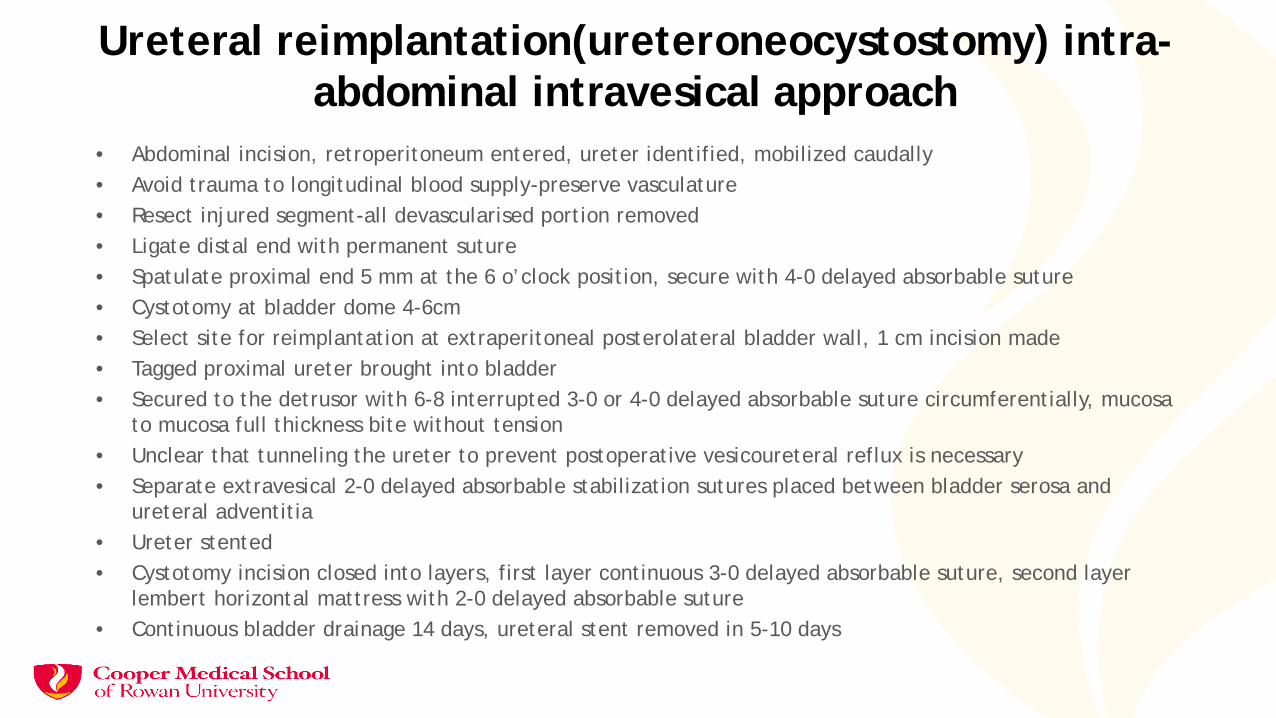
Ureteral reimplantation(ureteroneocystostomy) intra-abdominal intravesical approach
• Abdominal incision, retroperitoneum entered, ureter identified, mobilized caudally• Avoid trauma to longitudinal blood supply-preserve vasculature• Resect injured segment-all devascularised portion removed• Ligate distal end with permanent suture• Spatulate proximal end 5 mm at the 6 o’clock position, secure with 4-0 delayed absorbable suture• Cystotomy at bladder dome 4-6cm• Select site for reimplantation at extraperitoneal posterolateral bladder wall, 1 cm incision made• Tagged proximal ureter brought into bladder• Secured to the detrusor with 6-8 interrupted 3-0 or 4-0 delayed absorbable suture circumferentially, mucosa
to mucosa full thickness bite without tension• Unclear that tunneling the ureter to prevent postoperative vesicoureteral reflux is necessary• Separate extravesical 2-0 delayed absorbable stabilization sutures placed between bladder serosa and
ureteral adventitia• Ureter stented• Cystotomy incision closed into layers, first layer continuous 3-0 delayed absorbable suture, second layer
lembert horizontal mattress with 2-0 delayed absorbable suture• Continuous bladder drainage 14 days, ureteral stent removed in 5-10 days

Ureteral reimplantation(ureteroneocystostomy) extraperitoneal extra vesical approach
• Abdominal incision made, dissection proceeds extraperitonally only, abdominal muscles are split, not cut.
• Care taken to avoid injury to external iliac vein• Identification of the ureter assisted by starting at the pelvic brim, dissect down to the area of
injury, excise the injured portion and ligate the distal end with permanent suture• Retrograde fill the bladder & place 2 stay sutures adjacent to the selected site on the bladder• Spatulate the ureter at the distal end 5 mm at 6 o’clock position• Make a 1 cm incision full thickness in the bladder• Evert the bladder mucosa outwards• Place a single suture at 12 o’clock position• Place a stent within the ureter and into the bladder• Place 6-8 delayed absorbable 4-0 sutures through the ureter full thickness and through the
bladder mucosa and underlying detrusor• Place 3-0 delayed absorbable sutures in bladder detrusor anteriorly to cover the reimplant site• Place 2-0 delayed absorbable stabilization sutures in bladder serosa and ureteral adventitia• Continuous bladder drainage 14 days, ureteral stent removed in 5-10 days


Extraperitoneal approach

Extraperitoneal approach to left ureteral dissection

Psoas hitch
• Free bladder anteriorly contralateral side from the parietal peritoneal attachments in the space of Retzius
• Identify psoas muscle tendon and genitofemoral nerve lateral to the tendon
• Place 2-3 delayed absorbable 1-0 sutures in the muscle and tendon• Cystotomy in the bladder is completed and leading point of the outer
wall and muscular layer of the bladder is pushed towards the psoas• Tagged sutures are secured to the bladder to remove tension• Continue with the ureteral reimplantation


Boari-Ockerblad bladder flapWide base oblique bladder incision made at dome of bladder, with the wide base twice the diameter of the apexThe site of ureteral reimplantation already selectedAvoid narrowing the flap distallyEnd to side ureteral anastomosis made directly or a submucosal tunnel in the upper end is doneCystotomy closure of long linear defect donePsoas hitch performedConcerns –higher incidence of reflux & risk for compromised blood supply to the anastomotic site


Bilateral ureteral implantation in Boari flap

Bibliography
Hancock, B. Practical obstetric fistula surgery. London: Royal Society of Medicine Press, 2009Waaldijk, K. Step-by-step surgery of vesicovaginal fistulas. Edinburgh: Campion Press,1994Gebhart J. B. Urologic surgery for the gynecologist and urogynecologist. Philadelphia: Saunders Elsevier,2010Orr J. W. & H. M. Singleton. Complications in gynecologic surgery. Philadelphia: Lippincott, 1994Lee R. A. Atlas of gynecologic surgery. Philadelphia: Saunders, 1992Ostergard D. R., M. L. Berman & B. Yee. Atlas of gynecologic surgery. Philadelphia: Saunders, 2000