Auditory critical periods: A review from system’s...
17
NEUROSCIENCE FOREFRONT REVIEW AUDITORY CRITICAL PERIODS: A REVIEW FROM SYSTEM’S PERSPECTIVE I A. KRAL * Hearing4all Cluster of Excellence, Hannover School of Medicine, Feodor-Lynen-Str. 35, D-30625 Hannover, Germany Institute of Audioneurotechnology, Dept. of Experimental Otology, Hannover School of Medicine, Feodor-Lynen-Str. 35, D-30625 Hannover, Germany School of Behavioral and Brain Sciences, The University of Texas at Dallas, Richardson, TX 75083, USA Abstract—The article reviews evidence for sensitive periods in the sensory systems and considers their neuronal mech- anisms from the viewpoint of the system’s neuroscience. It reviews the essential cortical developmental steps and shows its dependence on experience. It differentiates fea- ture representation and object representation and their neu- ronal mechanisms. The most important developmental effect of experience is considered to be the transformation of a naive cortical neuronal network into a network capable of categorization, by that establishing auditory objects. The control mechanisms of juvenile and adult plasticity are fur- ther discussed. Total absence of hearing experience pre- vents the patterning of the naive auditory system with subsequent extensive consequences on the auditory func- tion. Additional to developmental changes in synaptic plas- ticity, other brain functions like corticocortical interareal couplings are also influenced by deprivation. Experiments with deaf auditory systems reveal several integrative effects of deafness and their reversibility with experience. Addi- tional to developmental molecular effects on synaptic plas- ticity, a combination of several integrative effects of deprivation on brain functions, including feature representa- tion (affecting the starting point for learning), categorization function, top–down interactions and cross-modal reorgani- zation close the sensitive periods and may contribute to their critical nature. Further, non-auditory effects of auditory deprivation are discussed. To reopen critical periods, removal of molecular breaks in synaptic plasticity and focused training therapy on the integrative effects are required. Ó 2013 The Author. Published by Elsevier Ltd. All rights reserved. Key words: sensitive periods, deprivation, development, plasticity, top–down, hearing loss. Contents Introduction 117 Sensory systems: from feature to object representation and back 118 Corticocortical interactions are the fundament of behavior 119 Development of the neocortex is in part regulated by experi- ence 120 Cochlear implants reveal a critical period for therapy of prelin- gual deafness 120 Auditory plasticity decreases with age 123 Juvenile and adult learning differ 126 Deafness affects non-auditory functions of the brain 128 Critical is not always critical: release of molecular breaks 128 Conclusions 129 Acknowledgements 129 References 129 INTRODUCTION Brain development includes periods of higher susceptibility to alterations by experience called sensitive periods (Kennard, 1938). Periods of higher plasticity allow the juvenile brain to cope with environmental demands and adapt to the conditions into which it was born to. Interestingly, in terms of behavior, some of these sensitive periods are called critical: absence of certain juvenile experiences cannot be fully compensated later in life. The best known examples have been observed during visual development (Cynader and Chernenko, 1976; Cynader and Mitchell, 1977; Daw et al., 1992; Daw, 2009a,b; Hubel and Wiesel, 1970; LeVay et al., 1980) and in the developmental process of visuo-auditory alignments in birds (Knudsen, 1998, 2004). Several types of sensitive periods have been observed: periods when experience is required for the develop- ment of a particular skill (sensitive periods for development); periods where the system is vulnerable by manipula- tion of experience like monocular deprivation (sensitive periods for damage); 0306-4522/13 $36.00 Ó 2013 The Author. Published by Elsevier Ltd. All rights reserved. http://dx.doi.org/10.1016/j.neuroscience.2013.05.021 q This is an open-access article distributed under the terms of the Creative Commons Attribution License, which permits unrestricted use, distribution, and reproduction in any medium, provided the original author and source are credited. * Correspondence to: Andrej Kral, Institute of Audioneurotechnology (VIANNA), Feodor-Lynen-Str. 35, D-30625 Hannover, Germany. Tel: +49-511-532-7272; fax: +49-511-532-7274. E-mail address: [email protected] Abbreviations: DZ, dorsal zone; PAF, posterior auditory field. Neuroscience 247 (2013) 117–133 117
Transcript of Auditory critical periods: A review from system’s...
Neuroscience 247 (2013) 117–133
NEUROSCIENCE FOREFRONT REVIEW
AUDITORY CRITICAL PERIODS: A REVIEW FROMSYSTEM’S PERSPECTIVEI
A. KRAL *
Hearing4all Cluster of Excellence, Hannover School of
Medicine, Feodor-Lynen-Str. 35, D-30625 Hannover, Germany
Institute of Audioneurotechnology, Dept. of Experimental
Otology, Hannover School of Medicine, Feodor-Lynen-Str. 35,
D-30625 Hannover, Germany
School of Behavioral and Brain Sciences, The University of Texas
at Dallas, Richardson, TX 75083, USA
Abstract—The article reviews evidence for sensitive periods
in the sensory systems and considers their neuronal mech-
anisms from the viewpoint of the system’s neuroscience. It
reviews the essential cortical developmental steps and
shows its dependence on experience. It differentiates fea-
ture representation and object representation and their neu-
ronal mechanisms. The most important developmental
effect of experience is considered to be the transformation
of a naive cortical neuronal network into a network capable
of categorization, by that establishing auditory objects. The
control mechanisms of juvenile and adult plasticity are fur-
ther discussed. Total absence of hearing experience pre-
vents the patterning of the naive auditory system with
subsequent extensive consequences on the auditory func-
tion. Additional to developmental changes in synaptic plas-
ticity, other brain functions like corticocortical interareal
couplings are also influenced by deprivation. Experiments
with deaf auditory systems reveal several integrative effects
of deafness and their reversibility with experience. Addi-
tional to developmental molecular effects on synaptic plas-
ticity, a combination of several integrative effects of
deprivation on brain functions, including feature representa-
tion (affecting the starting point for learning), categorization
function, top–down interactions and cross-modal reorgani-
zation close the sensitive periods and may contribute to
their critical nature. Further, non-auditory effects of auditory
deprivation are discussed. To reopen critical periods,
removal of molecular breaks in synaptic plasticity and
focused training therapy on the integrative effects are
required. � 2013 The Author. Published by Elsevier Ltd. All
rights reserved.
0306-4522/13 $36.00 � 2013 The Author. Published by Elsevier Ltd. All rights rehttp://dx.doi.org/10.1016/j.neuroscience.2013.05.021
q This is an open-access article distributed under the terms of theCreative Commons Attribution License, which permits unrestricteduse, distribution, and reproduction in any medium, provided theoriginal author and source are credited.*Correspondence to: Andrej Kral, Institute of Audioneurotechnology(VIANNA), Feodor-Lynen-Str. 35, D-30625 Hannover, Germany. Tel:+49-511-532-7272; fax: +49-511-532-7274.
E-mail address: [email protected]: DZ, dorsal zone; PAF, posterior auditory field.
117
Key words: sensitive periods, deprivation, development,
plasticity, top–down, hearing loss.
Contents
Introduction 117
Sensory systems: from feature to object representation and
back 118
Corticocortical interactions are the fundament of behavior 119
Development of the neocortex is in part regulated by experi-
ence 120
Cochlear implants reveal a critical period for therapy of prelin-
gual deafness 120
Auditory plasticity decreases with age 123
Juvenile and adult learning differ 126
Deafness affects non-auditory functions of the brain 128
Critical is not always critical: release of molecular breaks 128
Conclusions 129
Acknowledgements 129
References 129
INTRODUCTION
Brain development includes periods of higher
susceptibility to alterations by experience called
sensitive periods (Kennard, 1938). Periods of higher
plasticity allow the juvenile brain to cope with
environmental demands and adapt to the conditions into
which it was born to. Interestingly, in terms of behavior,
some of these sensitive periods are called critical:
absence of certain juvenile experiences cannot be fully
compensated later in life. The best known examples
have been observed during visual development
(Cynader and Chernenko, 1976; Cynader and Mitchell,
1977; Daw et al., 1992; Daw, 2009a,b; Hubel and
Wiesel, 1970; LeVay et al., 1980) and in the
developmental process of visuo-auditory alignments in
birds (Knudsen, 1998, 2004). Several types of sensitive
periods have been observed:
� periods when experience is required for the develop-
ment of a particular skill (sensitive periods for
development);
� periods where the system is vulnerable by manipula-
tion of experience like monocular deprivation (sensitive
periods for damage);
served.
1 Nonetheless, during development also sensitivity to new contrastmay appear (Lasky et al., 1975). This indicates that actually theauditory system can also ’’sharpen‘‘ some contrasts.
118 A. Kral / Neuroscience 247 (2013) 117–133
� periods when therapy (compensation of a deficit) is
only partially possible after some age has been missed
(sensitive periods for recovery);
� a distinct type of sensitive periods has to be addition-
ally differentiated: periods for recovery from total depri-
vation. Complete sensory deprivation from birth leaves
the sensory system functionally ‘‘naive’’, which is dis-
tinct from abnormal juvenile experience (like monocu-
lar deprivation or strabism). It leaves the deprived
sensory system functionally incompetent to perform
its function in controlling behavior, which differs from
consequences of abnormal experience. In abnormal
experience, the manipulated sensory system is still
used to control the behavior, but is subject to abnormal
input. The naive state opens the possibility for cross-
modal reorganization, degenerative processes (func-
tional and morphological) and other processes that
do not take place if the system remains functional
(albeit with abnormal input).
When auditory sensitive periods have been
investigated at the synaptic or single-cell level (see e.g.
Morishita and Hensch, 2008; Barkat et al., 2011; Yu
et al., 2012), their critical nature remained difficult to
explain: although synaptic plasticity generally decreases
with increasing age, it does not disappear completely.
Whereas some synapses lose substantial amount of
plasticity with age (Kotak et al., 2007; Barkat et al.,
2011), other do less and should be, in principle, able to
compensate the loss of plasticity in the former. Thus,
other participating processes have to be considered.
The present review focuses on auditory sensitive
periods for sensory deprivation from the system’s
perspective and uncovers the involvement of a
combination of integrative effects that can make sensitive
periods critical.
SENSORY SYSTEMS: FROM FEATURE TOOBJECT REPRESENTATION AND BACK
Neuroscientists often consider the sensory systems with
respect to their representational feature maps, in other
words they investigate how physical features of stimuli
are represented in the brain. In the auditory system,
such features are sound frequency and intensity,
binaural time and intensity differences, frequency
modulation, amplitude modulation, etc. In recent times
considerable interest has shifted also to the way how
the brain generalizes and abstracts from individual
physical features to generate auditory objects (Griffiths
and Warren, 2004; some authors call them events –
Blauert, 1997): did I hear a horn of a car, breaking of a
glass bottle fallen on the ground, ringing of a bell? The
auditory object can be defined as a neuronal
representation of a delimited acoustic pattern that is
subject to figure-background separation.
Sensitive periods are important for the development of
the brain that has to ‘‘bootstrap’’ its function from a
general inborn pattern of connectivity. Correspondingly,
discrimination performance improves during
development. The longest developmental behavioral
improvement can be traced for complex tasks like
discrimination of sounds (e.g. speech) in noise which
continues to improve after discrimination of simple
acoustic features has already matured (review in
Warner et al., 2012). This demonstrates that
representation of complex stimuli possibly requires more
experience than the establishment of feature maps,
although feature maps represent a precondition for the
classification of acoustic input into objects.
Some auditory functions show optimal performance if
learned early in life: e.g. musical experience has most
pronounced effects on performance during early
childhood (reviewed in Penhune, 2011). Also language
learning is easiest early in life. In fact, the best known
auditory examples of critical periods were observed in
language development (review in Kuhl, 2010; Friederici,
2012). Young children are able to discriminate phonetic
contrasts of all languages, however, they specialize in
mother language with increasing age and lose the ability
to discriminate phonetic contrasts that do not exist in
their mother language at around 8th–10th month after
birth. This has been observed for many languages
(Werker and Tees, 1992). Consequently, the newborn
brain is initially very sensitive to acoustic differences.
With time, it specializes and remains sensitive only to
so-called distinctive features of phonemes in their
mother language. Non-distinctive acoustic differences
are ignored (abstracted from)1.
This is an excellent example how the brain establishes
the world of auditory categories (objects). The brain aims
to withstand the enormous variability of the physical
world. It gains the ability to identify sounds that belong
to the same class of events in the physical world, and
learns to ignore their insignificant variability. This
increases the robustness of perception. If the acoustic
input is e.g. affected by several concurring sound
sources, even though some features of the objects are
masked, the object of interest remains discernible
because some of its other distinctive features are less
affected. A side-effect of the appearance of auditory
objects is the loss in sensitivity to contrasts that do not
contribute to the discrimination of the objects in the
given world of experiences. It has to be kept in mind
that several objects may in combination constitute
another object: phonemes are objects themselves, but
they combine into words that are objects on their own.
Which object level is appropriate in the given condition
may depend on the behavioral context.
One perceptional example of how auditory objects
help to cope with distorted input is the so-called
continuity illusion or filling-in phenomenon. Perceptual
filling-in appears when a portion of input is physically
occluded by a concurring stimulus, yet the input is
perceived undisturbed. For example, masking individual
phonemes in a sentence by brief noise does not
preclude the perception of these phonemes (Warren,
1970). More complex filling-in phenomena have been
described in the auditory system (Davis and Johnsrude,
A. Kral / Neuroscience 247 (2013) 117–133 119
2007; Petkov et al., 2007; Riecke et al., 2012; Wild et al.,
2012). The majority (if not all) of them may be related to
auditory objects. Presently, most scientists assume that
auditory objects are represented at the level of the
cerebral cortex (Nelken et al., 2003). For proper
functioning, the cortex requires interactions between
several levels of information representation (Hochstein
and Ahissar, 2002). Often, the higher-level
representation is required to interpret the lower-level
information, as suggested by the theory of inverse
hierarchy (Hochstein and Ahissar, 2002). This theory
assumes that feature representation and object
representations are in constant interaction, and higher-
order representations shape lower-level representations.
Consequently, an important part of the postnatal
developmental sequence is related to the establishment
of object representation. This involves complex
functional interactions at different levels of representation.
CORTICOCORTICAL INTERACTIONS ARE THEFUNDAMENT OF BEHAVIOR
As a prerequisite of these perceptional functions, the
cerebral cortex has to develop a complex architecture.
The auditory cortex contains several auditory fields with
distinct cytoarchitecture and distinct functions. Their
functions have been elucidated using different
experimental procedures. Behavioral functions can be
associated with the activity of distinct auditory fields
(Casseday and Diamond, 1977; Neff, 1977; Neff and
Casseday, 1977; Heffner, 1997; Malhotra et al., 2004).
Historically, cortical areas have been viewed as
hierarchically organized (Felleman and Van Essen,
1991), with lower order fields supplying the cortical input
to higher order fields. This hypothesis implicated that
information flow demonstrates a distinct stepwise
progression through these different fields. Indeed,
latencies of unit responses are largely in accord with
such a hypothesis. Recently, functional data revealing
cortical propagating waves passing through auditory
areal borders in a continuous fashion (among other
evidence) contradict such a hierarchical concept
(Reimer et al., 2011). Nonetheless, there is an
asymmetry of the anatomic projections between auditory
fields so that ‘‘lower-order’’ fields project in a different
way to ‘‘higher-order’’ fields than higher-order fields
project to the lower-order fields (Hackett, 2011). That
allows to still consider a ‘‘hierarchical order’’ in the
cortex, despite of the fact that some form of activity
does not respect areal borders. In what follows, we
understand the term ‘‘hierarchy’’ in this less strict sense;
the auditory cortex with all auditory fields forms a
functional unit.
Feedforward projections connect lower to higher-
order areas and feedback projections connect higher-
order to the lower-order areas (Rouiller et al., 1991).
Feedforward connections originate predominantly from
supragranular layers and target layer IV, feedback
projections target both infra- and supragranular layers,
avoiding layer IV (Felleman and Van Essen, 1991;
Rouiller et al., 1991). (Lateral connections, connecting
areas at the same level, form an intermediate pattern of
connections.) As mentioned above, based on layer-
specific morphological connectivity a ‘‘hierarchical’’ order
of cortical fields can be identified. To make the
interactions between cortical areas possible and thus
allow the appropriate integration of feedforward and
feedback information, the intrinsic cortical connections
forming the cortical column have to become functional.
Functional interactions and morphological connectivity
are different things: morphological connectivity represents
the prerequisite for functional interactions, but this
prerequisite may not be always functionally exploited.
To differentiate morphological connections from
functional interactions, the latter are referred to as
couplings or functional connectivity. In terms of
couplings, bottom–up interactions couple lower-order to
higher-order structures and top–down interactions vice
versa (Gilbert and Sigman, 2007; Kral and Eggermont,
2007). The integration of bottom–up and top–down
stream of information is a fast, in part instantaneous
process: if an activity in the feature map of a primary
auditory field does not correspond to the next higher
level of representation (does not ‘‘fit’’ to the patterns
stored in ‘‘secondary’’ areas), it will be difficult to
stabilize such activity due to the heavy reciprocal
interareal couplings. An instantaneous process of
correction of patterns in the form of e.g. ‘‘an adaptive
resonance’’, suggested by computational neuroscientists
(Grossberg, 1987), is likely to operate between different
level of representation and is likely to adapt activity in
lower-order fields to the representations stored in the
higher-order fields (Mumford, 1992).
An interesting feature of these interareal connections
is their asymmetry in another sense: feedforward
connections bind lower order fields to the next higher
level of hierarchy, whereas feedback connections often
cross several levels of hierarchies (de la Mothe et al.,
2006; Hackett, 2011). The top–down interactions from
high order associative fields can consequently directly
access the primary auditory cortex (de la Mothe et al.,
2006). Attentional top–down modulation is an additional
type of top–down interaction exploiting these long-range
top–down interactions.
The example of filling-in phenomena is strongly related
to top–down interactions, as has been shown in brain
imaging studies (Giraud et al., 2004; Davis and
Johnsrude, 2007; Riecke et al., 2009, 2012; Wild et al.,
2012). Through such interactions, word-level of
representation may affect phonemic level of
representation and fill-out gaps in auditory input (Warren,
1970; Riecke et al., 2009, 2012; comp. Petkov et al.,
2007). These phenomena are likely very important in
speech understanding in noisy environments and other
difficult listening conditions (Schofield et al., 2012; for
visual system, see Kok et al., 2012). In hearing children,
perceptual filling-in can be demonstrated only after the
2nd year of life has been reached (Newman, 2006). It is
therefore tempting to assume that these phenomena are
dependent on experience and consequently affected by
hearing impairment (Kral and Eggermont, 2007; Riecke
et al., 2012).

120 A. Kral / Neuroscience 247 (2013) 117–133
DEVELOPMENT OF THE NEOCORTEX IS INPART REGULATED BY EXPERIENCE
These complex cortical interconnection patterns appear
during development. The cerebral cortex develops in an
inside-out pattern, with deep-layer neurons arriving in
the cortex first, followed by the upper layers (Luskin and
Shatz, 1985; review in Kral and Pallas, 2010). These
migrations take place during early development, but
cortical interneuronal connections become functional
much later. First, neuronal branchings (dendritic and
axonal) establish the background for a first connectivity
matrix. Second, this matrix becomes functional after
synapses have gained basic functionality, which is at
relatively late stages of development. Although the
thalamic afferents arrive in the layer IV of human cortex
around postconceptual day 130 (Clancy et al., 2001),
the dendritic morphology continues to mature over long
periods (reviewed in Kral, 2007) and synaptic
development is not complete around birth and the years
after (Huttenlocher and Dabholkar, 1997).
In the visual system, extensive research on local
connections has revealed that the vertical connectivity
(between layers) develops precisely and specifically
before the visual system receives sensory input (Rakic
et al., 1986; Katz, 1991; Katz and Callaway, 1992).
Vertical connectivity seems to develop in a timescale
that is not related to the time of generation of neurons
(the typical inside–out pattern, Luskin and Shatz, 1985).
The data on the auditory system demonstrate that the
details of columnar intrinsic coupling do change in deaf
animals, although the general pattern is partially
preserved (Kral et al., 2000). Horizontal (tangential)
connections develop later than vertical connections,
within the first months after birth in the cat (Katz and
Callaway, 1992; Galuske and Singer, 1996; for indirect
evidence in the auditory system, see Kral et al., 2006).
Their first appearance is not dependent on sensory
input, but the phase of refinement (change from crude
to refined clusters of staining) depends on patterned
activity (Katz and Callaway, 1992; Galuske and Singer,
1996). Thalamocortical connections appear to precede
or develop in synchrony with the development of
corticothalamic connections, whereas the latter depend
on cortical activity (Shatz and Rakic, 1981; Arimatsu
and Ishida, 2002; Jacobs et al., 2007; Yoshida et al.,
2009). Further, feedforward cortical connections
precede the development of feedback connections
(Barone et al., 1996; Batardiere et al., 2002). The
existing evidence indicates that local cortical
connections develop before long-range cortical
connections appear (Dalva and Katz, 1994; Katz and
Shatz, 1996; Dye et al., 2011). Sensory deprivation in
the visual system does not eliminate feedforward
connectivity (Striem-Amit et al., 2012), and bottom–up
information is preserved up to the level of secondary
auditory cortex in congenital deafness, too (see below).
Top–down interactions are more affected by auditory
deprivation (Kral et al., 2005; Kral and Eggermont, 2007).
In the human cerebral cortex, the development of
cortical axons is very protracted, lasting into teen ages
(Paus et al., 1999; Moore and Guan, 2001).
Corticol–cortical interactions thus mature over very
extended periods in humans. Potentially, correlated
activity in different cortical areas, evoked by sensory
input and cortical activation by already established
thalamocortical afferents, through mechanisms of
synaptic plasticity, will strengthen the connections
between different areas of the same modality and by
that functionally couple together areas within the same
modality (function). Further, the immaturity of the
corticocortical connections indicates that the potential
for this plasticity is high in juvenile animals. The
protracted development of cortical phosphorylated
neurofilaments (Moore and Guan, 2001) does not
implicate that cortico–cortical interactions are not
functional at all, only that their structure is not fully
mature and potentially highly plastic (Pundir et al.,
2012). However, an axonal immaturity suggests that the
different auditory areas, receiving afferent input from
thalamic nuclei, can shape their interareal
communication by experience more than the thalamic
inputs to the cortex.
Development of neuronal (axonal and dendritic)
branching patterns generates the precondition for
forming functional circuits, yet the circuits can function
only once contacts (synapses) have been formed.
Therefore, mainly the most final phases of the
development of dendritic and axonal branches
(particularly in the cortex) and synaptic development are
assumed to be influenced by evoked activity. Indeed,
the adult auditory system of the cat is capable of relying
cochlear input up to the level of the auditory cortex even
in total absence of hearing (Hartmann et al., 1997;
Tillein et al., 2012). Thus, the connections of the
afferent auditory pathway up to the level of cortex
develop independently of experience.
These considerations are supported by functional
imaging of the human auditory cortex.
Electroencephalographic data demonstrate a massive
reorganization of the auditory system, from first large-
amplitude long-lasting responses to the more mature
responses of the adult individuals. First evoked
responses can be recorded from preterm infants (for
review, see Rotteveel, 1992), confirming that thalamic
input can activate the auditory cortex already before
birth (see above). However, the cortical P1/N1
response, generated by thalamic and cortical sources,
systematically decreases in latency within the first 12–
16 years of life (Ponton et al., 2000; Ponton and
Eggermont, 2001; Ceponiene et al., 2002).
In conclusion, the brain continues to mature during
many years of postnatal life, while it already interacts
with the acoustic world.
COCHLEAR IMPLANTS REVEAL A CRITICALPERIOD FOR THERAPY OF PRELINGUAL
DEAFNESS
Restoration of profound hearing loss (deafness) has
become possible using cochlear implants. Cochlear
implants are artificial electronic devices that bypass the

A. Kral / Neuroscience 247 (2013) 117–133 121
non-functional inner ear and electrically stimulate the
auditory nerve directly. They consist of a microphone, a
processor worn behind the ear, a transmitter coil, and
the intracorporal electrode carrier (Fig. 1). The electrode
carrier consists of a receiver coil, a feedthrough with
some electronics and the electrode contacts
themselves. The electrodes are inserted into the
cochlea (scala tympani) so that they come to lie
underneath the organ of Corti. The electrical current can
then stimulate the surviving neurons. Communication of
information and energy between the behind the ear
device and the intracorporal electrode is possible via
magnetic fields through the intact skin between the
transmitter and receiver coil.
As a rule, hearing loss is associated with very good
preservation of auditory nerve in humans (Nadol and
Eddington, 2006). Stimulation of these surviving fibers is
possible through cochlear implants also many years
after the onset of deafness (Dorman and Wilson, 2004).
Nowadays cochlear implants represent the standard of
therapy of congenital deafness in childhood (Kral and
O’Donoghue, 2010). Remarkably, in contrast to adult-
onset of deafness the final outcome of implantation in
‘‘prelingually’’ (congenitally) deaf children is optimal only
in early implantations (within the first 1–3 years of life,
Niparko et al., 2010). Clinical outcomes differentiate
early and late implantations with regard to auditory
performance. Very late implantation (in teen ages) in
prelingually deaf does not lead to speech understanding
and includes difficulties in more complex auditory tasks
(counting of auditory stimuli, electrode position
discrimination, and gap detection; Tong et al., 1988;
Busby et al., 1992, 1993). Even after years of
experience, performance is influenced by implantation
age (Yoshinaga-Itano et al., 1998; Geers, 2006; Niparko
Fig. 1. Schematic illustration of the position of a cochlear implant. Proporti
signal processor with a microphone and a transmitter coil. The intracorpora
inserted into the cochlea (the scala tympani). Sounds are split into frequency
hearing. The resulting envelope is transformed to pulse trains that are deliver
tonotopic cochlear organization.
et al., 2010; Geers and Sedey, 2011). Early implanted
children perform significantly better than later-implanted
children (review in Kral and O’Donoghue, 2010).
Consequently, a critical period for the therapy of human
prelingual deafness exists (Fryauf-Bertschy et al., 1997;
Niparko et al., 2010). Correspondingly, central neuronal
correlates of hearing confirm such a period: N1 waves
are dependent on hearing experience, as absence of
hearing prevents the development of this wave (Ponton
et al., 1996, 2000). The decrease in latency of the P1
component is dependent on hearing experience:
absence of hearing arrests this component (Ponton
et al., 1996), but early implantation normalizes it within
few months (Sharma et al., 2005, 2007). Late
implantation, on the other hand, is not longer able to
promote this fast development and the reorganization
arrests after a few months of hearing through a cochlear
implant (Sharma et al., 2005, 2007). Although late-
implanted subjects often profit from cochlear implant by
the awareness of sound, their ability to differentiate
complex acoustic patterns remains low even after many
years of hearing through cochlear implants.
In conclusion, data on prelingually-deaf children
convincingly demonstrate that loss of hearing cannot be
fully compensated late in life, and thus that this sensitive
period has a critical nature.
In close correspondence with these findings, the
human cerebral cortex develops synapses peri- and
postnatally (Conel, 1939; Huttenlocher and Dabholkar,
1997; for similar observation in cats, see Winfield,
1983). Therefore, information processing starts in the
cortex during the time when the sensory systems
already transduce information about the world. Synaptic
development is consequently dependent on activity
(Cragg, 1975a,b; O’Kusky and Colonnier, 1982;
ons are distorted. The cochlear implant consists of an extracorporal
l part contains a receiver coil, a processor and the electrode array
bands, low-pass filtered and adjusted to the dynamic range of electric
ed to the individual contacts of the electrode array with respect to the

122 A. Kral / Neuroscience 247 (2013) 117–133
Winfield, 1983), although the idea that synaptic
elimination is the key element adapting the microcircuits
to the environment (Changeux and Danchin, 1976),
supported by decreased synaptic counts in blind
animals (O’Kusky and Colonnier, 1982; Winfield, 1983),
seems to be only a part of the story: data from current
source density signals in the auditory cortex replicated
the early morphological data by showing that the
functional synaptic pruning is exaggerated in deafness,
nonetheless, demonstrated an even more extensive
effect on synaptogenesis (Fig. 2; Kral et al., 2005,
2006). Also at the level of individual synapses a similar
observation has been made recently: the emergence of
synapses is more dependent on activity than the
elimination (Kerschensteiner et al., 2009). In total, this
suggests that the period of synaptic development is
regulated by neuronal activity. Studies on rodents
demonstrate that plasticity needs to be considered from
the view of a constant synapse turnover (Trachtenberg
et al., 2002) that may lead to a net spine (synaptic) gain
in early development (Hofer et al., 2009), but not in
adults, where the overall synaptic counts are more
constant and the formation of new spines (synapses) is
connected with the loss of others (Trachtenberg et al.,
2002). Although the literature is not consistent in all
aspects of synapse formation by experience (review in
Holtmaat and Svoboda, 2009), one can currently
conclude:
(1) Cortical synapses develop under the influence of
neuronal activity.
Fig. 2. Developmental changes in mean synaptic activity in the primary audit
hearing controls (blue) and congenitally deaf (red) animals. The feline cochlea
day 180 after birth. Values for adult animals are grand mean averages from f
and O’Donoghue (2010), data from Kral et al. (2005) extended by animals in
using current source density analysis, i.e. the synaptic currents represe
microelectrode. The assessment was performed in six penetrations within
determined in a detailed mapping procedure. The data show an early increas
first 40 days after birth, with subsequent functional synaptic pruning, resultin
deaf animals, the functional synaptogenesis was delayed (1) and the prunin
gray bar (two-tailed Wilcoxon–Mann–Whitney test, a = 5%). The data demon
synapses (and neuronal networks) in the auditory cortex.
(2) When activity is absent, the synaptic development
is delayed and takes place later (Kral et al., 2005;
Kral and Sharma, 2012), regulated by other princi-
ples (not related to sensory input). This may lead
to lack of development of synapses that may be cru-
cial for adequate processing of sensory stimuli, to
the development of synapses that would normally
not develop – and also to pruning of synapses
essential for adequate cortical processing. Further,
balance changes in excitation and inhibition are
likely in deafness, as an increasing inhibitory influ-
ence with increasing age was observed in hearing
animals (data are, however, not entirely consistent
in all aspects, compare Gao et al., 1999; Dorrn
et al., 2010; Sun et al., 2010; Sanes and Kotak,
2011).
Analysis of the functional microcircuitry of the cortical
column identified several functional deficits in progression
of synaptic activity through cortical layers in congenitally
deaf adult animals (Kral et al., 2000). Not only was the
onset of synaptic activity desynchronized (delayed in
supragranular layers and infragranular layers), also
deep layers V/VI showed reduction of synaptic activity
following the first inputs at short latencies. These two
effects could be related: a desynchronization of cortical
inputs may lead to insufficient excitation of those
neurons at which the inputs converge extensively, which
include infragranular neurons. The reduced activity in
deep layers has led to the hypothesis that in primary
auditory field A1 these layers cannot perform their
ory cortex of cats evoked by stimulation through a cochlear implant in
becomes functional around day 10, sexual maturity is reached around
our hearing and four deaf cats. Reproduced with permission from Kral
vestigated before hearing onset. The synaptic activity was assessed
nt activity averaged from many synapses around the tip of the
the most active area of the cortex (‘‘hot-spot’’, size 1.5 � 1.0 mm)
e in synaptic activity (functional synaptogenesis) observed within the
g in adult-like activity between day 80 and 100 in hearing controls. In
g was exaggerated (2). Areas of statistical significance shown by the
strate extensive effects of sensory deprivation on the development of

A. Kral / Neuroscience 247 (2013) 117–133 123
function in congenital deafness, leading to reduction in the
corticofugal drive (e.g. corticothalamic interactions).
Cortical efferents are, among other functions, involved in
the control of subcortical plasticity (Ma and Suga, 2005;
Tang and Suga, 2008). Deficits in corticofugal
interactions will influence how information is processed
in temporal succession as well as how memory adapts
to individual needs of the subject. Deep layers further
modulate the function of supragranular layers,
conveying top–down effects to the intrinsic columnar
circuits (reviews in Raizada and Grossberg, 2003;
Callaway, 2004).
In conclusion, deafness has a disrupting effect on the
development of several control functions of the cortical
column.
AUDITORY PLASTICITY DECREASES WITHAGE
Ongoing (passive) presentation of a tone during
development leads to expansion of the representation
around this tone in the cortex, whereas the expansion is
larger if the presentation starts earlier (Stanton and
Harrison, 1996; Zhang et al., 2001). Indeed, auditory
plasticity is higher at juvenile age, allowing to
significantly reorganize the tonotopic organization of the
cortex with passive acoustic stimulation (Zhang et al.,
2002; Chang and Merzenich, 2003). The development
of tonotopic gradients can be affected and even
disrupted by ongoing presentation of tone sequences or
clicks (Zhang et al., 2002; Nakahara et al., 2004; for
complete deafness, compare Snyder et al., 1990;
Raggio and Schreiner, 1999, 2003; Fallon et al., 2009a).
‘Environmental noise’ (i.e. ongoing white noise of
moderate intensity), however, can delay the
developmental steps and extend the sensitive period for
plasticity of the tonotopic organization (Chang and
Merzenich, 2003), and this may even happen in
circumscribed cortical regions (de Villers-Sidani et al.,
2008). These studies in combination demonstrate that
an orchestrated sequence of sensitive developmental
period for auditory plasticity exists (de Villers-Sidani and
Merzenich, 2011).
But what is the consequence of profound hearing loss
on the sensitive periods? The absence of auditory input
may arrest of developmental processes and thus extend
the sensitive periods, similarly as e.g. environmental
noise. However, the fact that functional synaptogenesis,
although delayed, finally takes place even in complete
deafness (Fig. 2, Kral et al., 2005; Winfield, 1983),
demonstrates that such an arrest is not forever.
Environmental noise and profound deafness have
different consequences in the brain: in deafness, there
is no evoked and no spontaneous activity in the auditory
nerve. In contrast, masking noise reduces (but does not
eliminate) patterned auditory input, it preserves
spontaneous activity and increases the firing rate of
auditory nerve fibers. It thus initially increases the level
of peripheral input in the auditory system, whereas
deafness removes it. The central effects of noise and
deafness differ. Ongoing masking noise increases the
excitation thresholds in the central auditory system
(Chang and Merzenich, 2003; Norena et al., 2006).
Deafness, on the other hand, decreases cortical
thresholds (Kral et al., 2005; Fallon et al., 2009a,b). In
congenital deafness, the dynamic range of unit rate-
level functions decreases when tested with cochlear
implants (Tillein et al., 2010), a phenomenon not
observed with masking noise. Thus, congenital
deafness and noise exposure are different phenomena
triggering different pathophysiological processes in the
brain.
A cortical correlate of a developmental sensitive
period in auditory plasticity has been demonstrated in
deaf cats chronically stimulated with cochlear implants
(Kral et al., 2001, 2002). Using single-channel implants,
the cortical area activated with an electric pulse applied
to the auditory nerve expanded in course of the
stimulation and the responses decreased in latency
(Fig. 3; Klinke et al., 1999). Such plastic adaptation is
adequate for the single-channel stimulation, as it
attributes larger neuronal resources for processing of
the stimulus. Extracellular recordings of action potentials
in the hot spot demonstrated development of feature
sensitivity and indicate functional maturation of the
cortex (Klinke et al., 1999; Kral et al., 2006). This
reorganization had, however, a decreasing trend with
increasing implantation age (Fig. 3; Kral et al., 2002;
review in Kral and Sharma, 2012). Further, in hearing
animals cortical responses have shorter latency and
larger amplitude for stimulation at the contralateral ear,
a phenomenon that disappears in congenital deafness
(Kral et al., 2009). Asymmetry in hearing, caused either
by conductive hearing loss (Popescu and Polley, 2010)
or by single-sided deafness (Kral et al., 2013) may
further shift this relation in favor of the better hearing
ear, leading to a change in the ‘‘aural preference’’ at the
cortex ipsilateral to the ‘‘hearing’’ ear (Fig. 4; Kral et al.,
2013). This effect is strongest in congenital single-sided
deafness and ceases with increasing age of onset of
single-sided deafness, demonstrating a developmental
sensitive period (Figs. 4 and 5; Kral et al., 2013). In
children with sequential implantations and long delays
between implantation, the speech recognition at the
second-implanted ear lags behind the first implanted ear
even after years of bilateral hearing, and the
improvements are very slow (Graham et al., 2009; Illg
et al., 2013). Results consistent with this outcome were
obtained in human single-sided deafness with imaging
after stimulation of the hearing ear (Burton et al., 2012).
Hearing asymmetry in children is a factor that is under
investigation of clinicians, supporting the above
experimental studies in humans (Graham et al., 2009;
Gordon et al., 2010, 2011). Interestingly, although these
results appear similar to monocular deprivation in the
visual system, there is one important difference: in all
investigated cases the deaf ear remained capable of
substantial activation of the auditory cortex, which differs
from the situation found in amblyopia.
These results show that neuronal plasticity decreases
with age in congenital deafness, with decreasing
adaptability to sensory input. There is some indication
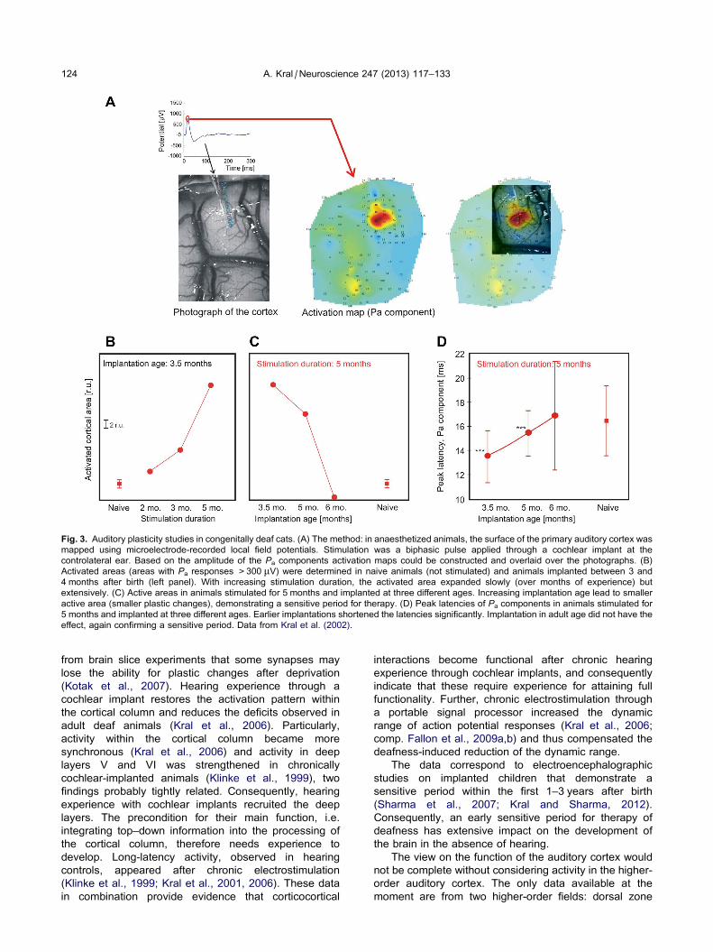
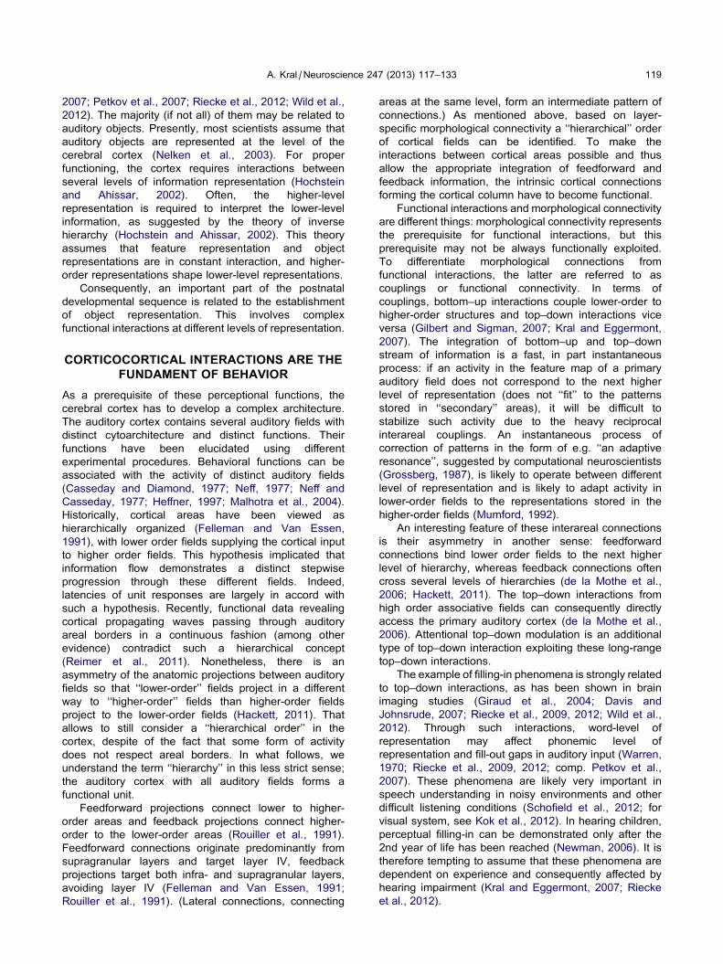
Fig. 3. Auditory plasticity studies in congenitally deaf cats. (A) The method: in anaesthetized animals, the surface of the primary auditory cortex was
mapped using microelectrode-recorded local field potentials. Stimulation was a biphasic pulse applied through a cochlear implant at the
controlateral ear. Based on the amplitude of the Pa components activation maps could be constructed and overlaid over the photographs. (B)
Activated areas (areas with Pa responses >300 lV) were determined in naive animals (not stimulated) and animals implanted between 3 and
4 months after birth (left panel). With increasing stimulation duration, the activated area expanded slowly (over months of experience) but
extensively. (C) Active areas in animals stimulated for 5 months and implanted at three different ages. Increasing implantation age lead to smaller
active area (smaller plastic changes), demonstrating a sensitive period for therapy. (D) Peak latencies of Pa components in animals stimulated for
5 months and implanted at three different ages. Earlier implantations shortened the latencies significantly. Implantation in adult age did not have the
effect, again confirming a sensitive period. Data from Kral et al. (2002).
124 A. Kral / Neuroscience 247 (2013) 117–133
from brain slice experiments that some synapses may
lose the ability for plastic changes after deprivation
(Kotak et al., 2007). Hearing experience through a
cochlear implant restores the activation pattern within
the cortical column and reduces the deficits observed in
adult deaf animals (Kral et al., 2006). Particularly,
activity within the cortical column became more
synchronous (Kral et al., 2006) and activity in deep
layers V and VI was strengthened in chronically
cochlear-implanted animals (Klinke et al., 1999), two
findings probably tightly related. Consequently, hearing
experience with cochlear implants recruited the deep
layers. The precondition for their main function, i.e.
integrating top–down information into the processing of
the cortical column, therefore needs experience to
develop. Long-latency activity, observed in hearing
controls, appeared after chronic electrostimulation
(Klinke et al., 1999; Kral et al., 2001, 2006). These data
in combination provide evidence that corticocortical
interactions become functional after chronic hearing
experience through cochlear implants, and consequently
indicate that these require experience for attaining full
functionality. Further, chronic electrostimulation through
a portable signal processor increased the dynamic
range of action potential responses (Kral et al., 2006;
comp. Fallon et al., 2009a,b) and thus compensated the
deafness-induced reduction of the dynamic range.
The data correspond to electroencephalographic
studies on implanted children that demonstrate a
sensitive period within the first 1–3 years after birth
(Sharma et al., 2007; Kral and Sharma, 2012).
Consequently, an early sensitive period for therapy of
deafness has extensive impact on the development of
the brain in the absence of hearing.
The view on the function of the auditory cortex would
not be complete without considering activity in the higher-
order auditory cortex. The only data available at the
moment are from two higher-order fields: dorsal zone
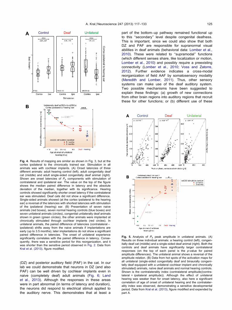
Fig. 4. Results of mapping are similar as shown in Fig. 3, but at the
cortex ipsilateral to the chronically trained ear. Stimulation in all
animals was with cochlear implants. (A) Onset latencies of three
different animals: adult hearing control (left), adult congenitally deaf
cat (middle) and adult single-sided congenitally deaf animal (right).
Shown are onset latencies of Pa components with stimulation of
contralateral and ipsilateral ear. The value on the top of the figure
shows the median paired difference in latency and the absolute
deviation of the median, together with its significance. Hearing
controls showed significantly shorter onset latency if the contralateral
ear was stimulated. Deaf cats did not show a significant difference.
Single-sided animals showed (at the cortex ipsilateral to the hearing
ear) a reversal of the latencies with shortest latencies with stimulation
of the ipsilateral (hearing) ear. (B) Presentation of seven naive
animals (red boxes), seven normal hearing controls (blue boxes) and
seven unilateral animals (circles), congenital unilaterally deaf animals
shown in green (green circles), the other animals were implanted at
chronically stimulated through cochlear implants (red circles). In
unilateral animals, the paired difference of latencies (contralateral –
ipsilateral) shifts away from the naive animals if implantations are
early (up to 3.5 months), later implantations do not show a significant
paired difference in latencies. The onset of unilateral experience
significantly correlates with the paired difference in latency. Conse-
quently, there was a sensitive period for this reorganization, and it
was shorter than the sensitive period observed in Fig. 2. Data from
Kral et al. (2013), figure modified.
Fig. 5. Analysis of Pa peak amplitude in unilateral animals. (A)
Results on three individual animals: a hearing control (left), congen-
itally deaf cat (middle) and a single-sided deaf animal (right). Both the
controls and deaf animals have significantly larger contralateral
responses (on the top of each panel is the p-value for paired
amplitude differences). The unilateral animal shows a reversal of the
amplitude relation. (B) Data from hot spots of the activation maps for
all unilateral (single-sided congenitally deaf and binaurally congeni-
tally deaf equipped with a unilateral cochlear implant and chronically
stimulated) animals, naıve deaf animals and normal hearing controls.
Shown is the contralaterality index (contralateral amplitude/(contra-
lateral + ipsilateral amplitude)). Although the effect of unilateral
hearing was weaker than for onset latency, also here a significant
correlation of age of onset of unilateral hearing and the contralater-
ality index was observed, demonstrating a sensitive developmental
period. Data from Kral et al. (2013), figure modified and expended by
part A.
A. Kral / Neuroscience 247 (2013) 117–133 125
(DZ) and posterior auditory field (PAF) in the cat. In our
lab we could demonstrate that neurons in DZ (and also
PAF) can be well driven by cochlear implants even in
naive (completely deaf) adult animals (Fig. 6; Land
et al., 2013). Although the responses in these areas
were in part abnormal (in terms of latency and duration),
the neurons did respond to electrical stimuli applied to
the auditory nerve. This demonstrates that at least a
part of the bottom–up pathway remained functional up
to this ‘‘secondary’’ level despite congenital deafness.
This is important, since we could also show that both
DZ and PAF are responsible for supranormal visual
abilities in deaf animals (behavioral data: Lomber et al.,
2010). These were related to ‘‘supramodal’’ functions
(which different senses share, like localization or motion,
Lomber et al., 2010) and possibly require a preexisting
connectivity (Lomber et al., 2010; Voss and Zatorre,
2012). Further evidence indicates a cross-modal
reorganization of field AAF by somatosensory modality
(Meredith and Lomber, 2011). Thus, other sensory
systems can make use of the deaf auditory system.
Two possible mechanisms have been suggested to
explain these findings: (a) growth of new connections
from other brain regions into auditory regions that recruit
these for other functions; or (b) different use of these
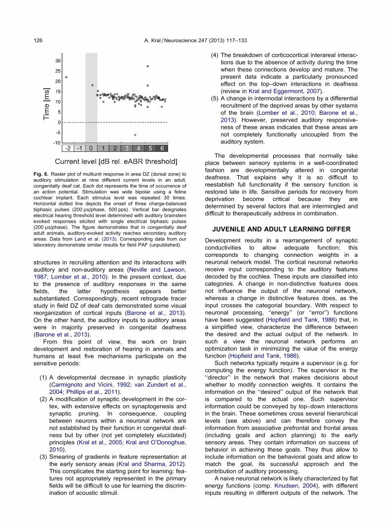
Fig. 6. Raster plot of multiunit response in area DZ (dorsal zone) to
auditory stimulation at nine different current levels in an adult,
congenitally deaf cat. Each dot represents the time of occurrence of
an action potential. Stimulation was wide bipolar using a feline
cochlear implant. Each stimulus level was repeated 30 times.
Horizontal dotted line depicts the onset of three charge-balanced
biphasic pulses (200 ls/phase, 500 pps). Vertical bar designates
electrical hearing threshold level determined with auditory brainstem
evoked responses elicited with single electrical biphasic pulses
(200 ls/phase). The figure demonstrates that in congenitally deaf
adult animals, auditory-evoked activity reaches secondary auditory
areas. Data from Land et al. (2013). Corresponding data from our
laboratory demonstrate similar results for field PAF (unpublished).
126 A. Kral / Neuroscience 247 (2013) 117–133
structures in recruiting attention and its interactions with
auditory and non-auditory areas (Neville and Lawson,
1987; Lomber et al., 2010). In the present context, due
to the presence of auditory responses in the same
fields, the latter hypothesis appears better
substantiated. Correspondingly, recent retrograde tracer
study in field DZ of deaf cats demonstrated some visual
reorganization of cortical inputs (Barone et al., 2013).
On the other hand, the auditory inputs to auditory areas
were in majority preserved in congenital deafness
(Barone et al., 2013).
From this point of view, the work on brain
development and restoration of hearing in animals and
humans at least five mechanisms participate on the
sensitive periods:
(1) A developmental decrease in synaptic plasticity
(Carmignoto and Vicini, 1992; van Zundert et al.,
2004; Phillips et al., 2011).
(2) A modification of synaptic development in the cor-
tex, with extensive effects on synaptogenesis and
synaptic pruning. In consequence, coupling
between neurons within a neuronal network are
not established by their function in congenital deaf-
ness but by other (not yet completely elucidated)
principles (Kral et al., 2005; Kral and O’Donoghue,
2010).
(3) Smearing of gradients in feature representation at
the early sensory areas (Kral and Sharma, 2012).
This complicates the starting point for learning: fea-
tures not appropriately represented in the primary
fields will be difficult to use for learning the discrim-
ination of acoustic stimuli.
(4) The breakdown of corticocortical interareal interac-
tions due to the absence of activity during the time
when these connections develop and mature. The
present data indicate a particularly pronounced
effect on the top–down interactions in deafness
(review in Kral and Eggermont, 2007).
(5) A change in intermodal interactions by a differential
recruitment of the deprived areas by other systems
of the brain (Lomber et al., 2010; Barone et al.,
2013). However, preserved auditory responsive-
ness of these areas indicates that these areas are
not completely functionally uncoupled from the
auditory system.
The developmental processes that normally take
place between sensory systems in a well-coordinated
fashion are developmentally altered in congenital
deafness. That explains why it is so difficult to
reestablish full functionality if the sensory function is
restored late in life. Sensitive periods for recovery from
deprivation become critical because they are
determined by several factors that are intermingled and
difficult to therapeutically address in combination.
JUVENILE AND ADULT LEARNING DIFFER
Development results in a rearrangement of synaptic
conductivities to allow adequate function; this
corresponds to changing connection weights in a
neuronal network model. The cortical neuronal networks
receive input corresponding to the auditory features
decoded by the cochlea. These inputs are classified into
categories. A change in non-distinctive features does
not influence the output of the neuronal network,
whereas a change in distinctive features does, as the
input crosses the categorial boundary. With respect to
neuronal processing, ‘‘energy’’ (or ‘‘error’’) functions
have been suggested (Hopfield and Tank, 1986) that, in
a simplified view, characterize the difference between
the desired and the actual output of the network. In
such a view the neuronal network performs an
optimization task in minimizing the value of the energy
function (Hopfield and Tank, 1986).
Such networks typically require a supervisor (e.g. for
computing the energy function). The supervisor is the
‘‘director’’ in the network that makes decisions about
whether to modify connection weights. It contains the
information on the ‘‘desired’’ output of the network that
is compared to the actual one. Such supervisor
information could be conveyed by top–down interactions
in the brain. These sometimes cross several hierarchical
levels (see above) and can therefore convey the
information from associative prefrontal and frontal areas
(including goals and action planning) to the early
sensory areas. They contain information on success of
behavior in achieving these goals. They thus allow to
include information on the behavioral goals and allow to
match the goal, its successful approach and the
contribution of auditory processing.
A naive neuronal network is likely characterized by flat
energy functions (comp. Knudsen, 2004), with different
inputs resulting in different outputs of the network. The
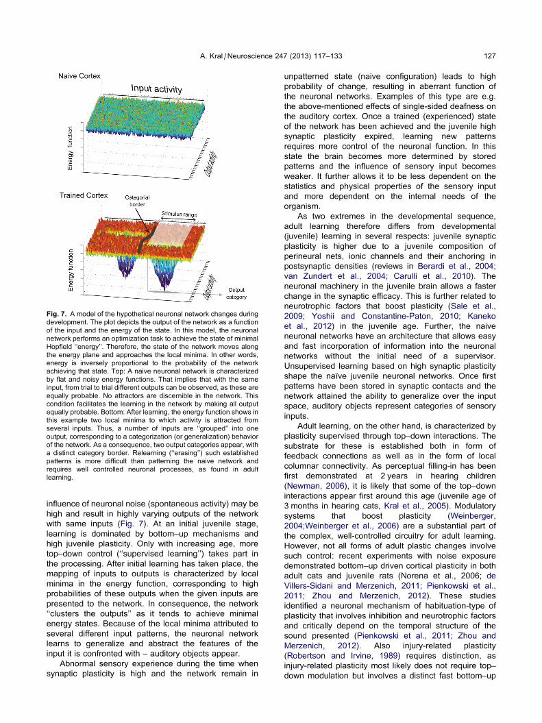
Fig. 7. A model of the hypothetical neuronal network changes during
development. The plot depicts the output of the network as a function
of the input and the energy of the state. In this model, the neuronal
network performs an optimization task to achieve the state of minimal
Hopfield ‘‘energy’’. Therefore, the state of the network moves along
the energy plane and approaches the local minima. In other words,
energy is inversely proportional to the probability of the network
achieving that state. Top: A naive neuronal network is characterized
by flat and noisy energy functions. That implies that with the same
input, from trial to trial different outputs can be observed, as these are
equally probable. No attractors are discernible in the network. This
condition facilitates the learning in the network by making all output
equally probable. Bottom: After learning, the energy function shows in
this example two local minima to which activity is attracted from
several inputs. Thus, a number of inputs are ‘‘grouped’’ into one
output, corresponding to a categorization (or generalization) behavior
of the network. As a consequence, two output categories appear, with
a distinct category border. Relearning (‘‘erasing’’) such established
patterns is more difficult than patterning the naive network and
requires well controlled neuronal processes, as found in adult
learning.
A. Kral / Neuroscience 247 (2013) 117–133 127
influence of neuronal noise (spontaneous activity) may be
high and result in highly varying outputs of the network
with same inputs (Fig. 7). At an initial juvenile stage,
learning is dominated by bottom–up mechanisms and
high juvenile plasticity. Only with increasing age, more
top–down control (‘‘supervised learning’’) takes part in
the processing. After initial learning has taken place, the
mapping of inputs to outputs is characterized by local
minima in the energy function, corresponding to high
probabilities of these outputs when the given inputs are
presented to the network. In consequence, the network
‘‘clusters the outputs’’ as it tends to achieve minimal
energy states. Because of the local minima attributed to
several different input patterns, the neuronal network
learns to generalize and abstract the features of the
input it is confronted with – auditory objects appear.
Abnormal sensory experience during the time when
synaptic plasticity is high and the network remain in
unpatterned state (naive configuration) leads to high
probability of change, resulting in aberrant function of
the neuronal networks. Examples of this type are e.g.
the above-mentioned effects of single-sided deafness on
the auditory cortex. Once a trained (experienced) state
of the network has been achieved and the juvenile high
synaptic plasticity expired, learning new patterns
requires more control of the neuronal function. In this
state the brain becomes more determined by stored
patterns and the influence of sensory input becomes
weaker. It further allows it to be less dependent on the
statistics and physical properties of the sensory input
and more dependent on the internal needs of the
organism.
As two extremes in the developmental sequence,
adult learning therefore differs from developmental
(juvenile) learning in several respects: juvenile synaptic
plasticity is higher due to a juvenile composition of
perineural nets, ionic channels and their anchoring in
postsynaptic densities (reviews in Berardi et al., 2004;
van Zundert et al., 2004; Carulli et al., 2010). The
neuronal machinery in the juvenile brain allows a faster
change in the synaptic efficacy. This is further related to
neurotrophic factors that boost plasticity (Sale et al.,
2009; Yoshii and Constantine-Paton, 2010; Kaneko
et al., 2012) in the juvenile age. Further, the naive
neuronal networks have an architecture that allows easy
and fast incorporation of information into the neuronal
networks without the initial need of a supervisor.
Unsupervised learning based on high synaptic plasticity
shape the naıve juvenile neuronal networks. Once first
patterns have been stored in synaptic contacts and the
network attained the ability to generalize over the input
space, auditory objects represent categories of sensory
inputs.
Adult learning, on the other hand, is characterized by
plasticity supervised through top–down interactions. The
substrate for these is established both in form of
feedback connections as well as in the form of local
columnar connectivity. As perceptual filling-in has been
first demonstrated at 2 years in hearing children
(Newman, 2006), it is likely that some of the top–down
interactions appear first around this age (juvenile age of
3 months in hearing cats, Kral et al., 2005). Modulatory
systems that boost plasticity (Weinberger,
2004;Weinberger et al., 2006) are a substantial part of
the complex, well-controlled circuitry for adult learning.
However, not all forms of adult plastic changes involve
such control: recent experiments with noise exposure
demonstrated bottom–up driven cortical plasticity in both
adult cats and juvenile rats (Norena et al., 2006; de
Villers-Sidani and Merzenich, 2011; Pienkowski et al.,
2011; Zhou and Merzenich, 2012). These studies
identified a neuronal mechanism of habituation-type of
plasticity that involves inhibition and neurotrophic factors
and critically depend on the temporal structure of the
sound presented (Pienkowski et al., 2011; Zhou and
Merzenich, 2012). Also injury-related plasticity
(Robertson and Irvine, 1989) requires distinction, as
injury-related plasticity most likely does not require top–
down modulation but involves a distinct fast bottom–up

128 A. Kral / Neuroscience 247 (2013) 117–133
mechanism starting with loss of lateral inhibition from the
injured regions (Snyder and Sinex, 2002).
In sensory deprivation, some developmental steps
transforming juvenile to adult learning have taken place
while others did not. Supervised learning is
compromised by dysfunctional cortical columns that
cannot integrate top–down modulations into the function
of infragranular layers. However, the phase of high
synaptic plasticity terminates, although in delayed time-
line. Some synapses even lose the ability for plastic
change. The naive network is then neither in the
juvenile highly-plastic state, nor in the state designed for
supervised learning. Adult plasticity cannot be properly
controlled and directed during learning, and therefore
adaptive learning is compromised. If we could find ways
to restore juvenile plasticity in the naive network, more
weight could be put on bottom–up mechanisms and
therefore more beneficial effect of sensory input could
be expected (Duffy and Mitchell, 2013). As the general
interareal (morphological) connectivity within the
auditory system is preserved in congenital deafness
(e.g. in A1 and DZ of deaf cats, Barone et al., 2013)
and the effects of deafness are more observed in
couplings, this might be a promising new strategy.
In conclusion, learning differs in juvenile and adult
brains. The developmental transition from juvenile to
adult learning requires steps that crucially depend on
experience. Complete deprivation from birth therefore
leaves an auditory system that has not the same
capacity for synaptic plasticity as the juvenile brain, but
also lacks the control mechanisms that boost learning in
adult age.
DEAFNESS AFFECTS NON-AUDITORYFUNCTIONS OF THE BRAIN
Finally, sensory systems are not completely equivalent:
the visual system provides excellent spatial information
(visual acuity reaching, depending on the type of task,
the level of few dozens of second of the arc), whereas
the temporal acuity is low (flicker fusion at �60 Hz). In
the auditory system, the spatial localization ability is low
(minimal audible angles in the order of 1–3�), yet the
temporal acuity is high (temporal code up to 3–4 kHz).
Therefore, different types of information are optimally
represented in different sensory systems, leading to
dominance of perception in sensory conflicts (auditory
or visual capture, Recanzone, 1998, 2003). The theory
has been put forward that the primary sensory areas
serve as a high-resolution buffer (‘‘blackboard’’) for
cognition (Mumford, 1992), whereas each sensory
system has a particular function with regard to certain
physical characteristics. In this regard it is tempting to
speculate that the auditory system has a particular
function in representing the timeline (sequence) of
events at high temporal resolution. Deafness,
particularly congenital deafness, would consequently
interfere with this ability and affect many cognitive
functions.
Indeed, absence of hearing affects more than hearing
itself (reviewed in Kral and O’Donoghue, 2010): deaf
subjects underperform hearing children in visual
sequence learning (Conway et al., 2011). This could be
related to reduced working memory, as observed in
signing subjects compared to hearing subjects (Pisoni
and Geers, 2000). Nonetheless, this type of reduced
working memory is most likely related to the linguistic
representation by visual signs (Boutla et al., 2004). An
alternative representation may be a reduced ability for
organizing sensory inputs along the temporal dimension.
Deaf children show alterations in fine motor coordination
(Horn et al., 2006, 2007). To what extent this finding
relates to the absence of auditory input remains to be
verified.
Finally, attention not only affects sensory systems,
also sensory systems can attract attention (if the stimuli
are salient). Hearing is particularly suitable for
controlling attention in situations when changes in
environment happen outside of the field of view. Most
interestingly, deaf children distribute more attentional
resources to the peripheral visual field (Bavelier et al.,
2000; Bottari et al., 2010), as if they would constantly
‘‘scan’’ the periphery. This leads to deficits in sustained
attention (Yucel and Derim, 2008; Barker et al., 2009).
Such condition affects the interaction with caretakers
and limits joint attention: the ability of children to orient
attention to the object attended by the caretaker. Joint
attention is an important process in the phase of
learning from parents. In consequence, even though the
deficit in sustained attention is alleviated with age, the
early juvenile learning phase must be extensively
affected by this deficit.
More focus needs to be put on these non-auditory
effects of deafness in the future. They clearly
demonstrate that the congenitally deaf brain differs from
a ‘‘hearing’’ brain by much more than the absence of
cochlear function. Training after restoration of the
peripheral hearing deficit may be necessary to
compensate the deficits that developed. The deficits
could be highly dependent on the subject and its mode
of exploitation of the cognitive resources of the brain.
These cognitive factors likely further contribute to the
closure of critical periods.
CRITICAL IS NOT ALWAYS CRITICAL:RELEASE OF MOLECULAR BREAKS
To demonstrate a critical nature of some sensitive periods
is not straightforward in an animal model. However,
experience with patients after sensory loss may serve
as a guide, confirming that some forms of monocular
deprivation, complete visual deprivation and complete
absence of hearing from birth are difficult to reverse
despite years-long sensory experience following therapy
of the defect.
Nevertheless, recent work indicates that release of
some molecular breaks of plasticity, particularly those
related to inhibition, may reopen some critical periods in
animal experiments (Pizzorusso et al., 2002; Morishita
and Hensch, 2008). Also, periods of constant low-level
noise or darkness appear to have the potential to delay
or even ‘reset’ developmental stages and reinstall
A. Kral / Neuroscience 247 (2013) 117–133 129
juvenile plasticity in some types of sensitive periods (Zhou
et al., 2011; Duffy and Mitchell, 2013). This is related to
molecular changes in the cortex that, after dark rearing,
increase plasticity (Duffy and Mitchell, 2013). In the
auditory system, the critical period for plasticity of
frequency tuning and tonotopic organization can
similarly be extended by stimulation with continuous
noise and terminated by tonal stimulation or just through
spontaneous recovery (Chang and Merzenich, 2003;
Zhou and Merzenich, 2012). In this regard the potential
for plastic reorganization is developmentally limited by
e.g. by neurofilament modification (Duffy and Mitchell,
2013), maturation of inhibitory transmission,
neurotrophic factors release or chondroitin-sulfate
(Pizzorusso et al., 2002; Carulli et al., 2010; Zhou and
Merzenich, 2012). Removal of such molecular breaks at
later age can restore juvenile synaptic plasticity
(Pizzorusso et al., 2002; Zhou et al., 2011; Duffy and
Mitchell, 2013) and potentially compensate many effects
of juvenile pathologic experience. However, so far these
studies concentrate on abnormal (pathological)
experience during development (monocular deprivation
or strabism, i.e. critical periods for damage, or disruption
of tonotopic organization in the auditory system).
Whether restoration of juvenile plasticity may
compensate also the here-described systemic effects of
complete sensory deprivation remains to be investigated
in the future. There is substantiated hope that
neuroscience will learn to counterbalance the
devastating effect of experience also in adult age. If the
here-reviewed systemic contributions to critical periods
should indeed be the key element, the critical periods
would not be set in stone. Focused manipulations in
sensory input, combined with training methods could
alleviate deprivation-induced deficits. At present, early
intervention remains the gold standard in therapy of
early hearing loss, enabled by neonatal hearing
screening (Kral and O’Donoghue, 2010).
CONCLUSIONS
The available evidence shows that many basic cerebral
functions are inborn. Learned, on the other hand, are
representations of sensory objects that are highly
individual and depend on the subject‘s experiences.
Related to it, corticocortical interactions and the function
of the cortical column depend on experience and are
shaped by sensory inputs. Periods of high susceptibility
to environmental manipulations are given by higher
synaptic plasticity and a naive state of neuronal
networks that may easily be patterned by sensory input.
Adult learning, on the other hand, is characterized by
weaker synaptic plasticity but the ability to control and
modulate plasticity by the need of the organism through
top–down interactions and modulatory systems.
Congenital deafness affects the development not only
by delaying it, but also by desynchronizing different
developmental steps. In its ultimate consequence,
congenital deafness results in an auditory system that
lacks the ability to supervise early sensory processing
and plasticity, but also lacks the high synaptic plasticity
of the juvenile brain. Critical developmental periods
result. It remains an open question whether restoring
juvenile plasticity by eliminating molecular breaks of
plasticity will reinstall functional connectivity in the
auditory cortex and bring a new therapy for complete
sensory deprivation in the future. Focus on integrative
aspects of critical periods will be required to counteract
the reorganization taken place in the deprived sensory
system and the other affected cerebral functions by
training procedures.
Acknowledgements—Supported by Deutsche Forschungsgeme-
inschaft (Kr 3370/1-3 and Cluster of Excellence Hearing4All). The
author wants to thank the anonymous reviewers for their helpful
comments.
REFERENCES
Arimatsu Y, Ishida M (2002) Distinct neuronal populations specified to
form corticocortical and corticothalamic projections from layer VI
of developing cerebral cortex. Neuroscience 114:1033–1045.
Barkat TR, Polley DB, Hensch TK (2011) A critical period for auditory
thalamocortical connectivity. Nat Neurosci 14:1189–1194.
Barker DH, Quittner AL, Fink NE, Eisenberg LS, Tobey EA, Niparko
JK, CDaCI Investigative Team (2009) Predicting behavior
problems in deaf and hearing children: the influences of
language, attention, and parent–child communication. Dev
Psychopathol 21:373–392.
Barone P, Dehay C, Berland M, Kennedy H (1996) Role of directed
growth and target selection in the formation of cortical pathways:
prenatal development of the projection of area V2 to area V4 in
the monkey. J Comp Neurol 374:1–20.
Barone P, Lecassagne L, Kral A (2013) Reorganization of the
connectivity of field DZ in congenitally deaf cat. PLoS One
8(4):e60093.
Batardiere A, Barone P, Knoblauch K, Giroud P, Berland M, Dumas
AM, Kennedy H (2002) Early specification of the hierarchical
organization of visual cortical areas in the macaque monkey.
Cereb Cortex 12:453–465.
Bavelier D, Tomann A, Hutton C, Mitchell T, Corina D, Liu G, Neville
H (2000) Visual attention to the periphery is enhanced in
congenitally deaf individuals. J Neurosci 20:RC93.
Berardi N, Pizzorusso T, Maffei L (2004) Extracellular matrix and
visual cortical plasticity: freeing the synapse. Neuron 44:905–908.
Blauert J (1997) Spatial hearing: the psychophysics of human spatial
localization. Cambridge, MA: MIT Press.
Bottari D, Nava E, Ley P, Pavani F (2010) Enhanced reactivity to
visual stimuli in deaf individuals. Restor Neurol Neurosci
28:167–179.
Boutla M, Supalla T, Newport EL, Bavelier D (2004) Short-term
memory span: insights from sign language. Nat Neurosci
7:997–1002.
Burton H, Firszt JB, Holden T, Agato A, Uchanski RM (2012)
Activation lateralization in human core, belt, and parabelt auditory
fields with unilateral deafness compared to normal hearing. Brain
Res 1454:33–47.
Busby PA, Tong YC, Clark GM (1992) Psychophysical studies using
a multiple-electrode cochlear implant in patients who were
deafened early in life. Audiology 31:95–111.
Busby PA, Tong YC, Clark GM (1993) Electrode position, repetition
rate, and speech perception by early- and late-deafened cochlear
implant patients. J Acoust Soc Am 93:1058–1067.
Callaway EM (2004) Feedforward, feedback and inhibitory
connections in primate visual cortex. Neural Network 17:625–632.
Carmignoto G, Vicini S (1992) Activity-dependent decrease in NMDA
receptor responses during development of the visual cortex.
Science 258:1007–1011.

130 A. Kral / Neuroscience 247 (2013) 117–133
Carulli D, Pizzorusso T, Kwok JC, Putignano E, Poli A, Forostyak S,
Andrews MR, Deepa SS, Glant T, Fawcett JW (2010) Animals
lacking link protein have attenuated perineuronal nets and
persistent plasticity. Brain 133:2331–2347.
Casseday JH, Diamond IT (1977) Symmetrical lateralization of
function in the auditory system of the cat: effects of unilateral
ablation of cortex. Ann N Y Acad Sci 299:255–263.
Ceponiene R, Rinne T, Naatanen R (2002) Maturation of cortical
sound processing as indexed by event-related potentials. Clin
Neurophysiol 113:870–882.
Chang EF, Merzenich MM (2003) Environmental noise retards
auditory cortical development. Science 300:498–502.
Changeux JP, Danchin A (1976) Selective stabilisation of developing
synapses as a mechanism for the specification of neuronal
networks. Nature 264:705–712.
Clancy B, Darlington RB, Finlay BL (2001) Translating developmental
time across mammalian species. Neuroscience 105:7–17.
Conel JL (1939). The postnatal development of human cerebral
cortex, Vol. I–VIII. Cambridge, MA: Harvard University Press.
Conway CM, Pisoni DB, Anaya EM, Karpicke J, Henning SC (2011)
Implicit sequence learning in deaf children with cochlear implants.
Dev Sci 14:69–82.
Cragg BG (1975a) The development of synapses in kitten visual
cortex during visual deprivation. Exp Neurol 46:445–451.
Cragg BG (1975b) The development of synapses in the visual system
of the cat. J Comp Neurol 160:147–166.
Cynader M, Chernenko G (1976) Abolition of direction selectivity in
the visual cortex of the cat. Science 193:504–505.
Cynader M, Mitchell DE (1977) Monocular astigmatism effects on
kitten visual cortex development. Nature 270:177–178.
Dalva MB, Katz LC (1994) Rearrangements of synaptic connections
in visual cortex revealed by laser photostimulation. Science
265:255–258.
Davis MH, Johnsrude IS (2007) Hearing speech sounds: top–down
influences on the interface between audition and speech
perception. Hear Res 229:132–147.
Daw NW (2009a) Critical periods: motion sensitivity is early in all
areas. Curr Biol 19:R336–R338.
Daw NW (2009b) The foundations of development and deprivation in
the visual system. J Physiol 587:2769–2773.
Daw NW, Fox K, Sato H, Czepita D (1992) Critical period for
monocular deprivation in the cat visual cortex. J Neurophysiol
67:197–202.
de la Mothe LA, Blumell S, Kajikawa Y, Hackett TA (2006) Cortical
connections of the auditory cortex in marmoset monkeys: core
and medial belt regions. J Comp Neurol 496:27–71.
de Villers-Sidani E, Merzenich MM (2011) Lifelong plasticity in the rat
auditory cortex: basic mechanisms and role of sensory
experience. Prog Brain Res 191:119–131.
de Villers-Sidani E, Simpson KL, Lu YF, Lin RC, Merzenich MM
(2008) Manipulating critical period closure across different sectors
of the primary auditory cortex. Nat Neurosci 11:957–965.
Dorman MF, Wilson BS (2004) The design and function of cochlear
implants. Am Sci 92:436–445.
Dorrn AL, Yuan K, Barker AJ, Schreiner CE, Froemke RC (2010)
Developmental sensory experience balances cortical excitation
and inhibition. Nature 465:932–936.
Duffy KR, Mitchell DE (2013) Darkness Alters Maturation of Visual
Cortex and Promotes Fast Recovery from Monocular Deprivation.
Curr Biol 23:382–386.
Dye CA, El Shawa H, Huffman KJ (2011) A lifespan analysis of
intraneocortical connections and gene expression in the mouse I.
Cereb Cortex 21:1311–1330.
Fallon JB, Irvine DR, Shepherd RK (2009a) Cochlear implant use
following neonatal deafness influences the cochleotopic
organization of the primary auditory cortex in cats. J Comp
Neurol 512:101–114.
Fallon JB, Irvine DR, Shepherd RK (2009b) Neural prostheses and
brain plasticity. J Neural Eng 6:65008.
Felleman DJ, Van Essen DC (1991) Distributed hierarchical
processing in the primate cerebral cortex. Cereb Cortex 1:1–47.
Friederici AD (2012) The cortical language circuit: from auditory
perception to sentence comprehension. Trends Cogn Sci
16:262–268.
Fryauf-Bertschy H, Tyler RS, Kelsay DM, Gantz BJ, Woodworth GG
(1997) Cochlear implant use by prelingually deafened children:
the influences of age at implant and length of device use. J
Speech Lang Hear Res 40:183–199.
Galuske RA, Singer W (1996) The origin and topography of long-
range intrinsic projections in cat visual cortex: a developmental
study. Cereb Cortex 6:417–430.
Gao WJ, Newman DE, Wormington AB, Pallas SL (1999)
Development of inhibitory circuitry in visual and auditory cortex
of postnatal ferrets: immunocytochemical localization of
GABAergic neurons. J Comp Neurol 409:261–273.
Geers AE (2006) Factors influencing spoken language outcomes in
children following early cochlear implantation. Adv
Otorhinolaryngol 64:50–65.
Geers AE, Sedey AL (2011) Language and verbal reasoning skills in
adolescents with 10 or more years of cochlear implant
experience. Ear Hear 32:39S–48S.
Gilbert CD, Sigman M (2007) Brain states: top–down influences in
sensory processing. Neuron 54:677–696.
Giraud AL, Kell C, Thierfelder C, Sterzer P, Russ MO, Preibisch C,
Kleinschmidt A (2004) Contributions of sensory input, auditory
search and verbal comprehension to cortical activity during
speech processing. Cereb Cortex 14:247–255.
Gordon KA, Wong DD, Papsin BC (2010) Cortical function in children
receiving bilateral cochlear implants simultaneously or after a
period of interimplant delay. Otol Neurotol 31:1293–1299.
Gordon KA, Jiwani S, Papsin BC (2011) What is the optimal timing for
bilateral cochlear implantation in children? Cochlear Implants Int
12(Suppl. 2):8–14.
Graham J, Vickers D, Eyles J, Brinton J, Al Malky G, Aleksy W, Martin
J, Henderson L, Mawman D, Robinson P, Midgley E, Hanvey K,
Twomey T, Johnson S, Vanat Z, Broxholme C, McAnallen C,
Allen A, Bray M (2009) Bilateral sequential cochlear implantation
in the congenitally deaf child: evidence to support the concept of a
‘critical age’ after which the second ear is less likely to provide an
adequate level of speech perception on its own. Cochlear
Implants Int 10:119–141.
Griffiths TD, Warren JD (2004) What is an auditory object? Nat Rev
Neurosci 5:887–892.
Grossberg S (1987) Competitive learning: from interactive activation
to adaptive resonance. Cogn Sci 11:23–63.
Hackett TA (2011) Information flow in the auditory cortical network.
Hear Res 271:133–146.
Hartmann R, Shepherd RK, Heid S, Klinke R (1997) Response of the
primary auditory cortex to electrical stimulation of the auditory
nerve in the congenitally deaf white cat. Hear Res 112:115–133.
Heffner HE (1997) The role of macaque auditory cortex in sound
localization. Acta Otolaryngol Suppl 532:22–27.
Hochstein S, Ahissar M (2002) View from the top: hierarchies and
reverse hierarchies in the visual system. Neuron 36:791–804.
Hofer SB, Mrsic-Flogel TD, Bonhoeffer T, Hubener M (2009)
Experience leaves a lasting structural trace in cortical circuits.
Nature 457:313–317.
Holtmaat A, Svoboda K (2009) Experience-dependent structural
synaptic plasticity in the mammalian brain. Nat Rev Neurosci
10:647–658.
Hopfield JJ, Tank DW (1986) Computing with neural circuits: a model.
Science 233:625–633.
Horn DL, Pisoni DB, Miyamoto RT (2006) Divergence of fine and
gross motor skills in prelingually deaf children: implications for
cochlear implantation. Laryngoscope 116:1500–1506.
Horn DL, Fagan MK, Dillon CM, Pisoni DB, Miyamoto RT (2007)
Visual-motor integration skills of prelingually deaf children:
implications for pediatric cochlear implantation. Laryngoscope
117:2017–2025.
Hubel DH, Wiesel TN (1970) The period of susceptibility to the
physiological effects of unilateral eye closure in kittens. J Physiol
206:419–436.

A. Kral / Neuroscience 247 (2013) 117–133 131
Huttenlocher PR, Dabholkar AS (1997) Regional differences in
synaptogenesis in human cerebral cortex. J Comp Neurol
387:167–178.
Illg A, Giourgas A, Kral A, Buchner A, Lesinski-Schiedat A, Lenarz T
(2013) Speech Comprehension in Children and Adolescents After
Sequential Bilateral Cochlear Implantation With Long Interimplant
Interval. Otol Neurotol 34:682–689.
Jacobs EC, Campagnoni C, Kampf K, Reyes SD, Kalra V, Handley V,
Xie YY, Hong-Hu Y, Spreur V, Fisher RS, Campagnoni AT (2007)
Visualization of corticofugal projections during early cortical
development in a tau-GFP-transgenic mouse. Eur J Neurosci
25:17–30.
Kaneko M, Xie Y, An JJ, Stryker MP, Xu B (2012) Dendritic BDNF
synthesis is required for late-phase spine maturation and recovery
of cortical responses following sensory deprivation. J Neurosci
32:4790–4802.
Katz LC (1991) Specificity in the development of vertical connections
in cat striate cortex. Eur J Neurosci 3:1–9.
Katz LC, Callaway EM (1992) Development of local circuits in
mammalian visual cortex. Annu Rev Neurosci 15:31–56.
Katz LC, Shatz CJ (1996) Synaptic activity and the construction of
cortical circuits. Science 274:1133–1138.
Kennard MA (1938) Reorganization of motor function in the cerebral
cortex of monkeys deprived of motor and premotor areas in
infancy. J Neurophysiol 1:477–496.
Kerschensteiner D, Morgan JL, Parker ED, Lewis RM, Wong RO
(2009) Neurotransmission selectively regulates synapse
formation in parallel circuits in vivo. Nature 460:1016–1020.
Klinke R, Kral A, Heid S, Tillein J, Hartmann R (1999) Recruitment of
the auditory cortex in congenitally deaf cats by long-term cochlear
electrostimulation. Science 285:1729–1733.
Knudsen EI (1998) Capacity for plasticity in the adult owl auditory
system expanded by juvenile experience. Science
279:1531–1533.
Knudsen EI (2004) Sensitive periods in the development of the brain
and behavior. J Cogn Neurosci 16:1412–1425.
Kok P, Jehee JF, de Lange FP (2012) Less is more: expectation
sharpens representations in the primary visual cortex. Neuron
75:265–270.
Kotak VC, Breithaupt AD, Sanes DH (2007) Developmental hearing
loss eliminates long-term potentiation in the auditory cortex. Proc
Natl Acad Sci U S A 104:3550–3555.
Kral A (2007) Unimodal and crossmodal plasticity in the ‘‘deaf’’
auditory cortex. Int J Audiol 46:479–493.
Kral A, Eggermont JJ (2007) What’s to lose and what’s to learn:
Development under auditory deprivation, cochlear implants and
limits of cortical plasticity. Brain Res Rev 56:259–269.
Kral A, O’Donoghue GM (2010) Profound deafness in childhood. N
Engl J Med 363:1438–1450.
Kral A, Pallas SL (2010) Development of the auditory cortex. In:
Schreiner C, Winer J, editors. The auditory cortex. New
York: Springer Verlag. p. 443–464.
Kral A, Sharma A (2012) Developmental neuroplasticity after
cochlear implantation. Trends Neurosci 35:111–122.
Kral A, Hartmann R, Tillein J, Heid S, Klinke R (2000) Congenital
auditory deprivation reduces synaptic activity within the auditory
cortex in a layer-specific manner. Cereb Cortex 10:714–726.
Kral A, Hartmann R, Tillein J, Heid S, Klinke R (2001) Delayed
maturation and sensitive periods in the auditory cortex. Audiol
Neurootol 6:346–362.
Kral A, Hartmann R, Tillein J, Heid S, Klinke R (2002) Hearing after
congenital deafness: central auditory plasticity and sensory
deprivation. Cereb Cortex 12:797–807.
Kral A, Tillein J, Heid S, Hartmann R, Klinke R (2005) Postnatal
cortical development in congenital auditory deprivation. Cereb
Cortex 15:552–562.
Kral A, Tillein J, Heid S, Klinke R, Hartmann R (2006) Cochlear
implants: cortical plasticity in congenital deprivation. Prog Brain
Res 157:283–313.
Kral A, Tillein J, Hubka P, Schiemann D, Heid S, Hartmann R,
Engel AK (2009) Spatiotemporal patterns of cortical activity with
bilateral cochlear implants in congenital deafness. J Neurosci
29:811–827.
Kral A, Hubka P, Heid S, Tillein J (2013) Single-sided deafness leads
to unilateral aural preference within an early sensitive period.
Brain 136:180–193.
Kuhl PK (2010) Brain mechanisms in early language acquisition.
Neuron 67:713–727.
Land R, Sprenger C, Baumhoff P, Hubka P, Tillein J, Kral A (2013)
Functional reorganization in area DZ of congenitally deaf cats.
Assoc Res Otolaryngol 36:317.
Lasky RE, Syrdal-Lasky A, Klein RE (1975) VOT discrimination by
four to six and a half month old infants from Spanish
environments. J Exp Child Psychol 20:215–225.
LeVay S, Wiesel TN, Hubel DH (1980) The development of ocular
dominance columns in normal and visually deprived monkeys. J
Comp Neurol 191:1–51.
Lomber SG, Meredith MA, Kral A (2010) Cross-modal plasticity in
specific auditory cortices underlies visual compensations in the
deaf. Nat Neurosci 13:1421–1427.
Luskin MB, Shatz CJ (1985) Neurogenesis of the cat’s primary visual
cortex. J Comp Neurol 242:611–631.
Ma X, Suga N (2005) Long-term cortical plasticity evoked by electric
stimulation and acetylcholine applied to the auditory cortex. Proc
Natl Acad Sci U S A 102:9335–9340.
Malhotra S, Hall AJ, Lomber SG (2004) Cortical control of sound
localization in the cat: unilateral cooling deactivation of 19
cerebral areas. J Neurophysiol 92:1625–1643.
Meredith MA, Lomber SG (2011) Somatosensory and visual
crossmodal plasticity in the anterior auditory field of early-deaf
cats. Hear Res 280:38–47.
Moore JK, Guan YL (2001) Cytoarchitectural and axonal maturation
in human auditory cortex. J Assoc Res Otolaryngol 2:297–311.
Morishita H, Hensch TK (2008) Critical period revisited: impact on
vision. Curr Opin Neurobiol 18:101–107.
Mumford D (1992) On the computational architecture of the
neocortex. II. The role of cortico-cortical loops. Biol Cybern
66:241–251.
Nadol JB, Eddington DK (2006) Histopathology of the inner
ear relevant to cochlear implantation. Adv Otorhinolaryngol
64:31–49.
Nakahara H, Zhang LI, Merzenich MM (2004) Specialization of
primary auditory cortex processing by sound exposure in the
‘‘critical period’’. Proc Natl Acad Sci U S A 101:7170–7174.
Neff WD (1977) The brain and hearing: auditory discriminations
affected by brain lesions. Ann Otol Rhinol Laryngol 86:500–506.
Neff WD, Casseday JH (1977) Effects of unilateral ablation of
auditory cortex on monaural cat’s ability to localize sound. J
Neurophysiol 40:44–52.
Nelken I, Fishbach A, Las L, Ulanovsky N, Farkas D (2003) Primary
auditory cortex of cats: feature detection or something else? Biol
Cybern 89:397–406.
Neville HJ, Lawson D (1987) Attention to central and peripheral visual
space in a movement detection task: an event-related potential
and behavioral study. I. Normal hearing adults. Brain Res
405:253–267.
Newman RS (2006) Perceptual restoration in toddlers. Percept
Psychophys 68:625–642.
Niparko JK, Tobey EA, Thal DJ, Eisenberg LS, Wang NY, Quittner
AL, Fink NE, CDaCI Investigative Team (2010) Spoken language
development in children following cochlear implantation. JAMA
303:1498–1506.
Norena AJ, Gourevitch B, Gourevich B, Aizawa N, Eggermont JJ
(2006) Spectrally enhanced acoustic environment disrupts
frequency representation in cat auditory cortex. Nat Neurosci
9:932–939.
O’Kusky J, Colonnier M (1982) Postnatal changes in the number of
neurons and synapses in the visual cortex (area 17) of the
macaque monkey: a stereological analysis in normal and
monocularly deprived animals. J Comp Neurol 210:291–306.
Paus T, Zijdenbos A, Worsley K, Collins DL, Blumenthal J, Giedd JN,
Rapoport JL, Evans AC (1999) Structural maturation of neural
132 A. Kral / Neuroscience 247 (2013) 117–133
pathways in children and adolescents: in vivo study. Science
283:1908–1911.
Penhune VB (2011) Sensitive periods in human development:
evidence from musical training. Cortex 47:1126–1147.
Petkov CI, O’Connor KN, Sutter ML (2007) Encoding of illusory
continuity in primary auditory cortex. Neuron 54:153–165.
Phillips MA, Colonnese MT, Goldberg J, Lewis LD, Brown EN,
Constantine-Paton M (2011) A synaptic strategy for consolidation
of convergent visuotopic maps. Neuron 71:710–724.
Pienkowski M, Munguia R, Eggermont JJ (2011) Passive exposure of
adult cats to bandlimited tone pip ensembles or noise leads to
long-term response suppression in auditory cortex. Hear Res
277:117–126.
Pisoni DB, Geers AE (2000) Working memory in deaf children with
cochlear implants: correlations between digit span and measures
of spoken language processing. Ann Otol Rhinol Laryngol Suppl
185:92–93.
Pizzorusso T, Medini P, Berardi N, Chierzi S, Fawcett JW, Maffei L
(2002) Reactivation of ocular dominance plasticity in the adult
visual cortex. Science 298:1248–1251.
Ponton CW, Eggermont JJ (2001) Of kittens and kids: altered cortical
maturation following profound deafness and cochlear implant use.
Audiol Neurootol 6:363–380.
Ponton CW, Don M, Eggermont JJ, Waring MD, Masuda A (1996)
Maturation of human cortical auditory function: differences
between normal-hearing children and children with cochlear
implants. Ear Hear 17:430–437.
Ponton CW, Eggermont JJ, Kwong B, Don M (2000) Maturation of
human central auditory system activity: evidence from multi-
channel evoked potentials. Clin Neurophysiol 111:220–236.
Popescu MV, Polley DB (2010) Monaural deprivation disrupts
development of binaural selectivity in auditory midbrain and
cortex. Neuron 65:718–731.
Pundir AS, Hameed LS, Dikshit PC, Kumar P, Mohan S, Radotra B,
Shankar SK, Mahadevan A, Iyengar S (2012) Expression of
medium and heavy chain neurofilaments in the developing human
auditory cortex. Brain Struct Funct 217:303–321.
Raggio MW, Schreiner CE (1999) Neuronal responses in cat primary
auditory cortex to electrical cochlear stimulation. III. Activation
patterns in short- and long-term deafness. J Neurophysiol
82:3506–3526.
Raggio MW, Schreiner CE (2003) Neuronal responses in cat primary
auditory cortex to electrical cochlear stimulation: IV. Activation
pattern for sinusoidal stimulation. J Neurophysiol 89:3190–3204.
Raizada RD, Grossberg S (2003) Towards a theory of the laminar
architecture of cerebral cortex: computational clues from the
visual system. Cereb Cortex 13:100–113.
Rakic P, Bourgeois JP, Eckenhoff MF, Zecevic N, Goldman-Rakic PS
(1986) Concurrent overproduction of synapses in diverse regions
of the primate cerebral cortex. Science 232:232–235.
Recanzone GH (1998) Rapidly induced auditory plasticity: the
ventriloquism aftereffect. Proc Natl Acad Sci U S A 95:869–875.
Recanzone GH (2003) Auditory influences on visual temporal rate
perception. J Neurophysiol 89:1078–1093.
Reimer A, Hubka P, Engel AK, Kral A (2011) Fast propagating waves
within the rodent auditory cortex. Cereb Cortex 21:166–177.
Riecke L, Esposito F, Bonte M, Formisano E (2009) Hearing illusory
sounds in noise: the timing of sensory-perceptual transformations
in auditory cortex. Neuron 64:550–561.
Riecke L, Vanbussel M, Hausfeld L, Baskent D, Formisano E,
Esposito F (2012) Hearing an illusory vowel in noise: suppression
of auditory cortical activity. J Neurosci 32:8024–8034.
Robertson D, Irvine DR (1989) Plasticity of frequency organization in
auditory cortex of guinea pigs with partial unilateral deafness. J
Comp Neurol 282:456–471.
Rotteveel JJ (1992) Development of brainstem, middle latency and
cortical auditory evoked responses in the human. In: Romand R,
editor. Development of auditory and vestibular systems.
Amsterdam: Elsevier Science Publishers B.V.. p. 321–356.
Rouiller EM, Simm GM, Villa AE, de Ribaupierre Y, de Ribaupierre F
(1991) Auditory corticocortical interconnections in the cat:
evidence for parallel and hierarchical arrangement of the
auditory cortical areas. Exp Brain Res 86:483–505.
Sale A, Berardi N, Maffei L (2009) Enrich the environment to
empower the brain. Trends Neurosci 32:233–239.
Sanes DH, Kotak VC (2011) Developmental plasticity of auditory
cortical inhibitory synapses. Hear Res 279:140–148.
Schofield TM, Penny WD, Stephan KE, Crinion JT, Thompson AJ,
Price CJ, Leff AP (2012) Changes in auditory feedback
connections determine the severity of speech processing
deficits after stroke. J Neurosci 32:4260–4270.
Sharma A, Dorman MF, Kral A (2005) The influence of a sensitive
period on central auditory development in children with unilateral
and bilateral cochlear implants. Hear Res 203:134–143.
Sharma A, Gilley PM, Dorman MF, Baldwin R (2007) Deprivation-
induced cortical reorganization in children with cochlear implants.
Int J Audiol 46:494–499.
Shatz CJ, Rakic P (1981) The genesis of efferent connections from
the visual cortex of the fetal rhesus monkey. J Comp Neurol
196:287–307.
Snyder RL, Sinex DG (2002) Immediate changes in tuning of inferior
colliculus neurons following acute lesions of cat spiral ganglion. J
Neurophysiol 87:434–452.
Snyder RL, Rebscher SJ, Cao KL, Leake PA, Kelly K (1990) Chronic
intracochlear electrical stimulation in the neonatally deafened cat.
I: Expansion of central representation. Hear Res 50:7–33.
Stanton SG, Harrison RV (1996) Abnormal cochleotopic organization
in the auditory cortex of cats reared in a frequency augmented
environment. Auditory Neurosci 2:97–107.
Striem-Amit E, Dakwar O, Reich L, Amedi A (2012) The large-scale
organization of ‘‘visual’’ streams emerges without visual
experience. Cereb Cortex 22:1698–1709.
Sun YJ, Wu GK, Liu BH, Li P, Zhou M, Xiao Z, Tao HW, Zhang LI
(2010) Fine-tuning of pre-balanced excitation and inhibition during
auditory cortical development. Nature 465:927–931.
Tang J, Suga N (2008) Modulation of auditory processing by cortico-
cortical feed-forward and feedback projections. Proc Natl Acad
Sci U S A 105:7600–7605.
Tillein J, Hubka P, Syed E, Hartmann R, Engel AK, Kral A (2010)
Cortical representation of interaural time difference in congenital
deafness. Cereb Cortex 20:492–506.
Tillein J, Heid S, Lang E, Hartmann R, Kral A (2012) Development of
brainstem-evoked responses in congenital auditory deprivation.
Neural Plast 2012:182767.
Tong YC, Busby PA, Clark GM (1988) Perceptual studies on cochlear
implant patients with early onset of profound hearing impairment
prior to normal development of auditory, speech, and language
skills. J Acoust Soc Am 84:951–962.
Trachtenberg JT, Chen BE, Knott GW, Feng G, Sanes JR, Welker E,
Svoboda K (2002) Long-term in vivo imaging of experience-
dependent synaptic plasticity in adult cortex. Nature
420:788–794.
van Zundert B, Yoshii A, Constantine-Paton M (2004) Receptor
compartmentalization and trafficking at glutamate synapses: a
developmental proposal. Trends Neurosci 27:428–437.
Voss P, Zatorre RJ (2012) Organization and reorganization of
sensory-deprived cortex. Curr Biol 22:R168–R173.
Warner L, Fay RR, Popper AN (2012) Human auditory development.
Springer Handbook of Auditory Research. New York: Springer
Verlag.
Warren RM (1970) Perceptual restoration of missing speech sounds.
Science 167:392–393.
Weinberger NM (2004) Specific long-term memory traces in primary
auditory cortex. Nat Rev Neurosci 5:279–290.
Weinberger NM, Miasnikov AA, Chen JC (2006) The level of
cholinergic nucleus basalis activation controls the specificity of
auditory associative memory. Neurobiol Learn Mem 86:270–285.
Werker JF, Tees RC (1992) The organization and reorganization of
human speech perception. Ann Rev Neurosci 15:377–402.
Wild CJ, Yusuf A, Wilson DE, Peelle JE, Davis MH, Johnsrude IS
(2012) Effortful listening: the processing of degraded speech
depends critically on attention. J Neurosci 32:14010–14021.
A. Kral / Neuroscience 247 (2013) 117–133 133
Winfield DA (1983) The postnatal development of synapses in the
different laminae of the visual cortex in the normal kitten and in
kittens with eyelid suture. Brain Res 285:155–169.
Yoshida M, Satoh T, Nakamura KC, Kaneko T, Hata Y (2009) Cortical
activity regulates corticothalamic synapses in dorsal lateral
geniculate nucleus of rats. Neurosci Res 64:118–127.
Yoshii A, Constantine-Paton M (2010) Postsynaptic BDNF-TrkB
signaling in synapse maturation, plasticity, and disease. Dev
Neurobiol 70:304–322.
Yoshinaga-Itano C, Sedey AL, Coulter DK, Mehl AL (1998) Language
of early- and later-identified children with hearing loss. Pediatrics
102:1161–1171.
Yu X, Chung S, Chen DY, Wang S, Dodd SJ, Walters JR, Isaac JT,
Koretsky AP (2012) Thalamocortical inputs show post-critical-
period plasticity. Neuron 74:731–742.
Yucel E, Derim D (2008) The effect of implantation age on
visual attention skills. Int J Pediatr Otorhinolaryngol
72:869–877.
Zhang LI, Bao S, Merzenich MM (2001) Persistent and specific
influences of early acoustic environments on primary auditory
cortex. Nat Neurosci 4:1123–1130.
Zhang LI, Bao S, Merzenich MM (2002) Disruption of primary auditory
cortex by synchronous auditory inputs during a critical period.
Proc Natl Acad Sci U S A 99:2309–2314.
Zhou X, Merzenich MM (2012) Environmental noise exposure
degrades normal listening processes. Nat Commun 3:843.
Zhou X, Panizzutti R, de Villers-Sidani E, Madeira C, Merzenich MM
(2011) Natural restoration of critical period plasticity in the
juvenile and adult primary auditory cortex. J Neurosci
31:5625–5634.
(Accepted 8 May 2013)(Available online 21 May 2013)