AUA - Cystography 2
41
Uroradiology For Medical Students Lesson 4: Cystography & Urethrography - Part 2 American Urological Association
-
Upload
prashant-bansal -
Category
Health & Medicine
-
view
93 -
download
2
Transcript of AUA - Cystography 2

UroradiologyFor Medical Students
Lesson 4:
Cystography & Urethrography - Part 2
American Urological Association

Review
• Cystography is useful in evaluating the bladder, the urethra and the competence of the ureteral valves that normally prevent reflux.
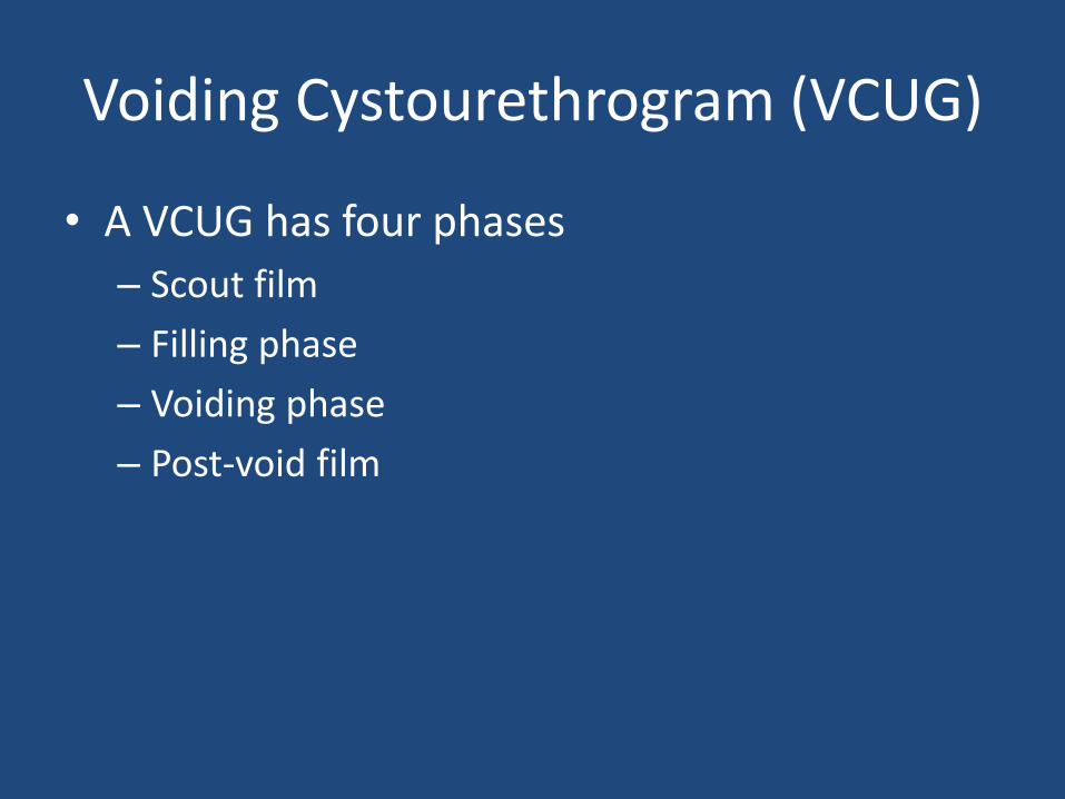
Voiding Cystourethrogram (VCUG)
• A VCUG has four phases
– Scout film
– Filling phase
– Voiding phase
– Post-void film

Vesicoureteral Reflux
• Abnormal retrograde leakage of urine from the bladder up the ureters
• Occurs when the ureterovesical valve is incompetent
• Associated with pyelonephritis

Reflux Grading
• I – Ureter only
• 2 – Ureter and pelvis, fine calyces
• 3 – Ureter, pelvis and blunted calyces
• 4 – Flattened or concave calyces
• 5 – Large, tortuous ureter
• Let’s see some more cases
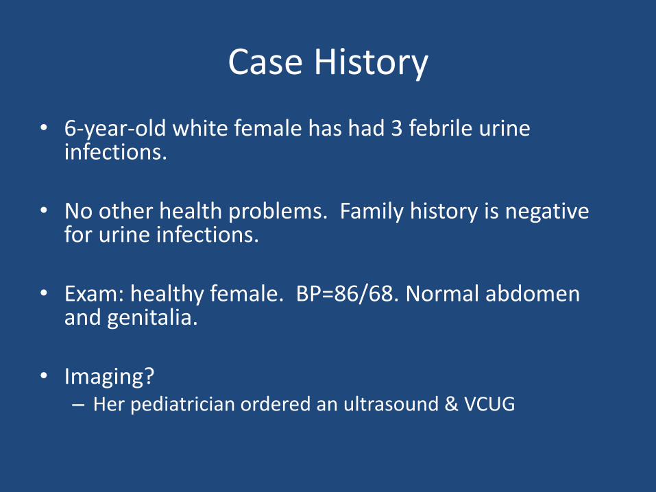
Case History
• 6-year-old white female has had 3 febrile urine infections.
• No other health problems. Family history is negative for urine infections.
• Exam: healthy female. BP=86/68. Normal abdomen and genitalia.
• Imaging? – Her pediatrician ordered an ultrasound & VCUG
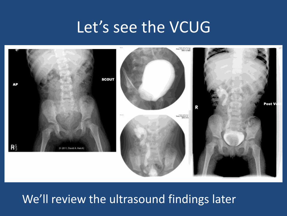
Let’s see the VCUG
We’ll review the ultrasound findings later
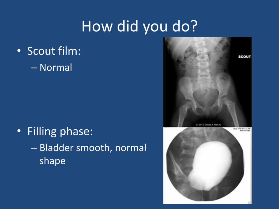
How did you do?
• Scout film:
– Normal
• Filling phase:
– Bladder smooth, normal shape

How did you do?
• Voiding phase:
– Grade V reflux on the right
– Grade I reflux on the left
• Post void:
– Reflux and a filling defect in the bladder
– What’s in the bladder?

Identification of Unknowns• Location:
– Inside the bladder
• Shape:– Almost round with regular, sharp
borders
• Density: – Low density; it doesn’t show up on
the scout film– It displaces contrast in the bladder
• Summary:– Circular structure in the bladder &
right reflux.
Voiding phase
Scout
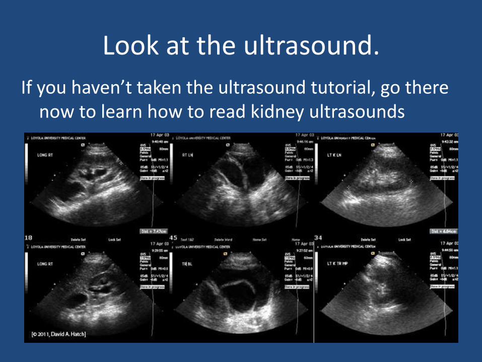
Look at the ultrasound.
If you haven’t taken the ultrasound tutorial, go there now to learn how to read kidney ultrasounds
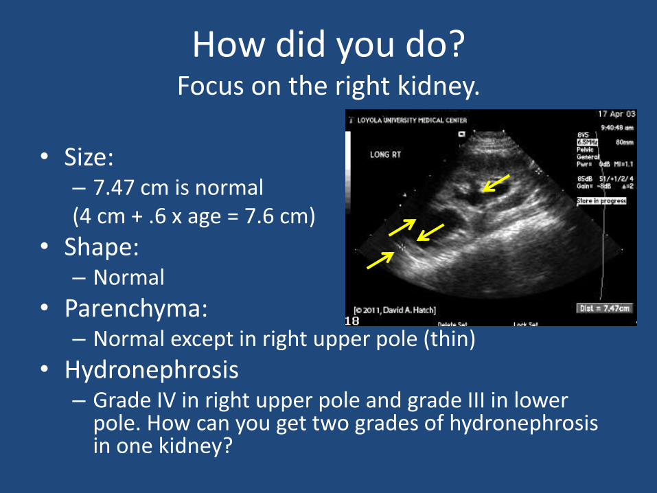
How did you do?Focus on the right kidney.
• Size:– 7.47 cm is normal (4 cm + .6 x age = 7.6 cm)
• Shape:– Normal
• Parenchyma:– Normal except in right upper pole (thin)
• Hydronephrosis– Grade IV in right upper pole and grade III in lower
pole. How can you get two grades of hydronephrosisin one kidney?

Ultrasound
• Bladder:
– What’s in the bladder?

• Location:
– Right side of the bladder
• Remember that on an ultrasound transverse image, the right side is always on the ‘x-ray’ left (like a CT scan)
• Shape:
– Round on longitudinal
– Round on transverse
– So it is spherical
Unknown Bladder Object
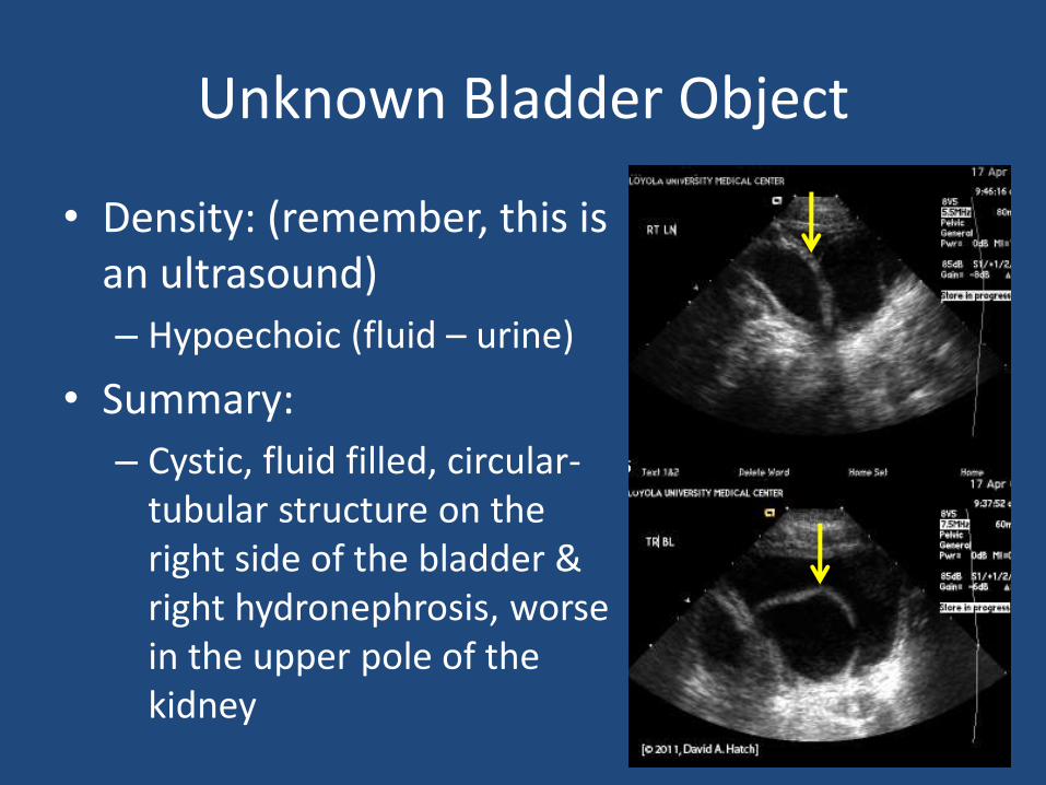
Unknown Bladder Object
• Density: (remember, this is an ultrasound)
– Hypoechoic (fluid – urine)
• Summary:
– Cystic, fluid filled, circular-tubular structure on the right side of the bladder & right hydronephrosis, worse in the upper pole of the kidney
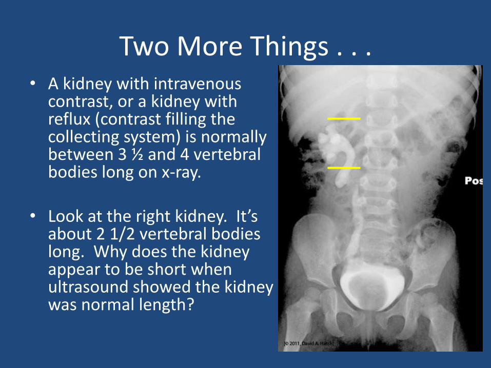
Two More Things . . .• A kidney with intravenous
contrast, or a kidney with reflux (contrast filling the collecting system) is normally between 3 ½ and 4 vertebral bodies long on x-ray.
• Look at the right kidney. It’s about 2 1/2 vertebral bodies long. Why does the kidney appear to be short when ultrasound showed the kidney was normal length?

Axis of the Kidney• Draw a line between the upper most calyx and the lower
most calyx. A normal kidney lies on an axis parallel to the psoas muscle (the upper most calyx is medial to the lower most calyx). On the VCUG of our patient, the upper pole calyx is more lateral than the lower pole.
Normal axis Abnormal axis
Abnormal Kidney Axis
• Whenever you see a kidney with an abnormal axis, you should consider these possibilities:
• Duplication: 2 separate collecting systems (You may be seeing only part of the kidney)
• Fusion anomaly of the kidney where the two kidneys are fused (horseshoe kidney, or other fusion anomaly)
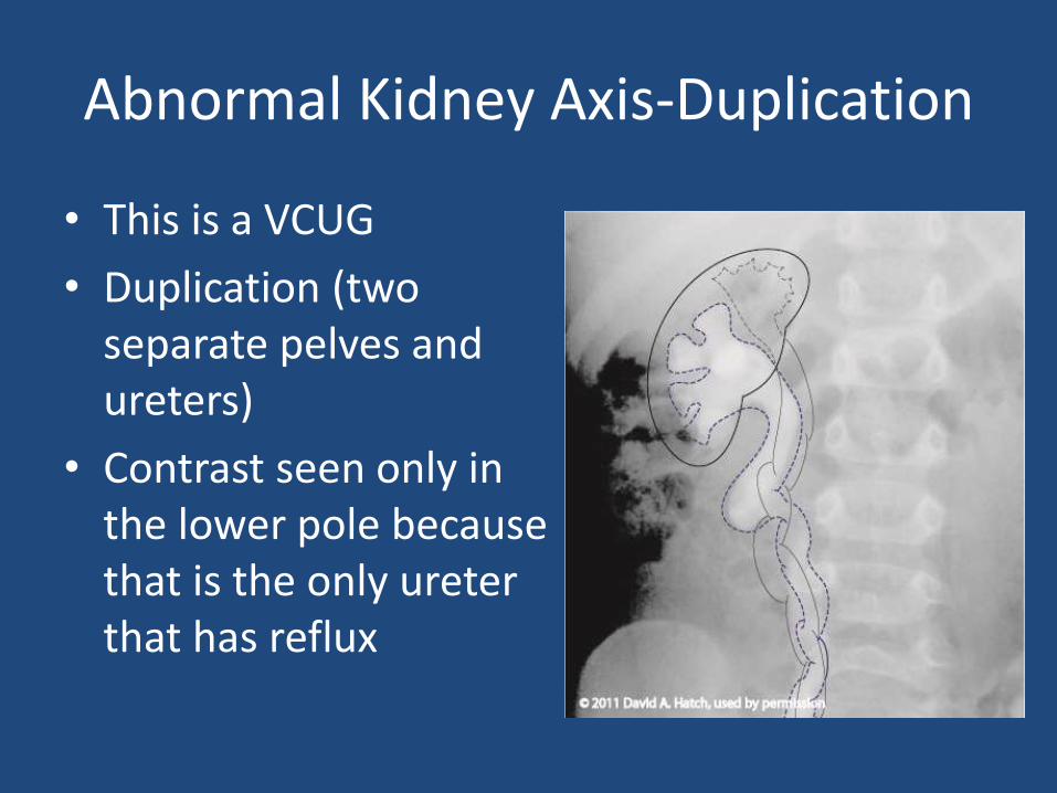
Abnormal Kidney Axis-Duplication
• This is a VCUG
• Duplication (two separate pelves and ureters)
• Contrast seen only in the lower pole because that is the only ureter that has reflux
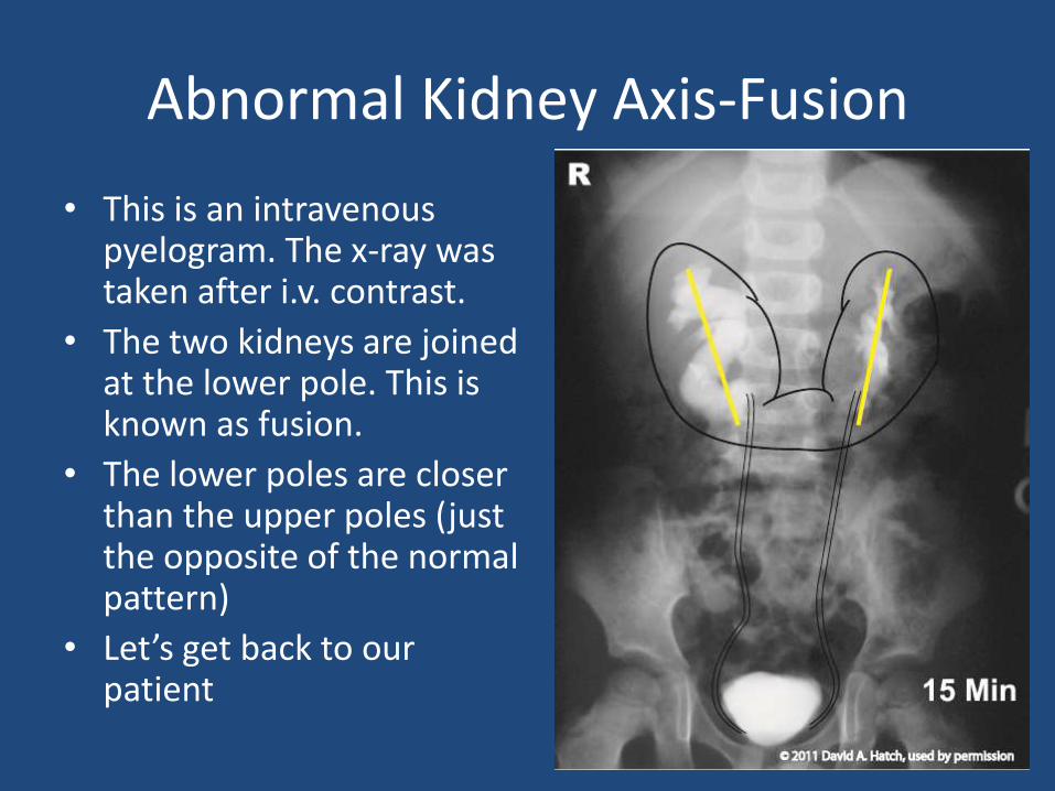
Abnormal Kidney Axis-Fusion
• This is an intravenous pyelogram. The x-ray was taken after i.v. contrast.
• The two kidneys are joined at the lower pole. This is known as fusion.
• The lower poles are closer than the upper poles (just the opposite of the normal pattern)
• Let’s get back to our patient
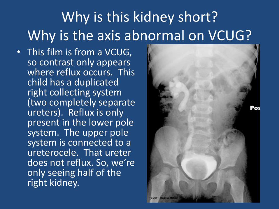
Why is this kidney short?Why is the axis abnormal on VCUG?
• This film is from a VCUG, so contrast only appears where reflux occurs. This child has a duplicated right collecting system (two completely separate ureters). Reflux is only present in the lower pole system. The upper pole system is connected to a ureterocele. That ureterdoes not reflux. So, we’re only seeing half of the right kidney.

This is a Ureterocele
• Intravesical dilation of the distal ureter
– Remember, it is within the bladder
• Thought to result from a delayed rupture of Chwalle’s membrane (structure that covers the connection of the ureter to the bladder). Essentially an obstruction of the ureter within the bladder.
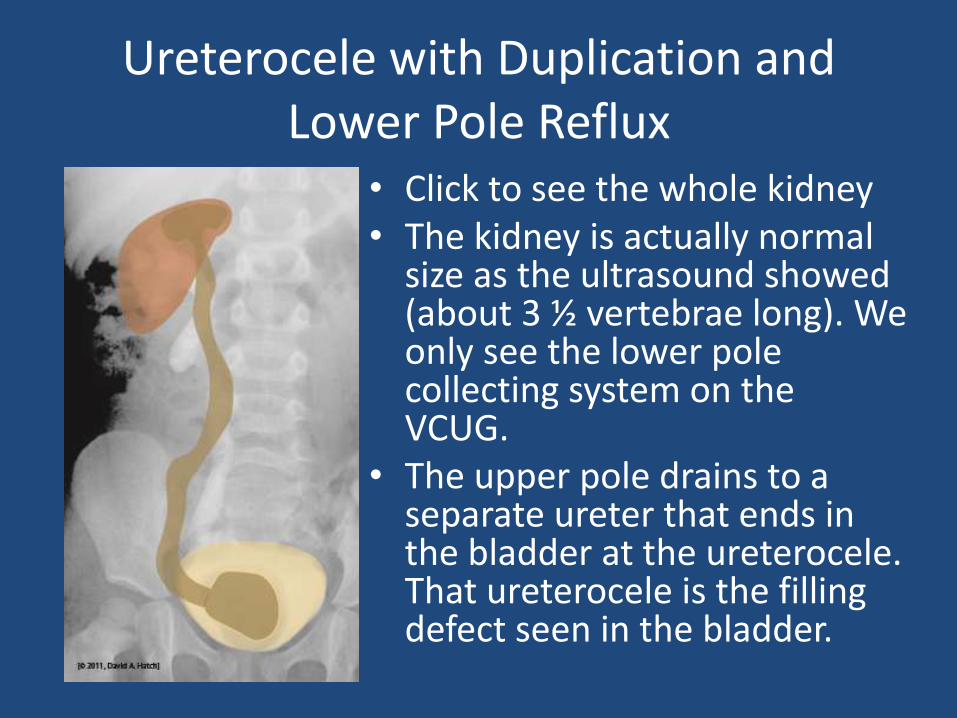
Ureterocele with Duplication and Lower Pole Reflux
• Click to see the whole kidney• The kidney is actually normal
size as the ultrasound showed (about 3 ½ vertebrae long). We only see the lower pole collecting system on the VCUG.
• The upper pole drains to a separate ureter that ends in the bladder at the ureterocele. That ureterocele is the filling defect seen in the bladder.
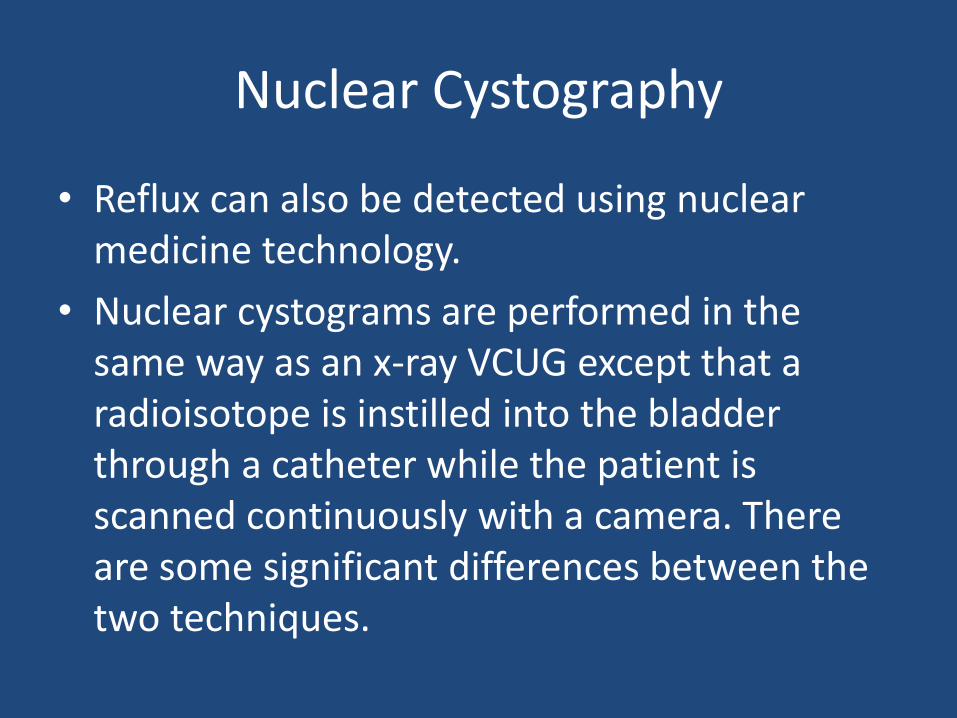
Nuclear Cystography
• Reflux can also be detected using nuclear medicine technology.
• Nuclear cystograms are performed in the same way as an x-ray VCUG except that a radioisotope is instilled into the bladder through a catheter while the patient is scanned continuously with a camera. There are some significant differences between the two techniques.
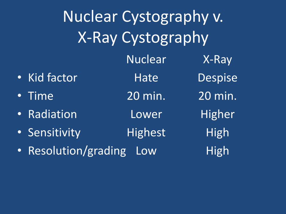
Nuclear Cystography v. X-Ray Cystography
Nuclear X-Ray
• Kid factor Hate Despise
• Time 20 min. 20 min.
• Radiation Lower Higher
• Sensitivity Highest High
• Resolution/grading Low High

Nuclear Cystography
[© 2010, David A. Hatch. Used by permission.]
Nuclear Cystogram
• As you saw, nuclear cystogram images are low resolution. They are very sensitive, but the detail is not sufficiently sharp to allow one to assign grading (International Reflux Study). Because the bladder and ureters are constantly observed during the study, unlike x-ray cystograms where x-rays are taken periodically, nuclear cystograms can detect transitory reflux. Many pediatric urologists prefer to order an x-ray VCUG for initial evaluation and then use nuclear cystography for subsequent follow-up.
Case History
• 10 year old boy was hit by a car while riding his bike. He suffered no head injury and did not lose consciousness. He has severe lower abdominal and pelvic pain. Vital signs are stable.
• “I feel like I’ve got to pee!”
• His attempts at voiding are unsuccessful. A little blood, but no urine comes out of his urethra.
• Imaging?
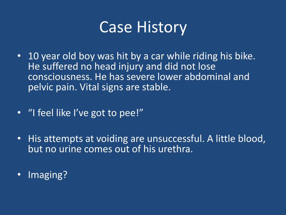
Trauma Imaging
• The question we need to answer is:– Does this boy have a significant injury to his urinary
tract?
• The trauma service ordered a contrast CT of the abdomen
• Your urology resident suggests that you evaluate this boy’s urethra and bladder. What imaging could define an injury?
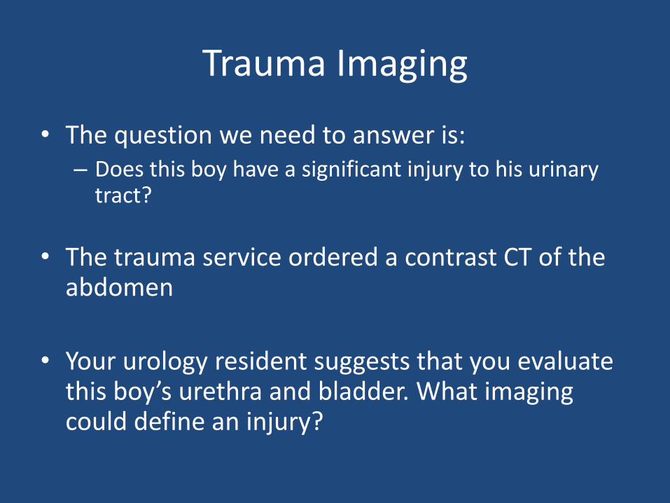
Film 1• Retrograde urethrogram (contrast
injected into the urethra)– Extravasation from urethral injury
• Contrast is seen in the kidney collecting system bilaterally because the patient was given contrast intravenously for the CT scan.
• Suprapubic catheter– He could not void and the CT
showed a full bladder, so a suprapubic catheter was placed.
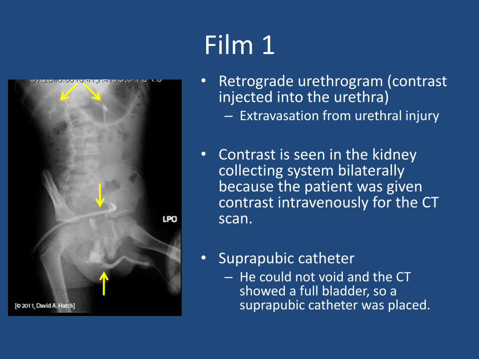
Film 2• The suprapubic catheter
was used to fill bladder for a cystogram– No extravasation with the
bladder full
– Notice that the bladder neck is higher than normal. It’s completely separated from the urethra. The bladder is ‘tear drop’ shape (long and narrow at base) because of blood accumulation in the pelvis.

Film 3• Drainage film
– No extravasation from the bladder (there was no bladder rupture)
– His urethra is completely separated from his bladder (urethral disruption).
– Management– With his other injuries the
best initial management is suprapubic catheter drainage for at least 2 months.
• Three months after his injury, your urology team repaired his urethra, reconnecting the two separated segments. We call this a urethroplasty. Today he has urinary continence. His mother gives you a big hug every time she sees you and she says, “I thank God every day for what you’ve done for my son.”
Case History
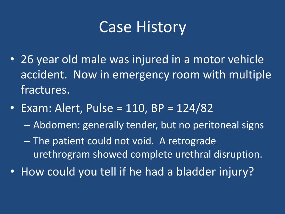
• 26 year old male was injured in a motor vehicle accident. Now in emergency room with multiple fractures.
• Exam: Alert, Pulse = 110, BP = 124/82
– Abdomen: generally tender, but no peritoneal signs
– The patient could not void. A retrograde urethrogram showed complete urethral disruption.
• How could you tell if he had a bladder injury?
Imaging?
• He needs a cystogram
• Because the urethra was separated from the bladder, a suprapubic catheter was placed
• A cystogram has films from three phases: scout film, filling/full and post drainage. In this case, of course, he can’t void, so his bladder is drained by the suprapubic catheter before the final film.

KUB – Scout Film

KUB – Scout Film
Bones?Pelvic fracture (see the
separation of the symphysis pubis). Pelvic fractures come in pairs. Where’s the second fracture?
Soft tissues?Little bowel gas
Foreign bodies?Surgical clampsDrain tube
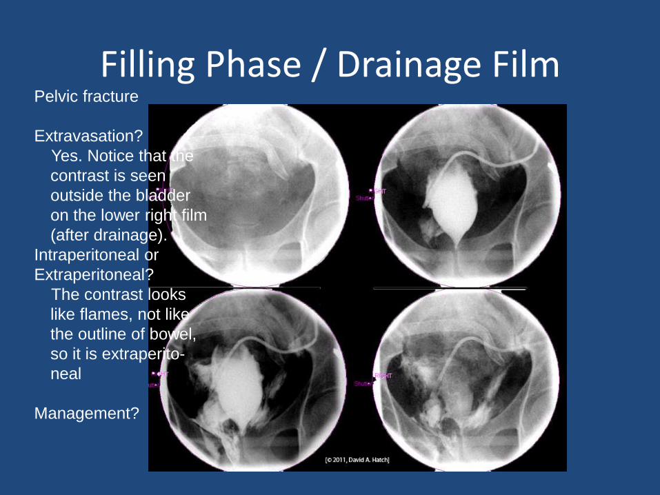
Filling Phase / Drainage FilmPelvic fracture
Extravasation?
Yes. Notice that the
contrast is seen
outside the bladder
on the lower right film
(after drainage).
Intraperitoneal or
Extraperitoneal?
The contrast looks
like flames, not like
the outline of bowel,
so it is extraperito-
neal
Management?

Traumatic Bladder Rupture
• Like the last patient, this man needs long-term catheter drainage. After six to eight weeks the cystogram should be repeated to see if the bladder rupture was sealed.
• If the rupture had extended into the peritoneal cavity, then the bladder tear would have been surgically closed.
Cystogram - Review
• Contrast is seen in the ureter or renal pelvis on VUCG only when reflux is present.
• If the axis of the kidney is abnormal (upper pole more medial than lower pole) consider either a duplication of the ureter and pelvis or horseshoe kidney.
• Normal kidneys are about 3 ½ to 4 vertebral bodies long when imaged by intravenous pyelogram or VCUG (if reflux is present).
Review
• Nuclear cystograms are sensitive, but don’t provide sufficient resolution to allow grading. Radiation exposure is lower with nuclear cystography.
• Trauma victims with hematuria and/or pelvic fractures need imaging of the lower urinary tract (cystogram and urethrogram) to detect urethral injury or bladder rupture.