Atypical Neural Responses During Face Processing in...
16
NEW RESEARCH Atypical Neural Responses During Face Processing in Female Adolescents With Conduct Disorder Graeme Fairchild, PhD, Cindy C. Hagan, PhD, Luca Passamonti, MD, Nicholas D. Walsh, PhD, Ian M. Goodyer, MD, FRCPsych, FMedSci, Andrew J. Calder, PhD Objective: Conduct disorder (CD) in females is associated with negative adult outcomes including mental health problems and personality disorders. Although recent neuroimaging studies have reported changes in neural activity during facial emotion processing in males with CD or callous-unemotional (CU) traits, there have been no neuroimaging studies specif- ically assessing females with CD. We addressed this gap by investigating whether female adolescents with CD show atypical neural activation when processing emotional or neutral faces. Method: We acquired functional magnetic resonance imaging (fMRI) data from 20 female adolescents with CD and 20 female control participants while they viewed angry, sad, and neutral faces. Results: An omnibus group (CD, control) by facial emotion (angry, sad, neutral) analysis of variance (ANOVA) revealed main effects of facial emotion in superior temporal cortex, fusiform gyrus, ventrolateral prefrontal cortex and insula, and main effects of group in medial orbitofrontal cortex (OFC) and right anterior insula. Female participants with CD showed reduced medial OFC and increased anterior insula responses relative to healthy controls. There were no significant group facial emotion interactions. Lifetime CD symp- toms were negatively correlated with amygdala, superior temporal cortex, fusiform gyrus, and dorsolateral prefrontal cortex activity for the contrast “all-faces versus fixation.” CU traits were negatively correlated with fusiform gyrus activity for the contrast sad versus neutral faces. Conclusion: Females with CD showed atypical neural activation during the processing of all facial expressions, irrespective of valence. Our results demonstrate that severity of CD symptoms and CU traits is important in explaining abnormal patterns of neural activity. J. Am. Acad. Child Adolesc. Psychiatry, 2014;53(6):677–687. Key Words: CD, CU traits, females, face processing, fMRI C onduct disorder (CD) is characterized by a pervasive pattern of antisocial and violent behavior in which the rights of others are violated. 1 CD is one of the most common dis- orders in adolescent females 2 and is associated with an increased risk of developing antisocial or borderline personality disorder, substance depen- dence, depression, and physical health problems in adulthood. 3-6 Despite this negative prognosis, we know relatively little about the neurobiological mechanisms underlying CD in females, as there have been few neuropsychological studies of this group, and as functional magnetic resonance imaging (fMRI) studies of CD have been largely restricted to males. Of the previous 26 fMRI stu- dies of CD, 17 have included only males, and the remaining 9 studies recruited mixed samples con- taining only a small number of females, resulting in an underrepresentation of females with CD (442 males versus 41 females pooled across 26 studies; Table S1, available online, provides references). Critically, none of these studies investigated brain activity in females with CD specifically. To address this gap in the literature, we in- vestigated neural responses to emotional and neutral facial expressions in female adolescents with CD relative to healthy controls. An earlier behavioral study found impaired recognition of facial expressions of anger and disgust in fe- male adolescents with CD, and an additional impairment in sadness (but not fear) recognition Supplemental material cited in this article is available online. JOURNAL OF THE AMERICAN ACADEMY OF CHILD & ADOLESCENT PSYCHIATRY VOLUME 53 NUMBER 6 JUNE 2014 www.jaacap.org 677
Transcript of Atypical Neural Responses During Face Processing in...

NEW RESEARCH
JOURNAL
VOLUM
Atypical Neural Responses During FaceProcessing in Female Adolescents With
Conduct DisorderGraeme Fairchild, PhD, Cindy C. Hagan, PhD, Luca Passamonti, MD,
Nicholas D. Walsh, PhD, Ian M. Goodyer, MD, FRCPsych, FMedSci, Andrew J. Calder, PhD
Objective: Conduct disorder (CD) in females is associated with negative adult outcomesincluding mental health problems and personality disorders. Although recent neuroimagingstudies have reported changes in neural activity during facial emotion processing in maleswith CD or callous-unemotional (CU) traits, there have been no neuroimaging studies specif-ically assessing females with CD. We addressed this gap by investigating whether femaleadolescents with CD show atypical neural activation when processing emotional or neutralfaces. Method: We acquired functional magnetic resonance imaging (fMRI) data from 20female adolescents with CD and 20 female control participants while they viewed angry,sad, and neutral faces. Results: An omnibus group (CD, control) by facial emotion (angry,sad, neutral) analysis of variance (ANOVA) revealed main effects of facial emotion in superiortemporal cortex, fusiform gyrus, ventrolateral prefrontal cortex and insula, and main effects ofgroup in medial orbitofrontal cortex (OFC) and right anterior insula. Female participants withCD showed reduced medial OFC and increased anterior insula responses relative to healthycontrols. There were no significant group � facial emotion interactions. Lifetime CD symp-toms were negatively correlated with amygdala, superior temporal cortex, fusiform gyrus,and dorsolateral prefrontal cortex activity for the contrast “all-faces versus fixation.” CU traitswere negatively correlated with fusiform gyrus activity for the contrast sad versus neutralfaces. Conclusion: Females with CD showed atypical neural activation during the processingof all facial expressions, irrespective of valence. Our results demonstrate that severity of CDsymptoms and CU traits is important in explaining abnormal patterns of neural activity. J. Am.Acad. Child Adolesc. Psychiatry, 2014;53(6):677–687. Key Words: CD, CU traits, females,face processing, fMRI
onduct disorder (CD) is characterized by apervasive pattern of antisocial and violent
C behavior in which the rights of others areviolated.1 CD is one of the most common dis-orders in adolescent females2 and is associatedwith an increased risk of developing antisocial orborderline personality disorder, substance depen-dence, depression, and physical health problemsin adulthood.3-6 Despite this negative prognosis,we know relatively little about the neurobiologicalmechanisms underlying CD in females, as therehave been few neuropsychological studies ofthis group, and as functional magnetic resonance
Supplemental material cited in this article is available online.
OF THE AMERICAN ACADEMY OF CHILD & ADOLESCENT PSYCHIATR
E 53 NUMBER 6 JUNE 2014
imaging (fMRI) studies of CD have been largelyrestricted to males. Of the previous 26 fMRI stu-dies of CD, 17 have included only males, and theremaining 9 studies recruited mixed samples con-taining only a small number of females, resultingin an underrepresentation of females with CD (442males versus 41 females pooled across 26 studies;Table S1, available online, provides references).Critically, none of these studies investigated brainactivity in females with CD specifically.
To address this gap in the literature, we in-vestigated neural responses to emotional andneutral facial expressions in female adolescentswith CD relative to healthy controls. An earlierbehavioral study found impaired recognitionof facial expressions of anger and disgust in fe-male adolescents with CD, and an additionalimpairment in sadness (but not fear) recognition
Y
www.jaacap.org 677

FAIRCHILD et al.
in females with CD and psychopathic traits.7
Similar deficits in anger and disgust recognitionwere observed in males with CD, 8 indicating thatCD in both sexes is associated with difficulties inprocessing these emotions. In contrast, psycho-pathic traits were associated with deficits in bothsadness and fear recognition in males with CD.8
To follow up these behavioral findings and cha-racterize the underlying neural processes, we pre-viously conducted an fMRI study and observedreduced amygdala, anterior insula, orbitofrontalcortex (OFC), and anterior superior temporalcortex responses to emotional versus neutral facesin male adolescents with CD.9
On the basis of these earlier results, we pre-dicted that female adolescents with CD wouldshow atypical neural responses when processingangry or sad relative to neutral facial expressions.Specifically, we predicted that females with CDwould show reduced activity in regions involvedin social cognition and emotion processing, suchas the amygdala, anterior insula, OFC and supe-rior temporal cortex, during the processing ofnegative facial expressions.10,11 This would bedemonstrated by significant interactions betweengroup and facial emotion in these regions, inwhich healthy controls would show greater neu-ral responses to angry and sad faces than neutralfaces. Meanwhile, participants with CD wouldshow weaker differentiation between these facialexpressions. However, it was also possible thatfemales with CD would show increased neuralresponses to neutral faces, as we previously foundthat male adolescents with CD showed increasedamygdala and insula responses to neutral faces.9
Our second aim was to examine relationshipsbetween brain activity and severity of CD, asquantified by number of CD symptoms. We pre-dicted that CD symptoms would be negativelycorrelated with amygdala, anterior insula, OFC andsuperior temporal cortex activity, given previousresearch in males showing negative relationshipsbetween activity in these regions and CD symp-toms9 or aggressive behavior.12
The presence of CU traits (such as emotionaldetachment) is thought to delineate a particularlysevere and persistent form of CD.13 Given theimportance of callous-unemotional (CU) traits forunderstanding heterogeneity within antisocialbehavior,14 our final aim was to investigatewhether CU traits would modulate neural activityduring facial emotion processing. The IntegratedEmotion Systems (IES) model15 proposes thatdistress cues, such as sad or fearful facial
JOURN
678 www.jaacap.org
expressions, play a critical role in the socializationprocess. According to this model, typically devel-oping children find distress cues aversive, so theylearn to stop engaging in aggressive behaviors thatelicit such cues. Individuals with CU traits areproposed to be insensitive to distress cues, whichdisrupts their socialization, rendering them atincreased risk for instrumental aggression. The IESmodel therefore predicts that CU traits would beassociated with impaired recognition of sad andfearful expressions, along with reduced neuralresponses to these facial expressions. Previousresearch has provided evidence for selective ordisproportionate impairments in sadness and fearrecognition in individuals with CU traits8,16,17
(although see Dawel et al.18 for a meta-analysisshowing pervasive emotion recognition deficitsin psychopathy). However, with the exception of 1study showing impaired sadness recognition infemale adolescents with CD and CU traits7 and astudy reporting deficits in sadness recognition infemale psychopaths,19 most previous studies ofCU traits or psychopathy have either focused onmales alone or have included small numbers offemales.16-18 fMRI studies have shown reducedamygdala responses to fearful facial expressionsin male children with conduct problems and CUtraits20 and in a group of adolescents with CUtraits and disruptive behavior disorder diagnoseswho were predominantly male.21 Studies in adultshave shown reduced amygdala or fusiform gyrusresponses to fearful faces in males with psycho-pathy.22,23 However, no comparable data exist onthe effects of CU traits on neural activation in fe-males. To further investigate the IES model, weassessed whether CU traits were associated withreduced brain activation during the processing ofsad facial expressions. Sadness, rather than fear,was selected, given previous behavioral resultsshowing that CU or psychopathic traits wereassociated with impaired recognition of sadnessbut not fear in females,7,19 and on the basis of ameta-analysis showing that CU traits are moststrongly linked with deficits in sadness recogni-tion.18 We predicted that CU traits would be ne-gatively correlated with amygdala, anterior insula,OFC and fusiform gyrus responses to sad versusneutral expressions.
METHODParticipantsTwenty-two female adolescents with CD were recruitedfrom schools, pupil referral units, and the Cambridge
AL OF THE AMERICAN ACADEMY OF CHILD & ADOLESCENT PSYCHIATRY
VOLUME 53 NUMBER 6 JUNE 2014

FACE PROCESSING IN FEMALE ADOLESCENTS WITH CD
Youth Offending Service. All participants gave writteninformed consent to participate in the study, whichwas approved by the local National Health Serviceresearch ethics committee. Exclusion criteria for theCD group included the following: IQ <80, as esti-mated using the Wechsler Abbreviated Scale of Intel-ligence,24 or presence of a pervasive developmentaldisorder (e.g., autism). A healthy control group (HC;no history of CD/oppositional defiant disorder [ODD]or current psychiatric illness) of 22 female adolescents,matched in age, handedness, ethnicity, and performanceIQ, was recruited from schools and colleges. This sampleoverlaps substantially (95%) with the female sampleincluded in an earlier structural MRI study.25
All participants were assessed for CD, ODD,attention-deficit/hyperactivity disorder (ADHD), majordepressive disorder (MDD), generalized anxiety disorder(GAD), obsessive-compulsive disorder (OCD), post-traumatic stress disorder (PTSD), and substance depen-dence, using the Schedule for Affective Disorders andSchizophrenia for School-Age Children (K-SADS).26
Diagnostic interviews were carried out separately withparticipants and caregivers. The majority (n ¼ 17) of thefemales with CD had the adolescence-onset form of CD(i.e., onset of CD symptoms only after age 10 years1).
Both CU and psychopathic traits were assessed us-ing the Callous-Unemotional dimension subscale andthe total score of the self-report Youth Psychopathictraits Inventory (YPI),27 respectively. CU traits datawere also obtained from parents using the Inventoryof Callous-Unemotional traits (ICU).28 The AdolescentAlcohol and Drug Involvement Scale (AADIS) mea-sured alcohol and substance use.29 Handedness wasassessed using the Edinburgh Handedness Inventory.30
Finally, socioeconomic status (SES) was quantified usingthe A Classification Of Residential Neighbourhoods(ACORN) geodemographic tool (http://www.caci.co.uk/acorn-classification.aspx).
To control for menstrual cycle phase effects on brainactivity,31 all participants with CD and the majority ofthe healthy control participants were scanned in themid-follicular phase of the cycle (i.e., within 5–10 daysof menstruation onset), as determined by self-report.
fMRI TaskParticipants categorized the gender of gray-scale pho-tographs of angry, sad, and neutral faces (half female)posed by 30 different identities, by pressing either theleft button on a button box to indicate that the face wasmale or the right button to indicate the face was female(Figure S1, available online). The faces were selectedfrom 2 stimulus sets32,33 on the basis of emotional rat-ings from an independent sample.34 In a mixed design,stimuli were presented in 17.5-second epochs containing5 faces from the same category (angry, sad, or neutral)interspersed with 5 null events (fixation cross). Eachface trial comprised a 1,000-millisecond presentation ofa face followed by a fixation cross (750 milliseconds).
JOURNAL OF THE AMERICAN ACADEMY OF CHILD & ADOLESCENT PSYCHIATR
VOLUME 53 NUMBER 6 JUNE 2014
Null events involved a 1,750-millisecond presentationof the fixation cross. The stimuli presented during eachepoch were pseudo-randomized with respect to trialtype (faces or null events) and facial gender and iden-tity; no more than 3 consecutive trials were of the sametrial type. This pseudo-randomization enhanced designefficiency while ensuring that the stimulus onsets andvalences were unpredictable for naive observers.9,35
Twelve epochs of each expression were presented (60angry, 60 sad, and 60 neutral faces). Reaction times (RTs)and accuracy were recorded throughout the experi-ment, which lasted 10 minutes 30 seconds. Subjectiveratings of the emotional intensity of the stimuli werealso obtained after scanning (participants rated eachof the facial expressions used in terms of anger andsadness intensity, using a scale from 1 ¼ not at all to9 ¼ very [angry or sad]).
Image Acquisition and PreprocessingMRI scanning was performed on a 3-Tesla Siemens TimTrio with a head coil gradient set at the MedicalResearch Council (MRC) Cognition and Brain SciencesUnit at University of Cambridge. Whole-brain datawere acquired with echo-planar T2*-weighted imaging(EPI), sensitive to blood-oxygenation-level–dependent(BOLD) signal contrast (32 axial slices, 3-mm thickness;repetition time ¼ 2,000 milliseconds; echo time ¼ 30milliseconds; voxel size ¼ 3 � 3 � 3 mm). Data wereanalyzed using SPM5 (www.fil.ion.ucl.ac.uk/spm/).EPIs were sinc-interpolated in time to correct for slicetime differences and were realigned to the first scan byrigid body transformations to correct for head move-ments. The mean EPI was computed for each partici-pant and inspected to ensure that no participantsshowed excessive signal dropout in medial temporaland OFC regions. EPIs were co-registered andnormalized to the T1 standard template in MontrealNeurological Institute (MNI) space using linear andnonlinear transformations, and smoothed with an8-mm Gaussian kernel of full width at half maximum.
fMRI AnalysesFor each participant, a general linear model (GLM)assessed regionally specific effects of task parameterson BOLD activation.36 The model included experi-mental factors (angry, sad, neutral face trials, and fix-ation trials) and 6 realignment parameters as effectsof no interest, to account for residual motion-relatedvariance. Low-frequency signal drift was removed us-ing a high-pass filter (cut-off at 128 seconds) and anautoregressive modelling [AR(1)] of temporal auto-correlations was applied.
We ran a group (CD, control) � facial emotion(angry, sad, neutral) analysis of variance (ANOVA) toinvestigate for main effects of group or emotion andinteractions between these factors. To follow up themain effects of group, we generated contrast imagesfor “all faces versus fixation” and included these in
Y
www.jaacap.org 679

FAIRCHILD et al.
regression analyses to assess whether individual dif-ferences in lifetime CD symptoms were correlated withneural activity. Given our a priori hypothesis that CUtraits would be associated with reduced neural re-sponses to sad facial expressions, we assessed whetherCU traits were correlated with neural activity for thecontrast sad versus neutral faces by using regressionanalysis. We also repeated the group-based analyses inSPM5, including lifetime or current ADHD symptoms
TABLE 1 Demographic and Clinical Characteristics of the Fe
Characteristics HC (n ¼ 20)
Age (y) 17.62 � 0.64Full-scale IQ 104.60 � 8.72Performance IQ 105.00 � 10.85Handedness (R/L) 20/0No. of current DSM-IV diagnoses
ADHD 0Substance abuse 0Panic disorder 0
Number of past DSM-IV diagnosesADHD 0MDD 3Substance abuse 0PTSD 0
No. of symptomsCurrent CD 0.13 � 0.34Lifetime CD 0.38 � 0.62Aggressive CD 0.06 � 0.25Current ADHD 1.55 � 1.88Lifetime ADHD 1.90 � 2.20Total psychopathic traits (total YPI) 1.65 � 0.35YPI CU traits subscale 0.53 � 0.11Inventory of CU traits 17.81 � 8.04
SES (ACORN)Wealthy achievers 9Urban prosperity 0Comfortably off 6Moderate means 1Hard-pressed 4
EthnicityWhite 20Nonwhite 0
fMRI task performanceAccuracy, %Angry 91 � 7Sad 96 � 3Neutral 93 � 5
RTs, msAngry 641 � 68Sad 635 � 70Neutral 635 � 69
Note: Data are presented as mean � SD or number in each group. ACORNdeficit/hyperactivity disorder; CD ¼ conduct disorder; CU ¼ callous-unemcontrol; L ¼ left; MDD ¼ major depressive disorder; PTSD ¼ posttraumatic sYPI ¼ Youth Psychopathic Traits Inventory.
JOURN
680 www.jaacap.org
or lifetime MDD diagnoses as covariates to examine theinfluence of these variables on the main findings.
Two approaches for thresholding second level mapswere applied. First, to conduct whole-brain analyses,we applied a threshold of p < .05, false-discovery ratewhole-brain correction.37 Second, in our a priori re-gions of interest (ROIs), the threshold used was p < .05,family-wise error correction for multiple comparisons insmall volumes (i.e., small volume correction [SVC]).38,39
male Participants
GroupGroup Comparisons
(p values)CD (n ¼ 20)
16.97 � 1.52 .0999.65 � 8.06 .07
101.50 � 9.99 .3019/1 .50
3 .232 .491 .99
4 .119 .084 .111 .99
2.70 � 2.58 <.0017.70 � 2.30 <.0012.70 � 1.08 <.0016.15 � 3.47 <.0018.30 � 3.85 <.0012.03 � 0.41 .0060.60 � 0.13 .069
28.78 � 14.11 .010
443 .06718
20 1.000
89 � 694 � 5 .2592 � 5
635 � 43633 � 45 .98644 � 45
¼ A Classification Of Residential Neighbourhoods; ADHD ¼ attention-otional; fMRI ¼ functional magnetic resonance imaging; HC ¼ healthytress disorder; R ¼ right; RT ¼ reaction time; SES ¼ socioeconomic status;
AL OF THE AMERICAN ACADEMY OF CHILD & ADOLESCENT PSYCHIATRY
VOLUME 53 NUMBER 6 JUNE 2014

FACE PROCESSING IN FEMALE ADOLESCENTS WITH CD
The amygdala, ventromedial prefrontal cortex, anteriorinsula, OFC, fusiform gyrus, and superior temporalgyrus were defined as our ROIs, given that severalprevious fMRI studies of CD have shown group dif-ferences in these regions.9,12,20,21,40-42 All ROIs wereanatomical regions defined using the “aal.02” atlas forautomated anatomical labelling.43 For completenessand to aid future meta-analyses, we also report allbrain regions that were significant at p < .001, un-corrected, �10 contiguous voxels, in Tables 1 to 3.
RESULTSThree participants (2 with CD and 1 control) wereexcluded because of excessive head movements(>2 mm) during scanning, and a further controlparticipant was excluded because of the presenceof neurological abnormalities as indicated by anaccompanying structural scan.
Demographic and Clinical VariablesThere were no significant group differences infull-scale IQ, age, or SES (Table 1). The groupswere matched in ethnicity, handedness, and
TABLE 2 Coordinates and Cluster Sizes for the Main Effects
Hemisphere LocaCerebral Region
Main Effects of Facial EmotionSuperior temporal gyrus/fusiform gyrus RFusiform gyrus L
RVentrolateral prefrontal cortex RMiddle temporal gyrus LPrecentral gyrus RPosterior insula RCuneus LAnterior insula R
Main Effects of GroupMedial orbitofrontal cortex RAnterior insula RPrecentral gyrus LPrecentral gyrus RCuneus L
RMedial frontal gyrus RMiddle temporal gyrus RCerebellum/fusiform gyrus RLingual gyrus RPostcentral gyrus R
Group � facial emotion interactionsNo significant activations
Note: Unless otherwise indicated, all regions were significant at p < .001, uncInstitute; R ¼ right.ap < .05, false-discovery rate whole-brain correction.bp < .05, family-wise error small-volume correction.
JOURNAL OF THE AMERICAN ACADEMY OF CHILD & ADOLESCENT PSYCHIATR
VOLUME 53 NUMBER 6 JUNE 2014
performance IQ. As in our previous research, thefemale CD participants had higher levels of psy-chopathic and CU traits relative to controls. Theparticipants with CD also reported more CD andADHD symptoms and lifetime MDD diagnosesthan did the healthy control participants.
Behavioral ResultsAccuracy and correct RTs during the genderdiscrimination task were entered into a 2 � 3ANOVA assessing for effects of group andemotion. Neither measure showed a group effect(accuracy, F1,38 ¼ 1.35, p ¼ .25; RT, F1,38 ¼ 0.001,p ¼ .98) or a group � emotion interaction (accu-racy, F2,76 ¼ 0.11, p ¼ .89; RT, F2,76 ¼ 1.87, p ¼ .16)(Table 1).
The rating data collected after scanning wereentered into separate 2 � 3 ANOVAs assessingfor effects of group and emotion on emotionalratings of sadness or anger intensity. There were nomain effects of group (anger, F1,38 ¼ 0.23, p ¼ .63;sadness, F1,38 ¼ 0.14, p ¼ .71) or group � emotion
of Emotion, Group, and Group � Emotion Interactions
l Maxima, FNo. of SignificantVoxels in Cluster
MNI Coordinates
X Y Z
23.58a 979 52 �38 617.87a 127 �42 �40 �1610.84a 73 44 �42 �1613.56a 131 52 34 012.87a 373 �52 �54 411.15a 138 30 �26 6410.34a 73 40 �12 209.04a 42 �16 �78 48.57a 16 36 26 6
11.46b 11 12 58 �212.08b 52 42 16 817.99 163 �28 �12 5818.12 201 28 �12 5218.12 128 �10 �102 211.95 27 10 �76 1615.33 87 10 12 5213.89 61 46 �58 �212.18 39 24 �82 �2011.86 32 28 �70 �211.51 16 46 �26 42
orrected, �10 contiguous voxels. L ¼ left; MNI ¼ Montreal Neurological
Y
www.jaacap.org 681
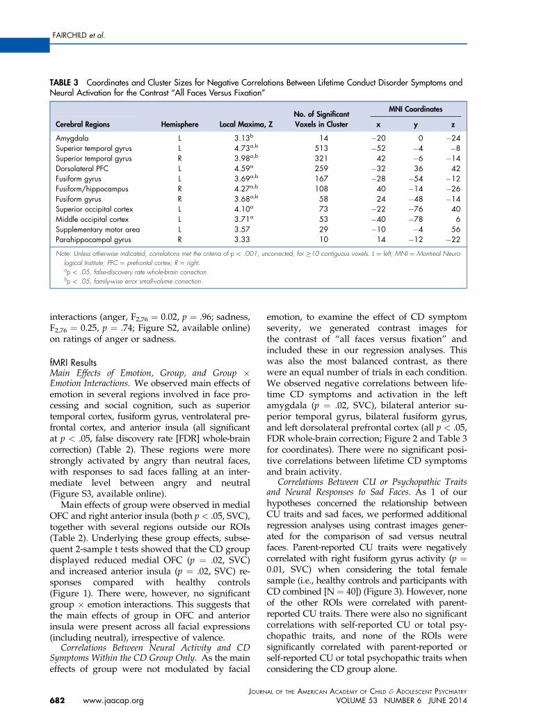
TABLE 3 Coordinates and Cluster Sizes for Negative Correlations Between Lifetime Conduct Disorder Symptoms andNeural Activation for the Contrast “All Faces Versus Fixation”
Hemisphere Local Maxima, ZNo. of SignificantVoxels in Cluster
MNI Coordinates
Cerebral Regions x y z
Amygdala L 3.13b 14 �20 0 �24Superior temporal gyrus L 4.73a,b 513 �52 �4 �8Superior temporal gyrus R 3.98a,b 321 42 �6 �14Dorsolateral PFC L 4.59a 259 �32 36 42Fusiform gyrus L 3.69a,b 167 �28 �54 �12Fusiform/hippocampus R 4.27a,b 108 40 �14 �26Fusiform gyrus R 3.68a,b 58 24 �48 �14Superior occipital cortex L 4.10a 73 �22 �76 40Middle occipital cortex L 3.71a 53 �40 �78 6Supplementary motor area L 3.57 29 �10 �4 56Parahippocampal gyrus R 3.33 10 14 �12 �22
Note: Unless otherwise indicated, correlations met the criteria of p < .001, uncorrected, for �10 contiguous voxels. L ¼ left; MNI ¼ Montreal Neuro-logical Institute; PFC ¼ prefrontal cortex; R ¼ right.ap < .05, false-discovery rate whole-brain correction.bp < .05, family-wise error small-volume correction.
FAIRCHILD et al.
interactions (anger, F2,76 ¼ 0.02, p ¼ .96; sadness,F2,76 ¼ 0.25, p ¼ .74; Figure S2, available online)on ratings of anger or sadness.
fMRI ResultsMain Effects of Emotion, Group, and Group �Emotion Interactions. We observed main effects ofemotion in several regions involved in face pro-cessing and social cognition, such as superiortemporal cortex, fusiform gyrus, ventrolateral pre-frontal cortex, and anterior insula (all significantat p < .05, false discovery rate [FDR] whole-braincorrection) (Table 2). These regions were morestrongly activated by angry than neutral faces,with responses to sad faces falling at an inter-mediate level between angry and neutral(Figure S3, available online).
Main effects of group were observed in medialOFC and right anterior insula (both p < .05, SVC),together with several regions outside our ROIs(Table 2). Underlying these group effects, subse-quent 2-sample t tests showed that the CD groupdisplayed reduced medial OFC (p ¼ .02, SVC)and increased anterior insula (p ¼ .02, SVC) re-sponses compared with healthy controls(Figure 1). There were, however, no significantgroup � emotion interactions. This suggests thatthe main effects of group in OFC and anteriorinsula were present across all facial expressions(including neutral), irrespective of valence.
Correlations Between Neural Activity and CDSymptoms Within the CD Group Only. As the maineffects of group were not modulated by facial
JOURN
682 www.jaacap.org
emotion, to examine the effect of CD symptomseverity, we generated contrast images forthe contrast of “all faces versus fixation” andincluded these in our regression analyses. Thiswas also the most balanced contrast, as therewere an equal number of trials in each condition.We observed negative correlations between life-time CD symptoms and activation in the leftamygdala (p ¼ .02, SVC), bilateral anterior su-perior temporal gyrus, bilateral fusiform gyrus,and left dorsolateral prefrontal cortex (all p < .05,FDR whole-brain correction; Figure 2 and Table 3for coordinates). There were no significant posi-tive correlations between lifetime CD symptomsand brain activity.
Correlations Between CU or Psychopathic Traitsand Neural Responses to Sad Faces. As 1 of ourhypotheses concerned the relationship betweenCU traits and sad faces, we performed additionalregression analyses using contrast images gener-ated for the comparison of sad versus neutralfaces. Parent-reported CU traits were negativelycorrelated with right fusiform gyrus activity (p ¼0.01, SVC) when considering the total femalesample (i.e., healthy controls and participants withCD combined [N ¼ 40]) (Figure 3). However, noneof the other ROIs were correlated with parent-reported CU traits. There were also no significantcorrelations with self-reported CU or total psy-chopathic traits, and none of the ROIs weresignificantly correlated with parent-reported orself-reported CU or total psychopathic traits whenconsidering the CD group alone.
AL OF THE AMERICAN ACADEMY OF CHILD & ADOLESCENT PSYCHIATRY
VOLUME 53 NUMBER 6 JUNE 2014

FIGURE 1 Main effects of group in the group � facial emotion analysis of variance. Note: The conduct disorder (CD)group showed significantly lower activation than the healthy control (HC) group in medial orbitofrontal cortex (panel A;circled in blue), whereas the CD group showed increased right anterior insula activity relative to the HC group (panel C;circled in blue). Color bars represent F statistics. The images are thresholded at p<0.005, uncorrected, for displaypurposes. Plots of the data extracted from the medial orbitofrontal cortex and right anterior insula are displayed in panelsB and D, respectively. These plots indicate that the main effects of group were independent of facial expression valence.Coordinates and statistics for the group effects are provided in Table 2.
FACE PROCESSING IN FEMALE ADOLESCENTS WITH CD
Potential Confounds. The medial OFC andanterior insula group effects were still present at atrend level when controlling for lifetime diagnosesof major depressive disorder (medial OFC: p ¼ .07,SVC; anterior insula: p ¼ .10, SVC, respectively).The group effect in medial OFC was also presentat a trend level when controlling for lifetimeADHD symptoms (p ¼ .08, SVC) or currentADHD symptoms (p ¼ .08, SVC), but the anteriorinsula finding was no longer present even as atrend in either case. However, when controllingfor lifetime ADHD symptoms, an additional maineffect of group emerged in right fusiform gyrus(p < .01, SVC). This effect was driven by partici-pants with CD showing increased right fusiformgyrus activity relative to healthy controls.
DISCUSSIONThe present study is, to our knowledge, the firstfMRI study to investigate brain activity specifically
JOURNAL OF THE AMERICAN ACADEMY OF CHILD & ADOLESCENT PSYCHIATR
VOLUME 53 NUMBER 6 JUNE 2014
in females with CD. Contrary to our hypothesis,we did not observe significant interactions bet-ween group and facial emotion such that femaleadolescents with CD showed weaker differentia-tion between negative and neutral facial expres-sions compared with healthy controls. Instead,we observed main effects of group in the medialOFC and anterior insula, which were presentirrespective of valence. These findings weredriven by females with CD showing reducedmedial OFC and increased anterior insula activityrelative to healthy controls. This pattern of resultsappears to differ from that observed in maleswith CD, who showed reduced neural responsesto angry and increased responses to neutral facialexpressions relative to healthy controls.9 Criti-cally, behavioral data collected during scanning(i.e., reaction times and accuracy of genderdiscrimination) ensured that group differencesin neural activation were not due to a failure to
Y
www.jaacap.org 683

FIGURE 2 Negative correlations between lifetime conduct disorder (CD) symptoms and neural activity for the contrast“all faces versus fixation” within the CD group alone. Note: Panel A shows the negative correlation between lifetime CDsymptoms and left amygdala activity (area circled in red), whereas Panel B shows the negative correlations betweenlifetime CD symptoms and bilateral anterior superior temporal gyrus and bilateral fusiform gyrus activity (circled in red).Coordinates and statistics for the correlations are provided in Table 3. The color bar represents T statistics. The imagesin panels A and B are thresholded at p<0.005, uncorrected, for display purposes. Panels C and D show scatter plots ofthe negative correlations between lifetime CD symptoms and left amygdala and left superior temporal gyrus activity,respectively. The regression lines are shown in black, whereas 95% confidence intervals are shown in red.
FAIRCHILD et al.
attend to the facial stimuli. In addition, the 2groups gave similar affective ratings of the angryand sad facial stimuli when asked to rate themafter scanning.
Rather than demonstrating an interaction bet-ween group and facial emotion that would indi-cate altered neural activity during the processingof negatively valenced facial expressions in CD,the present study found that females with CDshowed reduced medial OFC responses to allclasses of facial stimuli relative to controls. The
JOURN
684 www.jaacap.org
medial OFC plays an important role in socialcognitive processes, including facial expressiondecoding and mentalizing.44-46 The observationof increased right anterior insula responses to allfacial expressions in females with CD is also in-teresting, given previous work reporting reducedanterior insula gray matter volume in femaleswith CD.25 Although this structure–function re-lationship may appear to be contradictory, pre-vious studies have found that reduced volumemay lead to increased activity in the same region,
AL OF THE AMERICAN ACADEMY OF CHILD & ADOLESCENT PSYCHIATRY
VOLUME 53 NUMBER 6 JUNE 2014
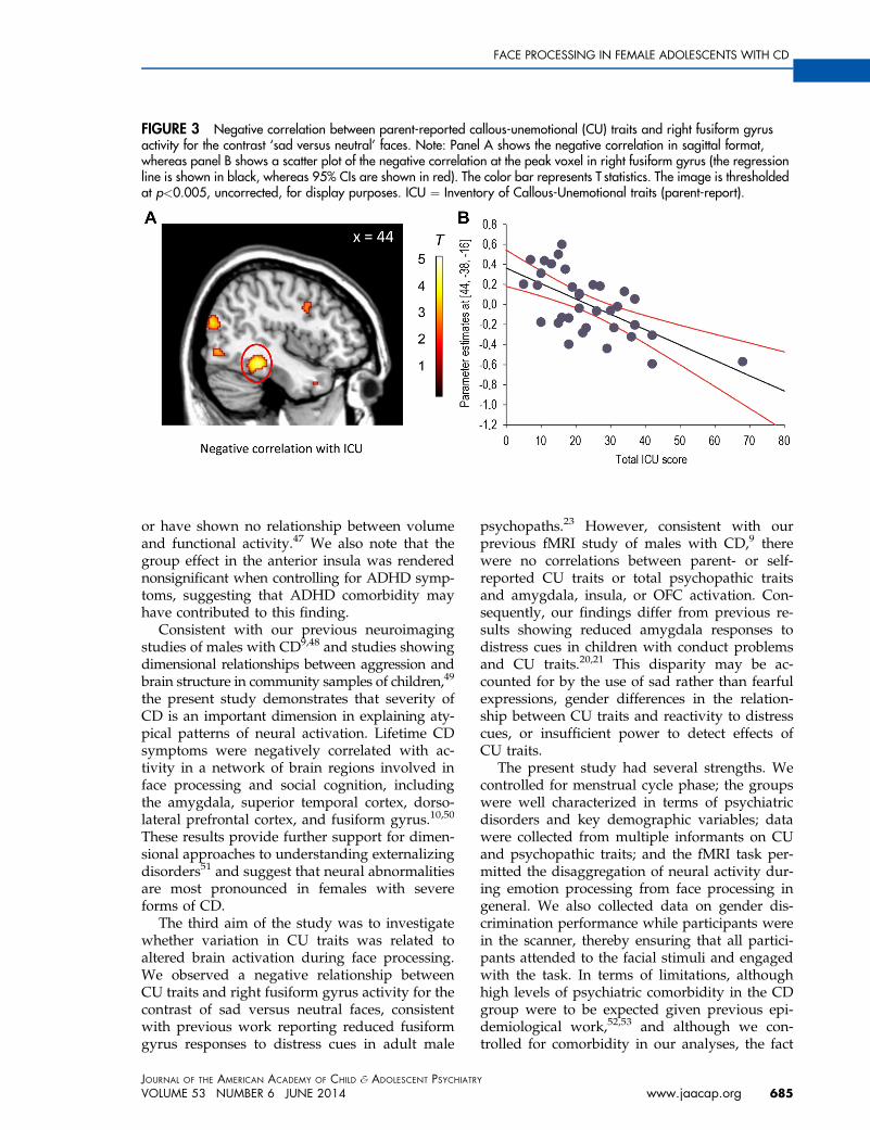
FIGURE 3 Negative correlation between parent-reported callous-unemotional (CU) traits and right fusiform gyrusactivity for the contrast ‘sad versus neutral’ faces. Note: Panel A shows the negative correlation in sagittal format,whereas panel B shows a scatter plot of the negative correlation at the peak voxel in right fusiform gyrus (the regressionline is shown in black, whereas 95% CIs are shown in red). The color bar represents T statistics. The image is thresholdedat p<0.005, uncorrected, for display purposes. ICU ¼ Inventory of Callous-Unemotional traits (parent-report).
FACE PROCESSING IN FEMALE ADOLESCENTS WITH CD
or have shown no relationship between volumeand functional activity.47 We also note that thegroup effect in the anterior insula was renderednonsignificant when controlling for ADHD symp-toms, suggesting that ADHD comorbidity mayhave contributed to this finding.
Consistent with our previous neuroimagingstudies of males with CD9,48 and studies showingdimensional relationships between aggression andbrain structure in community samples of children,49
the present study demonstrates that severity ofCD is an important dimension in explaining aty-pical patterns of neural activation. Lifetime CDsymptoms were negatively correlated with ac-tivity in a network of brain regions involved inface processing and social cognition, includingthe amygdala, superior temporal cortex, dorso-lateral prefrontal cortex, and fusiform gyrus.10,50
These results provide further support for dimen-sional approaches to understanding externalizingdisorders51 and suggest that neural abnormalitiesare most pronounced in females with severeforms of CD.
The third aim of the study was to investigatewhether variation in CU traits was related toaltered brain activation during face processing.We observed a negative relationship betweenCU traits and right fusiform gyrus activity for thecontrast of sad versus neutral faces, consistentwith previous work reporting reduced fusiformgyrus responses to distress cues in adult male
JOURNAL OF THE AMERICAN ACADEMY OF CHILD & ADOLESCENT PSYCHIATR
VOLUME 53 NUMBER 6 JUNE 2014
psychopaths.23 However, consistent with ourprevious fMRI study of males with CD,9 therewere no correlations between parent- or self-reported CU traits or total psychopathic traitsand amygdala, insula, or OFC activation. Con-sequently, our findings differ from previous re-sults showing reduced amygdala responses todistress cues in children with conduct problemsand CU traits.20,21 This disparity may be ac-counted for by the use of sad rather than fearfulexpressions, gender differences in the relation-ship between CU traits and reactivity to distresscues, or insufficient power to detect effects ofCU traits.
The present study had several strengths. Wecontrolled for menstrual cycle phase; the groupswere well characterized in terms of psychiatricdisorders and key demographic variables; datawere collected from multiple informants on CUand psychopathic traits; and the fMRI task per-mitted the disaggregation of neural activity dur-ing emotion processing from face processing ingeneral. We also collected data on gender dis-crimination performance while participants werein the scanner, thereby ensuring that all partici-pants attended to the facial stimuli and engagedwith the task. In terms of limitations, althoughhigh levels of psychiatric comorbidity in the CDgroup were to be expected given previous epi-demiological work,52,53 and although we con-trolled for comorbidity in our analyses, the fact
Y
www.jaacap.org 685

FAIRCHILD et al.
that almost half of the female CD group had acurrent or past internalizing disorder (e.g., de-pression or anxiety) may have influenced ourfindings. It will therefore be important to recruitlarger, noncomorbid CD samples in future re-search, although we note that there is a risk thatsuch samples would be unrepresentative, as levelsof psychiatric comorbidity are extremely high infemales with CD. The cross-sectional nature of thestudy means that we cannot infer that the atypicalneural activation observed plays a causal role inthe etiology of CD. Future studies should adoptlongitudinal designs to investigate whether aty-pical neural activation predicts the developmentof CD or resolves after successful treatment. Wealso note the possibility that females with CDmay have shown atypical neural responses to anycomplex visual stimulus. Consequently, future stu-dies should control for stimulus complexity by in-cluding complex but nonfacial stimuli or scrambledfaces. Finally, although the gender discriminationdata indicated that the participants with CDattended to the facial stimuli, their eye-scanningpatterns may have differed from those of healthycontrols.54 This may contribute to group differ-ences in brain activity as has been found instudies of autism spectrum disorders.55 In futurestudies, eye-tracking data could be collected dur-ing fMRI data acquisition to address this issue.
Our observation of atypical medial OFC andanterior insula responses during face processingin females with CD provides further evidencethat neurobiological factors may be involved in theetiology of severe antisocial behavior. These brainregions are involved in social cognitive processes,including the decoding of facial expressions andempathy, so our findings may help to explainprevious results showing impaired facial emotionrecognition in females with CD. These group
JOURN
686 www.jaacap.org
differences in neural activity were observed acrossall facial expressions (including neutral), and soappear to be related to face processing in generalrather than to emotion processing specifically. Wealso observed negative correlations between CDsymptoms and neural activity in a network of re-gions involved in social cognition and also betweenCU traits and fusiform gyrus responses to sadfacial expressions. These results support dimen-sional approaches to understanding externalizingdisorders, as they suggest that atypical brain ac-tivity is most evident in those individuals withsevere forms of CD or CU traits. &
Accepted March 5, 2014.
This article was reviewed under and accepted by deputy editor EllenLeibenluft, M.D.
Dr. Fairchild is with University of Southampton, UK. Dr. Hagan andDr. Goodyer are with University of Cambridge, UK. Dr. Passamonti iswith the Consiglio Nazionale delle Ricerche, Istituto di Bioimmagini eFisiologia Molecolare, Italy. Dr. Walsh is with University of EastAnglia, UK. Dr. Calder was with the Medical Research CouncilCognition and Brain Sciences Unit, Cambridge, UK.
This research was funded by project grant 083140 from the Well-come Trust (G.F. and I.M.G.) and Medical Research Council projectcode MC_US_A060_5PQ50 (A.J.C.). This work was completedwithin the National Institute of Health Research Collaboration forLeadership in Applied Health Research and Care for Cambridgeshireand Peterborough.
Rik Henson, PhD, of University of Cambridge served as a consultant onthe design of the functional magnetic resonance imaging task.
The authors thank the participants and their parents for taking part in thestudy. The authors also thank the schools, pupil referral units, and theCambridge Youth Offending Service for their help with recruitment.
Disclosures: Drs. Fairchild, Hagan, Passamonti, Walsh, Goodyer, andCalder report no biomedical financial interests or potential conflicts ofinterest.
Correspondence to Graeme Fairchild, PhD, Academic Unit of Psy-chology, University of Southampton, Southampton, SO17 1BJ, UK;e-mail: [email protected]
0890-8567/$36.00/ª2014 American Academy of Child andAdolescent Psychiatry
http://dx.doi.org/10.1016/j.jaac.2014.02.009
REFERENCES
1. American Psychiatric Association. Diagnostic and StatisticalManual of Mental Disorders. 4th Edition. Washington, DC:American Psychiatric Association; 1994.
2. Loeber R, Burke J, Pardini DA. Perspectives on oppositionaldefiant disorder, conduct disorder, and psychopathic features.J Child Psychol Psychiatry. 2009;50:133-142.
3. Pajer KA. What happens to "bad" girls? A review of the adultoutcomes of antisocial adolescent girls. Am J Psychiatry. 1998;155:862-870.
4. Bardone AM, Moffitt TE, Caspi A, Dickson N, Stanton WR,Silva PA. Adult physical health outcomes of adolescent girls withconduct disorder, depression, and anxiety. J Am Acad ChildAdolesc Psychiatry. 1998;37:594-601.
5. Odgers CL, Moffitt TE, Broadbent JM, et al. Female and maleantisocial trajectories: from childhood origins to adult outcomes.Dev Psychopathol. 2008;20:673-716.
6. Kasen S, Cohen P, Skodol AE, Johnson JG, Brook JS. Influence ofchild and adolescent psychiatric disorders on young adult per-sonality disorder. Am J Psychiatry. 1999;156:1529-1535.
7. Fairchild G, Stobbe Y, van Goozen SH, Calder AJ, Goodyer IM.Facial expression recognition, fear conditioning, and startle mod-ulation in female subjects with conduct disorder. Biol Psychiatry.2010;68:272-279.
8. Fairchild G, Van Goozen SH, Calder AJ, Stollery SJ, Goodyer IM.Deficits in facial expression recognition in male adolescents withearly-onset or adolescence-onset conduct disorder. J Child PsycholPsychiatry. 2009;50:627-636.
9. Passamonti L, Fairchild G, Goodyer IM, et al. Neural abnormalitiesin early-onset and adolescence-onset conduct disorder. Arch GenPsychiatry. 2010;67:729-738.
10. Calder AJ, Lawrence AD, Young AW. Neuropsychology of fearand loathing. Nat Rev Neurosci. 2001;2:352-363.
AL OF THE AMERICAN ACADEMY OF CHILD & ADOLESCENT PSYCHIATRY
VOLUME 53 NUMBER 6 JUNE 2014

FACE PROCESSING IN FEMALE ADOLESCENTS WITH CD
11. Kringelbach ML, Rolls ET. The functional neuroanatomy of thehuman orbitofrontal cortex: evidence from neuroimaging andneuropsychology. Prog Neurobiol. 2004;72:341-372.
12. Sterzer P, Stadler C, Krebs A, Kleinschmidt A, Poustka F.Abnormal neural responses to emotional visual stimuli in ado-lescents with conduct disorder. Biol Psychiatry. 2005;57:7-15.
13. Frick PJ, White SF. Research review: the importance of callous-unemotional traits for developmental models of aggressive andantisocial behavior. J Child Psychol Psychiatry. 2008;49:359-375.
14. Viding E, McCrory EJ. Genetic and neurocognitive contributionsto the development of psychopathy. Dev Psychopathol. 2012;24:969-983.
15. Blair RJ. Applying a cognitive neuroscience perspective to thedisorder of psychopathy. Dev Psychopathol. 2005;17:865-891.
16. Marsh AA, Blair RJ. Deficits in facial affect recognition amongantisocial populations: a meta-analysis. Neurosci Biobehav Rev.2008;32:454-465.
17. Blair RJ, Colledge E, Murray L, Mitchell DG. A selective impair-ment in the processing of sad and fearful expressions in childrenwith psychopathic tendencies. J Abnorm Child Psychol. 2001;29:491-498.
18. Dawel A, O’Kearney R, McKone E, Palermo R. Not just fear andsadness:meta-analytic evidence of pervasive emotion recognitiondeficits for facial and vocal expressions in psychopathy. NeurosciBiobehav Rev. 2012;36:2288-2304.
19. Eisenbarth H, Alpers GW, Segre D, Calogero A, Angrilli A.Categorization and evaluation of emotional faces in psychopathicwomen. Psychiatry Res. 2008;159:189-195.
20. Jones AP, Laurens KR, Herba CM, Barker GJ, Viding E.Amygdala hypoactivity to fearful faces in boys with conductproblems and callous-unemotional traits. Am J Psychiatry. 2009;166:95-102.
21. Marsh AA, Finger EC, Mitchell DG, et al. Reduced amygdalaresponse to fearful expressions in children and adolescents withcallous-unemotional traits and disruptive behavior disorders. AmJ Psychiatry. 2008;165:712-720.
22. Dolan MC, Fullam RS. Psychopathy and functional magneticresonance imaging blood oxygenation level-dependent responsesto emotional faces in violent patients with schizophrenia. BiolPsychiatry. 2009;66:570-577.
23. Deeley Q, Daly E, Surguladze S, et al. Facial emotion processing incriminal psychopathy. Preliminary functional magnetic resonanceimaging study. Br J Psychiatry. 2006;189:533-539.
24. Wechsler D. Wechsler Abbreviated Scale of Intelligence. SanAntonio, TX: Psychological Corporation; 1999.
25. Fairchild G, Hagan CC, Walsh ND, Passamonti L, Calder AJ,Goodyer IM. Brain structure abnormalities in adolescent girls withconduct disorder. J Child Psychol Psychiatry. 2013;54:86-95.
26. Kaufman J, Birmaher B, Brent D, et al. Schedule for AffectiveDisorders and Schizophrenia for School-Age Children–Presentand Lifetime Version (K-SADS-PL): initial reliability and validitydata. J Am Acad Child Adolesc Psychiatry. 1997;36:980-988.
27. Andershed H, Kerr M, Stattin H, Levander S. Psychopathic traitsin non-referred youths: a new assessment tool. In: Blaauw E,Sheridan L, eds. Psychopaths: Current International Perspectives.Hague, Netherlands: Elsevier; 2002:131-158.
28. Essau CA, Sasagawa S, Frick PJ. Callous-unemotional traits in acommunity sample of adolescents. Assessment. 2006;13:454-469.
29. Moberg DP. The Adolescent Alcohol and Drug Involvement Scale.Madison, WI: Center for Health Policy and Program Evalua-tion; 2000.
30. Oldfield RC. The assessment and analysis of handedness: theEdinburgh inventory. Neuropsychologia. 1971;9:97-113.
31. Dreher JC, Schmidt PJ, Kohn P, Furman D, Rubinow D,Berman KF. Menstrual cycle phase modulates reward-relatedneural function in women. Proc Natl Acad Sci U S A. 2007;104:2465-2470.
32. Tottenham N, Tanaka JW, Leon AC, et al. The NimStim Set offacial expressions: judgments from untrained research partici-pants. Psychiatry Res. 2009;168:242-249.
JOURNAL OF THE AMERICAN ACADEMY OF CHILD & ADOLESCENT PSYCHIATR
VOLUME 53 NUMBER 6 JUNE 2014
33. Lundqvist D, Flykt A, Ohman A. The Karolinska DirectedEmotional Faces (KDEF). Stockholm: Department of Neurosci-ences Karolinska Hospital; 1998.
34. Ewbank MP, Lawrence AD, Passamonti L, Keane J, Peers PV,Calder AJ. Anxiety predicts a differential neural response toattended and unattended facial signals of anger and fear. Neuro-image. 2009;44:1144-1151.
35. Passamonti L, Rowe JB, Ewbank M, Hampshire A, Keane J,Calder AJ. Connectivity from the ventral anterior cingulate to theamygdala is modulated by appetitive motivation in response tofacial signals of aggression. Neuroimage. 2008;43:562-570.
36. Friston KJ, Holmes AP, Worsley KJ, Poline J-P, Frith CD,Frackowiak RSJ. Statistical parametric maps in functional imaging:a general linear approach. Hum Brain Mapp. 1994;2:189-210.
37. Genovese CR, Lazar NA, Nichols T. Thresholding of statisticalmaps in functional neuroimaging using the false discovery rate.Neuroimage. 2002;15:870-878.
38. Friston KJ. Testing for anatomically specified regional effects.Hum Brain Mapp. 1997;5:133-136.
39. Worsley KJ, Marrett S, Neelin P, Vandal AC, Friston KJ, Evans AC.A unified statistical approach for determining significant signalsin images of cerebral activation. Hum Brain Mapp. 1996;4:58-73.
40. Herpertz SC, Huebner T, Marx I, et al. Emotional processing inmale adolescents with childhood-onset conduct disorder. J ChildPsychol Psychiatry. 2008;49:781-791.
41. Finger EC, Marsh AA, Mitchell DG, et al. Abnormal ventromedialprefrontal cortex function in children with psychopathic traitsduring reversal learning. Arch Gen Psychiatry. 2008;65:586-594.
42. Finger EC, Marsh AA, Blair KS, et al. Disrupted reinforcementsignalling in the orbitofrontal cortex and caudate in youths withconduct disorder or oppositional defiant disorder and a high levelof psychopathic traits. Am J Psychiatry. 2011;168:152-162.
43. Tzourio-Mazoyer N, Landeau B, Papathanassiou D, et al. Auto-mated anatomical labeling of activations in SPM using a macro-scopic anatomical parcellation of the MNI MRI single-subjectbrain. Neuroimage. 2002;15:273-289.
44. Gallagher HL, Frith CD. Functional imaging of ‘theory of mind’.Trends Cogn Sci. 2003;7:77-83.
45. Amodio DM, Frith CD. Meeting of minds: the medial frontalcortex and social cognition. Nat Rev Neurosci. 2006;7:268-277.
46. Adolphs R. Neural systems for recognizing emotion. Curr OpinNeurobiol. 2002;12:169-177.
47. Pizzagalli DA, Oakes TR, Fox AS, et al. Functional but not struc-tural subgenual prefrontal cortex abnormalities in melancholia.Mol Psychiatry. 2004;9:393-405.
48. Fairchild G, Passamonti L, Hurford G, et al. Brain structure ab-normalities in early-onset and adolescent-onset conduct disorder.Am J Psychiatry. 2011;168:624-633.
49. Ducharme S, Hudziak JJ, Botteron KN, et al. Right anteriorcingulate cortical thickness and bilateral striatal volume correlatewith Child Behavior Checklist aggressive behavior scores inhealthy children. Biol Psychiatry. 2011;70:283-290.
50. Haxby JV, Hoffman EA, Gobbini MI. The distributed human neuralsystem for face perception. Trends Cogn Sci. 2000;4:223-233.
51. Krueger RF, Markon KE, Patrick CJ, Iacono WG. Externalizingpsychopathology in adulthood: a dimensional-spectrum concep-tualization and its implications for DSM-V. J Abnorm Psychol.2005;114:537-550.
52. Wasserman GA, McReynolds LS, Ko SJ, Katz LM, Carpenter JR.Gender differences in psychiatric disorders at juvenile probationintake. Am J Public Health. 2005;95:131-137.
53. Lehto-Salo P, Narhi V, Ahonen T, Marttunen M. Psychiatric co-morbidity more common among adolescent females with CD/ODD than among males. Nord J Psychiatry. 2009;63:308-315.
54. Dadds MR, El Masry Y, Wimalaweera S, Guastella AJ. Reducedeye gaze explains “fear blindness” in childhood psychopathictraits. J Am Acad Child Adolesc Psychiatry. 2008;47:455-463.
55. Perlman SB, Hudac CM, Pegors T, Minshew NJ, Pelphrey KA.Experimental manipulation of face-evoked activity in the fusiformgyrus of individuals with autism. Soc Neurosci. 2011;6:22-30.
Y
www.jaacap.org 687

FIGURE S1 Schematic representation of the functional magnetic resonance imaging (fMRI) task and examples of thefacial stimuli used in the experiment. Note: All participants were shown gray-scale photographs of angry, sad, or neutralfacial expressions (12 epochs of each) intermixed with null events (fixation cross) within 17.5-second epochs. In each facetrial, participants had to indicate whether the face was male or female by pressing either the left or right key on a buttonbox. Each trial lasted 1,750 milliseconds, and total scanning time was 10 minutes 30 seconds.
JOURNAL OF THE AMERICAN ACADEMY OF CHILD & ADOLESCENT PSYCHIATRY
687.e1 www.jaacap.org VOLUME 53 NUMBER 6 JUNE 2014
FAIRCHILD et al.

FIGURE S2 Ratings for anger and sadness intensity for the 3 classes of facial expressions used in the functionalmagnetic resonance imaging (fMRI) experiment, by group, as obtained after the fMRI scanning session. Note: Participantswere asked to rate each facial expression in terms of anger intensity, and then rate each expression again in terms ofsadness intensity. There were no group differences in ratings of emotion intensity or group by expression interactions ineither the anger or the sadness rating conditions. CD ¼ conduct disorder.
JOURNAL OF THE AMERICAN ACADEMY OF CHILD & ADOLESCENT PSYCHIATRY
VOLUME 53 NUMBER 6 JUNE 2014 www.jaacap.org 687.e2
FACE PROCESSING IN FEMALE ADOLESCENTS WITH CD

FIGURE S3 Main effects of facial emotion in the group � facial emotion analysis of variance. Note: Panel A showsthe significant main effect of emotion in right posterior superior temporal gyrus/sulcus extending into fusiform gyrus(circled in blue), whereas Panel C depicts the main effect of emotion in right ventrolateral prefrontal cortex/inferior frontalgyrus (circled in blue). The color bar, which ranges from red to white, represents F statistics. Images are thresholded atp < .005, uncorrected, for display purposes only. Panels B and D display the plots of the data extracted from the peakvoxels in superior temporal gyrus and ventrolateral prefrontal cortex, respectively. These plots show that both regionswere most strongly activated by angry faces and least activated by neutral faces, with sad faces also tending to activatethese regions to a greater degree than neutral faces. CD ¼ conduct disorder; HC ¼ healthy control.
JOURNAL OF THE AMERICAN ACADEMY OF CHILD & ADOLESCENT PSYCHIATRY
687.e3 www.jaacap.org VOLUME 53 NUMBER 6 JUNE 2014
FAIRCHILD et al.

TABLE S1 Sample Characteristics of Previous Functional Magnetic Resonance Imaging (fMRI) Studies of ConductDisorder (CD), Conduct Problems, or Callous-Unemotional (CU) Traits
Authors, Year, Reference Sample Description
No. of Male andFemale CD Participants
(Male:Female) Task UsedPsychiatricComorbidity
Sterzer et al. (2005)1 Childhood-onset CD 13:0 Passive viewing ofIAPS pictures
8/13 Had comorbid ADHD;elevated anxiety anddepression scores
Stadler et al. (2007)2 Childhood-onset CD 13:0 Passive viewing ofIAPS pictures
8/13 Had comorbid ADHD;elevated anxiety anddepression scores
Banich et al. (2007)3 Conduct problemsand substance use
12:0 Stroop task 2/12 Had comorbid ADHD
Marsh et al. (2008)4 Disruptive behaviordisorders þ CU
7:5 Fear, anger, andneutral faceprocessing
7/12 Had comorbid ADHD
Herpertz et al. (2008)5 Childhood-onset CD 22:0 Passive viewing ofIAPS pictures
16/22 Had comorbid ADHD;elevated anxiety anddepression scores
Finger et al. (2008)6 Disruptive behaviordisorders þ CU
9:5 Probabilistic reversallearning
10/14 Had comorbid ADHD
Jones et al. (2009)7 Conductproblems þ CU
17:0 Fear and neutralface processing
Not assessed
Decety et al. (2009)8 Childhood-onset CD 8:0 Empathy for paintask
7/8 Had comorbid ADHD
Rubia et al. (2008)9 Childhood-onset CD 13:0 Stop task NoneRubia et al. (2009a)10 Childhood-onset CD 14:0 Rewarded continuous
performance taskNone
Rubia et al. (2009b)11 Childhood-onset CD 13:0 Simon interferencetask
None
Gatzke-Kopp et al.(2009)12
Mixed externalizingdisorders
19:0 Monetary incentivedelay
16/19 Had ADHD diagnoses;elevated depression scores
Bjork et al. (2010)13 Mixed externalizingdisorders
9:3 Monetary incentivedelay
3/12 Had comorbidADHD; elevatedinternalizing scores
Passamonti et al.(2010)14
Childhood- andadolescent-onset CD
40:0 Angry, sad, andneutral faceprocessing
9/40 Had comorbidADHD
Rubia et al. (2010)15 Childhood-onset CD 14:0 Switch task NoneCrowley et al. (2010)16 CD and substance
use disorders20:0 Risk taking task Not assessed with DSM-IV
criteriaFinger et al. (2011)17 Disruptive behavior
disorders þ CU9:6 Passive avoidance
learning10/15 Had comorbid ADHD
Marsh et al. (2011)18 Disruptive behaviordisorders þ CU
8:6 Implicit associationtest with moralwords
9/14 Had comorbid ADHD
White et al. (2012a)19 Disruptive behaviordisorders þ CU
13:4 Spatial attention witheye gaze cues
9/17 Had comorbid ADHD
White et al. (2012b)20 Disruptive behaviordisorders þ CU
12:3 Face processingunder high or lowattentional load
8/15 Had comorbid ADHD
Sebastian et al. (2012)21 Conduct problemswith or without CU
31:0 Affective theory ofmind task
Not assessed with DSM-IVcriteria; elevated anxietyand depression scores
Viding et al. (2012)22 Conduct problemswith or without CU
30:0 Subliminal faceprocessing task
Not assessed with DSM-IVcriteria; elevated anxietyand depression scores
JOURNAL OF THE AMERICAN ACADEMY OF CHILD & ADOLESCENT PSYCHIATRY
VOLUME 53 NUMBER 6 JUNE 2014 www.jaacap.org 687.e4
FACE PROCESSING IN FEMALE ADOLESCENTS WITH CD

TABLE S1 Continued
Authors, Year, Reference Sample Description
No. of Male andFemale CD Participants
(Male:Female) Task UsedPsychiatricComorbidity
White et al. (2013)23 Disruptive behaviordisorders þ CU
17:3 Passive avoidancetask
4/20 Had comorbid ADHD
Marsh et al. (2013)24 Disruptive behaviordisorders þ CU
8:6 Empathy for paintask
8/14 Had comorbid ADHD
Lockwood et al. (2013)25 Conduct problemswith or without CU
37:0 Empathy for paintask
Not assessed with DSM-IVcriteria; elevated anxietyand depression scores
Sebastian et al. (2014)26 Conduct problemswith or without CU
34:0 Processing emotionalfaces and eyes
Not Assessed with DSM-IVcriteria; elevated anxietyand depression scores
Note: ADHD ¼ attention deficit/ hyperactivity disorder; IAPS ¼ International Affective Picture System.
FAIRCHILD et al.
SUPPLEMENTAL REFERENCES
1. Sterzer P, Stadler C, Krebs A, Kleinschmidt A, Poustka F.Abnormal neural responses to emotional visual stimuli in ado-lescents with conduct disorder. Biol Psychiatry. 2005;57:7-15.
2. Stadler C, Sterzer P, Schmeck K, Krebs A, Kleinschmidt A,Poustka F. Reduced anterior cingulate activation in aggressivechildren and adolescents during affective stimulation: associationwith temperament traits. J Psychiatr Res. 2007;41:410-417.
3. Banich MT, Crowley TJ, Thompson LL, et al. Brain activationduring the Stroop task in adolescents with severe substance andconduct problems: A pilot study. Drug Alcohol Depend. 2007;90:175-182.
4. Marsh AA, Finger EC, Mitchell DG, et al. Reduced amygdalaresponse to fearful expressions in children and adolescents withcallous-unemotional traits and disruptive behavior disorders. AmJ Psychiatry. 2008;165:712-720.
5. Herpertz SC, Huebner T, Marx I, et al. Emotional processing inmale adolescents with childhood-onset conduct disorder. J ChildPsychol Psychiatry. 2008;49:781-791.
6. Finger EC, Marsh AA, Mitchell DG, et al. Abnormal ventromedialprefrontal cortex function in children with psychopathic traitsduring reversal learning. Arch Gen Psychiatry. 2008;65:586-594.
7. Jones AP, Laurens KR, Herba CM, Barker GJ, Viding E. Amygdalahypoactivity to fearful faces in boys with conduct problems andcallous-unemotional traits. Am J Psychiatry. 2009;166:95-102.
8. Decety J, Michalska KJ, Akitsuki Y, Lahey BB. Atypical empathicresponses in adolescents with aggressive conduct disorder: afunctional MRI investigation. Biol Psychol. 2009;80:203-211.
9. Rubia K, Halari R, Smith AB, et al. Dissociated functional brainabnormalities of inhibition in boys with pure conduct disorderand in boys with pure attention deficit hyperactivity disorder. AmJ Psychiatry. 2008;165:889-897.
10. Rubia K, Halari R, Smith AB, Mohammad M, Scott S,Brammer MJ. Shared and disorder-specific prefrontal abnormal-ities in boys with pure attention-deficit/hyperactivity disordercompared to boys with pure CD during interference inhibition andattention allocation. J Child Psychol Psychiatry. 2009;50:669-678.
11. Rubia K, Smith AB, Halari R, et al. Disorder-specific dissociation oforbitofrontal dysfunction in boys with pure conduct disorderduring reward and ventrolateral prefrontal dysfunction in boyswith pure ADHD during sustained attention. Am J Psychiatry.2009;166:83-94.
12. Gatzke-Kopp LM, Beauchaine TP, Shannon KE, et al. Neurologicalcorrelates of reward responding in adolescents with and withoutexternalizing behavior disorders. J Abnorm Psychol. 2009;118:203-213.
13. Bjork JM, Chen G, Smith AR, Hommer DW. Incentive-elicitedmesolimbic activation and externalizing symptomatology in ado-lescents. J Child Psychol Psychiatry. 2010;51:827-837.
JOURN
687.e5 www.jaacap.org
14. Passamonti L, Fairchild G, Goodyer IM, et al. Neural abnormalitiesin early-onset and adolescence-onset conduct disorder. Arch GenPsychiatry. 2010;67:729-738.
15. Rubia K, Halari R, Cubillo A, Mohammad AM, Scott S,Brammer M. Disorder-specific inferior prefrontal hypofunction inboys with pure attention-deficit/hyperactivity disorder comparedto boys with pure conduct disorder during cognitive flexibility.Hum Brain Mapp. 2010;31:1823-1833.
16. Crowley TJ, Dalwani MS, Mikulich-Gilbertson SK, et al. Riskydecisions and their consequences: neural processing by boys withAntisocial Substance Disorder. PLoS One. 2010;5:e12835.
17. Finger EC, Marsh AA, Blair KS, et al. Disrupted reinforcementsignaling in the orbitofrontal cortex and caudate in youths withconduct disorder or oppositional defiant disorder and a high levelof psychopathic traits. Am J Psychiatry. 2011;168:152-162.
18. Marsh AA, Finger EC, Fowler KA, et al. Reduced amygdala-orbitofrontal connectivity during moral judgments in youthswith disruptive behavior disorders and psychopathic traits. Psy-chiatry Res. 2011;194:279-286.
19. White SF, Marsh AA, Fowler KA, et al. Reduced amygdala responsein youths with disruptive behavior disorders and psychopathic traits:decreased emotional response versus increased top-down attentionto nonemotional features. Am J Psychiatry. 2012;169:750-758.
20. White SF, Williams WC, Brislin SJ, et al. Reduced activity withinthe dorsal endogenous orienting of attention network to fearfulexpressions in youth with disruptive behavior disorders andpsychopathic traits. Dev Psychopathol. 2012;24:1105-1116.
21. Sebastian CL, McCrory EJ, Cecil CA, et al. Neural responses toaffective and cognitive theory of mind in children with conductproblems and varying levels of callous-unemotional traits. ArchGen Psychiatry. 2012;69:814-822.
22. Viding E, Sebastian CL, Dadds MR, et al. Amygdala response topreattentive masked fear in children with conduct problems: therole of callous-unemotional traits. Am J Psychiatry. 2012;169:1109-1116.
23. White SF, Pope K, Sinclair S, et al. Disrupted expected value andprediction error signaling in youths with disruptive behaviordisorders during a passive avoidance task. Am J Psychiatry. 2013;170:315-323.
24. Marsh AA, Finger EC, Fowler KA, et al. Empathic responsivenessin amygdala and anterior cingulate cortex in youths with psy-chopathic traits. J Child Psychol Psychiatry. 2013;54:900-910.
25. Lockwood PL, Sebastian CL, McCrory EJ, et al. Association ofcallous traits with reduced neural response to others’ pain inchildren with conduct problems. Curr Biol. 2013;23:901-905.
26. Sebastian CL, McCrory EJ, Dadds MR, et al. Neural responses tofearful eyes in children with conduct problems and varying levelsof callous-unemotional traits. Psychol Med. 2014;44:99-109.
AL OF THE AMERICAN ACADEMY OF CHILD & ADOLESCENT PSYCHIATRY
VOLUME 53 NUMBER 6 JUNE 2014