ATROPA BELLADONNA L. WATER EXTRACT: MODULATOR OF ... · 1 ATROPA BELLADONNA L. WATER EXTRACT:...
25
1 ATROPA BELLADONNA L. WATER EXTRACT: MODULATOR OF EXTRACELLULAR MATRIX FORMATION IN VITRO AND IN VIVO Peter Gál 1,2,4 , Tomáš Vasilenko 2,3 , Ivan Kováč 1 , Martina Kostelníková 1 , Ján Jakubčo 1 , Pavol Szabo 5 , Barbora Dvořánková 5 , František Sabol 6 , Hans-Joachim Gabius 7 , Karel Smetana Jr. 5 1 Department for Biomedical Research, and 6 Clinic of Heart Surgery, East-Slovak Institute of Cardiovascular Diseases, Košice, Slovak Republic 2 Department of Pathological Anatomy, University of Veterinary Medicine and Pharmacy, Košice, Slovak Republic 3 3 rd Clinic of Surgery, Slovak Health University and 1 st Private Hospital Košice-Šaca, Košice, Slovak Republic 4 Institute of Histology and Embryology, and 5 Institute of Anatomy, 1 st Faculty of Medicine, Charles University, Prague, Czech Republic 7 Institute of Physiological Chemistry, Faculty of Veterinary Medicine, Ludwig-Maximilian- University, Munich, Germany Corresponding author: Peter Gál East-Slovak Institute of Cardiovascular Diseases, Department for Biomedical Research Ondavská ul. č. 8 040 11 Košice Slovak Republic e-mail: [email protected] Running title: Atropa belladonna and extracellular matrix
Transcript of ATROPA BELLADONNA L. WATER EXTRACT: MODULATOR OF ... · 1 ATROPA BELLADONNA L. WATER EXTRACT:...
1
ATROPA BELLADONNA L. WATER EXTRACT: MODULATOR OF
EXTRACELLULAR MATRIX FORMATION IN VITRO AND IN VIVO
Peter Gál1,2,4, Tomáš Vasilenko2,3, Ivan Kováč1, Martina Kostelníková1, Ján Jakubčo1, Pavol
Szabo5, Barbora Dvořánková5, František Sabol6, Hans-Joachim Gabius7, Karel Smetana Jr.5
1Department for Biomedical Research, and 6Clinic of Heart Surgery, East-Slovak Institute of
Cardiovascular Diseases, Košice, Slovak Republic
2Department of Pathological Anatomy, University of Veterinary Medicine and Pharmacy,
Košice, Slovak Republic
33rd Clinic of Surgery, Slovak Health University and 1st Private Hospital Košice-Šaca, Košice,
Slovak Republic
4Institute of Histology and Embryology, and 5Institute of Anatomy, 1st Faculty of Medicine,
Charles University, Prague, Czech Republic
7Institute of Physiological Chemistry, Faculty of Veterinary Medicine, Ludwig-Maximilian-
University, Munich, Germany
Corresponding author: Peter Gál
East-Slovak Institute of Cardiovascular Diseases, Department for Biomedical Research
Ondavská ul. č. 8
040 11 Košice
Slovak Republic
e-mail: [email protected]
Running title: Atropa belladonna and extracellular matrix
physres
Nové razítko
2
Summary
Previously, we found that treatment of cutaneous wounds with Atropa belladonna L. (AB)
revealed shortened process of acute inflammation as well as increased tensile strength and
collagen deposition in healing skin wounds (Wound Repair Regen 17: 378-386, 2009). To
better understand AB effect on skin wound healing male Sprague-Dawley rats were submitted
to one round full thickness skin wound on the back. In two experimental groups two different
concentrations of AB extract were daily applied whereas the control group remained
untreated. For histological evaluation samples were removed on day 21 after surgery and
stained for wide spectrum cytokeratin, collagen III, fibronectin, galectin-1, and vimentin. In
addition, in the in vitro study different concentration of AB extract were used to evaluate
differences in HaCaT keratinocytes proliferation and differentiation by detection of Ki67 and
keratin-19 expressions. Furthermore, to assess ECM formation of human dermal fibroblasts
on the in vitro level fibronectin and galectin-1 were visualized. Our study showed that AB
induces fibronectin and galectin-1 rich ECM formation in vitro and in vivo. In addition, the
proliferation of keratinocytes was also increased. In conclusion, AB is an effective modulator
of skin wound healing. Nevertheless, further research is needed to find optimal therapeutic
concentration and exact underlying mechanism of action.
Key words: phytotherapy; aqueous extract; wound healing; fibroblast; keratinocyte,
inflammation
3
Introduction
The quest for finding ways to improve wound healing warrants to test new approaches.
Toward this end, experimental studies are exploring the potential for benefit, examining for
example physical (Gurdol et al. 2010; Lacjakova et al. 2010; Toporcer et al. 2006; Vasilenko
et al. 2010) and pharmacological methods (Chu et al. 2010; Gál et al. 2010; Novotný et al.
2011), stem cell therapy (Luo et al. 2010; Nishino et al. 2011) or phytotherapy (Priya et al.
2004; Skórkowska-Telichowska et al. 2010). Considering economic aspects, applying natural
products represents a feasible option of treatment in many regions of the world. In this
respect, the use of Atropa belladonna L. (AB) aqueous extract for improving skin-wound
healing has a long tradition in the folk medicine of “Spiš region” located in the Eastern
Slovakia. As often encountered, the effect of this herb has never rigorously been
experimentally verified, prompting us to perform our previous investigation. We found that
AB-treated wounds have a shortened process of inflammationas well as present enhanced
collagen deposition and increased tensile strength when compared with their untreated
controls (Gál et al. 2009). Furthermore, our in vitro study revealed that keratinocytes in the
presence of AB expressed keratin-19.
Poorly differentiated cells have a large potential to stimulate new tissue development
and/or formation (Chen et al. 2009; Fu and Li 2009), an aspect of crucial significance for
wound healing (Lau et al. 2010). Herein, several (glyco)proteins, especially components of
the extracellular matrix (ECM), play important roles to facilitate cell-cell and cell-matrix
interactions, what is essential for an effective course of healing (Suzuki et al. 2003; Nečas et
al. 2010; Dvořánková et al. 2011). It has already been shown that an ECM rich in fibronectin
and galectin-1 can serve as active substratum substituting for feeder cells in the case of
keratinocytes (Dvořánková et al. 2011). In addition, galectin-1, an endogenous
adhesion/growth-regulatory lectin (Gabius 2006; Villalobo et al. 2006; Gabius et al. 2011)
4
was found to be up-regulated during the early phases of healing (Klíma et al. 2009) as well as
to be capable to act anti-inflammatorily (Cooper et al. 2010). Of note, both fibronectin and
galectin-1 production have been detected in our in vivo and in vitro investigations. To extend
the scope of study, collagen-3, keratin-19, Ki67, vimentin, and wide-spectrum keratin were
investigated.
In principle, in order to achieve swift repair and regeneration of injured tissues it
would be ideal to prime cells for optimal cell biological properties and for suitable ECM
production. In our previous investigation, it was observed that AB has the capacibility to
modulate these processes (Gál et al. 2009). This study was therefore designed as a combined
in vivo (conducted on rats) and in vitro (conducted on keratinocytes and fibroblasts)
investigation to better understand the mechanisms behind AB-dependent modulatory effects
on skin wound healing. Tissue and cell specimens were processed under identical conditions
to exclude any factor other than AB treatment to affect signal occurrence and intensity.
5
Methods
Plant material – Atropa belladonna L. (AB)
AB (Solanaceae) was collected in August 2006 from the vicinity of mast on Čertova sihoť,
Slovak Paradise, Slovak Republic. The plant was unequivocally identified by Dr. Pavol
Mártonfi from the Department of Botany, Institute of Biology and Ecology, Šafárik
University in Košice. Herb (overground parts) of the plant was dried at room temperature in
the dark. A voucher specimen (KO-30301) was deposited in the Herbarium of the Botanical
Garden of the Šafárik University in Košice.
Preparation of the aqueous extract of AB
The water extract for both in vitro and in vivo experiments was prepared by pouring 10 g of a
powder of dried AB leaves into 100 mL of boiling distilled water. The suspension was then
left for 10 minutes at room temperature. Consecutively, the extract was filtered (0.2 µm).
For the animal study two concentrations were used: the original solution obtained by
extraction (AB-10%) and a 10-times diluted concentration (AB-1%). For the in vitro study the
extract was 10-times diluted in culture medium and the obtained concentration was
considered to be the highest concentration (AB-1%). In addition to this concentration tested,
AB extract was 256 times diluted (AB-0.00390625%). Preliminary dose-response testing
included 0.25%, 0.0625%, and 0.015626% concentrations (unpublished data).
Animal model
The experimental conditions were in compliance with the requirements of the European rules
of ethical standards of animal treatment and welfare. Hence, our experiment was approved by
the Ethics Committee of the Faculty of Medicine of the Šafárik University in Košice and by
the State Veterinary and Food Administration of the Slovak Republic.
6
Male Sprague-Dawley rats (n=12; 8-10 months of age) were used for experiments and
allocated into 3 groups (control – untreated; AB-10% – treated with the high AB
concentration, AB-0.00390625% – treated with the low AB concentration). For general
anesthesia a combination of 33 mg/kg ketamine (Calypsol; Richter Gedeon, Budapest,
Hungary), 11 mg/kg xylazine (Rometar a.u.v.; Spofa, Prague, Czech Republic) and 5 mg/kg
tramadol (Tramadol-K; Krka, Novo Mesto, Slovenia) was intramuscularly administered to the
rats. One round – 1 cm in diameter – full thickness skin wound was performed under aseptic
conditions on the back of each rat. All rats were sacrificed by ether inhalation 21d after
surgery.
Wound treatment
During the treatment all rats were restrained individually in a Plexiglas cage with a circular
opening over the wound. In the control group, the aqueous AB extract was not applied. In the
experimental groups, the extract was topically applied (by means of an eye dropper) three
times a day during the first three days of healing. New extract was prepared each day of
wound treatment.
Basic histology and immunohistochemistry
The first half of skin-wound specimens was processed routinely for light microscopy, i.e.
fixation in 4% buffered formaldehyde, dehydration, embedding, cutting, and staining with
hematoxylin-eosin.
The second half of wound specimens was cryoprotected by Tissue-Tek (Sakura,
Zoeterwoude, Netherlands) and deeply frozen in liquid nitrogen. Cryocut sections (Reichert-
Jung, Vienna, Austria) were first mounted on the surface of poly-L-lysine-treated glasses
(Sigma-Aldrich, St Louis, MO, USA), then fixed in 2% (w/v) paraformaldehyde in
7
phosphate-buffered saline (PBS; pH 7.2). Any binding of the used secondary-step antibody
preparations was precluded by pre-incubation with normal swine serum (DAKO, Glostrup,
Denmark) diluted in PBS for 30 min. Both first- and second-step antibodies were diluted as
recommended by supplier, the sources given in Table 1. The specificity of
immunohistochemical reactions was verified by the replacement of used antibody by an
irrelevant antibody and/or by the omission of the first-step antibody. The lack of cross-
reactivity of our homemade anti-galectin-1 antibody was ascertained, if required, after affinity
depletion on resin with conjugated galectins (Kaltner et al., 2002; Saal et al., 2005).
The nuclei of cells were counterstained with the DAPI fluorochrome (Sigma-Aldrich,
St Louis, MO, USA), specifically recognizing DNA. The specimens were mounted to
Vectashield (Vector Laboratories, Burlingame, CA, USA).
Wound area measurement
The area of healing wounds was measured from standardized photographs as follows.
Wounds were photographed with a scale immediately after surgery and at 21 using an
Olympus E330 digital camera equipped with a digital ED 50 mm f 2.0 macro objective and a
ring set flash SRF-11 (Olympus, Tokyo, Japan). The wound area was then measured on the
images using Quick Photo Micro 2.2 software (Premiere, Prague, Czech Republic) and
expressed as a percentage of the original wound area created on the day of surgery.
Isolation and in vitro cultivation of human dermal fibroblasts (HDF)
Fibroblasts were isolated from residual skin samples. They were obtained from the
Department of Aesthetic Surgery of the 3rd Faculty of Medicine of Charles University
according to the criteria of the Helsinki Declaration with informed consent of patients and
approved by local Ethical Committee. Cells were cultured in Dulbecco’s Modified Eagle's

8
Medium (DMEM) (Biochrom, Berlin, Germany) supplemented with 10% fetal bovine serum
(FBS; Biochrom) and antibiotics (streptomycin and penicillin; Biochrom).
Cells were seeded on glass coverslips at a density of 3 000 cells/cm2 and cultured for
24 hours. Medium containing the tested concentration of AB extract was then added to the
cells, which were cultured thereafter for three days.
In vitro cultivation of HaCaT
The HaCaT (human keratinocytes cell line) cell line was obtained from Cell Lines Service
(Eppelheim, Germany) (Boukamp et al. 1988). Cells were cultured in the Dulbecco’s
Modified Eagle's Medium (DMEM; Biochrom) supplemented with 10% FBS (Biochrom) and
antibiotics (streptomycin and penicillin; Biochrom).
Cells were seeded on a glass coverslips at a density of 5 000 cells/cm2 and cultured for
24 hours. Medium containing the tested concentration of AB extract was then added to cells,
which were cultured thereafter for four days (medium was changed 1 x during the
experiment).
Immunocytochemistry of in vitro cultured keratinocytes and fibroblasts
The adherent cells on the coverslips were washed in PBS and fixed briefly with 2% (w/v)
paraformaldehyde diluted in PBS (pH = 7.2). Any binding of the used second-step antibody
was precluded by pre-incubation with normal swine serum (DAKO, Glostrup, Denmark)
diluted in PBS for 30 min. Both the primary and secondary antibodies were diluted as
recommended by supplier and are described in Table 1. Their specificity was tested by
replacement of a distinct antibody by another polyclonal or monoclonal antibody of the same
animal and isotype, but against antigens not present in studied cells. The nuclei of cells were
9
routinely counterstained with DAPI (Sigma-Aldrich). The specimens were mounted to
Vectashield (Vector Laboratories).
Fluorescence intensities measurement, cell counting, and image analysis
Both skin sections and coverslips containing cultured cells were analyzed by fluorescence
microscopy using a Nikon Eclipse 90i apparatus (Nikon, Tokyo, Japan) equipped with filter-
blocks specific for FITC, TRITC and DAPI, respectively, a high-resolution CCD camera
Cool-1300Q (Vosskühler, Osnabrück, Germany) and a LUCIA 5.1 computer-assisted image
analysis system (Laboratory Imaging, Prague, Czech Republic). Fluorescence intensity was
measured under standardized conditions (Dubový et al. 2002, Klíma et al. 2009) using the
software given above.
By evaluating Ki67 expression all cells were counted in three visualization field of one
coverslip following by counting the Ki67 positive cells. The proliferation activity was than
expressed as the percentage of Ki67 positive cells to total no. of cells.
Statistical analysis
Data from the measurement of fluorescence intensities, wound areas, and cell counting were
compared by one-way ANOVA followed by Tukey-Kramer post-hoc test. Significance was
accepted at p<0.05.
10
Results
Wound histology
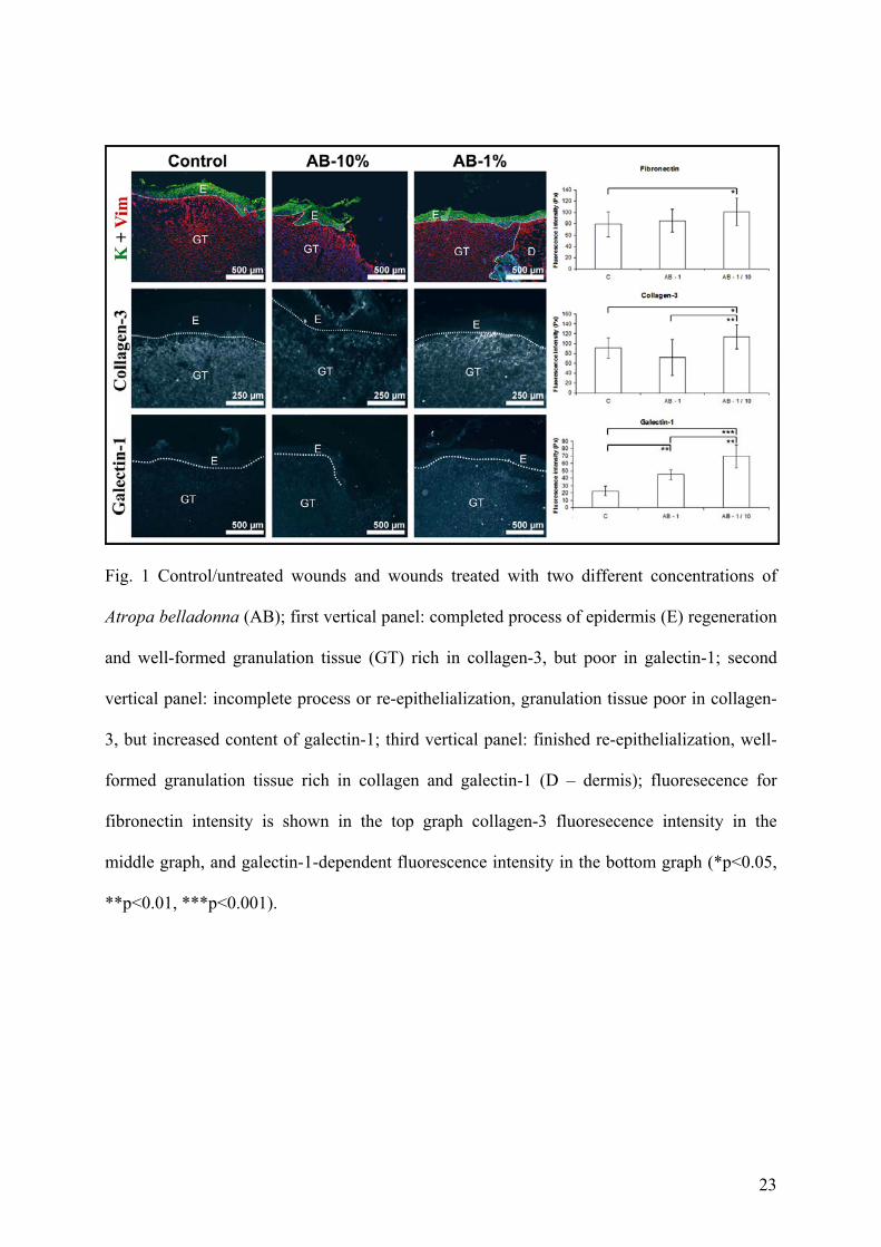
At the period of 21 days post surgery regeneration of the epidermis was completely finished
in the control and AB-1% groups, but not in the AB-10% group (Fig. 1). Only a limited
number of luminized vessels was present in the granulation tissues of control and treated
wounds (not shown). The level of fibronectin presence in the granulation tissue was
comparable in untreated wounds (control) and wounds treated with the undiluted AB extract
(AB-10%) (not shown). However, treatment of wounds with the diluted AB extract (AB-1%)
resulted in significantly increased production of fibronectin (Fig. 1). Interestingly, granulation
tissue of the treated wounds contained a significantly increased amount of galectin-1 when
compared to the control (Fig. 1). Of note, the presence of collagen-3 was enhanced in wounds
treated with the diluted AB extract, while high extract concentration decreased collagen
deposition when compared to the other two groups (Fig. 1).
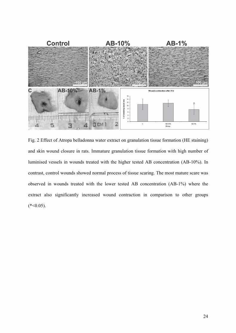
Wound contraction
Measuring the extent of wound contraction revealed significant effect of lower tested AB
concentration (AB-1%) when compared to control (Fig. 2), while the higher tested AB
concentration (AB-10%) did not significantly modulated wound closure.
HDF
The presence of AB in the culture medium led to the formation of a ECM scaffold which
consisted of fibronectin and galectin-1 (Fig. 2). The most prominent newly synthesized ECM
network was observed on the coverslips with cells exposed to the higher tested extract
concentration (AB-1%). ECM production was then leveling off in a concentration dependent
manner (AB-0.00390625%).
11
The presence of AB in the culture medium did not stimulate any transition of
fibroblasts into myofibroblasts (not shown). As consequence, no or only a very limited
number (up to one per visualization field) of myofibroblasts was present.
HaCaT
All cells expressed wide-spectrum keratin. In contrast to ECM production the most prominent
expression of Ki67 was observed in cells exposed to the lower tested AB concentrations (AB-
0.00390625%). With increasing plant concentration Ki67 expression decreased (Fig. 3). No
differences were observed in keratin-19 expression between control and AB stimulated cells
(not shown).
12
Discussion
In our previous investigation we found keratinocyte, cultured on 3T3 feeder fibroblasts,
expressing keratin-19 in cultures which were supplemented with the highest tested
concentration of AB (Gál et al. 2009). Such a cell phenotype is evocative of stem-like features
(Dvořánková et al. 2005), implying a beneficial role for wound healing. In further study we
have also shown that keratinocytes cultured on substratum, rich in fibronectin and galectin-1,
induced by several galectins, have a poorly differentiated phenotype with keratin-19
expression (Dvořánková et al. 2011). In the current investigation, we used the HaCaT cell line
and revealed that AB increases Ki67 expression, but did not affect keratin-19 expression.
Obviously, previously observed keratin-19 expression may depend on 3T3 feeder fibroblasts
which might be affected by AB. Along this line, it was found that wound treatment with
Datura alba L. (Solanaceae) extract led to accelerated epidermis regeneration and increased
fibroblast proliferation (Priya et al. 2002).
Following AB treatment we previously described increased wound tensile strength and
presence of collagen in healing skin wounds (Gál et al. 2009). Formation and reorganization
of the ECM are crucial processes involved in ensuring sufficient wound stiffness and
successful healing. It is well known that fibronectin plays an important role herein.
Fibroblasts as a component of granulation tissue secrete both proteases, which are able to
proteolytically process surrounding ECM, and new constituents of the ECM including
fibronectin and collagen that establish the insoluble matrix (Kumar et al. 2003). Therefore, we
have monitored fibronectin production after cells and/or wounds were exposed to aequeous
AB extract. We found that this treatment of wounds/cells enhanced fibronectin production. Of
note, the effect of AB on collagen-3 expression revealed a concentration-dependent manner
(AB-10% decreased, whereas AB-1% increased collagen deposition in wounds).
13
Interestingly, newly formed ECM was also rich in galectin-1 as well. This lectin can
directly affect cytokine production such as induced IL-10 secretion and decreased IFN-γ
production in activated T cells locally (Stowell et al. 2008) and is a potent effector in T cell
communication (Wang et al. 2009) and its expression is elevated during the inflammatory
phase of healing (Gál et al. 2011). In addition, pharmacological treatment with galectin-1
inhibited leukocyte recruitment into the peritoneal cavity (Gil et al. 2006). Reduction of the
inflammatory process by means of lower number of infiltrated PMNLs, also seen for CD45-
positive lymphocytes (Saussez et al. 2009), and scarce degranulated mast cells were observed
when galectin-1 was administered locally (Rabinovich et al. 2000). Obviously, the lectin
intimately partakes in the regulation of the inflammatory process (Schwartz-Albiez, 2009).
The results with experimentally applied galectin-1 or upon its in situ up-regulation in tumors
can have relevant results for us, in which AB significantly reduced the process of
inflammation (Gál et al. 2009), a process associated with increased production of galectin-1.
Moreover, anti-inflammatory properties of AB were documented by significantly increased
wound tensile strength in both treated groups just with unimpressive treatment duration
differences (Gál et al. 2009). From this point of view, anti-inflammatory effects of AB extract
may engage enhanced generation of galectin-1.
Based on our previous (Gál et al. 2009) and current investigations we may conclude
that the plant extract’s effect is probably based on the acceleration of several processes
occurring during wound healing. In particular, AB is able to stimulate ECM production,
endothelial cells proliferation (as previously shown in HUVECs and may indicate accelerated
angiogenesis), and has anti-inflammatory effects (as previously shown in rats). In our
previous study, detectable concentrations of tropane alkaloids in water extract of leaves of
Atropa belladonna L. were confirmed (Gál et al. 2009). Concentration of atropine has been
more than ten times higher when compared to scopolamine. This is in accordance with results
14
of Zárate et al. (1997) which found in leaves of Atropa beatica higher amount of atropine than
scopolamine, with atropine/scopolamine ratio similar to results obtained in this study. Of note
the pharmacological effect on wound healing may; however, be modulated either positively or
negatively by other additional ballast compounds of the extract. Therefore, the main limitation
of presented study is in omission of biological active compounds isolation following their
wound healing effect evaluation. Accordingly, further research is needed to explain the
underlying mechanisms of action and find optimal therapeutic concentration for any use in
clinical practice. But the presented data indicate the potential for further study of this extract,
also in defining the possible regulatory pathways.
Acknowledgements
We thank Magdaléna Majnušová, Iva Burdová, and Vít Hajdúch for their expert technical
assistance. This study was supported by the Charles University in Prague (program for the
Specific university research No. 260510), by the Ministry of Education, Science, Research
and Sport of the Slovak Republic (VEGA No. 1/1095/11 and No. 1/0152/12), by the Safarik
University in Kosice (Institutional student Grants No. 5/GSŠ/2011, No. 6/GSŠ/2011, and No.
VVGS-39/12-13), and by the EC Glyco-HIT consortium (No. 260600). These results were
partially presented at the 4th Young Biomedical Engineers and Researchers Conference –
YBERC 2010, held in Košice (Slovak Republic) on July 1-3, 2010.
15
References
BOUKAMP P, PETRUSSEVSKA RT, BREITKREUTZ D, HORNUNG J, MARKHAM A,
FUSENIG NE. Normal keratinization in a spontaneously immortalized aneuploid human
keratinocyte cell line. J Cell Biol 106: 761-771, 1988.
CHEN M, PRZYBOROWSKI M, BERTHIAUME F: Stem cells for skin tissue engineering
and wound healing. Crit Rev Biomed Eng 37: 399-421, 2009.
CHU Y, YU D, WANG P, XU J, LI D, DING M: Nanotechnology promotes the full-
thickness diabetic wound healing effect of recombinant human epidermal growth factor in
diabetic rats. Wound Repair Regen 18: 499-505, 2010.
COOPER D, ILARREGUI JM, PESOA SA, CROCI DO, PERRETTI M, RABINOVICH
GA: Multiple functional targets of the immunoregulatory activity of galectin-1: control of
immune cell trafficking, dendritic cell physiology, and T-cell fate. Methods Enzymol 480:
199-244, 2010.
DVOŘÁNKOVÁ B, SMETANA K JR, CHOVANEC M, LACINA L, ŠTORK J,
PLZÁKOVÁ Z, GALOVIČOVÁ M, GABIUS H-J: Transient expression of keratin 19 is
induced in originally negative interfollicular epidermal cells by adhesion of suspended cells.
Int J Mol Med 16: 525-531, 2005.
DVOŘÁNKOVÁ B, SZABO P, LACINA L, GÁL P, UHROVA J, ZIMA T, KALTNER H,
ANDRÉ S, GABIUS H-J, SYKOVÁ E, SMETANA K JR: Human galectins induce
conversion of dermal fibroblasts into myofibroblasts and production of extracellular matrix:
16
potential application in tissue engineering and wound repair. Cells Tissues Organs 194: 469-
480.
FU X, LI H: Mesenchymal stem cells and skin wound repair and regeneration: possibilities
and questions. Cell Tissue Res 335: 317-321, 2009.
GABIUS H-J: Cell surface glycans: the why and how of their functionality as biochemical
signals in lectin-mediated information transfer. Crit Rev Immunol 26:43-80, 2006.
GABIUS H-J, ANDRÉ S, JIMÉNEZ-BARBERO J, ROMERO A, SOLÍS D: From lectin
structure to functional glycomics: principles of the sugar code. Trends Biochem Sci:36: 298-
313, 2011
GÁL P, TOPORCER T, GRENDEL T, VIDOVÁ Z, SMETANA K JR, DVOŘÁNKOVÁ B,
GÁL T, MOZEŠ S, LENHARDT L, LONGAUER F, SABOL M, SABO J, BAČKOR M:
Effect of Atropa belladonna L. on skin wound healing: biomechanical and histological study
in rats and in vitro study in keratinocytes, 3T3 fibroblasts, and human umbilical vein
endothelial cells. Wound Repair Regen 17: 378-386, 2009.
GÁL P, NOVOTNÝ M, VASILENKO T, DEPTA F, ŠULLA I, TOMORI Z: Decrease in
wound tensile strength following post-surgical estrogen replacement therapy in
ovariectomized rats during the early phase of healing is mediated via ER-α rather than ER-β:
a preliminary report. J Surg Res 159: e25-28, 2010.
17
GÁL P, VASILENKO T, KOSTELNÍKOVÁ M, JAKUBČO J, KOVÁČ I, SABOL F,
ANDRÉ S, KALTNER H, GABIUS HJ, SMETANA K JR: Open wound healing in vivo:
monitoring binding and presence of adhesion/growth-regulatory galectins in rat skin during
the course of complete re-epithelialization. Acta Histochem Cytochem 44: 191-199, 2011.
GIL CD, COOPER D, ROSIGNOLI G, PERRETTI M, OLIANI SM: Inflammation-induced
modulation of cellular galectin-1 and -3 expression in a model of rat peritonitis. Inflamm Res
55: 99-107, 2006.
GURDOL F, CIMSIT M, ONER-IYIDOGAN Y, KOCAK H, SENGUN S, YALCINKAYA-
DEMIRSOZ S: Collagen synthesis, nitric oxide and asymmetric dimethylarginine in diabetic
subjects undergoing hyperbaric oxygen therapy. Physiol Res 59: 423-429, 2010.
KALTNER H, SEYREK K, HECK A, SINOWATZ F, GABIUS HJ: Galectin-1 and galectin-
3 in fetal development of bovine respiratory and digestive tracts. Comparison of cell type-
specific expression profiles and subcellular localization. Cell Tissue Res 307: 35–46, 2002.
KLÍMA J, LACINA L, DVORÁNKOVÁ B, HERRMANN D, CARNWATH JW,
NIEMANN H, KALTNER H, ANDRÉ S, MOTLÍK J, GABIUS HJ, SMETANA K JR:
Differential regulation of galectin expression/reactivity during wound healing in porcine skin
and in cultures of epidermal cells with functional impact on migration. Physiol Res 58: 873-
884, 2009
KUMAR V, COTRAN RZ, ROBBINS SL: Basic Pathology 7th Ed. Saunders, Philadelphia,
London, Toronto, Montreal, Sydney, Tokyo, 2003, pp 873
18
LACJAKOVÁ K, BOBROV N, POLÁKOVÁ M, SLEZÁK M, VIDOVÁ M, VASILENKO
T, NOVOTNÝ M, LONGAUER F, LENHARDT L, BOBER J, LEVKUT M, SABOL F,
GÁL P: Effects of equal daily doses delivered by different power densities of low-level laser
therapy at 670 nm on open skin wound healing in normal and corticosteroid-treated rats: a
brief report. Lasers Med Sci 25: 761-766, 2010.
LAU K, PAUS R, TIEDE S, DAY P, BAYAT A: Exploring the role of stem cells in
cutaneous wound healing. Exp Dermatol 18: 921-933, 2009
LUO G, CHENG W, HE W, WANG X, TAN J, FITZGERALD M, LI X, WU J: Promotion
of cutaneous wound healing by local application of mesenchymal stem cells derived from
human umbilical cord blood. Wound Repair Regen 18: 506-13, 2010.
MICHEL M, TÖRÖK N, GODBOUT MJ, LUSSIER M, GAUDREAU P, ROYAL A,
GERMAIN L: Keratin 19 as a biochemical marker of skin stem cells in vivo and in vitro:
keratin 19 expressing cells are differentially localized in function of anatomic sites, and their
number varies with donor age and culture stage. J Cell Sci 109: 1017-1028, 1996.
NEČAS A, PLÁNKA L, SRNEC R, CRHA M, HLUCILOVÁ J, KLÍMA J, STARÝ D,
KREN L, AMLER E, VOJTOVÁ L, JANCÁR J, GÁL P: Quality of newly formed
cartilaginous tissue in defects of articular surface after transplantation of mesenchymal stem
cells in a composite scaffold based on collagen I with chitosan micro- and nanofibres. Physiol
Res 59: 605-614, 2010.
19
NICKE B, DETJEN K., LOGSDON CD: Muscarinic cholinergic receptors activate both
inhibitory and stimulatory growth mechanisms in NIH3T3 cells. J Biol Chem 274: 21701-
21706, 1999.
NISHINO Y, YAMADA Y, EBISAWA K, NAKAMURA S, OKABE K, UMEMURA E,
HARA K, UEDA M: Stem cells from human exfoliated deciduous teeth (SHED) enhance
wound healing and the possibility of novel cell therapy. Cytotherapy 13: 598-605, 2011.
NOVOTNÝ M, VASILENKO T, VARINSKÁ L, SMETANA K JR, SZABO P, SARIŠSKÝ
M, DVOŘÁNKOVÁ B, MOJŽIŠ J, BOBROV N, TOPORCEROVÁ S, SABOL F,
MATTHEWS BJ, GÁL P: ER-α agonist induces conversion of fibroblasts into
myofibroblasts, while ER-β agonist increases ECM production and wound tensile strength of
healing skin wounds in ovariectomised rats. Exp Dermatol 20: 703-708, 2011.
PRIYA KS, GNANAMANI A, RADHAKRISHNAN N, BABU M: Healing potential of
Datura alba on burn wounds in albino rats. J Ethnopharmacol 83: 193-199, 2002.
PRIYA KS, ARUMUGAM G, RATHINAM B, WELLS A, BABU M: Celosia argentea
Linn. leaf extract improves wound healing in a rat burn wound model. Wound Repair Regen
12: 618-625, 2004.
RABINOVICH GA, SOTOMAYOR CE, RIERA CM, BIANCO I, CORREA SG: Evidence
of a role for galectin-1 in acute inflammation. Eur J Immunol 30: 1331-1339, 2000.
20
SAAL I, NAGY N, LENSCH M, LOHR M, MANNING JC, DECAESTECKER C, ANDRÉ
S, KISS R, SALMON I, GABIUS HJ: Human galectin-2:expression profiling by RT-
PCR/immunohistochemistry and its introduction as histochemical tool for ligand localization.
Histol Histopathol 20: 1191–1208, 2005.
SAUSSEZ S, DECAESTECKER C, CLUDTS S, ERNOUX P, CHEVALIER D, SMETANA
K, JR., ANDRE S, LEROY X, GABIUS HJ: Adhesion/growth-regulatory tissue lectin
galectin-1 in relation to angiogenesis/lymphocyte infiltration and prognostic relevance of
stromal up-regulation in laryngeal carcinomas. Anticancer Res 29: 59-65, 2009.
SCHWARTZ-ALBIEZ R. Inflammation and glycosciences. In: Gabius H-J, ed. The Sugar
Code Fundamentals of glycosciences. Weinheim, Germany: Wiley-VCH; 2009:447-67.
SKÓRKOWSKA-TELICHOWSKA K, ZUK M, KULMA A, BUGAJSKA-PRUSAK A,
RATAJCZAK K, GASIOROWSKI K, KOSTYN K, SZOPA J: New dressing materials
derived from transgenic flax products to treat long-standing venous ulcers--a pilot study.
Wound Repair Regen 18: 168-179, 2010.
STOWELL SR, QIAN Y, KARMAKAR S, KOYAMA NS, DIAS-BARUFFI M, LEFFLER
H, MCEVER RP, CUMMINGS RD: Differential roles of galectin-1 and galectin-3 in
regulating leukocyte viability and cytokine secretion. J Immunol 180: 3091-3102, 2008.
SUZUKI K, SAITO J, YANAI R, YAMADA N, CHIKAMA T, SEKI K, NISHIDA T: Cell-
matrix and cell-cell interactions during corneal epithelial wound healing. Prog Retin Eye Res
22: 113-133, 2003.
21
TOPORCER T, RADONÁK J: Vacuum assisted wound closure --overview of lesson and
applications. Čas Lek Česk 145: 702-707, 2006.
VASILENKO T, SLEZÁK M, KOVÁC I, BOTTKOVÁ Z, JAKUBCO J, KOSTELNÍKOVÁ
M, TOMORI Z, GÁL P: The effect of equal daily dose achieved by different power densities
of low-level laser therapy at 635 and 670 nm on wound tensile strength in rats: a short report.
Photomed Laser Surg 28: 281-283, 2010.
VILLALOBO A, NOGALES-GONZALÉS A, GABIUS H-J: A guide to signaling pathways
connecting protein-glycan interaction with the emerging versatile effector functionality of
mammalian lectins. Trends Glycosci Glycotechnol 18:1-37, 2006.
WANG J, LU ZH, GABIUS H-J, ROHOWSKY-KOCHAN C, LEDEEN RW, WU G: Cross-
linking of GM1 ganglioside by galectin-1 mediates regulatory T cell activity involving
TRPC5 channel activation: possible role in suppressing experimental autoimmune
encephalomyelitis. J Immunol 182:4036-4045, 2009.
Zérate, R, Hermosin B, Cantos M, Troncoso A. Tropane alkaloid distribution in Atropa
beatica plants. J Chem Ecol 23:2059-2066, 1997.
22
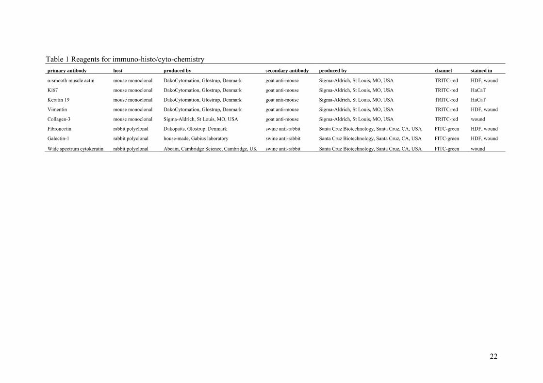
Table 1 Reagents for immuno-histo/cyto-chemistry primary antibody host produced by secondary antibody produced by channel stained in
α-smooth muscle actin mouse monoclonal DakoCytomation, Glostrup, Denmark goat anti-mouse Sigma-Aldrich, St Louis, MO, USA TRITC-red HDF, wound
Ki67 mouse monoclonal DakoCytomation, Glostrup, Denmark goat anti-mouse Sigma-Aldrich, St Louis, MO, USA TRITC-red HaCaT
Keratin 19 mouse monoclonal DakoCytomation, Glostrup, Denmark goat anti-mouse Sigma-Aldrich, St Louis, MO, USA TRITC-red HaCaT
Vimentin mouse monoclonal DakoCytomation, Glostrup, Denmark goat anti-mouse Sigma-Aldrich, St Louis, MO, USA TRITC-red HDF, wound
Collagen-3 mouse monoclonal Sigma-Aldrich, St Louis, MO, USA goat anti-mouse Sigma-Aldrich, St Louis, MO, USA TRITC-red wound
Fibronectin rabbit polyclonal Dakopatts, Glostrup, Denmark swine anti-rabbit Santa Cruz Biotechnology, Santa Cruz, CA, USA FITC-green HDF, wound
Galectin-1 rabbit polyclonal house-made, Gabius laboratory swine anti-rabbit Santa Cruz Biotechnology, Santa Cruz, CA, USA FITC-green HDF, wound
Wide spectrum cytokeratin rabbit polyclonal Abcam, Cambridge Science, Cambridge, UK swine anti-rabbit Santa Cruz Biotechnology, Santa Cruz, CA, USA FITC-green wound
23
Fig. 1 Control/untreated wounds and wounds treated with two different concentrations of
Atropa belladonna (AB); first vertical panel: completed process of epidermis (E) regeneration
and well-formed granulation tissue (GT) rich in collagen-3, but poor in galectin-1; second
vertical panel: incomplete process or re-epithelialization, granulation tissue poor in collagen-
3, but increased content of galectin-1; third vertical panel: finished re-epithelialization, well-
formed granulation tissue rich in collagen and galectin-1 (D – dermis); fluoresecence for
fibronectin intensity is shown in the top graph collagen-3 fluoresecence intensity in the
middle graph, and galectin-1-dependent fluorescence intensity in the bottom graph (*p<0.05,
**p<0.01, ***p<0.001).
24
Fig. 2 Effect of Atropa belladonna water extract on granulation tissue formation (HE staining)
and skin wound closure in rats. Immature granulation tissue formation with high number of
luminised vessels in wounds treated with the higher tested AB concentration (AB-10%). In
contrast, control wounds showed normal process of tissue scaring. The most mature scare was
observed in wounds treated with the lower tested AB concentration (AB-1%) where the
extract also significantly increased wound contraction in comparison to other groups
(*<0.05).
25
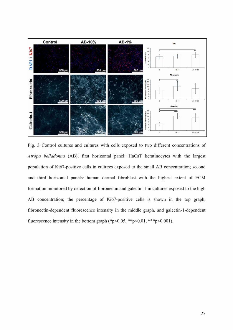
Fig. 3 Control cultures and cultures with cells exposed to two different concentrations of
Atropa belladonna (AB); first horizontal panel: HaCaT keratinocytes with the largest
population of Ki67-positive cells in cultures exposed to the small AB concentration; second
and third horizontal panels: human dermal fibroblast with the highest extent of ECM
formation monitored by detection of fibronectin and galectin-1 in cultures exposed to the high
AB concentration; the percentage of Ki67-positive cells is shown in the top graph,
fibronectin-dependent fluorescence intensity in the middle graph, and galectin-1-dependent
fluorescence intensity in the bottom graph (*p<0.05, **p<0.01, ***p<0.001).