Associations With Immunity in Clear Cell Renal Cell ...
21
Page 1/21 High Expression of CDCA7 Predicts Poor Prognosis in Clear Cell Renal Cell Carcinoma and Its Associations With Immunity Shouyong Liu Jiangsu Province Hospital and Nanjing Medical University First Aliated Hospital Yi Wang Aliated Hospital of Nantong University Chenkui Miao Jiangsu Province Hospital and Nanjing Medical University First Aliated Hospital Qianwei Xing Aliated Hospital of Nantong University Zengjun Wang ( [email protected] ) Jiangsu Province Hospital and Nanjing Medical University First Aliated Hospital https://orcid.org/0000-0002-7583-4750 Primary research Keywords: CDCA7, Clear cell renal cell carcinoma, Prognosis, Survival, Immunity Posted Date: January 14th, 2021 DOI: https://doi.org/10.21203/rs.3.rs-143767/v1 License: This work is licensed under a Creative Commons Attribution 4.0 International License. Read Full License Version of Record: A version of this preprint was published on March 1st, 2021. See the published version at https://doi.org/10.1186/s12935-021-01834-x.
Transcript of Associations With Immunity in Clear Cell Renal Cell ...
Page 1/21
High Expression of CDCA7 Predicts Poor Prognosisin Clear Cell Renal Cell Carcinoma and ItsAssociations With ImmunityShouyong Liu
Jiangsu Province Hospital and Nanjing Medical University First A�liated HospitalYi Wang
A�liated Hospital of Nantong UniversityChenkui Miao
Jiangsu Province Hospital and Nanjing Medical University First A�liated HospitalQianwei Xing
A�liated Hospital of Nantong UniversityZengjun Wang ( [email protected] )
Jiangsu Province Hospital and Nanjing Medical University First A�liated Hospitalhttps://orcid.org/0000-0002-7583-4750
Primary research
Keywords: CDCA7, Clear cell renal cell carcinoma, Prognosis, Survival, Immunity
Posted Date: January 14th, 2021
DOI: https://doi.org/10.21203/rs.3.rs-143767/v1
License: This work is licensed under a Creative Commons Attribution 4.0 International License. Read Full License
Version of Record: A version of this preprint was published on March 1st, 2021. See the published versionat https://doi.org/10.1186/s12935-021-01834-x.
Page 2/21
AbstractBackground
Cell division cycle-associated 7 (CDCA7), as a member of the cell division cycle associated family, wasreported to be aberrantly expressed in both solid tumors and hematological tumors, suggesting itsessential role in promoting tumorigenesis. Hence, we aimed to explore its comprehensive role of overallsurvival (OS) in clear cell renal cell carcinoma (ccRCC) and emphasis on immunity.
Methods
The RNA sequencing data and corresponding clinical information were downloaded from The CancerGenome Atlas (TCGA) database. Gene set enrichment analysis (GSEA) was adopted to explore CDCA7associated signaling pathways. Univariate and multivariate Cox regression analyses were carried out toassess independent prognostic factors. Furthermore, roles of CDCA7 in human immunity were alsoinvestigated.
Results
Our results suggested that CDCA7 was overexpressed in ccRCC and its elevated expression was related toshorter OS (P<0.01). Univariate and multivariate Cox regression analyses identi�ed CDCA7 as anindependent prognostic factor (both P<0.05). The prognostic nomogram integrating CDCA7 expressionlevel and clinicopathologic variables was constructed to predict 1-, 3- and 5-year OS. GSEA indicated thathigh CDCA7 expression was related to the apoptosis pathway, cell cycle pathway, JAK-STAT pathway,NOD like receptor pathway, P53 pathway, T cell receptor pathway and toll like receptor pathway, etc. Asfor immunity, CDCA7 was signi�cantly associated with tumor mutational burden (TMB), immunecheckpoint molecules, tumor microenvironment and immune in�ltration.
Conclusions
CDCA7 could serve as an independent prognostic factor for ccRCC and it was closely related to immunity
IntroductionAccording to Cancer Statistics 2020 in the United States, renal cell carcinoma (RCC) is the 6th and 8thmost common cancer in males and females respectively[1]. It is estimated that there will be 73,750 newlydiagnosed cases with the male-to-female ratio approximately being 1.6:1.0, and 14,830 newly estimateddeaths in 2020 [1]. Clear cell RCC (ccRCC) is the most common histological subtype, accounts for 75% ofall kidney tumors [2, 3]. From 2009 to 2015, the 5-year relative survival rate for RCC in the United States is74% [2]. In general, RCC is increasingly recognized as a serious, worldwide public health concern.
Cell division cycle-associated 7 (CDCA7) gene, also called JPO1, located on chromosome 2q31, is amember of the cell division cycle associated family of genes and encodes a 47 kDa nuclear protein

Page 3/21
consisting of 371 amino acids [4, 5]. JPO1 was �rst discovered and named by Dang et al. as adifferentially expressed gene in �broblasts transfected with c-Myc gene in a representational differenceanalysis (RDA) carried out to explore novel putative c-Myc target genes [5, 6]. Later the Human GenomeNomenclature Committee o�cially named the gene JPO1 as CDCA7 according to its periodic expressionin the cell cycle, reaching its peak at the G1 to S phase transition [7]. In human normal tissues, highexpression levels of CDCA7 were found in small intestine, thymus, and colon whereas relatively low inbone marrow, lymph node, spleen, and peripheral leukocytes [4]. It was reported that CDCA7 wasaberrantly expressed in both solid tumors and hematological tumors, including lung cancer, stomachcancer, breast cancer, colorectal cancer, lymphoma, acute myelogenous leukemia and so on [5, 8–13],suggesting its essential role in promoting tumorigenesis. CDCA7 was identi�ed to behave as a direct c-Myc target gene [4], containing g a leucine zipper motif and a cysteine rich region that suggested it couldfunction as a DNA binding protein [5, 9]. Besides, CDCA7 was also reported to be a direct transcriptionaltarget of transcription factor E2F1 [11]. Intriguingly, Ye et al. demonstrated that CDCA7 could increase theexpression of EZH2, an important regulator in TNBC, by binding the promoter of EZH2 to enhancing itstranscriptional activity [9]. These indicated the complex functional mechanism of CDCA7 and moreefforts are required to elucidate its role in tumorigenesis.
So far, however, there has been no detailed investigation of the role of CDCA7 in RCC. In the present study,via comprehensive and systematic bioinformatic analysis of RNA sequence data downloaded from TheCancer Genome Atlas (TCGA) database, we found CDCA7 was upregulated in ccRCC and signi�cantlyassociated with some clinicopathologic parameters of patients. CDCA7 also could serve as anindependent prognostic factor of ccRCC patients. Gene Set Enrichment Analysis (GSEA) and KyotoEncyclopedia of Genes and Genomes (KEGG) were carried out to explore related enrichment genes andsignal pathways. Furthermore, we performed immune-related analysis of CDCA7 in ccRCC to reveal itspotential function.
Materials And Methods
Data acquisitionThe RNA sequence data and related clinical information of kidney renal clear cell carcinoma (KIRC)patients were searched from The Cancer Genome Atlas (TCGA) Data Portal(http://cancergenome.nih.gov/). A total of 531 ccRCC tissues and 72 normal kidney tissues were enrolledin this study, including 72 pairs of ccRCC tissues and matched corresponding adjacent normal tissues.
RNA extraction, reverse transcription and quantitative real-time PCR (qRT-PCR)16 pairs of ccRCC and corresponding adjacent normal kidney tissues were obtained from patient withprimary ccRCC who had underwent radical nephrectomy at the Department of Urology of the FirstA�liated Hospital of Nanjing Medical University. Total RNA was extracted from clinical samples using

Page 4/21
TRIzol reagent (Invitrogen, Carlsbad, CA, USA) and cDNA was synthesized using HiScript III RT SuperMixfor qPCR (+ gDNA wiper) (Vazyme, Nanjing, China) according to the manufacturer's instructions. The qRT-PCR was performed by using StepOne Plus Real-time PCR system (Applied Biosystems, Foster City, CA,USA) with ChamQ SYBR qPCR Master Mix (High ROX Premixed) (Vazyme). The following primers wereused for qRT-PCR: CDCA7, forward:5'- GGGTGGCGATGAAGTTTCCA − 3', reverse: 5'-GGGGATGTCTTCCACG
GAAC − 3'; β-actin, forward: 5'- CTCGCCTTTGCCGATCC − 3', reverse: 5'- TTCTCCATGTCGT
CCCAGTT − 3'. Data analysis was performed with ABI Step One Software version 2.1 and the relativemRNA level was calculated using 2−ΔΔCt method.
The Kaplan Meier survival analysis and the receiveroperating characteristic analysisThe median expression value of CDCA7 in the enrolled 531 ccRCC patients was set as cut-off value andthe patients were divided into a high-risk group and a low-risk group. The Kaplan Meier survival curve wasplotted to analyze the different survival outcomes of the two groups. The receiver operating characteristiccurves (ROC) were generated by means of the "survivalROC" package, and the area under the curve (AUC)values were calculated to assess the speci�city and sensitivity of CDCA7 and associatedclinicopathologic parameters including age, gender, race, grade, stage, and T, N, M.
Univariate and multivariate Cox hazard regression analysisIn order to identify signi�cantly OS-related independent factors, univariate and multivariate Coxregression analyses were conducted to exclude clinical characteristics with little prognostic values fromage, gender, ethnicity, grade, stage, T, N, M and CDCA7 expression level by R package.
Construction of nomogram modelIn order to predict the possibility of OS and visualize the correlation between individual predictors (age,gender, ethnicity, grade, stage, T, M, N) and survival outcomes, we applied a nomogram model to helpclinicians observe and predict the prognosis of ccRCC patients by using the R “rms” package. Points weredivided to every parameter by performing the point scale in the nomogram, and we calculated the wholepoints by summing up the points of all factors.
Gene set enrichment analysis (GSEA)To analyze the signaling pathways of CDCA7, Kyoto Encyclopedia of Genes and Genomes (KEGG)pathway enrichment analysis was performed with the help of “clusterPro�ler” R package. Gene SetEnrichment Analysis (GSEA) is a powerful analytical method used to interpret gene expression data andanalyze statistically signi�cant and consistent differences between different groups with differentbiological states [14]. The permutation tests were carried out 1000 times for discovering signi�cant
Page 5/21
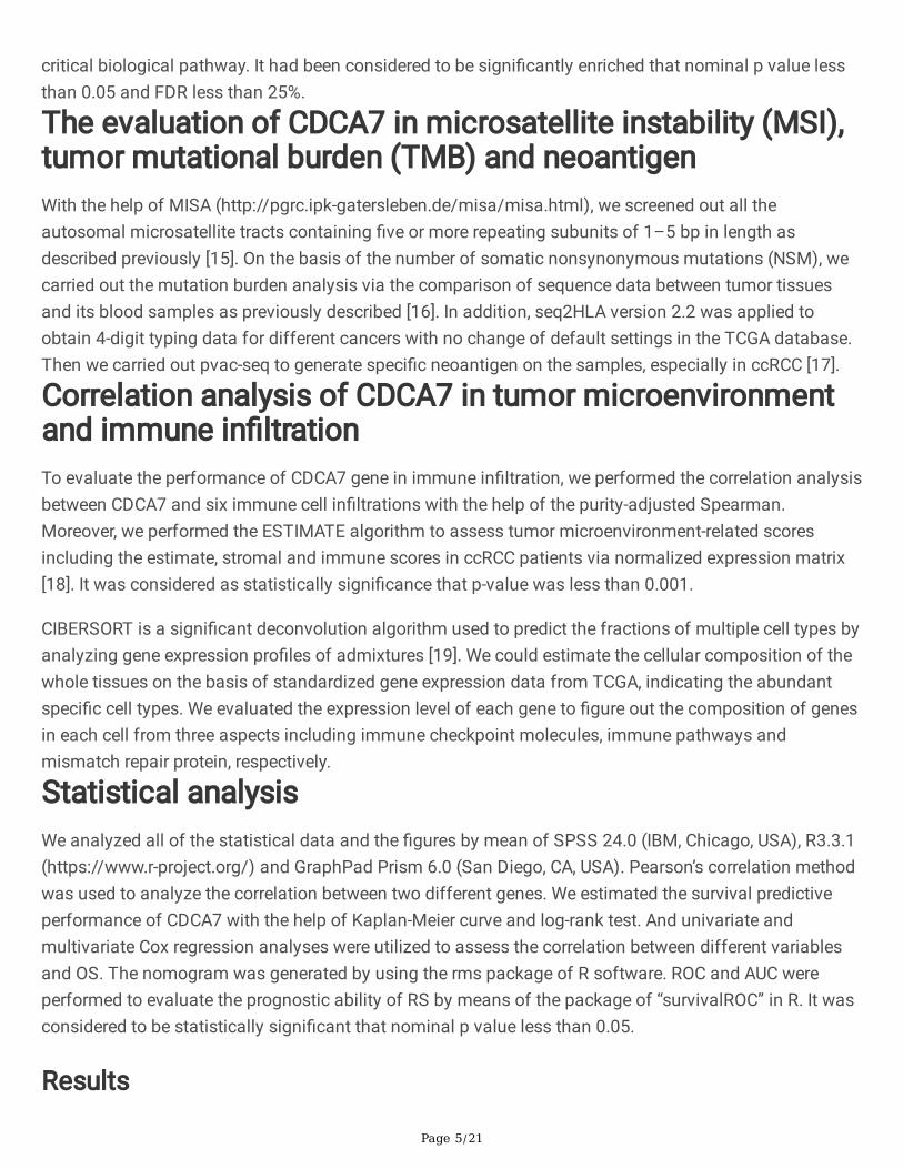
critical biological pathway. It had been considered to be signi�cantly enriched that nominal p value lessthan 0.05 and FDR less than 25%.
The evaluation of CDCA7 in microsatellite instability (MSI),tumor mutational burden (TMB) and neoantigenWith the help of MISA (http://pgrc.ipk-gatersleben.de/misa/misa.html), we screened out all theautosomal microsatellite tracts containing �ve or more repeating subunits of 1–5 bp in length asdescribed previously [15]. On the basis of the number of somatic nonsynonymous mutations (NSM), wecarried out the mutation burden analysis via the comparison of sequence data between tumor tissuesand its blood samples as previously described [16]. In addition, seq2HLA version 2.2 was applied toobtain 4-digit typing data for different cancers with no change of default settings in the TCGA database.Then we carried out pvac-seq to generate speci�c neoantigen on the samples, especially in ccRCC [17].
Correlation analysis of CDCA7 in tumor microenvironmentand immune in�ltrationTo evaluate the performance of CDCA7 gene in immune in�ltration, we performed the correlation analysisbetween CDCA7 and six immune cell in�ltrations with the help of the purity-adjusted Spearman.Moreover, we performed the ESTIMATE algorithm to assess tumor microenvironment-related scoresincluding the estimate, stromal and immune scores in ccRCC patients via normalized expression matrix[18]. It was considered as statistically signi�cance that p-value was less than 0.001.
CIBERSORT is a signi�cant deconvolution algorithm used to predict the fractions of multiple cell types byanalyzing gene expression pro�les of admixtures [19]. We could estimate the cellular composition of thewhole tissues on the basis of standardized gene expression data from TCGA, indicating the abundantspeci�c cell types. We evaluated the expression level of each gene to �gure out the composition of genesin each cell from three aspects including immune checkpoint molecules, immune pathways andmismatch repair protein, respectively.
Statistical analysisWe analyzed all of the statistical data and the �gures by mean of SPSS 24.0 (IBM, Chicago, USA), R3.3.1(https://www.r-project.org/) and GraphPad Prism 6.0 (San Diego, CA, USA). Pearson’s correlation methodwas used to analyze the correlation between two different genes. We estimated the survival predictiveperformance of CDCA7 with the help of Kaplan-Meier curve and log-rank test. And univariate andmultivariate Cox regression analyses were utilized to assess the correlation between different variablesand OS. The nomogram was generated by using the rms package of R software. ROC and AUC wereperformed to evaluate the prognostic ability of RS by means of the package of “survivalROC” in R. It wasconsidered to be statistically signi�cant that nominal p value less than 0.05.
Results
Page 6/21
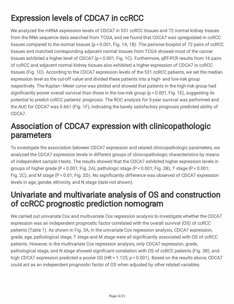
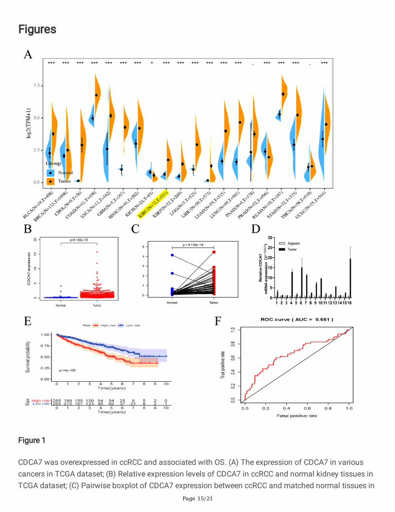
Expression levels of CDCA7 in ccRCCWe analyzed the mRNA expression levels of CDCA7 in 531 ccRCC tissues and 72 normal kidney tissuesfrom the RNA sequence data searched from TCGA, and we found that CDCA7 was upregulated in ccRCCtissues compared to the normal tissues (p < 0.001, Fig. 1A, 1B). The pairwise boxplot of 72 pairs of ccRCCtissues and matched corresponding adjacent normal tissues from TCGA showed most of the cancertissues exhibited a higher level of CDCA7 (p < 0.001, Fig. 1C). Furthermore, qRT-PCR results from 16 pairsof ccRCC and adjacent normal kidney tissues also exhibited a higher expression of CDCA7 in ccRCCtissues (Fig. 1D). According to the CDCA7 expression levels of the 531 ccRCC patients, we set the medianexpression level as the cut-off value and divided these patients into a high- and low-risk grouprespectively. The Kaplan–Meier curve was plotted and showed that patients in the high-risk group hadsigni�cantly poorer overall survival than those in the low-risk group (p < 0.001, Fig. 1E), suggesting itspotential to predict ccRCC patients’ prognosis. The ROC analysis for 5-year survival was performed andthe AUC for CDCA7 was 0.661 (Fig. 1F), indicating the barely satisfactory prognosis predicted ability ofCDCA7.
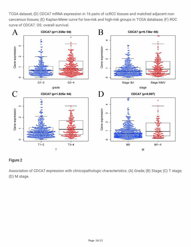
Association of CDCA7 expression with clinicopathologicparametersTo investigate the association between CDCA7 expression and related clinicopathologic parameters, weanalyzed the CDCA7 expression levels in different groups of clinicopathologic characteristics by meansof independent sample t-tests. The results showed that the CDCA7 exhibited higher expression levels ingroups of higher grade (P < 0.001; Fig. 2A), pathologic stage (P < 0.001; Fig. 2B), T stage (P < 0.001;Fig. 2C), and M stage (P < 0.01; Fig. 2D). No signi�cantly difference was observed of CDCA7 expressionlevels in age, gender, ethnicity, and N stage (date not shown).
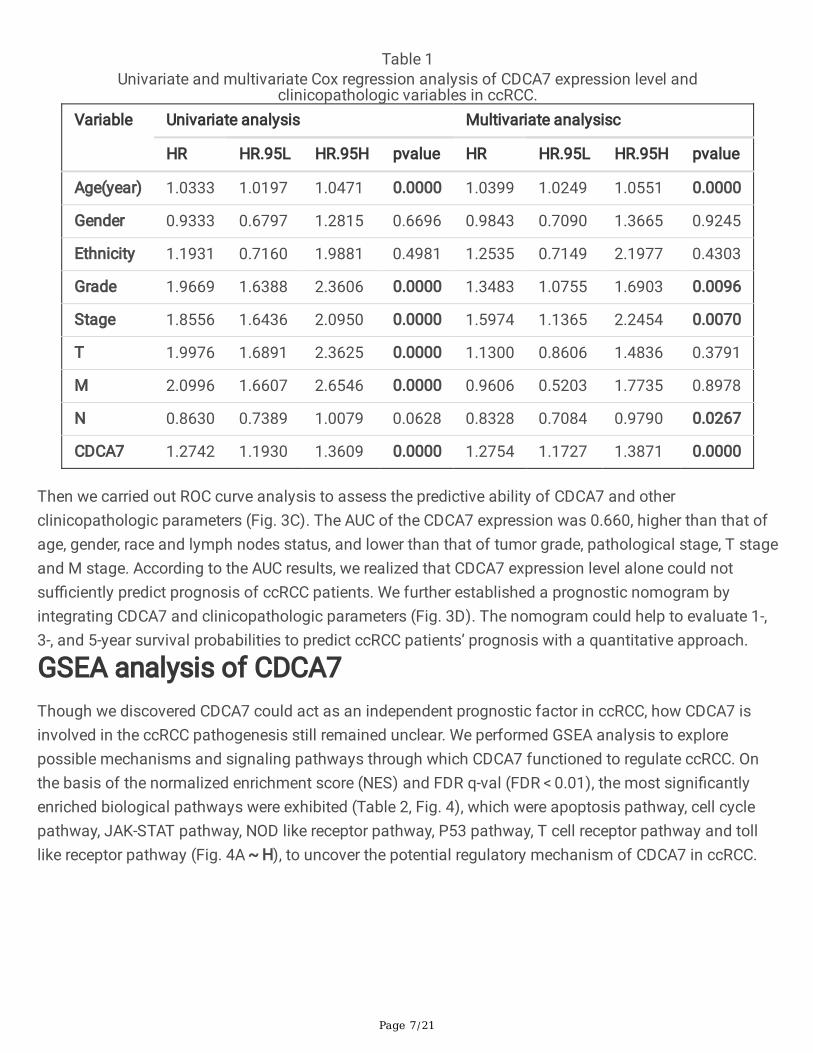
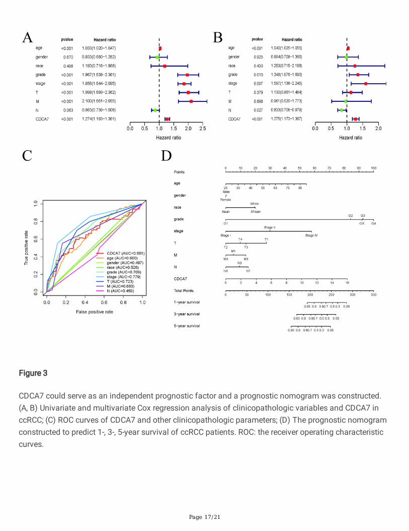
Univariate and multivariate analysis of OS and constructionof ccRCC prognostic prediction nomogramWe carried out univariate Cox and multivariate Cox regression analysis to investigate whether the CDCA7expression was an independent prognostic factor correlated with the overall survival (OS) of ccRCCpatients (Table 1). As shown in Fig. 3A, in the univariate Cox regression analysis, CDCA7 expression,grade, age, pathological stage, T stage and M stage were all signi�cantly associated with OS of ccRCCpatients. However, in the multivariate Cox regression analysis, only CDCA7 expression, grade,pathological stage, and N stage showed signi�cant correlation with OS of ccRCC patients (Fig. 3B), andhigh CDCA7 expression predicted a poorer OS (HR = 1.125; p < 0.001). Based on the results above, CDCA7could act as an independent prognostic factor of OS when adjusted by other related variables.
Page 7/21
Table 1Univariate and multivariate Cox regression analysis of CDCA7 expression level and
clinicopathologic variables in ccRCC.Variable Univariate analysis Multivariate analysisc
HR HR.95L HR.95H pvalue HR HR.95L HR.95H pvalue
Age(year) 1.0333 1.0197 1.0471 0.0000 1.0399 1.0249 1.0551 0.0000
Gender 0.9333 0.6797 1.2815 0.6696 0.9843 0.7090 1.3665 0.9245
Ethnicity 1.1931 0.7160 1.9881 0.4981 1.2535 0.7149 2.1977 0.4303
Grade 1.9669 1.6388 2.3606 0.0000 1.3483 1.0755 1.6903 0.0096
Stage 1.8556 1.6436 2.0950 0.0000 1.5974 1.1365 2.2454 0.0070
T 1.9976 1.6891 2.3625 0.0000 1.1300 0.8606 1.4836 0.3791
M 2.0996 1.6607 2.6546 0.0000 0.9606 0.5203 1.7735 0.8978
N 0.8630 0.7389 1.0079 0.0628 0.8328 0.7084 0.9790 0.0267
CDCA7 1.2742 1.1930 1.3609 0.0000 1.2754 1.1727 1.3871 0.0000
Then we carried out ROC curve analysis to assess the predictive ability of CDCA7 and otherclinicopathologic parameters (Fig. 3C). The AUC of the CDCA7 expression was 0.660, higher than that ofage, gender, race and lymph nodes status, and lower than that of tumor grade, pathological stage, T stageand M stage. According to the AUC results, we realized that CDCA7 expression level alone could notsu�ciently predict prognosis of ccRCC patients. We further established a prognostic nomogram byintegrating CDCA7 and clinicopathologic parameters (Fig. 3D). The nomogram could help to evaluate 1-,3-, and 5-year survival probabilities to predict ccRCC patients’ prognosis with a quantitative approach.
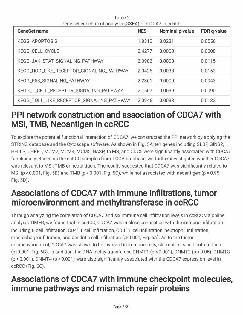
GSEA analysis of CDCA7Though we discovered CDCA7 could act as an independent prognostic factor in ccRCC, how CDCA7 isinvolved in the ccRCC pathogenesis still remained unclear. We performed GSEA analysis to explorepossible mechanisms and signaling pathways through which CDCA7 functioned to regulate ccRCC. Onthe basis of the normalized enrichment score (NES) and FDR q-val (FDR < 0.01), the most signi�cantlyenriched biological pathways were exhibited (Table 2, Fig. 4), which were apoptosis pathway, cell cyclepathway, JAK-STAT pathway, NOD like receptor pathway, P53 pathway, T cell receptor pathway and tolllike receptor pathway (Fig. 4A ~ H), to uncover the potential regulatory mechanism of CDCA7 in ccRCC.
Page 8/21
Table 2Gene set enrichment analysis (GSEA) of CDCA7 in ccRCC.
GeneSet name NES Nominal p-value FDR q-value
KEGG_APOPTOSIS 1.8310 0.0231 0.0556
KEGG_CELL_CYCLE 2.4277 0.0000 0.0008
KEGG_JAK_STAT_SIGNALING_PATHWAY 2.0902 0.0000 0.0115
KEGG_NOD_LIKE_RECEPTOR_SIGNALING_PATHWAY 2.0426 0.0038 0.0153
KEGG_P53_SIGNALING_PATHWAY 2.2361 0.0000 0.0043
KEGG_T_CELL_RECEPTOR_SIGNALING_PATHWAY 2.1507 0.0039 0.0090
KEGG_TOLL_LIKE_RECEPTOR_SIGNALING_PATHWAY 2.0946 0.0038 0.0132
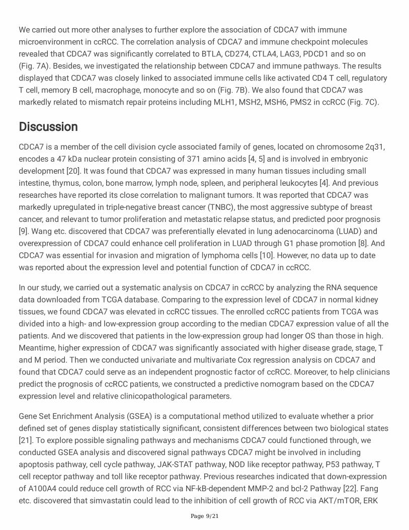
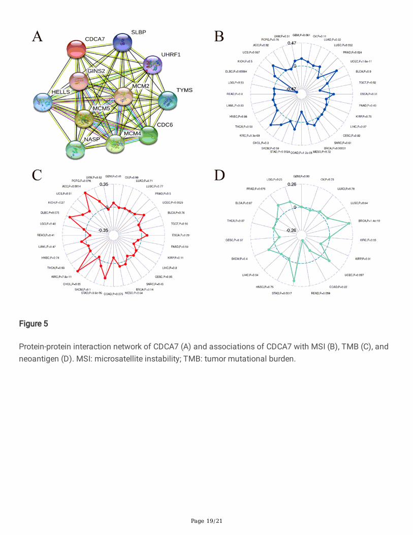
PPI network construction and association of CDCA7 withMSI, TMB, Neoantigen in ccRCCTo explore the potential functional interaction of CDCA7, we constructed the PPI network by applying theSTRING database and the Cytoscape software. As shown in Fig. 5A, ten genes including SLBP, GINS2,HELLS, UHRF1, MCM2, MCM4, MCM5, NASP, TYMS, and CDC6 were signi�cantly associated with CDCA7functionally. Based on the ccRCC samples from TCGA database, we further investigated whether CDCA7was relevant to MSI, TMB or neoantigen. The results suggested that CDCA7 was signi�cantly related toMSI (p < 0.001, Fig. 5B) and TMB (p < 0.001, Fig. 5C), while not associated with neoantigen (p = 0.95,Fig. 5D).
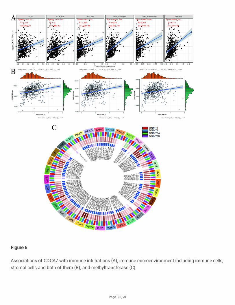
Associations of CDCA7 with immune in�ltrations, tumormicroenvironment and methyltransferase in ccRCCThrough analyzing the correlation of CDCA7 and six immune cell in�ltration levels in ccRCC via onlineanalysis TIMER, we found that in ccRCC, CDCA7 was in close connection with the immune in�ltrationincluding B cell in�ltration, CD4+ T cell in�ltration, CD8+ T cell in�ltration, neutrophil in�ltration,macrophage in�ltration, and dendritic cell in�ltration (p 0.001, Fig. 6A). As to the tumormicroenvironment, CDCA7 was shown to be involved in immune cells, stromal cells and both of them(p 0.001, Fig. 6B). In addition, the DNA methyltransferase DNMT1 (p < 0.001), DNMT2 (p < 0.05), DNMT3(p < 0.001), DNMT4 (p < 0.001) were also signi�cantly associated with the CDCA7 expression level inccRCC (Fig. 6C).
Associations of CDCA7 with immune checkpoint molecules,immune pathways and mismatch repair proteins
Page 9/21
We carried out more other analyses to further explore the association of CDCA7 with immunemicroenvironment in ccRCC. The correlation analysis of CDCA7 and immune checkpoint moleculesrevealed that CDCA7 was signi�cantly correlated to BTLA, CD274, CTLA4, LAG3, PDCD1 and so on(Fig. 7A). Besides, we investigated the relationship between CDCA7 and immune pathways. The resultsdisplayed that CDCA7 was closely linked to associated immune cells like activated CD4 T cell, regulatoryT cell, memory B cell, macrophage, monocyte and so on (Fig. 7B). We also found that CDCA7 wasmarkedly related to mismatch repair proteins including MLH1, MSH2, MSH6, PMS2 in ccRCC (Fig. 7C).
DiscussionCDCA7 is a member of the cell division cycle associated family of genes, located on chromosome 2q31,encodes a 47 kDa nuclear protein consisting of 371 amino acids [4, 5] and is involved in embryonicdevelopment [20]. It was found that CDCA7 was expressed in many human tissues including smallintestine, thymus, colon, bone marrow, lymph node, spleen, and peripheral leukocytes [4]. And previousresearches have reported its close correlation to malignant tumors. It was reported that CDCA7 wasmarkedly upregulated in triple-negative breast cancer (TNBC), the most aggressive subtype of breastcancer, and relevant to tumor proliferation and metastatic relapse status, and predicted poor prognosis[9]. Wang etc. discovered that CDCA7 was preferentially elevated in lung adenocarcinoma (LUAD) andoverexpression of CDCA7 could enhance cell proliferation in LUAD through G1 phase promotion [8]. AndCDCA7 was essential for invasion and migration of lymphoma cells [10]. However, no data up to datewas reported about the expression level and potential function of CDCA7 in ccRCC.
In our study, we carried out a systematic analysis on CDCA7 in ccRCC by analyzing the RNA sequencedata downloaded from TCGA database. Comparing to the expression level of CDCA7 in normal kidneytissues, we found CDCA7 was elevated in ccRCC tissues. The enrolled ccRCC patients from TCGA wasdivided into a high- and low-expression group according to the median CDCA7 expression value of all thepatients. And we discovered that patients in the low-expression group had longer OS than those in high.Meantime, higher expression of CDCA7 was signi�cantly associated with higher disease grade, stage, Tand M period. Then we conducted univariate and multivariate Cox regression analysis on CDCA7 andfound that CDCA7 could serve as an independent prognostic factor of ccRCC. Moreover, to help clinicianspredict the prognosis of ccRCC patients, we constructed a predictive nomogram based on the CDCA7expression level and relative clinicopathological parameters.
Gene Set Enrichment Analysis (GSEA) is a computational method utilized to evaluate whether a priorde�ned set of genes display statistically signi�cant, consistent differences between two biological states[21]. To explore possible signaling pathways and mechanisms CDCA7 could functioned through, weconducted GSEA analysis and discovered signal pathways CDCA7 might be involved in includingapoptosis pathway, cell cycle pathway, JAK-STAT pathway, NOD like receptor pathway, P53 pathway, Tcell receptor pathway and toll like receptor pathway. Previous researches indicated that down-expressionof A100A4 could reduce cell growth of RCC via NF-kB-dependent MMP-2 and bcl-2 Pathway [22]. Fangetc. discovered that simvastatin could lead to the inhibition of cell growth of RCC via AKT/mTOR, ERK
Page 10/21
and JAK2/STAT3 pathway [23]. It was also reported that Tropomyosin-1, a widely expressed actin-bindingprotein, could promotes cancer cell apoptosis via the p53-mediated mitochondrial pathway in ccRCC [24],and the MDM2 inhibitor MI-319 could induce RCC cell apoptosis mainly dependent on p53overexpression [25].
Then we constructed the PPI network of CDCA7 to explore potential protein-protein interaction. It showedthat ten genes were signi�cantly functional associated with CDCA7, including SLBP, GINS2, HELLS,UHRF1, MCM2, MCM4, MCM5, NASP, TYMS, and CDC6. Researchers have discovered the important rolesof these genes in physiological process and tumor progression. For instance, SLBP (Stem-loop-bindingprotein) is evolutionarily conserved and involved in the processing, translation, and degradation ofcanonical histones mRNAs including H1, H2A, H2B, H3, and H4 [26]. With regard to GINS2, it was reportedthat GINS2 could promote cancer cell proliferation, migration and invasion of non-small-cell lung cancer(NSCLC) via facilitating epithelial-to-mesenchymal transition and modulating PI3K/Akt and MEK/ERKsignal pathway [27]. Previous reports revealed that mutations in CDCA7 and HELLS respectively causeimmunode�ciency, centromeric instability, and facial anomalies (ICF) syndrome types 3 and 4, thatencode a CXXC-type zinc �nger protein and an SNF2 family chromatin remodeler [28]. And the ZBTB24-CDCA7 axis could facilitate DNA methylation by regulating HELLS enrichment at centromeric satelliterepeats [29]. As to the minichromosome maintenance (MCM) proteins family, they were mainly involvedin the initiation and elongation of DNA replication [30] and the formation of the pre-replicative complex(preRC), the replication fork and the initial steps of DNA synthesis [31]. MCM2, MCM4, and MCM5 servedas essential components of a hexameric, ring-shaped complex that acted as one of the pre-replicationfactors and were related to the chromatin and the proteins of the origin recognition complex at the M-G1transition [30]. The important roles of these genes in the PPI network of CDCA7 all indicated the possibleessential functions of CDCA7.
We also discovered the association of CDCA7 with immunity. CDCA7 was in close connection with theimmune in�ltration including B cell in�ltration, CD4+ T cell in�ltration, CD8+ T cell in�ltration, neutrophilin�ltration, macrophage in�ltration, and dendritic cell in�ltration. Moreover, CDCA7 was signi�cantlycorrelated to tumor-related immunosuppressive molecules including PDCD1, CD274, CTLA4, BTLA andLAG3. PDCD1 (programmed cell death 1), CTLA-4 (cytotoxic T-lymphocyte-associated antigen 4) andBTLA (B and T lymphocyte attenuator) are members of immunoglobulin-related receptors familyassociated with various aspects of T cell immune regulation [32]. PDCD1 (PD-1, CD279) and CD274 (PD-L1) axis has been discovered as a worthy therapeutic target for its important role not only inphysiological immune homoeostasis, but also in the way through which cancer cells evade the immunesystem [33]. Overexpression of PD-1 and PD-L1 in tumors is closely correlated with poor disease outcomein some human cancers [34]. The research of PD-1 or PD-L1 inhibitors have greatly promoted thedevelopment of the treatment of cancer [35]. As to CTLA4, it is a transmembrane receptor with inhibitoryfunction expressed by T lymphocytes. CTLA4 could inhibit costimulation through competing for CD28ligands [36]. In addition to CTLA4 and PD1, BTLA is also an immune checkpoint related to suppressingimmune responses [37], containing two immunoreceptor tyrosine-based inhibitory motifs in its
Page 11/21
cytoplasmic region. BTLA could modulate T cell responses and attenuate B cell function to paly itsinhibitory roles in multiple diseases [38, 39]. Lymphocyte Activation Gene-3 (LAG3, CD223) served asanother potential cancer immunotherapeutic target. It could inhibit T cells function and mediate a state ofexhaustion in combination with PD1 [40]. The association of CDCA7 with these tumor-relatedimmunosuppressive molecules suggested its important potential functions in human immunity.
To conclude, our study elucidated that CDCA7 could act as a favorable prognostic factor for ccRCC.Moreover, apoptosis pathway, cell cycle pathway, JAK-STAT pathway, NOD like receptor pathway, P53pathway, T cell receptor pathway and toll like receptor pathway might be the primary pathways regulatedby CDCA7. Furthermore, CDCA7 could serve as an independent prognostic factor for ccRCC and it wasclosely related to immunity. And advanced researches were required to verify our �ndings in vivo and invitro.
DeclarationsFunding
This study was supported by the National Natural Science Foundation of China (81771640).
Author's Contribution
ZJ.W: Conceptualization, Methodology, Project administration;
QW.X: Data collection and management, Formal analysis;
CK.M: Formal analysis, Writing - Review & Editing;
SY.L, Y.W: Software, Writing - Original Draft;
Con�icts of Interest
The authors con�rm that there are no con�icts of interest.
Consent for publication
Not applicable.
Ethics approval and consent to participate
Not applicable.
Data Availability Statement
The RNA-sequencing data and corresponding clinical information were downloaded from the
Cancer Genome Atlas (TCGA) database (https://portal.gdc.cancer.gov/).
Page 12/21
Acknowledgements
We would like to thank the researchers and study participants for their contributions.
References1. Siegel RL, Miller KD, Jemal A: Cancer statistics, 2020. CA Cancer J Clin 2020, 70(1):7-30.
2. Moch H, Cubilla AL, Humphrey PA, Reuter VE, Ulbright TM: The 2016 WHO Classi�cation of Tumoursof the Urinary System and Male Genital Organs-Part A: Renal, Penile, and Testicular Tumours.European urology 2016, 70(1):93-105.
3. Ferlay J, Colombet M, Soerjomataram I, Mathers C, Parkin DM, Piñeros M, Znaor A, Bray F:Estimating the global cancer incidence and mortality in 2018: GLOBOCAN sources and methods. IntJ Cancer 2019, 144(8):1941-1953.
4. Prescott JE, Osthus RC, Lee LA, Lewis BC, Shim H, Barrett JF, Guo Q, Hawkins AL, Gri�n CA, Dang CV:A novel c-Myc-responsive gene, JPO1, participates in neoplastic transformation. The Journal ofbiological chemistry 2001, 276(51):48276-48284.
5. Osthus RC, Karim B, Prescott JE, Smith BD, McDevitt M, Huso DL, Dang CV: The Myc target geneJPO1/CDCA7 is frequently overexpressed in human tumors and has limited transforming activity invivo. Cancer research 2005, 65(13):5620-5627.
�. Lewis BC, Shim H, Li Q, Wu CS, Lee LA, Maity A, Dang CV: Identi�cation of putative c-Myc-responsivegenes: characterization of rcl, a novel growth-related gene. Molecular and cellular biology 1997,17(9):4967-4978.
7. Whit�eld ML, Sherlock G, Saldanha AJ, Murray JI, Ball CA, Alexander KE, Matese JC, Perou CM, HurtMM, Brown PO et al: Identi�cation of genes periodically expressed in the human cell cycle and theirexpression in tumors. Molecular biology of the cell 2002, 13(6):1977-2000.
�. Wang H, Ye L, Xing Z, Li H, Lv T, Liu H, Zhang F, Song Y: CDCA7 promotes lung adenocarcinomaproliferation via regulating the cell cycle. Pathology, research and practice 2019, 215(11):152559.
9. Ye L, Li F, Song Y, Yu D, Xiong Z, Li Y, Shi T, Yuan Z, Lin C, Wu X et al: Overexpression of CDCA7predicts poor prognosis and induces EZH2-mediated progression of triple-negative breast cancer.International journal of cancer 2018, 143(10):2602-2613.
10. Martín-Cortázar C, Chiodo Y, Jiménez RP, Bernabé M, Cayuela ML, Iglesias T, Campanero MR: CDCA7�nely tunes cytoskeleton dynamics to promote lymphoma migration and invasion. Haematologica2020, 105(3):730-740.
11. Goto Y, Hayashi R, Muramatsu T, Ogawa H, Eguchi I, Oshida Y, Ohtani K, Yoshida K: JPO1/CDCA7, anovel transcription factor E2F1-induced protein, possesses intrinsic transcriptional regulator activity.Biochimica et biophysica acta 2006, 1759(1-2):60-68.
12. Gill RM, Gabor TV, Couzens AL, Scheid MP: The MYC-associated protein CDCA7 is phosphorylated byAKT to regulate MYC-dependent apoptosis and transformation. Molecular and cellular biology 2013,33(3):498-513.
Page 13/21
13. Li D, Jiang X, Zhang X, Cao G, Wang D, Chen Z: Long noncoding RNA FGD5-AS1 promotes colorectalcancer cell proliferation, migration, and invasion through upregulating CDCA7 via sponging miR-302e. In vitro cellular & developmental biology Animal 2019, 55(8):577-585.
14. Subramanian A, Tamayo P, Mootha VK, Mukherjee S, Ebert BL, Gillette MA, Paulovich A, Pomeroy SL,Golub TR, Lander ES et al: Gene set enrichment analysis: a knowledge-based approach forinterpreting genome-wide expression pro�les. Proceedings of the National Academy of Sciences ofthe United States of America 2005, 102(43):15545-15550.
15. Timmermann B, Kerick M, Roehr C, Fischer A, Isau M, Boerno ST, Wunderlich A, Barmeyer C, SeemannP, Koenig J et al: Somatic mutation pro�les of MSI and MSS colorectal cancer identi�ed by wholeexome next generation sequencing and bioinformatics analysis. PLoS One 2010, 5(12):e15661.
1�. Chalmers ZR, Connelly CF, Fabrizio D, Gay L, Ali SM, Ennis R, Schrock A, Campbell B, Shlien A,Chmielecki J et al: Analysis of 100,000 human cancer genomes reveals the landscape of tumormutational burden. Genome Med 2017, 9(1):34.
17. Hundal J, Carreno BM, Petti AA, Linette GP, Gri�th OL, Mardis ER, Gri�th M: pVAC-Seq: A genome-guided in silico approach to identifying tumor neoantigens. Genome Med 2016, 8(1):11.
1�. Yoshihara K, Shahmoradgoli M, Martínez E, Vegesna R, Kim H, Torres-Garcia W, Treviño V, Shen H,Laird PW, Levine DA et al: Inferring tumour purity and stromal and immune cell admixture fromexpression data. Nat Commun 2013, 4:2612.
19. Newman AM, Liu CL, Green MR, Gentles AJ, Feng W, Xu Y, Hoang CD, Diehn M, Alizadeh AA: Robustenumeration of cell subsets from tissue expression pro�les. Nat Methods 2015, 12(5):453-457.
20. Guiu J, Bergen DJ, De Pater E, Islam AB, Ayllón V, Gama-Norton L, Ruiz-Herguido C, González J,López-Bigas N, Menendez P et al: Identi�cation of Cdca7 as a novel Notch transcriptional targetinvolved in hematopoietic stem cell emergence. J Exp Med 2014, 211(12):2411-2423.
21. Subramanian A, Kuehn H, Gould J, Tamayo P, Mesirov JP: GSEA-P: a desktop application for GeneSet Enrichment Analysis. Bioinformatics 2007, 23(23):3251-3253.
22. Yang XC, Wang X, Luo L, Dong DH, Yu QC, Wang XS, Zhao K: RNA interference suppression ofA100A4 reduces the growth and metastatic phenotype of human renal cancer cells via NF-kB-dependent MMP-2 and bcl-2 pathway. Eur Rev Med Pharmacol Sci 2013, 17(12):1669-1680.
23. Fang Z, Tang Y, Fang J, Zhou Z, Xing Z, Guo Z, Guo X, Wang W, Jiao W, Xu Z et al: Simvastatininhibits renal cancer cell growth and metastasis via AKT/mTOR, ERK and JAK2/STAT3 pathway.PLoS One 2013, 8(5):e62823.
24. Tang C, Wang J, Wei Q, Du YP, Qiu HP, Yang C, Hou YC: Tropomyosin-1 promotes cancer cellapoptosis via the p53-mediated mitochondrial pathway in renal cell carcinoma. Oncol Lett 2018,15(5):7060-7068.
25. Liu QJ, Shen HL, Lin J, Xu XH, Ji ZG, Han X, Shang DH, Yang PQ: Synergistic roles of p53 and HIF1αin human renal cell carcinoma-cell apoptosis responding to the inhibition of mTOR and MDM2signaling pathways. Drug Des Devel Ther 2016, 10:745-755.
Page 14/21
2�. Marzluff WF, Wagner EJ, Duronio RJ: Metabolism and regulation of canonical histone mRNAs: lifewithout a poly(A) tail. Nat Rev Genet 2008, 9(11):843-854.
27. Liu X, Sun L, Zhang S, Zhang S, Li W: GINS2 facilitates epithelial-to-mesenchymal transition in non-small-cell lung cancer through modulating PI3K/Akt and MEK/ERK signaling. J Cell Physiol 2019.
2�. Thijssen PE, Ito Y, Grillo G, Wang J, Velasco G, Nitta H, Unoki M, Yoshihara M, Suyama M, Sun Y et al:Mutations in CDCA7 and HELLS cause immunode�ciency-centromeric instability-facial anomaliessyndrome. Nat Commun 2015, 6:7870.
29. Hardikar S, Ying Z, Zeng Y, Zhao H, Liu B, Veland N, McBride K, Cheng X, Chen T: The ZBTB24-CDCA7axis regulates HELLS enrichment at centromeric satellite repeats to facilitate DNA methylation.Protein Cell 2020, 11(3):214-218.
30. Dubois ML, Bastin C, Lévesque D, Boisvert FM: Comprehensive Characterization of MinichromosomeMaintenance Complex (MCM) Protein Interactions Using A�nity and Proximity Puri�cations Coupledto Mass Spectrometry. J Proteome Res 2016, 15(9):2924-2934.
31. Giaginis C, Vgenopoulou S, Vielh P, Theocharis S: MCM proteins as diagnostic and prognostic tumormarkers in the clinical setting. Histol Histopathol 2010, 25(3):351-370.
32. Rowshanravan B, Halliday N, Sansom DM: CTLA-4: a moving target in immunotherapy. Blood 2018,131(1):58-67.
33. Sun C, Mezzadra R, Schumacher TN: Regulation and Function of the PD-L1 Checkpoint. Immunity2018, 48(3):434-452.
34. Ohaegbulam KC, Assal A, Lazar-Molnar E, Yao Y, Zang X: Human cancer immunotherapy withantibodies to the PD-1 and PD-L1 pathway. Trends Mol Med 2015, 21(1):24-33.
35. Ribas A, Wolchok JD: Cancer immunotherapy using checkpoint blockade. Science 2018,359(6382):1350-1355.
3�. Khailaie S, Rowshanravan B, Robert PA, Waters E, Halliday N, Badillo Herrera JD, Walker LSK,Sansom DM, Meyer-Hermann M: Characterization of CTLA4 Tra�cking and Implications for ItsFunction. Biophys J 2018, 115(7):1330-1343.
37. Pardoll DM: The blockade of immune checkpoints in cancer immunotherapy. Nat Rev Cancer 2012,12(4):252-264.
3�. Watanabe N, Gavrieli M, Sedy JR, Yang J, Fallarino F, Loftin SK, Hurchla MA, Zimmerman N, Sim J,Zang X et al: BTLA is a lymphocyte inhibitory receptor with similarities to CTLA-4 and PD-1. NatImmunol 2003, 4(7):670-679.
39. Iwata A, Watanabe N, Oya Y, Owada T, Ikeda K, Suto A, Kagami S, Hirose K, Kanari H, Kawashima S etal: Protective roles of B and T lymphocyte attenuator in NKT cell-mediated experimental hepatitis. JImmunol 2010, 184(1):127-133.
40. Turnis ME, Andrews LP, Vignali DA: Inhibitory receptors as targets for cancer immunotherapy. Eur JImmunol 2015, 45(7):1892-1905.
Page 15/21
Figures
Figure 1
CDCA7 was overexpressed in ccRCC and associated with OS. (A) The expression of CDCA7 in variouscancers in TCGA dataset; (B) Relative expression levels of CDCA7 in ccRCC and normal kidney tissues inTCGA dataset; (C) Pairwise boxplot of CDCA7 expression between ccRCC and matched normal tissues in
Page 16/21
TCGA dataset; (D) CDCA7 mRNA expression in 16 pairs of ccRCC tissues and matched adjacent non-cancerous tissues; (E) Kaplan-Meier curve for low-risk and high-risk groups in TCGA database; (F) ROCcurve of CDCA7. OS: overall survival.
Figure 2
Association of CDCA7 expression with clinicopathologic characteristics. (A) Grade; (B) Stage; (C) T stage;(D) M stage.
Page 17/21
Figure 3
CDCA7 could serve as an independent prognostic factor and a prognostic nomogram was constructed.(A, B) Univariate and multivariate Cox regression analysis of clinicopathologic variables and CDCA7 inccRCC; (C) ROC curves of CDCA7 and other clinicopathologic parameters; (D) The prognostic nomogramconstructed to predict 1-, 3-, 5-year survival of ccRCC patients. ROC: the receiver operating characteristiccurves.

Page 18/21
Figure 4
Enrichment plots from gene set enrichment analysis (GSEA). (A) Apoptosis pathway; (B) Cell cyclepathway; (C) JAK-STAT pathway; (D) NOD like receptor pathway; (E) P53 pathway; (F) T cell receptorpathway; (G) Toll like receptor pathway; (H) The seven most signi�cantly enriched signaling pathwaysbased on their normalized enrichment score and the expression map.
Page 19/21
Figure 5
Protein-protein interaction network of CDCA7 (A) and associations of CDCA7 with MSI (B), TMB (C), andneoantigen (D). MSI: microsatellite instability; TMB: tumor mutational burden.
Page 20/21
Figure 6
Associations of CDCA7 with immune in�ltrations (A), immune microenvironment including immune cells,stromal cells and both of them (B), and methyltransferase (C).

Page 21/21
Figure 7
Associations of CDCA7 with immune checkpoint molecules (A), immune cells (B), and mismatch repairproteins (C).