ASSOCIATIONS BETWEEN DENTAL CONSONANT...
58
ASSOCIATIONS BETWEEN DENTAL CONSONANT ARTICULATION, OROFACIAL MORPHOLOGY AND FUNCTION IN CLEFT LIP/PALATE JAANA LAITINEN HELSINKI 1999
Transcript of ASSOCIATIONS BETWEEN DENTAL CONSONANT...

ASSOCIATIONS BETWEEN DENTAL CONSONANT ARTICULATION,
OROFACIAL MORPHOLOGY AND FUNCTION IN CLEFT LIP/PALATE
JAANA LAITINEN
HELSINKI 1999

2
From the Cleft Center, Department of Plastic
Surgery, Helsinki University Central Hospital
and
Department of Pedodontics and Orthodontics,
Institute of Dentistry, University of Helsinki
ASSOCIATIONS BETWEEN DENTAL CONSONANT ARTICULATION,
OROFACIAL MORPHOLOGY AND FUNCTION IN CLEFT LIP/PALATE
Jaana Laitinen
Academic dissertation
to be publicly discussed with the assent of the Faculty of Medicine of the University of
Helsinki for public examination in Auditorium XII of the University Main Building,
on 3 December, 1999, at 12 noon.
HELSINKI 1999

3
Supervised by
Docent Reijo Ranta, DDS, PhD
Docent Marja-Leena Haapanen, MD, PhD
Cleft Center, Department of Plastic Surgery,
Helsinki University Central Hospital
Reviewed by
Docent Pirjo Korpilahti, PhD
Department of Phonetics,
University of Helsinki
and
Professor Juha Varrela, DDS, PhD
Department of Oral Development and Orthodontics,
Institute of Dentistry, University of Turku
ISBN 951-45-8751-0 (PDF version)
Helsingin yliopiston verkkojulkaisut
Helsinki 1999

4
CONTENTS
1. LIST OF ORIGINAL PUBLICATIONS.................................................................................6
2. ABBREVIATIONS.................................................................................................................7
3. SUMMARY............................................................................................................................8
4. INTRODUCTION AND REVIEW OF THE LITERATURE ................................................9
4.1. Introduction..........................................................................................................................9
4.2. Review of the literature......................................................................................................10
4.2.1. General aspects of speech and articulation in subjects with cleft palate.........................10
4.2.2. Articulation of the Finnish /r/, /s/ and /l/ consonants......................................................12
4.2.3. Prevalence of articulatory errors in /r/, /s/ and /l/ sounds in Finnish pre-school andschoolchildren ...........................................................................................................................13
4.2.4. Articulatory development in children aged 6 to 8 years .................................................13
4.2.5. Dental arch dimensions and misarticulations..................................................................14
4.2.6. Occlusion and misarticulations .......................................................................................15
5. AIMS OF THE STUDY .......................................................................................................17
6. SUBJECTS AND METHODS .............................................................................................18
6.1. Subjects ..............................................................................................................................18
6.2. Methods..............................................................................................................................20
6.2.1. Speech analysis (I-VI) .....................................................................................................20
6.2.2. Measurements of speech and breathing aerodynamics, oralsomatosensoric sensibility, and strength of the tongue muscles (V).........................................21
6.2.3. Dental arch measurements (III).......................................................................................22
5
6.2.4. Analysis of occlusion and definition of spacing of the maxillary dental arch(II,IV) ........................................................................................................................................22
6.2.5. Lateral cephalometric measurements (VI) ......................................................................23
6.2.6. Statistical procedures ......................................................................................................25
6.2.7. Reliability of the methods ...............................................................................................25
7. RESULTS .............................................................................................................................27
7.1. Occurrence of misarticulations at the age of 6 years and changes in articulationbetween 6 to 8 years of age (I,II)...............................................................................................27
7.2. Dental arch dimensions, occlusion and misarticulations at the age of 6years (III,IV)..............................................................................................................................30
7.3. Correlations between speech and breathing aerodynamics, oralsomatosensoric sensibility, and strength of the tongue muscles, andskeletal/pharyngeal morphology and the Finnish /r/ sound (V,VI)...........................................33
8. DISCUSSION .......................................................................................................................35
8.1. Subjects and methods.........................................................................................................35
8.2. Findings..............................................................................................................................36
8.2.1. Articulatory abilities in cleft-children.............................................................................36
8.2.2. Dentofacial morphology and articulation........................................................................40
8.2.3. /R/ distortion, speech aerodynamics and pharyngeal morphology..................................42
9. CONCLUSIONS...................................................................................................................45
10. ACKNOWLEDGEMENTS................................................................................................46
11. REFERENCES ...................................................................................................................49
12. ORIGINAL PUBLICATIONS

6
1. LIST OF ORIGINAL PUBLICATIONS
The thesis is based on following original articles, which are referred to in the text by
their Roman numerals:
I Laitinen J, Haapanen M-L, Paaso M, Pulkkinen J, Heliövaara A, Ranta R. Occurrence
of dental consonant misarticulations in different cleft types. Folia Phoniatr Logop
1998;50:92-100.
II Laitinen J, Ranta R, Pulkkinen J, Paaso M, Haapanen M-L. Changes in Finnish dental
consonant articulation in cleft lip/palate children between 6 and 8 years of age. Folia
Phoniatr Logop; (in press).
III Laitinen J, Ranta R, Pulkkinen J, Haapanen M-L. The association between dental
arch dimensions and occurrence of Finnish dental consonant misarticulations in cleft
lip/palate children. Acta Odontol Scand 1998;56:308-312.
IV Laitinen J, Ranta R, Pulkkinen J, Haapanen M-L. Associations between dental
occlusion and misarticulations of Finnish dental consonants in cleft lip/palate children.
Eur J Oral Sci 1999;107:109-113.
V Laitinen J, Sarmas R, Haapanen M-L, Ranta R. Speech and breathing aerodynamics,
oral somatosensoric sensibility, and strength of tongue muscles correlated with
articulation of the Finnish /r/-sound in subjects with and without oral clefts. Med Sci
Res 1998;26:813-815.
VI Laitinen J, Hurmerinta K, Ranta R, Sarmas R, Haapanen M-L. Comparisons of
speech aerodynamics and lateral craniofacial cephalometrics in cleft lip and palate
patients with and without Finnish /r/ sound distortion. Folia Phoniatr Logop; (in press).

7
2. ABBREVIATIONS
CP isolated cleft palate
CL(A) cleft lip with or without cleft alveolus
UCLP unilateral cleft lip and palate
BCLP bilateral cleft lip and palate
NONC non-cleft
LARE laryngeal resistance
NASA the smallest nasal crossectional area
NASAR nasal resistance
VEPA velopharyngeal orifice area

8
3. SUMMARY
The occurrence of articulatory problems of /r/, /s/ and /l/ sounds as well as their relations
to oral and pharyngeal morphology were studied in subjects with different cleft types.
Altogether 44% of 6-year-old cleft children had studied misarticulations and the
number of errors increased with the severity of the cleft. Boys outnumbered girls in the
occurrence of misarticulations of all the studied sounds. Problems with the /r/ sound
predominated, and one-fourth needed treatment mostly because of severe /r/ distortions.
The frequency of /l/ distortions was surprisingly high.
By the age of 8 years four-fifths still misarticulated at least one of the studied
sounds. There were no differences due to gender according to unchanged or changed
articulation by the age of 8 years. Nearly one third of the /l/ and /s/ misarticulations but
only one fifth of the /r/ errors were corrected. New misarticulations appeared similarly
whether speech therapy was given or not.
Shorter and especially narrower maxillary dental arches and shallower palates as
well as posterior crossbites were related to misarticulations in all the studied sounds.
Mandibular dental arch dimensions, anterior crossbite alone, large maxillary overjet, or
deep bite were not related to the studied misarticulations.
/R/ distortion was related to low laryngeal resistance, and anteriorly positioned
hyoid bone but not to the smallest nasal cross-sectional area, nasal resistance or
velopharyngeal orifice area or dentofacial morphology.

9
4. INTRODUCTION AND REVIEW OF THE LITERATURE
4.1. Introduction
Clefts of the lip and/or palate are the most prevalent group of congenital malformations
affecting morphology and growth of the cranium and middle face. The clefting
incidence is approximately 1 per 500 live births with variation in cleft type, sex, and
ethnic origin. The cleft lip with or without cleft palate and the isolated cleft palate
deviate both epidemiologically and etiologically. The etiology of clefting seems to be
multifactorial, including hereditary and environmental factors (specific drugs, maternal
smoking).
The articulation skills of cleft children are reported to be clearly inferior and mature
later than those of non-cleft children (McWilliams and Musgrave, 1977; Karling et al.,
1993). Improvement of articulation skills of cleft individuals continues past 8 years of
age (Van Demark et al., 1979; Riski and Delong, 1984) when most non-cleft children
have already achieved mature articulation. Especially errors in dental consonants /r/, /s/
and /l/ are frequent for cleft palate speakers (Philips and Harrison, 1969; McWilliams
and Musgrave, 1977; Van Demark et al., 1979). Altogether, articulatory speech
disorders are very common among children in general, and are more common in boys
than in girls, probably due to a slower development of articulatory skills in boys.
More than half of the children with clefts are estimated to need treatment because of
severe articulatory errors (Ross and Johnston, 1972). About 10% of Finnish non-cleft
schoolchildren are considered to need speech therapy (Pietarinen, 1987). Van Riper
(1978) maintained that since articulatory speech disorders represent 50-60% of all
speech disorders they are relatively important in terms of channelling resources of
speech therapy. In Finland early speech therapy rests with the community health service
system, and often several speech therapy sessions over several years are needed to
rehabilitate the speech especially in the case of cleft palate individual. This is costly for
society, and at first, it is often hard for a child and his/her parents.

10
Especially /r/ disorders are reported to be fairly resistant to early speech therapy in
Finland (Qvarnström et al., 1991; Luotonen, 1995). Despite speech therapy as many as
one-third of young Finnish adults still have articulatory disorders, one-fifth of them
misarticulating severe enough to need further treatment (Laine, 1987). It is essential to
find out the conditions, which improve articulation as much as possible in order to use
resources better and time treatment optimally.
4.2. Review of the literature
4.2.1. General aspects of speech and articulation in subjects with cleft palate
Motor speech functions can be divided into four events: breathing, phonation,
resonance, and articulation. Besides supplying the oxygen needs for the circulatory
system, breathing produces the airflow needed for the voice and sound production.
Various muscles control the expiratory phase of breathing during speaking. Phonation is
accomplished by controlling the airflow by moving the vocal folds in the larynx, which
makes the air pressure above the larynx fluctuate. When the fluctuating air passes
through the vocal tract, it resonates in the oro-nasal cavities and is heard as the voice of
speech (Darley et al., 1975; Hardcastle, 1976). The larynx itself is responsible for
separating voiced and unvoiced sounds (Broad, 1973), and regulating the airflow during
breathing and speech (Zajac, 1995). During speech different vocal tract actions are
sequenced to produce linguistically relevant sounds (Gracco, 1991). To sum up briefly,
whilst voice is generated by the larynx, it is shaped by the pharynx, the mouth and the
nose (Wyatt et al., 1996). Speech sound production is a rapid motor act requiring finely
tuned neurological regulation of the central nervous system (Darley et al., 1975).
Articulation disorders may result from organic, neuromuscular or functional faults (Van
Dyke et al., 1984) as well as from sensory abnormalities or disturbances of language
development, or other developmental factors (Van Riper and Emerick, 1984). Organic
disorders may be found in the speech articulators or in the articulating and resonating as
well as in orofacial skeletal structures, and even in dental occlusion. The function of the
tongue is the most essential compared to other articulators, since most speech sounds are

11
produced by altering the shape of the tongue. Neurogenic articulatory disorders result
from weakness, slowness or incoordination in muscular control due to damage of the
central or peripheral nervous system or both. Disabilities without any obvious organic or
neurogenic cause constitute 75% of all articulatory disorders and form a group of
functional disorders (McReynolds and Elbert, 1982; Van Dyke et al., 1984).
Clefts in the lip and/or palate affect the structures and growth of the orofacial area.
Repetitive surgical procedures bring their own hazards by inhibiting the growth, and
reducing muscular strength in the middle face (Folkins, 1985; Enemark et al., 1990;
Wada et al., 1997). Since the severity of articulation deficit has been found to increase
with increasing severity of clefting (Ross and Johnston, 1972; Riski and Delong, 1984),
abnormal dentofacial morphology obviously is partly responsible for a higher risk for
articulatory problems in cleft affected individuals. Many cleft palate children have
articulatory errors associated with velopharyngeal incompetence (Haapanen and
Rantala, 1992) or other conditions that result from cleft palate. While the soft palate as
well as the pharyngeal wall regulates the air flow and the laryngeal voice through the
oral and nasal airways, an adequate velopharyngeal sphincter provides a barrier to the
nasal cavity so that the voice is shaped by the mouth and oropharynx. Excess resonance
in the nasal cavity results in hypernasality. Velopharyngeal incompetence indicates a
lack of separation between the oral and nasal cavities resulting, for example, from a
short palate or an unusually deep nasopharynx (Wyatt et al., 1996). The production of
Finnish /r/ and /l/ sounds, however, do not require high intraoral air pressures, and that
is why disorders of these sounds are difficult to explain only in terms of velopharyngeal
incompetence.
Dento-alveolar dysmorphology due to cleft lip and/or palate is associated with atypical
cephalometric dimensions and abnormalities in vocal tract anatomy (Horowitz et al.,
1976; Smahel et al., 1991; Hoenig and Schoener, 1992). Abnormalities in vocal tract
probably result in abnormal functions and physiology during breathing and phonation
(Ruscello et al., 1985; Scaf et al., 1991). Many cleft palate children attempt to regulate
intraoral pressure by increasing the airflow rate (Warren et al., 1969a; 1985; Warren,
1986). Maxillary growth deficits due to palatal and velopharyngeal surgery may
constrict the nasal floor, reduce airway size, and increase airway resistance (Warren et

12
al., 1969b; Smahel et al., 1991). The size of the pharynx itself has been concluded to be
more important fact affecting speech than timing and the method of surgery in cleft
children (Shprintzen and Goldberg, 1995). The production of the Finnish /r/ sound
requires modulation of phonatory air so that the physical properties of the tremulating
air stream associated with the effect of Bernoull is perceived. Thus, it is important to
find out dysfunctions as well as abnormal morphology in pharyngolaryngeal region,
which may be possible causes for inadequate articulation. Not all speech disorders
present in cleft palate speakers are, however, directly related to the cleft (Wyatt et al.,
1996). For this reason, ”unintelligible speech” requires detailed assessment to establish
whether errors are predominantly cleft or non-cleft related or, as often occurs, a
combination of both.
4.2.2. Articulation of the Finnish /r/, /s/ and /l/ consonants
Finnish /r/ and /s/ sounds are regarded as the most difficult sounds to produce in the
Finnish language. These sounds together with the /l/ sound are dental consonants which
are correctly produced by the help of the tip of the tongue with the anterior palatal
alveolus. The Finnish /r/ is a voiced linguomedioalveolar tremulant, which between two
vowels can be a one-tap trill, and after the /s/ sound, a fricative (Wiik, 1981).
Maddieson (1984) described the Finnish /r/ as a voiced or long voiced dental trill, and
Karlsson (1982) as a dental tremulant. This sound differs from that of most other
languages. In Finnish there is only one sibilant, /s/ phoneme, which is an unvoiced
dental sibilant (Wiik, 1981) produced by the tip of the tongue forming a narrow
constriction against the medioalveolar area. The Finnish /l/ is a voiced dental lateral
sound (Karlsson, 1982). Comparison of misarticulations between different languages is
very difficult because production of the sounds deviate in most languages. However, the
Finnish /r/ and /s/ are probably more difficult to produce than the /r/ and /s/ in Germanic
languages (Arnold, 1965).

13
4.2.3. Prevalence of articulatory errors in /r/, /s/ and /l/ sounds in Finnish pre-school and
schoolchildren
Finnish children are estimated to produce correctly all sounds of their mother language
at the age of 5 years (Sarmavuori, 1982). However, articulatory problems especially in
/r/ and /s/ sounds, are very common in 7 to 8 years old Finnish non-cleft children.
Nearly 10% have /r/ disorders, and 30% /s/ disorders of various degrees (Sonninen and
Sonninen, 1976; Qvarnström et al., 1991). Altogether /r/ and /s/ sounds make up 95% of
all errors at the age of 7 years (Luotonen, 1995). Fortunately, only 1% of non-cleft
schoolchildren have /l/ disorders (Qvarnström et al., 1991). Misarticulations are mainly
distortions among children older than 5 years, substitutions occurring only in 1% of the
schoolchildren (Qvarnström et al., 1991).
Boys are reported to have more often problems with articulation than girls (Qvarnström
et al., 1991; Luotonen, 1995). Slower speech development and fine motor maturation in
boys are suggested to explain the difference in articulatory abilities between gender in
pre-school and the first school years (Stevenson and Richman, 1976; Silva, 1980;
Beitchman et al., 1986). Boys have more /r/ disorders than girls, while /s/ disorders
seem to be more common among girls (Qvarnström et al., 1991).
4.2.4. Articulatory development in children aged 6 to 8 years
Several authors have reported the frequency of articulatory speech disorders decreasing
spontaneously with age (Milisen, 1971; Alstam-Malcus et al., 1976; Leske, 1981). In
Finnish non-cleft children /s/ sound disorders seem spontaneously to decrease at the age
of 7 years, whereas later, between 7 to 10 years of age /s/ disorders increase markedly
even in subjects receiving speech therapy or special education (Qvarnström et al., 1991;
1993a). Instead /r/ disorders do not disappear and seem to be rather resistant to early
speech therapy (Qvarnström et al., 1991). In the case of the non-cleft children the results
of the effectiveness of early speech therapy have not altogether been obvious according

14
to earlier studies (Qvarnström et al., 1991; Luotonen, 1995). While articulatory speech
disorders in 6-8-year old children have been stated to be associated with inaccurate
tongue movements (Speirs and Maktabi, 1990), and problems in coordinating those
movements (Yoss and Darley, 1974; Ferry et al., 1975; Cermak et al., 1986), tongue
skills in normally developing children are known to improve markedly with advancing
age up to the age of 11 years (Tingley and Allen, 1975; Watkin and Fromm, 1984;
Sharkey and Folkins, 1985; Robbins and Klee, 1987; Wood and Smith, 1992;
Qvarnström et al., 1994). Both efficiency in moving the tongue into articulatory
positions and precision in achieving the desired articulatory effect increase with age
(Fletcher, 1989).
4.2.5. Dental arch dimensions and misarticulations
Operated cleft of the palate diminishes the growth of the maxilla and upper dental arch
(Ranta, 1990; Nyström and Ranta, 1992) causing crowding, malocclusions and
incomplete development in the middle face. Deviant oral structures, including the dental
and skeletal relationship between maxilla and mandible, are suggested to affect the
precise coordination between the lips, tongue, and mandible needed for correct speech
sound articulation (Lindblom and Sundberg, 1971; Powers, 1971; Starr, 1971; Van
Dyke et al., 1984). In addition, variations of oral structures are suggested to affect
speech by influencing the function and relation of the tongue and the soft palate (Bishara
et al., 1975), as well as the position of the tongue and hyoid bone (Laine, 1987).
Subjects with noticeable articulatory problems have been found to have a tendency to
have smaller oral dimensions than correctly articulating subjects (Oliver and Evans,
1986). Maxillary arch width and length dimensions at different ages have been found to
be smaller in children with clefts than in subjects without clefts (Friede et al., 1993;
Nyström and Ranta, 1994). Defective articulation increases (Ross and Johnston, 1972;
Friede et al., 1993), and maxillary dento-alveolar dimensions decrease with the severity
of the cleft (Hellquist et al., 1978; Nyström and Ranta, 1992).

15
4.2.6. Occlusion and misarticulations
According to the literature of both orthodontics and speech pathology, the anterior part
of the dentition is important in articulation. The position of the upper and lower incisors
is thought to be the most important occlusal variable influencing the production of
certain sounds, especially the presence of an anterior open bite being frequently
associated with distortions of the unvoiced /s/ sound (Lewis, 1971; Guay et al., 1978;
Laine, 1987, 1992). Some studies on the relationships between articulatory problems
and malocclusions have dealt with the occurrence of /s/ sounds produced too anteriorly
(lisping) in subjects with tongue-thrust swallowing and open bite (Proffit and Norton,
1970; Lewis 1971). An open bite of more than 6 mm is found to impair the maintaining
of oral pressure, which is especially vital for the speech of subjects with oral clefts
(Warren, 1986). In cases of increased overjet or decreased overbite a correct /s/ sound is
found to be more difficult to produce, while /r/ and /l/ misarticulations are not reported
to be associated with changes in overjet or overbite (Laine et al., 1985).
Articulatory problems are reported to be more common in subjects with maxillary
frontal spacing (Bankson and Byrne, 1962; Starr, 1971; Laine et al., 1987; Pahkala et al.,
1991), although earlier results concerning the spacing or eruption stage of the maxillary
incisors and articulation of /r/, /s/ and /l/ sounds are somewhat contradictory in both
non-cleft and cleft affected subjects. Laine et al. (1985) stated that even if maxillary
spacing was associated with difficulties in producing Finnish /r/, /s/, and /l/ sounds, it
was less important in inter-incisal occlusion than increased overjet or decreased
overbite.
Both distal molar occlusion and/or large maxillary overjet (Blyth, 1959; Subtelny et al.,
1964; Laine ,1987) and mesial molar occlusion and/or mandibular overjet (Lubit, 1967;
Guay et al., 1978; Ruscello et al., 1986; Laine 1987) have been reported to affect
articulation. In some cases lateral cross bite has been found to predispose individuals to
misarticulations of certain medio-alveolar sounds (Laine 1987). Although compensatory
mechanisms are able to correct structural deviations in some individuals so that they

16
produce accurate speech sounds, morphology may play a role in the etiology of
articulatory disorders.

17
5. AIMS OF THE STUDY
The purpose of the study was to elucidate misarticulations of dental (alveolar)
consonants and their relationships in dental occlusion and pharyngolaryngeal function
and morphology in cleft lip/palate children.
The specific aims were to
- find out the occurrence of /r/, /s/ and /l/ misarticulations at the age of 6 years in
different cleft types and changes in articulation of these sounds between 6 and 8 years of
age
- examine the associations between oral morphology in terms of dental arch dimensions
and dental occlusion and /r/, /s/ and /l/ misarticulation
- investigate correlations between the pharyngolaryngeal morphology and function and
/r/ sound distortion

18
6. SUBJECTS AND METHODS
6.1. Subjects
The subjects comprised 280 (115 girls and 165 boys) Finnish speaking children with
clefts born between 1980-1988. The patients included 82 children with isolated cleft
palate (CP), 82 with cleft lip with (34) or without (48) cleft alveolus [CL(A)], 85
unilateral (UCLP) and 31 bilateral cleft lip and palate (BCLP). All the subjects were
treated at the Cleft Center, Department of Plastic Surgery, Helsinki University Central
Hospital. None of them had documented persistent hearing defect or psychomotor
retardation, and no known syndrome or associated anomaly possibly affecting speech.
None of the subjects had had orthodontic treatment. The cleft palate was closed at the
mean age of 1.4 years with one-stage closure of the cleft palate with either the Veau-
Wardill-Kilner (32%) V to Y pushback procedure or (66%) the Cronin modification
(Heliövaara et al., 1993). Pharyngeal flap surgery was performed in 33 children before
the 6-year-old follow-up speech examination.
In the first phase of the study the patients attended regular follow-up examinations at the
age of six years when speech analysis was carried out and dental casts were taken (I).
The subjects with acceptable dental plaster casts comprised 263 (109 girls and 154 boys)
out of the 280 (III). The occlusion was diagnosed from the dental casts of 260 of them
(108 girls and 152 boys) (IV). The second phase of the study included examination of
the hospital records of children with adequate speech data and without orthodontic
treatment at the age of 6 and 8 years. 133 children (47 girls, 86 boys) out of 280 patients
were included (II).
Speech aerodynamics, somatosensory sensibility and the strength of tongue muscles and
/r/ distortion were examined and lateral cephalograms were taken in 20 (13 females, 7
males) consecutive adult patients treated surgically and orthodontically at the Cleft
Center, Department of Plastic Surgery, Helsinki University Central Hospital. The mean
age was 25 years. One of the patients had cleft lip and alveolus, 8 had isolated cleft
palate, 8 unilateral and 3 bilateral cleft lip and palate. In all cases anomalies of dental

19
occlusion were corrected. Ten students of logopedics (speech therapy) served as controls
for the aerodynamic measurements, as well as 10 healthy persons from the staff of the
Cleft Center for the somatosensoric and muscle strength measurements (V). Eighteen
(12 females, 6 males) out of these 20 subjects were included in the study of speech
aerodynamics, lateral cephalometric craniofacial and pharyngeal morphology and /r/
distortion, since 2 patients (1 CP and 1 BCLP patient) were excluded due to the lack of
an acceptable lateral cephalometric radiograph (VI). Table 1 provides a summary of the
subjects in the present studies (I-VI).
Table 1. Summary of the subjects in the present study.
Study Analysis Age Cleft type n (male/female)
Mean (SD) CP CL(A) UCLP BCLP Total
I Speech 6.1 (0.2) 82 (37/45) 82 (49/33) 85 (55/30) 31 (24/7) 280 (165/115)
II Speech 6.1 (0.3) 34 (16/18) 49 (32/17) 33 (23/10) 17 (15/2) 133 (86/47)
8.1 (0.4)
III Speech
Dental arches 6.1 (0.2) 79 (35/44) 77 (46/31) 80 (53/27) 27 (20/7) 263 (154/109)
IV Speech
Occlusion 6.1 (0.2) 79 (35/44) 76 (46/30) 78 (51/27) 27 (20/7) 260 (152/108)
V /r/ sound
Aerodynamics 25 (7.0) 8 (0/8) 1 (0/1) 8 (4/4) 3 (3/0) 20 (7/13)
VI /r/ sound
Aerodynamics 20 (7.0) 8 (0/8) 1 (0/1) 7 (4/3) 2 (2/0) 18 (6/12)
Cephalograms

20
6.2. Methods
6.2.1. Speech analysis (I-VI)
Evaluation of speech at the age of 6 years was made by one of the two experienced
speech pathologists of the cleft team, and the data was registered in the hospital records.
The history of the speech development before the age of 6 years and the speech therapy
given was also available in the hospital records. These data served as a document of the
speech problem, even though the speech assessment was based on the speech condition
at the age of 6 years. Misarticulations of sounds /r/, /s/ and /l/ were evaluated
immediatly in spontaneous speech and words elicited by the Remes Articulation test for
the Finnish language (Remes, 1975). Sounds were categorised into correct, distorted or
substituted sounds. Distortions were judged according to a 3 point scale, where 0=
normal production, 1= slightly distorted, speech therapy not necessary, 2= clearly
distorted, unaccepted quality, speech therapy required. Substitutions and clearly
distorted sounds were classified as severe misarticulations. Omissions and additions
were not examined in the present study because they are extremely rare in 6-year-old
children. Information of given speech therapy before the six-year-follow-up visit was
collected from the hospital records and by interviewing the subjects’ parents (I).
The speech analysis at the age of 8 years was carried out in a similar manner as the
analysis when the child was 6 years old. However, at the age of 6 years the /r/ sounds
produced with a slightly immature anterior trill were categorised as correct, whereas
similar errors were categorised as slight distortions at the age of 8 years. At the age of 6
years some inconsistency in the production of /s/ and /l/ sounds was allowed. If these
sounds were mainly produced normally, even though sometimes slightly too anteriorly,
they were categorised as correct. The patients were divided into 2 groups. Subjects in
Group 1 distorted clearly or substituted at least one studied sound at the age of 6 years,
and subjects in Group 2 articulated correctly all the studied sounds at the same age. In
Group 1 48% and in Group 2 14% of the subjects had received speech therapy before

21
the age of 6 years. Sixty-four percent of the subjects in Group 1 and none in Group 2
had received speech therapy between 6 to 8 years of age (II).
The /r/ sound of adult patients participating in studies of speech aerodynamics was tape
recorded and perceptually analyzed during the clinical examination period by three
speech experts. The distortion was identified by asking the patient to repeat words with
a /r/ sound in the beginning and in the middle of the word (riisu, raasu, ruusu, siiri, saari,
suuri, paras, parrat, pörröt). The /r/ sound was assessed as disordered if at least two of
the examiners agreed upon the presence of distortion (V,VI).
6.2.2. Measurements of speech and breathing aerodynamics, oral
somatosensoric sensibility, and strength of the tongue muscles (V)
Aerodynamic parameters were measured with a pressure-flow technique called PERCI
(Palatal Efficiency Rating Computed Instantaneously; Microtronics Corporation, Chapel
Hill, North Caroline, USA), which measures nasal airflow and air pressure as well as
oral air pressure while breathing out and speaking. The smallest nasal crossectional area
and nasal resistance while breathing out, the velopharyngeal orifice area and laryngeal
resistance while speaking were estimated (Warren, 1979, Smitheran and Hixon, 1981).
The basic components of these aerodynamic measuring systems are flowmeters, which
record volume rates of airflow, and pressure transducers, which record airway pressures
within the vocal tract. The measurement of parameters depends on the placement of one
or two catheters and mask. The selected flow value was 250 mls. Although pharyngeal
flap surgery is stated to limit the air flow capacity (Smahel et al., 1991), the effects of
velopharyngeal function and surgery were not included in the present study, because our
unpublished data excluded the effect of velopharyngeal incompetence as an explanation
for the frequency for the /r/, /s/ and /l/ sound misarticulations. The peak expiratory flow
was measured by a Peak Flow-measurement device SPIRA.
The somatosensoric assessment was carried out as a two-point discrimination test with a
Pressure Specifying Sensory Device (NK Biotechnical Engineering Co., p.O. Box
26335, Minneapolis, USA). Two points 2-7 mm from each other were tested from the
medioalveolar zone of the palate, and from the tip and the sides of the tongue. The

22
strength of the tongue muscles was tested by modifying the fingertip pressure power
measurement device included with the above mentioned device.
6.2.3. Dental arch measurements (III)
Width and length dimensions of maxillary and mandibular dental arches were measured
using the method described by Moorrees (1959). The anterior palatal height was the
perpendicular distance between the mid-palatal surface, and a line connecting the palatal
interproximal points of maxillary first and second deciduous molars. The posterior
palatal height was measured similarly, distal to the second deciduous molars (Nyström
and Ranta, 1994). In this study also maxillary and mandibular intermolar arch width
between the first deciduous molars (width 54,64) and width 74,84 were measured
equally as width 55,65 and width 75,85. The measurements were done by two dentists
of the team with a sliding digital caliper (MITUTOYO) and were read to the nearest
tenth of a millimetre.
6.2.4. Analysis of occlusion and definition of spacing of the maxillary dental arch
(II,IV)
Occlusal anomalies were evaluated from dental plaster casts by a senior orthodontist.
Posterior crossbites were registered by the intermaxillary relationship of the permanent
first molars (dd 16, 26), the second deciduous molars (dd 55, 65), and the deciduous
canines (dd 53, 63). Posterior crossbite included uni- or bilateral crossbites. Unilateral
crossbite included crossbites of molar and/or canine tooth. Bilateral crossbite included
crossbites of molar and canine on both sides or at least one of them on both sides.
Anterior crossbites were registered by the deciduous or permanent central incisors (dd
51, 61, 11, 21). An edge to edge situation was considered as crossbite as well. Anterior
crossbite included crossbite of one or both central incisors. Maxillary overjet was
categorised as normal (less than 6 mm) or large (6 mm or more). Overbite was
categorised as normal (1/4-3/4 incisal coverage) or deep bite (more than 3/4 incisal
coverage). Dental arch spacing due to changing of the maxillary incisors at the age of 6
and 8 years was defined from panoramic x-rays.

23
6.2.5. Lateral cephalometric measurements (VI)
Lateral cephalometric radiographs were taken with the patient´s head positioned in
accordance with the Frankfort horizontal plane with her/his teeth completely occluded in
the intercuspal position. Reference points and reference lines and their definitions are
presented in Fig. 1. These points have been used to landmark the skeletal structure,
pharyngeal airway diameters, and position of the hyoid bone. The cephalometric
landmarks were digitised with a computer-connected digitiser (x-metrics) and 24
cephalometric linear and angular variables were calculated. All the linear dimensions
were corrected for the radiographic enlargement (9%).
Fig 1.

24
Fig. 1. Reference points and reference lines used in cephalometric analysis. Abbreviated and
full names, and definitions in alphabetic order.
Points: A = most posterior point on anterior contour of upper alveolar process; Aa = (atlas
anterior), foremost point of atlas; ad1 = intersection of line PNS-basion and posterior
nasopharyngeal wall; ad2 = intersection of the line from midpoint of line sella-basion to PNS
and posterior nasopharyngeal wall; ANS = tip of anterior nasal spine; Ar = (articulare),
intersection between external contour of cranial base and dorsal contour of mandible; B = most
posterior point on anterior contour of lower alveolar process; Ba = (basion), most inferior point
on clivus of occipital bone; Cv3ai = lowest anterior point on 3. cervical vertebra; Gn =
(gnathion), most anterior and inferior point of bony chin; H = (hyoid bone), foremost and upper
most point of hyoid bone; Me = (menton), most inferior point on mandibular symphysis; N =
(nasion), most anterior point of nasofrontal suture; p1 = (palate), tip of soft palate; p2 = point
on dorsal pharyngeal wall closest to p1; pas1 = intersection between line B-gonion and anterior
pharyngeal wall; pas2 = intersection between line B -gonion and posterior pharyngeal wall;
ph1 = point of anterior pharyngeal wall above 4. vertebra, closest to posterior pharyngeal wall;
ph2 = point of posterior pharyngeal wall closest to ph1; PNS = posterior nasal spine; RGn =
(retrognathion), most posterior point of mandibular symphysis; S = (sella), center of sella
turcica; sp1, sp2 = points for soft palate width; va1 = (vallecula), base of epiglottis; va2 = point
of posterior pharyngeal wall closest to va1; ve1 = (velum palati), point on soft palate closest to
posterior pharyngeal wall; ve2 = point on posterior pharyngeal wall closest to ve1.
Lines/distances/angles: H-Cv3ai = distance between hyoid and cervical vertebra 3
anteroinferior; H-Gn = distance between hyoid and gnathion; H-ML = perpendicular distance
of hyoid point from mandibular line; H-PNS = distance between hyoid point and posterior
nasal spine; H-RGn = distance between hyoid point and retrognathion; Go-angle = angle
between the tangent to the lower border of mandible through Me and tangent to the mandibular
ramus through Ar; ML = (mandibular line), tangent to the lower border of mandible through
Me; NL = (nasal line),line through points ANS and PNS; p1-p2 = distance between the tip of
soft palate and posterior pharyngeal wall; pas = width of pharynx (pas1-pas2); ph1-ph2 =
shortest pharyngeal width above 4. cervical vertebra; PNS -Aa = bony length of nasopharynx;
PNS-p1 = length of soft palate; PNS-S = bony height of nasopharynx; Sp1-Sp2 = width of soft
palate; va1-va2 = distance of vallecula from posterior pharyngeal wall; ve1- ve2 = shortest
distance of soft palate from posterior pharyngeal wall.

25
6.2.6. Statistical procedures
The data were entered into a microcomputer and analysed using NCSS 6.0 for
Windows. Pearson’s Chi-square or Fisher’s exact statistics were calculated to test
differences in occurrence of articulatory errors between genders, cleft types and different
ages. These statistics were also used to examine differences in the occurrence of
articulatory errors and occlusal characteristics. Dimensions of dental arches were
analysed using unpaired Student’s t-test and the Mann-Whitney U-test. Comparisons of
/r/ sound distortion and speech and breathing aerodynamics, somatosensoric sensibility
and the strength of tongue muscles were carried out by calculating the Pearson
correlation, and regression analysis, ANOVA procedure as well as Student’s t-test using
SAS version 6 (SAS Institute Inc. 1987). Using a statistical computer program
(Statview), Student`s t-test and the simple lineal regression were used for calculating the
differences between subjects with or without /r/ sound distortions and their lateral
cephalometric as well as speech aerodynamic variables. For all analysis, a p-value of
less than 0.05 was accepted as significant.
6.2.7. Reliability of the methods
Interjudge agreement was assumed to ensure that the interpretation of the speech data in
hospital records was identical for the team’s two speech pathologists. The categorisation
of the /r/, /s/ and /l/ errors to distortions and substitutions was based on a 100%
consensus between the judges.
To test the intra- and interexaminer consistency in dental arch measurements, all 14
measurements were done twice by two authors with dental casts of BCLP-patients
(n=27). Analysis of variance by Winer (1971) showed high inter- and intrajudge
reliability (in width and length dimensions r2= 0.96 and 1.00, and in palatal height
dimensions r2= 0.82 and 0.98).
The reliability of estimations of crossbite, overbite and overjet was tested by estimating
35 subjects occlusions twice (20 CP and 15 UCLP cases) with a one month interval.

26
Intra-judge agreement for crossbite estimations was 97.4%, 95.3% for maxillary overjet
and 92.6% for overbite, respectively.
All cephalograms were digitised twice by the same author with a two-week interval in
order to calculate the method error in landmark identification. The error of method was
determined by the Dahlberg statistic using the formula: dN
2
2∑ where d is the
difference between first and second tracing measurements and N is the number of
cephalograms studied (Dahlberg, 1948). The range of error between two registrations
was 0.3-1.1 mm for the linear and 0.2-1.5 degrees for the angular measurements.
27
7. RESULTS
7.1. Occurrence of misarticulations at the age of 6 years and changes in articulationbetween 6 to 8 years of age (I,II)
At the age of 6 years 44% of the subjects had at least one misarticulation of the sounds
/r/, /s/ or /l/. Distortions were produced by 41% and substitutions by 5% of the subjects.
The /r/ sound was misarticulated by 36%, the /s/ sound by 23%, and the /l/ sound by
18% of the children. One-fourth had problems with at least two sounds. Too far
palatally produced sounds appeared in 9% of the subjects.
The number of the subjects with at least one misarticulation increased significantly with
the severity of the cleft. The distribution of the misarticulations of /r/, /s/ or /l/ by the
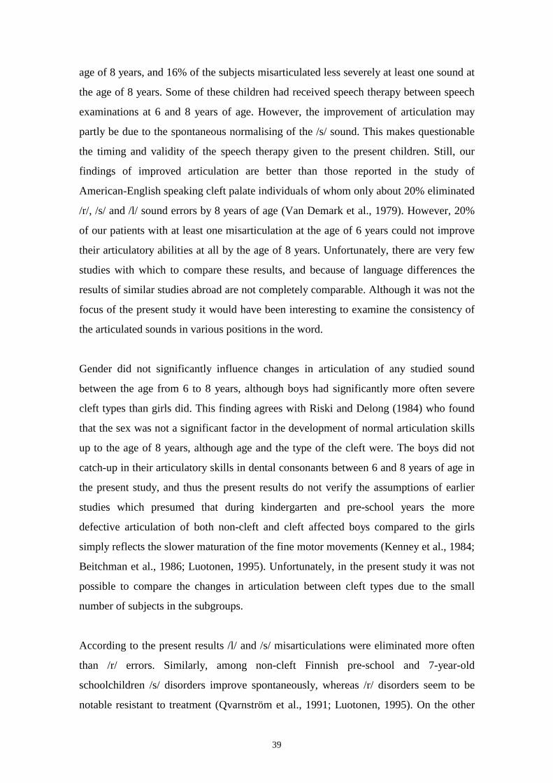
type of the cleft is shown in Fig 2. Boys outnumbered girls in misarticulating all of the
studied sounds (Fig. 3). Boys also misarticulated significantly more often (31%) two or
more sounds than girls (12%).

28
29
22
46
61
13
8
38
45
16
7
22
42
0
10
20
30
40
50
60
70
CP CL(A) UCLP BCLP
Cleft types
%
/r/ /s/ /l/
Fig. 2. The distribution of the misarticulations of /r/, /s/ or /l/ sounds by the type of the cleft in
6-year-old children.
53
44
30
22
31
23
13 12
0
10
20
30
40
50
60
/r/, /s/ or /l/ /r/ /s/ /l/
Misarticulations
%
Boys Girls
Fig. 3. Occurrence of at least one misarticulation or their combinations of /r/, /s/ or /l/ sounds
and separately for at least /r/, at least /s/ or at least /l/ misarticulations between boys and girls at
the age of 6 years.
29
Altogether, 26% of all the 6-year-old subjects required speech therapy. As many as 70%
of the children who had previously received speech therapy still misarticulated at least
one of the sounds, and almost half of them (43%) still needed treatment at the age of 6
years. The need for speech therapy increased with the severity of the cleft. Boys required
significantly more often (34%) speech therapy than girls (14%). The most resistant to
the treatment was the /r/ sound, since 64% of previously treated children still
misarticulated it. Also the /s/ and /l/ sounds were rather resistant to treatment because
the /s/ sound was still misarticulated by 38% and the /l/ sound distorted by 35% of
treated subjects.
The genders did not differ statistically significantly according to the number of
unchanged or changed articulations from 6 to 8 years of age. Altogether 81% of
subjects with misarticulations at the age of 6 years still misarticulated at least one sound
at the age of 8 years. In the group of the subjects with at least one misarticulation at 6
years of age one-fifth of the patients’ articulations remained unchanged, but 57% of
them could eliminate at least one studied misarticulation. One, two or all three
misarticulations were eliminated in 67%, 27% and 6% of the subjects, respectively, by
the age of 8 years. /S/ and /l/ sounds were corrected more often than the /r/ sound.
Fourteen percent of the subjects with at least one misarticulation at the age of 6 years
and 16% of the subjects without any misarticulation at the age of 6 years had a new
misarticulation by the age of 8 years.
At the age of 8 years new /r/, /s/ and /l/ errors were diagnosed almost equally often in
those patients with misarticulations at the age of 6 years (5%, 5%, 3%, respectively),
whereas in patients without misarticulations at 6 years of age the figures were 7%, 12%
and 1%, respectively. Subjects with and without speech therapy before the age of 6 years
produced as often new misarticulations by 8 years of age. Misarticulation of the /s/
sound appeared altogether in 12 patients, of which 9 subjects had unchanged spacing of
the maxillary incisors from the age of 6 to 8 years, and only 3 had spacing at the age of 8
years but not at 6 years of age. The situation was similar for new /r/ and /l/ errors and
spacing.

30
7.2. Dental arch dimensions, occlusion and misarticulations at the age of 6
years (III,IV)
All dimensions, and especially maxillary ones, were smaller in cleft children than in
non-cleft Finnish children. Dental arch dimensions showed statistically significant
differences between genders in general and in different cleft types. The boys had larger
values in all dimensions in each cleft group. Maxillary widths decreased both anteriorly
and posteriorly with the severity of the cleft but maxillary lengths and heights as well as
lower arch dimensions did not differ so clearly. CL(A)-children had the widest and
BCLP-children the narrowest dimensions. Table 2 shows statistical comparisons of the
mean values of the dental arch dimensions between subjects misarticulating one or more
of the sounds /r/, /s/ or /l/ and subjects with correct /r/, /s/ and /l/ articulation genders
separated and cleft types combined. In general both boys and girls with all studied
articulatory errors had significantly narrower maxillary arches than subjects without
these errors. In addition, girls with /r/, /s/ and /l/ errors had significantly shallower
palates than girls with correct /r/, /s/ and /l/ production, and boys with the /s/ and /l/
errors, and girls with /r/ errors had also significantly shorter maxillary arches than
subjects without studied errors. The biggest differences in maxillary width and palatal
height dimensions were found between girls with and without /l/ errors. Only one
mandibular arch dimension differed significantly in the corresponding comparisons.

31
Table 2. Statistical comparisons (Student's t-test) of maxillary mean values (d, mm) of the dental archdimensions between subjects with /r/ or /s/ or /l/ misarticulation including their combinations (n1) andsubjects without them (n2) separately for genders in all cleft types.
Boys (n=154) Girls (n=109)
/r/ /s/ /l/ /r/ /s/ /l/
(n1=67, (n1=45, (n1=33, (n1=26, (n1=14, (n1=13,
Dental arch n2=87) n2=109) n2=121) n2=83) n2=95) n2=96)dimensions p d p d p d p d p d p d
Width 53,63 0.000*** 2.3 0.000*** 2.9 0.004** 2.3 0.000*** 4.2 0.005** 4.0 0.000*** 6.5
Width 55,65 0.016* 1.4 0.000*** 2.6 0.002** 2.2 0.000*** 2.3 0.000*** 3.1 0.000*** 3.3
Maxillary archlength ns 0.023* 1.0 0.034* 1.0 0.000*** 1.2 ns ns
Palatal heightposterior ns ns ns 0.000*** 2.1 0.008** 1.9 0.002** 2.2
*p<0.050, **p<0.010, ***p<0.001, ns= not significant, d= difference of the mean values (x2-x1, mm) of the dental arch dimensions between subjects without (n2) and with misarticulations (n1).
Fifty-seven percent of the subjects had anterior and/or posterior crossbite, 8% had large
maxillary overjet, and 29% had deep bite. The number of subjects with crossbite
increased with the severity of the cleft; 26% in CL(A)-, 34% in CP-, and 96% in both
UCLP- and BCLP-groups. The occurrence of crossbites, large maxillary overjet, or deep
bite did not reveal any significant sex differences. Subjects with misarticulations of at
least one studied sound had significantly more dental crossbites (73%) than subjects
with correct /r/, /s/ and /l/ sound production, of whom 45% had crossbites. Table 3
shows statistical comparisons of the occurrence of at least one misarticulation of /r/, /s/
or /l/ sounds, and separately for at least /r/, at least /s/ or at least /l/ sound between
subjects with and without different types of crossbites. Subjects with any of /r/ or /s/ or
/l/ misarticulation or their combinations had significantly more posterior crossbites than
subjects with correct /r/, /s/ and /l/ sound articulation. Subjects with at least /s/ errors
had significantly more often anterior crossbite associated with posterior crossbite in one
side than subjects with correct /s/ production. There were no significant differences
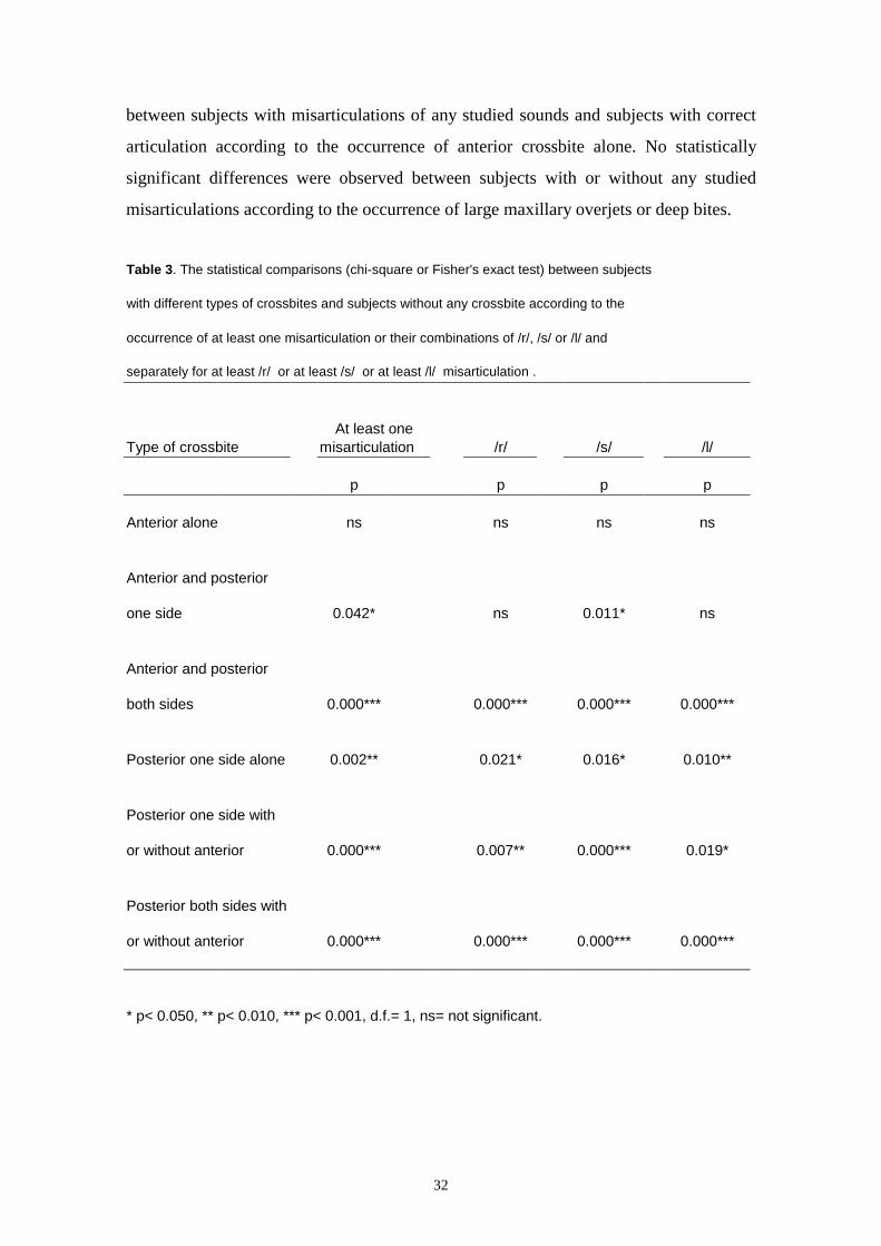
32
between subjects with misarticulations of any studied sounds and subjects with correct
articulation according to the occurrence of anterior crossbite alone. No statistically
significant differences were observed between subjects with or without any studied
misarticulations according to the occurrence of large maxillary overjets or deep bites.
Table 3. The statistical comparisons (chi-square or Fisher's exact test) between subjects
with different types of crossbites and subjects without any crossbite according to the
occurrence of at least one misarticulation or their combinations of /r/, /s/ or /l/ and
separately for at least /r/ or at least /s/ or at least /l/ misarticulation .
At least oneType of crossbite misarticulation /r/ /s/ /l/
p p p p
Anterior alone ns ns ns ns
Anterior and posterior
one side 0.042* ns 0.011* ns
Anterior and posterior
both sides 0.000*** 0.000*** 0.000*** 0.000***
Posterior one side alone 0.002** 0.021* 0.016* 0.010**
Posterior one side with
or without anterior 0.000*** 0.007** 0.000*** 0.019*
Posterior both sides with
or without anterior 0.000*** 0.000*** 0.000*** 0.000***
* p< 0.050, ** p< 0.010, *** p< 0.001, d.f.= 1, ns= not significant.

33
7.3. Correlations between speech and breathing aerodynamics, oral
somatosensoric sensibility, and strength of the tongue muscles, and
skeletal/pharyngeal morphology and the Finnish /r/ sound (V,VI)
Males had more frequently /r/ distortions and significantly more severe cleft types than
females. In subjects with /r/ distortion laryngeal resistance was significantly lower than
in subjects without /r/ distortion (p= 0.000), and significantly lower in cleft subjects
without /r/ distortion than in the controls (p= 0.000). There were no significant
differences in the velopharyngeal orifice area and the smallest nasal crossectional area
between subjects with and without /r/ distortion. However, the smallest nasal
crossectional area was significantly smaller in subjects with /r/ distortion than in the
controls. Nasal resistance did not differ between cleft groups or the controls. The peak
expiratory flow values, somatosensoric measurements and strength of tongue muscles
did not differ between subjects with and without /r/ distortion. The sensitivity in the
sides of their tongues was similar for all the subjects.
In the cephalometric comparisons the position of the hyoid bone was significantly more
anterior in subjects with /r/ distortion. No significant differences in the cranial base or in
the relationship of the jaws or in their relationship to the cranial base were found
between the groups with and without /r/ sound distortions. The nasopharyngeal,
oropharyngeal and hypopharyngeal linear measurements did not differ significantly
between the groups. However, in the hypopharyngeal region all linear measurements
were 12-26% wider, but in the oro- and nasopahynx it was the same or smaller in
subjects with /r/ distortion than in subjects without distortion.
Comparison of the aerodynamic measurements with the craniofacial and
pharyngolaryngeal measurements indicated that laryngeal resistance showed significant
negative correlation to the maxillary prognathy (SNA, p= 0.020), and to the sagittal
maxillo-mandibular relationship (ANB, p= 0.020). Also a positive correlation between
laryngeal resistance and the distance from the posterior nasal spine to the sella-basion
line on the pharyngeal wall (ad 2, p= 0.037) was observed. In other words, low laryngeal
resistance was associated with a narrower nasopharyngeal region. Laryngeal resistance

34
also correlated positively with the anterior position of the hyoid bone (H-RGn, H-Gn, p=
0.024 and p= 0.022, respectively). Low laryngeal resistance was thus associated with
more anteriorly positioned hyoid bone.

35
8. DISCUSSION
8.1. Subjects and methods
The treatment of cleft children in Finland is centralised in the Cleft Center in Helsinki
University Central Hospital, and therefore all socio-economic groups were represented
in the study groups. Non-cleft controls were not used because there are several recent
methodically comparable studies on articulatory abilities of non-cleft Finnish children
aged from 5 to 9 years (Qvarnström et al., 1991; Luotonen, 1995). Although the number
and consistency of our material should be considered representative for examining the
association between dental arch sizes, crossbites and misarticulations, too small
subgroups complicated the conclusions.
In the present study quantity and quality of speech material was designed to be
practically carried out in every patient in Cleft Center. Tape-recorded strictly structured
speech sample collection was not used because it would have reduced the quantity of the
material. The interjudge agreement between the cleft team’s two speech pathologists in
rating and categorising misarticulations and estimating the need of speech therapy was
adequate. The criteria used to evaluate the misarticulations may, however, vary in
different ages and in different language groups. Therefore, the results of this study
cannot be completely compared with similar studies. Methods in evaluation the sizes of
dental arches (Moorrees, 1959; Nyström and Ranta, 1994) as well as measurement of
speech and breathing aerodynamics (Warren, 1979; Smitheran and Hixon, 1981) are
widely used. Intra-judge agreement for estimation of occlusion was high, and the range
error between two tracings of cephalometric landmarks was adequate. Only angular
skeletal lateral cephalometric measurements were used so that the genders could be
combined. The measurements of the sensitivity and the strength of the tongue must be
taken with caution due to the limitations of the instrumentation. The method to assess
tongue muscle strength was modified for the present study and not standardized. The
sensitivity was possibly measured too roughly. The distance of two points should have
been 1 mm instead of the 2 mm allowed by the instrumentation.

36
8.2. Findings
8.2.1. Articulatory abilities in cleft-children
At the age of 6 years 44% of the Finnish cleft-children have articulatory problems with
/r/, /s/ or /l/ sound, while only 30% of 5-8-year-old non-cleft Finnish children have the
same misarticulations (Sonninen and Sonninen, 1976; Qvarnström et al., 1991;
Luotonen, 1995). Most of the present misarticulations were distortions of the sounds,
whereas substitutions were rather rare. This parallels the results of Powers (1971) who
reported articulatory speech disorders being mainly distortions in schoolchildren,
substitutions being common among kindergarten children. According to Shelton and
McReynolds (1979) distortions are the least severe articulatory disorders.
For cleft-children the /r/ sound was the most difficult to produce; altogether 36%
misarticulated this sound. Also /s/ and /l/ errors were common, since as many as 23%
misarticulated /s/ and 18% /l/ sound. /R/ and /s/ sounds are the most difficult ones to
produce for Finnish non-cleft children, too, since together these two sounds make up
89% and 95% of all errors at the age of 5 and 7 years (Luotonen, 1995), /l/ disorders
being very rare (1%) (Qvarnström et al., 1991). Earlier Finnish studies are partly at odds
with regard to the frequency of /r/ or /s/ disorders in Finnish non-cleft pre-school and
schoolchildren. According to Sonninen and Sonninen (1976) and Qvarnström et al.
(1991) /s/ disorders are the most common (30%), /r/ disorders being rather uncommon
(7-8%), whereas Luotonen (1995) reported /r/ disorders accounting 61% and /s/
disorders 28% of all errors. Luotonen’s (1995) results for non-cleft children agree with
ours, although the definitions and criteria used for the correctness of the /s/ sound may
partly vary between the studies. The most distinguishing finding was the high frequency
of /l/ distortions among cleft-children compared with non-cleft Finnish children.
Although the production of correct /l/ sound seem to be clearly easier than /r/ and /s/
production for non-cleft children, the production of /l/ sound is difficult enough for cleft
affected individuals.
Although articulation disorders are suggested to be psychologically based, learned
phenomena, articulation defects may have both organic and functional origins (Nichols,

37
1981; Van Dyke et al., 1984). Accurate articulation requires precise movements of
articulators involving fine motor control, and close coordination of the lips, tongue, and
mandible (Lindblom and Sundberg, 1971). Dental consonants are the most difficult
sounds to produce, requiring precise placement and function of both of the tip and the
blade of the tongue as well as space enough to the tongue tip in the most anterior part of
the palate. A deviant structure or volume of the posterior part of the oropharynx may
complicate adequate placement of the tongue (Laine, 1992). In addition, since especially
the production of the /r/ sound requires modulation of phonatory air, abnormalities in
the vocal tract structure may affect this air stream modulation resulting in distorted /r/ or
/s/ sound. Different dentofacial morphology may also explain the high frequency of /l/
distortions in cleft children, and further studies are needed to find out which structures
complicate most the /l/ sound production.
The number of subjects with misarticulations increased with cleft severity, and this
result is in agreement with earlier studies (Ross and Johnston, 1972; Riski and Delong,
1984). Also the need for speech therapy increased obviously with the severity of the
cleft. Boys outnumbered girls in the more severe cleft types (UCLP, BCLP). Hence the
correlation between the occurrence and severity of the misarticulations and the cleft type
or the gender is not completely clear. However, patients with complete clefts may have
maxillary collapse, and/or retrusion or protrusion of the premaxilla which present their
own hazards to articulation. The fact that both the number and severity of
misarticulations clearly increased with the severity of the cleft may reflect the negative
effect of occlusal problems for speech development.
Boys outnumbered girls in articulatory disorders according to the present as well as
earlier studies (Kenney et al., 1984; Beitchman et al., 1986; Qvarnström et al., 1991;
Luotonen, 1995). Boys were also more often treated because of severe misarticulations
than girls were. Early speech development as well as early progress of fine motor
movements have been reported to be slower among boys (Stevenson and Richman,
1976; Silva, 1980; Kirkpatrick and Ward, 1984; Beitchman et al., 1986; Luotonen,
1995). Boys are found to have more inaccurate tongue movements and problems in
coordinating those movements than girls, especially among children with articulatory
speech defects (Qvarnström et al., 1993b). Disorders in speech sound production in

38
general are reported to reflect immaturity in fine motor control of the orofacial muscles
in 6-8-year-old children (Pahkala et al., 1991). Interestingly, while non-cleft boys have
more /r/ disorders (Qvarnström et al., 1991; Luotonen, 1995), but fewer /s/ disorders
than non-cleft girls (Qvarnström et al., 1991), cleft-boys outnumbered cleft-girls in both
/r/, /s/ and /l/ misarticulations at the age of 6 years.
Unfortunately, there is only a little information on changes in misarticulations during the
maturation of speech articulation. Although children are considered to be able to
produce all sounds correctly long before school age (Sarmavuori, 1982), the final
control of the oral articulatory movements is thought to be achieved as late as the age of
10 to11 years (Smith and McLean-Muse, 1986). Articulatory disorders are known to
decrease with age (Milisen, 1971; Alstam-Malcus et al., 1976; Leske, 1981; Qvarnström
et al., 1991), but they seem to correct more often spontaneously in non-cleft
schoolchildren than cleft-children, since half of the Swedish (Alstam-Malcus et al.,
1976), and as many as 82% of American first grade schoolchildren correct their
articulation without speech therapy up to grades 2 and 3 (Irwin et al., 1974). Half of
misarticulations in Finnish non-cleft children decreased between the ages of 7 to 9, but
these children had received treatment (Luotonen, 1995). Articulation of dental
consonants are distinguishly difficult for cleft-children, since 81% of subjects with at
least one error at the age of 6 years still misarticulated at least one sound at the age of 8
years.
Altogether 14% to 16% of the subjects generated a new misarticulation between the age
6 to 8 years. New misarticulations were diagnosed whether speech therapy was given or
not. This may partly, however, be a consequence of the fact that some inconsistent
distortions were categorised as immature but not as misarticulations at the age of 6
years, and they were expected to correct spontaneously by the age of 8 years. However,
persisting dysmature articulation was considered as a misarticulation at the age of 8
years. Abnormalities in dentofacial growth may partly explain appearance of new
misarticulations in cleft children.
As many as 57% of the 6-year-old patients with /r/, /s/ or /l/ misarticulations could
improve their articulation to some extent and eliminate at least one of the errors by the
39
age of 8 years, and 16% of the subjects misarticulated less severely at least one sound at
the age of 8 years. Some of these children had received speech therapy between speech
examinations at 6 and 8 years of age. However, the improvement of articulation may
partly be due to the spontaneous normalising of the /s/ sound. This makes questionable
the timing and validity of the speech therapy given to the present children. Still, our
findings of improved articulation are better than those reported in the study of
American-English speaking cleft palate individuals of whom only about 20% eliminated
/r/, /s/ and /l/ sound errors by 8 years of age (Van Demark et al., 1979). However, 20%
of our patients with at least one misarticulation at the age of 6 years could not improve
their articulatory abilities at all by the age of 8 years. Unfortunately, there are very few
studies with which to compare these results, and because of language differences the
results of similar studies abroad are not completely comparable. Although it was not the
focus of the present study it would have been interesting to examine the consistency of
the articulated sounds in various positions in the word.
Gender did not significantly influence changes in articulation of any studied sound
between the age from 6 to 8 years, although boys had significantly more often severe
cleft types than girls did. This finding agrees with Riski and Delong (1984) who found
that the sex was not a significant factor in the development of normal articulation skills
up to the age of 8 years, although age and the type of the cleft were. The boys did not
catch-up in their articulatory skills in dental consonants between 6 and 8 years of age in
the present study, and thus the present results do not verify the assumptions of earlier
studies which presumed that during kindergarten and pre-school years the more
defective articulation of both non-cleft and cleft affected boys compared to the girls
simply reflects the slower maturation of the fine motor movements (Kenney et al., 1984;
Beitchman et al., 1986; Luotonen, 1995). Unfortunately, in the present study it was not
possible to compare the changes in articulation between cleft types due to the small
number of subjects in the subgroups.
According to the present results /l/ and /s/ misarticulations were eliminated more often
than /r/ errors. Similarly, among non-cleft Finnish pre-school and 7-year-old
schoolchildren /s/ disorders improve spontaneously, whereas /r/ disorders seem to be
notable resistant to treatment (Qvarnström et al., 1991; Luotonen, 1995). On the other

40
hand, with advancing age from 7 to 10 years /r/ disorders are reported to decrease, and
/s/ disorders to increase markedly, although most of these subjects had received speech
therapy before or at school (Qvarnström et al., 1993a). Probably the Finnish /r/ sound is
more difficult to produce correctly than it is in other languages, since, for example, in
American-English speaking cleft populations the degree of success for /r/ correction was
similar to corrections for /s/ and /l/ between 6 to 8 years of age (Van Demark et al.,
1979). Unfortunately, this study does not mention if the subjects had been treated with
speech therapy. Since cleft affected subjects have articulation deficits that are apparently
strongly influenced by phonological maturation, it is impossible to conclude whether the
decrease in the occurrence of the studied misarticulations is due to more mature tongue
movements or to the effects of speech therapy or both. Cleft children also have recurrent
otitis media, which is known to affect speech development. This may also have an
impact on the articulatory skills of cleft children. Unfortunately, it was not possible to
control for the otitis condition in the present children. Altogether, at least the timing and
intensity of speech therapy used with cleft children warrants re-evaluation so that the
treatment would have related more to maturation and developmental factors.
8.2.2. Dentofacial morphology and articulation
According to present results a narrow and shorter maxillary arch as well as a shallower
palate seems to explain partly the occurrence of dental consonant misarticulations.
These findings are in agreement with results of earlier studies with cleft-patients (Starr,
1971; Okazaki et al., 1991), and non-cleft individuals (Oliver and Evans, 1986). Powers
(1971) stated that subjects with /r/ or /l/ distortions keep their tongue posteriorly and
inferiorly instead of raising it to the alveolar ridge, and Laine et al. (1987) suggested that
certain sounds may be produced too far posteriorly due to a deficiency in maxillary
anterior space. Wilcox et al. (1985) and Wyatt et al. (1996) concluded that subjects with
reduced maxillary arches may have insufficient space for their tongues to obstruct the air
steam adequately in order to produce the /s/ sound correctly. On the other hand, Bishara
et al. (1975) found that subjects with isolated clefts palate with good or poor articulation
did not differ systematically in their dental relations.

41
Our findings revealed clearly that cleft patients with posterior crossbites are at high risk
for defective dental consonant articulation, while anterior crossbite was related to
articulatory errors only when it was combined with posterior crossbite. Although only
weak association between /s/ disorders and lateral crossbite has been earlier found
(Laine, 1987), several authors have concluded that both anteriorly or posteriorly
decreased intermaxillary space of the oral cavity due to occlusal anomalies prevents the
precise placement and function of the tongue required for correct /s/ production (Heinz
and Stevens, 1961; Wilcox et al, 1985; Howell, 1987; Laine, 1987). In distal (Class II)
and mesial (Class III) molar occlusion /s/ production is found to be too anterior due to
the protrusion of the tongue (Ruscello et al., 1985; Wyatt et al., 1996), although also
retruded tongue posture in Class III subjects with distorted /s/ phonation has been found
(Guay et al., 1978). Altogether, subjects with Class III malocclusion are reported to
misarticulate more than those with incisal open bite (Laine, 1992). Probably because
cleft patients’ occlusion only resembles Class III occlusion due to the retruded maxilla,
anterior crossbite alone did not seem to explain the articulatory errors of cleft patients.
According to the present study neither a large maxillary overjet nor a deep bite did not
influence on the occurrence of misarticulations. These findings support earlier results
concerning incisor relationship and articulatory defects in non-cleft adults (Oliver and
Evans, 1986; Laine, 1987), although weak associations between /r/ disorders and large
maxillary overjet have been reported (Laine, 1992).
The present results regarding the association between spacing of the maxillary incisors
and misarticulations partly disagree with earlier studies, since several authors have
found subjects with misarticulations having more often spacing of the maxillary dental
arch than subjects without misarticulations (Snow, 1961; Laine et al., 1987). On the
other hand, other reports have found that other developmental factors have a greater
influence on correct speech sound production than the eruption stage of the incisors
(Ingervall and Sarnäs, 1962; Pahkala et al., 1991). Altogether some individuals can
functionally compensate for maxillary frontal spacing and control the airflow adequately
to produce acoustically correct consonants (Weinberg, 1968). In this study the changing
of the maxillary incisors occurred between the studied ages. The varying spacing
through time due to changing may influence the appearance of new sound errors in these
42
subjects. However, more detailed information is needed to find out if spacing or severe
malpositions of the maxillary incisors have an influence on articulation.
Since many of the present subjects with posterior crossbites had correct /r/, /s/ and /l/
articulation, and many subjects without posterior crossbites still misarticulated at least
one studied consonant, a smaller oral cavity should be considered only as a possible
contributing hazard to clear articulation in cleft palate patients. So, even in the presence
of severe structural deviations good articulation can exist due to the highly adaptable
speech mechanisms and compensatory behaviour (Starr, 1971; Hardcastle et al., 1987).
Studies dealing with the developmental aspects of speech in relation to occlusion and
compensatory speech responses to the presence of structural deviations are clearly
lacking, partly due to methodological difficulties. Longitudinal studies to determine
whether children with occlusal anomalies and articulation disorders have the same faulty
sounds at a later age or whether articulatory speech disorders appear later during
development of the dentition, would clarify these issues. Finally, if misarticulations of
especially /s/ and /r/ sounds are so called ”residual speech errors” dating from the early
stages of speech development (Hardcastle et al., 1987), any other malocclusion except
those existing before the age of 5 years could not possibly have any effect on speech
sound production. On the other hand, it is generally recognised that both a narrow upper
dental arch and posterior crossbite are sometimes seen in connection with pathological
conditions causing obstruction of the nasopharyngeal airway, which may complicate
articulation (Solow and Greve, 1979).
8.2.3. /R/ distortion, speech aerodynamics and pharyngeal morphology
The present results showed that cleft subjects with /r/ distortion had lower laryngeal
resistance and more anterior position of the hyoid bone than subjects without /r/
distortion. Low laryngeal resistance has been found to increase the risk for different
voice disorders also in earlier studies (Zajac, 1995). Velopharyngeal orifice size, the
smallest nasal crossectional area or nasal resistance as well as the sensitivity of the
palatal alveolus or the tongue or strength of tongue muscles were not related to /r/
distortions.

43
Although the cleft type per se is probably not the determinant of distorted /r/ sound
articulation, the effect of the cleft type on dento-alveolar morphology and other
cephalometric dimensions may be reflected both in respiration and phonation. Phonation
requires precise function of the so-called physiologic larynx, i.e. tongue base, the
position of the hyoid bone and the glottis (Vilkman et al., 1996), and this function may
be distorted in patients with orofacial abnormalities. Dysmorphology in the vocal tract
probably negatively affect the modulation of the speech air stream resulting in a
distorted /r/ sound.
The present results showed no significant differences in lateral cephalometric skeletal
morphology between subjects with and without /r/ sound distortion. These results
parallel those of Bishara et al. (1975) who could not establish any consistent trends
between dentofacial angular or linear relations and phonemes in patients with isolated
cleft palate. However, some pharyngeal horizontal dimensions were slightly but not
significantly bigger in our subjects with /r/ distortion. A slightly wider pharynx in
subjects with /r/ distortion may alter airflow and air pressure in /r/ sound production and
thus contribute to /r/ distortion. Laryngeal resistance was lower in subjects with /r/
distortion than in subjects without /r/ distortion. Although it has been previously
suggested that some occlusal anomalies may affect the position of the tongue and that of
the hyoid bone which may increase the risk for defective articulation (Laine, 1987),
Haralabakis et al. (1993) stated that the position of the hyoid bone moves in close
conjunction with the pharynx and mandibular plane even in patients with entirely
different skeletal patterns. Since the position of the hyoid bone is a reflection of the
relative tensions of the muscles, ligaments, and fasciae attached to it, the reason for
hyoid bone moving ”in concert” with the adjacent anatomical structures is concluded to
be the need to maintain the patency of the upper airway at the level of the base of the
tongue (Opdebeeck et al., 1978; Haralabakis et al., 1993). Thus, it is possible that the
anterior position of the hyoid bone increases the pharyngolaryngeal volume and is
associated with decreased laryngeal resistance in subjects with /r/ distortion. In addition,
if the hyoid bone is positioned too anteriorly, it may be difficult and perhaps impossible
to place the tongue tip in the proper position for the correct production of the /r/ sound.

44
Even though, the smallest nasal cross-sectional area and nasal resistance correlated with
each other, neither of them correlated significantly with the dentofacial or pharyngeal
cephalometric measurements, except for the fact that the smallest nasal cross-sectional
area decreased significantly with the more inferior position of the hyoid bone. Although
anatomical abnormalities of the pharynx in cleft palate speakers can be evaluated
reliable with cephalometric radiography (Hoenig and Schoener, 1992), Kluemper et al.
(1995) stated that facial morphology and respiratory mode are unrelated, and that
cephalometric analyses are poor indicators of nasal impairment. Nasopharyngeal depth
has been reported to be shorter and thus nasopharyngeal airway to be reduced in cleft
affected subjects than in non-cleft individuals (Scaf et al., 1991; Smahel et al., 1991;
Wada et al., 1997), although also deviant results have been presented (Hoenig et
Schoener, 1992). In individual cleft patients nasopharyngeal width may compensate for
smaller nasopharyngeal depth (Smahel et al., 1991). According to present results the
smallest nasal cross-sectional area and nasal resistance did not correlate with laryngeal
resistance, and thus do not probably explain the /r/ distortion in cleft affected subjects.
Velopharyngeal orifice area did not correlate with lateral cephalometric skeletal
morphology, hyoid measurements, pharyngolaryngeal dimensions, the smallest nasal
cross-sectional area, nasal or laryngeal resistance or /r/ distortion. In our present subjects
the distance from the posterior nasal spine to the pharyngeal wall (ad2) were on the
average shorter than 20 mm indicating the absence of velopharyngeal incompetence
(Haapanen et al., 1991). To sum up, the velopharyngeal area was not associated with
any parameters that correlated with /r/ sound distortions.
The present associations between /r/ distortion and low laryngeal resistance as well as
anterior position of the hyoid bone are new and suggest the need for more research to
resolve treatment possibilities of orofacial structures to allow normal speech.

45
9. CONCLUSIONS
Altogether 44% of 6-year-old cleft children had /r/, /s/ or /l/ misarticulations and these
errors increased with the severity of the cleft. The frequency of /l/ distortions was
surprisingly high. Dental sound disorders were more common in boys than in girls.
As many as 81% of the subjects still misarticulated /r/, /s/ or /l/ sound at the age of 8
years. New misarticulations were diagnosed at the age of 8 years in the same way in the
groups with (14%) and without (16%) misarticulations at the age of 6 years.
The decreasing size of the maxillary dental arch as well as posterior crossbites seem to
be related to the increasing incidence of the studied misarticulations.
/R/ distortion is significantly correlated with low laryngeal resistance, and tends to occur
with a wider pharynx, and an anteriorly positioned hyoid bone.

46
10. ACKNOWLEDGEMENTS
The present thesis was carried out at the Cleft Center, Department of Plastic Surgery,
Helsinki University Central Hospital, headed by Professor Sirpa Asko-Seljavaara, M.D.,
Ph.D., and at the Department of Pedodontics and Orthodontics, University of Helsinki,
headed by Acting Professor Sinikka Pirinen, D.D.S., Ph.D.
My deepest gratitude goes to my supervisor Docent Reijo Ranta, D.D.S., Ph.D., Chief
Orthodontist of the Cleft Center, Department of Plastic Surgery, Helsinki University
Central Hospital, for proposing and introducing me the topic of this research and for his
friendly guidance and encouragement throughout all stages of this work.
I am also very grateful to my other supervisor Docent Marja-Leena Haapanen, M.A.,
M.D., Ph.D., who introduced me to the world of phoniatrics and friendly guided me
through these years.
I wish to thank co-workers of this study Marjukka Paaso and Riitta Sarmas, speech
therapists, Arja Heliövaara, D.D.S., Ph.D., and Kirsti Hurmerinta, D.D.S., Ph.D., for
their valuable cooperation. My special thanks belong to my co-worker Joonas
Pulkkinen, D.D.S., for his friendship and help throughout this work.
I thank the official referees of this work Docent Pirjo Korpilahti, Ph.D., and Professor
Juha Varrela, D.D.S., Ph.D., for their careful and precise work with constructive
criticism and suggestions to improve the form and contents of the manuscript. In
addition, Docent Korpilahti deserves special thanks for her valuable logopedic
knowledge.
My warm thanks to Timo Pessi, M.Sc., for helping me in biostatistical problems.
I wish to thank sincerely Donald Smart, B.Sc., M.A., for his professional linguistic
revision of this manuscript.

47
I am also grateful to my cousin Helena and her family Hannu, Laura and Aino.
Especially Hannu helped me editing this thesis.
My very special thanks belong to my parents, my brother Jukka and his family Arja,
Sanna and Simo for their warm support through these years.
This research was supported by grants from the Emil Aaltonen Foundation and the
Finnish Women Dentists Association.
Helsinki, December 1999
Jaana Laitinen

48
11. REFERENCES
Alstam-Malcus A, Fritzell B, Fritzell M, Svantesson F. Talsvårigheter vid skolstarten
och ett år senare. Nord Tidskr Logop Foniat 1976;1:15-25.
Arnold GE. Physiology and pathology of speech and language; in Luchsinger R and
Arnold GE (eds): Voice, speech, language. Wadsworth, Belmont 1965, pp 337-790.
Bankson NW, Byrne MC. The relationship between missing teeth and selected
consonant sounds. J Speech Hearing Dis 1962;27:341-348.
Beitchman JH, Nair R, Clegg M, Patel PG. Prevalence of speech and language disorders
in 5-year-old kindergarten children in the Ottawa-Carleton region. J Speech Hear Disord
1986;51:98-110.
Bishara SE, VanDemark DR, Henderson WG. Relation between speech production and
oro-facial structures in individuals with isolated clefts of the palate. Cleft Palate J
1975;12:452-460.
Blyth P. The relationship between speech, tongue behavior, and occlusal abnormalities.
Dent Pract 1959;10:11-20.
Broad W. Phonation; in Minifie F, Hixon T, Williams F (eds): Normal aspects of
speech, hearing and language. Prentice-Hall Inc, New Jersey 1973, pp 127-149.
Cermak SA, Ward EA, Ward LM. The relationship between articulation disorders and
motor coordination in children. Am J Occup Ther 1986;40:546-550.
Dahlberg G. Statistical methods for medical and biological students. Allen and Unwin,
London 1948, p 232.

49
Darley FL, Aronson AE, Brown JR. Motor speech disorders W B Saunders,
Philadelphia 1975, pp 3-6.
Enemark H, Bolund S, Jorgensen I. Evaluation of unilateral cleft lip and palate
treatment: long term results. Cleft Palate J 1990;27:354-361.
Ferry PC, Hall SM, Hicks JL. ‘Dilapidated’ speech: Developmental verbal dyspraxia.
Dev Med Child Neurol 1975;17:749-756.
Fletcher SG. Palatometric specification of stop, affricate, and sibilant sounds. J Speech
Hear Res 1989;32:736-748.
Folkins JW. Issues in speech motor control and their relation to the speech of
individuals with cleft palate. Cleft Palate J 1985;22:106-119.
Friede H, Persson E-C, Lilja J, Elander A, Lohmander-Agerskov A, Söderpalm E.
Maxillary dental arch and occlusion in patients with repaired clefts of the secondary
palate. Scand J Plast Reconstr Hand Surg 1993;27:297-305.
Gracco VL. Sensorimotor mechanisms in speech motor control; in Peters HFM et al.
(eds): Speech motor control and stuttering. Proceedings of the 2nd Conference on
Speech motor control and stuttering. June 13-16, 1990 Nijmegen, Netherlands. Excerpta
Medica, Amsterdam 1991, pp 53-76.
Guay AH, Maxwell DL, Beecher R. A radiographic study of tongue posture at rest and
during the phonation of /s/ in Class III malocclusion. Angle Orthodontist 1978;48:10-22.
Haapanen M-L, Heliövaara A, Ranta R.. Hypernasality and the nasopharyngeal space. A
cephalometric study. J Craniomaxfac Surg 1991;19:77-80.
Haapanen M-L, Rantala SL. Correlation between the age at repair and speech outcome
in patients with isolated cleft palate. Scand J Plast Reconstr Hand Surg 1992;26:71-78.

50
Haralabakis NB, Toutountzakis NM, Yiagtzis SC. The hyoid bone position in adult
individuals with open bite and normal occlusion. Eur J Orthod 1993;15:265-271.
Hardcastle WJ. Physiology of speech production; An introduction for speech scientists.
Academic Press, London 1976, pp 83-86.
Hardcastle WJ, Morgan Barry RA, Clark CJ. An instrumental phonetic study of lingual
activity in articulation-disordered children. J Speech Hear Res 1987;30:171-184.
Heinz JM, Stevens KN. On the properties of voiceless fricative consonants. J Acoust
Soc Am 1961;33:589-596.
Heliövaara A, Rintala A, Ranta R. One-stage closure of isolated cleft palate with the
Veau-Wardill-Kilner V to Y pushback procedure or the Cronin modification. Scand J
Plast Reconstr Hand Surg 1993;27:49-54.
Hellquist R, Ponten B, Skoog T. The influence of cleft length and palatoplasty on the
dental arch and the deciduous occlusion in cases of clefts of the secondary palate. Scand
J Plast Reconstr Surg 1978;12:45-54.
Hoenig JF, Schoener WF. Radiological survey of the cervical spine in cleft lip and
palate. Dentomaxillofac Radiol 1992;21:36-39.
Horowitz SL, Graf-Pinthus B, Bettex M, Vinkka H, Gerstman LJ. Factor analysis of
craniofacial morphology in cleft lip and palate in man. Arch Oral Biol 1976;21:465-472.
Howell PGT. The variation in the size and shape of the human speech pattern with
incisor-tooth relation. Arch Oral Biol 1987;32:587-592.
Ingervall B, Sarnäs K-V. Comparisons of dentition in lispers and non-lispers. Odont
revy 1962;13:344-354.

51
Irwin JV, Huskey R, Knight N, Oltman S. A longitudinal study of the spontaneous
remission of articulatory defects of 1665 school children in grades 1, 2 and 3. Acta
Symbolica 1974;5:1-17.
Karling J, Larson O, Leanderson R, Henningsson G. Speech in unilateral and bilateral
cleft palate patients from Stockholm. Cleft Palate-Craniofacial J 1993;30:73-77.
Karlsson F. Suomen kielen äänne- ja muotorakenne. WSOY, Porvoo-Helsinki-Juva
1982, pp 56-80.
Kenney KW, Prather EM, Mooney MA, Jeruzal NC. Comparison among three
articulation sampling procedures with pre-school children. J Speech Hear Res
1984;27:226-231.
Kirkpatrick E, Ward J. Prevalence of articulation errors in New South Wales primary
school pupils. Aust J Hum Commun Disord 1984;12:55-62.
Kluemper GT, Vig PS, Vig KW. Nasorespiratory characteristics and craniofacial
morphology. Eur J Orthod 1995;17:491-495.
Laine T, Jaroma M, Linnasalo A-L. Articulatory disorders in speech related to the
position of the incisors. Eur J Orthod 1985;7:260-266.
Laine T. Associations between articulatory disorders in speech and occlusal anomalies.
Eur J Orthod 1987;9:144-150.
Laine T, Jaroma M, Linnasalo A-L. Relationships between interincisal occlusion and
articulatory components of speech. Folia Phoniat 1987;39:78-86.
Laine T. Malocclusion traits and articulatory components of speech. Eur J Orthod
1992;14:302-309.

52
Leske MC. Prevalence estimates of communicative disorders in the U.S. Speech
disorders. ASHA 1981;23:217-224.
Lewis R. Tongue thrust and protrusion lisp. J Canad Dent Assoc 1971;37:381-383.
Lindblom BEF, Sundberg EF. Acoustical consequences of lip, tongue, jaw, and larynx
movement. J Acoust Soc Am 1971;50:1166-1179.
Lubit EC. The relationship of malocclusion and faulty speech articulation. J Oral Med
1967;22:47-55.
Luotonen M. Early speech development, articulation and reading ability up to the age of
9. Folia Phoniatr Logop 1995;47:310-317.
Maddieson I. Patterns of sounds. Cambridge University Press, Cambridge 1984, p 275.
McReynolds LV, Elbert MF. Articulation disorders of unknown etiology ant their
remediation; in Lass N J et al. (eds): Speech, language, and hearing, W B Saunders,
Philadelphia 1982, pp 591-610.
McWilliams BJ, Musgrave RH. Diagnosis of speech problems in patients with cleft
palate. Br J Disord Commun 1977;6:26-32.
Milisen R. The incidence of speech disorders; in Travis LE (ed): Handbook of speech
pathology and audiology, Appleton-Century-Crofts, New York 1971, pp 619-634.
Moorrees CFA. Dentition of the growing child; A longitudinal study of dental
development between 3 and 18 years of age. Harvard University Press, Cambridge,
Mass. 1959, p 245.
Nichols AC. Articulation-Organic factors; in: Rieber RW (ed): Communication
Disorders. Plenum Press, New York and London, 1981, pp 3-19.

53
Nyström M, Ranta R, Kataja M. Sizes of dental arches and general body growth up to 6
years of age in children with isolated cleft palate. Scand J Dent Res 1992;100:123-129.
Nyström M, Ranta R. Effect of timing and method of closure of isolated cleft palate on
development of dental arches from 3 to 6 years of age. Eur J Orthod 1994;16:377-383.
Okazaki K, Kato M, Onizuka T. Palate morphology in children with cleft palate with
palatalized articulation. Ann Plast Surg 1991;26:156-163.
Oliver RG, Evans SP. Tongue size, oral cavity size and speech. The Angle Orthodontist
1986;56:234-243.
Opdebeeck H, Bell WH, Eisenfeld J, Michelevich D. Comparative study between the
SFS and LFS rotation as a possible morphogenic mechanism. Am J Orthod
1978;74:509-521.
Pahkala R, Laine T, Lammi S. Developmental stage of the dentition and speech sound
production in a series of first-grade schoolchildren. J Craniofac Gen Dev Biol
1991;11:170-175.
Philips BJ, Harrison RJ. Articulation patterns of pre-school cleft palate children. Cleft
Palate J 1969;6:245-253.
Pietarinen A. Vantaalaisten v. 1980 syntyneiden lasten viisivuotisseulalla mitatut
kielelliset häiriöt ja niiden yhteydet kehityksen muihin osatekijöihin. Helsingin yliopisto
1987, pp 38-47.
Powers MH. Functional disorders of articulation-symptomatology and etiology; in:
Travis LE (ed): Handbook of speech pathology and audiology. Appleton-Century-
Crofts, New York 1971, pp 837-875.
Proffit WR, Norton LA. The tongue and oral morphology: Influences of tongue activity
during speech and swallowing. ASHA report 1970;5:106-115.

54
Qvarnström MJ, Laine MT, Jaroma SM. Prevalence of articulatory disorders of different
sounds in a group of Finnish first-graders. J Commun Disord 1991;24:381-392.
Qvarnström MJ, Laine MT, Jaroma SM. Changes in speech articulation in Finnish
children from the age of 7 to 10 years. Scand J Log Phon 1993a;18:9-14.
Qvarnström M, Jaroma M, Laine T. Accuracy of articulatory movements of speech in a
group of first-graders. Folia Phoniatr 1993b;45:214-222.
Qvarnström MJ, Jaroma SM, Laine MT. Changes in peripheral speech mechanisms of
children from the age of 7 to 10 years. Folia Phoniatr Logop 1994;46:193-202.
Ranta R. Orthodontic treatment alternatives for unilateral cleft lip and palate patients; in
Bardach J, Morris HL (eds): Multidisciplinary Management of Cleft Lip and Palate. WB
Saunders, Philadelphia 1990, pp 637-641.
Remes K. Artikulaatiotesti. Kuvakortisto puheenkehityksen ja äännevirheisyyden
tutkimiseen ja tarkkailuun. Koulun Erityispalvelu Oy, Helsinki 1975.
Riski JE, Delong E. Articulation development in children with cleft lip/palate. Cleft
Palate J 1984;21:57-64.
Robbins J, Klee T. Clinical assessment of oropharyngeal motor development in young
children. J Speech Hear Disord 1987;52:271-277.
Ross BR, Johnston MC. Cleft Lip and Palate. Williams and Wilkins, Baltimore 1972,
pp 206-226.
Ruscello DM, Tekieli ME, Van Sickels JE. Speech production before and after
orthognathic surgery: A review. Oral Surg Oral Med Oral Pathol 1985;59:10-14.

55
Ruscello DM, Tekieli ME, Jakomis T, Cook L, Van Sickels JE. The effects of
orthognathic surgery on speech production. Am J Orthod 1986;89:237-241.
Sarmavuori K. Lasten kielen oppiminen. Gaudeamus, Helsinki 1982, p 90.
Scaf G, Capelozza Filho L, Freitas JA. Assessment of the nasopharyngeal area by
cephalometry in cases of cleft lip and palate. J Nihon Univ Sch Dent 1991;33:98-107.
Sharkey SG, Folkins JW. Variability of jaw and lip movements in children and adults:
Implications of the development of speech motor control. J Speech Hear Res 1985;28:8-
15.
Shelton RL, McReynolds LV. Functional articulation disorders; in Lass NJ (ed): Speech
and Language-Advances in Basic Research and Practise. Academic Press, New York
1979, pp 82-88.
Shprintzen RJ, Goldberg D. The genetics of clefting and associated syndromes; in
Shprintzen RJ, Bardach J (eds): Cleft palate speech management. A multidisciplinary
approach. Mosby-Year Book Inc., Missouri 1995, pp 16-43.
Silva PA. The prevalence, stability and significance of developmental language delay in
pre-school children. Dev Med Child Neurol 1980;22:768-777.
Smahel Z, Kasalova P, Skvarilova B. Morphometric nasopharyngeal characteristics in
facial clefts. J Craniofac Genet Dev Biol 1991;11:24-32.
Smith BL, McLean-Muse A. Articulatory movement characteristics of labial consonant
productions by children and adults. J Acoust Soc Am 1986;80:1321-1328.
Smitheran JR, Hixon TJ. A clinical method for estimating laryngeal airway resistance
during vowel production. Speech Hear Disord 1981;46:138-146.

56
Snow K. Articulation proficiency in relation to certain dental abnormalities. J Speech
Hear Disord 1961;26:209-212.
Solow B, Greve E. Craniocervical angulation and nasal respiratory resistance; in
McNamara J A Jr (ed): Naso-respiratory function and craniofacial growth. Craniofacial
growth series, monograph, vol. 9, Ann Arbor, Michigan, 1979, pp 87-119.
Sonninen A, Sonninen E. Logopedic-phoniatric observations on 8-year-old
schoolchildren in the town of Jyväskylä. Nord Tidskr Logop Foniat 1976;1:74-82.
Speirs RL, Maktabi MA. Tongue skills and clearance of toffee in two age-groups and in
children with problems of speech articulation. J Dent Child 1990;57:356-360.
Starr CD. Dental and occlusal hazards to normal speech production; in: Grapp WC,
Rosenstein SW, Bzoch KR, (eds): Cleft lip and palate: Surgical, Dental and Speech
Aspects. Little, Brown and Company; Boston 1971, pp 670-680.
Stevenson J, Richman N. The prevalence of language delay in a population of three-
year-old children and its association with general retardation. Dev Med Child Neurol
1976;18:431-441.
Subtelny JD, Mestre JC, Subtelny JD. Comparative study of normal and defective
articulation of /s/ as related to malocclusion and deglutition. J Speech Hear Disord
1964;29:269-285.
Tingley BM, Allen GD. Development of speech timing control in children. J Child
Develop 1975;46:186-194.
Wada T, Satoh K, Tachimura T, Tatsuta U. Comparison of nasopharyngeal growth
between patients with clefts and noncleft controls. Cleft Palate Craniofac J
1997;34:405-409.

57
Van Demark DR, Morris HL,VandeHaar C. Patterns of articulation abilities in speakers
with cleft palate. Cleft Palate J 1979;16:230-239.
Van Dyke DC, Yeacer D, McInerney JF, Schellinger D, Fox AA. Speech and language
disorders in children. Am Fam Physician 1984; 29: 257-268.
Van Riper C. Speech Correction. Principles and methods. Prentice-Hall, Englewood
Cliffs, NJ, 1978, 35-39.
Van Riper C, Emerick L. Speech correction: An introduction to speech pathology and
audiology , 7th edition. Prentice-Hall, New Jersey 1984.
Warren DW, Wood MT, Bradley DP. Respiratory volumes in normal and cleft palate
speech. Cleft Palate J 1969a;6:449-460.
Warren DW, Duany LF, Fischer ND. Nasal airway resistance in normal and cleft palate
children. Cleft Palate J 1969b;6:134-140.
Warren, DW. Perci: A method for rating palatal efficiency. Cleft Palate J 1979;16:279-
285.
Warren DW, Dalston RM, Trier WC, Holder MB. A pressure-flow technique for
quantifying temporal patterns of palatopharyngeal closure. Cleft Palate J 1985;22:11-19.
Warren DW. Compensatory speech behaviors in individuals with cleft palate: A
regulation/control phenomenon. Cleft Palate J 1986;23:251-260.
Watkin KL, Fromm D. Labial coordination in children: Preliminary considerations. J
Acoust Soc Am 1984;75:629-632.
Weinberg B. A cephalometric study of normal and defective /s/ articulation and
variations in incisor dentition. J Speech Hear Res 1968;11:288-300.

58
Wiik K. Fonetiikan perusteet. WSOY, Juva 1981, pp 70-71.
Vilkman E, Sonninen A, Hurme P, Körkkö P. External laryngeal frame function in
voice production revisited: a review. J Voice 1996;10:78-92.
Wilcox KA, Stephens MI, Daniloff RG. Mandibular position during children’s defective
/s/ productions. J Commun Disord 1985;18:273-283.
Winer BJ. Statistical principles in experimental design. 2nd ed. McGrew-Hill, New
York 1971, pp 283-296.
Wood JL, Smith A. Cutaneous oral-motor reflexes of children with normal and
disordered speech. Dev Med Child Neurol 1992;34:797-812.
Wyatt R, Sell D, Russel J, Harding A, Harland K, Albery E. Cleft palate speech
dissected.: a review of current knowledge and analysis. Br J Plast Surg 1996;49:143-
149.
Yoss KA, Darley FL. Developmental apraxia of speech in children with defective
articulation. J Speech Hear Res 1974;17:399-416.
Zajac DJ. Laryngeal airway resistance in children with cleft palate and adequate
velopharyngeal function. Cleft Palate Craniofac J 1995;32:138-144.
![SSC - prepadda.comprepadda.com/wp-content/uploads/english/ARTICLE IMPORTANT NOTES[].pdf Means to say ( ) Vowel Consonant Consonant Vowel Vowel = Vowel Consonant = Consonant ... I had](https://static.fdocuments.net/doc/165x107/5e4437036ae6ba6d743ded6b/ssc-prepaddacomprepaddacomwp-contentuploadsenglisharticle-important-notes.jpg)