Association of synovial inflammation and inflammatory...
9
ORIGINAL ARTICLE Association of synovial inflammation and inflammatory mediators with glenohumeral rotator cuff pathology Geoffrey D. Abrams, MD a,b,1, *, Ayala Luria, PhD b,1 , Rebecca A. Carr, BA b , Christopher Rhodes, BS b , William H. Robinson, MD, PhD b,c , Jeremy Sokolove, MD b,c a Department of Orthopedic Surgery, Stanford University, Stanford, CA, USA b VA Palo Alto Healthcare System, Palo Alto, CA, USA c Division of Immunology/Rheumatology, Stanford University, Stanford, CA, USA Hypothesis: We hypothesized that patients with full-thickness rotator cuff tears would have greater sy- novial inflammation compared with those without rotator cuff tear pathology, with gene expression relating to histologic findings. Methods: Synovial sampling was performed in 19 patients with full-thickness rotator cuff tears (RTC group) and in 11 patients without rotator cuff pathology (control group). Cryosections were stained and exam- ined under light microscopy and confocal fluorescent microscopy for anti-cluster CD45 (common leukocyte antigen), anti-CD31 (endothelial), and anti-CD68 (macrophage) cell surface markers. A grading system was used to quantitate synovitis under light microscopy, and digital image analysis was used to quantify the immunofluorescence staining area. Quantitative polymerase chain reaction was performed for vali- dated inflammatory markers. Data were analyzed with analysis of covariance, Mann-Whitney U, and Spearman rank order testing, with significance set at α= .05. Results: The synovitis score was significantly increased in the RTC group compared with controls. Im- munofluorescence demonstrated significantly increased staining for CD31, CD45, and CD68 in the RTC vs control group. CD45+/68– cells were found perivascularly, with CD45+/68+ cells toward the joint lining edge of the synovium. Levels of matrix metalloproteinase-3 (MMP-3) and interleukin-6 were significantly increased in the RTC group, with a positive correlation between the synovitis score and MMP-3 expression. Conclusions: Patients with full-thickness rotator cuff tears have greater levels of synovial inflammation, angiogenesis, and MMP-3 upregulation compared with controls. Gene expression of MMP-3 correlates with the degree of synovitis. Level of evidence: Basic Science Study; Molecular Biology Published by Elsevier Inc. on behalf of Journal of Shoulder and Elbow Surgery Board of Trustees. Keywords: Rotator cuff; synovium; synovitis; inflammation; cytokine; MMP Rotator cuff tears are the most common cause of shoul- der disability in the upper extremity 8 and account for most of the 4.5 million annual office visits for shoulder pain in the USA. 32 Extrinsic and intrinsic processes have both been pro- posed as the underlying cause of rotator cuff tears, but the The Standard University Institutional Review Board approved this study (Stan- ford IRB Protocol 30479). *Reprint requests: Geoffrey D. Abrams, MD, Department of Orthopedic Surgery, Stanford University, 450 Broadway St. MC 6342, Redwood City, CA 94063, USA. E-mail address: [email protected] (G.D. Abrams). 1 These authors contributed equally to this work. www.elsevier.com/locate/ymse ARTICLE IN PRESS 1058-2746/$ - see front matter Published by Elsevier Inc. on behalf of Journal of Shoulder and Elbow Surgery Board of Trustees. http://dx.doi.org/10.1016/j.jse.2015.10.011 J Shoulder Elbow Surg (2015) ■■, ■■–■■
Transcript of Association of synovial inflammation and inflammatory...

ORIGINAL ARTICLE
Association of synovial inflammation andinflammatory mediators with glenohumeralrotator cuff pathology
Geoffrey D. Abrams, MDa,b,1,*, Ayala Luria, PhDb,1, Rebecca A. Carr, BAb,Christopher Rhodes, BSb, William H. Robinson, MD, PhDb,c, Jeremy Sokolove, MDb,c
aDepartment of Orthopedic Surgery, Stanford University, Stanford, CA, USAbVA Palo Alto Healthcare System, Palo Alto, CA, USAcDivision of Immunology/Rheumatology, Stanford University, Stanford, CA, USA
Hypothesis: We hypothesized that patients with full-thickness rotator cuff tears would have greater sy-novial inflammation compared with those without rotator cuff tear pathology, with gene expression relatingto histologic findings.Methods: Synovial sampling was performed in 19 patients with full-thickness rotator cuff tears (RTC group)and in 11 patients without rotator cuff pathology (control group). Cryosections were stained and exam-ined under light microscopy and confocal fluorescent microscopy for anti-cluster CD45 (common leukocyteantigen), anti-CD31 (endothelial), and anti-CD68 (macrophage) cell surface markers. A grading systemwas used to quantitate synovitis under light microscopy, and digital image analysis was used to quantifythe immunofluorescence staining area. Quantitative polymerase chain reaction was performed for vali-dated inflammatory markers. Data were analyzed with analysis of covariance, Mann-Whitney U, and Spearmanrank order testing, with significance set at α = .05.Results: The synovitis score was significantly increased in the RTC group compared with controls. Im-munofluorescence demonstrated significantly increased staining for CD31, CD45, and CD68 in the RTCvs control group. CD45+/68– cells were found perivascularly, with CD45+/68+ cells toward the joint liningedge of the synovium. Levels of matrix metalloproteinase-3 (MMP-3) and interleukin-6 were significantlyincreased in the RTC group, with a positive correlation between the synovitis score and MMP-3 expression.Conclusions: Patients with full-thickness rotator cuff tears have greater levels of synovial inflammation,angiogenesis, and MMP-3 upregulation compared with controls. Gene expression of MMP-3 correlateswith the degree of synovitis.Level of evidence: Basic Science Study; Molecular BiologyPublished by Elsevier Inc. on behalf of Journal of Shoulder and Elbow Surgery Board of Trustees.
Keywords: Rotator cuff; synovium; synovitis; inflammation; cytokine; MMP
Rotator cuff tears are the most common cause of shoul-der disability in the upper extremity8 and account for mostof the 4.5 million annual office visits for shoulder pain in theUSA.32 Extrinsic and intrinsic processes have both been pro-posed as the underlying cause of rotator cuff tears, but the
The Standard University Institutional Review Board approved this study (Stan-ford IRB Protocol 30479).
*Reprint requests: Geoffrey D. Abrams, MD, Department of OrthopedicSurgery, Stanford University, 450 Broadway St. MC 6342, Redwood City,CA 94063, USA.
E-mail address: [email protected] (G.D. Abrams).1These authors contributed equally to this work.
www.elsevier.com/locate/ymse
ARTICLE IN PRESS
1058-2746/$ - see front matter Published by Elsevier Inc. on behalf of Journal of Shoulder and Elbow Surgery Board of Trustees.http://dx.doi.org/10.1016/j.jse.2015.10.011
J Shoulder Elbow Surg (2015) ■■, ■■–■■
exact etiology leading to the disease pathology remainsunknown.10,35 According to Neer,30 impingement of the rotatorcuff on the undersurface of the acromion can cause abra-sion and tearing. Others have emphasized the role of intrinsictendon degeneration caused by a variety of factors, includ-ing systemic patient factors and inflammatory mediators.16,24,31
Chronic inflammation has long been known to be a con-tributing factor in pathologies such as cardiovasculardisease,18,26 chronic gingivitis,17,29 and rheumatoid arthritis.4
Historically, chronic inflammation as a source of joint painhas been thought to encompass only the inflammatory ar-thropathies. Chronic inflammation has more recently beenrecognized as a source of joint pain and dysfunctionin those with pathologies previously considered purelydegenerative.37,38
Although many animal and basic science studies have in-vestigated inflammatory mediators in rotator cuffpathology,1,5,11,15,27 the study of this condition in an in vivomodel has been limited because control samples have oftenbeen obtained from adjacent, nondiseased tendon within thesame shoulder in those with known rotator cuff pathology6,7,36,40
or from cadaveric specimens.24 Other studies have exam-ined only the synovial fluid34,49 or the subacromial bursa,3,45
often with conflicting results. Because the synovium is a majorlocation for the production of inflammatory mediators, it iscritical to understand the association between synovial in-flammation and the generation of proinflammatory moleculesto better understand the mechanisms behind the develop-ment rotator cuff pathology.
No prior studies have examined and correlated the amountof synovial inflammation with inflammatory mediator pro-duction in patients with full-thickness rotator cuff tears vs thosewith arthroscopically normal rotator cuff tissue. We hypoth-esized that patients with full-thickness rotator cuff tears woulddemonstrate increased synovial inflammation on microsco-py and increased inflammatory mediators on gene expressionanalysis compared with those without rotator cuff pathology.
Methods
The study enrolled 30 patients. Inclusion criteria consistedof a history of nontraumatic onset of shoulder pain of >6months and age ≥18 years. The study excluded patients witha history of inflammatory arthritis, prior surgery to the in-volved shoulder, previous trauma coincident with the onsetof shoulder pain, those receiving glucocorticoid or otherintravenous/intramuscular anti-inflammatory medication within6 weeks of surgery, and those receiving oral anti-inflammatorymedication within 2 weeks of surgery.
Patients were divided into 2 groups. The control group con-sisted of those with shoulder pain with no radiographicglenohumeral arthritis (Weinstein grade I) as well as an intactrotator cuff confirmed by magnetic resonance imaging (MRI)and arthroscopy. The rotator cuff tear (RTC) group con-sisted of patients without radiographic glenohumeral arthritis
(Weinstein grade I) but with MRI and arthroscopically con-firmed full-thickness rotator cuff tear.
All patients underwent surgical treatment by a singlesurgeon, with a synovial biopsy specimen obtained from acommon site within the rotator interval after anterior portalestablishment. A diagnostic arthroscopy was performed in allpatients to ensure there were no nonbiopsy location sites thatdemonstrated nonrepresentative areas of abnormal synovitis.
The preoperative MRI was examined for tear size and thedegree of fatty infiltration using the MRI modification ofthe Goutallier classification.12 Tear size was determined in theanterior-posterior (AP) dimension and by the amount of re-traction. AP tear size was determined on sagittal oblique T2sequences, and the largest dimension noted in medial-to-lateral scrolling was recorded. The amount of tendon retractionwas determined on coronal oblique T2 sequences and wasrecorded as the maximum distance of any rotator cuff tendonedge during anterior-to-posterior scrolling as measured fromthe medial aspect of the rotator cuff footprint immediatelyadjacent to the humeral head articular cartilage. All mea-surements were made using the IntelliSpace 4.4 digital picturearchiving and communication system (Phillips, Amsterdam,The Netherlands).
Light microscopy analysis
Biopsy specimens were frozen at −80°C in optimum cuttingtemperature compound (Tissue-Tek, Torrance, CA, USA), and10-μm cryosections were cut and affixed on glass slides. Sec-tions from 3 different depths of the sample were used to obtaina representative sample of the entire specimen.
Tissue was stained with hematoxylin and counterstainedwith eosin, as previously detailed.23 Light microscopy wasused to calculate a synovitis score, which has been previ-ously described and validated.20,21 The synovitis score consistsof 3 components—lining cell layer, synovial stroma, and in-flammatory infiltrate—each graded on a scale of 0 to 3 points.Scores are added to achieve a final synovitis score. Sampleswere blinded and scored twice by 2 observers.
Immunofluorescence analysis
Cryosections were cut and placed on slides, as described above.Slides were fixed with 4% paraformaldehyde in 1× phosphate-buffered saline. Blocking was performed with 1% bovineserum albumin and 1% normal goat serum. Slides were in-cubated with monoclonal anti-human CD31 (Thermo FisherScientific, Waltham, MA, USA) and anti-human CD45 an-tibodies (BioLegend, San Diego, CA, USA) overnight,followed by secondary conjugated antibodies of anti-mouseimmunoglobulin (Ig)G1 and IgG2a, respectively (Alexa Fluor555 and 488; Life Technologies, Carlsbad, CA, USA). Anotherset of slides was incubated with monoclonal anti-human CD45and anti-human CD68 antibodies (Abcam, Cambridge, UK),followed by conjugated secondary antibodies of anti-mouse
ARTICLE IN PRESS2 G.D. Abrams et al.
IgG2a and IgG2b, respectively (Alexa Fluor 555 and 488).A third set was incubated with monoclonal anti-human matrixmetalloproteinase 3 (MMP-3) antibodies (BioLegend, SanDiego, CA, USA). All slides were mounted with anti-fadegold containing 4′,6-diamidino-2-phenylindole (Promega,Madison, WI, USA) for visualization of nuclei. Slides wereexamined under a confocal laser microscope (Zeiss, Göttingen,Germany). ImageJ image analysis software (National Insti-tutes of Health, Bethesda, MD, USA) was used to calculateimmunostaining area for CD31, CD45, and CD68. Three sep-arate areas of maximal staining were identified using theconfocal microscope. The total area of pixel representationwas recorded for each wavelength to assess the staining areafor each image.
Quantitative polymerase chain reaction analysis
Human synovial tissue obtained at the time of surgery wasimmediately placed in RNAlater (Thermo Fisher, Carlsbad,CA, USA) and stored at −80°C until processing. Total RNAwas extracted according to the manufacturer’s instructionsusing the RNeasy Kit (Qiagen N.V., Venlo, Netherlands) andTRIzol reagent (Thermo Fisher). To generate complimenta-ry DNA (cDNA), 1-μg total RNA was reverse transcribedusing a high-capacity cDNA reverse-transcriptase kit (AppliedBiosystems, Foster City, CA, USA). All cDNA samples werealiquoted and stored at −80°C.
The fold-change in messenger RNA (mRNA) expressionwas determined using TaqMan (Applied Biosystems) geneexpression assays. Validated gene primers and TaqMan probeswere obtained for MMP-3, MMP-13, interleukin (IL)-1β, IL-6, IL-8, tumor necrosis factor (TNF)-α, vascular endothelialgrowth factor (VEGF), and glyceraldehyde 3-phosphate de-hydrogenase. These primers were chosen based on priorexperience with inflammatory mediators in our laboratory aswell as published results in other investigations.10,36,40 Quan-titative polymerase chain reaction experiments were run onthe same day in triplicate and were normalized to glyceral-dehyde 3-phosphate dehydrogenase using the delta (Δ) method.
Statistics
An a priori power analysis with preliminarily data was per-formed using our primary outcome measure, synovitis score,and our secondary outcome measure, MMP-3 expression. Withstandardized assumptions of α = .05 and calculated effect sizesof 2.0 and 1.9 for the primary and secondary outcome mea-sures, respectively, we required 6 patients in each group toachieve 80% power and 8 patients in each group for 95%power. Data were checked for normality using the Shapiro-Wilk test (SPSS 22; IBM Corp, Armonk, NY, USA). Thesynovitis score was calculated as the average of the 4 scoresand compared using an analysis of covariance (ANCOVA).Intraobserver and interobserver reliability was assessed withthe Cohen κ statistic. A pooled mean was calculated for im-munofluorescence data and then analyzed with ANCOVA forbetween-group comparisons. Quantitative polymerase chainreaction was compared using the Mann-Whitney U test andan ANCOVA after log(10) transformation of data to controlfor covariates and non-normality of data, respectively. Cor-relation was examined with the Spearman rank correlationcoefficient. Values are reported as mean ± standard devia-tion. An α = .05 was set as significant.
Results
A total of 30 patients (19 RTC group, 11 control group) agreedto participate and were enrolled in the investigation. No patientwho was offered enrollment declined to participate. Averageage was 55 ± 11 years (range, 33-69 years; Table I). All pa-tients in the RTC group had arthroscopically confirmed full-thickness rotator cuff tears. Sixteen patients in the RCT groupunderwent arthroscopic repair of their rotator cuff tear, 2 un-derwent débridement and biceps tenodesis for unrepairablemassive rotator cuff tears, and 1 patient underwent a reversetotal shoulder arthroplasty. MRI characteristics of the rotatorcuff tears are detailed in Table I.
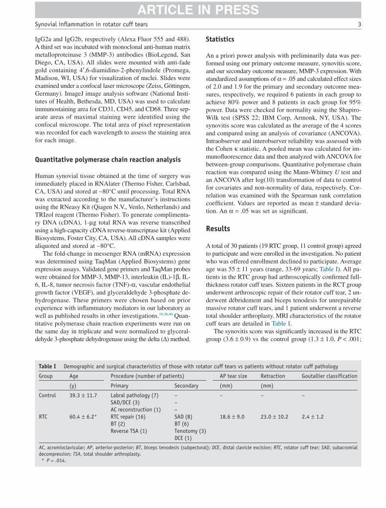
The synovitis score was significantly increased in the RTCgroup (3.6 ± 0.9) vs the control group (1.3 ± 1.0, P < .001;
Table I Demographic and surgical characteristics of those with rotator cuff tears vs patients without rotator cuff pathology
Group Age Procedure (number of patients) AP tear size Retraction Goutallier classification
(y) Primary Secondary (mm) (mm)
Control 39.3 ± 11.7 Labral pathology (7)SAD/DCE (3)AC reconstruction (1)
–––
– – –
RTC 60.4 ± 6.2* RTC repair (16)BT (2)Reverse TSA (1)
SAD (8)BT (6)Tenotomy (3)DCE (1)
18.6 ± 9.0 23.0 ± 10.2 2.4 ± 1.2
AC, acromioclavicular; AP, anterior-posterior; BT, biceps tenodesis (subpectoral); DCE, distal clavicle excision; RTC, rotator cuff tear; SAD, subacromialdecompression; TSA, total shoulder arthroplasty.* P = .014.
ARTICLE IN PRESSSynovial inflammation in rotator cuff tears 3
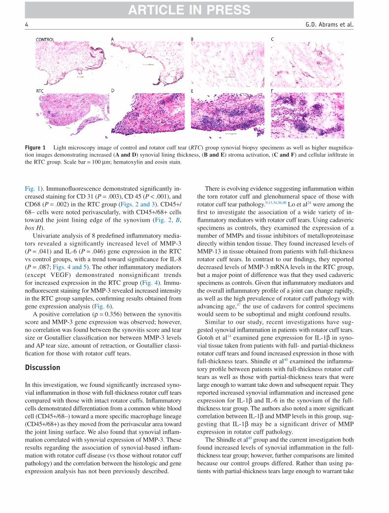
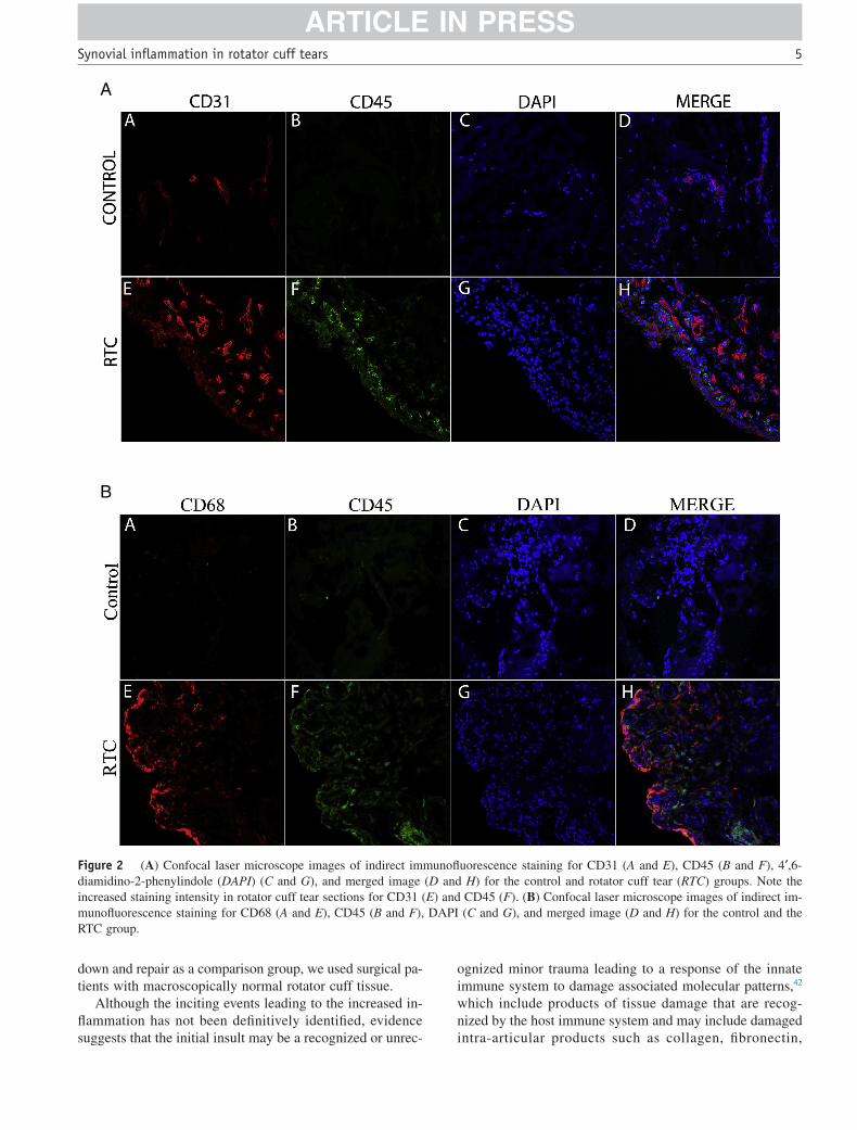
Fig. 1). Immunofluorescence demonstrated significantly in-creased staining for CD 31 (P = .003), CD 45 (P < .001), andCD68 (P = .002) in the RTC group (Figs. 2 and 3). CD45+/68– cells were noted perivascularly, with CD45+/68+ cellstoward the joint lining edge of the synovium (Fig. 2, B,box H).
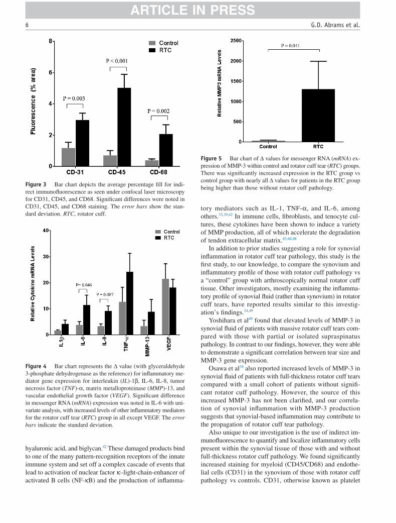
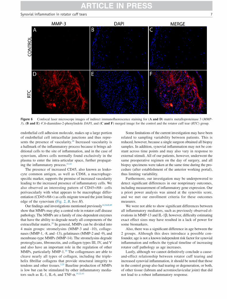
Univariate analysis of 8 predefined inflammatory media-tors revealed a significantly increased level of MMP-3(P = .041) and IL-6 (P = .046) gene expression in the RTCvs control groups, with a trend toward significance for IL-8(P = .087; Figs. 4 and 5). The other inflammatory mediators(except VEGF) demonstrated nonsignificant trendsfor increased expression in the RTC group (Fig. 4). Immu-nofluorescent staining for MMP-3 revealed increased intensityin the RTC group samples, confirming results obtained fromgene expression analysis (Fig. 6).
A positive correlation (ρ = 0.356) between the synovitisscore and MMP-3 gene expression was observed; however,no correlation was found between the synovitis score and tearsize or Goutallier classification nor between MMP-3 levelsand AP tear size, amount of retraction, or Goutallier classi-fication for those with rotator cuff tears.
Discussion
In this investigation, we found significantly increased syno-vial inflammation in those with full-thickness rotator cuff tearscompared with those with intact rotator cuffs. Inflammatorycells demonstrated differentiation from a common white bloodcell (CD45+/68–) toward a more specific macrophage lineage(CD45+/68+) as they moved from the perivascular area towardthe joint lining surface. We also found that synovial inflam-mation correlated with synovial expression of MMP-3. Theseresults regarding the association of synovial-based inflam-mation with rotator cuff disease (vs those without rotator cuffpathology) and the correlation between the histologic and geneexpression analysis has not been previously described.
There is evolving evidence suggesting inflammation withinthe torn rotator cuff and glenohumeral space of those withrotator cuff tear pathology.9,13,34,36,40 Lo et al24 were among thefirst to investigate the association of a wide variety of in-flammatory mediators with rotator cuff tears. Using cadavericspecimens as controls, they examined the expression of anumber of MMPs and tissue inhibitors of metalloproteinasedirectly within tendon tissue. They found increased levels ofMMP-13 in tissue obtained from patients with full-thicknessrotator cuff tears. In contrast to our findings, they reporteddecreased levels of MMP-3 mRNA levels in the RTC group,but a major point of difference was that they used cadavericspecimens as controls. Given that inflammatory mediators andthe overall inflammatory profile of a joint can change rapidly,as well as the high prevalence of rotator cuff pathology withadvancing age,47 the use of cadavers for control specimenswould seem to be suboptimal and might confound results.
Similar to our study, recent investigations have sug-gested synovial inflammation in patients with rotator cuff tears.Gotoh et al14 examined gene expression for IL-1β in syno-vial tissue taken from patients with full- and partial-thicknessrotator cuff tears and found increased expression in those withfull-thickness tears. Shindle et al40 examined the inflamma-tory profile between patients with full-thickness rotator cufftears as well as those with partial-thickness tears that werelarge enough to warrant take down and subsequent repair. Theyreported increased synovial inflammation and increased geneexpression for IL-1β and IL-6 in the synovium of the full-thickness tear group. The authors also noted a more significantcorrelation between IL-1β and MMP levels in this group, sug-gesting that IL-1β may be a significant driver of MMPexpression in rotator cuff pathology.
The Shindle et al40 group and the current investigation bothfound increased levels of synovial inflammation in the full-thickness tear group; however, further comparisons are limitedbecause our control groups differed. Rather than using pa-tients with partial-thickness tears large enough to warrant take
Figure 1 Light microscopy image of control and rotator cuff tear (RTC) group synovial biopsy specimens as well as higher magnifica-tion images demonstrating increased (A and D) synovial lining thickness, (B and E) stroma activation, (C and F) and cellular infiltrate inthe RTC group. Scale bar = 100 μm; hematoxylin and eosin stain.
ARTICLE IN PRESS4 G.D. Abrams et al.
down and repair as a comparison group, we used surgical pa-tients with macroscopically normal rotator cuff tissue.
Although the inciting events leading to the increased in-flammation has not been definitively identified, evidencesuggests that the initial insult may be a recognized or unrec-
ognized minor trauma leading to a response of the innateimmune system to damage associated molecular patterns,42
which include products of tissue damage that are recog-nized by the host immune system and may include damagedintra-articular products such as collagen, fibronectin,
A
B
Figure 2 (A) Confocal laser microscope images of indirect immunofluorescence staining for CD31 (A and E), CD45 (B and F), 4′,6-diamidino-2-phenylindole (DAPI) (C and G), and merged image (D and H) for the control and rotator cuff tear (RTC) groups. Note theincreased staining intensity in rotator cuff tear sections for CD31 (E) and CD45 (F). (B) Confocal laser microscope images of indirect im-munofluorescence staining for CD68 (A and E), CD45 (B and F), DAPI (C and G), and merged image (D and H) for the control and theRTC group.
ARTICLE IN PRESSSynovial inflammation in rotator cuff tears 5
hyaluronic acid, and biglycan.42 These damaged products bindto one of the many pattern-recognition receptors of the innateimmune system and set off a complex cascade of events thatlead to activation of nuclear factor κ–light-chain-enhancer ofactivated B cells (NF-κB) and the production of inflamma-
tory mediators such as IL-1, TNF-α, and IL-6, amongothers.33,39,42 In immune cells, fibroblasts, and tenocyte cul-tures, these cytokines have been shown to induce a varietyof MMP production, all of which accelerate the degradationof tendon extracellular matrix.43,44,48
In addition to prior studies suggesting a role for synovialinflammation in rotator cuff tear pathology, this study is thefirst study, to our knowledge, to compare the synovium andinflammatory profile of those with rotator cuff pathology vsa “control” group with arthroscopically normal rotator cufftissue. Other investigators, mostly examining the inflamma-tory profile of synovial fluid (rather than synovium) in rotatorcuff tears, have reported results similar to this investig-ation’s findings.34,49
Yoshihara et al49 found that elevated levels of MMP-3 insynovial fluid of patients with massive rotator cuff tears com-pared with those with partial or isolated supraspinatuspathology. In contrast to our findings, however, they were ableto demonstrate a significant correlation between tear size andMMP-3 gene expression.
Osawa et al34 also reported increased levels of MMP-3 insynovial fluid of patients with full-thickness rotator cuff tearscompared with a small cohort of patients without signifi-cant rotator cuff pathology. However, the source of thisincreased MMP-3 has not been clarified, and our correla-tion of synovial inflammation with MMP-3 productionsuggests that synovial-based inflammation may contribute tothe propagation of rotator cuff tear pathology.
Also unique to our investigation is the use of indirect im-munofluorescence to quantify and localize inflammatory cellspresent within the synovial tissue of those with and withoutfull-thickness rotator cuff pathology. We found significantlyincreased staining for myeloid (CD45/CD68) and endothe-lial cells (CD31) in the synovium of those with rotator cuffpathology vs controls. CD31, otherwise known as platelet
Figure 3 Bar chart depicts the average percentage fill for indi-rect immunofluorescence as seen under confocal laser microscopyfor CD31, CD45, and CD68. Significant differences were noted inCD31, CD45, and CD68 staining. The error bars show the stan-dard deviation. RTC, rotator cuff.
Figure 4 Bar chart represents the Δ value (with glyceraldehyde3-phosphate dehydrogenase as the reference) for inflammatory me-diator gene expression for interleukin (IL)-1β, IL-6, IL-8, tumornecrosis factor (TNF)-α, matrix metalloproteinase (MMP)-13, andvascular endothelial growth factor (VEGF). Significant differencein messenger RNA (mRNA) expression was noted in IL-6 with uni-variate analysis, with increased levels of other inflammatory mediatorsfor the rotator cuff tear (RTC) group in all except VEGF. The errorbars indicate the standard deviation.
Figure 5 Bar chart of Δ values for messenger RNA (mRNA) ex-pression of MMP-3 within control and rotator cuff tear (RTC) groups.There was significantly increased expression in the RTC group vscontrol group with nearly all Δ values for patients in the RTC groupbeing higher than those without rotator cuff pathology.
ARTICLE IN PRESS6 G.D. Abrams et al.
endothelial cell adhesion molecule, makes up a large portionof endothelial cell intracellular junctions and thus repre-sents the presence of vascularity.46 Increased vascularity isa hallmark of the inflammatory process because it brings ad-ditional cells to the site of inflammation, and in the case ofsynovium, allows cells normally found exclusively in theplasma to enter the intra-articular space, further propagat-ing the inflammatory process.41,42
The presence of increased CD45, also known as leuko-cyte common antigen, as well as CD68, a macrophage-specific marker, supports the premise of increased vascularityleading to the increased presence of inflammatory cells. Wealso observed an interesting pattern of CD45+/68– cellsperivascularly with what appears to be macrophage differ-entiation (CD45+/68+) as cells migrate toward the joint liningedge of the synovium (Fig. 2, B, box H).
Our findings and investigations mentioned previously14,24,40,49
show that MMPs may play a central role in rotator cuff diseasepathology. The MMPs are a family of zinc-dependent enzymesthat have the ability to degrade nearly all components of theextracellular matrix.28 In general, MMPs can be divided into4 main groups: stromelysins (MMP-3 and -10), collage-nases (MMP-1, -8, and -13), gelatinases (MMP-2 and -9), andmembrane-type MMPs (MMP-14). The stromelysins degradeproteoglycans, fibronectin, and collagen types III, IV, and Vand also have an important role in the regulation of otherMMPs, particularly MMP-1.10 The collagenases are able tocleave nearly all types of collagen, including the triple-helix fibrillar collagens that provide structural integrity totendons and other tissues.2,25 Baseline production of MMPsis low but can be stimulated by other inflammatory media-tors such as IL-1, IL-6, and TNF-α.19,22,43
Some limitations of the current investigation may have beenrelated to sampling variability between patients. This isreduced, however, because a single surgeon obtained all biopsysamples. In addition, synovial inflammation may not be con-stant across time points and may also vary in response toexternal stimuli. All of our patients, however, underwent thesame preoperative regimen on the day of surgery, and allbiopsy specimens were taken at the same time during the pro-cedure (after establishment of the anterior working portal),thus limiting variability.
Furthermore, our investigation may be underpowered todetect significant differences in our nonprimary outcomes,including measurement of inflammatory gene expression. Oura priori power analysis was aimed at the synovitis score,and we met our enrollment criteria for these outcomesmeasures.
We were not able to show significant differences betweenall inflammatory mediators, such as previously observed el-evations in MMP-13 and IL-1β; however, difficulty estimatingexact effect sizes may have resulted in a lack of power forsome biomarkers.
Also, there was a significant difference in age between the2 groups. Although this does introduce a possible con-founder, age is not a known independent risk factor for synovialinflammation and reflects the typical timeline of increasingrotator cuff pathology as age increases.
Lastly, although we cannot definitively conclude a cause-and-effect relationship between rotator cuff tearing andincreased synovial inflammation, it should be noted that thosein the control group also had tearing or degeneration, or both,of other tissue (labrum and acromioclavicular joint) that didnot lead to a robust inflammatory response.
Figure 6 Confocal laser microscope images of indirect immunofluorescence staining for (A and D) matrix metalloproteinase 3 (MMP-3), (B and E) 4′,6-diamidino-2-phenylindole DAPI, and (C and F) merged image for the control and the rotator cuff tear (RTC) group.
ARTICLE IN PRESSSynovial inflammation in rotator cuff tears 7

The relatively small sample size required to observe sig-nificance for MMP-3 provides evidence for the potential roleof MMP-3 in the development or, or both, propagation ofrotator cuff pathology. Although prior studies have not re-ported increased MMP-3 levels in the setting of full-thicknessrotator cuff tears, these studies often used tendon tissue, sy-novial fluid, or synovium from full thickness tears comparedwith those with partial-thickness tears.36,40,49 Given the evi-dence for MMP-3 as not only a protease but also a regulatorof other MMP activity,10 this may represent an early stagewithin the inflammatory cascade that leads to activation ofother MMPs and propagation of rotator cuff tendon destruction.
Conclusion
We observed robust synovial inflammation and increasedinflammatory gene expression in those with full-thicknessrotator cuff tears vs those without rotator cuff pathology.These data identify the synovium as a potential player inthe pathogenesis of rotator cuff pathology and may guidethe development of therapies to reduce pain and preventprogression of pathology associated with RCT disease.
Disclaimer
The authors, their immediate families, and any researchfoundations with which they are affiliated have not re-ceived any financial payments or other benefits from anycommercial entity related to the subject of this article.
References
1. Bedi A, Fox AJ, Kovacevic D, Deng XH, Warren RF, Rodeo SA.Doxycycline-mediated inhibition of matrix metalloproteinases improveshealing after rotator cuff repair. Am J Sports Med 2010;38:308-17.http://dx.doi.org/10.1177/0363546509347366
2. Bertini I, Fragai M, Luchinat C, Melikian M, Toccafondi M, Lauer JL,et al. Structural basis for matrix metalloproteinase 1-catalyzedcollagenolysis. J Am Chem Soc 2012;134:2100-10. http://dx.doi.org/10.1021/ja208338j
3. Blaine TA, Kim YS, Voloshin I, Chen D, Murakami K, Chang SS, et al.The molecular pathophysiology of subacromial bursitis in rotator cuffdisease. J Shoulder Elbow Surg 2005;14:84S-89S. http://dx.doi.org/10.1016/j.jse.2004.09.022
4. Burrage PS, Mix KS, Brinckerhoff CE. Matrix metalloproteinases: rolein arthritis. Front Biosci 2006;11:529-43.
5. Cabuk H, Avci A, Durmaz H, Cabuk FK, Ertem F, Muhittin Sener I.The effect of diclofenac on matrix metalloproteinase levels in the rotatorcuff. Arch Orthop Trauma Surg 2014;134:1739-44. http://dx.doi.org/10.1007/s00402-014-2099-0
6. Castagna A, Cesari E, Garofalo R, Gigante A, Conti M, MarkopoulosN, et al. Matrix metalloproteases and their inhibitors are altered in tornrotator cuff tendons, but also in the macroscopically and histologicallyintact portion of those tendons. Muscles Ligaments Tendons J2013;3:132-8.
7. Castagna A, Cesari E, Gigante A, Conti M, Garofalo R. Metalloproteasesand their inhibitors are altered in both torn and intact rotator cuff tendons.
Musculoskelet Surg 2013;97(Suppl 1):39-47. http://dx.doi.org/10.1007/s12306-013-0264-1
8. Chakravarty K, Webley M. Shoulder joint movement and its relationshipto disability in the elderly. J Rheumatol 1993;20:1359-61.
9. Choi HR, Kondo S, Hirose K, Ishiguro N, Hasegawa Y, Iwata H.Expression and enzymatic activity of MMP-2 during healing processof the acute supraspinatus tendon tear in rabbits. J Orthop Res2002;20:927-33. http://dx.doi.org/10.1016/S0736-0266(02)00016-5
10. Del Buono A, Oliva F, Longo UG, Rodeo SA, Orchard J, Denaro V,et al. Metalloproteases and rotator cuff disease. J Shoulder Elbow Surg2012;21:200-8. http://dx.doi.org/10.1016/j.jse.2011.10.020
11. Fan L, Sarkar K, Franks DJ, Uhthoff HK. Estimation of total collagenand types I and III collagen in canine rotator cuff tendons. Calcif TissueInt 1997;61:223-9.
12. Fuchs B, Weishaupt D, Zanetti M, Hodler J, Gerber C. Fatty degenerationof the muscles of the rotator cuff: assessment by computed tomographyversus magnetic resonance imaging. J Shoulder Elbow Surg 1999;8:599-605.
13. Gotoh M, Hamada K, Yamakawa H, Nakamura M, Yamazaki H, UeyamaY, et al. Perforation of rotator cuff increases interleukin 1beta productionin the synovium of glenohumeral joint in rotator cuff diseases. JRheumatol 2000;27:2886-92.
14. Gotoh M, Hamada K, Yamakawa H, Yanagisawa K, Nakamura M,Yamazaki H, et al. Interleukin-1-induced glenohumeral synovitis andshoulder pain in rotator cuff diseases. J Orthop Res 2002;20:1365-71.http://dx.doi.org/10.1016/S0736-0266(02)00063-3
15. Gulotta LV, Kovacevic D, Montgomery S, Ehteshami JR, Packer JD,Rodeo SA. Stem cells genetically modified with the developmental geneMT1-MMP improve regeneration of the supraspinatus tendon-to-boneinsertion site. Am J Sports Med 2010;38:1429-37. http://dx.doi.org/10.1177/0363546510361235
16. Gumina S, Arceri V, Carbone S, Albino P, Passaretti D, Campagna V,et al. The association between arterial hypertension and rotator cuff tear:the influence on rotator cuff tear sizes. J Shoulder Elbow Surg2013;22:229-32. http://dx.doi.org/10.1016/j.jse.2012.05.023
17. Gupta N, Gupta ND, Gupta A, Khan S, Bansal N. Role of salivary matrixmetalloproteinase-8 (MMP-8) in chronic periodontitis diagnosis. FrontMed 2015;9:72-6. http://dx.doi.org/10.1007/s11684-014-0347-x
18. Johnson JL, Jenkins NP, Huang WC, Di Gregoli K, Sala-Newby GB,Scholtes VP, et al. Relationship of MMP-14 and TIMP-3 expressionwith macrophage activation and human atherosclerotic plaquevulnerability. Mediators Inflamm 2014;2014:276457. http://dx.doi.org/10.1155/2014/276457
19. Kelley MJ, Rose AY, Song K, Chen Y, Bradley JM, Rookhuizen D,et al. Synergism of TNF and IL-1 in the induction of matrixmetalloproteinase-3 in trabecular meshwork. Invest Ophthalmol Vis Sci2007;48:2634-43. http://dx.doi.org/10.1167/iovs.06-1445
20. Krenn V, Morawietz L, Burmester GR, Kinne RW, Mueller-Ladner U,Muller B, et al. Synovitis score: discrimination between chroniclow-grade and high-grade synovitis. Histopathology 2006;49:358-64.http://dx.doi.org/10.1111/j.1365-2559.2006.02508.x
21. Krenn V, Morawietz L, Haupl T, Neidel J, Petersen I, Konig A. Gradingof chronic synovitis–a histopathological grading system for molecularand diagnostic pathology. Pathol Res Pract 2002;198:317-25.http://dx.doi.org/10.1078/0344-0338-5710261
22. Kusano K, Miyaura C, Inada M, Tamura T, Ito A, Nagase H, et al.Regulation of matrix metalloproteinases (MMP-2, -3, -9, and -13) byinterleukin-1 and interleukin-6 in mouse calvaria: association of MMPinduction with bone resorption. Endocrinology 1998;139:1338-45.
23. Lillie RD, Pizzolato P, Welsh RA, Holmquist ND, Donaldson PT, BergerC. A consideration of substitutes for alum hematoxylin in routinehistologic and cytologic diagnostic procedures. Am J Clin Pathol1973;60:817-9.
24. Lo IK, Marchuk LL, Hollinshead R, Hart DA, Frank CB. Matrixmetalloproteinase and tissue inhibitor of matrix metalloproteinase mRNAlevels are specifically altered in torn rotator cuff tendons. Am J SportsMed 2004;32:1223-9. http://dx.doi.org/10.1177/0363546503262200
ARTICLE IN PRESS8 G.D. Abrams et al.
25. Manka SW, Carafoli F, Visse R, Bihan D, Raynal N, Farndale RW, et al.Structural insights into triple-helical collagen cleavage by matrixmetalloproteinase 1. Proc Natl Acad Sci U S A 2012;109:12461-6.http://dx.doi.org/10.1073/pnas.1204991109
26. Martinez-Aguilar E, Gomez-Rodriguez V, Orbe J, Rodriguez JA,Fernandez-Alonso L, Roncal C, et al. Matrix metalloproteinase 10 isassociated with disease severity and mortality in patients with peripheralarterial disease. J Vasc Surg 2015;61:428-35. http://dx.doi.org/10.1016/j.jvs.2014.09.002
27. Millar NL, Wei AQ, Molloy TJ, Bonar F, Murrell GA. Cytokines andapoptosis in supraspinatus tendinopathy. J Bone Joint Surg Br2009;91:417-24. http://dx.doi.org/10.1302/0301-620X.91B3.21652
28. Nagase H, Woessner JF Jr. Matrix metalloproteinases. J Biol Chem1999;274:21491-4.
29. Nagasupriya A, Rao DB, Ravikanth M, Kumar NG, Ramachandran CR,Saraswathi TR. Immunohistochemical expression of matrixmetalloproteinase 13 in chronic periodontitis. Int J PeriodonticsRestorative Dent 2014;34:e79-84. http://dx.doi.org/10.11607/prd.1922
30. Neer CS 2nd. Impingement lesions. Clin Orthop Relat Res 1983;70-7.
31. Neviaser A, Andarawis-Puri N, Flatow E. Basic mechanisms of tendonfatigue damage. J Shoulder Elbow Surg 2012;21:158-63.http://dx.doi.org/10.1016/j.jse.2011.11.014
32. Oh LS, Wolf BR, Hall MP, Levy BA, Marx RG. Indications for rotatorcuff repair: a systematic review. Clin Orthop Relat Res 2007;455:52-63.http://dx.doi.org/10.1097/BLO.0b013e31802fc175
33. Okamura Y, Watari M, Jerud ES, Young DW, Ishizaka ST, Rose J, et al.The extra domain A of fibronectin activates Toll-like receptor 4. J BiolChem 2001;276:10229-33.
34. Osawa T, Shinozaki T, Takagishi K. Multivariate analysis of biochemicalmarkers in synovial fluid from the shoulder joint for diagnosis of rotatorcuff tears. Rheumatol Int 2005;25:436-41. http://dx.doi.org/10.1007/s00296-004-0509-2
35. Rhee YG, Cho NS, Yoo JH. Clinical outcome and repair integrity afterrotator cuff repair in patients older than 70 years versus patients youngerthan 70 years. Arthroscopy 2014;30:546-54. http://dx.doi.org/10.1016/j.arthro.2014.02.006
36. Robertson CM, Chen CT, Shindle MK, Cordasco FA, Rodeo SA, WarrenRF. Failed healing of rotator cuff repair correlates with alteredcollagenase and gelatinase in supraspinatus and subscapularis tendons.Am J Sports Med 2012;40:1993-2001. http://dx.doi.org/10.1177/0363546512456519
37. Scanzello CR, Albert AS, DiCarlo E, Rajan KB, Kanda V, AsomughaEU, et al. The influence of synovial inflammation and hyperplasia onsymptomatic outcomes up to 2 years post-operatively in patientsundergoing partial meniscectomy. Osteoarthritis Cartilage 2013;21:1392-9. http://dx.doi.org/10.1016/j.joca.2013.05.011
38. Scanzello CR, McKeon B, Swaim BH, DiCarlo E, Asomugha EU, KandaV, et al. Synovial inflammation in patients undergoing arthroscopicmeniscectomy: molecular characterization and relationship to symptoms.Arthritis Rheum 2011;63:391-400. http://dx.doi.org/10.1002/art.30137
39. Schaefer L, Babelova A, Kiss E, Hausser HJ, Baliova M, KrzyzankovaM, et al. The matrix component biglycan is proinflammatory and signalsthrough Toll-like receptors 4 and 2 in macrophages. J Clin Invest2005;115:2223-33. http://dx.doi.org/10.1172/JCI23755
40. Shindle MK, Chen CC, Robertson C, DiTullio AE, Paulus MC, ClintonCM, et al. Full-thickness supraspinatus tears are associated with moresynovial inflammation and tissue degeneration than partial-thickness tears.J Shoulder Elbow Surg 2011;20:917-27. http://dx.doi.org/10.1016/j.jse.2011.02.015
41. Sohn DH, Sokolove J, Sharpe O, Erhart JC, Chandra PE, Lahey LJ, et al.Plasma proteins present in osteoarthritic synovial fluid can stimulatecytokine production via Toll-like receptor 4. Arthritis Res Ther2012;14:R7. http://dx.doi.org/10.1186/ar3555
42. Sokolove J, Lepus CM. Role of inflammation in the pathogenesis ofosteoarthritis: latest findings and interpretations. Ther Adv MusculoskeletDis 2013;5:77-94. http://dx.doi.org/10.1177/1759720X12467868
43. Tsuzaki M, Guyton G, Garrett W, Archambault JM, Herzog W,Almekinders L, et al. IL-1 beta induces COX2, MMP-1, -3 and -13,ADAMTS-4, IL-1 beta and IL-6 in human tendon cells. J Orthop Res2003;21:256-64. http://dx.doi.org/10.1016/S0736-0266(02)00141-9
44. Vincenti MP, Brinckerhoff CE. Transcriptional regulation of collagenase(MMP-1, MMP-13) genes in arthritis: integration of complex signalingpathways for the recruitment of gene-specific transcription factors.Arthritis Res 2002;4:157-64.
45. Voloshin I, Gelinas J, Maloney MD, O’Keefe RJ, Bigliani LU, BlaineTA. Proinflammatory cytokines and metalloproteases are expressed inthe subacromial bursa in patients with rotator cuff disease. Arthroscopy2005;21:1076.e1-9. http://dx.doi.org/10.1016/j.arthro.2005.05.017
46. Xie Y, Muller WA. Fluorescence in situ hybridization mapping of themouse platelet endothelial cell adhesion molecule-1 (PECAM1) to mousechromosome 6, region F3-G1. Genomics 1996;37:226-8.
47. Yamaguchi K, Ditsios K, Middleton WD, Hildebolt CF, Galatz LM,Teefey SA. The demographic and morphological features of rotator cuffdisease. A comparison of asymptomatic and symptomatic shoulders.J Bone Joint Surg Am 2006;88:1699-704. http://dx.doi.org/10.2106/JBJS.E.00835
48. Yang G, Im HJ, Wang JH. Repetitive mechanical stretching modulatesIL-1beta induced COX-2, MMP-1 expression, and PGE2 production inhuman patellar tendon fibroblasts. Gene 2005;363:166-72.http://dx.doi.org/10.1016/j.gene.2005.08.006
49. Yoshihara Y, Hamada K, Nakajima T, Fujikawa K, Fukuda H.Biochemical markers in the synovial fluid of glenohumeral joints frompatients with rotator cuff tear. J Orthop Res 2001;19:573-9.
ARTICLE IN PRESSSynovial inflammation in rotator cuff tears 9