
ASSOCIATE EDITOR: ANNETTE C. DOLPHIN...
33
ASSOCIATE EDITOR: ANNETTE C. DOLPHIN Unravelling the Mystery of Capsaicin: A Tool to Understand and Treat Pain Jessica O’Neill, Christina Brock, Anne Estrup Olesen, Trine Andresen, Matias Nilsson, and Anthony H. Dickenson Neuroscience, Physiology and Pharmacology, University College London (J.O., A.H.D.); and Mech-Sense, Department of Gastroenterology, Aalborg Hospital, Aarhus University, Denmark (C.B., A.E.O., T.A., M.N.) Abstract............................................................................... 940 I. Introduction ........................................................................... 940 II. Physical and chemical properties of capsaicin ............................................. 942 III. Pharmacokinetics of capsaicin ........................................................... 942 A. Oral administration ................................................................. 942 B. Systemic administration ............................................................. 943 C. Topical administration .............................................................. 943 D. Intradermal administration .......................................................... 945 IV. Capsaicin metabolism .................................................................. 945 V. Capsaicin elimination .................................................................. 945 VI. Capsaicin pharmacology ................................................................ 945 VII. Transient receptor potential channels .................................................... 946 VIII. Transient receptor potential vanilloid 1 .................................................. 946 A. Introduction ........................................................................ 946 B. Transient receptor potential vanilloid 1-associated molecules ............................ 947 1. Phosphatidylinositol 4,5-bisphosphate .............................................. 947 2. Cytoskeleton .................................................................... 947 C. In the periphery .................................................................... 947 D. In the viscera ...................................................................... 949 E. In the spinal cord ................................................................... 949 F. Supraspinal ........................................................................ 950 G. Non-neuronal....................................................................... 950 IX. Transient receptor potential vanilloid 1 splice variants .................................... 951 X. Transient receptor potential vanilloid 1 polymorphisms .................................... 951 XI. Transient receptor potential vanilloid 1 receptor expression in humans in the airways, skin, and viscera ....................................................................... 952 A. Airways............................................................................ 952 B. Skin ............................................................................... 952 C. Gastrointestinal tract ............................................................... 952 XII. Experimental pain models .............................................................. 952 A. Animals ........................................................................... 952 B. Humans ........................................................................... 955 C. Superficial somatic pain ............................................................. 955 D. Experimental deep somatic pain...................................................... 957 E. Human visceral studies ............................................................. 960 XIII. Sensitizing or desensitizing? ............................................................ 963 A. Altered skin innervation after transient receptor potential vanilloid 1 activation .......... 964 Address correspondence to: Jessica O’Neill, Neuroscience, Physiology and Pharmacology, University College London, Gower Street, London WC1E 6BT. E-mail: [email protected] This article is available online at http://pharmrev.aspetjournals.org. http://dx.doi.org/10.1124/pr.112.006163. 1521-0081/12/6404-939 –971$25.00 PHARMACOLOGICAL REVIEWS Vol. 64, No. 4 Copyright © 2012 by The American Society for Pharmacology and Experimental Therapeutics 6163/3789549 Pharmacol Rev 64:939 –971, 2012 939 by guest on May 12, 2018 Downloaded from
Transcript of ASSOCIATE EDITOR: ANNETTE C. DOLPHIN...

ASSOCIATE EDITOR: ANNETTE C. DOLPHIN
Unravelling the Mystery of Capsaicin: A Tool toUnderstand and Treat Pain
Jessica O’Neill, Christina Brock, Anne Estrup Olesen, Trine Andresen, Matias Nilsson, and Anthony H. Dickenson
Neuroscience, Physiology and Pharmacology, University College London (J.O., A.H.D.); and Mech-Sense, Department of Gastroenterology,Aalborg Hospital, Aarhus University, Denmark (C.B., A.E.O., T.A., M.N.)
Abstract. . . . . . . . . . . . . . . . . . . . . . . . . . . . . . . . . . . . . . . . . . . . . . . . . . . . . . . . . . . . . . . . . . . . . . . . . . . . . . . 940I. Introduction . . . . . . . . . . . . . . . . . . . . . . . . . . . . . . . . . . . . . . . . . . . . . . . . . . . . . . . . . . . . . . . . . . . . . . . . . . . 940
II. Physical and chemical properties of capsaicin . . . . . . . . . . . . . . . . . . . . . . . . . . . . . . . . . . . . . . . . . . . . . 942III. Pharmacokinetics of capsaicin . . . . . . . . . . . . . . . . . . . . . . . . . . . . . . . . . . . . . . . . . . . . . . . . . . . . . . . . . . . 942
A. Oral administration . . . . . . . . . . . . . . . . . . . . . . . . . . . . . . . . . . . . . . . . . . . . . . . . . . . . . . . . . . . . . . . . . 942B. Systemic administration. . . . . . . . . . . . . . . . . . . . . . . . . . . . . . . . . . . . . . . . . . . . . . . . . . . . . . . . . . . . . 943C. Topical administration . . . . . . . . . . . . . . . . . . . . . . . . . . . . . . . . . . . . . . . . . . . . . . . . . . . . . . . . . . . . . . 943D. Intradermal administration . . . . . . . . . . . . . . . . . . . . . . . . . . . . . . . . . . . . . . . . . . . . . . . . . . . . . . . . . . 945
IV. Capsaicin metabolism . . . . . . . . . . . . . . . . . . . . . . . . . . . . . . . . . . . . . . . . . . . . . . . . . . . . . . . . . . . . . . . . . . 945V. Capsaicin elimination . . . . . . . . . . . . . . . . . . . . . . . . . . . . . . . . . . . . . . . . . . . . . . . . . . . . . . . . . . . . . . . . . . 945
VI. Capsaicin pharmacology . . . . . . . . . . . . . . . . . . . . . . . . . . . . . . . . . . . . . . . . . . . . . . . . . . . . . . . . . . . . . . . . 945VII. Transient receptor potential channels . . . . . . . . . . . . . . . . . . . . . . . . . . . . . . . . . . . . . . . . . . . . . . . . . . . . 946
VIII. Transient receptor potential vanilloid 1 . . . . . . . . . . . . . . . . . . . . . . . . . . . . . . . . . . . . . . . . . . . . . . . . . . 946A. Introduction. . . . . . . . . . . . . . . . . . . . . . . . . . . . . . . . . . . . . . . . . . . . . . . . . . . . . . . . . . . . . . . . . . . . . . . . 946B. Transient receptor potential vanilloid 1-associated molecules. . . . . . . . . . . . . . . . . . . . . . . . . . . . 947
1. Phosphatidylinositol 4,5-bisphosphate. . . . . . . . . . . . . . . . . . . . . . . . . . . . . . . . . . . . . . . . . . . . . . 9472. Cytoskeleton . . . . . . . . . . . . . . . . . . . . . . . . . . . . . . . . . . . . . . . . . . . . . . . . . . . . . . . . . . . . . . . . . . . . 947
C. In the periphery . . . . . . . . . . . . . . . . . . . . . . . . . . . . . . . . . . . . . . . . . . . . . . . . . . . . . . . . . . . . . . . . . . . . 947D. In the viscera . . . . . . . . . . . . . . . . . . . . . . . . . . . . . . . . . . . . . . . . . . . . . . . . . . . . . . . . . . . . . . . . . . . . . . 949E. In the spinal cord . . . . . . . . . . . . . . . . . . . . . . . . . . . . . . . . . . . . . . . . . . . . . . . . . . . . . . . . . . . . . . . . . . . 949F. Supraspinal . . . . . . . . . . . . . . . . . . . . . . . . . . . . . . . . . . . . . . . . . . . . . . . . . . . . . . . . . . . . . . . . . . . . . . . . 950G. Non-neuronal. . . . . . . . . . . . . . . . . . . . . . . . . . . . . . . . . . . . . . . . . . . . . . . . . . . . . . . . . . . . . . . . . . . . . . . 950
IX. Transient receptor potential vanilloid 1 splice variants . . . . . . . . . . . . . . . . . . . . . . . . . . . . . . . . . . . . 951X. Transient receptor potential vanilloid 1 polymorphisms . . . . . . . . . . . . . . . . . . . . . . . . . . . . . . . . . . . . 951
XI. Transient receptor potential vanilloid 1 receptor expression in humans in the airways,skin, and viscera . . . . . . . . . . . . . . . . . . . . . . . . . . . . . . . . . . . . . . . . . . . . . . . . . . . . . . . . . . . . . . . . . . . . . . . 952A. Airways. . . . . . . . . . . . . . . . . . . . . . . . . . . . . . . . . . . . . . . . . . . . . . . . . . . . . . . . . . . . . . . . . . . . . . . . . . . . 952B. Skin . . . . . . . . . . . . . . . . . . . . . . . . . . . . . . . . . . . . . . . . . . . . . . . . . . . . . . . . . . . . . . . . . . . . . . . . . . . . . . . 952C. Gastrointestinal tract . . . . . . . . . . . . . . . . . . . . . . . . . . . . . . . . . . . . . . . . . . . . . . . . . . . . . . . . . . . . . . . 952
XII. Experimental pain models . . . . . . . . . . . . . . . . . . . . . . . . . . . . . . . . . . . . . . . . . . . . . . . . . . . . . . . . . . . . . . 952A. Animals . . . . . . . . . . . . . . . . . . . . . . . . . . . . . . . . . . . . . . . . . . . . . . . . . . . . . . . . . . . . . . . . . . . . . . . . . . . 952B. Humans . . . . . . . . . . . . . . . . . . . . . . . . . . . . . . . . . . . . . . . . . . . . . . . . . . . . . . . . . . . . . . . . . . . . . . . . . . . 955C. Superficial somatic pain . . . . . . . . . . . . . . . . . . . . . . . . . . . . . . . . . . . . . . . . . . . . . . . . . . . . . . . . . . . . . 955D. Experimental deep somatic pain. . . . . . . . . . . . . . . . . . . . . . . . . . . . . . . . . . . . . . . . . . . . . . . . . . . . . . 957E. Human visceral studies . . . . . . . . . . . . . . . . . . . . . . . . . . . . . . . . . . . . . . . . . . . . . . . . . . . . . . . . . . . . . 960
XIII. Sensitizing or desensitizing? . . . . . . . . . . . . . . . . . . . . . . . . . . . . . . . . . . . . . . . . . . . . . . . . . . . . . . . . . . . . 963A. Altered skin innervation after transient receptor potential vanilloid 1 activation . . . . . . . . . . 964
Address correspondence to: Jessica O’Neill, Neuroscience, Physiology and Pharmacology, University College London, Gower Street,London WC1E 6BT. E-mail: [email protected]
This article is available online at http://pharmrev.aspetjournals.org.http://dx.doi.org/10.1124/pr.112.006163.
1521-0081/12/6404-939–971$25.00PHARMACOLOGICAL REVIEWS Vol. 64, No. 4Copyright © 2012 by The American Society for Pharmacology and Experimental Therapeutics 6163/3789549Pharmacol Rev 64:939–971, 2012
939
by guest on May 12, 2018
Dow
nloaded from
XIV. Capsaicin as a therapeutic pharmacological agent . . . . . . . . . . . . . . . . . . . . . . . . . . . . . . . . . . . . . . . . . 964XV. Conclusion . . . . . . . . . . . . . . . . . . . . . . . . . . . . . . . . . . . . . . . . . . . . . . . . . . . . . . . . . . . . . . . . . . . . . . . . . . . . 967
Acknowledgments . . . . . . . . . . . . . . . . . . . . . . . . . . . . . . . . . . . . . . . . . . . . . . . . . . . . . . . . . . . . . . . . . . . . . . 967References . . . . . . . . . . . . . . . . . . . . . . . . . . . . . . . . . . . . . . . . . . . . . . . . . . . . . . . . . . . . . . . . . . . . . . . . . . . . 967
Abstract——A large number of pharmacologicalstudies have used capsaicin as a tool to activate manyphysiological systems, with an emphasis on pain re-search but also including functions such as the cardio-vascular system, the respiratory system, and the uri-nary tract. Understanding the actions of capsaicin ledto the discovery its receptor, transient receptor poten-tial (TRP) vanilloid subfamily member 1 (TRPV1), partof the superfamily of TRP receptors, sensing externalevents. This receptor is found on key fine sensory af-ferents, and so the use of capsaicin to selectively acti-vate pain afferents has been exploited in animal stud-ies, human psychophysics, and imaging studies. Itseffects depend on the dose and route of administrationand may include sensitization, desensitization, with-
drawal of afferent nerve terminals, or even overtdeath of afferent fibers. The ability of capsaicin togenerate central hypersensitivity has been valuable inunderstanding the consequences and mechanisms be-hind enhanced central processing of pain. In addition,capsaicin has been used as a therapeutic agent whenapplied topically, and antagonists of the TRPV1 recep-tor have been developed. Overall, the numerous usesfor capsaicin are clear; hence, the rationale of thisreview is to bring together and discuss the differenttypes of studies that exploit these actions to shed lightupon capsaicin working both as a tool to understandpain but also as a treatment for chronic pain. Thisreview will discuss the various actions of capsaicinand how it lends itself to these different purposes.
I. Introduction
Capsicum frutescens is a vegetable used daily, and thesubstance capsaicin is responsible for its hot and spicyflavor, sought after in gastronomy. Capsaicin and sev-eral related molecules are known by the collective namecapsaicinoids, and they are produced by all plants of thegenus Capsicum, with the exception of the bell pepper(Capsicum annum), which produces no capsaicin.
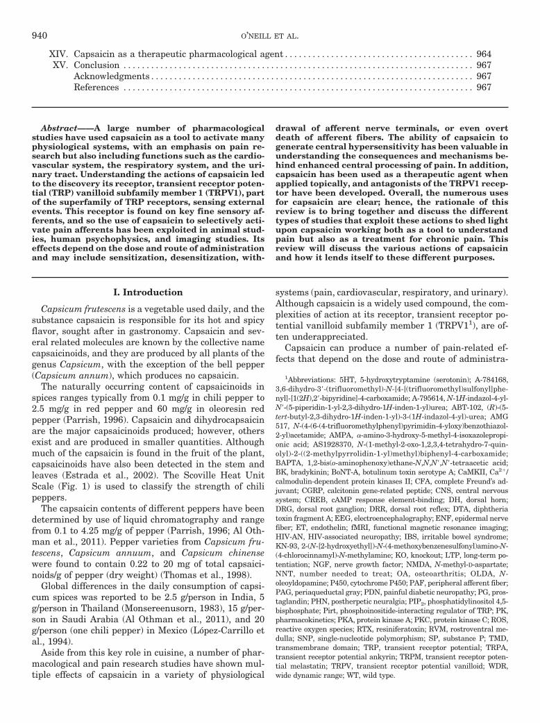
The naturally occurring content of capsaicinoids inspices ranges typically from 0.1 mg/g in chili pepper to2.5 mg/g in red pepper and 60 mg/g in oleoresin redpepper (Parrish, 1996). Capsaicin and dihydrocapsaicinare the major capsaicinoids produced; however, othersexist and are produced in smaller quantities. Althoughmuch of the capsaicin is found in the fruit of the plant,capsaicinoids have also been detected in the stem andleaves (Estrada et al., 2002). The Scoville Heat UnitScale (Fig. 1) is used to classify the strength of chilipeppers.
The capsaicin contents of different peppers have beendetermined by use of liquid chromatography and rangefrom 0.1 to 4.25 mg/g of pepper (Parrish, 1996; Al Oth-man et al., 2011). Pepper varieties from Capsicum fru-tescens, Capsicum annuum, and Capsicum chinensewere found to contain 0.22 to 20 mg of total capsaici-noids/g of pepper (dry weight) (Thomas et al., 1998).
Global differences in the daily consumption of capsi-cum spices was reported to be 2.5 g/person in India, 5g/person in Thailand (Monsereenusorn, 1983), 15 g/per-son in Saudi Arabia (Al Othman et al., 2011), and 20g/person (one chili pepper) in Mexico (Lopez-Carrillo etal., 1994).
Aside from this key role in cuisine, a number of phar-macological and pain research studies have shown mul-tiple effects of capsaicin in a variety of physiological
systems (pain, cardiovascular, respiratory, and urinary).Although capsaicin is a widely used compound, the com-plexities of action at its receptor, transient receptor po-tential vanilloid subfamily member 1 (TRPV11), are of-ten underappreciated.
Capsaicin can produce a number of pain-related ef-fects that depend on the dose and route of administra-
1Abbreviations: 5HT, 5-hydroxytryptamine (serotonin); A-784168,3,6-dihydro-3�-(trifluoromethyl)-N-[4-[(trifluoromethyl)sulfonyl]phe-nyl]-[1(2H),2�-bipyridine]-4-carboxamide; A-795614, N-1H-indazol-4-yl-N�-(5-piperidin-1-yl-2,3-dihydro-1H-inden-1-yl)urea; ABT-102, (R)-(5-tert-butyl-2,3-dihydro-1H-inden-1-yl)-3-(1H-indazol-4-yl)-urea; AMG517, N-(4-(6-(4-trifluoromethylphenyl)pyrimidin-4-yloxy)benzothiazol-2-yl)acetamide; AMPA, �-amino-3-hydroxy-5-methyl-4-isoxazolepropi-onic acid; AS1928370, N-(1-methyl-2-oxo-1,2,3,4-tetrahydro-7-quin-olyl)-2-((2-methylpyrrolidin-1-yl)methyl)biphenyl-4-carboxamide;BAPTA, 1,2-bis(o-aminophenoxy)ethane-N,N,N�,N�-tetraacetic acid;BK, bradykinin; BoNT-A, botulinum toxin serotype A; CaMKII, Ca2�/calmodulin-dependent protein kinases II; CFA, complete Freund’s ad-juvant; CGRP, calcitonin gene-related peptide; CNS, central nervoussystem; CREB, cAMP response element-binding; DH, dorsal horn;DRG, dorsal root ganglion; DRR, dorsal root reflex; DTA, diphtheriatoxin fragment A; EEG, electroencephalography; ENF, epidermal nervefiber; ET, endothelin; fMRI, functional magnetic resonance imaging;HIV-AN, HIV-associated neuropathy; IBS, irritable bowel syndrome;KN-93, 2-(N-[2-hydroxyethyl])-N-(4-methoxybenzenesulfonyl)amino-N-(4-chlorocinnamyl)-N-methylamine; KO, knockout; LTP, long-term po-tentiation; NGF, nerve growth factor; NMDA, N-methyl-D-aspartate;NNT, number needed to treat; OA, osteoarthritis; OLDA, N-oleoyldopamine; P450, cytochrome P450; PAF, peripheral afferent fiber;PAG, periaqueductal gray; PDN, painful diabetic neuropathy; PG, pros-taglandin; PHN, postherpetic neuralgia; PIP2, phosphatidylinositol 4,5-bisphosphate; Pirt, phosphoinositide-interacting regulator of TRP; PK,pharmacokinetics; PKA, protein kinase A; PKC, protein kinase C; ROS,reactive oxygen species; RTX, resiniferatoxin; RVM, rostroventral me-dulla; SNP, single-nucleotide polymorphism; SP, substance P; TMD,transmembrane domain; TRP, transient receptor potential; TRPA,transient receptor potential ankyrin; TRPM, transient receptor poten-tial melastatin; TRPV, transient receptor potential vanilloid; WDR,wide dynamic range; WT, wild type.
940 O’NEILL ET AL.

tion. The consequent effects may be sensitization, desen-sitization, withdrawal of afferent nerve terminals, oreven overt death of afferents when given to neonatalanimals.
This review will first explore the physical and chem-ical properties of capsaicin, including its structure, phar-macology, and, importantly, pharmacokinetics. We willthen give a brief overview of the TRP family ion chan-nels, which are not only one of the largest families butalso are involved in a myriad of physiological processes.From their discovery in 1969, they have been exten-sively studied in many laboratories to elucidate theirroles and mechanisms. Here, we will focus on the TRPV1receptor within the pain pathway, which is required forthe detection of heat, protons, and of course, capsaicin. Itis located in the periphery and spinal cord, in addition tosome supraspinal sites. This review examines the func-
tion, activation, and modulation at each. In addition,splice variants and polymorphisms identified in bothanimals and humans are discussed. Finally, TRPV1 ex-pression in human peripheral and visceral tissues areexplored.
We then consider the use of capsaicin in models ofpain on the basis of its ability to activate pain-sensingafferents. To understand signaling between the periph-eral fibers and the central nervous system, it is impor-tant to be able to assess the roles of receptors, channels,and associated molecules in the complex processes thattransduce external stimuli to electrical and chemicalsignals. Sensory inputs from the periphery terminate inthe spinal cord, where integration and hypersensitivitycan be established. Spinal outputs run to limbic struc-tures, where the affective component of pain is estab-lished and in parallel to cortical areas via the thalamus,where the coding mapping of the body on the cortex andcortical homunculus allows the location and intensity ofpain to be generated. Centers of the brain important inemotional and aversive responses to pain are then re-cruited. These centers in the brain will be activated notonly by nociceptive input but also by “top-down” pro-cesses, such as fear, anxiety, and other life events. De-scending controls from the midbrain and brainstem al-low the spinal cord to be regulated by descendingpathways from the brain (Fig. 2).
Administration of capsaicin in animals was originallyused to elucidate the function of TRPV1 as well as to aidknowledge regarding pain processing and modulation.The intraplantar injection is commonly used to studymechanisms involved in central sensitization, and thesemodels have been developed extensively. Capsaicin-in-
FIG. 1. The Scoville Scale. The Scoville rating indicates how “spicy” apepper is, which depends on its respective capsaicin content. Pure cap-saicin is more than 3 times hotter than pepper spray used in the UnitedStates and 200 times hotter than the average jalapeno!
FIG. 2. Pain pathways. Incoming peripheral afferent fibers input intothe DH of the spinal cord. Spinal projection neurons extend and synapsein regions such as the thalamus and brainstem. From these sites, inter-actions are also made with the limbic system and cortical structures.Descending pathways originate in the RVM and PAG and may act on bothprojection neurons and afferent fibers to modulate the pain signal.
CAPSAICIN: TO UNDERSTAND AND TREAT PAIN 941

duced secondary hyperalgesia (a consequence of its abil-ity to induce central sensitization—a key event in thetransformation of afferent pain messages to a higherlevel of onward transmission via spinal cord mecha-nisms) has been used to investigate the roles not only ofsecond-messenger cascades but also transmitters in-volved in descending modulation. The models are cur-rently also used to assess new TRPV1 antagonists beforeclinical trails.
In humans, capsaicin has been used in numerous ex-perimental pain studies, from somatic to visceral mod-els; it can be used to assess both primary and secondaryhyperalgesia. The latter is a result of central sensitiza-tion and mimics the symptoms associated with neuro-pathic pain, such as allodynia, secondary hyperalgesia,referred pain area, and viscerovisceral hyperalgesia. Re-producibility of different models has been investigatedto verify their usefulness in unraveling mechanistic ac-tions or testing of analgesic compounds. The models wewill discuss range from intradermal capsaicin injectionto topical application and oral administration.
An interesting phenomenon associated with capsaicinis its opposing effects after application. Although it iswell recognized to cause pain and sensitization of bothperipheral and central nerves, it can also lead to desen-sitization and withdrawal of epidermal nerve fibers. Weconsider the mechanisms behind these opposing actions,as well as how the effect is determined by method ofadministration, repeated application, and the dose.
It is important to recognize that the doses of capsaicinused in various studies encompass a large range. Thisarises from capsaicin’s use as an experimental agent toactivate TRPV1 in different conditions and with differentaims, such as sensitization or desensitization. Because ofthese experimental studies, activation of TRPV1 in thismanner may not be physiologically relevant. Applicationmethods and doses used therapeutically produce only alocal effect, avoiding systemic actions.
Finally, we review the uses of capsaicin not just as aninvestigative tool but also as a treatment for a number ofneuropathic pain disorders. For example, the 8% patchis currently used in the treatment of localized neuro-
pathic conditions, such as postherpetic neuralgia (PHN).The use of an antagonist at TRPV1 is still a possibilitybut has yet to be used in the clinic. Here we discuss bothcurrent and possible future treatments (Fig. 3).
II. Physical and Chemical Propertiesof Capsaicin
Capsaicin (trans-8-methyl-N-vanillyl-6-nonenamide) isa naturally occurring alkaloid derived from plants of thegenus Capsicum, better known as chili pepper fruit. It is amember of the vanilloid family of compounds (e.g., vanillinfrom vanilla, eugenol from bay leaves, cloves, and zing-erone from ginger) (Hayman and Kam, 2008). Like othervanilloids, capsaicin has a benzene ring and a long hydro-phobic carbon tail with a polar amide group (Fig. 4). Cap-saicin is a hydrophobic, colorless, odorless, crystalline com-pound with the molecular formula C18H27NO3; the meltingpoint is 62–65°C, and the molar mass is 305.4 g/mol (Hay-man and Kam, 2008; Reyes-Escogido et al., 2011). Becausecapsaicin is not water-soluble, alcohols and other organicsolvents are used to solubilize capsaicin in topical prepa-rations and sprays. This liposolubility is likely to explainwhy an oral excess of capsaicin in food is not alleviated bydrinking water, whereas a yogurt-based drink, such as alassi, is able to remove the vanilloid from the mouth.
III. Pharmacokinetics of Capsaicin
Capsaicin can be administered by number of differentroutes, including, in humans, exposure to the ingredientthrough consumption and in self-defending actions (pep-per sprays), as well as topical analgesics. However, inbasic science studies, the main methods are intrader-mal/intraplantar injections.
A. Oral Administration
Capsaicin is absorbed by a nonactive process from thestomach and whole intestine (Leelahuta et al., 1983;Kawada et al., 1984), where the total absorption capac-ity varies between 50 and 90% in different animal stud-ies (Leelahuta et al., 1983; Kawada et al., 1984; Don-nerer et al., 1990). Maximum blood concentration is seen1 h after administration (Suresh and Srinivasan, 2010)(Fig. 5). To a certain extent, capsaicin is absorbed in itsintact form, and the amount in the portal blood is mea-
FIG. 3. Uses of capsaicin. Capsaicin is used as both an investigativetool into pain mechanisms and a treatment for chronic pain. Currently,although basic science exploits the sensitizing effects of the compound,pharmacological interventions usually rely on the desensitizing.
FIG. 4. The molecular structure of capsaicin.
942 O’NEILL ET AL.
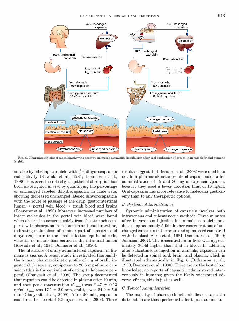
surable by labeling capsaicin with [3H]dihydrocapsaicinradioactivity (Kawada et al., 1984; Donnerer et al.,1990). However, the role of gut-epithelial absorption hasbeen investigated in vivo by quantifying the percentageof unchanged labeled dihydrocapsaicin in male rats,showing decreased unchanged labeled dihydrocapsaicinwith the route of passage of the drug (gastrointestinallumen � portal vein blood � trunk blood and brain)(Donnerer et al., 1990). Moreover, increased numbers ofintact molecules in the portal vein blood were foundwhen absorption occurred solely from the stomach com-pared with absorption from stomach and small intestine,indicating metabolism of a minor part of capsaicin anddihydrocapsaicin in the small intestine epithelial cells,whereas no metabolism occurs in the intestinal lumen(Kawada et al., 1984; Donnerer et al., 1990).
The literature of orally administered capsaicin in hu-mans is sparse. A recent study investigated thoroughlythe human pharmacokinetic profile of 5 g of orally in-gested C. frutescens, equipotent to 26.6 mg of pure cap-saicin (this is the equivalent of eating 15 habanero pep-pers!) (Chaiyasit et al., 2009). The group documentedthat capsaicin could be detected in plasma after 10 min,and that peak concentration (Cmax) was 2.47 � 0.13ng/ml, tmax was 47.1 � 2.0 min, and t1/2 was 24.9 � 5.0min (Chaiyasit et al., 2009). After 90 min, capsaicincould not be detected (Chaiyasit et al., 2009). These
results suggest that Bernard et al. (2008) were unable tocreate a pharmacokinetic profile of capsaicinoids afteradministration of 15 and 30 mg of capsaicin /person,because they used a lower detection limit of 10 ng/ml.Oral capsaicin has more relevance to molecular gastron-omy than to any therapeutic options.
B. Systemic Administration
Systemic administration of capsaicin involves bothintravenous and subcutaneous methods. Three minutesafter intravenous injection in animals, capsaicin pro-duces approximately 5-fold higher concentrations of un-changed capsaicin in the brain and spinal cord comparedwith the blood (Saria et al., 1981; Donnerer et al., 1990;Johnson, 2007). The concentration in liver was approx-imately 3-fold higher than that in blood. In addition,after subcutaneous injection in animals, capsaicin canbe detected in spinal cord, brain, and plasma, which isillustrated schematically in Fig. 6 (Dickenson et al.,1990; Donnerer et al., 1990). There are, to the best of ourknowledge, no reports of capsaicin administered intra-venously in humans; given the likely widespread ad-verse effects, this is just as well.
C. Topical Administration
The majority of pharmacokinetic studies on capsaicindistribution are those performed after topical administra-
FIG. 5. Pharmacokinetics of capsaicin showing absorption, metabolism, and distribution after oral application of capsaicin in rats (left) and humans(right).
CAPSAICIN: TO UNDERSTAND AND TREAT PAIN 943

tion because of the important therapeutic implications ofthis route. In vitro percutaneous absorption of vanillyl-nonamides (capsaicin analogs) in animals has been shownin dorsal skin collected in shaved hairy rats and “fuzzy”rats (Kasting et al., 1997), exemplifying the use of thisroute to deliver the vanilloid to peripheral neurons.
Topical capsaicin in humans is rapidly and well ab-sorbed through the skin (Hayman and Kam, 2008), andmany low concentrations of capsaicin (0.025–0.1%) areavailable over the counter as creams or patches. A study of
12 subjects evaluated the topical application of 3% solu-tions of capsaicin (55% capsaicin, 35% hydrocapsaicin, and105 other analogs) using three different vehicle prepara-tions (70% isopropyl alcohol, mineral oil, and propyleneglycol containing 20% alcohol). Capsaicinoids were de-tected in the stratum corneum within 1 min after applica-tion, and a steady state was reached shortly thereafter.Maximum concentration was nearly three times greater inthe subjects who received 70% isopropyl alcohol comparedwith the mineral oil or propylene glycol preparations. Thehalf-life of capsaicin is approximately 24 h (Pershing et al.,2004; Hayman and Kam, 2008).
A prescribed 8% topical capsaicin patch (NGX-4010)has been introduced and labeled for pain treatment,indicated for the management of neuropathic pain asso-ciated with postherpetic neuralgia. The patch contains640 �g/cm2, meaning that each patch contains a total of179 mg of capsaicin (Jones et al., 2011). To determinesystemic capsaicin exposure after single 60- or 90-minNGX-4010 applications, plasma samples were collectedfrom 173 patients with PHN, painful HIV-associatedneuropathy (HIV-AN), and painful diabetic neuropathy(PDN). The percentages of patients with quantifiablelevels of capsaicin at any time point were 31% for PHN(30/96), 7% for HIV-AN (3/44), and 3% for PDN (1/33).The maximum plasma concentration observed in anypatient was 17.8 ng/ml (Babbar et al., 2009). Because ofthe limited number of quantifiable levels, a populationanalysis was performed to characterize the pharmacoki-netics (PK) of capsaicin. Plasma concentrations werefitted adequately using a one-compartment model withfirst-order absorption and linear elimination. Capsaicinlevels declined very rapidly, with a mean populationelimination half-life of 1.64 h. Mean area under thecurve and Cmax values after a 60-min application were7.42 ng � ml�1 � h�1 and 1.86 ng/ml, respectively (Babbaret al., 2009). Correlations between calculated PK param-eters and patient characteristics were observed. Dura-tion and area of application of the patch were detected assignificant covariates explaining the PK of capsaicin.Ninety-minute applications of NGX-4010 resulted incapsaicin area under the curve and Cmax values approx-imately 1.78- and 2.15-fold higher than those observedin patients treated for 60 min. Treatment on the feet(patients with HIV-AN and PDN) produced far lowersystemic exposure than treatment on the trunk (pa-tients with PHN). The low systemic exposure and veryrapid elimination half-life of capsaicin after NGX-4010 administration are unlikely to result in systemiceffects and support the overall safety profile of thisinvestigational cutaneous patch (Babbar et al., 2009).The medical use of topical capsaicin (which, as theabove-mentioned studies show, acts almost exclu-sively at peripheral local sites) is discussed in furtherdetail in section XIV.
FIG. 6. The distribution of capsaicin in various tissues after subcuta-neous administration in rats. The numerical values refer to concentra-tions after distribution to brain, blood, and skin. In the case of the spinalcord, the values refer to concentrations after local administration. Be-cause administration is subcutaneous, the values cannot be comparedwith the experimental pain models in which capsaicin is administeredintradermally.
944 O’NEILL ET AL.
D. Intradermal Administration
Chanda et al. (2008) investigated the in vitro metab-olism of capsaicin in human skin and found that bio-transformation in human skin was very slow. Duringincubation, capsaicin was slowly metabolized over 20 h.In addition, two metabolites were detected, vanillyl-amine and vanillic acid.
When capsaicin is injected intradermally in humans,it is associated with a spontaneous burning pain thatsubsides within few minutes (Jantsch et al., 2009). Theinjection leads to primary and secondary hyperalgesicareas. Sensitivity to heat has been reported to be con-fined to an area approximately 1 cm in diameter, cen-tered approximately on the injection site. In addition, amuch larger dose-dependent area of hyperalgesia to me-chanical stimuli develops around the injection site(LaMotte et al., 1992). This study further demonstrateddose-dependent sensitivity, with lower doses leading tosensitization and higher doses to desensitization—thismay account for the presence of analgesia to pinprick atthe site of injection. Studies have reported a large num-ber of nonresponders regarding the presence of hyperal-gesia (Park et al., 1995; Liu et al., 1998) A number ofvariables could be responsible for this variation; it istherefore important to keep in mind that several factorscan influence the size of the area [e.g., dose, stimulusused to test (size of von Frey hair, cotton swab, brush),time of assessment, and temperature of skin before in-jection]. However, the model has proved to be reliableand reproducible and has been widely used in clinicalstudies investigating pain and analgesia (Staahl et al.,2009a,b). In general, intradermal capsaicin is used toinduce central sensitization and altered pain sensitivity,which will be discussed in section XII.
IV. Capsaicin Metabolism
Capsaicin metabolism after oral administration is be-lieved to be similar in the human, rat, and canine mi-crosome. When capsaicin and dihydrocapsaicin reachesthe liver, a major part is metabolized; however, theproportion that undergoes metabolism is unclear. Invitro experiments show that the amount of capsaicinand dihydrocapsaicin is greatly reduced after incubationwith liver homogenates (Donnerer et al., 1990; Chandaet al., 2008). In situ experiments in rats have shown thatthe intestinal elimination rate of capsaicin and dihydro-capsaicin is approximately equal to the concentration ofradioactivity in mesenteric venous blood, indicating thatpresumably no metabolism take place in the gut lumen(Kawada et al., 1984).
An in vitro human investigation with hepatic micro-somes and S9 fractions (used to investigate involvementof phase 2 metabolisms), showed that capsaicin wasrapidly metabolized, producing three major metabolites,16-hydroxycapsaicin, 17-hydroxycapsaicin, and 16,17-hydroxycapsaicin, whereas vanillin was a minor metab-
olite (Chanda et al., 2008). Moreover, Chanda et al.(2008) established a model to elucidate the metabolismof capsaicin at various concentrations physiologicallyequivalent to those obtained after oral ingestion of pep-per fruits. They showed that capsaicin metabolism wasless extensive at a concentration of 10 �M than at 1 �M(more than 50% direct inhibition of CYP1A2, CYP2C9,and CYP2C19), and the authors suggest therefore thatthe rate of metabolism is saturable. Although manyenzymes may play some role in hepatic drug metabo-lism, cytochrome P450 (P450) enzymes are quantita-tively the most important, and many drug-drug interac-tions result from the alteration (increase or decrease) inthe activities of these enzymes. At the much lowerplasma capsaicin concentrations occurring after topicaladministration, such as via the 8% patch, direct inhibi-tion of any P450 enzyme has not been shown. Becauseinhibition of CYP2E1 is thought to prevent the meta-bolic activation of several carcinogens, and because cap-saicin is believed to possess anticancer properties, it hasbeen widely inferred that capsaicin is a CYP2E1 inhib-itor. This implication, however, appears in only one pub-lication (Reilly and Yost, 2006). There is no informationon the ability of any of these compounds to modulateP450 activity and, to the best of our knowledge, theactions of the metabolites 16-hydroxycapsaicin and 17-hydroxycapsaicin are not known.
In vitro studies of capsaicin in human skin haveshown slow biotransformation, and most capsaicin re-mains unchanged; only a small fraction is metabolized tovanillylamine and vanillyl acid (Chanda et al., 2008).This suggests that cytochrome P450 enzymes partici-pate minimally in capsaicin transformation in skin com-pared with their key role in hepatic metabolism.
V. Capsaicin Elimination
Animal studies have shown that capsaicin is eliminatedmainly by the kidneys, with a small untransformed pro-portion excreted in the feces and urine (Leelahuta et al.,1983; Kawada et al., 1984; Surh et al., 1995). It is excretedin both free form and glucuronide form. In vivo animalstudies have shown that after 48 h, only a small amount(�10%) of an administered dose was found in feces (Leela-huta et al., 1983; Kawada et al., 1984).
VI. Capsaicin Pharmacology
Capsaicin depolarizes nociceptors and increases theircytosolic free Ca2� concentration (Dickenson et al.,1990). The gene encoding the capsaicin receptor wasisolated by using a Ca2� imaging-based expressionstrategy (Caterina et al., 1997). A functional screeningassay was used to isolate mRNA from the dorsal rootganglion (DRG) and create a cDNA library, which wasdivided into pools of clones and then transfected intohuman embryonic kidney (293) cells. Capsaicin inducedchanges in intracellular calcium levels were measured
CAPSAICIN: TO UNDERSTAND AND TREAT PAIN 945

within the transfected human embryonic kidney 293cells and used to identify the vanilloid receptor (Ca-terina et al., 1997). The cloned receptor was designatedvanilloid receptor subtype 1, or transient receptor poten-tial vanilloid subfamily member 1, because a vanilloidmoiety constitutes an essential chemical component ofcapsaicin. It is now well known that capsaicinoids exerttheir effects by activating the TRPV1 receptor (Haymanand Kam, 2008; Reyes-Escogido et al., 2011).
VII. Transient Receptor Potential Channels
TRP channels are one of the largest families of ion chan-nels and have a wide variety of functional roles. In 1969,Cosens and Manning isolated a mutant photoreceptor fromDrosophila melanogaster that caused the specimen to be-come blind upon exposure to bright light. This was the firstTRP channel to be discovered; since then, 28 mammalianisoforms have been identified, which are split into sevendifferent subfamilies (Clapham, 2003). They are made upof six transmembrane domain (TMD) polypeptide subunitsthat assemble as tetramers that can form pores in the cellmembrane.
TRPs are one of the most extensively studied fami-lies of ion channels present in sensory neurons. Thesix subfamilies include TRPV, TRPC (canonical),TRPM (melastatin), TRPML (mucolipins), TRPA (ankyrin),and TRPP (polycystin) (Venkatachalam and Montell,2007). TRPM8 and -A1 are thought to be involved incold sensing, whereas seven others are activated byheat, over a distinct range of temperatures: TRPV1– 4,TRPM2, TRPM4, and TRPM5. Collectively, these ninechannels are known as thermo-TRPs and are acti-vated at different ranges of temperature, both noxiousand innocuous. TRPV2 has the highest threshold foractivation (over 52°C) (Caterina et al., 1999). It hasbeen suggested that TRPV1 and TRPA1 are the firstto detect noxious hot and cold stimuli, respectively,with activation thresholds in humans of 42°C forTRPV1 and 14°C for TRPA1 (Dhaka et al., 2006); thus,activation of these receptors is proposed to lead to theperception of hot or cold thermal pain, respectively.
Another member of the TRP family, TRPM8, is be-lieved to be responsible for the detection of cooling (30–15°C) and noxious cold (�15°C). TRPM8 may also be thereceptor for menthol, which has been shown to causecooling and eventually irritation and pain (Wasner etal., 2004). It is a nonselective cation channel, and acti-vation generates currents required for cold sensing.TRPM8 knockout mice show deficiencies in cold detec-tion as well as impaired development of cold hypersen-sitivity (Colburn et al., 2007; Dhaka et al., 2007). IfTRPV1 neurons are knocked out during embryonic de-velopment, the mice also lack TRPM8-expressing neu-rons, suggesting that the two hot and cold receptors arecolocalized during development (Mishra et al., 2011).This overview of the TRP family of channels now sets the
scene for concentration of the TRPV1 receptor, the tar-get for capsaicin.
VIII. Transient Receptor Potential Vanilloid 1
A. Introduction
The TRPV1 receptor is a nonselective ligand-gatedcation channel. It is an integrator of many physical andchemical stimuli, including capsaicin and noxious heat(�43°C), as well as being activated by protons (pH�5.2), endogenous lipids, and certain inflammatory me-diators (Szallasi and Blumberg, 1999). All compoundsare lipophilic and therefore act at intracellular bindingsites (Yang et al., 2003). Stimuli are detected and trans-duced through opening of the ion channel, which resultsin entry of cations such as Na� and Ca2� to the neuron;although the channel is nonselective, it has been shownto have a high preference for Ca2� (Caterina et al.,1997).
TRPV1 has a pore-forming hydrophobic stretch be-tween TMDs 5 and 6 and is believed to exist as a homo-or heteromeric complex consisting of four subunits (Ca-terina et al., 1997; Kedei et al., 2001; Moiseenkova-Bellet al., 2008). The presence of specific amino acid residuesis required for sensitivity to different stimuli; it is be-lieved that Tyr511 and Ser512 located between TMDs 2and 3 dictate vanilloid/capsaicin sensitivity because mu-tations to tyrosine or alanine render the channel capsa-icin-insensitive (Jordt and Julius, 2002). As-yet unre-ported polymorphisms at these sites could underliedifferential pain sensitivities.
There are a number of modulators of TRPV1, includ-ing certain enzymes and inflammatory mediators (Szal-lasi et al., 2007). Sensitization of TRPV1 is achievedthrough phosphorylation by certain kinases (Premku-mar and Ahern, 2000; Bhave et al., 2003; Jung et al.,2004). It is believed that inflammatory mediators, dis-cussed later, activate these enzymes through second-messenger cascades. When sensitized, the activationthreshold of TRPV1 is lowered (Moriyama et al., 2005).On the other hand, phosphatases such as calcineurinand protein phosphatases 2A and 2B cause desensitiza-tion and thus an increase in activation thresholds(Zhang et al., 2006; Por et al., 2010). This is achieved viadephosphorylation in a Ca2�-dependent manner (Junget al., 2004; Mohapatra and Nau, 2005). Activity ofTRPV1 can therefore be dictated by the state of phos-phorylation of the channel.
TRPV1 has a number of functions and is involved indifferent physical processes depending on its location.One main role on peripheral nerve endings has beensuggested to involve the detection of noxious heat andchemicals (Caterina et al., 1997; Jung et al., 2004), al-though it is also believed to play an important role inthermoregulation (Caterina, 2007). Its role in the cen-tral nervous system (CNS), although it still involves
946 O’NEILL ET AL.

pain processing and modulation, is currently less wellunderstood.
The receptor also seems to play an important role incertain chronic pain conditions such as neuropathicpain, osteoarthritis (OA), bone cancer pain, inflamma-tory bowl disease (IBD) and migraine (Szallasi et al.,2007; Alawi and Keeble, 2010). In these conditionsmediators such as ATP and nerve growth factor (NGF)are capable of augmenting TRPV1 activity (Schum-acher, 2010). Most effects occur in the periphery at thesite of inflammation, which will be discussed in fur-ther detail in section C.
B. Transient Receptor Potential Vanilloid1-Associated Molecules
In addition to phosphorylation of the channel, TRPV1is reported to associate with a number of intracellularproteins that may also modulate receptor activity andtrafficking to the membrane—and thus affect function ofthe channel. Reported interactions include phosphati-dylinositol 4,5-bisphosphate (PIP2), phosphoinositide-in-teracting regulator of TRP (Pirt), GABA-receptor-asso-ciated protein, soluble N-ethylmaleimide-sensitive factorattachment protein receptor, A-kinase anchoring pro-tein 150, and components of the cytoskeleton. Here wewill focus on PIP2 and the cytoskeleton.
1. Phosphatidylinositol 4,5-Bisphosphate. PIP2 is amembrane phospholipid required for a number of intra-cellular signaling pathways. It has been shown to beassociated with TRPV1 in the plasma membrane,through an interaction at the C-terminal (Prescott andJulius, 2003; Ufret-Vincenty et al., 2011). Two opposingactions have been proposed with regard to how PIP2regulates TRPV1 function. Although it was originallybelieved that binding of PIP2 to TRPV1 was inhibitory,later studies suggested the contrary. Prescott and Julius(2003) reported that a mutation in the C-terminal ofTRPV1, inhibiting the binding of PIP2, resulted in largercapsaicin currents. However, this finding relied on anabsence of extracellular Ca2�, and further in vitro and invivo studies have produced opposing findings. Stein at aldemonstrated that polylysine (an agent that sequestersPIP2) had an inhibitory effect on TRPV1, whereas Liu etal. (1998) found that replenishing PIP2 aided recoveryafter desensitization. More recently, Sowa et al. (2010)demonstrated in a number of in vivo experiments thatPIP2 was required both for normal sensing of noxiousheat and for the development of sensitization. PIP2 en-hanced thermosensation for up to 2 h and increasedthermal hypersensitivity and mechanical allodynia inmodels of inflammation and nerve injury. Taken to-gether, this evidence suggests there is an overridingpronociceptive role of PIP2.
It has been suggested that imperative for this inter-action with PIP2 is the regulatory subunit Pirt. It hasbeen shown that the C-terminal of Pirt binds to bothTRPV1 and PIP2 and that Pirt is required for enhancing
TRPV1 signaling through PIP2 (Kim et al., 2008). Inaddition, Pirt(�/�) mice were found to have impairedresponse to heat and capsaicin. This suggests that asso-ciation with Pirt may also be required for optimum func-tioning of TRPV1. However, this has not been repro-duced in subsequent studies, and it has been suggestedthat the phosphoinositide sensitivity of TRPV1 is notaltered by Pirt (Ufret-Vincenty et al., 2011). Furtherinvestigation into the small molecules that associatewith TRPV1 and may modulate activity will be of greatinterest. Novel modulation of this channel, targetingassociated molecules, could be useful when designingdrugs to alter the function of TRPV1 but keep sideeffects minimal.
2. Cytoskeleton. Recent evidence suggests that thecytoskeleton is not just important for structural integ-rity but may also play a role in signal transduction andtransmission of nociceptive information between neu-rons (Bhave et al., 2003). A number of studies havefurther shown that TRPV1 forms a signaling complexwith elements of the cytoskeleton, allowing the integra-tion of signals and exchange of synaptic information.This suggests that TRPV1 is important for structuraland functional regulation as well as for neuronal com-munication and network formation, in addition to thepivotal role already discussed in the detection of noxiousstimuli and transduction of pain signaling (Goswami,2010). Through a number of investigations, Goswami etal. (2004, 2006) have demonstrated that TRPV1 inter-acts with tubulin, in particular polymerized dynamicmicrotubules. Microtubules are made up of polymers oftubulin, which are integral for the maintenance of cellstructure and intracellular transport. Activation ofTRPV1 may therefore lead to cytoskeletal reorganiza-tion, such as rapid disassembly of dynamic microtu-bules, which may be involved in the detraction of sen-sory afferents (Goswami, 2010). In addition, they foundthat TRPV1 was often localized with growth cones andfilopodia tips and suggested that it may regulate themorphology and function of these structures and thusplay a role in neuronal communication (Goswami, 2010).Although interaction with microtubules is believed toarise from the C terminus of TRPV1, the N terminus isthought to be important for regulation filopodial dynam-ics. It remains to be determined whether these interac-tions have functional importance in vivo in terms ofsensory function.
C. In the Periphery
TRPV1 receptors are mainly expressed on primarysensory neurons. They have been detected on terminalsof small- to medium-diameter nociceptors, such as pep-tidergic and nonpeptidergic C fibers, as well as some A�fibers (Caterina et al., 1997; Tominaga et al., 1998).These fibers generally project into the superficial layersof the dorsal horn (DH) including laminae I and II (To-
CAPSAICIN: TO UNDERSTAND AND TREAT PAIN 947

minaga et al., 1998). Projections may also extend intolaminae V and X (Tominaga et al., 1998).
As discussed, TRPV1 is a polymodal signal trans-ducer, imperative for the detection of capsaicin and par-ticularly important for detection of noxious heat. Acti-vation of receptors causes depolarization of nociceptors(Bevan and Szolcsanyi, 1990), which allows the painsignal to be transduced and relayed through the spinalcord to the brain, where it is processed and perceived.Caterina et al. (2000) demonstrated that capsaicin sen-sitivity was eliminated in TRPV1(�/�) mice; interest-ingly, however, they had only impaired heat detectionand reduced thermal hypersensitivity in response toinflammatory agents. This also highlights the impor-tance of TRPV1 for the induction of thermal hyperalge-sia in inflammatory states, but incomplete loss suggeststhere may be additional channels involved in normalheat sensing or compensatory changes.
However, more recently, the function of TRPV1 posi-tive afferents has further been elucidated with the pro-filing of TRPV1-DTA mice generated by Mishra et al.(2011). TRPV1-Cre animals were crossed with a ROSA-stop-diphtheria toxin fragment A (DTA) line to ablate aspecific population of TRPV1-expressing fibers. This en-ables the study of the function of the neuronal popula-tion rather than just TPV1 itself. As noted, it was shownpreviously that TRPV1 KO mice maintained some ther-mosensation; it was impaired only over 50°C (Caterinaet al., 2000). However, these mice, whose TRPV1 affer-ents are completely ablated and have no response tocapsaicin, are also totally insensitive to both hot andcold (Mishra et al., 2011). The mice also exhibited nohypothermia in response to intraplantar capsaicin,which is seen in normal animals. This suggests thatTRPV1-positive afferents may be more imperative in thethermoregulation and detection of noxious heat thanwas previously thought.
Thermal hyperalgesia develops in certain pathologicalstates in which peripheral TRPV1 may be sensitized bya number of mediators acting through intracellular sig-naling pathways (Kanai et al., 2007). For example, pro-tons are able to both directly activate and potentiateactivity of TRPV1. During a state of tissue injury orischemia, when proton levels may be elevated, hydrogenions are thought to act at an extracellular site to in-crease the potential of channel opening (Jordt et al.,2000; Huang et al., 2006). On the other hand, mediatorssuch as prostaglandins, including PGE2 and PGI2, act atEP1 or IP receptors, respectively, which are coupled toGs. They have been demonstrated to interact withTRPV1 through PKA-dependent pathways, resulting inlowering of the temperature activation threshold to aslow as 35°C (Smith et al., 2000; Moriyama et al., 2005).Other mediators, such as bradykinin (BK), ATP, andendothelin (ET)-1 act at Gq-coupled receptors— B1/B2,P2Y2 and ETA, respectively. This is believed to activatethe diacylglycerol-PKC pathway (Vellani et al., 2004),
therefore once again resulting in phosphorylationof TRPV1. PKC� has been implicated in phosphorylationof TRPV1 at serine residues 502 and 800 because cellscontaining the mutations S502A or S800A are unable tosensitize (Numazaki et al., 2002; Kawamata et al.,2008). Finally, an influx of Ca2� through TRPV1 itselfand release from intracellular stores can result in theactivation of CaMKII (Fig. 7).
This aforementioned sensitization, intriguingly, couldallow the receptor to become active at body temperature,so it not only contributes toward thermal hypersensitiv-ity but also is possibly a substrate for ongoing burningpain that patients often report. It has been suggestedthat the pain that is felt from mediators, such as BKalone, may be as a result of this interaction withTRPV1—suggesting that in addition to the traditionalview of TRPV1 directly gating nociceptive signals, itmay also indirectly gate other signals through intracel-lular signaling pathways (McMahon and Wood, 2006).This would involve the binding of BK to B1/B2 receptorsand initiation of PKC-dependent signaling pathways,resulting in phosphorylation of TRPV1 and a reductionin heat pain threshold to body temperature, and thus an“ongoing” pain is felt.
FIG. 7. Phosphorylation of peripheral TRPV1. There are a number ofendogenous inflammatory mediators, such as PGE2, ATP, BK, and ET-1,which are able to act at their respective receptors located on TRPV1-expressing afferent fibers. Through various intracellular signaling cas-cades, they are able to phosphorylate, and sensitize, TRPV1 (althoughcertain lipids and acid may act directly on TRPV1 itself)—resulting in thelowering of activation thresholds and heightened activity to further stim-uli. Furthermore, this effect may also be achieved through Ca2�-depen-dent activation of CaMKII, which also phosphorylates TRPV1. This phos-phorylation may lead to both thermal and mechanical hypersensitivity atthis site. AC, adenylyl cyclase; PLC13, phospholipase C13. (See sectionVIII.C for details.)
948 O’NEILL ET AL.

The theory that TRPV1 integrates various signalsfrom inflammatory molecules that are not direct ago-nists of the channel is supported by the finding that BKresponses can be modulated by capsazepine and are alsodepleted in TRPV1 KO mice (Huang et al., 2006). Fur-thermore, in a study regarding PIP2 modulation ofTRPV1, the authors found that, in addition to the ex-pected decrease in activity of TRPV1 itself, there wasalso a reduction in response to both BK and ATP and aninhibition of ATP-mediated thermal hyperalgesia (Sowaet al., 2010). The authors suggest that this reduction inresponse may be a direct effect of B1/2 and P2Y2 requir-ing PIP2; however, it is also plausible that it is due toattenuation of the proposed interaction with TRPV1.This would strongly suggest that BK currents are initi-ated through TRPV1; therefore, a role for TRPV1 inongoing pain is not unreasonable to suggest.
A second method of potentiating the actions of TRPV1,rather than phosphorylation, is by increasing the sur-face expression of receptors, either through an increasein transport or in number of receptors produced. It hasbeen suggested that in inflammatory conditions such asOA, NGF is released and contributes toward painthrough actions at TrkA receptors, which are expressedon specific sensory neurons such as C and A� fibers(McMahon et al., 1991). Injection of complete Freund’sadjuvant (CFA) and the iodoacetate model of OA havebeen shown to result in increased TRPV1 expression inDRG (Ji et al., 2002; Fernihough et al., 2005). Zhang etal. (2005) demonstrated that TrkA induced activation ofPI3 kinase/Src kinase, causing phosphorylation of theTyr200 residue, which resulted in increased membraneexpression of TRPV1. In addition, Xue et al. (2007)Ji etal. (2002) found that it could also induce translation viap38 mitogen-activated protein kinase activation. Con-sidering these possible roles in sensitization and poten-tial contribution to chronic pain states, it can be inferredthat TRPV1 may be an important drug target.
D. In the Viscera
Weller et al. (2011) found evidence for functional ex-pression in viscera of excitatory TRPV1, TRPA1, andinhibitory cannabinoid 1 receptors along the sensoryfibers of the vagus nerve. This finding has pathophysi-ological relevance to the axonal membrane and the con-trol of neuropeptide release that may become importantin cases of inflammation or neuropathy. Sensitizationand possible ectopic discharge may contribute to thedevelopment of autonomic dysregulation in visceral tis-sues that are innervated by the vagus nerve (Weller etal., 2011).
E. In the Spinal Cord
TRPV1 is found not only on peripheral nerve endingsbut also in the spinal cord. Expression seems to bemainly restricted to the central branches of small andmedium-sized fibers located in the dorsal root ganglia
and in the superficial DH (Szallasi et al., 1994; Guo etal., 1999). Both Valtschanoff et al. (2001) and Doly et al.(2004a) detected TRPV1 on postsynaptic second-orderneurons in the rat DH. In the spinal cord, TRPV1 isbelieved to play a role in both the modulation and trans-mission of pain.
Because the spinal cord is not subject to the samechanges in temperature and pH that occur in the periph-ery to activate TRPV1, it was assumed that endogenousagonists must be present within the CNS. Studies havehighlighted several possible substances (Fig. 8), includ-ing anandamide (Zygmunt et al., 1999), metabolites oflipoxygenases (Hwang et al., 2000), �-3 polyunsaturatedfatty acids (Matta et al., 2007), N-arachidonoyldopamine(Huang et al., 2002), and N-oleoyldopamine (OLDA)(Spicarova and Palecek, 2009). Application of high con-centrations of OLDA to superficial DH neurons in ratspinal cord slices increased miniature excitatory post-synaptic currents, which in turn were blocked by theaddition of TRPV1 antagonists (Spicarova and Palecek,2009). Activation of presynaptic TRPV1 receptors in thespinal cord was originally thought to result in excitationof central fibers via increased release of glutamate andother excitatory neuropeptides, such as substance P (SP)and calcitonin gene-related peptide (CGRP). This wasshown by Ueda et al. (1994), who demonstrated thatapplication of capsaicin to slices of rat DH evoked re-lease of glutamate and depleted stores of SP. In addition,electrophysiological studies showed that capsaicin appli-cation to DH slices from adult rats resulted in increasedminiature excitatory postsynaptic currents that could beblocked by application of the TRPV1 competitive antag-onist capsazepine (Yang et al., 1998).
However, spinal intrathecal administration of capsa-icin results in a rapid attenuation of C-fiber input when
FIG. 8. Spinal activators TRPV1. The endogenous agonists of spinalTRPV1 remain unclear; however, a number of possible substances havebeen suggested, including anandamide, metabolites of lipoxygenases,�-3 polyunsaturated fatty acids, N-arachidonoyldopamine (NADA), andN-oleoyldopamine (OLDA).
CAPSAICIN: TO UNDERSTAND AND TREAT PAIN 949
recording from wide dynamic range (WDR) cells. Theysuggest that systemic capsaicin, which had previouslybeen shown to be antinociceptive in behavioral tests,could selectively attenuate C-fiber inputs to the DH(Dickenson et al., 1990). They concluded that the effectwas unlikely to be the result of an inactivation of C-fi-bers at the peripheral terminals and was more likely tobe the result of the actions directly on the spinal cord; SPor other substances released from primary afferentnerve terminals play a prominent role in such desensi-tization. Thus suggesting activation of receptors directlyin the spinal cord has an inhibitory effect on the nocice-ptive pathway.
Ferrini et al. (2007) showed that administration ofcapsaicin to lamina II neuron slices from mice pupsresulted in an increase of spontaneous inhibitory post-synaptic currents. The authors postulated that GABAergicinhibitory interneurons in laminae I, III, and IV areexcited because of the release of SP, which in turn re-duces activity in lamina II neurons. Thus provides fur-ther evidence that there is an inhibitory, as well asexcitatory, role for TRPV1 in the spinal cord.
The distribution and therefore the role of spinal TRPV1is believed to alter during pathological pain states. Dom-Bourian et al. (2006) showed that there was an up-regulation of TRPV1 in the DH of rats after spinal cordinjury, and Tohda et al. (2001) demonstrated that carra-geenan-induced inflammation resulted in an increasedtransport of TRPV1 mRNA to neuron terminals in the DH.Both models were associated with thermal hyperalgesia,which has been shown to be blocked by intrathecal admin-istration of TRPV1 antagonists such as 3,6-dihydro-3�-(trifluoromethyl)-N-[4-[(trifluoromethyl)sulfonyl]phenyl]-[1(2H),2�-bipyridine]-4-carboxamide (A-784168) andN-1H-indazol-4-yl-N�-(5-piperidin-1-yl-2,3-dihydro-1H-inden-1-yl)urea (A-795614) (Wang and Woolf, 2005; Cui etal., 2006; Yu et al., 2008). Studies have also shown thatspinal TRPV1 is likely to be involved in the development ofmechanical hyperalgesia (Kanai et al., 2007). Intrathecalinjection of antisense oligonucleotides against TRPV1, in arat spinal nerve ligation model, resulted in decreased me-chanical hypersensitivity (Christoph et al., 2007). The re-sults therefore strongly suggest that activation of TRPV1has an over-riding pronociceptive effect in chronic painstates.
In contrast, the cannabinoid receptor ligand anand-amide, which has also been shown to activate TRPV1,has an antinociceptive effect (which was reduced bycapsazepine) when administered intrathecally in a ratmodel of carrageenan-induced inflammation (Horvath etal., 2008). This effect is most likely to occur throughTRPV1 activation of spinal inhibitory interneurons, incombination with activation of cannabinoid receptors,resulting in decreased pain. Thus, pharmacological evi-dence also suggests there are two opposing actions ofTRPV1 in the spinal cord.
F. Supraspinal
In addition to expression in the periphery and in theDH, TRPV1 receptors have also been detected at numer-ous supraspinal sites within the CNS, where it is be-lieved to be involved in pain processing. Locations in-clude the rostroventral medulla (RVM), periaqueductalgray (PAG), and amygdala (Millan, 2002; Liapi andWood, 2005), as well as the nucleus tractus solitarius(Doyle et al., 2002), the somatosensory cortex, the ante-rior cingulated cortex, and the insula (Millan, 2002). It isimportant to note that endogenous agonists of supraspi-nal TRPV1 are unknown, and additional roles (besidesthe potential in pain processing) are largely unclear.Capsaicin has not been used in many studies to studysupraspinal TRPV1, and further elucidation of its rolewill be important, including information on the endoge-nous activators of the channel within the CNS.
Compared with peripheral and spinal TRPV1, expres-sion of supraspinal receptors was thought to exclusivelyplay an analgesic role. A microinjection of capsaicinwithin the ventrolateral PAG was demonstrated to havean antinociceptive effect in rats (Palazzo et al., 2002;Starowicz et al., 2007). This suggests that the analgesicproperties are due to an interaction with descendingmodulatory pain pathways.
Starowicz et al. (2007) also found that the capsaicininjection increased thermal pain thresholds. This ap-peared to be the result of an increase in glutamaterelease from neurons in the PAG, which activated mGluand NMDA receptors, causing an eventual downstreamincrease in OFF cell activity in the RVM. Injection ofa TRPV1 competitive antagonist, iodo-resiniferatoxin(RTX), to the ventrolateral PAG was also associatedwith an increase in ON cell activity (Starowicz et al.,2007), suggesting that TRPV1 activation in the PAGdoes lead to an increase in descending inhibition of pro-nociceptive pathways.
On the other hand, it has been previously demon-strated that capsaicin injection into the dorsolateralPAG resulted in hyperalgesia before analgesia set in; theauthors therefore suggested that capsaicin-induced an-algesia may be due to TRPV1 desensitization (McGa-raughty et al., 2003). At present, the more importantmechanism of supraspinal TRPV1-mediated antinocice-ption is unclear, but much focus has been placed on therole of its activation of descending inhibitory pathways.
G. Non-Neuronal
It has also been found that a number of non-neuronalcells express TRPV1, including Schwann cells, astro-cytes, and mast cells (Doly et al., 2004b; Stander et al.,2004). It has been suggested that whereas astrocyticexpression seen in the rat DH may be involved in painmodulation and central sensitization, activation ofTRPV1 on the surface of mast cells may contribute to theinflammatory response through enhanced production of
950 O’NEILL ET AL.
interleukin-4 (Doly et al., 2004b; Stander et al., 2004).Further discussion of non-neuronal TRPV1 expressionand its contribution to pain signaling is outside thescope of this review.
IX. Transient Receptor Potential Vanilloid 1Splice Variants
Splice variants of TRPV1 have been identified in bothrats and humans. In rodents, there are three mainsplice variants: TRPV1 5�, TRPV1VAR, and TRPV1�(Szallasi et al., 2007). TRPV1 5� is missing the first 0.5kilobases, which is the N-terminal region of wild-type(WT) TRPV1 and was shown by Sanchez et al. (2001) tobe expressed in the rat DRG and the CNS. In the DRG,its expression is reported to be 12 times lower than theWT, although in the CNS levels are similar for bothvariants (Sanchez et al., 2001). TRPV1 5� does not forma functioning ion channel and thus is nonresponsive tovanilloid agonists (Jara-Oseguera et al., 2008).TRPV1VAR is also nonfunctional, unless expressed withTRPV1 WT, where it seems to act as a negative regula-tor (Tian et al., 2006). TRPV1� is also believed to be anegative modulator of TRPV1 WT, despite also re-sponding to capsaicin alone (Wang et al., 2004). Inhumans the splice variant TRPV1b, which is found inthe DRG and CNS, lacks exon 7, and once again hasbeen demonstrated to act as an endogenous negativeregulator of TRPV1 (Lu et al., 2005; Vos et al., 2006).Vos et al. (2006) found that TRPV1b is expressed onthe cell surface and forms a complex with TRPV1 toinhibit activity. The physiological relevance ofTRPV1b, with regard to pain, remains unknown. How-ever, it would be interesting to examine a possibledown-regulation during chronic pain states.
It is interesting to note that it has recently beenshown that alternative splicing of the TRPV1 gene in thetrigeminal ganglia of vampire bats, to create the isoformTRPV1-S, can lower the activation threshold to 30°C(Gracheva et al., 2011). This ganglion specific splicingallows a modification of TRPV1 function so that thespecies can use it to detect infrared radiation given off bywarm-blooded prey, yet the bats otherwise maintainnormal function of TRPV1 in somatic afferent fibers.Thus highlighting the importance of splice variants,with regard to channel function.
X. Transient Receptor PotentialVanilloid 1 Polymorphisms
There are at least six nonsynonymous polymorphismsof the human TRPV1 gene, though relatively little isknown about their consequences—particularly with re-gard to pain (Xu et al., 2007). Single-nucleotide poly-morphisms (SNPs) include: TRPV1I585V, TRPV1T505A,TRPV1T469I, TRPV1I315M, TRPV1P91S, and TRPV1K2N
(Xu et al., 2007). Although most seem to have littlefunctional significance, studies have shown that
TRPV1I585V, TRPV1I315M, and TRPV1P91S may affectreceptor activity (Kim et al., 2004; Cantero-Recasens etal., 2010). TRPV1I585V involves the substitution of iso-leucine for valine at position 585 in exon 11 and thusaffects a TMD (Xu et al., 2007). Kim et al. (2004) dem-onstrated that people carrying the mutation had longerwithdrawal times to cold pain. This discovery was unex-pected because TRPV1 is mainly believed to be involvedin the detection of heat and therefore suggests thatdifferent TRP channels may interact during tempera-ture sensing. An increased withdrawal time would sug-gest the SNP results in a damping of receptor function.This is yet to be shown animal models of pain; however,investigation of the role of TRPV1I585V in childhoodasthma has begun to elucidate the mechanism. A studyby Cantero-Recasens et al. (2010) found that the substi-tution for valine resulted in decreased channel activity/Ca2� entry in response to both capsaicin and heat, re-sulting in a 20 to 30% loss of channel function (this wasassociated with a lower risk of active asthma/coughing).It is plausible that this decreased activity of TRPV1could explain people with higher heat pain thresholdsand why certain subjects seem unable to develop astrong central sensitization and mechanical hyperalge-sia in response to intradermal capsaicin injection, de-spite still perceiving the initial pain and developingthermal hyperalgesia.
In a separate study Xu et al. (2007) found that theSNPs TRPV1I315M and TRPV1P91S resulted in greatersurface expression of the receptors. TRPV1I315M andTRPV1P91S SNPs effect exons 5 and 1, respectively; be-cause TRPV1I315M is in a region that is believed toencode protein binding, this may effect ligand interac-tions and thus agonist responses. Both TRPV1I315M andTRPV1P91S were demonstrated had a slight increase inmaximum response to capsaicin and anandamide (Xu etal., 2007), thus suggesting that this mutation results ina gain of function of the receptor and may render carri-ers more sensitive to painful stimuli that are transducedby TRPV1.
A study from the German Research Network on Neu-ropathic Pain, assessing the impact of 11 select TRPSNPs in healthy volunteers and patients with neuro-pathic pain, has shown that the TRPV1 mutation1911A�G (TRPV1I585V) was associated with a decreasein heat hyperalgesia, pinprick hyperalgesia, and me-chanical hypoesthesia in the patients with neuropathicpain. In addition, TRPV1I315M was associated with coldhypoesthesia (Binder et al., 2011). Although they foundno evidence that TRP genetic variants are associatedspecifically with the prevalence of neuropathic pain, thepolymorphisms contributed significantly to the somato-sensory abnormalities that are experienced by patientswith neuropathic pain (Binder et al., 2011). Furtherinvestigation into TRPV1 SNPs and neuropathic painconditions may be useful, but these very recent studiesshow that variations in TRPV1 can explain some of the
CAPSAICIN: TO UNDERSTAND AND TREAT PAIN 951

variability in pain seen in patients with pathophysiolog-ical conditions.
XI. Transient Receptor Potential Vanilloid 1Receptor Expression in Humans in the Airways,
Skin, and Viscera
Outside of the CNS, TRPV1-expressing afferents havebeen reported to be present in different human tissuesincluding vascular smooth muscle, bronchial epithelialcells (Seki et al., 2007), the epidermis (Lee et al., 2009),and the gastrointestinal tract (Yiangou et al., 2001;Chan et al., 2003; Matthews et al., 2004). This suggestsother roles for TRPV1 besides nociception (for a fullreview on the subject, see Fernandes et al., 2012).
A. Airways
It is well know that TRPV1 is expressed in the air-ways, on vagal and nonmyelinated C-fiber afferents (Hoet al., 2001), and it is believed to be involved in bothairway constriction and cough. Lundberg and Saria(1982) found that stimulation of capsaicin-sensitive neu-rons resulted in smooth muscle contraction, which wasconfirmed by Forsberg et al. (1988) who additionallyhighlighted the role in cough. It was further demon-strated that TRPV1 antagonists can suppress cough,therefore confirming its pivotal role in this reflex(Trevisani et al., 2004). Recent studies have also re-ported potential up-regulation of TRPV1 in patientswith chronic cough, suggesting that increased activitywould underlie the hypersensitivity leading to this con-dition (Mitchell et al., 2005).
B. Skin
Studies suggest that in the epidermis, heat- and UV-induced matrix metalloproteinases-1 expression may bepartly mediated by TRPV1 activation in human keratin-ocytes (Lee et al., 2012) and showed that TRPV1 proteinwas expressed at higher levels in the sun-protected skinof older subjects than in that of young people. However,photoaged skin of older subjects showed increased ex-pression of TRPV1 mRNA and protein compared withthat of the sun-protected skin of the same people. There-fore, the expression of TRPV1 is affected by both theintrinsic aging and photoaging processes (Lee et al.,2009). The increased TRPV1 expression in skin fromolder people implies that TRPV1 may be related to senileskin symptoms, such as senile pruritus and neurogenicinflammation; therefore TRPV1 has been proposed as anintriguing novel target for the prevention of skin agingand inflammation.
C. Gastrointestinal Tract
TRPV1-immunoreactive nerves were found to be dis-tributed within the lamina propria from esophageal mu-cosal biopsies in healthy subjects and in patients withesophagitis. The percentage of papillae positive for
TRPV1 was elevated in patients with esophagitis com-pared with control subjects (Matthews et al., 2004). Sup-porting this, TRPV1 has been localized in the humancolon innervations, where TRPV1 immunoreactivity isgreatly increased in colonic nerve fibers of patients withactive IBD than in healthy bowel (Yiangou et al., 2001).The increase might be mediated by NGF, which, asdiscussed previously, regulates the capsaicin sensitivityof human sensory neurons and is itself increased ininflamed tissues, such as in IBD (Yiangou et al., 2001).Chan et al. (2003) compared full-thickness rectal biopsysamples from nine patients with physiologically charac-terized rectal hypersensitivity with tissue samples from12 control subjects. In rectal hypersensitivity, nerve fi-bers immunoreactive to TRPV1 were increased in mus-cle, submucosal, and mucosal layers. The increase inTRVP1 correlated significantly with decrease in rectalheat and distension sensory thresholds, therefore sug-gesting that TRPV1 may be important in certain statesof visceral inflammation.
XII. Experimental Pain Models
A. Animals
The use of capsaicin in animal models is important inthe study of both processing and modulation of painsignals. As mentioned previously, capsaicin was origi-nally used in animal models to elucidate the function ofthe TRPV1 receptor and its interactions in the periph-ery. Cloning of the capsaicin receptor by Caterina et al.(1997) was followed by in vivo analysis of its mecha-nisms using an intradermal capsaicin injection into theplantar skin of the hind paw in mice. It was initiallygiven to wild-type animals before being administered toTRPV1(�/�) variants and DTA KOs, which highlightedthe importance of the receptor in detection of capsaicinitself, as well as noxious heat (Caterina et al., 1997). Thedevelopment of thermal and mechanical hyperalgesiawas also impaired, highlighting the role of TRPV1 in thedevelopment of these symptoms (Caterina et al., 1997).
Although thermal hypersensitivity is regarded as aperipheral phenomenon discussed previously, sensitiza-tion to mechanical stimulation is thought to be under-pinned by central mechanisms such as an increasedexcitability of DH neurons. Ongoing stimulation of pe-ripheral C and A� fibers—for example, because of cap-saicin injection or large areas of topical application—canresult in wind-up, central sensitization, and long-termpotentiation (LTP). The increased input into the DHcauses an increase in release of glutamate and neuro-peptides, such as SP and CGRP, at the central synapticterminal. This in turn results in an increased activationof postsynaptic receptors and a slow depolarization ofsecond-order projection neurons, thus relieving theusual block of NMDA receptors (Woolf and Thompson,1991). Once NMDA receptors are also activated, changesmay occur within the second-order projection neurons,
952 O’NEILL ET AL.

including an increased surface expression of postsynap-tic receptors. This may be due to a number of mecha-nisms involving changes in transcription and transla-tion, as well as post-translational modifications (Fig. 9).These changes are believed to be responsible for thesymptoms associated with secondary hyperalgesia inboth humans and animals. Although wind-up is only ashort-term phenomenon and is reversible, central sensi-tization and LTP can induce long-term changes.
Before the discovery of the receptor, intradermal in-jection of capsaicin was shown to produce secondaryhypersensitivity to noxious and innocuous stimuli inboth monkeys and rats (Baumann et al., 1991; Slukaand Willis, 1997). As mentioned, these symptoms (whichin humans are known as secondary hyperalgesia andallodynia) are markers of a state known as central sen-sitization, where DH neurons have become hyperexcit-able and elicit greater responses than usual. It is asso-ciated with many chronic inflammatory and neuropathicpain states and therefore is imperative that mechanismsare understood. Dougherty and Willis (1992) gave intra-dermal capsaicin to monkeys and showed 1) that dis-charge rate of second-order neurons in the DH increasedafter injection and 2) an increase in release of the excit-atory amino acids glutamate and aspartate. They alsofound an increase in activation of NK1 receptors onsecond-order neurons, which, they hypothesized, mightbe contributing to the sensitization (Dougherty et al.,1994).
Subsequently, a rodent model was used, and onceagain it was shown that intradermal injection of capsa-icin could lead to increased response of second-orderneurons (Fig. 10); this was associated with an increasedrelease of neurotransmitters in the DH such as gluta-mate and SP (Willis, 1997; Yan et al., 2006). Willis, 2001demonstrated that a heightened release of neurotrans-mitters and neuropeptides in response to capsaicin re-sulted in an increased activation of AMPA, NMDA,mGlu and NK1 receptors on second-order neurons. Itwas later shown that SP synthesis, as well as release,was amplified after injection and is also believed tocontribute to sensitization of second-order neurons (Yanet al., 2005).
Sun et al. (2003) have since shown that CGRP isanother important neuropeptide contributing to centralsensitization. TRPV1 is expressed on peripheral pepti-dergic C fibers, some of which contain CGRP—a peptidethat is believed to be released in the DH after a pro-longed activation of the receptor. Administration of theCGRP receptor antagonist CGRP8–37 into the DH 1 hafter injection partially reduced mechanical hyperalge-sia and allodynia, whereas giving it before intradermalcapsaicin injection resulted in a clear reduction of bothabnormal responses (Sun et al., 2003). Thus, it wasconcluded that CGRP is involved in both the develop-ment and maintenance of hyperalgesia.
Intradermal injection of capsaicin in rats was also usedto demonstrate the importance of second-messenger cas-cades in central sensitization. Sluka and Willis (1997)found that capsaicin-evoked mechanical allodynia could bereversed by both G protein and protein kinase inhibitors.Kinase activity can result in post-translational modifica-tion of ion channels and receptors as well as increasedtrafficking and cell-surface expression. Further studies ofspecific kinase inhibitors after capsaicin injection hashelped define precise mechanisms that may underlie cen-tral sensitization; for example, Zou et al., (2002, 2004)revealed that capsaicin-evoked PKA and PKC activationresulted in phosphorylation of the NMDA receptor subunitNR1 at Ser890/897 and Ser896, respectively, which couldbe blocked with specific inhibitors. A role for PKB/Akt wasalso found using the intradermal capsaicin model whenSun et al. (2007) showed elevated levels 5 min after injec-tion in the DRG. This resulted in sensitization of WDRcells in the DH—leading to mechanical hypersensitivity—that could be prevented with a PKB inhibitor.
Fang et al. (2005) demonstrated a role for anotherkinase—CaMKII—in central sensitization. They showedthat phosphorylation of cAMP response element-bindingprotein (CREB) 20 min after capsaicin injection could beblocked by the intrathecal CaMKII inhibitor 2-(N-[2-hydroxyethyl])-N-(4-methoxybenzenesulfonyl)amino-N-(4-chlorocinnamyl)-N-methylamine (KN-93). CaMKIImay phosphorylate the glutamate receptor 1 subunit ofAMPA receptor, resulting in increased surface expres-sion and conductance of the channel. Up-regulation ofphosphorylated CREB may also lead to enhanced geneexpression and transcription of products that contributeto central sensitization.
Protein phosphatases are believed to oppose kinaseaction and can therefore control the duration of statessuch as central sensitization. Zhang et al. (2006) usedinhibitors of protein phosphatase type 2A after capsaicininjection, which increased the duration of capsaicin-evoked hypersensitivity to mechanical stimuli by up to3 h, thus suggesting that phosphatases indeed play arole in regulating central sensitization.
Another possible modulator of central sensitizationthat has been proposed is reactive oxygen species (ROS).The intradermal capsaicin model was used to examinetheir involvement in secondary hyperalgesia by admin-istering systemic and intrathecal ROS scavengers afterinjection. Although systemic administration had no ef-fect on either primary or secondary hyperalgesia, theintrathecal injection reduced activity in WDR cells andsigns of secondary hyperalgesia (Lee et al., 2007). Thestudy suggests that ROS may have a role in the induc-tion or maintenance of central sensitization at the levelof the spinal cord.
An additional mechanism believed to be involved inboth peripheral and central sensitization, dorsal rootreflexes (DRRs), have also been examined using intra-dermal capsaicin. Zou et al. (2001) showed that capsai-
CAPSAICIN: TO UNDERSTAND AND TREAT PAIN 953

cin-evoked NMDA receptor activation could in turn in-crease spinal GABA-ergic activity. It is believed thatGABA release from spinal interneurons may induceDRRs through activation of receptors on the peripheralafferent fibers (PAFs) and thus depolarize the cells (Li etal., 2008). Li et al. (2008) further suggested that DRRsmay be responsible for release of neuropeptides such asCGRP and thus contribute to the sensitization of PAFs.Removal of DRRs inhibited capsaicin-evoked sensitiza-tion, which was restored by administration of CGRP.Therefore, it can be concluded that DRRs are induced bycentral mechanisms and involved in sensitization of pe-ripheral fibers.
Central sensitization is not only a phenomenon ofascending pain tracts but also is believed to be contrib-uted to by the descending pathways. Serotonin (5HT) isreleased from descending neurons into the DH, and oneaction occurs via 5HT7 receptors to evoke an antinocice-ptive affect. Capsaicin-induced hypersensitivity was re-versed in mice with the addition of a 5HT7 agonist andpromoted when combined with a 5HT7 antagonist (Bren-chat et al., 2009). This implies that 5HT is important inthe development of mechanical hypersensitivity. Fur-thermore, administration of a 5HT7 receptor agonist inrats with capsaicin-induced sensitization showed a re-duction in mechanical hypersensitivity when given sys-temically or intrathecally—however, intraplantar injec-tion of the agonist increased hypersensitivity (Brenchatet al., 2012). This reduction suggests that during a stateof central sensitization, although at the spinal level5HT7 is antinociceptive, in the periphery, it is pronoci-ceptive. Because oral administration of the agonist re-sulted in a decrease of mechanical hyperalgesia, it wasconcluded that the over-riding effects of 5HT7 activationare antinociceptive.
It is important to note that although 5HT is known tobe both anti- and pronociceptive within the spinal cord(depending on the subtype of receptors activated), thepredominant CNS role is thought to be in the facilitationof nociceptive signaling (Bannister et al., 2009). Thisaction is believed to be largely modulated by 5HT3 re-ceptors located mainly on afferent fibers terminating inthe superficial laminae of the spinal cord, activated by5HT released from descending pathways originating inthe brain stem as well as on spinal inhibitory interneu-rons. 5HT3 is a ligand-gated ion channel, and activationresults in an influx of Ca2� to the peripheral fiber, whichin turn may lead to increased release of neuropeptidessuch as SP, neurokinin A, and CGRP. In contrast, nor-adrenaline released from descending neurons is ac-cepted to have a predominantly inhibitory role, via its
FIG. 9. Cellular events underpinning central sensitization. A, an ex-ample of wind up in a single neuron, which is believed to be a key eventfor the induction of central sensitization. B, the flow of neuronal changesand their behavioral correlates. C, intracellular mechanisms play a rolein the development of central sensitization. Excess Ca2� entry into pro-jection neurons, mainly through the NMDA receptor, results in activationof Ca2�-dependent enzymes such as PKA, PKB, PKC, and CamKII,which phosphorylate NMDA and AMPA receptors, both enhancing activity
directly by promoting channel opening as well as increasing traffickingand insertion into the postsynaptic membrane. Furthermore, activationof the transcription factor CREB may also be involved, which results inincreased gene expression and thus production of receptors. (Other het-erosynaptic mechanisms are outside the scope of this review.).
954 O’NEILL ET AL.

activation at �2 adrenergic receptors in the DH. Thebalance between the two may be altered in chronic painstates and has been suggested to play a key role insupporting ongoing pain (Bannister et al., 2009). It iscurrently unclear how descending facilitation contrib-utes to capsaicin-induced central sensitization, but itcould be interesting to further investigate—particularlygiven that it is not essential for induction of LTP but hasbeen shown to modulate its expression (Rygh et al.,2006).
The intradermal capsaicin model has yet to be usedextensively to study supraspinal mechanisms, which arecurrently less well understood than the periphery interms of pain processing. Moylan Governo et al. (2006)used fMRI to examine the brain response up to 120 minafter injection. They found that initially the injectionincreased activity in the thalamus, PAG, parabrachial
nucleus, and superior colliculus. Mechanical stimuli ap-plied after injection elicited a reduced blood-oxygen-leveldependence signal in the PAG (Moylan Governo et al.,2006), which is unexpected because it is generally ac-cepted that there is enhanced activity in the PAG in astate of central sensitization (Zambreanu et al., 2005). Itwas suggested this might simply be the oxygen demandoutweighing the supply. Further studies of the effects ofcapsaicin-evoked central sensation on supraspinal sitescould be of key interest.
The capsaicin model in animals clearly evokes centralphenomena such as mechanical hypersensitivity, whichare likely to be underpinned by the mechanisms in-volved in central sensitization detailed above. These arelikely to be important with regard to chronic pain disor-ders, which involve changes in central processing ofpain, including enhanced responses to peripheral stim-uli. Of particular interest is the observation that it hasbeen shown to evoke similar sensory changes in healthyhuman volunteers. Thus, this model is of great clinicalrelevance.
B. Humans
The use of capsaicin in experimental human painmodels is imperative in the study of both processing andmodulation of pain signals, and it has been used widelyin both basic and clinical studies. It provides a uniquereversible drive for central sensitization. Major advan-tages of experimental models are the possibility to con-trol localization, intensity, frequency, and duration ofstimuli, hereby providing quantitative measures of thepsychophysical or neurophysiologic responses (Dreweset al., 2003a; Arendt-Nielsen et al., 2007; Brock et al.,2009). Such valid and reproducible experimental painmodels, which mimic the clinical situation to a highdegree, are valuable tools in investigating basic mecha-nisms as well as investigating effects and mechanisms ofanalgesics in humans (Arendt-Nielsen et al., 2007). Theuse of capsaicin has been developed to simulate aspectsof clinical pain, because it leads to central sensitizationand the associated symptoms—hyperalgesia and allo-dynia—that are both essential characteristics in chronicpain (Fig. 11. Capsaicin has mostly been administeredtopically, by intradermal or intramuscular injections, ororally in visceral models (LaMotte et al., 1991; Liu et al.,1998; Witting et al., 2000; Dirks et al., 2003; Brock et al.,2010).
C. Superficial Somatic Pain
Topical and intradermal administration of capsaicinproduces a remarkable set of sensory alterations, includ-ing ongoing burning and aching pain, thermal and me-chanical hypersensitivity, and allodynia in the sur-rounding skin (see Table 1 (Simone et al., 1989; LaMotteet al., 1991; Koltzenburg et al., 1992; Kilo et al., 1994).The models involve development of both primary andsecondary hyperalgesia. Although primary hyperalgesia
FIG. 10. Capsaicin-induced hyperexcitability of dorsal horn neurons.Peripheral application of capsaicin (such as by intradermal injection)excites afferent A� and C fibers. The ongoing input into the dorsal horn ofthe spinal cord is believed to underlie the development of hyperexcitabil-ity of spinal neurons and leads to secondary hyperalgesia and allodynia.
CAPSAICIN: TO UNDERSTAND AND TREAT PAIN 955

is believed to be the net result of combined peripheraland central sensitization, secondary allodynia and hy-peralgesia, by contrast, are believed to exclusively rep-resent the phenomenon of central sensitization. It isbelieved this requires ongoing input from the primaryarea, to be maintained (Petersen and Rowbotham,1999). The necessity of ongoing input to maintain sen-sitivity has been demonstrated in a human experimen-tal hyperalgesic pain model in which rekindling withheat was crucial to sustain secondary hyperalgesia (Pe-tersen and Rowbotham, 1999). This has further beenconfirmed in other studies demonstrating that reducingor terminating the ongoing input—such as by cooling orlocal anesthetics cause the area of hyperalgesia toshrink (LaMotte et al., 1991; Koltzenburg et al., 1992;Dahl et al., 1993).
As mentioned previously, it can be hypothesized thatdescending facilitation may play a role when the periph-eral drive wears off after capsaicin-induced hyperalgesia;however, this is yet to be addressed in human models. It istempting to consider equivalent the central sensitizationunderlying hyperalgesia and temporal summation (wind-up) (McMahon et al., 1993; Urban et al., 1994; Price, 1996);however, this has been questioned because different syn-apses are involved (Treede and Magerl, 1995). Magerl et al.(1998) suggest that the developed secondary hyperalgesiais a result of heterosynaptic facilitation as the enhancedresponsiveness of spinal neurons mediated by low-thresh-old mechanoreceptors are not involved in the intense C-fi-ber input. This is independent of the mechanism involvedin temporal summation, which affects only the same syn-aptic input that has transmitted the intense stimulus,
indicating homosynaptic facilitation. Intradermal capsai-cin administration in humans has been investigated re-garding reliability, reproducibility, and sensitization, andanalysis of underlying mechanisms has been extensive(Hughes et al., 2002). Scanlon et al. (2006) investigatedcapsaicin-induced spontaneous pain corresponding to C-fi-ber activity, pin-prick hyperalgesia (quantified and evokedby use of e.g., von Frey filaments), which is mediated by C-and A�-fibers, and finally allodynia (evoked by gentlebrush stimulation) primarily mediated by A�-fibers. Inaddition, electrophysiological studies have provided strongevidence against any sensitization of primary nociceptorsin undamaged skin (Scanlon et al., 2006).
Intradermal capsaicin is often used as a tool to inducecentral sensitization and its physiological counterpartsecondary hyperalgesia. However, spontaneous painduring intradermal capsaicin injection has been thoughtto influence the results. To address this issue, Jantsch etal. (2009) investigated the characteristics of memory forthe sensory-discriminative components of the model.The study demonstrated reliable memory for magnitudeand duration of intradermal capsaicin, supporting valid-ity of the model. In addition, Lee et al. (2008) studied theblood-oxygen-level dependence fMRI response to intra-dermal capsaicin, in which all subjects experienced in-tense burning pain immediately after injection, and 80%(12/15) developed secondary mechanical punctate hyper-algesia, caused by central sensitization. The authorssuggest that, similar to findings in animal models,brainstem activity is involved in maintenance of centralsensitization, whereas cortical activity, primarily so-matosensory cortex, reflects the increased intensity ofpain experienced during capsaicin-induced central sen-sitization (Lee et al., 2008). To investigate secondaryhyperalgesia, Torebjork et al. (1992) applied an intra-dermal capsaicin injection combined with selectivelystimulating intraneural low-threshold mechanorecep-tors. The group showed a sensation of touch elicited innormal skin, whereas the same stimulus became painfulwhen the receptive field was part of the secondary hy-peralgesic area. In addition, intradermal capsaicin inhumans was used to characterize the supraspinal con-tributions to central sensitization using functional mag-netic resonance imaging technique. Zambreanu et al.(2005) found increased activity after intradermal injec-tion in the contralateral brainstem, cerebellum, bilat-eral thalamus, contralateral primary and secondary cor-tices, bilateral posterior insula, cingulated cortex, rightmiddle frontal gyrus, and right parietal association cor-tex. Brainstem activation was localized to two distinctareas of the midbrain reticular formation, nucleus cu-neiformis and PAG, which are the major sources to com-municate with RVM and therefore in an ideal position tomodulate its output (Zambreanu et al., 2005).
Another role of intradermal capsaicin was studied byGazerani et al. (2005), who applied capsaicin in theforehead to induce trigeminal sensitization as a model of
FIG. 11. Pathophysiological pain conditions in which central sensiti-zation has been implicated. Central sensitization is believed to be a keyphenomenon contributing to the development of a number of thesechronic pain conditions. Increased knowledge of CS may therefore lead tothe development of new compounds, which can help improve the outcomeof such conditions.
956 O’NEILL ET AL.
human migraine (Fig. 12). Capsaicin induced trigeminalsensitization and evoked gender-specific sensory and va-somotor responses; women generally showed the stron-gest manifestation during menstruation (Gazerani etal., 2005). It has been hypothesized that botulinum toxintype A (BoNT-A) inhibits peripheral sensitization of no-ciceptive fibers and indirectly reduces central sensitiza-tion. Hence, BoNT-A administration was incorporated inthe above-mentioned migraine model, showing reduc-tion of capsaicin-evoked pain, flare, skin temperature,blood flow, and area of secondary hyperalgesia, reflect-ing BoNT-A attenuation of neurogenic pain mechanisms(Gazerani et al., 2006).
Finally, the fact that a nonpainful stimuli (von Frey/brush) leads to painful sensation is a phenomenon be-lieved to be produced as a consequence of central sensi-tization, and it is present in many clinical pain states(e.g., postherpetic neuralgia, complex regional syn-drome). A model of intradermal capsaicin-induced allo-dynia is therefore highly useful in clinical studies inves-tigating analgesic effects. However, it is crucial that thedose of capsaicin produces consistent neurosensory mea-sures. In addition, use of 100 �g has shown to be repro-ducible and valid over time, important factors in clinicalstudies investigating mechanisms and effects of analge-sics and antihyperalgesics (Simone et al., 1989; Hugheset al., 2002).
Because of the ability of intradermal capsaicin to pro-duce symptoms attributed to a number of differentchronic pain conditions, it has also been widely used instudies testing analgesic effect of pharmacological com-pounds. However, it is well recognized that healthy vol-unteer responses are greatly variable in their develop-ment of central sensitization—these differences may bedue to differences in enzymes required for the break-down of capsaicin or in the TRPV1 receptors themselves,among a number of other possible mechanisms. There-fore, to optimize a pharmacological study design, Kleinet al. (2008) used enriched enrolment, in which theyincluded only 78% of those subjects who did develophyperalgesia. The remaining 22% did not develop therequired hyperalgesia, which was defined as at least a2-fold increase in pain rating to pin-prick stimuli after a40-�g intradermal capsaicin injection (Klein et al.,2008).
Andresen et al. (2011) demonstrated that red-hairedwomen were less sensitized to capsaicin-induced hyper-algesia compared with blonde/dark haired women. Be-cause this phenotype is known to come from mutationsin the MC1 receptor, which is expressed in the brain andis believed to be involved in the response to endorphins,it suggests that some central opioid mechanisms maybe involved in the modulation of the capsaicin responseand therefore may be of importance in future studiesinvestigating antihyperalgesic drugs as well as treat-ment of pain (Andresen et al., 2011).
Topical and intradermal injection of capsaicin havebeen shown to be valid models for testing antihyperal-gesic effect of analgesics (Scanlon et al., 2006; Staahl etal., 2009a,b). Even though Magerl et al. (1998) demon-strated that hyperalgesia and wind-up are independentregarding synaptic facilitation, numerous human exper-imental pain studies with ketamine—an NMDA recep-tor antagonist—have shown effects of experimentallyinduced hyperalgesia as well as temporal summation(wind-up) (Staahl et al., 2009b). It may be speculatedthat temporal summation is partly needed to develophyperalgesia; therefore, it is not surprising that repeti-tive electrical stimulation (temporal summation) canlead to hyperalgesia.
An important imaging study with fMRI in normalvolunteers studied effects of gabapentin, effective inneuropathic pain, on brain processing of pain signals innormal and central sensitization states, the latter beinginduced by capsaicin. The effect of gabapentin was tomodulate activation in cortex, independently of the pres-ence of central sensitization, whereas the drug reducedbrainstem activity but only during central sensitization(Iannetti et al., 2005). This suggests changes in descend-ing controls as discussed previously (Bannister et al.,2009). Capsaicin also produced deactivation, a mecha-nism wherein the baseline brain activity is reduced,potentially highlighting the pain signals—this was sup-pressed by gabapentin only during central sensitization.This effect was more robust than the effect on brainactivation. Thus, in healthy volunteers, central sensiti-zation has brain correlates and gabapentin can modu-late this activity.
It is important to keep in mind that intradermal in-jection of capsaicin is—as mentioned above—associatedwith extreme pain, and the topical heat/capsaicin modelhas been developed to achieve a noninvasive paradigmto generate stable, long-lasting, and reproducible pri-mary and secondary hyperalgesia. This model is further-more suitable for testing of analgesics with longer ab-sorption than intravenous administration (Dirks et al.,2003).
D. Experimental Deep Somatic Pain
This section will focus on capsaicin evoked muscle-related pain. Clinically, muscle pain may cause local(e.g., injury, myofascial pain) or widespread (whiplashinjury, fibromyalgia) pain, which can be presented withor without referred pain (Arendt-Nielsen and Yarnitsky,2009); consequently, experimental models are needed tomimic the clinical setting.
As stated, capsaicin excites the nociceptive afferentsand causes neurogenic inflammation, primary heat andmechanical hyperalgesia, and secondary mechanical hy-peralgesia (Baumann et al., 1991; LaMotte et al., 1991;Gazerani et al., 2005). Short-lasting (minutes) musclehyperalgesia can be studied after intramuscular injec-tions of capsaicin (Arima et al., 2000; Sohn et al., 2000;
CAPSAICIN: TO UNDERSTAND AND TREAT PAIN 957
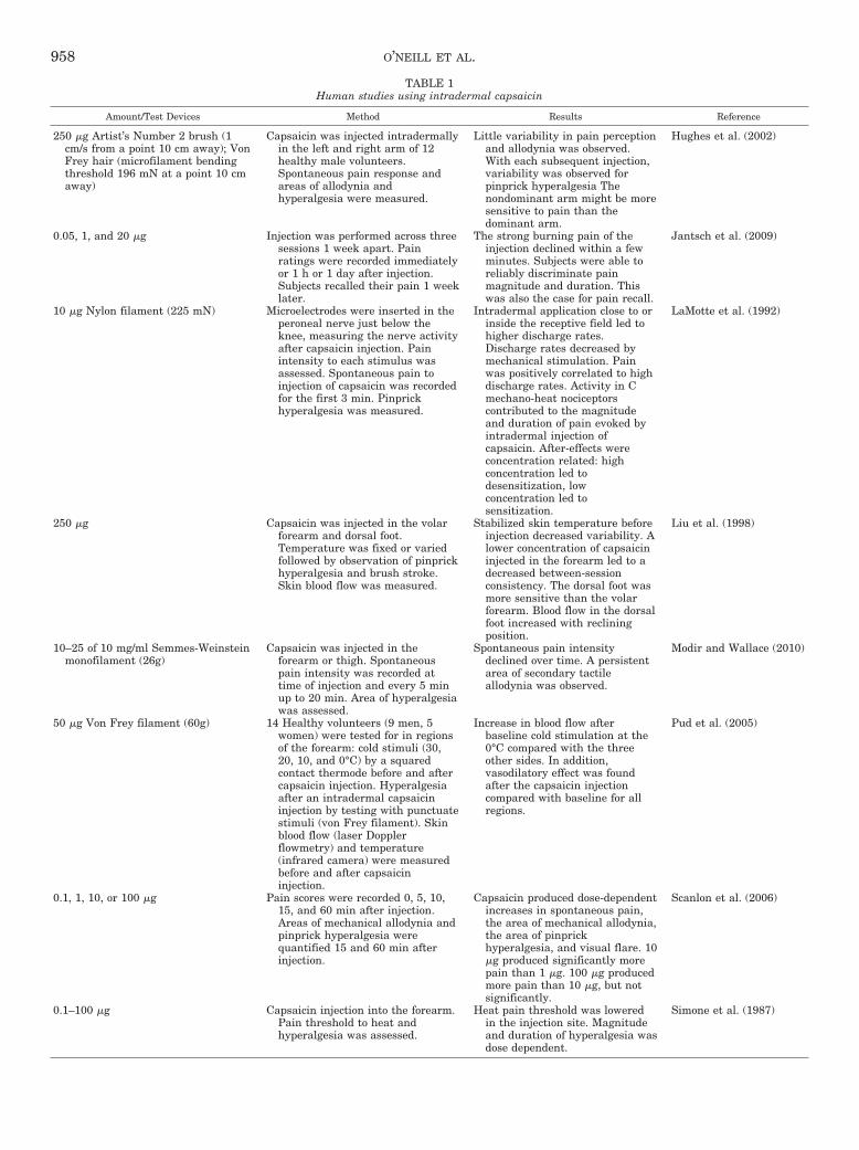
TABLE 1Human studies using intradermal capsaicin
Amount/Test Devices Method Results Reference
250 �g Artist’s Number 2 brush (1cm/s from a point 10 cm away); VonFrey hair (microfilament bendingthreshold 196 mN at a point 10 cmaway)
Capsaicin was injected intradermallyin the left and right arm of 12healthy male volunteers.Spontaneous pain response andareas of allodynia andhyperalgesia were measured.
Little variability in pain perceptionand allodynia was observed.With each subsequent injection,variability was observed forpinprick hyperalgesia Thenondominant arm might be moresensitive to pain than thedominant arm.
Hughes et al. (2002)
0.05, 1, and 20 �g Injection was performed across threesessions 1 week apart. Painratings were recorded immediatelyor 1 h or 1 day after injection.Subjects recalled their pain 1 weeklater.
The strong burning pain of theinjection declined within a fewminutes. Subjects were able toreliably discriminate painmagnitude and duration. Thiswas also the case for pain recall.
Jantsch et al. (2009)
10 �g Nylon filament (225 mN) Microelectrodes were inserted in theperoneal nerve just below theknee, measuring the nerve activityafter capsaicin injection. Painintensity to each stimulus wasassessed. Spontaneous pain toinjection of capsaicin was recordedfor the first 3 min. Pinprickhyperalgesia was measured.
Intradermal application close to orinside the receptive field led tohigher discharge rates.Discharge rates decreased bymechanical stimulation. Painwas positively correlated to highdischarge rates. Activity in Cmechano-heat nociceptorscontributed to the magnitudeand duration of pain evoked byintradermal injection ofcapsaicin. After-effects wereconcentration related: highconcentration led todesensitization, lowconcentration led tosensitization.
LaMotte et al. (1992)
250 �g Capsaicin was injected in the volarforearm and dorsal foot.Temperature was fixed or variedfollowed by observation of pinprickhyperalgesia and brush stroke.Skin blood flow was measured.
Stabilized skin temperature beforeinjection decreased variability. Alower concentration of capsaicininjected in the forearm led to adecreased between-sessionconsistency. The dorsal foot wasmore sensitive than the volarforearm. Blood flow in the dorsalfoot increased with recliningposition.
Liu et al. (1998)
10–25 of 10 mg/ml Semmes-Weinsteinmonofilament (26g)
Capsaicin was injected in theforearm or thigh. Spontaneouspain intensity was recorded attime of injection and every 5 minup to 20 min. Area of hyperalgesiawas assessed.
Spontaneous pain intensitydeclined over time. A persistentarea of secondary tactileallodynia was observed.
Modir and Wallace (2010)
50 �g Von Frey filament (60g) 14 Healthy volunteers (9 men, 5women) were tested for in regionsof the forearm: cold stimuli (30,20, 10, and 0°C) by a squaredcontact thermode before and aftercapsaicin injection. Hyperalgesiaafter an intradermal capsaicininjection by testing with punctuatestimuli (von Frey filament). Skinblood flow (laser Dopplerflowmetry) and temperature(infrared camera) were measuredbefore and after capsaicininjection.
Increase in blood flow afterbaseline cold stimulation at the0°C compared with the threeother sides. In addition,vasodilatory effect was foundafter the capsaicin injectioncompared with baseline for allregions.
Pud et al. (2005)
0.1, 1, 10, or 100 �g Pain scores were recorded 0, 5, 10,15, and 60 min after injection.Areas of mechanical allodynia andpinprick hyperalgesia werequantified 15 and 60 min afterinjection.
Capsaicin produced dose-dependentincreases in spontaneous pain,the area of mechanical allodynia,the area of pinprickhyperalgesia, and visual flare. 10�g produced significantly morepain than 1 �g. 100 �g producedmore pain than 10 �g, but notsignificantly.
Scanlon et al. (2006)
0.1–100 �g Capsaicin injection into the forearm.Pain threshold to heat andhyperalgesia was assessed.
Heat pain threshold was loweredin the injection site. Magnitudeand duration of hyperalgesia wasdose dependent.
Simone et al. (1987)
958 O’NEILL ET AL.
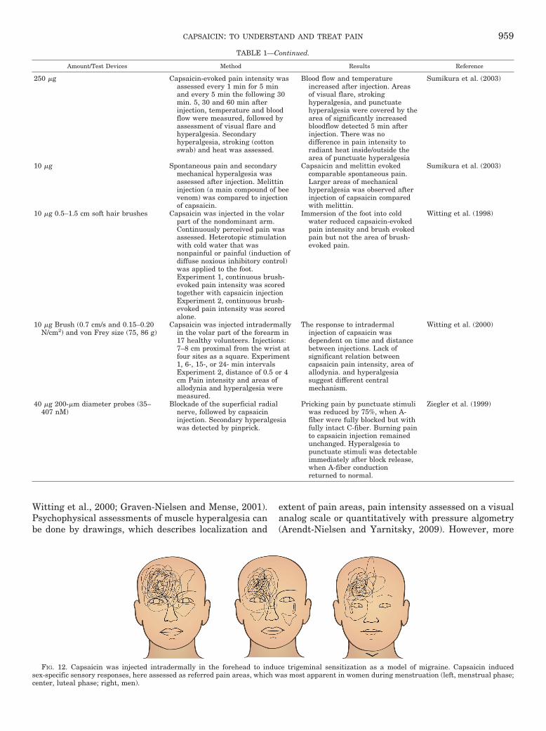
Witting et al., 2000; Graven-Nielsen and Mense, 2001).Psychophysical assessments of muscle hyperalgesia canbe done by drawings, which describes localization and
extent of pain areas, pain intensity assessed on a visualanalog scale or quantitatively with pressure algometry(Arendt-Nielsen and Yarnitsky, 2009). However, more
FIG. 12. Capsaicin was injected intradermally in the forehead to induce trigeminal sensitization as a model of migraine. Capsaicin inducedsex-specific sensory responses, here assessed as referred pain areas, which was most apparent in women during menstruation (left, menstrual phase;center, luteal phase; right, men).
TABLE 1—Continued.
Amount/Test Devices Method Results Reference
250 �g Capsaicin-evoked pain intensity wasassessed every 1 min for 5 minand every 5 min the following 30min. 5, 30 and 60 min afterinjection, temperature and bloodflow were measured, followed byassessment of visual flare andhyperalgesia. Secondaryhyperalgesia, stroking (cottonswab) and heat was assessed.
Blood flow and temperatureincreased after injection. Areasof visual flare, strokinghyperalgesia, and punctuatehyperalgesia were covered by thearea of significantly increasedbloodflow detected 5 min afterinjection. There was nodifference in pain intensity toradiant heat inside/outside thearea of punctuate hyperalgesia
Sumikura et al. (2003)
10 �g Spontaneous pain and secondarymechanical hyperalgesia wasassessed after injection. Melittininjection (a main compound of beevenom) was compared to injectionof capsaicin.
Capsaicin and melittin evokedcomparable spontaneous pain.Larger areas of mechanicalhyperalgesia was observed afterinjection of capsaicin comparedwith melittin.
Sumikura et al. (2003)
10 �g 0.5–1.5 cm soft hair brushes Capsaicin was injected in the volarpart of the nondominant arm.Continuously perceived pain wasassessed. Heterotopic stimulationwith cold water that wasnonpainful or painful (induction ofdiffuse noxious inhibitory control)was applied to the foot.Experiment 1, continuous brush-evoked pain intensity was scoredtogether with capsaicin injectionExperiment 2, continuous brush-evoked pain intensity was scoredalone.
Immersion of the foot into coldwater reduced capsaicin-evokedpain intensity and brush evokedpain but not the area of brush-evoked pain.
Witting et al. (1998)
10 �g Brush (0.7 cm/s and 0.15–0.20N/cm2) and von Frey size (75, 86 g)
Capsaicin was injected intradermallyin the volar part of the forearm in17 healthy volunteers. Injections:7–8 cm proximal from the wrist atfour sites as a square. Experiment1, 6-, 15-, or 24- min intervalsExperiment 2, distance of 0.5 or 4cm Pain intensity and areas ofallodynia and hyperalgesia weremeasured.
The response to intradermalinjection of capsaicin wasdependent on time and distancebetween injections. Lack ofsignificant relation betweencapsaicin pain intensity, area ofallodynia. and hyperalgesiasuggest different centralmechanism.
Witting et al. (2000)
40 �g 200-�m diameter probes (35–407 nM)
Blockade of the superficial radialnerve, followed by capsaicininjection. Secondary hyperalgesiawas detected by pinprick.
Pricking pain by punctuate stimuliwas reduced by 75%, when A-fiber were fully blocked but withfully intact C-fiber. Burning painto capsaicin injection remainedunchanged. Hyperalgesia topunctuate stimuli was detectableimmediately after block release,when A-fiber conductionreturned to normal.
Ziegler et al. (1999)
CAPSAICIN: TO UNDERSTAND AND TREAT PAIN 959

objective methods such as measuring blood flow, record-ing electromyography, and electroencephalography (EEG),also exist (Chang et al., 2004; Arima et al., 2009). Amodel of deep tonic pain originating from the massetermuscle, mimicking clinical pain (such as, for example, intemporomandibular joint disease), was established byinjection of 0.2 ml of a 100 �g/ml capsaicin preparationinto the deep mid-portion of the muscle (Arendt-Nielsenand Yarnitsky, 2009). The capsaicin-induced pain wasdescribed as “sharp”, “pulsing,” and “shooting.”
After capsaicin administration, subjects tolerated sig-nificantly lower pressure stimulation of the masseter,which demonstrates sensitization to mechanical stimuliafter the intramuscular injection (Arendt-Nielsen et al.,2008a). The finding is supported by other studies(LaMotte et al., 1991; Witting et al., 2000; Sluka, 2002;Arima et al., 2009). Central integration has also beenshown: if intramuscular chemical stimulations are re-peated, pain intensity and the referred pain area areincreased, indicating temporal summation (Graven-Nielsen et al., 1997).
Arima et al. (2009) investigated the effect of capsaicin-evoked masseter-muscle pain on intramuscular bloodflow at rest and during contractions. Blood flow wasinvestigated by a single-fiber laser Doppler probe in-serted to the masseter muscle and on the skin before andafter administration of intramuscular capsaicin and per-formance of isometric voluntary muscle contractions(Arima et al., 2009). Intramuscular blood flow increasedimmediately after capsaicin injection but decreased rap-idly (30 s) to preinjection values. Cutaneous blood flowabove injection site was increased for 10 min, whereasperipheral blood flow (finger) was decreased. Musclecontractions were associated with increases in intra-muscular blood flow before and after the capsaicin injec-tion (Arima et al., 2009). The study demonstrated thatincreased blood flow by muscle and neurogenic inflam-mation in muscles could possibly be mediated via anti-dromical effects and local release of vasoactive sub-stances (Arima et al., 2009). It is well known that themuscular glutamate receptor is involved in muscle pain,because glutamate-evoked pain can be reduced by coad-ministration of an NMDA antagonist (Cairns et al.,2006; Castrillon et al., 2008a,b). Hence, to explore theinteraction between glutamate and capsaicin, a gluta-mate injection before or after the capsaicin administra-tion was incorporated in the model (Arendt-Nielsen etal., 2008b). The findings showed that glutamate injec-tion after capsaicin evoked significantly less pain com-pared with its injection after isotonic saline. In contrast,the capsaicin injection after glutamate evoked signifi-cantly stronger pain compared with its injection afterisotonic saline (Arendt-Nielsen and Yarnitsky, 2009).Hence, the authors suggest a strong interaction betweenthe peripheral glutamate excitatory receptors andTRPV1, which possibly could influence the magnitude ofdevelopment of clinical musculoskeletal pain (Arendt-
Nielsen and Yarnitsky, 2009). In another study by Wanget al. (2010), the interaction between glutamate andcapsaicin-evoked muscle pain on human jaw motor func-tions was assessed. Objective assessments such as rest-ing electromyography, maximum voluntary bite force,and maximum voluntary jaw opening were recorded be-fore and after administration of glutamate or capsaicinor isotonic saline, in a paired-sequence order. Psycho-physical pain intensity before injection was recorded ona 0 to 10 numerical rating scale during each maximumvoluntary bite force and jaw opening and subsequentlyat 5-min intervals for 50 min (Wang et al., 2010). Glu-tamate followed by capsaicin injection evoked increasedresting electromyography activity and higher peak pain.Saline/glutamate followed by capsaicin evoked de-creased bite force and bite opening. The results indicatethat intramuscular administration of glutamate andcapsaicin induces muscle pain, which has the potentialto perturb some normal jaw motor functions (Wang etal., 2010).
Finally nociceptive input to the brain deriving fromintradermal or intramuscular injection of capsaicin tothe forearm has been investigated. Visual Analog Scalescore, EEG topography, and power spectra were ac-quired before and during the vehicle/capsaicin injection(Chang et al., 2004). Visual Analog Scale profiles forskin and muscle pain were highly similar despite dis-tinct qualities perceived, and both applications produceda similar but not identical EEG topographic pattern.However, muscle pain induced increased �-2 activity inthe extensive frontal, parietal, and occipital areas com-pared with skin pain, and the authors suggest that thenociceptive inputs from muscle and skin are processeddifferently in the similar neural matrix of the brain(Chang et al., 2004).
E. Human Visceral Studies
An overview of studies in which capsaicin was used toinvestigate human experimentally induced visceral hy-peralgesia is given in Table 2. Results obtained fromhuman tissues strongly differ from those obtained fromanimal preparations (Bartho et al., 2004). For example,Hammer et al., (1998) stated that the ability of intraje-junal capsaicin to give rise to pain was unexpected,because exposure of the rat gastric mucosa to 5-foldhigher concentrations of capsaicin failed to evoke pseu-doaffective reactions indicative of pain. Unfortunately,results obtained on human tissues are sparse, and thecapsaicin mystery is therefore somehow still unsolvedregarding specific effects in humans. However, interest-ing knowledge on general effects has been gained fromhuman studies.
Lee et al. (2004) found no effect of capsaicin on thefirst perception of intragastric distension, but capsa-icin infusion decreased the pressures and correspond-ing wall tensions at the threshold of discomfort but, incontrast to the findings of Gonzalez et al. (1998), with-
960 O’NEILL ET AL.
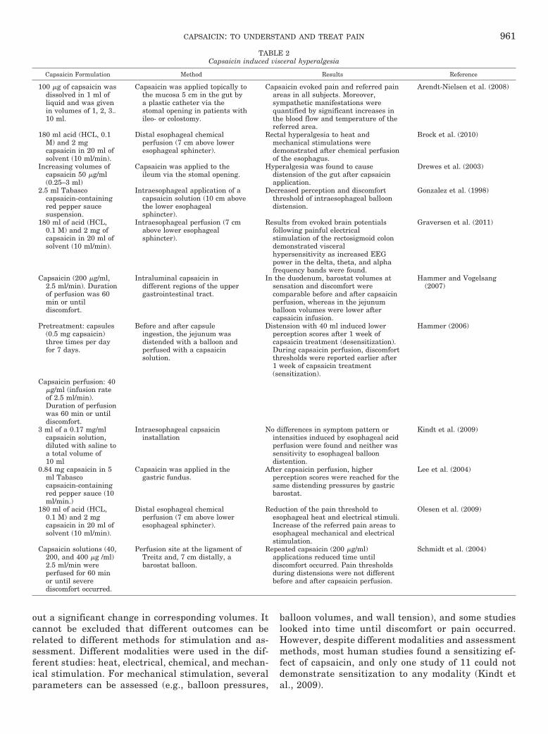
out a significant change in corresponding volumes. Itcannot be excluded that different outcomes can berelated to different methods for stimulation and as-sessment. Different modalities were used in the dif-ferent studies: heat, electrical, chemical, and mechan-ical stimulation. For mechanical stimulation, severalparameters can be assessed (e.g., balloon pressures,
balloon volumes, and wall tension), and some studieslooked into time until discomfort or pain occurred.However, despite different modalities and assessmentmethods, most human studies found a sensitizing ef-fect of capsaicin, and only one study of 11 could notdemonstrate sensitization to any modality (Kindt etal., 2009).
TABLE 2Capsaicin induced visceral hyperalgesia
Capsaicin Formulation Method Results Reference
100 �g of capsaicin wasdissolved in 1 ml ofliquid and was givenin volumes of 1, 2, 3�10 ml.
Capsaicin was applied topically tothe mucosa 5 cm in the gut bya plastic catheter via thestomal opening in patients withileo- or colostomy.
Capsaicin evoked pain and referred painareas in all subjects. Moreover,sympathetic manifestations werequantified by significant increases inthe blood flow and temperature of thereferred area.
Arendt-Nielsen et al. (2008)
180 ml acid (HCL, 0.1M) and 2 mgcapsaicin in 20 ml ofsolvent (10 ml/min).
Distal esophageal chemicalperfusion (7 cm above loweresophageal sphincter).
Rectal hyperalgesia to heat andmechanical stimulations weredemonstrated after chemical perfusionof the esophagus.
Brock et al. (2010)
Increasing volumes ofcapsaicin 50 �g/ml(0.25–3 ml)
Capsaicin was applied to theileum via the stomal opening.
Hyperalgesia was found to causedistension of the gut after capsaicinapplication.
Drewes et al. (2003)
2.5 ml Tabascocapsaicin-containingred pepper saucesuspension.
Intraesophageal application of acapsaicin solution (10 cm abovethe lower esophagealsphincter).
Decreased perception and discomfortthreshold of intraesophageal balloondistension.
Gonzalez et al. (1998)
180 ml of acid (HCL,0.1 M) and 2 mg ofcapsaicin in 20 ml ofsolvent (10 ml/min).
Intraesophageal perfusion (7 cmabove lower esophagealsphincter).
Results from evoked brain potentialsfollowing painful electricalstimulation of the rectosigmoid colondemonstrated visceralhypersensitivity as increased EEGpower in the delta, theta, and alphafrequency bands were found.
Graversen et al. (2011)
Capsaicin (200 �g/ml,2.5 ml/min). Durationof perfusion was 60min or untildiscomfort.
Intraluminal capsaicin indifferent regions of the uppergastrointestinal tract.
In the duodenum, barostat volumes atsensation and discomfort werecomparable before and after capsaicinperfusion, whereas in the jejunumballoon volumes were lower aftercapsaicin infusion.
Hammer and Vogelsang(2007)
Pretreatment: capsules(0.5 mg capsaicin)three times per dayfor 7 days.
Before and after capsuleingestion, the jejunum wasdistended with a balloon andperfused with a capsaicinsolution.
Distension with 40 ml induced lowerperception scores after 1 week ofcapsaicin treatment (desensitization).During capsaicin perfusion, discomfortthresholds were reported earlier after1 week of capsaicin treatment(sensitization).
Hammer (2006)
Capsaicin perfusion: 40�g/ml (infusion rateof 2.5 ml/min).Duration of perfusionwas 60 min or untildiscomfort.
3 ml of a 0.17 mg/mlcapsaicin solution,diluted with saline toa total volume of10 ml
Intraesophageal capsaicininstallation
No differences in symptom pattern orintensities induced by esophageal acidperfusion were found and neither wassensitivity to esophageal balloondistention.
Kindt et al. (2009)
0.84 mg capsaicin in 5ml Tabascocapsaicin-containingred pepper sauce (10ml/min.)
Capsaicin was applied in thegastric fundus.
After capsaicin perfusion, higherperception scores were reached for thesame distending pressures by gastricbarostat.
Lee et al. (2004)
180 ml of acid (HCL,0.1 M) and 2 mgcapsaicin in 20 ml ofsolvent (10 ml/min).
Distal esophageal chemicalperfusion (7 cm above loweresophageal sphincter).
Reduction of the pain threshold toesophageal heat and electrical stimuli.Increase of the referred pain areas toesophageal mechanical and electricalstimulation.
Olesen et al. (2009)
Capsaicin solutions (40,200, and 400 �g /ml)2.5 ml/min wereperfused for 60 minor until severediscomfort occurred.
Perfusion site at the ligament ofTreitz and, 7 cm distally, abarostat balloon.
Repeated capsaicin (200 �g/ml)applications reduced time untildiscomfort occurred. Pain thresholdsduring distensions were not differentbefore and after capsaicin perfusion.
Schmidt et al. (2004)
CAPSAICIN: TO UNDERSTAND AND TREAT PAIN 961

Intraesophageal application of capsaicin-containingred pepper sauce (Tabasco) has been found to decreasethe distension volume for perception and discomfortthresholds of intraesophageal balloon distension. Never-theless, the sensitizing effect of capsaicin disappeared30 min after infusion. This return to basal level could becaused by limited local activity or by desensitization(Gonzalez et al., 1998). Desensitization was also foundby Hammer (2006) in a timing study in which distensionwith 40 ml capsaicin induced lower perception scoresafter 1 week of treatment. However, the same studyfound that during capsaicin perfusion, discomfort thresholdswere reported earlier after 1 week of capsaicin treat-ment, indicating sensitization. This could indicate thatmechanoreceptors and chemoreceptors are affected dif-ferently by 7 days of oral ingestion of capsaicin capsules(Hammer, 2006).
Later, a purified solution of capsaicin has been used indifferent doses at different sites of the gastrointestinaltract. It has been concluded that capsaicin evokes ab-dominal sensations by stimulation of chemoreceptorsand not mechanoreceptors (Schmidt et al., 2004). How-ever, several studies have found reduction in painthresholds to mechanical, electrical, and thermal stim-ulation (Gonzalez et al., 1998; Drewes et al., 2003b; Leeet al., 2004; Hammer and Vogelsang, 2007; Olesen et al.,2009; Brock et al., 2010). This indicates that capsaicinmust affect a variety of receptors, and sensitization/desensitization is not necessarily dependent on the qual-ity of receptor activation but more the quantity or dura-tion. Nevertheless, this is still not clear from humanstudies, and further investigation may be useful. In thiscontext, it should be noted that visceral innervation isweak and diffuse, and so the ability of capsaicin to acti-vate sufficient numbers of TRPV1 afferents to inducechanges may be more difficult in visceral tissue com-pared with somatic and muscular tissues (Sikandar andDickenson, 2012).
Administration of capsaicin by itself or in a combina-tion with acid has been shown to induce visceral hyper-algesia, which has led to hypersensitivity to heat andmechanical, electrical, and chemical stimulation (Gon-zalez et al., 1998; Drewes et al., 2003a; Lee et al., 2004;Hammer, 2006; Hammer and Vogelsang, 2007; Olesenet al., 2009; Brock et al., 2010). These stimuli are likelyto reflect both peripheral and central sensitization.Therefore, quantification of referred pain areas repre-senting the viscerosomatic convergence to esophagealacid and capsaicin perfusion has been studied, and in-dications for central effects have been found (Olesen etal., 2010). These models of central sensitization werefurther developed to investigate viscerovisceral hyperal-gesia, which manifests as extra segmental hypersensi-tivity to mechanical and heat stimulation of the rectumafter esophageal infusion (Brock et al., 2010). Graversenet al. (2011) have, for the first time in humans, directlydemonstrated central neuronal changes as a result of
chemical irritation by intraesophageal acid and capsai-cin perfusion. This was demonstrated as an increasedEEG power in the delta, theta, and alpha frequencybands. The authors found a correlation when taking allfrequency bands into consideration simultaneously,which showed that central sensitization was directlyreflected by the alteration of the CNS response afternoxious stimulations, whereas no correlation to changesin pain score was seen for the placebo response. It isnoteworthy that the correlation was evident eventhough some subjects developed hyposensitivity, wherethey tolerated higher stimulus intensity after perfusionwith acid and capsaicin than before. This phenomenon ofmixed hypo- and hypersensitivity to the same treatmenthas been demonstrated previously (Hammer, 2006) andcan be explained by the two competing mechanismsinvolved with capsaicin administration—sensitizationor desensitization, which will be discussed in the nextsection. It should be noted, however, that a combinationof acid and capsaicin was applied. Therefore, it cannot beconcluded whether the central effect was caused by cap-saicin, acid, or the combinations. Further human studiesare needed to distinguish between capsaicin and acideffects.
It has been discussed whether capsaicin causes painby directly stimulating nociceptive afferents in theintestinal mucosa or by stimulating motility as mightbe deduced from the cramping nature of the sensation.Stimulation of motility may result in pain from exces-sive contractions or movement of the inflated balloonalong the intestine. Excessive contraction or disten-sion of the gut is well established to give rise to pain(Hammer et al., 1998). A first-in-human study hasdemonstrated that afferent nerves from human guttissue ex vivo are stimulated directly by capsaicin ashighly increased firing rates (Peiris et al., 2011).Hence, capsaicin can cause pain as a combination ofdirect stimulation of nociceptive afferents and stimu-lation of motility.
It is noteworthy that there appears to be a dramaticinteraction with the sympathetic response in referredpain areas, which has been studied by application ofcapsaicin to human ileo- or colostomy (Arendt-Nielsen etal., 2008a). Capsaicin-evoked pain and referred painareas in all subjects and sympathetic manifestationswere quantified by increased temperature and bloodflow of the referred area (Arendt-Nielsen et al., 2008a).Bowel disorders are also accompanied by sympatheticmanifestations. Schmidt et al. (2004) demonstrated thattheir model of intraluminal capsaicin can modulate per-ception and cause discomfort or pain in a normal-ap-pearing gut and thus provides a model for the study offunctional bowel disorders.
Few human studies in which visceral sensitization wasused as models for the study of disease have been used tostudy analgesic effects or mechanisms. Olesen et al. (2010)investigated the effects of morphine and oxycodone in ex-
962 O’NEILL ET AL.

perimentally evoked hyperalgesia in healthy volunteers.Hyperalgesia was induced by esophageal perfusion withacid and capsaicin. They found that morphine and oxy-codone exerted different effects in the sensitized pain sys-tem. The evoked hyperalgesia bridged findings from stud-ies in healthy volunteers to patients, and new fundamentalknowledge on different analgesic effects in hyperalgesiawas found.
Another approach can be to use these human painmodels of sensitization as specific tools to investigateanalgesic mechanisms. For example, Krarup et al. (2011)investigated the efficacy of a new transient receptorpotential vanilloid 1 antagonist AZD1386 in a model ofhuman esophageal pain where sensitization and activa-tion of TRPV1 receptors was evoked by acid. The resultsdemonstrated a dose-dependent increase in the esopha-geal heat pain threshold after administration of theTRPV1 antagonist (Krarup et al., 2011).
Willert et al. have used a model of human visceralpain hypersensitivity to determine whether the devel-opment and maintenance of human visceral hypersen-sitivity is NMDA receptor mediated. This was per-formed using the NMDA receptor antagonist ketamineeither before or after acid esophageal infusion of acid.Acid-induced esophageal hypersensitivity was pre-vented by ketamine and ketamine administered afteracid reversed the induction of esophageal hypersensi-tivity induced by acid (Willert et al., 2004). As capsa-icin induces similar hypersensitivity and central sen-sitization, it might also be used in human pain modelsas a model of functional gastrointestinal disorders toconfirm effects of NMDA antagonists or other putativeanalgesics.
XIII. Sensitizing or Desensitizing?
Activation of the TRPV1 receptor, in rats and humans,can cause both sensitization—as previously discussed—but may also result in desensitization of nerve cells. In1977, Szolcsanyi showed that capsaicin applicationcould result in a long-lasting insensitivity to mechanicalpain, which was believed to be due to peripheral mech-anisms. In addition, when capsaicin is systemically ad-ministrated in supratherapeutic doses to neonatal ro-dents, capsaicin has been found not only to desensitizesensory neurons but also to induce degeneration of theDRG sensory afferents (Jancso et al., 1977). It has laterbeen revealed that desensitization is a dose-dependentphenomenon, whereby repeated application of low doseor a single high dose of injected or topical capsaicin led toimmediate desensitization, whereas single lower dosescause activation (LaMotte et al., 1991; Simone et al.,1998; Kennedy et al., 2010). It is also well characterizedthat although a single application of low-dose capsaicincream (�1%) results in an activation of PAFs and bothprimary and secondary hyperalgesia, repeated applica-tion of topical capsaicin over a few weeks dampens re-
sponses to both heat and mechanical stimuli (Nolano etal., 1999; Mason et al., 2004). This desensitization seemsselective for A and C fibers and hence supports the useof capsaicin as a tool to study nociceptive afferents andconsequences of their deactivation.
There are a number of ways in which capsaicin canresult in desensitization of TRPV1-positive afferents.Activation may cause an acute rapid desensitization,long-term tachyphylaxis, and could eventually be asso-ciated with a withdrawal of epidermal nerve fibers(ENFs) (Backonja et al., 2008). Both types of desensiti-zations occur in the area localized to the site of injection/application; however, the time frame for each varies; theinitial acute desensitization occurs within the first 20 safter agonist binding, whereas tachyphylaxis and ENFwithdrawal occur over much longer periods up to 72 hafter application (Simone et al., 1998; Touska et al.,2011). The type of desensitization will depend on theconcentration of capsaicin, length of application time,and the penetration through to the dermis (Touska etal., 2011).
Rapid desensitization first involves capsaicin bindingof TRPV1, which not only activates PAFs and pain path-ways but also results in release of neuropeptides fromthe C fiber terminals in the periphery. This includesCGRP and SP, which are involved in local neurogenicinflammation and thus an area of flare around the ap-plication site. Once released, terminals are depleted andthe nerve may become desensitized (Maggi and Meli,1988; Simone et al., 1998).
Second, voltage-gated ion channels are inactivated,which also results in a rapid, short-lasting, desensitiza-tion as further action potentials cannot be generated(Simone et al., 1998; Tominaga and Tominaga, 2005). Itis believed that the desensitization of fibers is a protec-tive mechanism to inhibit excessive calcium influx,which leads to excitotoxic cell death. As already dis-cussed, activation of TRPV1 and entry of Ca2� ions tothe nerves activates a number of enzymes, which maylead to sensitization. However, a second effect is theactivation of other Ca2�-dependent enzymes such asprotein phosphatase 2B/calcineurin, which is likely tolead to tachyphyaxis (Docherty et al., 1996; Mohapatraand Nau, 2005). Calcineurin dephosphorylates TRPV1with a key site believed to be Thr370 and thereforedecreases sensitivity to agonists such as capsaicin (Mo-hapatra and Nau, 2005).
The importance of Ca2� in capsaicin-induced desensiti-zation was highlighted by Docherty et al. (1996), whofound that desensitization in cultured rat DRG neuronswas strongly inhibited by the calcium chelator 1,2-bis(o-aminophenoxy)ethane-N,N,N�,N�-tetraacetic acid (BAPTA).The study also showed that inhibition of calcineurin bycyclosporin A complexed to cyclophilin greatly reduceddesensitization (Docherty et al., 1996). Koplas et al.(1997) demonstrated that reducing the external Ca2�
concentration resulted in lower levels of acute desensi-
CAPSAICIN: TO UNDERSTAND AND TREAT PAIN 963

tization and tachyphylaxis. However, caffeine-inducedrelease of intracellular Ca2� resulted in desensitization,even in the absence of extracellular Ca2� (Koplas et al.,1997), therefore confirming that desensitization is de-pendent on both intra- and extracellular Ca2� and asubsequent dephosphorylation of TRPV1. Desensitiza-tion can therefore involve a number of mechanisms and,as such, underlies some of therapeutic use of low-dosecapsaicin cream.
A. Altered Skin Innervation after Transient ReceptorPotential Vanilloid 1 Activation
Other than receptor desensitization, another mecha-nism can be brought into play by which the peripheralsensory innervation is altered by high levels of activa-tion of the TRPV1 receptor. Simone et al. (1998) showedthat intradermal injection of capsaicin into the skinoverlying the shoulder results in a decrease of ENFswithin a radius of 1 to 2 mm away from the injection site,up to 72 h after injection, therefore suggesting thatwithdrawal of ENFs from the dermis is restricted tofibers that have come into direct contact with capsaicin.However, based on assessment of ENF density in skinbiopsies after topical exposure to an occlusive 35 �50-mm bandage containing 1.8 g of 0.1% capsaicincream, Polydefkis et al. (2004) found that capsaicin ap-plication produced denervation of both the epidermisand the subepidermal plexus, with few or no ENFs re-maining after 48 h in both healthy control subjects andpatients with diabetes. This withdrawal may be due tothe previously described rearrangement of microtubulesor simply to mitochondrial dysfunction. The rate ofnerve regeneration was associated with the baselineENF density, but not age, sex, epidermal thickness, orpostcapsaicin treatment density. ENF regeneration, asdetermined by recovery of ENF density, occurred at arate of 0.18 � 0.08 fibers/mm per day in healthy controlsubjects, which was significantly reduced in subjectswith diabetes (0.07 � 0.06 fibers/mm per day, P � 0.001)after adjusting for changes in baseline ENF density(Polydefkis et al., 2004).
A loss of ENFs would result not only in a decreasedperipheral activity through selective drawing back ofpain fibers but might also decrease central sensitizationdriven by these fibers; therefore, the rationale of apply-ing topical capsaicin to achieve analgesia may extendbeyond receptor events. The clinical application of theseapproaches is discussed in section XIV.
XIV. Capsaicin As a TherapeuticPharmacological Agent
Capsaicin has been used all over the world for manyyears as a pharmacological intervention for a plethora ofailments. It is used to treat fevers, nausea, vomiting,malaria, and different pain conditions. In regions ofcentral Peru, the leaves are burned or used to create
steam in the bath to cure headaches; in the DominicanRepublic, the leaves are ingested to treat painful men-ses; and in the Phillipines, the fruit is used to treatarthritis (Meghvansi et al., 2010). This final section con-siders the use of capsaicin as a therapeutic option, butfirst discusses the use of TRPV1 modulation to elucidatethe role of different fibers in the behavioral changes thataccompany models of nerve injury. Two studies haveemployed the desensitization of TRPV1-positive fiberswith systemic RTX to tease out the roles of TRPV1-positive and -negative fibers in animal models. Althoughmany models of different pain conditions monitor andmanipulate responses to evoked stimuli, many patientswith neuropathic pain report stimulus-independentspontaneous pain, often described as burning. King etal., (2011) used RTX to prevent nerve injury-inducedthermal hypersensitivity and spontaneous pain (as-sessed by novel place aversion), but the treatment hadno effect on tactile hypersensitivity. Destruction of adefined population of spinal NK-1 receptor-expressingneurons, cells that respond to SP found in TRPV-1 af-ferents, blocked the neuropathic thermal and tactile hy-persensitivity as well as spontaneous pain. Further ma-nipulations revealed that the large-diameter dorsalcolumn projection can mediate nerve injury-induced tac-tile hypersensitivity but does not contribute to sponta-neous pain. This would suggest not only that differentpathways from the periphery through the spinal cordconvey different components of the neuropathic pain inthis model but also that the therapeutic actions ofagents acting through TRPV1 might be selective.
It is well known that both nerve and tissue injuryproduce hypersensitivity to evoked stimuli and ongoing,stimulus-independent pain. Using their validated ap-proach based on pain relief elicits reward in nerve-in-jured rats, in a 4-day model of inflammatory pain, Okunet al. (2011) showed that the ongoing pain, driven by theperipheral site of injury (blocked by lidocaine appliedhere), is dependent on TRPV1-positive fiber, but theselective TRPV1 receptor antagonist AMG9810, al-though able to reverse the CFA-induced thermal hyper-algesia, had no effect at all on the CFA-induced ongoingpain or guarding behavior, suggesting that TRV1 recep-tor mechanisms do not contribute to spontaneous painafter inflammation. These novel approaches will shedmuch needed light on the processes that drive stimulus-evoked and stimulus-independent pains.
Because of the problems of the central effects of anal-gesics and the need for sustained pain control, there hasbeen an increasing focus on development of new routesof drug administration to control persistent pain in atolerable way and with acceptable patient compliance.Patches have been used for a wide variety of drugs—opioids for their central actions but at stable prolongedplasma levels or topical use of nonsteroidal anti-inflam-matory agents and capsaicin where the aim is to targetthe local pain generators. Accordingly, recent applica-
964 O’NEILL ET AL.
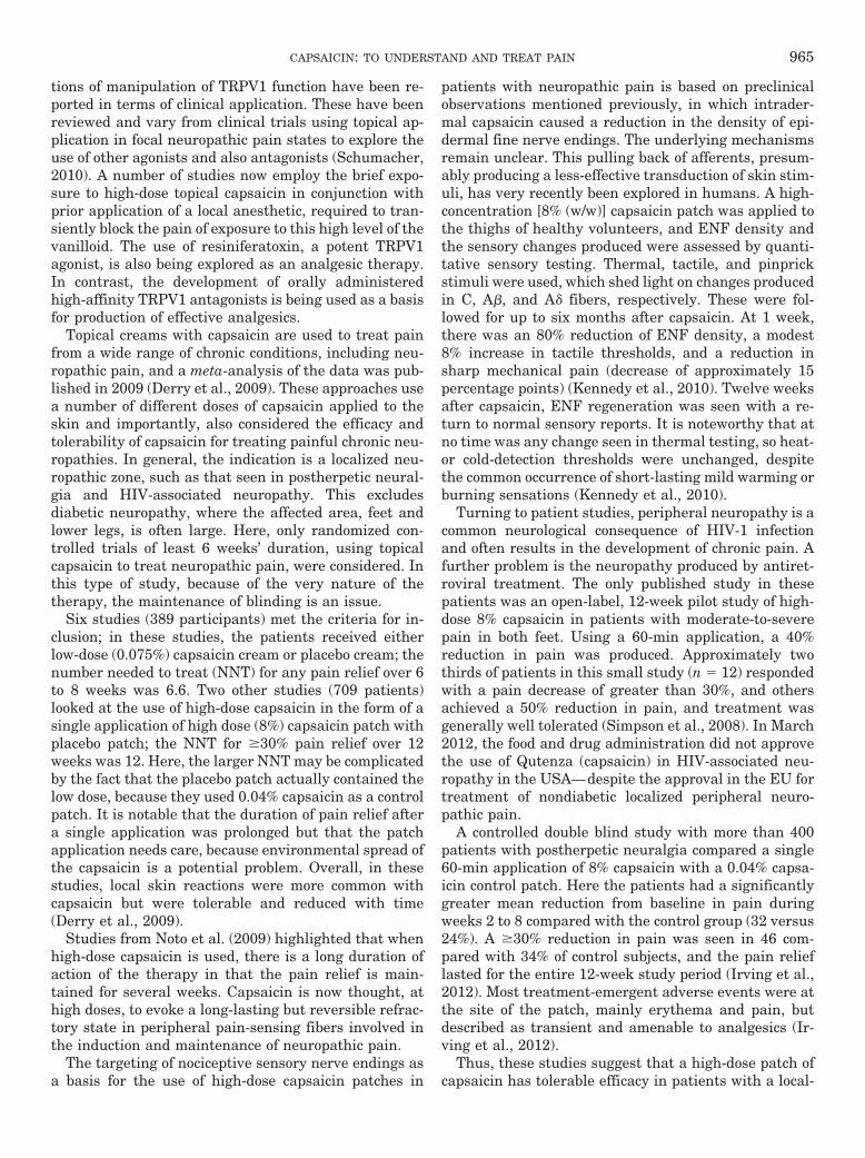
tions of manipulation of TRPV1 function have been re-ported in terms of clinical application. These have beenreviewed and vary from clinical trials using topical ap-plication in focal neuropathic pain states to explore theuse of other agonists and also antagonists (Schumacher,2010). A number of studies now employ the brief expo-sure to high-dose topical capsaicin in conjunction withprior application of a local anesthetic, required to tran-siently block the pain of exposure to this high level of thevanilloid. The use of resiniferatoxin, a potent TRPV1agonist, is also being explored as an analgesic therapy.In contrast, the development of orally administeredhigh-affinity TRPV1 antagonists is being used as a basisfor production of effective analgesics.
Topical creams with capsaicin are used to treat painfrom a wide range of chronic conditions, including neu-ropathic pain, and a meta-analysis of the data was pub-lished in 2009 (Derry et al., 2009). These approaches usea number of different doses of capsaicin applied to theskin and importantly, also considered the efficacy andtolerability of capsaicin for treating painful chronic neu-ropathies. In general, the indication is a localized neu-ropathic zone, such as that seen in postherpetic neural-gia and HIV-associated neuropathy. This excludesdiabetic neuropathy, where the affected area, feet andlower legs, is often large. Here, only randomized con-trolled trials of least 6 weeks’ duration, using topicalcapsaicin to treat neuropathic pain, were considered. Inthis type of study, because of the very nature of thetherapy, the maintenance of blinding is an issue.
Six studies (389 participants) met the criteria for in-clusion; in these studies, the patients received eitherlow-dose (0.075%) capsaicin cream or placebo cream; thenumber needed to treat (NNT) for any pain relief over 6to 8 weeks was 6.6. Two other studies (709 patients)looked at the use of high-dose capsaicin in the form of asingle application of high dose (8%) capsaicin patch withplacebo patch; the NNT for �30% pain relief over 12weeks was 12. Here, the larger NNT may be complicatedby the fact that the placebo patch actually contained thelow dose, because they used 0.04% capsaicin as a controlpatch. It is notable that the duration of pain relief aftera single application was prolonged but that the patchapplication needs care, because environmental spread ofthe capsaicin is a potential problem. Overall, in thesestudies, local skin reactions were more common withcapsaicin but were tolerable and reduced with time(Derry et al., 2009).
Studies from Noto et al. (2009) highlighted that whenhigh-dose capsaicin is used, there is a long duration ofaction of the therapy in that the pain relief is main-tained for several weeks. Capsaicin is now thought, athigh doses, to evoke a long-lasting but reversible refrac-tory state in peripheral pain-sensing fibers involved inthe induction and maintenance of neuropathic pain.
The targeting of nociceptive sensory nerve endings asa basis for the use of high-dose capsaicin patches in
patients with neuropathic pain is based on preclinicalobservations mentioned previously, in which intrader-mal capsaicin caused a reduction in the density of epi-dermal fine nerve endings. The underlying mechanismsremain unclear. This pulling back of afferents, presum-ably producing a less-effective transduction of skin stim-uli, has very recently been explored in humans. A high-concentration [8% (w/w)] capsaicin patch was applied tothe thighs of healthy volunteers, and ENF density andthe sensory changes produced were assessed by quanti-tative sensory testing. Thermal, tactile, and pinprickstimuli were used, which shed light on changes producedin C, A�, and A� fibers, respectively. These were fol-lowed for up to six months after capsaicin. At 1 week,there was an 80% reduction of ENF density, a modest8% increase in tactile thresholds, and a reduction insharp mechanical pain (decrease of approximately 15percentage points) (Kennedy et al., 2010). Twelve weeksafter capsaicin, ENF regeneration was seen with a re-turn to normal sensory reports. It is noteworthy that atno time was any change seen in thermal testing, so heat-or cold-detection thresholds were unchanged, despitethe common occurrence of short-lasting mild warming orburning sensations (Kennedy et al., 2010).
Turning to patient studies, peripheral neuropathy is acommon neurological consequence of HIV-1 infectionand often results in the development of chronic pain. Afurther problem is the neuropathy produced by antiret-roviral treatment. The only published study in thesepatients was an open-label, 12-week pilot study of high-dose 8% capsaicin in patients with moderate-to-severepain in both feet. Using a 60-min application, a 40%reduction in pain was produced. Approximately twothirds of patients in this small study (n 12) respondedwith a pain decrease of greater than 30%, and othersachieved a 50% reduction in pain, and treatment wasgenerally well tolerated (Simpson et al., 2008). In March2012, the food and drug administration did not approvethe use of Qutenza (capsaicin) in HIV-associated neu-ropathy in the USA—despite the approval in the EU fortreatment of nondiabetic localized peripheral neuro-pathic pain.
A controlled double blind study with more than 400patients with postherpetic neuralgia compared a single60-min application of 8% capsaicin with a 0.04% capsa-icin control patch. Here the patients had a significantlygreater mean reduction from baseline in pain duringweeks 2 to 8 compared with the control group (32 versus24%). A �30% reduction in pain was seen in 46 com-pared with 34% of control subjects, and the pain relieflasted for the entire 12-week study period (Irving et al.,2012). Most treatment-emergent adverse events were atthe site of the patch, mainly erythema and pain, butdescribed as transient and amenable to analgesics (Ir-ving et al., 2012).
Thus, these studies suggest that a high-dose patch ofcapsaicin has tolerable efficacy in patients with a local-
CAPSAICIN: TO UNDERSTAND AND TREAT PAIN 965

ized pain as a result of nerve injury and that the modestreductions in pain might be clinically useful, especiallybecause they last for months after application. Side ef-fects are common but not problematic. This is explicablein terms of a “pulling back” of afferent fibers in the areaof the patch. What is difficult to explain is the observa-tion that heat and cold thresholds were unaltered, be-cause clearly TRPV heat sensors are on fine fibers, asare TRPM8 cold receptors. This disconnect is also seenin the small but clear change in tactile sensitivity, be-cause this modality is conveyed by large fibers that arenot changed on visual inspection after high-dose capsa-icin and do not possess TRPV1 receptors. These datamay be explained by TRPV1 containing afferents drivingcentral sensitization, so that loss of the ability of thesefibers to be activated causes a “wind-down” of centralresponsivity. The ability of other modalities to accesspain pathways centrally may be limited in the absence ofcentral hypersensitivity.
So what other approaches might be possible? One obvi-ous potential is the production of blockers of the TRPV1receptor. A number of recent studies have been publishedon this topic. Based on the observation that patients withgastroesophageal reflux disease are hypersensitive to heatand acid, a TRPV1 antagonist (AZD1386) was tested onresponses to painful heat, distension, electrical current,and acid in the esophagus in healthy volunteers (Krarup etal., 2011). It is noteworthy that both heat and pressurepain applied on the arm was used to gauge possible effectson somatic stimuli. Almost all of the 21 participants com-pleted the protocol. In the esophagus, AZD1386 increasedpain thresholds to heat stimuli by 23 to 28%, depending onthe dose. The drug also increased skin heat tolerance, andthe effects on thermal stimuli (which were selective, inthat the mechanical pain tests were unaltered) lasted for2.5 h. Half of the subjects reported “feeling cold” and bodytemperature was elevated by 0.4 to 0.7°C, depending onthe dose; however, no other severe adverse-event problemswere reported (Krarup et al., 2011).
Another study in healthy volunteers used the TRPV1antagonist (R)-(5-tert-butyl-2,3-dihydro-1H-inden-1-yl)-3-(1H-indazol-4-yl)-urea (ABT-102) with proven efficacyin a number of preclinical pain models. Here, the sen-sory effects with somatic stimuli were tested, as was thepotential of drug-induced thermosensory impairment.ABT-102 was tested in a three-dose, double-blind, pla-cebo-controlled, randomized trial. The drug caused sig-nificant dose-dependent increases in the heat painthreshold and reduced pain scores for suprathresholdheat. These were seen over the weeklong experimentalduration and were present for both oral and cutaneousstimuli with selectivity, in that cutaneous cold detectiondid not alter. All effects were fully reversed by day 10,and no safety issues were seen; body temperature stayedbelow 39°C in all subjects. Thus, in this randomizedcontrolled trial, this selective TRPV1 antagonist ABT-102 potently and reversibly increased the threshold for
heat pain but also reduced the pain from suprathresholdoral and cutaneous heat. Thus, this study adds to theprevious data, suggesting that these antagonists areboth effective and tolerable (Rowbotham et al., 2011).
Thus far, based on its distribution and functions,TRPV1 can be considered a highly suitable target fordeveloping small-molecule antagonists, although the al-terations in body temperature could lead to the threat ofabandonment of such antagonists. Furthermore, asmentioned previously, this receptor is not found only inneurons, so blockade could also be problematic. Some ofthese issues have been reviewed by Premkumar andBishnoi (2011), but the thermal changes as a potentialproblem were noted several years ago.
In one sense, it is reassuring that, in terms of trans-lation from animals to the clinic, in all species tested(except birds—thus a chicken jalfrezzi is a crime againstnature, but still rather tasty), the agonists at TRPV1cause pain and hypothermia—when delivered systemi-cally; thus, not surprisingly, several of the various an-tagonists of TRPV1 block pain in rat and mouse modelsof inflammation, OA, and cancer-induced pain. But,from rodents to primates, body temperature also in-creases. This ancient conserved function of TRPV1 inbody-temperature maintenance was a hurdle for clinicaldevelopment of one antagonist, N-(4-(6-(4-trifluorometh-ylphenyl)pyrimidin-4-yloxy)benzothiazol-2-yl)acetamide(AMG 517) (Gavva et al., 2008).
However, several other TRPV1 antagonists are cur-rently being evaluated, and a very recently publishedarticle sheds light on the problem of antagonist-inducedhyperthermia while developing therapeutic agents(Watabiki, 2011). With several new antagonists produc-ing the same elevation of body temperature—is this aclass effect or not? The TRPV1 receptor is activated by avariety of stimuli, such as endogenous ligands and lowpH. This range may be important in terms of hyperther-mia in animals and humans.
One important issue is that TRPV1 antagonists dif-ferentially block three modes of TRPV1 activation: byheat, protons, and chemical ligands (e.g., capsaicin). Astudy compared potencies in these three modes ofTRPV1 activation with the potency of a TRPV1 antago-nist to cause hyperthermia in rats, mice, and guinea pigsusing eight TRPV1 antagonists with different pharma-cological profiles. With the use of mathematical model-ing, it became clear that the hyperthermic effect ishighly related to the extent of TRPV1 blockade in theproton mode with no to moderate sensitivity in the cap-saicin mode and no sensitivity in the heat mode. Thus,hyperthermia-free TRPV1 antagonists that do not blockTRPV1 activation by protons and have low potency toblock the capsaicin mode may be of use, but are thesetypes of compounds still effective?
The TRPV1 antagonist N-(1-methyl-2-oxo-1,2,3,4-tetrahydro-7-quinolyl)-2-((2-methylpyrrolidin-1-yl)methyl)biphenyl-4-carboxamide (AS1928370) binds to the res-
966 O’NEILL ET AL.

iniferatoxin-binding site on TRPV1 and so, as expected,will reduce capsaicin-mediated inward currents.AS1928370 also inhibits the capsaicin-induced Ca2� fluxin human and rat TRPV1-expressing cells yet has a verysmall effect on the proton-induced flux (Watabiki et al.,2011). AS1928370 attenuated capsaicin-induced second-ary hyperalgesia and mechanical allodynia after neuropa-thy and was effective on inflammatory pain. AS1928370had no effect on body temperature up to 10 mg/kg p.o. buthad a hypothermic effect at higher doses.
XV. Conclusion
Capsaicin administered topically, intradermally, ororally has proven to produce complex effects but remainsa reliable and reproducible way to investigate peripheraland central mechanisms underlying certain hypersensi-tivities. In addition, we have discussed how it may alsobe a useful treatment of certain pain states, includingneuropathies.
In animal models, capsaicin has been used extensivelyto elucidate the molecular mechanisms that underpincentral sensitization as well as investigating potentialpharmacological interventions. In human models, it hasbeen shown to evoke similar signs and symptoms ofcentral sensitization, including hyperalgesia and allo-dynia. This suggests that similar mechanisms are in-volved in changes of pain processing after capsaicin ad-ministration in both animals and humans. Furthercharacterization of capsaicin as a tool and its use as areliable translational model, using similar evoked re-sponses and time frames, with consideration of the dose,would be useful to confirm this translation. However,from this review we conclude that agents acting onTRPV1 receptors, as well as capsaicin itself, will stillgenerate plenty of interest in the future and will shedlight on pain mechanisms. There is plenty of excitementto come with this vanilloid.
Acknowledgments
This work was funded by the Wellcome Trust London Pain Con-sortium; The Obel Family Foundation (Det Obelske Familiefond);and a UCL Grand Challenges PhD studentship (to J.O.N.).
Authorship Contributions
Wrote or contributed to the writing of the manuscript: O’Neill,Brock, Estrup Olesen, Andresen, Nilsson, and Dickenson.
ReferencesAlawi K and Keeble J (2010) The paradoxical role of the transient receptor potential
vanilloid 1 receptor in inflammation. Pharmacol Ther 125:181–195.Al Othman ZA, Ahmed YB, Habila MA, and Ghafar AA (2011) Determination of
capsaicin and dihydrocapsaicin in Capsicum fruit samples using high performanceliquid chromatography. Molecules 16:8919–8929.
Andresen T, Lunden D, Drewes AM, and Arendt-Nielsen L (2011) Pain sensitivityand experimentally induced sensitisation in red haired females. Scand J Pain2:3–6.
Arendt-Nielsen L, Curatolo M, and Drewes A (2007) A Human experimental painmodels in drug development: translational pain research. Curr Opin InvestigDrugs 8:41–53.
Arendt-Nielsen L, Schipper KP, Dimcevski G, Sumikura H, Krarup AL, Giambe-rardino MA, and Drewes AM (2008a) Viscero-somatic reflexes in referred painareas evoked by capsaicin stimulation of the human gut. Eur J Pain 12:544–551.
Arendt-Nielsen L, Svensson P, Sessle BJ, Cairns BE, and Wang K (2008b) Interac-
tions between glutamate and capsaicin in inducing muscle pain and sensitizationin humans. Eur J Pain 12:661–670.
Arendt-Nielsen L and Yarnitsky D (2009) Experimental and clinical applications ofquantitative sensory testing applied to skin, muscles and viscera. J Pain 10:556–572.
Arima T, Arendt-Nielsen L, Minagi S, and Svensson P (2009) Effect of capsaicin-evoked jaw-muscle pain on intramuscular blood-flow. Arch Oral Biol 54:241–249.
Arima T, Svensson P, and Arendt-Nielsen L (2000) Capsaicin-induced muscle hy-peralgesia in the exercised and non-exercised human masseter muscle. J OrofacPain 14:213–223.
Babbar S, Marier JF, Mouksassi MS, Beliveau M, Vanhove GF, Chanda S, and BleyK (2009) Pharmacokinetic analysis of capsaicin after topical administration of ahigh-concentration capsaicin patch to patients with peripheral neuropathic pain.Ther Drug Monit 31:502–510.
Backonja M, Wallace MS, Blonsky ER, Cutler BJ, Malan P Jr, Rauck R, Tobias J,and NGX-4010 C116 Study Group (2008) NGX-4010, a high-concentration capsa-icin patch, for the treatment of postherpetic neuralgia: a randomised, double-blindstudy. Lancet Neurol 7:1106–1112.
Bannister K, Bee LA, and Dickenson AH (2009) Preclinical and early clinical inves-tigations related to monoaminergic pain modulation. Neurotherapeutics 6:703–712.
Bartho L, Benko R, Patacchini R, Petho G, Holzer-Petsche U, Holzer P, Lazar Z,Undi S, Illenyi L, Antal A, et al. (2004) Effects of capsaicin on visceral smoothmuscle: a valuable tool for sensory neurotransmitter identification. Eur J Phar-macol 500:143–157.
Baumann TK, Simone DA, Shain CN, and LaMotte RH (1991) Neurogenic hyperal-gesia: the search for the primary cutaneous afferent fibers that contribute tocapsaicin-induced pain and hyperalgesia. J Neurophysiol 66:212–227.
Bernard BK, Tsubuku S, Kayahara T, Maeda K, Hamada M, Nakamura T, Shirai Y,Nakayama A, Ueno S, and Mihara R (2008) Studies of the toxicological potentialof capsinoids: X. Safety assessment and pharmacokinetics of capsinoids in healthymale volunteers after a single oral ingestion of CH-19 Sweet extract. Int J Toxicol27 (Suppl 3):137–147.
Bevan S and Szolcsanyi J (1990) Sensory neuron-specific actions of capsaicin: mech-anisms and applications. Trends Pharmacol Sci 11:330–333.
Bhave G, Hu HJ, Glauner KS, Zhu W, Wang H, Brasier DJ, Oxford GS, and GereauRW 4th (2003) Protein kinase C phosphorylation sensitizes but does not activatethe capsaicin receptor transient receptor potential vanilloid 1 (TRPV1). Proc NatlAcad Sci USA 100:12480–12485.
Binder A, May D, Baron R, Maier C, Tolle TR, Treede RD, Berthele A, Faltraco F,Flor H, Gierthmuhlen J, et al. (2011) Transient receptor potential channel poly-morphisms are associated with the somatosensory function in neuropathic painpatients. PLoS One 6:e17387.
Brenchat A, Zamanillo D, Hamon M, Romero L, and Vela JM (2012) Role of periph-eral versus spinal 5-HT(7) receptors in the modulation of pain undersensitizingconditions. Eur J Pain 16:72–81.
Brenchat A, Romero L, García M, Pujol M, Burgueno J, Torrens A, Hamon M,Baeyens JM, Buschmann H, Zamanillo D, et al. (2009) 5-HT7 receptor activationinhibits mechanical hypersensitivity secondary to capsaicin sensitization in mice.Pain 141:239–247.
Brock C, Andresen T, Frøkjaer JB, Gale J, Olesen AE, Arendt-Nielsen L, and DrewesAM (2010) Central pain mechanisms following combined acid and capsaicin per-fusion of the human oesophagus. Eur J Pain 14:273–281.
Brock C, Arendt-Nielsen L, Wilder-Smith O, and Drewes AM (2009) Sensory testingof the human gastrointestinal tract. World J Gastroenterol 15:151–159.
Cairns BE, Svensson P, Wang K, Castrillon E, Hupfeld S, Sessle BJ, and Arendt-Nielsen L (2006) Ketamine attenuates glutamate-induced mechanical sensitiza-tion of the masseter muscle in human males. Exp Brain Res 169:467–472.
Cantero-Recasens G, Gonzalez JR, Fandos C, Duran-Tauleria E, Smit LA, Kauff-mann F, Anto JM, and Valverde MA (2010) Loss of function of transient receptorpotential vanilloid 1 (TRPV1) genetic variant is associated with lower risk of activechildhood asthma. J Biol Chem 285:27532–27535.
Castrillon EE, Cairns BE, Ernberg M, Wang K, Sessle B, Arendt-Nielsen L, andSvensson P (2008a) Glutamate-evoked jaw muscle pain as a model of persistentmyofascial TMD pain? Arch Oral Biol 53:666–676.
Castrillon EE, Cairns BE, Ernberg M, Wang K, Sessle BJ, Arendt-Nielsen L, andSvensson P (2008b) Effect of peripheral NMDA receptor blockade with ketamineon chronic myofascial pain in temporomandibular disorder patients: a randomized,double-blinded, placebo-controlled trial. J Orofac Pain 22:122–130.
Caterina MJ (2007) Transient receptor potential ion channels as participants inthermosensation and thermoregulation. Am J Physiol Regul Integr Comp Physiol292:R64–R76.
Caterina MJ, Leffler A, Malmberg AB, Martin WJ, Trafton J, Petersen-Zeitz KR,Koltzenburg M, Basbaum AI, and Julius D (2000) Impaired nociception and painsensation in mice lacking the capsaicin receptor. Science 288:306–313.
Caterina MJ, Rosen TA, Tominaga M, Brake AJ, and Julius D (1999) A capsaicin-receptor homologue with a high threshold for noxious heat. Nature 398:436–441.
Caterina MJ, Schumacher MA, Tominaga M, Rosen TA, Levine JD, and Julius D(1997) The capsaicin receptor: a heat-activated ion channel in the pain pathway.Nature 389:816–824.
Chaiyasit K, Khovidhunkit W, and Wittayalertpanya S (2009) Pharmacokinetic andthe effect of capsaicin in Capsicum frutescens on decreasing plasma glucose level.J Med Assoc Thai 92:108–113.
Chan CL, Facer P, Davis JB, Smith GD, Egerton J, Bountra C, Williams NS, andAnand P (2003) Sensory fibres expressing capsaicin receptor TRPV1 in patientswith rectal hypersensitivity and faecal urgency. Lancet 361:385–391.
Chanda S, Bashir M, Babbar S, Koganti A, and Bley K (2008) In vitro hepatic andskin metabolism of capsaicin. Drug Metab Dispos 36:670–675.
Chang PF, Arendt-Nielsen L, Graven-Nielsen T, Svensson P, and Chen AC (2004)Comparative EEG activation to skin pain and muscle pain induced by capsaicininjection. Int J Psychophysiol 51:117–126.
CAPSAICIN: TO UNDERSTAND AND TREAT PAIN 967
Christoph T, Gillen C, Mika J, Grunweller A, Schafer MK, Schiene K, Frank R,Jostock R, Bahrenberg G, Weihe E, et al. (2007) Antinociceptive effect of antisenseoligonucleotides against the vanilloid receptor VR1/TRPV1. Neurochem Int 50:281–290.
Clapham DE (2003) TRP channels as cellular sensors. Nature 426:517–524.Colburn RW, Lubin ML, Stone DJ, Jr., Wang Y, Lawrence D, D’Andrea MR, Brandt
MR, Liu Y, Flores CM, and Qin N (2007) Attenuated cold sensitivity in TRPM8null mice. Neuron 54:379–386.
Cosens DJ and Manning A (1969) Abnormal electroretinogram from a Drosophilamutant. Nature 224:285–287.
Cui M, Honore P, Zhong C, Gauvin D, Mikusa J, Hernandez G, Chandran P,Gomtsyan A, Brown B, Bayburt EK, et al. (2006) TRPV1 receptors in the CNS playa key role in broad-spectrum analgesia of TRPV1 antagonists. J Neurosci 26:9385–9393.
Dahl JB, Brennum J, Arendt-Nielsen L, Jensen TS, and Kehlet H (1993) The effectof pre- versus postinjury infiltration with lidocaine on thermal and mechanicalhyperalgesia after heat injury to the skin. Pain 53:43–51.
Derry S, Lloyd R, Moore RA, and McQuay HJ (2009) Topical capsaicin for chronicneuropathic pain in adults. Cochrane Database Syst Rev (4):CD007393.
Dhaka A, Murray AN, Mathur J, Earley TJ, Petrus MJ, and Patapoutian A (2007)TRPM8 is required for cold sensation in mice. Neuron 54:371–378.
Dhaka A, Viswanath V, and Patapoutian A (2006) Trp ion channels and temperaturesensation. Annu Rev Neurosci 29:135–161.
Dickenson A, Ashwood N, Sullivan AF, James I, and Dray A (1990) Antinociceptionproduced by capsaicin: spinal or peripheral mechanism? Eur J Pharmacol 187:225–233.
Dirks J, Petersen KL, and Dahl JB (2003) The heat/capsaicin sensitization model: amethodologic study. J Pain 4:122–128.
Docherty RJ, Yeats JC, Bevan S, and Boddeke HW (1996) Inhibition of calcineurininhibits the desensitization of capsaicin-evoked currents in cultured dorsal rootganglion neurones from adult rats. Pflugers Arch 431:828–837.
Doly S, Fischer J, and Conrath M (2004a) The vanilloid receptor-1 (TRPV1) isexpressed in some rat dorsal horn NK1 cells. Brain Res 1004:203–207.
Doly S, Fischer J, Salio C, and Conrath M (2004b) The vanilloid receptor-1 isexpressed in rat spinal dorsal horn astrocytes. Neurosci Lett 357:123–126.
DomBourian MG, Turner NA, Gerovac TA, Vemuganti R, Miranpuri GS, Tureyen K,Satriotomo I, Miletic V, and Resnick DK (2006) B1 and TRPV-1 receptor genes andtheir relationship to hyperalgesia following spinal cord injury. Spine 31:2778–2782.
Donnerer J, Amann R, Schuligoi R, and Lembeck F (1990) Absorption and metabo-lism of capsaicinoids following intragastric administration in rats. NaunynSchmiedebergs Arch Pharmacol 342:357–361.
Dougherty PM, Palecek J, Paleckova V, and Willis WD (1994) Neurokinin 1 and 2antagonists attenuate the responses and NK1 antagonists prevent the sensitiza-tion of primate spinothalamic tract neurons after intradermal capsaicin. J Neu-rophysiol 72:1464–1475.
Dougherty PM and Willis WD (1992) Enhanced responses of spinothalamic tractneurons to excitatory amino acids accompany capsaicin-induced sensitization inthe monkey. J Neurosci 12:883–894.
Doyle MW, Bailey TW, Jin YH, and Andresen MC (2002) Vanilloid receptors pre-synaptically modulate cranial visceral afferent synaptic transmission in nucleustractus solitarius. J Neurosci 22:8222–8229.
Drewes AM, Schipper KP, Dimcevski G, Petersen P, Andersen OK, Gregersen H, andArendt-Nielsen L (2003a) Multi-modal induction and assessment of allodynia andhyperalgesia in the human oesophagus. Eur J Pain 7:539–549.
Drewes AM, Schipper KP, Dimcevski G, Petersen P, Gregersen H, Funch-Jensen P,and Arendt-Nielsen L (2003b) Gut pain and hyperalgesia induced by capsaicin: ahuman experimental model. Pain 104:333–341.
Estrada B, Bernal MA, Díaz J, Pomar F, and Merino F (2002) Capsaicinoids invegetative organs of Capsicum annuum L. in relation to fruiting. J Agric FoodChem 50:1188–1191.
Fang L, Wu J, Zhang X, Lin Q, and Willis WD (2005) Calcium/calmodulin dependentprotein kinase II regulates the phosphorylation of cyclic AMP-responsive element-binding protein of spinal cord in rats following noxious stimulation. Neurosci Lett374:1–4.
Fernandes ES, Fernandes MA, and Keeble JE (2012) The functions of TRPA1 andTRPV1: moving away from sensory nerves. Br J Pharmacol 166:510–521.
Fernihough J, Gentry C, Bevan S, and Winter J (2005) Regulation of calcitoningene-related peptide and TRPV1 in a rat model of osteoarthritis. Neurosci Lett388:75–80.
Ferrini F, Salio C, Vergnano AM, and Merighi A (2007) Vanilloid receptor-1(TRPV1)-dependent activation of inhibitory neurotransmission in spinal substan-tia gelatinosa neurons of mouse. Pain 129:195–209.
Forsberg K, Karlsson JA, Theodorsson E, Lundberg JM, and Persson CG (1988)Cough and bronchoconstriction mediated by capsaicin-sensitive sensory neuronsin the guinea-pig. Pulm Pharmacol 1:33–39.
Gavva NR, Treanor JJ, Garami A, Fang L, Surapaneni S, Akrami A, Alvarez F, BakA, Darling M, Gore A, et al. (2008) Pharmacological blockade of the vanilloidreceptor TRPV1 elicits marked hyperthermia in humans. Pain 136:202–210.
Gazerani P, Andersen OK, and Arendt-Nielsen L (2005) A human experimentalcapsaicin model for trigeminal sensitization. Gender-specific differences. Pain118:155–163.
Gazerani P, Staahl C, Drewes AM, and Arendt-Nielsen L (2006) The effects ofBotulinum Toxin type A on capsaicin-evoked pain, flare, and secondary hyperal-gesia in an experimental human model of trigeminal sensitization. Pain 122:315–325.
Gonzalez R, Dunkel R, Koletzko B, Schusdziarra V, and Allescher HD (1998) Effectof capsaicin-containing red pepper sauce suspension on upper gastrointestinalmotility in healthy volunteers. Dig Dis Sci 43:1165–1171.
Goswami C (2010) Structural and functional regulation of growth cone, filopodia andsynaptic sites by TRPV1. Commun Integr Biol 3:614–618.
Goswami C, Dreger M, Jahnel R, Bogen O, Gillen C, and Hucho F (2004) Identifi-cation and characterization of a Ca2�x-sensitive interaction of the vanilloid recep-tor TRPV1 with tubulin. J Neurochem 91:1092–1103.
Goswami C, Dreger M, Otto H, Schwappach B, and Hucho F (2006) Rapid disassem-bly of dynamic microtubules upon activation of the capsaicin receptor TRPV1.J Neurochem 96:254–266.
Gracheva EO, Cordero-Morales JF, Gonzalez-Carcacía JA, Ingolia NT, Manno C,Aranguren CI, Weissman JS, and Julius D (2011) Ganglion-specific splicing ofTRPV1 underlies infrared sensation in vampire bats. Nature 476:88–91.
Graven-Nielsen T and Mense S (2001) The peripheral apparatus of muscle pain:evidence from animal and human studies. Clin J Pain 17:2–10.
Graven-Nielsen T, Arendt-Nielsen L, Svensson P, and Jensen TS (1997) Quantifica-tion of local and referred muscle pain in humans after sequential i.m. injections ofhypertonic saline. Pain 69:111–117.
Graversen C, Brock C, Drewes AM, and Farina D (2011) Biomarkers for visceralhypersensitivity identified by classification of electroencephalographic frequencyalterations. J Neural Eng 8:056014.
Guo A, Vulchanova L, Wang J, Li X, and Elde R (1999) Immunocytochemicallocalization of the vanilloid receptor 1 (VR1): relationship to neuropeptides, theP2X3 purinoceptor and IB4 binding sites. Eur J Neurosci 11:946–958.
Hammer J (2006) Effect of repeated capsaicin ingestion on intestinal chemosensa-tion and mechanosensation. Aliment Pharmacol Ther 24:679–686.
Hammer J, Hammer HF, Eherer AJ, Petritsch W, Holzer P, and Krejs GJ (1998)Intraluminal capsaicin does not affect fluid and electrolyte absorption in thehuman jejunum but does cause pain. Gut 43:252–255.
Hammer J and Vogelsang H (2007) Characterization of sensations induced by cap-saicin in the upper gastrointestinal tract. Neurogastroenterol Motil 19:279–287.
Hayman M and Kam P (2008) Capsaicin: a review of its pharmacology and clinicalapplications. Curr Anaesth Crit Care 19:338–343.
Ho CY, Gu Q, Lin YS, and Lee LY (2001) Sensitivity of vagal afferent endings tochemical irritants in the rat lung. Respir Physiol 127:113–124.
Horvath G, Kekesi G, Nagy E, and Benedek G (2008) The role of TRPV1 receptors inthe antinociceptive effect of anandamide at spinal level. Pain 134:277–284.
Huang J, Zhang X, and McNaughton PA (2006) Inflammatory pain: the cellular basisof heat hyperalgesia. Curr Neuropharmacol 4:197–206.
Huang SM, Bisogno T, Trevisani M, Al-Hayani A, De Petrocellis L, Fezza F, TognettoM, Petros TJ, Krey JF, Chu CJ, et al. (2002) An endogenous capsaicin-like sub-stance with high potency at recombinant and native vanilloid VR1 receptors. ProcNatl Acad Sci USA 99:8400–8405.
Hughes A, Macleod A, Growcott J, and Thomas I (2002) Assessment of the repro-ducibility of intradermal administration of capsaicin as a model for inducinghuman pain. Pain 99:323–331.
Hwang SW, Cho H, Kwak J, Lee SY, Kang CJ, Jung J, Cho S, Min KH, Suh YG, KimD, et al. (2000) Direct activation of capsaicin receptors by products of lipoxyge-nases: endogenous capsaicin-like substances. Proc Natl Acad Sci USA 97:6155–6160.
Iannetti GD, Zambreanu L, Wise RG, Buchanan TJ, Huggins JP, Smart TS, VennartW, and Tracey I (2005) Pharmacological modulation of pain-related brain activityduring normal and central sensitization states in humans. Proc Natl Acad Sci USA102:18195–18200.
Irving G, Backonja M, Rauck R, Webster LR, Tobias JK, and Vanhove GF (2012)NGX-4010, a capsaicin 8% dermal patch, administered alone or in combinationwith systemic neuropathic pain medications, reduces pain in patients with post-herpetic neuralgia. Clin J Pain 28:101–107.
Jancso G, Kiraly E, and Jancso-Gabor A (1977) Pharmacologically induced selectivedegeneration of chemosensitive primary sensory neurones. Nature 270:741–743.
Jantsch HH, Gawlitza M, Geber C, Baumgartner U, Kramer HH, Magerl W, TreedeRD, and Birklein F (2009) Explicit episodic memory for sensory-discriminativecomponents of capsaicin-induced pain: immediate and delayed ratings. Pain 143:97–105.
Jara-Oseguera A, Simon SA, and Rosenbaum T (2008) TRPV1: on the road to painrelief. Curr Mol Pharmacol 1:255–269.
Ji RR, Samad TA, Jin SX, Schmoll R, and Woolf CJ (2002) p38 MAPK activation byNGF in primary sensory neurons after inflammation increases TRPV1 levels andmaintains heat hyperalgesia. Neuron 36:57–68.
Johnson W Jr (2007) Final report on the safety assessment of capsicum annuumextract, capsicum annuum fruit extract, capsicum annuum resin, capsicum an-nuum fruit powder, capsicum frutescens fruit, capsicum frutescens fruit extract,capsicum frutescens resin, and capsaicin. Int J Toxicol 26 (Suppl 1):3–106.
Jones VM, Moore KA, and Peterson DM (2011) Capsaicin 8% topical patch(Qutenza)—a review of the evidence. J Pain Palliat Care Pharmacother 25:32–41.
Jordt SE and Julius D (2002) Molecular basis for species-specific sensitivity to “hot”chili peppers. Cell 108:421–430.
Jordt SE, Tominaga M, and Julius D (2000) Acid potentiation of the capsaicinreceptor determined by a key extracellular site. Proc Natl Acad Sci USA 97:8134–8139.
Jung J, Shin JS, Lee SY, Hwang SW, Koo J, Cho H, and Oh U (2004) Phosphorylationof vanilloid receptor 1 by Ca2�/calmodulin-dependent kinase II regulates its va-nilloid binding. J Biol Chem 279:7048–7054.
Kanai Y, Hara T, Imai A, and Sakakibara A (2007) Differential involvement ofTRPV1 receptors at the central and peripheral nerves in CFA-induced mechanicaland thermal hyperalgesia. J Pharm Pharmacol 59:733–738.
Kasting GB, Francis WR, Bowman LA, and Kinnett GO (1997) Percutaneous ab-sorption of vanilloids: in vivo and in vitro studies. J Pharm Sci 86:142–146.
Kawada T, Suzuki T, Takahashi M, and Iwai K (1984) Gastrointestinal absorptionand metabolism of capsaicin and dihydrocapsaicin in rats. Toxicol Appl Pharmacol72:449–456.
Kawamata T, Ji W, Yamamoto J, Niiyama Y, Furuse S, and Namiki A (2008)Contribution of transient receptor potential vanilloid subfamily 1 to endothelin-1-induced thermal hyperalgesia. Neuroscience 154:1067–1076.
Kedei N, Szabo T, Lile JD, Treanor JJ, Olah Z, Iadarola MJ, and Blumberg PM
968 O’NEILL ET AL.
(2001) Analysis of the native quaternary structure of vanilloid receptor 1. J BiolChem 276:28613–28619.
Kennedy WR, Vanhove GF, Lu SP, Tobias J, Bley KR, Walk D, Wendelschafer-CrabbG, Simone DA, and Selim MM (2010) A randomized, controlled, open-label study ofthe long-term effects of NGX-4010, a high-concentration capsaicin patch, on epi-dermal nerve fiber density and sensory function in healthy volunteers. J Pain11:579–587.
Kilo S, Schmelz M, Koltzenburg M, and Handwerker HO (1994) Different patterns ofhyperalgesia induced by experimental inflammation in human skin. Brain 117:385–396.
Kim AY, Tang Z, Liu Q, Patel KN, Maag D, Geng Y, and Dong X (2008) Pirt, aphosphoinositide-binding protein, functions as a regulatory subunit of TRPV1. Cell133:475–485.
Kim H, Neubert JK, San Miguel A, Xu K, Krishnaraju RK, Iadarola MJ, Goldman D,and Dionne RA (2004) Genetic influence on variability in human acute experimen-tal pain sensitivity associated with gender, ethnicity and psychological tempera-ment. Pain 109:488–496.
Kindt S, Vos R, Blondeau K, and Tack J (2009) Influence of intra-oesophagealcapsaicin instillation on heartburn induction and oesophageal sensitivity in man.Neurogastroenterol Motil 21:1032–e82.
King T, Qu C, Okun A, Mercado R, Ren J, Brion T, Lai J, and Porreca F (2011)Contribution of afferent pathways to nerve injury-induced spontaneous pain andevoked hypersensitivity. Pain 152:1997–2005.
Klein T, Magerl W, Hanschmann A, Althaus M, and Treede RD (2008) Antihyper-algesic and analgesic properties of the N-methyl-D-aspartate (NMDA) receptorantagonist neramexane in a human surrogate model of neurogenic hyperalgesia.Eur J Pain 12:17–29.
Koltzenburg M, Lundberg LE, and Torebjork HE (1992) Dynamic and static compo-nents of mechanical hyperalgesia in human hairy skin. Pain 51:207–219.
Koplas PA, Rosenberg RL, and Oxford GS (1997) The role of calcium in the desen-sitization of capsaicin responses in rat dorsal root ganglion neurons. J Neurosci17:3525–3537.
Krarup AL, Ny L, Astrand M, Bajor A, Hvid-Jensen F, Hansen MB, Simren M,Funch-Jensen P, and Drewes AM (2011) Randomised clinical trial: the efficacy ofa transient receptor potential vanilloid 1 antagonist AZD1386 in human oesoph-ageal pain. Aliment Pharmacol Ther 33:1113–1122.
LaMotte RH, Lundberg LE, and Torebjork HE (1992) Pain, hyperalgesia and activityin nociceptive C units in humans after intradermal injection of capsaicin. J Physiol448:749–764.
LaMotte RH, Shain CN, Simone DA, and Tsai EF (1991) Neurogenic hyperalgesia:psychophysical studies of underlying mechanisms. J Neurophysiol 66:190–211.
Lee YM, Kang SM, and Chung JH (2012) The role of TRPV1 channel in aged humanskin. J Dermatol Sci 65:81–85.
Lee I, Kim HK, Kim JH, Chung K, and Chung JM (2007) The role of reactive oxygenspecies in capsaicin-induced mechanical hyperalgesia and in the activities ofdorsal horn neurons. Pain 133:9–17.
Lee KJ, Vos R, and Tack J (2004) Effects of capsaicin on the sensorimotor functionof the proximal stomach in humans. Aliment Pharmacol Ther 19:415–425.
Lee MC, Zambreanu L, Menon DK, and Tracey I (2008) Identifying brain activityspecifically related to the maintenance and perceptual consequence of centralsensitization in humans. J Neurosci 28:11642–11649.
Lee YM, Kim YK, and Chung JH (2009) Increased expression of TRPV1 channel inintrinsically aged and photoaged human skin in vivo. Exp Dermatol 18:431–436.
Leelahuta Y, Glinsukon T, and Wangpanish W (1983) In vitro capsaicin metabolismin the rat, mouse and hamster. Toxicon 21 (Suppl 3):245–248.
Li D, Ren Y, Xu X, Zou X, Fang L, and Lin Q (2008) Sensitization of primary afferentnociceptors induced by intradermal capsaicin involves the peripheral release ofcalcitonin gene-related peptide driven by dorsal root reflexes. J Pain 9:1155–1168.
Liapi A and Wood JN (2005) Extensive co-localization and heteromultimer formationof the vanilloid receptor-like protein TRPV2 and the capsaicin receptor TRPV1 inthe adult rat cerebral cortex. Eur J Neurosci 22:825–834.
Liu M, Max MB, Robinovitz E, Gracely RH, and Bennett GJ (1998) The humancapsaicin model of allodynia and hyperalgesia: sources of variability and methodsfor reduction. J Pain Symptom Manage 16:10–20.
Lopez-Carrillo L, Hernandez Avila M, and Dubrow R (1994) Chili pepper consump-tion and gastric cancer in Mexico: a case-control study. Am J Epidemiol 139:263–271.
Lu G, Henderson D, Liu L, Reinhart PH, and Simon SA (2005) TRPV1b, a functionalhuman vanilloid receptor splice variant. Mol Pharmacol 67:1119–1127.
Lundberg JM and Saria A (1982) Bronchial smooth muscle contraction induced bystimulation of capsaicin-sensitive sensory neurons. Acta Physiol Scand 116:473–476.
Magerl W, Wilk SH, and Treede RD (1998) Secondary hyperalgesia and perceptualwind-up following intradermal injection of capsaicin in humans. Pain 74:257–268.
Maggi CA and Meli A (1988) The sensory-efferent function of capsaicin-sensitivesensory neurons. Gen Pharmacol 19:1–43.
Mason L, Moore RA, Derry S, Edwards JE, and McQuay HJ (2004) Systematicreview of topical capsaicin for the treatment of chronic pain. BMJ 328:991.
Matta JA, Miyares RL, and Ahern GP (2007) TRPV1 is a novel target for omega-3polyunsaturated fatty acids. J Physiol 578:397–411.
Matthews PJ, Aziz Q, Facer P, Davis JB, Thompson DG, and Anand P (2004)Increased capsaicin receptor TRPV1 nerve fibres in the inflamed human oesoph-agus. Eur J Gastroenterol Hepatol 16:897–902.
McGaraughty S, Chu KL, Bitner RS, Martino B, El Kouhen R, Han P, Nikkel AL,Burgard EC, Faltynek CR, and Jarvis MF (2003) Capsaicin infused into the PAGaffects rat tail flick responses to noxious heat and alters neuronal firing in theRVM. J Neurophysiol 90:2702–2710.
McMahon SB, Lewin G, and Bloom SR (1991) The consequences of long-term topicalcapsaicin application in the rat. Pain 44:301–310.
McMahon SB, Lewin GR, and Wall PD (1993) Central hyperexcitability triggered bynoxious inputs. Curr Opin Neurobiol 3:602–610.
McMahon SB and Wood JN (2006) Increasingly irritable and close to tears: TRPA1in inflammatory pain. Cell 124:1123–1125.
Meghvansi MK, Siddiqui S, Khan MH, Gupta VK, Vairale MG, Gogoi HK, and SinghL (2010) Naga chilli: a potential source of capsaicinoids with broad-spectrumethnopharmacological applications. J Ethnopharmacol 132:1–14.
Millan MJ (2002) Descending control of pain. Prog Neurobiol 66:355–474.Mishra SK, Tisel SM, Orestes P, Bhangoo SK, and Hoon MA (2011) TRPV1-lineage
neurons are required for thermal sensation. EMBO J 30:582–593.Mitchell JE, Campbell AP, New NE, Sadofsky LR, Kastelik JA, Mulrennan SA,
Compton SJ, and Morice AH (2005) Expression and characterization of the intra-cellular vanilloid receptor (TRPV1) in bronchi from patients with chronic cough.Exp Lung Res 31:295–306.
Modir JG and Wallace MS (2010) Human experimental pain models 3: heat/capsaicinsensitization and intradermal capsaicin models. Methods Mol Biol 617:169–174.
Mohapatra DP and Nau C (2005) Regulation of Ca2�-dependent desensitization inthe vanilloid receptor TRPV1 by calcineurin and cAMP-dependent protein kinase.J Biol Chem 280:13424–13432.
Moiseenkova-Bell VY, Stanciu LA, Serysheva II, Tobe BJ, and Wensel TG (2008)Structure of TRPV1 channel revealed by electron cryomicroscopy. Proc Natl AcadSci USA 105:7451–7455.
Monsereenusorn Y (1983) Subchronic toxicity studies of capsaicin and capsicum inrats. Res Commun Chem Pathol Pharmacol 41:95–110.
Moriyama T, Higashi T, Togashi K, Iida T, Segi E, Sugimoto Y, Tominaga T,Narumiya S, and Tominaga M (2005) Sensitization of TRPV1 by EP1 and IPreveals peripheral nociceptive mechanism of prostaglandins. Mol Pain 1:3.
Moylan Governo RJ, Morris PG, Prior MJ, Marsden CA, and Chapman V (2006)Capsaicin-evoked brain activation and central sensitization in anaesthetised rats:a functional magnetic resonance imaging study. Pain 126:35–45.
Nolano M, Simone DA, Wendelschafer-Crabb G, Johnson T, Hazen E, and KennedyWR (1999) Topical capsaicin in humans: parallel loss of epidermal nerve fibers andpain sensation. Pain 81:135–145.
Noto C, Pappagallo M, and Szallasi A (2009) NGX-4010, a high-concentration cap-saicin dermal patch for lasting relief of peripheral neuropathic pain. Curr OpinInvestig Drugs 10:702–710.
Numazaki M, Tominaga T, Toyooka H, and Tominaga M (2002) Direct phosphory-lation of capsaicin receptor VR1 by protein kinase Cepsilon and identification oftwo target serine residues. J Biol Chem 277:13375–13378.
Okun A, DeFelice M, Eyde N, Ren J, Mercado R, King T, and Porreca F (2011)Transient inflammation-induced ongoing pain is driven by TRPV1 sensitive affer-ents. Mol Pain 7:4.
Olesen AE, Staahl C, Brock C, Arendt-Nielsen L, and Drewes AM (2009) Evokedhuman oesophageal hyperalgesia: a potential tool for analgesic evaluation? BasicClin Pharmacol Toxicol 105:126–136.
Olesen AE, Staahl C, Arendt-Nielsen L, and Drewes AM (2010) Different effects ofmorphine and oxycodone in experimentally evoked hyperalgesia: a human trans-lational study. Br J Clin Pharmacol 70:189–200.
Palazzo E, de Novellis V, Marabese I, Cuomo D, Rossi F, Berrino L, Rossi F, andMaione S (2002) Interaction between vanilloid and glutamate receptors in thecentral modulation of nociception. Eur J Pharmacol 439:69–75.
Park KM, Max MB, Robinovitz E, Gracely RH, and Bennett GJ (1995) Effects ofintravenous ketamine, alfentanil, or placebo on pain, pinprick hyperalgesia, andallodynia produced by intradermal capsaicin in human subjects. Pain 63:163–172.
Parrish M (1996) Liquid chromatographic method for determining capsaicinoids incapsicums and their extractives: collaborative study. J AOAC Int 79:738–745.
Peiris M, Bulmer DC, Baker MD, Boundouki G, Sinha S, Hobson A, Lee K, Aziz Q,and Knowles CH (2011) Human visceral afferent recordings: preliminary report.Gut 60:204–208.
Pershing LK, Reilly CA, Corlett JL, and Crouch DJ (2004) Effects of vehicle on theuptake and elimination kinetics of capsaicinoids in human skin in vivo. ToxicolAppl Pharmacol 200:73–81.
Petersen KL and Rowbotham MC (1999) A new human experimental pain model: theheat/capsaicin sensitization model. Neuroreport 10:1511–1516.
Polydefkis M, Hauer P, Sheth S, Sirdofsky M, Griffin JW, and McArthur JC (2004)The time course of epidermal nerve fibre regeneration: studies in normal controlsand in people with diabetes, with and without neuropathy. Brain 127:1606–1615.
Por ED, Samelson BK, Belugin S, Akopian AN, Scott JD, and Jeske NA (2010)PP2B/calcineurin-mediated desensitization of TRPV1 does not require AKAP150.Biochem J 432:549–556.
Premkumar LS and Ahern GP (2000) Induction of vanilloid receptor channel activityby protein kinase C. Nature 408:985–990.
Premkumar LS and Bishnoi M (2011) Disease-related changes in TRPV1 expressionand its implications for drug development. Curr Top Med Chem 11:2192–2209.
Prescott ED and Julius D (2003) A modular PIP2 binding site as a determinant ofcapsaicin receptor sensitivity. Science 300:1284–1288.
Price DD (1996) Selective activation of A-delta and C nociceptive afferents bydifferent parameters of nociceptive heat stimulation: a tool for analysis of centralmechanisms of pain. Pain 68:1–3.
Pud D, Andersen OK, Arendt-Nielsen L, Eisenberg E, and Yarnitsky D (2005)Vasomotor response to cold stimulation in human capsaicin-induced hyperalgesicarea. Exp Brain Res 164:334–340.
Reilly CA and Yost GS (2006) Metabolism of capsaicinoids by P450 enzymes: areview of recent findings on reaction mechanisms, bio-activation, and detoxifica-tion processes. Drug Metab Rev 38:685–706.
Reyes-Escogido Mde L, Gonzalez-Mondragon EG, and Vazquez-Tzompantzi E (2011)Chemical and pharmacological aspects of capsaicin. Molecules 16:1253–1270.
Rowbotham MC, Nothaft W, Duan WR, Wang Y, Faltynek C, McGaraughty S, ChuKL, and Svensson P (2011) Oral and cutaneous thermosensory profile of selectiveTRPV1 inhibition by ABT-102 in a randomized healthy volunteer trial. Pain152:1192–1200.
Rygh LJ, Suzuki R, Rahman W, Wong Y, Vonsy JL, Sandhu H, Webber M, Hunt S,
CAPSAICIN: TO UNDERSTAND AND TREAT PAIN 969

and Dickenson AH (2006) Local and descending circuits regulate long-term poten-tiation and zif268 expression in spinal neurons. Eur J Neurosci 24:761–772.
Sanchez JF, Krause JE, and Cortright DN (2001) The distribution and regulation ofvanilloid receptor VR1 and VR1 5� splice variant RNA expression in rat. Neuro-science 107:373–381.
Saria A, Lembeck F, and Skofitsch G (1981) Determination of capsaicin in tissuesand separation of capsaicin analogues by high-performance liquid chromatogra-phy. J Chromatogr 208:41–46.
Scanlon GC, Wallace MS, Ispirescu JS, and Schulteis G (2006) Intradermal capsaicincauses dose-dependent pain, allodynia, and hyperalgesia in humans. J InvestigMed 54:238–244.
Schmidt B, Hammer J, Holzer P, and Hammer HF (2004) Chemical nociception inthe jejunum induced by capsaicin. Gut 53:1109–1116.
Schumacher MA (2010) Transient receptor potential channels in pain and inflam-mation: therapeutic opportunities. Pain Pract 10:185–200.
Seki N, Shirasaki H, Kikuchi M, and Himi T (2007) Capsaicin induces the productionof IL-6 in human upper respiratory epithelial cells. Life Sci 80:1592–1597.
Sikandar S and Dickenson AH (2012) Visceral pain: the ins and outs, the ups anddowns. Curr Opin Support Palliat Care 6:17–26.
Simone DA, Baumann TK, and LaMotte RH (1989) Dose-dependent pain and me-chanical hyperalgesia in humans after intradermal injection of capsaicin. Pain38:99–107.
Simone DA, Ngeow JY, Putterman GJ, and LaMotte RH (1987) Hyperalgesia to heatafter intradermal injection of capsaicin. Brain Res 418:201–203.
Simone DA, Nolano M, Johnson T, Wendelschafer-Crabb G, and Kennedy WR (1998)Intradermal injection of capsaicin in humans produces degeneration and subse-quent reinnervation of epidermal nerve fibers: correlation with sensory function.J Neurosci 18:8947–8959.
Simpson DM, Brown S, Tobias J, and NGX-4010 C107 Study Group (2008) Con-trolled trial of high-concentration capsaicin patch for treatment of painful HIVneuropathy. Neurology 70:2305–2313.
Sluka KA (2002) Stimulation of deep somatic tissue with capsaicin produces long-lasting mechanical allodynia and heat hypoalgesia that depends on early activa-tion of the CAMP pathway. J Neurosci 22:5687–5693.
Sluka KA and Willis WD (1997) The effects of G-protein and protein kinase inhibi-tors on the behavioral responses of rats to intradermal injection of capsaicin. Pain71:165–178.
Smith JA, Davis CL, and Burgess GM (2000) Prostaglandin E2-induced sensitizationof bradykinin-evoked responses in rat dorsal root ganglion neurons is mediated bycAMP-dependent protein kinase A. Eur J Neurosci 12:3250–3258.
Sohn MK, Graven-Nielsen T, Arendt-Nielsen L, and Svensson P (2000) Inhibition ofmotor unit firing during experimental muscle pain in humans. Muscle Nerve23:1219–1226.
Sowa NA, Street SE, Vihko P, and Zylka MJ (2010) Prostatic acid phosphatasereduces thermal sensitivity and chronic pain sensitization by depleting phospha-tidylinositol 4,5-bisphosphate. J Neurosci 30:10282–10293.
Spicarova D and Palecek J (2009) The role of the TRPV1 endogenous agonistN-oleoyldopamine in modulation of nociceptive signaling at the spinal cord level.J Neurophysiol 102:234–243.
Staahl C, Olesen AE, Andresen T, Arendt-Nielsen L, and Drewes AM (2009a)Assessing analgesic actions of opioids by experimental pain models in healthyvolunteers - an updated review. Br J Clin Pharmacol 68:149–168.
Staahl C, Olesen AE, Andresen T, Arendt-Nielsen L, and Drewes AM (2009b)Assessing efficacy of non-opioid analgesics in experimental pain models in healthyvolunteers: an updated review. Br J Clin Pharmacol 68:322–341.
Stander S, Moormann C, Schumacher M, Buddenkotte J, Artuc M, Shpacovitch V,Brzoska T, Lippert U, Henz BM, Luger TA, et al. (2004) Expression of vanilloidreceptor subtype 1 in cutaneous sensory nerve fibers, mast cells, and epithelialcells of appendage structures. Exp Dermatol 13:129–139.
Starowicz K, Maione S, Cristino L, Palazzo E, Marabese I, Rossi F, de Novellis V, andDi Marzo V (2007) Tonic endovanilloid facilitation of glutamate release in brain-stem descending antinociceptive pathways. J Neurosci 27:13739–13749.
Stein AT, Ufret-Vincenty CA, Hua L, Santana LF, and Gordon SE (2006) Phospho-inositide 3-kinase binds to TRPV1 and mediates NGF-stimulated TRPV1 traffick-ing to the plasma membrane. J Gen Physiol 128:509–522.
Sumikura H, Andersen OK, Drewes AM, and Arendt-Nielsen L (2003a) A compari-son of hyperalgesia and neurogenic inflammation induced by melittin and capsa-icin in humans. Neurosci Lett 337:147–150.
Sun R, Yan J, and Willis WD (2007) Activation of protein kinase B/Akt in theperiphery contributes to pain behavior induced by capsaicin in rats. Neuroscience144:286–294.
Sun RQ, Lawand NB, and Willis WD (2003) The role of calcitonin gene-relatedpeptide (CGRP) in the generation and maintenance of mechanical allodynia andhyperalgesia in rats after intradermal injection of capsaicin. Pain 104:201–208.
Suresh D and Srinivasan K (2010) Tissue distribution & elimination of capsaicin,piperine & curcumin following oral intake in rats. Indian J Med Res 131:682–691.
Surh YJ, Ahn SH, Kim KC, Park JB, Sohn YW, and Lee SS (1995) Metabolism ofcapsaicinoids: evidence for aliphatic hydroxylation and its pharmacological impli-cations. Life Sci 56:PL305–PL311.
Szallasi A and Blumberg PM (1999) Vanilloid (Capsaicin) receptors and mecha-nisms. Pharmacol Rev 51:159–212.
Szallasi A, Blumberg PM, Nilsson S, Hokfelt T, and Lundberg JM (1994) Visualiza-tion by [3H]resiniferatoxin autoradiography of capsaicin-sensitive neurons in therat, pig and man. Eur J Pharmacol 264:217–221.
Szallasi A, Cortright DN, Blum CA, and Eid SR (2007) The vanilloid receptor TRPV1:10 years from channel cloning to antagonist proof-of-concept. Nat Rev Drug Discov6:357–372.
Szolcsanyi J (1977) A pharmacological approach to elucidation of the role of differentnerve fibres and receptor endings in mediation of pain. J Physiol (Paris) 73:251–259.
Thomas BV, Schreiber AA, and Weisskopf CP (1998) Simple method for quantitation
of capsaicinoids in peppers using capillary gas chromatography. J Agric FoodChem 46:2655–2663.
Tian W, Fu Y, Wang DH, and Cohen DM (2006) Regulation of TRPV1 by a novelrenally expressed rat TRPV1 splice variant. Am J Physiol Renal Physiol 290:F117–F126.
Tohda C, Sasaki M, Konemura T, Sasamura T, Itoh M, and Kuraishi Y (2001) Axonaltransport of VR1 capsaicin receptor mRNA in primary afferents and its participa-tion in inflammation-induced increase in capsaicin sensitivity. J Neurochem 76:1628–1635.
Tominaga M, Caterina MJ, Malmberg AB, Rosen TA, Gilbert H, Skinner K, Rau-mann BE, Basbaum AI, and Julius D (1998) The cloned capsaicin receptor inte-grates multiple pain-producing stimuli. Neuron 21:531–543.
Tominaga M and Tominaga T (2005) Structure and function of TRPV1. Pflugers Arch451:143–150.
Torebjork HE, Lundberg LE, and LaMotte RH (1992) Central changes in processingof mechanoreceptive input in capsaicin-induced secondary hyperalgesia in hu-mans. J Physiol 448:765–780.
Touska F, Marsakova L, Teisinger J, and Vlachova V (2011) A “cute” desensitizationof TRPV1. Curr Pharm Biotechnol 12:122–129.
Treede RD and Magerl W (1995) Modern concepts of pain and hyperalgesia: beyondthe polymodal C-nociceptor. News Physiol Sci 10:216–228.
Trevisani M, Milan A, Gatti R, Zanasi A, Harrison S, Fontana G, Morice AH, andGeppetti P (2004) Antitussive activity of iodo-resiniferatoxin in guinea pigs. Tho-rax 59:769–772.
Ueda M, Kuraishi Y, Sugimoto K, and Satoh M (1994) Evidence that glutamate isreleased from capsaicin-sensitive primary afferent fibers in rats: study with on-line continuous monitoring of glutamate. Neurosci Res 20:231–237.
Ufret-Vincenty CA, Klein RM, Hua L, Angueyra J, and Gordon SE (2011) Localiza-tion of the PIP2 sensor of TRPV1 ion channels. J Biol Chem 286:9688–9698.
Urban L, Thompson SW, and Dray A (1994) Modulation of spinal excitability:co-operation between neurokinin and excitatory amino acid neurotransmitters.Trends Neurosci 17:432–438.
Valtschanoff JG, Rustioni A, Guo A, and Hwang SJ (2001) Vanilloid receptor VR1 isboth presynaptic and postsynaptic in the superficial laminae of the rat dorsal horn.J Comp Neurol 436:225–235.
Vellani V, Zachrisson O, and McNaughton PA (2004) Functional bradykinin B1receptors are expressed in nociceptive neurones and are upregulated by the neu-rotrophin GDNF. J Physiol 560:391–401.
Venkatachalam K and Montell C (2007) TRP channels. Annu Rev Biochem 76:387–417.
Vos MH, Neelands TR, McDonald HA, Choi W, Kroeger PE, Puttfarcken PS, Fal-tynek CR, Moreland RB, and Han P (2006) TRPV1b overexpression negativelyregulates TRPV1 responsiveness to capsaicin, heat and low PH in HEK293 cells.J Neurochem 99:1088–1102.
Wang C, Hu HZ, Colton CK, Wood JD, and Zhu MX (2004) An alternative splicingproduct of the murine trpv1 gene dominant negatively modulates the activity ofTRPV1 channels. J Biol Chem 279:37423–37430.
Wang H and Woolf CJ (2005) Pain TRPs. Neuron 46:9–12.Wang K, Svensson P, Sessle BJ, Cairns BE, and Arendt-Nielsen L (2010) Interac-
tions of glutamate and capsaicin-evoked muscle pain on jaw motor functions ofmen. Clin Neurophysiol 121:950–956.
Wasner G, Schwarz K, Schattschneider J, Binder A, Jensen TS, and Baron R (2004)Interaction between histamine-induced itch and experimental muscle pain. Eur JPain 8:179–185.
Watabiki T, Kiso T, Kuramochi T, Yonezawa K, Tsuji N, Kohara A, Kakimoto S, AokiT, and Matsuoka N (2011) Amelioration of neuropathic pain by novel transientreceptor potential vanilloid 1 antagonist AS1928370 in rats without hyperthermiceffect. J Pharmacol Exp Ther 336:743–750.
Weller K, Reeh PW, and Sauer SK (2011) TRPV1, TRPA1, and CB1 in the isolatedvagus nerve—axonal chemosensitivity and control of neuropeptide release. Neu-ropeptides 45:391–400.
Willert RP, Woolf CJ, Hobson AR, Delaney C, Thompson DG, and Aziz Q (2004) Thedevelopment and maintenance of human visceral pain hypersensitivity is depen-dent on the N-methyl-D-aspartate receptor. Gastroenterology 126:683–692.
Willis WD (2001) Role of neurotransmitters in sensitization of pain responses. AnnNY Acad Sci 933:142–156.
Willis WD (1997) Central sensitization following intradermal injection of capsaicin.Behav Brain Sci 20:471.
Witting N, Svensson P, Arendt-Nielsen L, and Jensen TS (1998) Differential effect ofpainful heterotopic stimulation on capsaicin-induced pain and allodynia. BrainRes 801:206–210.
Witting N, Svensson P, Gottrup H, Arendt-Nielsen L, and Jensen TS (2000) Intra-muscular and intradermal injection of capsaicin: a comparison of local and referredpain. Pain 84:407–412.
Woolf CJ and Thompson SW (1991) The induction and maintenance of centralsensitization is dependent on N-methyl-D-aspartic acid receptor activation; impli-cations for the treatment of post-injury pain hypersensitivity states. Pain 44:293–299.
Xu H, Tian W, Fu Y, Oyama TT, Anderson S, and Cohen DM (2007) Functionaleffects of nonsynonymous polymorphisms in the human TRPV1 gene. Am JPhysiol Renal Physiol 293:F1865–F1876.
Xue Q, Jong B, Chen T, and Schumacher MA (2007) Transcription of rat TRPV1utilizes a dual promoter system that is positively regulated by nerve growth factor.J Neurochem 101:212–222.
Yan J, Sun R, Nie L, Hughes M, Dunsmore J, McAdoo D, and Willis W (2005)Substance P contributes to spinal cord central sensitization induced by capsaicin(Abstract). J Pain 6:S9.
Yan JY, Sun RQ, Hughes MG, McAdoo DJ, and Willis WD (2006) Intradermalinjection of capsaicin induces acute substance P release from rat spinal cord dorsalhorn. Neurosci Lett 410:183–186.
970 O’NEILL ET AL.

Yang BH, Piao ZG, Kim YB, Lee CH, Lee JK, Park K, Kim JS, and Oh SB (2003)Activation of vanilloid receptor 1 (VR1) by eugenol. J Dent Res 82:781–785.
Yang K, Kumamoto E, Furue H, and Yoshimura M (1998) Capsaicin facilitatesexcitatory but not inhibitory synaptic transmission in substantia gelatinosa of therat spinal cord. Neurosci Lett 255:135–138.
Yiangou Y, Facer P, Dyer NH, Chan CL, Knowles C, Williams NS, and Anand P(2001) Vanilloid receptor 1 immunoreactivity in inflamed human bowel. Lancet357:1338–1339.
Yu L, Yang F, Luo H, Liu FY, Han JS, Xing GG, and Wan Y (2008) The role of TRPV1in different subtypes of dorsal root ganglion neurons in rat chronic inflammatorynociception induced by complete Freund’s adjuvant. Mol Pain 4:61.
Zambreanu L, Wise RG, Brooks JC, Iannetti GD, and Tracey I (2005) A role for thebrainstem in central sensitisation in humans. evidence from functional magneticresonance imaging. Pain 114:397–407.
Zhang X, Huang J, and McNaughton PA (2005) NGF rapidly increases membraneexpression of TRPV1 heat-gated ion channels. EMBO J 24:4211–4223.
Zhang X, Wu J, Fang L, and Willis WD (2006) The effects of protein phosphatase
inhibitors on the duration of central sensitization of rat dorsal horn neuronsfollowing injection of capsaicin. Mol Pain 2:23.
Ziegler EA, Magerl W, Meyer RA, and Treede RD (1999) Secondary hyperalgesia topunctate mechanical stimuli. Central sensitization to A-fibre nociceptor input.Brain 122:2245–2257.
Zou X, Lin Q, and Willis WD (2001) NMDA or non-NMDA receptor antagonistsattenuate increased fos expression in spinal dorsal horn GABAergic neurons afterintradermal injection of capsaicin in rats. Neuroscience 106:171–182.
Zou X, Lin Q, and Willis WD (2002) Role of protein kinase A in phosphorylation ofNMDA receptor 1 subunits in dorsal horn and spinothalamic tract neurons afterintradermal injection of capsaicin in rats. Neuroscience 115:775–786.
Zou X, Lin Q, and Willis WD (2004) Effect of protein kinase C blockade on phosphor-ylation of NR1 in dorsal horn and spinothalamic tract cells caused by intradermalcapsaicin injection in rats. Brain Res 1020:95–105.
Zygmunt PM, Petersson J, Andersson DA, Chuang H, Sørgård M, Di Marzo V, JuliusD, and Hogestatt ED (1999) Vanilloid receptors on sensory nerves mediate thevasodilator action of anandamide. Nature 400:452–457.
CAPSAICIN: TO UNDERSTAND AND TREAT PAIN 971

![Index [pharmrev.aspetjournals.org]pharmrev.aspetjournals.org/content/pharmrev/30/4/local/... · 2005-07-26 · INDEX 469 intissues withremote involvement withnervous system, 80 inbloodcells,93](https://static.fdocuments.net/doc/165x107/5f7f8afccf762d253719f201/index-2005-07-26-index-469-intissues-withremote-involvement-withnervous-system.jpg)

![Index [pharmrev.aspetjournals.org]pharmrev.aspetjournals.org/content/pharmrev/43/4/local/back-matter.pdf · primary structure, kidney (fig.), 497 ... pathophysiology, ... schematic](https://static.fdocuments.net/doc/165x107/5b82cde67f8b9a315b8bcf1a/index-primary-structure-kidney-fig-497-pathophysiology-schematic.jpg)

![Index [pharmrev.aspetjournals.org]pharmrev.aspetjournals.org/content/pharmrev/42/4/local/back-matter.pdf · Benzomorphan aromatic substituents ofmolecules (fig.), 365 non-benzomorphan-binding](https://static.fdocuments.net/doc/165x107/605cd54e20643b2bbb76713d/index-benzomorphan-aromatic-substituents-ofmolecules-fig-365-non-benzomorphan-binding.jpg)


![Adolescent Alcohol Exposure Persistently Impacts …pharmrev.aspetjournals.org/.../pharmrev/68/4/1074.full.pdfAA019767, AA11605, AA007573, and AA021040], Neurobiology of Adolescent](https://static.fdocuments.net/doc/165x107/5f0f015d7e708231d4420425/adolescent-alcohol-exposure-persistently-impacts-aa019767-aa11605-aa007573-and.jpg)