

Assessment of Postural Control, Dizziness and ...
180
Assessment of Postural Control, Dizziness and Musculoskeletal Impairments in Post-Concussion Children and Adolescents by Devashish Tiwari PT, DPT, NCS A dissertation submitted in partial fulfilment of the requirements for the degree of Doctor of Philosophy (Physical Therapy) College of Health Sciences University of Michigan-Flint 2018 Doctoral Committee: Associate Professor Bara Alsalaheen, Chair Professor Allon Goldberg Associate Professor Amy Yorke Associate Professor Gregory Marchetti, Duquesne University
Transcript of Assessment of Postural Control, Dizziness and ...

Assessment of Postural Control, Dizziness and Musculoskeletal Impairments
in Post-Concussion Children and Adolescents
by
Devashish Tiwari PT, DPT, NCS
A dissertation submitted in partial fulfilment of the requirements for the degree of
Doctor of Philosophy (Physical Therapy)
College of Health Sciences University of Michigan-Flint
2018
Doctoral Committee:
Associate Professor Bara Alsalaheen, Chair Professor Allon Goldberg Associate Professor Amy Yorke Associate Professor Gregory Marchetti, Duquesne University

ii
ACKNOWLEDGEMENTS
During my journey through the doctoral program, I have received guidance and
encouragement from many people. I would like to express my deepest gratitude to my doctoral
chair, Dr. Bara Alsalaheen PT, PhD for the guidance, encouragement and timely advise to make
this dissertation into a meaningful research. He has been a constant source of strength and
motivation for me throughout this process.
I would like to present my sincere gratitude to my committee members Dr. Allon
Goldberg PT, PhD, Dr. Amy Yorke PT, PhD, NCS and Dr. Gregory Marchetti PT, PhD for their
invaluable inputs, guidance and support throughout this period.
A very special thank you to my wife Dr. Shweta Gore PT, PhD, GCS, who has been a
strong pillar of strength and courage throughout this journey. She inspired me all along and kept
me going on this path even at my lowest points. I sincerely thank you for always standing strong
by me.
To my mother, who firmly believed in me and always blessed me to pursue my dreams
and continue to work hard no matter what challenges the circumstances present. I thank you
mom.
I thank the following physical therapists of the NeuroSport clinic: Kari Alsager, PT, Nina
Reynolds PT, DPT, CSCS, Patricia Connors PT, Ryan strang PT, DPT, OCS, Cynthia Munday

iii
PT, Pam Knickerbocker MS, PT, OCS, OMPT, Julia Okuly PT, DPT, OMPT and Diane Rufe
PT. I would like to thank Ryan Bean PT, DPT, OMPT, OCS, Becky Rodda PT, DPT, OMPT and
Laura Smith PT, PhD, DPT, OCS, MTC, FAAOMPT.
I would like I would like to acknowledge Dr. Shyamala Nagraj from the Center of
Statistical Consulting and Research (CSCAR) for her invaluable guidance and support with the
statistical analysis involved in this dissertation.
Finally, I thank and all my friends and colleagues for their love, support and constant
encouragement.

iv
TABLE OF CONTENTS
ACKNOWLEDGEMENTS ⅱ LIST OF TABLES ⅶ LIST OF FIGURES ⅷ LIST OF APPENDICES ⅸ PUBLICATION STATUS ⅹ BROAD ABSTRACT ⅺi CHAPTER I Background
Concussion: Definition and Prevalence 1
Concussion in Youth 2
Clinical Presentation and Assessment 5
Postural Control and Concussion 7
Cervical Spine Function and Concussion 10
Dizziness and Concussion 12
Limitations of Previous Work and Rationale for the proposed studies
15
References 20

v
Ⅱ Measurement Error of Postural Control Measures in Typically Developing Children: A systematic Review
25
Abstract 26
Introduction 28
Methods 30
Quality Assessment 32
Results 33
Discussion 39
Limitations 44
Conclusion 45
References 46
Ⅲ Characterization of Cervical Spine Impairments in Post-Concussion Children and Adolescents
63
Abstract 64
Introduction 66
Methods 69
Analysis 78
Results 79
Discussion 83
Limitations 86
Conclusion 88
References 89

vi
Ⅳ Measurement properties of the Dizziness Handicap Inventory- Children and Adolescents in children and adolescents post-concussion
102
Abstract 103
Introduction 105
Methods 107
Analysis 111
Results 115
Discussion 119
Limitations 123
Conclusion 125
References 126
Ⅴ Overview
137
Summary of Research Design and Results 138
Discussion of Results 142
Limitations 145
Recommendations for Future Research 147
Conclusion and Clinical Implications 148
Future Research Agenda 150
References 151

vii
LIST OF TABLES
Ⅱ.1 Classification of postural control measures 53
Ⅱ.2 Reliability coefficients for static postural control measures 55
Ⅱ.3 Reliability coefficients for dynamic postural control measures 57
Ⅱ.4 Minimal detectable change (MDC-95) and standard error of
measurement (SEM) for postural control measures
58
Ⅲ.1 Demographic, injury and care characteristics of participants 95
Ⅲ.2 Frequency of patients exhibiting with impairments in the six
assessment categories
96
Ⅲ.3 Impairment frequencies in posture, movement quality & generalized
joint hypermobility
97
Ⅲ.4 Myofascial tension to palpation
98
Ⅲ.5 Percentile distribution for cervical active range of motion and pain
with range of motion testing
99
Ⅲ.6 Segmental spine and rib mobility results
100

viii
Ⅲ.7 Muscle strength and endurance results
101
Ⅳ.1 Demographic, injury and care characteristics of participants 131
Ⅳ.2 Relationship between Dizziness Handicap Inventory-Children and
Adolescents (DHI-CA) & SCAT III measures and Near point
convergence (NPC)
132
Ⅳ.3 Relationship between Dizziness Handicap Inventory-Children and
Adolescents (DHI-CA) and dizziness scores VOMS at initial
vestibular physical therapy evaluation
133
Ⅳ.4 Exploratory factor analysis of the DHI-CA 134
Ⅳ.5 Internal consistency of the DHI-CA 135

ix
LIST OF FIGURES
PRISMA Flow Diagram 51
Assessment categories 94
Scree plot indicating factors to be retained 129
Receiver operating characteristic (ROC) curve 130

x
LIST OF APPENDICES
Ⅱ.A Reasons for exclusion of full text articles after review 60
Ⅱ.B Assessment of reporting quality of the studies using STROBE 61
Ⅱ.C Methodological quality assessment of studies (QAREL) 62
IV.A Dizziness Handicap Inventory – Children and adolescents (DHI-CA) 136
Ⅴ.A Approval Letter from the Institutional Review Board, University of
Michigan-Flint
152
Ⅴ.B PEERRS certification 160
xi
PUBLICATION STATUS
Ⅱ Measurement Error of Postural Control Measures in Typically
Developing Children: A systematic Review
Not yet submitted
Ⅲ Characterization of Cervical Spine Impairments in Post-
Concussion Children and Adolescents
Accepted in
IJSPT: in press
Ⅳ Measurement properties of the Dizziness Handicap Inventory-
Children and Adolescents in children and adolescents post-
concussion
Not yet submitted

xii
BROAD ABSTRACT
This three-paper, five-chapter dissertation aimed to examine the three commonly seen
impairment categories post-concussion i.e. postural control, musculoskeletal impairments and
dizziness to provide clinicians with clinically useful information regarding these impairment
categories. This dissertation also provides the details on psychometric properties of a recently
developed patient reported measure to evaluate perceived disability due to post-concussion
dizziness in children and adolescents.
There were three independent studies that were associated with this dissertation. The first
study provides details on the relative and absolute reliability postural control measures in
typically developing children and adolescents. The second study describes the various
impairment categories that were observed in children and adolescents post-concussion. This
study will aid towards formulation of a population specific structured tool for the cervical spine
impairments following concussion. The final study evaluated the psychometric properties of the
Dizziness Handicap Inventory- children and adolescents (DHI-CA) in post-concussion children
and adolescents. This study will aid clinicians in making informed clinical decisions while
evaluating perceived disability due to dizziness following a concussion in children and
adolescents.
xiii
Chapter I of this dissertation provides background information for each of the three
studies and chapter V describes the integrated discussion and a broad conclusion.
1
CHAPTER I
Background
Concussion: definition and prevalence
Several definitions of concussion have been proposed over the years. Milder forms of
traumatic brain injury (TBI) have been previously described in literature by different overlapping
terms such as concussion, mild TBI or mild closed head injury.1
One of the earliest definitions of concussion dates back to 1966 where the committee on
head injury nomenclature of neurologic surgeons defined it as “a clinical syndrome characterized
by the immediate and transient posttraumatic impairment of neural function such as alteration of
consciousness, disturbance of vision or equilibrium, etc., due to brain stem dysfunction”.2 This
definition was widely recognized and used until 1997, when the American Academy of
Neurology proposed another definition as “any trauma induced alteration in mental status that
may or may not include loss of consciousness”.3
The centers for disease control and prevention (CDC) defined concussion as “a type of
TBI caused by a bump, blow or jolt to the head or by a hit to the body that causes the head and
brain to move rapidly back and forth.”4 Most recently, in the 2017 Concussion in Sport Group
consensus statement, concussion was defined as “a TBI induced by biomechanical forces that
may be caused either by a direct blow to the head, face, neck or elsewhere in body with an
impulsive force transmitted by the head.”5

2
Concussion can occur via several mechanisms including a direct blow to head, neck, face
or elsewhere on the body associated with impulsive forces transmitted to the head.1,5
Concussion has been reported to produce “graded set of clinical symptoms that may or
may not involve loss of consciousness” with sequential resolution.5 The symptoms produced by
concussion are rapid onset, short lived and present mostly as functional disturbances rather than
structural injury.1
Of all the TBIs, concussions are the most common with up to 3.8 million recreation or
sport related concussions occurring annually in the United States.1 It is noteworthy that, this
number may actually be lower than the actual incidence since many concussions go
unrecognized.6,7
Concussion in the youth
TBI is one of the major causes of mortality and morbidity in children and adolsecents.8,9
Concussion in children and adolescents can occur from various mechanisms and activities that
vary by age.10 In younger population, i.e. 15 years or below, incidence of TBI is 180 per 100,000
children per year out of which 85% are classified as mild injuries.11 It has been estimated that
over 1 million children sustain TBI annually and TBI is responsible for more than 250,000
pediatric hospital admissions.12
It has been reported that rate of concussion is higher in high school athletes than that of
older athletes.13 According to the CDC, from 2001 to 2009, there has been a 62% increase in
number of ED visits by persons 19 years or younger following a sport related concussion.14 An
increase of up to half a million emergency department visits for concussions was reported in
children aged 0-14 years in the last decade.15 In any given year, 43200 to 67200 of the 1.2

3
million total high school football players sustain concussions with adolescents 15-19 years being
most suceptible.15-17
Children and adolescents may be at a higher risk for concussion with longer recovery
periods and increased severity as compared to adults.13,18 Bey and Ostick reported that sports and
bicycle accidents were the most prominent causes of sustaining concussion in 5-14 year age
group.19 It has been suggested that younger athletes demonstrate considerable differences from
adult athletes in terms of biomechanical properties of injury, variations in pathophysiological
responses to injury, neurobehavioral outcomes and contextual expectations.20 Additionally,
factors such as weight gain during adolescent growth spurt may increase the force and
momentum during collision.21
Collins and colleagues reported that high school athletes may take longer to recover as
compared to collegiate athletes based on neuropsychologic test results.22 Following concussion,
there is a drastic increase in the amount of Glutamate and other excitatory neurotransmitters such
as N-methyl-D-Aspartate (NMDA) and 2-amino-3-propanoic acid (AMPA) receptors that results
in massive influx of Sodium and Calcium ions. This in turn leads to upregulation of sodium-
potassium pump to restore normal resting membrane potential.23
Disturbances in cerebral blood flow in terms of autoregulation and vascular reactivity
impairments have been observed following concussion.23 It has been proposed that children and
adolescents may experience more prolonged and diffused cerebral edema and an acute increase
in cerebral blood flow (CBF) following a concussion as compared to adults, suggesting that age
may play a role in modulation of the CBF.23,24This alteration in the CBF in turn can lead to an
increased risk for secondary intracranial hypertension and ischemia.25 A combination of these

4
factors may lead to longer recovery periods and could increase the likelihood of severe or
permanent neurological deficits.22,25
It has been suggested that teenage and high school age might be the most vulnerable to
demonstrate slow recovery.26 Iverson and colleagues reported that following a concussion,
professional athletes recovered faster as compared to college athletes and high school athletes,
who demonstrated most delay in recovery.27 Additionally, it was reported that children with prior
history of mental health problems or migraines may be at a greater risk for prolonged
recovery.13,20,27 Pre-existing co-morbidities including learning disabilities and Attention Deficit
Hyperactivity Disorder (ADHD) have been identified as risk factors that contribute to prolonged
recovery following concussion in children and adolescents.13,20 It is also noteworthy that younger
children may have less ability to conceptualize and verbalize their symptoms as compared to
teenage and high school population.26
It has been a common misinterpretation that injury from concussion is considered less
severe than mild TBI, which may result in premature return to school or activity.28 On the
contrary, concussion in children and adolescents may lead to acute and long-term physical,
behavioral and neurocognitive effects that may impact learning and school performance.13,29-31
Additionally, managing a child or adolescent with concussion requires active involvement of the
parent as the parent is an important participant in the process of recovery, return to school, sports
and everyday home and social activity.20 Taking the above factors into consideration, systematic
tracking of young athletes through conducting a comprehensive physical examination and
administering standardized symptom inventories has been recommended.20
Previous concussion guidelines did not include age and developmental considerations
while determining return to play criteria.32 In a recent study, Davis and colleagues indicated that

5
management of sport-related concussion in children must be different from adults.33 Also, it was
recommended that post-concussion management for children may need to be more specific
according to the age groups and they proposed three age groups i.e. 5-8 years, 9-21 years and 12
years.33
Clinical presentation and assessment
A range of clinical symptoms, physical signs and neurobehavioral features characterize
concussions. The clinical presentation of concussion may include somatic features such as
headache, neck pain, nausea, vomiting, dizziness, visual problems, sensitivity to light and noise;
physical signs such as postural control and gait impairments and fatigue, cognitive features such
as feeling like in a fog, difficulty concentrating, difficulty remembering and feeling slowed
down; emotional symptoms including irritability, sadness, nervousness and sleep/wake
disturbances including insomnia, difficulty falling asleep, and drowsiness.5
Diagnosis of concussion involves numerous areas of assessments including clinical
symptoms, physical signs, cognitive and sleep impairments and neurobehavioral deficits.5 The
sports concussion assessment tool-5 (SCAT-5) which incorporates the Maddocks' questions and
standardized assessment of concussion (SAC) represents a comprehensive and the most well-
established instrument available for sideline assessment.5
The Berlin guidelines recommend a comprehensive assessment following a concussion.
The assessment must include a comprehensive history, neurological examination that consists of
mental status, cognitive functioning, ocular & vestibular function, gait, postural control and
sleep/ wake disturbances.5,34 Evaluation of different phenotypes of concussion is now considered
an important part of clinical assessment.34 Key components of concussion phenotypes include
cognitive function, ocular manifestations, affective function, cervical spine function,

6
cardiovascular system and vestibular system.5,34 A significant overlap has been documented
between the clinical phenotypes and it has been recommended that the healthcare providers must
consider each potential phenotype in patients with delayed recovery.34 The signs and symptoms
following concussions can range anywhere from several minutes to months or even longer.35,36

7
Postural control and concussion
Definition
Postural control has been identified to be associated with maintenance of specified
postures, voluntary movements and reaction to an external disturbance such as perturbation.37-39
Together, posture and equilibrium components are responsible for maintaining stability of the body
during various functional activities.40,41 Nashner suggested that a global scheme for combining
information from various sensory systems throughout the body is essential for interpreting
orientation and motion information during movement.42 Upright bipedal stance depends on vision,
vestibular and somatosensory inputs to provide postural control and appropriate alignment of body
segments with respect to gravity.39,42
Postural control impairments in children post-concussion
Post-concussion postural instability has been reported in multiple studies.9,43,44 Numerous
structures ranging from peripheral sensory receptors to central structures including the
cerebellum, cerebral cortex and brain stem have been involved in the perception and integration
of sensory information.44 Sensory interaction impairments, including interactions between visual,
somatosensory and vestibular systems, has been identified as one of the primary contributing
factors towards postural instability.44 Previous research has reported that postural impairments
may most likely occur secondary to the inability to resolve sensory conflict that comes from
unstable surfaces or from inaccurate inputs by visual cues.44,45
Examination of postural control in children may reveal subtle motor deficits.46
Persistence of these deficits in later childhood and adolescence can indicate motor dysfunction
and may be associated with atypical neurological function.46,47 These post-concussion postural
8
control impairments limit children’s ability to participate in school related activities and return to
sports.16,48 Regaining clinically normal postural control has been identified as one of the
indicators of post-concussion symptom resolution.5 Examining postural stability, therefore, has
been identified as an indirect means of identifying neurophysiological abnormalities post-
concussion and serves as an essential tool to determine recovery.43,44
Assessment of postural control
A comprehensive assessment of postural control is essential in clinical practice both for
diagnostic and therapeutic reasons.49-52 Both instrumented and clinical measures are available for
assessment of postural control.
Instrumented measures assess the amplitude of center of pressure (COP) while
maintaining the center of gravity (COG) within the base of support (BOS).53 Larger amplitude
COP indicates greater motion of the COG and greater muscle activity requirements to maintain
postural control.53 These measures include posturography using various force platforms.
Clinical measures include both static and dynamic assessment of postural control. Since
performance of a task is influenced by task, environmental and individual constraints, postural
control requires continual adjustment to carry out a successful motor task.54 Hence, clinical
measures evaluate postural control in terms of task performance.54 Numerous clinical measures
are available currently including timed up and go, functional reach test, balance error scoring
system, balance evaluation systems test etc. as well as batteries of tests including movement
assessment battery and Bruninks Oseretsky test of motor proficiency 2.
While instrumented measures focus on sensory organization tests and limits of stability,
clinical tests focus on motor and cognitive systems (dynamic postural control and dual tasking)
along with sensory organization.55,56

9
Clinical utility of a postural control measure depends on its ability to reproduce reliable
and error free scores.57 However, research on measurement properties of postural control measures
in children and adolescents remain sparse at this time.
10
Cervical Spine function and concussion
Cervical musculature is responsible for managing 80% of mechanical load and providing
stability to the cervical spine.58 Upper cervical spine provides afferent input for head and neck
position to the central nervous system (CNS) and has neurophysiologic interaction with the
sensory and motor nuclei of the brain stem. Additionally, somatosensory information from the
cervical spine in combination with visual and vestibular inputs contributes towards postural and
oculomotor regulation.59,60
Injury to the cervical spine may result from acceleration-deceleration and rotational
forces that are sustained in concussion.61,62 Axial loading, hyperflexion and hyperextension of
cervical spine are the most frequently reported mechanisms of injury to the cervical spine
associated with various sports such as football, hockey and wrestling.9,63 Cervical spine injuries
including muscle strain, facet joint injuries and nerve root injuries may result from the neck
being forced to excessive range of motion during collisions.64,65
A variety of signs and symptoms are observed post-concussion, some of which can be
associated with injury to the cervical spine.5,66 Cervical spine injury can be structural or
functional and is associated with symptoms such as dizziness, headache, neck pain and blurred
vision.67-70 Zygapophyseal joints have been identified as the most common source of neck pain
post-injury.71 Also, factors such as tension in cervical muscles, bad posture and physical activity
performed with faulty motor strategy contribute towards neck pain and restricting movement.71
These symptoms can negatively impact the life of an individual in regards to participation in
sports, activities of daily living, socializing and overall quality of life (QOL).72
Children and adolescents may be at higher risk for concussion as they have greater head
mass to body ratio and weaker neck musculature as compared to adults.73 Compared to adults,
11
the reduced development of neck and shoulder muscles in children and adolescents can
potentially contribute to the ineffective energy dissipation from the head impact to the rest of the
body.13 Also, immaturity of the developing CNS, larger head to body ratio, thinner cranial bones,
larger arachnoid space and difference in the cerebral blood volume have been reported as risk
factors in terms of differences in vulnerability to concussion and recovery post-concussion
between the pediatric and adult population.13
Poor neck strength is a potentially modifiable risk factor that contributes to higher
concussion risk in athletes.74 Weak neck musculature may lead the athletes to experience greater
linear and angular head displacements, velocities and acceleration after impact.75 It has also been
reported that an athlete with stronger neck muscles and normal neck mobility can generate
greater absolute tensile forces and produce greater neck stiffness as compared to an athlete with
weaker neck muscles or limited ROM.76 Higher neck strength and greater tensile force have been
reported as a protective mechanism as they may potentially reduce risk of sustaining
concussion.75 Additionally, as compared to young adults, adolescents have been found to have
decreased active cervical spine rotation. This decreased cervical spine range of motion limits the
ability of the athlete, during an impact, to move out of the way of the path of the torso, thereby
increasing the risk of injury.77 A detailed evaluation of the cervical spine, therefore may
contribute towards identifying targeted interventions for these athletes post-concussion.

12
Dizziness and concussion
Dizziness has been defined as “a constellation of symptoms including vertigo and
lightheadedness with motion as a result of concomitant vestibular injury following
concussion.”78 Vertigo is defined as the “hallucination of movement” whereas lightheadedness is
caused by diminished cerebral perfusion or brief autonomic dysfunction.79 However, recent
evidence suggests that post-concussion, both vestibular and cervical spine involvement may
contribute towards lasting dizziness.69,80 Post-concussion dizziness can be explained by a central
functional disturbance, peripheral vestibular dysfunction or impairments in cervical
proprioception.81 Causes for peripheral vestibular dysfunction can be attributed to unilateral
vestibular weakness, benign paroxysmal positional vertigo, perilymphatic fistula, otolithic injury
or superior canal dehiscence.78 Also, cervical spine mechanoreceptor dysfunction along with
dysfunction in cervico-collic reflex, vestibulo-collic reflex and vestibulo-ocular reflex has been
reported to contribute towards cervicogenic dizziness.82,83 Anatomic proximity of the vestibular
nuclei to cervical vertebrae may explain the mismatch in sensory information that may cause an
interplay in symptoms.81,82,84 Injury to cervical spine can affect numerous structures including
cervical nerve roots, cervico-thoracic and cervico-scapular musculature and cervical inter-
vertebral discs along with zygapophyseal joints. An injury to these structures can contribute
towards post-concussion dizziness, postural control impairments and neck pain.34,85,86
Up to 80% of all cases with concussion report dizziness during the first few days post-
injury.14,87 Following concussion, the natural course of recovery from dizziness is longer as
compared to the non-dizziness oriented symptoms and may last for several years after the
event.36,81 It was reported by Lau and colleagues that athletes who reported on-field dizziness

13
were more likely to have post-concussion symptoms after 21 days as compared to athletes who
denied dizziness (Odds ratio = 6.3).88
Dizziness following concussion could be related to symptoms such as lightheadedness,
weakness, imbalance, faintness or perception of moving or vertigo.79 Chamelian and colleagues
reported that dizziness was an adverse prognostic indicator and was an independent predictor of
return to work in adults with mild to moderate TBI.5,89 In children, this may manifest in the
school affecting their school performance, communication abilities, and psychological frame of
mind. Additionally, dizziness may result in nausea and vomiting, poor postural control,
coordination impairments, difficulty with visuospatial orientation, falls during participation in
play activities or sports. These impairments can significantly can negatively impact the life of an
individual in regards to participation in sports, activities of daily living, academic performance,
socializing and overall Quality of Life (QOL).72
Assessment of dizziness
Several assessment methods are currently available for evaluating vestibular function in
adults ranging from complex instrumented measures to simple self-reported subjective measures.
Objective measures: A variety of lab-based objective measures are currently available to
test the both vestibular and postural control systems that may be involved with dizziness. These
measures include video nystagmography (VNG) recording of eye movement, Vestibular Evoked
Myogenic Potential (VEMP)caloric testing, rotary chair, platform posturographic testing and
electronystagmography.90,91 92Caloric testing and rotatory chair test primarily focus on evaluating
vestibulo-ocular reflex function.92,93 Traditionally. electronystagmography has been considered
the ‘gold standard’ for testing dizzy patients. However, the advent of video nystagmography has

14
offered several advantages over traditional protocols in terms of its ability to record eye
movements using digital video image technology.92
Subjective measures: Since dizziness is a highly subjective construct, subjective medical
history and self-reported measures are widely used and are of great importance in determining
the cause of dizziness as these provide valuable insight on exact descriptions and triggers of
dizziness.92,94 Previous research has recommended that age appropriate, validated symptom
rating scales should be utilized as a part of diagnostic evaluation in children who present with
suspected concussion.33
The Vertigo Symptom Scale (VSS) is a disease-specific subjective questionnaire used to
quantify balance disorders, somatic anxiety, and autonomic severity symptoms and consists of
two subscales.95 The Vertigo scale (VSS-VER) subscale primarily assesses vestibular system and
the Anxiety and Autonomic symptom subscale (VSS-AA) assesses symptoms associated with
autonomic arousal or somatic system.95
The Dizziness Handicap Inventory (DHI) was developed and validated by Jacobson and
Newman as a self-reported questionnaire for dizziness. The DHI evaluates the impact of
dizziness on quality of life under various domains including physical, functional and emotional
domains. The DHI is one of the most widely accepted and commonly used tool for evaluation
dizziness. The DHI has been adapted in several languages and for different age groups.96-99
Recently, DeSoussa and colleagues adapted the Brazilian Portuguese version of the adult DHI to
the children and adolescent population which was named DHI- Children and Adolescent (DHI-
CA).91

15
Limitations of previous work and rationale for the proposed studies
Physical therapists are instrumental members in multidisciplinary management of
concussion as they contribute significantly to the process of evaluation and treatment post-
concussion. Considerable differences exist between children and adolescents and adults’ post-
concussion.
Clinical practice guidelines contribute towards continuous enhancement of the quality
and process of care. They are instrumental in assisting healthcare providers in making well
informed decisions under specific clinical circumstances.100 The clinical practice guidelines for
peripheral vestibular hypofunction highlight the paucity of research on assessment and
management of vestibular dysfunction in children.101 Also, since postural control is still
developing in children, the rehabilitation strategies might differ with age and further research on
assessment of postural control in this population has been recommended.101 It is also noteworthy
that practice guidelines for management of cervical spine impairments are currently unavailable
for individuals below 18 years of age. Recommendations have been made in the past that this
population must be evaluated and managed differently from adults due to considerable
anatomical and physiological differences.13 However, currently there is a gap in literature on the
characterization of cervical spine impairments, as well as information regarding valid and
reliable evaluation tools for children and adolescents post-concussion.
To maximize the applicability of clinical practice guidelines in improving the continuum
of care over a wider age spectrum, it is important to fill the gaps in evidence that currently exist
in this subset of children and adolescent population. By addressing the limitations above,

16
physical therapists will be able to evaluate population specific impairments and provide
impairment-specific interventions.

17
Rationale for the first study
Although, there are several measures of postural control available to the clinician, the
choice of the right outcome measure depends, in part, on how reliable the measure is in assessing
postural control in the population subset. Children and adolescents post-concussion represent a
unique group of individuals as these individuals are otherwise typically developing prior to
concussion. The neuromotor impairments including balance deficits that are seen post-concussion
may be reversible unlike other neuromuscular disorders such as cerebral palsy, muscular dystrophy
etc.23,102 Post-concussion children and adolescents therefore may relate better to outcome measures
that have been tested in the typically developing children. At this time, the literature regarding
reliability assessment of postural control outcome measures for typically developing children and
adolescents is scant.
Hence, the purpose of the first study was to describe the reliability, minimal detectable
change and standard error of measurement of postural control measures along with reporting the
methodological qualities of the studies that investigated these parameters in typically developing
children and adolescents.

18
Rationale for the second study
The mechanism of injury in concussion and whiplash is almost identical, with a
significant overlap in symptom expression.34 Recent evidence suggests that post-concussion
symptoms demonstrate overlapping in terms of cervical spine and vestibular system
involvement. Also, documentation exists on the fact that children and adolescents demonstrate
considerable differences in cervical spine anatomy and physiology as compared to adults. The
evidence is still sparse in terms of identifying impairment patterns related to cervical spine that
are specifically seen in this population.
Examination and treatment methods for concussion and cervical spine injury are different
despite of almost identical symptoms.60 Currently, the prevalence of cervical spine pathology in
concussed patients is unknown.34 There are no standardized physical therapy evaluation formats
available for evaluating cervical spine post-concussion for children and adolescents. There exists
very limited data on the patterns of clinical presentation, impairments and examination measures
that are currently utilized for post-concussion children and adolescents, establishing the need for
further description of these patterns in children and adolescents. Ability to accurately identify
symptoms and impairments will not only provide the necessary information to develop
standardized evaluation tools, but will also be a step towards allowing therapists to provide
appropriate prescription for cervical physical therapy that can be instrumental in reducing
symptoms intensity and speeding up the process of recovery.
The purpose of the second study therefore was to provide a description and
characterization of the impairments relevant to the cervical spine observed post-concussion in
children and adolescents.

19
Rationale for the third study
The impact of dizziness in individuals post- concussion on QoL and participation
warrants finding ways to systematically assess dizziness. Among the currently available
measures of dizziness, the DHI is widely recognized and used to assess dizziness. The DHI-CA
was recently developed from the original DHI for the assessment of dizziness in children and
adolescents.
The validity, responsiveness and internal consistency of the DHI-CA has not been
examined in post-concussion children and adolescents. The purpose of the third study, therefore,
was to evaluate the validity, responsiveness and internal consistency of DHI-CA in children and
adolescents post-concussion. The results of this study would contribute towards meeting the need
to have population and diagnosis specific measures to accurately evaluate dizziness.
20
References
1. Halstead ME, Walter KD, Council on Sports M, Fitness. American Academy of Pediatrics. Clinical report--sport-related concussion in children and adolescents. Pediatrics 2010;126(3):597-615.
2. Practice parameter: the management of concussion in sports (summary statement). Report of the Quality Standards Subcommittee. Neurology 1997;48(3):581.
3. CoN. S. Committee on head injury nomenclature: Glossary of head injury. Clin Neurosurg 1966;12:384-386.
4. What is a concussion? https://www.cdc.gov/headsup/basics/concussion_whatis.html. Accessed 9/22, 2018.
5. McCrory P, Meeuwisse W, Dvořák J, et al. Consensus statement on concussion in sport—the 5th international conference on concussion in sport held in Berlin, October 2016. Br J Sports Med 2017;51(11):838.
6. Langlois JA, Rutland-Brown W, Wald MM. The epidemiology and impact of traumatic brain injury: a brief overview. J Head Trauma Rehabil 2006;21(5):375-378.
7. Scorza KA, Raleigh MF, O'Connor FG. Current Concepts in Concussion: Evaluation and Management. Am Fam Physician 2012;85(2):123-132.
8. McKinlay A, Grace RC, Horwood LJ, Fergusson DM, Ridder EM, MacFarlane MR. Prevalence of traumatic brain injury among children, adolescents and young adults: prospective evidence from a birth cohort. Brain Inj 2008;22(2):175.
9. McCrory P, Meeuwisse W, Aubry M, et al. Consensus Statement on Concussion in Sport—the 4th International Conference on Concussion in Sport Held in Zurich, November 2012. Clin J Sport Med 2013;23(2):89-117.
10. Haarbauer-Krupa J, Arbogast KB, Metzger KB, et al. Variations in Mechanisms of Injury for Children with Concussion. J Pediatr 2018;197:241-248.e241.
11. Petitpas A, Danish SJ. Caring for injured athletes. In: Murphy SM, ed. Sport psychology interventions. Champaign, IL: Human Kinetics1995:225-281.
12. Yeates KO, Luria J, Bartkowski H, Rusin J, Martin L, Bigler ED. Postconcussive symptoms in children with mild closed head injuries. J Head Trauma Rehabil 1999;14(4):337-350.
13. Karlin AM. Concussion in the pediatric and adolescent population: "different population, different concerns". PM R 2011;3(10):S369.
14. Gilchrist J, Thomas KE, Xu L, McGuire LC, Coronado V. Nonfatal Traumatic Brain Injuries Related to Sports and Recreation Activities Among Persons Aged ≤19 Years—United States, 2001-2009. JAMA 2011;306(21):2318-2320.
15. Faul M XL, Wald MM, Coronado VG. Traumatic Brain Injury in the United States: Emergency Department Visits, Hospitalizations and Deaths 2002–2006. Atlanta (GA). National Center for Injury Prevention and Control 2010.
16. Guskiewicz KM, Weaver NL, Padua DA, William E. Garrett, Jr. Epidemiology of Concussion in Collegiate and High School Football Players. Am J Sports Med 2000;28(5):643.
17. Powell JW, Barber-Foss KD. Traumatic Brain Injury in High School Athletes. JAMA 1999;282(10):958-963.
18. Zhang AL, Sing DC, Rugg CM, Feeley BT, Senter C. The Rise of Concussions in the Adolescent Population. Orthop J Sports Med 2016;4(8):2325967116662458.
19. Bey T, Ostick B. Second Impact Syndrome. West J Emerg Med 2009;10(1):6-10. 20. Gioia GA, Schneider JC, Vaughan CG, Isquith PK. Which symptom assessments and approaches
are uniquely appropriate for paediatric concussion? Br J Sports Med 2009;43 Suppl 1(Suppl_1):i13-i22.

21
21. Buzzini SRR, Guskiewicz KM. Sport-related concussion in the young athlete. Curr Opin Pediatr 2006;18(4):376-382.
22. Collins MW, Hawn KL. The clinical management of sports concussion. Curr Sports Med Rep 2002;1(1):12-22.
23. Choe MC, Babikian T, DiFiori J, Hovda DA, Giza CC. A pediatric perspective on concussion pathophysiology. Curr Opin Pediatr 2012;24(6):689.
24. Mandera M, Larysz D, Wojtacha M. Changes in cerebral hemodynamics assessed by transcranial Doppler ultrasonography in children after head injury. Childs Nerv Syst 2002;18(3):124-128.
25. Bruce DA, Alavi A, Bilaniuk L, Dolinskas C, Obrist W, Uzzell B. Diffuse cerebral swelling following head injuries in children: the syndrome of "malignant brain edema". J Neurosurg 1981;54(2):170.
26. Zemek R, Barrowman N, Freedman SB, et al. Clinical Risk Score for Persistent Postconcussion Symptoms Among Children With Acute Concussion in the ED. JAMA 2016;315(10):1014-1025.
27. Iverson GL, Gardner AJ, Terry DP, et al. Predictors of clinical recovery from concussion: a systematic review. Br J Sports Med 2017;51:941-948.
28. DeMatteo CA, Hanna SE, Mahoney WJ, et al. "My Child Doesn't Have a Brain Injury, He Only Has a Concussion". Pediatrics 2010;125(2):327.
29. Patel DR, Reddy V. Sport-related Concussion in Adolescents. Pediatr Clin North Am 2010;57(3):649-670.
30. Marar M, McIlvain NM, Fields SK, Comstock RD. Epidemiology of Concussions Among United States High School Athletes in 20 Sports. Am J Sports Med 2012;40(4):747-755.
31. McCrory P, Collie A, Anderson V, Davis G. Can we manage sport related concussion in children the same as in adults? Br J Sports Med 2004;38(5):516-519.
32. Reddy CCMD, Collins MWP, Gioia GAP. Adolescent Sports Concussion. Phys Med Rehabil Clin N Am 2008;19(2):247-269.
33. Davis GA, Anderson V, Babl FE, et al. What is the difference in concussion management in children as compared with adults? A systematic review. Br J Sports Med 2017;51(12):949-957.
34. Craton N, Ali H, Lenoski S. COACH CV: The Seven Clinical Phenotypes of Concussion. Brain sciences 2017;7(9):119.
35. Centers for Disease C. Signs and symptoms. Centers for Disease Control and Prevention 2016. 36. Alsalaheen BA, Mucha A, Morris LO, et al. Vestibular Rehabilitation for Dizziness and Balance
Disorders After Concussion. J Neurol Phys Ther 2010;34(2):87-93. 37. King MB, Judge JO, Wolfson L. Functional base of support decreases with age. J Gerontol
1994;49(6):M258-M263. 38. Berg K, Wood-Dauphinee S, Williams J, Gayton D. Measuring balance in the elderly: preliminary
development of an instrument. Physiother Can 1989;41:304-311. 39. Ivanenko Y, Gurfinkel VS. Human Postural Control. Front Neurosci 2018;12:171. 40. Goldie PA, Huxham FE, Patla AE. Theoretical considerations in balance assessment. Aust J
Physiother 2001;47(2):89-100. 41. Kegel AD, Dhooge I, Peersman W, et al. Construct Validity of the Assessment of Balance in
Children Who Are Developing Typically and in Children With Hearing Impairments. Phys Ther 2010;90(12):1783-1794.
42. Nashner LM, McCollum G. The organization of human postural movements: A formal basis and experimental synthesis. Behav Brain Sci 1985;8(1):135-150.
43. Cripps A, Livingston SC. The value of balance-assessment measurements in identifying and monitoring acute postural instability among concussed athletes. J Sport Rehabil 2013;22(1):67.
44. Guskiewicz KM, Ross SE, Marshall SW. Postural Stability and Neuropsychological Deficits After Concussion in Collegiate Athletes. J Athl Train 2001;36(3):263.
22
45. Guskiewicz KM, Riemann BL, Perrin DH, Nashner LM. Alternative approaches to the assessment of mild head injury in athletes. Med Sci Sports Exerc 1997;29(7 Suppl):S213.
46. Gidley Larson JC, Mostofsky SH, Goldberg MC, Cutting LE, Denckla MB, Mahone EM. Effects of Gender and Age on Motor Exam in Typically Developing Children. Dev Neuropsychol 2007;32(1):543-562.
47. Mostofsky SH, Newschaffer CJ, Denckla MB. Overflow movements predict impaired response inhibition in children with ADHD. Percept Mot Skills 2003;97(3 Pt 2):1315.
48. Moran LM, Taylor HG, Rusin J, et al. Quality of life in pediatric mild traumatic brain injury and its relationship to postconcussive symptoms. J Pediatr Psychol 2012;37(7):736-744.
49. Mancini M, Horak FB. The relevance of clinical balance assessment tools to differentiate balance deficits. Eur J Phys Rehabil Med 2010;46:239-248.
50. Condon C, Cremin K. Static balance norms in children. Physiother Res Int 2014;19(1):1-7. 51. Emck C, Beek PJ, Vries dEM, Stins JF, Doop S. Attentional and sensory contributions to postural
sway in children with autism spectrum disorder. Gait Posture 2015;42(2):199-203. 52. Westcott SL, Lowes LP, Richardson PK. Evaluation of postural stability in children: current
theories and assessment tools. Phys Ther 1997;77:629-645. 53. Cavanaugh JT, Guskiewicz KM, Giuliani C, Marshall S, Mercer VS, Stergiou N. Recovery of
postural control after cerebral concussion: new insights using approximate entropy. J Athl Train 2006;41(3):305-313.
54. Verbecque E, Lobo Da Costa PH, Vereeck L, Hallemans A. Psychometric properties of functional balance tests in children: a literature review. Dev Med Child Neurol 2015;57(6):521-529.
55. Guskiewicz KMPATC. Balance Assessment in the Management of Sport-Related Concussion. Clin Sports Med 2011;30(1):89-102.
56. Shumway-Cook A, Woollacott MH. Motor control: translating research into clinical practice. 4th ed. Philadelphia: Wolters Kluwer Health/Lippincott Williams & Wilkins; 2012.
57. Portney L, Watkins M. Foundations of clinical research: applications to practice. 3rd Edition ed. . Vol 3rd. Upper Saddle River, NJ: Prentice-Hall,Inc; 2009.
58. Schmidt JD, Guskiewicz KM, Blackburn JT, Mihalik JP, Siegmund GP, Marshall SW. The Influence of Cervical Muscle Characteristics on Head Impact Biomechanics in Football. Am J Sports Med 2014;42(9):2056-2066.
59. Kandel ER. Principles of neural science. 5th ed. New York: McGraw-Hill Medical; 2013. 60. Cheever K, Kawata K, Tierney R, Galgon A. Cervical Injury Assessments for Concussion
Evaluation: A Review. J Athl Train 2016;51(12):1037-1044. 61. Barth JT, Freeman JR, Broshek DK, Varney RN. Acceleration-Deceleration Sport-Related
Concussion: The Gravity of It All. J Athl Train 2001;36(3):253. 62. Armstrong B, McNair P, Taylor D. Head and Neck Position Sense. Sports Med 2008;38(2):101-
117. 63. Bailes JE, Petschauer M, Guskiewicz KM, Marano G. Management of cervical spine injuries in
athletes. J Athl Train 2007;42(1):126-134. 64. Naish R, Burnett A, Burrows S, Andrews W, Appleby B. Can a Specific Neck Strengthening
Program Decrease Cervical Spine Injuries in a Men's Professional Rugby Union Team? A Retrospective Analysis. J Sports Sci Med 2013;12(3):542-550.
65. Swain MS, Lystad RP, Pollard H, Bonello R. Incidence and severity of neck injury in Rugby Union: A systematic review. J Sci Med Sport 2010;14(5):383-389.
66. Leslie O, Craton N. Concussion: Purely a Brain Injury? Clin J Sport Med 2013;23(5):331-332. 67. Endo K, Ichimaru K, Komagata M, Yamamoto K. Cervical vertigo and dizziness after whiplash
injury. Eur Spine J 2006;15(6):886-890.

23
68. Treleaven J, Jull G, Atkinson L. Cervical Musculoskeletal Dysfunction in Post-Concussional Headache. Cephalalgia 1994;14(4):273-279.
69. Schneider KJ, Meeuwisse WH, Nettel-Aguirre A, et al. Cervicovestibular rehabilitation in sport-related concussion: a randomised controlled trial. Br J Sports Med 2014;48(17):1294-1298.
70. Leddy JJ, Baker JG, Merchant A, et al. Brain or Strain? Symptoms Alone Do Not Distinguish Physiologic Concussion From Cervical/Vestibular Injury. Clin J Sport Med 2015;25(3):237-242.
71. Morin M, Langevin P, Fait P. Cervical Spine Involvement in Mild Traumatic Brain Injury: A Review. J Sports Med (Hindawi Publ Corp) 2016;2016:1-20.
72. Yang CC, Tu, Y.K., Hua, M.S., Huang, S.J. The Association Between the Postconcussion Symptoms and Clinical Outcomes for Patients With Mild Traumatic Brain Injury. J Trauma 2007;62:657-663.
73. Broglio SP, Sosnoff JJ, Shin S, He X, Alcaraz C, Zimmerman J. Head impacts during high school football: a biomechanical assessment. J Athl Train 2009;44(4):342-349.
74. Collins CL, Fletcher EN, Fields SK, et al. Neck Strength: A Protective Factor Reducing Risk for Concussion in High School Sports. J Prim Prev 2014;35(5):309-319.
75. Eckner JT, Oh YK, Joshi MS, Richardson JK, Ashton-Miller JA. Effect of Neck Muscle Strength and Anticipatory Cervical Muscle Activation on the Kinematic Response of the Head to Impulsive Loads. Am J Sports Med 2014;42(3):566-576.
76. Smith L, Ruediger T, Alsalaheen B, Bean R. Performance of high school football players on clinical measures of deep cervical flexor endurance and cervical active range of motion: Is history of concussion a factor? Int J Sports Phys Ther 2016;11(2):156.
77. Nyland J, Johson D. Collegiate football players display more active cervical spine mobility than high school football players. J Athl Train 2004;39(2):146-150.
78. Doettl Steven M. Sports Concussions (TBI), Imbalance, and Dizziness. Perspect Neurophysiol Neurogenic Speech Lang Disord 2015;25(1):36-41.
79. Heyer GL, Young JA, Fischer AN. Lightheadedness After Concussion: Not All Dizziness is Vertigo. Clin J Sport Med 2018;28(3):272-277.
80. Maskell F, Chiarelli P, Isles R. Dizziness after traumatic brain injury: overview and measurement in the clinical setting. Brain Inj 2006;20(3):293.
81. Reneker JC, Cook CE. Dizziness after sports-related concussion: can physiotherapists offer better treatment than just 'physical and cognitive rest'? Br J Sports Med 2015;49(8):491-492.
82. Treleaven J, Gwndolen J, Sterling M. Dizziness and unsteadiness following whiplash injury: Characteristic features and relationship with cervical joint position error. J Rehabil Med 2003;35(1):36-43.
83. Kristjansson E, Treleaven J. Sensorimotor function and dizziness in neck pain: implications for assessment and management. J Orthop Sports Phys Ther 2009;39(5):364-377.
84. Nacci A, Ferrazzi M, Berrettini S, et al. Vestibular and stabilometric findings in whiplash injury and minor head trauma. Acta Otorhinolaryngol Ital 2011;31(6):378.
85. Bogduk NMD, Govind JM. Cervicogenic headache: an assessment of the evidence on clinical diagnosis, invasive tests, and treatment. Lancet Neurology, The 2009;8(10):959-968.
86. Piovesan EJ, Kowacs PA, Oshinsky ML. Convergence of cervical and trigeminal sensory afferents. Curr Pain Headache Rep 2003;7(5):377-383.
87. Wright DW, Kellermann A, McGuire LC, et al. CDC grand rounds: reducing severe traumatic brain injury in the United States. MMWR Morbidity and mortality weekly report 2013;62(27):549.
88. Lau BC, Kontos AP, Collins MW, Mucha A, Lovell MR. Which On-field Signs/Symptoms Predict Protracted Recovery From Sport-Related Concussion Among High School Football Players? Am J Sports Med 2011;39(11):2311-2318.
89. Chamelian L, Feinstein A. Outcome after mild to moderate traumatic brain injury: The role of dizziness. Arch Phys Med Rehabil 2004;85(10):1662-1666.
24
90. Jacobson GP, Newman CW. The Development of the Dizziness Handicap Inventory. Arch Otolaryngol Head Neck Surg 1990;116(4):424-427.
91. Maria da Gloria Canto de Sousa OC, Amanda Nery Santos, Cristina Gananca, Leandro Almeida, Eduardo Pondé de Sena. Brazilian adaptation of the dizziness handicap inventory for the pediatric population: reliability of the results. Audiol Commun Res 2015;20:327-335.
92. Al Saif A, Alsenany S. Sensitivity and specificity of the amer dizziness diagnostic scale (adds) for patients with vestibular disorders. J Phys Ther Sci 2015;27(1):91-96.
93. Zhou G, Brodsky JR. Objective Vestibular Testing of Children with Dizziness and Balance Complaints Following Sports-Related Concussions. Otolaryngol Head Neck Surg 2015;152(6):1133-1139.
94. Herdman SJ, Clendaniel RA, Waltner P, O'Brien C, ProQuest. Vestibular rehabilitation. Fourth;4th;4; ed. Philadelphia, Pennsylvania: F. A. Davis Company; 2014.
95. Yardley L, Masson E, Verschuur C, Haacke N, Luxon L. Symptoms, anxiety and handicap in dizzy patients: Development of the Vertigo symptom scale. J Psychosom Res 1992;36(8):731-741.
96. Jafarzadeh S, Bahrami E, Pourbakht A, Jalaie S, Daneshi A. Validity and reliability of the Persian version of the dizziness handicap inventory. J Res Med Sci 2014;19(8):769-775.
97. Jarlsater S, Mattsson E. Test of reliability of the Dizziness Handicap Inventory and The Activities-specific Balance Confidence Scale for Use in Sweden. Adv Physiother 2003;5(3):137-144.
98. Kurre A, van Gool CJAW, Bastiaenen CHG, Gloor-Juzi T, Straumann D, de Bruin ED. Translation, cross-cultural adaptation and reliability of the german version of the dizziness handicap inventory. Otol Neurotol 2009;30(3):359-367.
99. Nola G, Mostardini C, Salvi C, Ercolani AP, Ralli G. Validity of Italian adaptation of the Dizziness Handicap Inventory (DHI) and evaluation of the quality of life in patients with acute dizziness. Acta Otorhinolaryngol Ital 2010;30(4):190.
100. Graham ID, Harrison MB. Evaluation and adaptation of clinical practice guidelines. Evid Based Nurs 2005;8(3):68-72.
101. Hall CD, Herdman SJ, Whitney SL, et al. Vestibular Rehabilitation for Peripheral Vestibular Hypofunction: An Evidence-Based Clinical Practice Guideline. J Neurol Phys Ther 2016;40(2):124-155.
102. Choe MC. The Pathophysiology of Concussion. Curr Pain Headache Rep 2016;20(6):1-10.
25
CHAPTER Ⅱ
Measurement error in postural control measures in typically developing children: A
systematic review

26
Abstract
Background. A comprehensive clinical assessment of postural control is essential for
both diagnostic and therapeutic reasons in clinical practice. Use of non-reproducible postural
control outcome measures may result in over or underestimation of performance.
Purpose. The purpose of this study was to, 1) report the test-retest, intra-rater and inter-
rater reliability of postural control outcome measures, to 2) report the minimal detectable change
and standard error of measurement (SEM) of these outcome measures and to 3) describe
methodological and reporting qualities of the studies that examined the reliability of postural
control outcome measures in typically developing children with a mean age of 8-18 years.
Methods. An electronic database search of PubMed and CINAHL was performed for
literature published between 1985 until February, 2018 using search terms for reliability,
children and balance. Quality of reporting was assessed with the Strengthening the reporting of
observational studies in epidemiology (STROBE) checklist. Methodological quality was
assessed using the modified Quality Appraisal tool for Reliability studies (QAREL). MDC and
SEM were calculated from the information available in the studies.
Results. Of the 5820 studies screened, 25 were included in the final qualitative analysis.
Twenty-two different postural control measures (8 static, 14 dynamic) were identified. Among
static measures, the Clinical Test of Sensory Interaction in Balance (CTSIB) demonstrated
highest test retest reliability for sway velocity across all 4 test conditions when used with the
AccuGait force plate (ICC = 0.72-0.91, CI = 0.54-0.95). The instrumented Balance Error Scoring
System (BESS), BESS and modified BESS demonstrated moderate to good test-retest reliability
(ICC = 0.5-0.88 respectively). Among the dynamic measures, the modified functional reach
(mFRT) and one leg hop tests demonstrated highest test-retest reliability (ICC = 0.95 (0.91-

27
0.99); 0.95 (0.91-0.97)). MDC values for TUG ranged from 0.6-0.9 seconds. MDC for limits of
stability ranged from 0.1-0.3 seconds for reaction time, and 1.5-3.4 degrees/sec for movement
velocity. MDC values for other measures were not comparable across studies.
Conclusion. The BESS demonstrates acceptable reliability across studies and may be
used for evaluation of static postural control in typically developing children. The CTSIB may
provide reliable data when used with the AccuGait. The mFRT, one leg hop, Balance Evaluation
Systems Test (BEST and mini BEST) provide promising findings. Single study observations and
methodological inconsistencies warrant cautious interpretation of findings.
Clinical Relevance. This review discusses the assessment of absolute and relative
reliabilities, and MDC values in the typically developing children for static and dynamic postural
control measures as well as the assessment of methodological quality and clarity of reporting in
the included studies. By providing a comprehensive description of the reliability, MDC and
study quality, this review will aid the clinician in making informed decisions when selecting an
outcome measure.
Keywords: reliability, balance, typically developing

28
Introduction
Postural control or postural stability is the foundation for all voluntary motor skills
essential for everyday function and is a prerequisite for normal motor development in children.1,2
Static postural control or stability has been defined by Nashner as the ability to limit the
movement of center of gravity (COG) when the base of support remained fixed.3 Static postural
control differs from dynamic postural control in that it pertains to the ability to shift and control
the COG within a fixed base of support, whereas dynamic postural control describes the ability
to move and control the COG within a changing base of support.3
A comprehensive clinical assessment of postural control is essential for both diagnostic
and therapeutic reasons in clinical practice. Sequential administration of postural control tests is
instrumental in measuring change attributed to progression of disease, recovery or
rehabilitation.4-7
Clinical utility of a postural control measure depends, in part, on its ability to reproduce
reliable and error free scores. Reliability of outcome measures is also required to track efficacy
of treatments and to monitor progression following an intervention.8,9 Use of non-reproducible
postural control outcome measures may lead the clinicians to over or under estimate the
performance.10 Therefore, accurate quantification of measurement error in postural control
outcome measures in typically developing children is warranted.
Postural control in typically developing children is quantified using a myriad of outcome
measures ranging from relatively simple clinical measures to more complex and instrumented
measures.11 Although previous reviews have assessed the reliability of postural control outcomes
in different neuromuscular conditions,12-14 evidence for reliability of postural control outcomes in
typically developing children remains sparse. There is a critical need to evaluate the reliability of

29
postural control outcome measures in typically developing children before they can be utilized in
heterogeneous clinical populations with varying degree of severity.
Even though values such as intraclass correlation coefficient (ICC) and kappa statistic are
used to quantify reliability, translation of this information in to clinical practice remains unclear.
Minimal detectable change (MDC) provides a means to translate the reliability of an outcome
measure in to clinical practice, making the information more meaningful to the clinician. MDC is
defined as the smallest real change that falls outside the measurement error.15,16 MDC is required
to distinguish between a true performance change and a change due to measurement error.15,16
Lower MDC values ascertain higher reliability of a measure. At this time, there is a lack of MDC
data on the various postural control outcome measures in typically developing children.
The objectives of this review were to 1) report the test-retest, intra-rater and inter-rater
reliability of postural control outcome measures, to 2) report the minimal detectable change and
standard error of measurement (SEM) of these outcome measures and to 3) describe
methodological and reporting qualities of the studies that examined the reliability of postural
control outcome measures in typically developing children with a mean age of 8-18 years.
30
Methods
Search strategy
An electronic search of PubMed and CINAHL was performed for literature published
between 1985 until February, 2018 using search terms including “balance OR standing OR
equilibrium OR walking OR postural control OR postural stability OR postural sway OR
steadiness AND children OR child OR toddler OR pediatric OR pre-pubertal OR young OR
youth OR kid OR young children OR school aged OR adolescent AND reliability OR
responsiveness OR validity OR development OR validation”. The search was further
supplemented by manual search of the references of articles that were initially selected.
Study selection
Studies in this review were included if they met the following inclusion criteria: 1)
published in the English language, 2) examined the reliability of static and/or dynamic postural
control outcome measures of typically developing children or adolescents with mean age
between 8-18 years. Studies were excluded if they met at least one of the following criteria 1) the
mean age over 18 or below 8 years, 2) participants with neuromuscular, musculoskeletal or
cardiopulmonary impairments or conditions that may have impacted performance (e.g. history of
recent concussion), 3) any grey literature including reports, dissertations, non-peer reviewed
publications, conference proceedings, non-commercial translations and bibliographies as these
often lack sufficient details required for data extraction and assessment of methodological and
reporting quality.

31
Data extraction
Two independent reviewers (DT and CT) reviewed titles and abstracts. The same two
reviewers independently performed data extraction for each study included in the review.
Participants’ age group, sample size and gender were extracted from the studies for descriptive
purposes. Types of reliability assessed, reliability coefficients (e.g. ICC, Pearson’s/ Spearman’s
r, Kappa statistics), confidence intervals and test-retest intervals were also extracted. For
Intraclass correlation coefficients, the models (one-way random (model 1), two-way random
(model 2) and two-way mixed (model 3)), and forms (single measures (1) or averaged (k)
measures) were extracted whenever specified. All ICCs and confidence intervals reported within
a valid range from 0 to 1 were reported. The ICCs with values above 0.75 were interpreted as
demonstrating good reliability, 0.50 and 0.75 as moderate reliability and coefficients below 0.50
indicating poor reliability.15
Due to lack of specific guidelines for interpretation of the Pearson’s “r” and Spearman’s
rho for reliability analysis, these values were interpreted following the same guidelines as
reported for ICC described by Portney & Watkins.15 MDC values and SEM scores were also
extracted from studies whenever reported. When the MDC and SEM were not reported, these
values were calculated from the available information on ICC for the test-retest reliability and
initial standard deviation. SEM was calculated using the formula SEM = Standard deviation X
√(1-ICC). Using this SEM value, MDC was calculated as SEM x √2 x 1.96.15
Data classification and interpretation
Owing to the variable nomenclature of postural control, postural control measures were
classified according to the construct they assessed, i.e. static or dynamic. Operationally, static

32
postural control was defined as “the ability to maintain postural control in upright standing while
standing still“17 whereas dynamic postural control was defined as “ability to maintain postural
control during functional movements such as reaching and walking.”18 If a measure contained
both static and dynamic postural control items, then the measure was reported under both static
and dynamic postural control sections. Static and dynamic postural control measures were
further classified as instrumented and non-instrumented.
Assessment of reporting and methodological quality
The same two reviewers (DT & CT) rated the studies for quality of reporting as well as
methodological quality. The reporting quality was assessed using the Strengthening the
Reporting of Observational studies in Epidemiology (STROBE) checklist,19 whereas the
methodological quality was assessed using the Quality Appraisal for Reliability Studies
(QAREL) statement.20 The STROBE checklist includes 22 items addressing different aspects of
reporting in observational studies.19 Each item on the STROBE was rated with a yes or no
response and was assigned a numerical value of 0 or 1 respectively with a maximum total score
of 22. Higher scores indicated better reporting quality. QAREL is an 11 item tool including
seven major domains addressing various aspects of the methodological quality such as blinding,
subjects, assessors, use of appropriate statistics, sample representation, order of examination and
suitability of time interval between repeated measurements, with higher scores indicating better
methodological quality.20 Similar to the STROBE, the items on the QAREL were rated as 0 or 1,
with higher scores indicating better methodological quality. Any disagreement between the two
reviewers was resolved by mutual consensus. If disagreement remained, a third reviewer (BA)
was consulted. Inter-rater reliability among the reviewers was tested using the ICC and was
found to be good (ICC3,1 = 0.98, 95% CI = 0.95-0.99)

33
Results
Initial Yield
The electronic search yielded 5,820 studies with 19 additional studies that were found via
manual search of references. After removing of duplicates (n=16) and exclusion of studies by
reviewing titles and abstract (n=5743), 80 studies were included for full-text reviews. Following
a review of full-texts, twenty-five 25 studies that were included in the final qualitative analysis.
(Appendix Ⅱ A). The reasons for excluding studies after full text review (n=55) are detailed in
Figure 1 and Appendix Ⅱ B.
Insert figure 1 about here
Quality of studies
The studies included in this review demonstrated high variability in quality of reporting
as indicated by the STROBE scores that ranged from 13-22 out of 22 with a median score of 20.
Out of the twenty-two items on the STROBE statement, sample size calculation, limitations of
the study and measures to control bias were the least reported items (Appendix Ⅱ B). QAREL
scores ranged between 8-11, with lowest scores observed for items 1 (representative sample) and
8 (order of examination). (Appendix Ⅱ C).
Characteristics of included studies
Twenty-two different postural control measures were identified from the 25 studies
included in the final review. These 22 measures represented 8 static and 14 dynamic measures.

34
Eleven studies examined static postural control measures, 11 studies examined dynamic postural
control measures, and 3 studies examined both static and dynamic measures.
Static postural control measures comprised of both instrumented and non-instrumented
measures. Instrumented measures included the Balance Error Scoring System (BESS), Clinical
Test of Sensory Interaction in Balance (CTSIB) using SVeP stabilometric platform, Neurocom
balance master (BM) and AccuGait, Laboratory System and Clinical System (lab sys & clin sys),
Limits of Stability (LOS), single leg stance (SLS) and tandem stance. The non-instrumented
measures included the original BESS, modified BESS, single leg mini squat test, SLS and Zurich
Neuromotor Assessment (ZNA). Of all the static postural control measures, BESS was the most
commonly examined measure (6 studies),21-26 followed by CTSIB (3 studies),27-29 LOS (2
studies),28,30 and SLS (2 studies).29,31 Other measures included tandem stance,29 ZNA,32 lab sys
& clin sys,33 and single leg mini squat test,34 (1 study per outcome measure). (Table Ⅱ.1)
The dynamic measures comprised of both individual measures and test batteries.
Individual measures included the balance beam walk, Five Times Sit to Stand (FTSTS),
Functional Reach Test (FRT), modified FRT, one leg hop, Standardized Walking Obstacle
Course (SWOC), Star Excursion Balance Test (SEBT), Timed Up and Down Stairs (TUDS) and
Timed Up and Go (TUG) (Table Ⅱ.1). Test batteries included were the Balance Evaluation
Systems Test (BEST), mini BEST, Dynamic Gait Index (DGI), mini BEST, Movement
Assessment Battery (MAB) and ZNA. Among the dynamic postural control measures, the TUG
was most commonly examined (3 studies),21,35,36 followed by FRT,37,38 and SEBT.39,40 Balance
beam walk,29 FTSTS,21 mFRT,35 one leg hop,29 SWOC,41TUDS,42 BEST,43 mini BEST,43 DGI,44
MAB,45 and ZNA,32 were examined in separate single studies (Table Ⅱ.2).

35
The sample size of the included studies was largely variable (9 - 459 participants).
Variable representation from both genders was noted, with one study including only female
participants37 and four studies with missing information on gender distribution22,25,33,40 (Table Ⅱ.
2 and Table Ⅱ.3).
Reliability types and outcomes
Test-retest reliability was the most commonly examined type of reliability (20 studies)
followed by inter-rater (10 studies) and intra-rater reliability (9 studies). High variability was
observed in the test-retest intervals ranging from 15 minutes to 60 days from the first test.
Twenty-four studies examined reliability using the Intraclass correlation coefficient
(ICC). Nine studies utilized two-way random model with single measures
(ICC2,1),23,30,33,37,41,42,44,45 four studies utilized two-way mixed model with single measure
(ICC3,1),21,24,31,39 one study utilized two-way mixed model with two-way mixed model with
average measures (ICC3,2),41 and two studies utilized one-way random model with single
measures (ICC1,1).35,38 Specifications on the model and/or form of the ICC used was not provided
in nine studies.22,25,27-29,32,36,40,43 Percent agreement44 and linear weighted kappa34 were used in
one study each.
Overall, MDC scores were available for 16 outcome measures (6 static, 10 dynamic).
MDC was reported in five studies21,24,30,39,43 and was calculated using SEM values26,29,33,35,36,38
and/or standard deviations28,44 in 8 studies. The MDC for the TUG was the most documented (3
studies)21,35,36 followed by BESS (2 studies),24,26 and LOS.28,30

36
Static postural control
The test-retest reliability for the CTSIB across various force platforms was examined
under four conditions, including eyes open (EO) and eyes closed (EC) on firm and foam surfaces
for sway velocity and sway area. Sway area demonstrated lower reliability scores as compared to
sway velocity for all force platforms. Of these, CTSIB with the AccuGait force plate
demonstrated highest test retest reliability across all 4 test conditions (ICC = 0.72-0.91, CI =
0.54-0.95) for sway velocity.29 Test retest reliability scores were comparable between AccuGait
and stabilometric platform posturography (ICC = 0.48-0.65, CI = 0.21-0.79) for sway area.27,29
Tandem stance on AccuGait also demonstrated good test-retest reliability both in EO and EC
conditions (ICC = 0.83-0.87).29
The LOS demonstrated highest test-retest reliability scores in the 14-18-year age group
(ICC = 0.73-0.96) with end-point excursion and maximum excursion as the most reliable
parameters.30 The instrumented BESS also demonstrated moderate test-retest reliability scores
(ICC = 0.74, CI = 0.5-0.87).24 Clin sys & lab sys, on the other hand, demonstrated lowest test-
retest reliability. Poor to good reliability values were observed for SLS across different force
platforms (Table Ⅱ.2).
Among the non-instrumented static postural control measures, BESS and modified BESS
demonstrated highest test-retest reliability values (ICC = 0.6-0.74 & 0.73 respectively).22,24 Test-
retest reliability for SLS ranged from ICC of 0.46-0.86, with higher reliability scores noted for
EO condition.29,31 The BESS and the ZNA tests both demonstrated high inter-rater reliability
(ICC = 0.95 and 0.98 respectively)23,32 The single leg mini squat test demonstrated moderate to

37
good inter-rater reliability (Linear weighted kappa = 0.54-0.86).34 The intra rater reliability was
found to be highest for the ZNA (ICC = 1.0) followed closely by the BESS (ICC = 0.99).23,25,32
Of all the available static postural control measures, BESS, CTSIB, LOS, SLS, tandem
stance and lab sys & clin sys had available MDC values. For these measures, the MDC values
were reported in 2 studies (BESS24 and LOS30) and were computed from the data available in 4
studies (BESS,26 LOS,28 CTSIB,29 SLS,29 tandem stance29 and lab sys & clin sys,33). The MDC
of LOS ranged from 0.1-0.3 seconds for reaction time, and 1.5-3.4 degrees per second for
movement velocity, with smaller values noted for older age groups. For the BESS, MDC values
were available for both instrumented and non- instrumented versions (instrumented = 0.6
degrees/second; non-instrumented = 9.1 errors). Details on MDC and SEM values are reported in
Table Ⅱ.4.
Dynamic postural control
Among the individual dynamic postural control measures, modified FRT and one leg hop
demonstrated highest test-retest reliability values (ICC = 0.95 (0.91-0.99); 0.95 (0.91-0.97)
respectively)29,35 followed by FTSTS,21 TUDS,42 balance beam and TUG.21,35,36 The SEBT
demonstrated moderate to good test-retest reliability (ICC = 0.53-0.93) with highest reliability
observed for posterolateral direction.39 Among test batteries, both BEST and mini BEST
demonstrated moderate to good test-retest reliability scores.43 On the other hand, reliability
scores were found to have high variability for measures including the FRT, DGI and MAB
(Table Ⅱ.3). Test-retest reliability was not available for SWOC and MAB.
The SWOC, ZNA, DGI and BEST demonstrated good inter-rater reliability (ICC = 0.99;
ICC = 0.96 – 0.99; ICC = 0.90; percent agreement = 90 and ICC = 0.87). Both ZNA and BEST

38
demonstrated good intra-rater reliability (ICC = 1.0 and 0.96 respectively).32,43 Dynamic postural
control component of ZNA demonstrated good reliability in all three forms ( ICC = 0.86-1.0).32
Balance component of MAB demonstrated high test-retest reliability (ICC= 0.84-0.91) but poor
to moderate inter-rater (ICC = 0-0.58) and intra-rater (ICC = 0.15-0.72) reliability 46
Of the available dynamic postural control measures, MDC values were available for the
TUG, mFRT, FRT, FTSTS, BEST, miniBEST, SEBT and DGI. Out of these, the MDC values
were reported in 3 studies (SEBT, 39 BEST,43 miniBEST,43 FTSST21 and TUG21) and were
calculated from 5 studies (mFRT,35 FRT,38 TUG35,36 one leg hop,29 balance beam walk,29 and
DGI,44). For TUG scores, MDC values ranged from 0.6-0.9 seconds. The MDC for FRT ranged
from 7.1 to 9.8 centimeters (cm) as compared to 3.9 cm for the mFRT. The MDC values for
BEST and miniBEST were calculated both in real time and video settings. For real time, the
values were 1.3 points for miniBEST and 6.6 for BEST whereas for video the values were 2.4 for
miniBEST and 4.9 for the BEST. Details on the MDC and SEM values are reported in table Ⅱ.4.

39
Discussion
The purpose of this systematic review was to provide a comprehensive overview of
measurement error of static and dynamic postural control measures in typically developing
children. To the author’s knowledge, this is the first systematic review that reported
measurement error of static and dynamic postural control measures in typically developing
children. This review describes the absolute and relative reliabilities, and MDC values in the
typically developing children for various available static and dynamic postural control measures
as well as the assessment of methodological quality and clarity of reporting in the included
studies. Evaluation of the methodological quality of a study is important to determine if the
findings of the study are to be considered generalizable. By providing a comprehensive
description of the reliability, MDC and study quality, this review will aid the clinician user in
making informed decisions when selecting an outcome measure.
Results of this study indicate that instrumented measures such as AccuGait, and non-
instrumented measures such as BESS and TUG demonstrated highest reliability scores and may
prove useful for evaluation of static and dynamic postural control in the clinical settings
respectively.
Static Postural Control Measures
Both instrumented and non-instrumented versions of the BESS demonstrated comparable
reliability despite previous research suggesting that higher sensitivity of the instrumented BESS
in recording minor non-meaningful changes may result in increased error thereby lowering the
reliability scores.30 This warrants a need for further exploration of the clinical utility of the
instrumented BESS in terms of cost effectiveness in this population.

40
A comparison of different force platforms to evaluate CTSIB revealed highest reliability
for the AccuGait force platform. Better performance of the AccuGait may be explained by the
fact that the test results are not affected by foot positioning on the force platform as is true with
the other measures.28,29 In contrast, the Neurocom BM appears to have a higher potential for
errors as the protocol for measuring stability requires the exact same foot positioning on the
force plate.28 In terms of specific parameters, it was observed that sway velocity was a better
measure as compared to sway area for reliability.
Higher reliability of the LOS in the older age groups (≥ 14 years) could be explained by
the fact that postural control in children does not reach the adult level by the age of 13-14 years.
Reliability values were also noted to change with the number of trials, and test-retest intervals.
LOS appeared to demonstrate higher reliability when one practice trial was given instead of
multiple practice trials before recording the data,28,30 which could be attributed to the onset of
fatigue with multiple trials prior to the test resulting in lower reliability. Emery et al noted that
using a test-retest interval of 1 week could affect the reliability by allowing time to practice
balance and from the possible effect of other physical activities on balance during this interval.31
Other static postural control measures such as SLS and ZNA on the other hand
demonstrated variability in their performance in terms of reliability.31,32 The ZNA utilized
occurrence of first failure to characterize performance instead of a mean of trials which may
have resulted in errors.32 Variability in performance for these measures could also be attributed
to personal differences including attention span, concentration and motivation levels. Baker et al
postulated that variability in static postural control measurements could be due to individual
variability in the utilization of visual, vestibular and proprioceptive inputs in maintaining
postural control.33 However, single study findings limit the generalizability of the results.

41
Dynamic postural control measures
Among the dynamic measures, the TUG.35,36 was most widely studied and demonstrated
good test-retest reliability. Other measures including modified FRT, one leg hop, FTSTS,21
TUDS,42 demonstrated high reliability, however were reported only in single studies, indicating a
need for further research. The ease of administration, simplicity of instructions and brevity of
TUG could explain its higher reliability. In addition, minimal equipment, time and practice
requirements could have contributed in providing stable results.35 The FRT demonstrated good
reliability values in 15-16-year age group as compared to younger age groups. This variation in
reliability could be explained by differences in concentration, motivation and fatigue as well as
higher variation in postural control development in the younger age groups.37,38 The difference in
the reliability values between mFRT and FRT could be explained by the smaller number of
practice trials and longer rest intervals between trials during mFRT.35 Additionally, mFRT
demonstrated a smaller MDC (3.9 cm) as compared to FRT (7-9.8 cm) indicating the possibility
of better clinical applicability in children and adolescents. However, single study observations on
mFRT limit the strength of the findings. Similarly, despite moderate to good reliability scores on
the SEBT (ICC = 0.51-0.93), ZNA (ICC = 0.86) and TUDS (ICC = 0.94), single study
observations and lack of confidence intervals warrant cautious interpretation of findings.
Among test batteries, both BESTest versions demonstrated moderate to good agreement
over days43 indicating that both versions may be appropriate to be used for monitoring of
postural control development in children over time. The DGI on the other hand, demonstrated
wide confidence interval in the test-retest reliability scores. A possible explanation for this
finding may be that the instructions for individual DGI items were difficult for children to
understand and appeared to increase the cognitive load. The authors have recommended

42
modifications to the DGI items to make it suitable for pediatric population.44 The ZNA and
BESTest also demonstrated good inter and intra-rater reliability in addition to good test-retest
reliability scores. However, single study findings warrant cautious interpretation of these
findings and highlight need for further research.
The findings of this review indicated methodological inconsistencies in the studies
evaluating reliability of postural control measures. Diversity was observed in the age range and
sample size reported across various studies that were included in this review. Four out of eight
static and 11 out of 14 dynamic measures were reported in single studies thereby warranting
further research in typically developing population (Table Ⅱ.2 and Table Ⅱ.3). Sample size
calculation has been identified as an essential component of reporting in reliability studies in
order to achieve a specified width of confidence interval.47 Eighty four percent of the studies did
not report methods that were used to calculate sample size, thereby making the generalizability
of their results questionable. There is a need for use of more robust statistical tests for power
analysis. Hence, the lack in the quality and quantity of studies makes it difficult to generalize the
reliability of these measures.
Confidence intervals (CI) provide estimates of reliability for any outcome measure with
narrower CI indicating better reliability.48 This review revealed that CI was not reported in
twelve out of 25 studies. Also, inconsistencies in reliability coefficients, type and model of ICC
used further contributed to limited generalizability of these results. Use of ICC has been reported
to decrease systematic error and also to account for performance consistency between tests over
time.15 In spite of a consensus regarding use of ICC as a measure of reliability, variations in
models and forms of ICCs may yield considerably different ICC values.49 Also, 36 % (9 out of

43
25) did not report information on model and type of ICC, which may have affected the overall
range of reliability coefficients reported in the reviewed studies.
In addition to measuring the relative reliability of instruments, the assessment of absolute
reliability is needed to evaluate the response stability of a measure.15 Whereas relative reliability
can be measured with statistical measures such as ICC, absolute reliability involves measurement
of MDC and SEM.50 MDC might be considered as an estimation of an outcome measure’s ability
to detect true change and is often considered a more meaningful tool in clinical practice.51
However, this review found that MDC was only reported in five studies. Additionally, for the
measures that had MDC values, the scores could not be compared due to methodological
differences and limited number of studies. Therefore, future reliability studies should also
include measurement of MDC and SEM, given that the postural control measures are generally
used to examine postural control over time to evaluate response to an intervention.
This review provides details on the reliability and MDC of postural control measures in
typically developing children. The results from this review, recommend cautious use of BESS
for static and TUG for dynamic postural control assessment. Several other dynamic postural
control measures demonstrated promising results but further investigation is warranted. Diversity
existed among the studies in terms of reliability calculation and reporting, thereby limiting
generalizability of the findings. Additionally, it is essential to consider other measurement
properties including various forms of validity and responsiveness while making informed
decision about selection of an outcome measure.
The findings of this review suggest that a wide variety of clinical measures is available
for assessment of postural control. However, most of these measures have not been studied

44
extensively in terms of their measurement properties, thereby limiting the confidence in making
strong recommendations for their clinical applicability at this time. Therefore, selection of
outcome measure must depend on the purpose and specificity of task in question. It is also
important to note that other measurement properties such as validity and responsiveness must be
considered when making a choice of assessment tool. Outcome measures with excellent
reliability will not be useful if these are not valid in measuring the desired construct. Finally,
clinicians must be aware of the reliability of different measures when deciding on the selection
of outcome measures, however, must make an informed decision based on information gathered
from multiple assessments, and not just rely on one measure.
Limitations
The heterogeneity in the sample studied and limited number of studies assessing
reliability in different age groups contributed to the limitations of this review. Lack of reporting
of power analysis and confidence intervals, inconsistencies in the use of appropriate reliability
coefficients and limitations in methodological quality of the studies limit the findings of this
review. Hence, caution is advised when interpreting the findings.

45
Conclusion
The findings of this review suggest that among the static postural control measures,
BESS demonstrates acceptable reliability across studies and may be used for evaluation of static
postural control in typically developing children. The CTSIB may provide reliable data when
used with the AccuGait force platform. Among the dynamic postural control measures, TUG
demonstrated good reliability and can be used cautiously to evaluate dynamic postural control.
Other measures including the mFRT, one leg hop, BEST and mini BEST provide promising
findings. However, single study observations and methodological inconsistencies warrant
cautious interpretation of findings. Studies with stronger methodological design in future are
needed to draw meaningful conclusions.

46
References
1. Goldie PA, Huxham FE, Patla AE. Theoretical considerations in balance assessment. Aust J Physiother 2001;47(2):89-100.
2. Kegel AD, Dhooge I, Peersman W, et al. Construct Validity of the Assessment of Balance in Children Who Are Developing Typically and in Children With Hearing Impairments. Phys Ther 2010;90(12):1783-1794.
3. Nashner LM, McCollum G. The organization of human postural movements: A formal basis and experimental synthesis. Behav Brain Sci 1985;8(1):135-150.
4. Mancini M, Horak FB. The relevance of clinical balance assessment tools to differentiate balance deficits. Eur J Phys Rehabil Med 2010;46:239-248.
5. Condon C, Cremin K. Static balance norms in children. Physiother Res Int 2014;19(1):1-7. 6. Emck C, Beek PJ, Vries dEM, Stins JF, Doop S. Attentional and sensory contributions to postural
sway in children with autism spectrum disorder. Gait Posture 2015;42(2):199-203. 7. Westcott SL, Lowes LP, Richardson PK. Evaluation of postural stability in children: current theories
and assessment tools. Phys Ther 1997;77:629-645. 8. Kembhavi G, Darrah J, Evans JM, Loomis J. Use of Berg Balance Scale to distinguish balance abilities
in children with cerebral palsy. Pediatr Phys Ther 2002;14:92-99. 9. Rehabilitation Research: Principles and Applications, 5th Edition. Vol 2. Beaverton: Ringgold Inc;
2015. 10. Reedman SE, Beagley S, Sakzewski L, Boyd RN. The Jebsen Taylor Test of Hand Function: A Pilot
Test-Retest Reliability Study in Typically Developing Children. Phys Occup Ther Pediatr 2016;36(3):292-304.
11. Humphriss R, Hall A, May M, Macleod J. Balance ability of 7 and 10 year old children in the population: Results from a large UK birth cohort study. Int J Pediatr Otorhinolaryngol 2011;75(1):106-113.
12. Ketelaar M, Vermeer A, Helders PJ. Functional motor abilities of children with cerebral palsy: a systematic literature review of assessment measures. Clin Rehabil 1998;12(5):369-380.
13. Saether R, Helbostad JL, Riphagen, II, Vik T. Clinical tools to assess balance in children and adults with cerebral palsy: a systematic review. Dev Med Child Neurol 2013;55(11):988-999.
14. Verbecque E, Lobo Da Costa PH, Vereeck L, Hallemans A. Psychometric properties of functional balance tests in children: a literature review. Dev Med Child Neurol 2015;57(6):521-529.
15. Portney L, Watkins M. Foundations of clinical research: applications to practice. 3rd Edition ed. . Vol 3rd. Upper Saddle River, NJ: Prentice-Hall,Inc; 2009.
16. de Vet HC, Terwee CB, Ostelo RW, Beckerman H, Knol DL, Bouter LM. Minimal changes in health status questionnaires: Distinction between minimally detectable change and minimally important change. Health Qual Life Outcomes 2006;4(1):54-54.
17. Ragnarsdóttir M. The Concept of Balance. Physiotherapy 1996;82(6):368-375. 18. Butz SM, Sweeney JK, Roberts PL, Rauh MJ. Relationships among age, gender, anthropometric
characteristics, and dynamic balance in children 5 to 12 years old. Pediatric physical therapy : the official publication of the Section on Pediatrics of the American Physical Therapy Association 2015;27(2):126.
19. Elm EV, Altman DG, Egger M, Pocock SJ, Gotzsche PC, Vandenbroucke JP. The Strengthening the Reporting of Observational Studies in Epidemiology (STROBE) Statement: Guidelines for Reporting Observational Studies. Ann Intern Med 2007;147:573-577.
20. Lucas N, Macaskill P, Moran R, Rickards L, Turner R, Bogduk N. The reliability of a quality appraisal tool for studies of diagnostic reliability (QAREL). BMC Med Res Methodol 2013;13:1-6.

47
21. Alsalaheen BA, Whitney SL, Marchetti GF, et al. Performance of high school adolescents on functional gait and balance measures. Pediatr Phys Ther 2014;26(2):191-199.
22. Hunt TN, Ferrara MS, Bornstein RA, Baumgartner TA. The reliability of the modified Balance Error Scoring System. Clin J Sport Med 2009;19(6):471-475.
23. McLeod Valovich TC PD, Guskiewicz KM, Shultz SJ, Diamond R, Gansneder BM. Serial Administration of Clinical Concussion Assessments and Learning Effects in Healthy Young Athletes. Clin J Sport Med 2004;14:287-295.
24. Alsalaheen BA, Haines J, Yorke A, Stockdale K, Broglio SP. Reliability and concurrent validity of instrumented balance error scoring system using a portable force plate system. Phys Sportsmed 2015;43(3):221.
25. Khanna NK, Baumgartner K, LaBella CR. Balance Error Scoring System Performance in Children and Adolescents With No History of Concussion. Sports Health 2015;7(4):341-345.
26. Valovich McLeod TC, Barr WB, McCrea M, Guskiewicz KM. Psychometric and measurement properties of concussion assessment tools in youth sports. J Athl Train 2006;41(4):399.
27. Barozzi S, Socci M, Soi D, et al. Reliability of postural control measures in children and young adolescents. Eur Arch Otorhinolaryngol 2014;271(7):2069-2077.
28. Geldhof E, Cardon G, De Bourdeaudhuij I, et al. Static and dynamic standing balance: test-retest reliability and reference values in 9 to 10 year old children. Eur J Pediatr 2006;165(11):779-786.
29. De Kegel A, Dhooge I, Cambier D, Baetens T, Palmans T, Van Waelvelde H. Test–retest reliability of the assessment of postural stability in typically developing children and in hearing impaired children. Gait & Posture 2011;33(4):679-685.
30. Alsalaheen B, Haines J, Yorke A, Broglio SP. Reliability and Construct Validity of Limits of Stability Test in Adolescents Using a Portable Forceplate System. Arch Phys Med Rehabil 2015;96(12):2194-2200.
31. Emery CA CD, Klassen TP, Rosychuk RJ, Rowe BH. Development of a clinical static and dynamic standing balance measurement tool appropriate for use in adolescents. Phys Ther 2005;85:502-514.
32. Rousson V, Gasser T, Caflisch J, Largo R. Reliability of the Zurich Neuromotor Assessment. Clin Neuropsychol 2008;22(1):60-72.
33. Baker CP NA, Mossberg KA, NIcodemus CL. Reliability of static balance in children in non disabled children: comparison of two methods of measurement. Pediatric rehabilitation 1998;2(1):15-20.
34. Junge T, Balsnes S, Runge L, Juul-Kristensen B, Wedderkopp N. Single leg mini squat: an inter-tester reproducibility study of children in the age of 9-10 and 12-14 years presented by various methods of kappa calculation. BMC Musculoskelet Disord 2012;13(1):203-203.
35. Katz-Leurer M, Rotem H, Lewitus H, Keren O, Meyer S. Functional balance tests for children with traumatic brain injury: within-session reliability. Pediatr Phys Ther 2008;20(3):254-258.
36. Nicolini-Panisson RD, Donadio MV. Normative values for the Timed 'Up and Go' test in children and adolescents and validation for individuals with Down syndrome. Dev Med Child Neurol 2014;56(5):490-497.
37. Donahoe B TD, Worrell T The use of functional reach as a measurement of balance in boys and girls without disabilities ages 5 to 15 years. Pediatr Phys Ther 1994;6:189-193.
38. Volkman KG, Stergiou N, Stuberg W, Blanke D, Stoner J. Methods to improve the reliability of the functional reach test in children and adolescents with typical development. Pediatr Phys Ther 2007;19(1):20-27.
39. Calatayud J, Borreani S, Colado JC, Martin F, Flandez J. Test-retest reliability of the Star Excursion Balance Test in primary school children. Phys Sportsmed 2014;42(4):120-124.
40. Shaikh AA, Walunjkar RN. Reliability of the Star Excursion Balance test (SEBT) in Healthy Children of 12–16 Years. Indian J Physiother Occup Ther 2014;8(2):29.

48
41. Held SL, Kott KM, Young BL. Standardized Walking Obstacle Course (SWOC): reliability and validity of a new functional measurement tool for children. Pediatr Phys Ther 2006;18(1):23-30.
42. Zaino CA, Marchese VG, Westcott SL. Timed up and down stairs test: preliminary reliability and validity of a new measure of functional mobility. Pediatr Phys Ther 2004;16(2):90-98.
43. Dewar R, Claus AP, Tucker K, Ware R, Johnston LM. Reproducibility of the Balance Evaluation Systems Test (BESTest) and the Mini-BESTest in school-aged children. Gait & Posture 2017;55:68-74.
44. Lubetzky-Vilnai A, Jirikowic TL, McCoy SW. Investigation of the Dynamic Gait Index in children: a pilot study. Pediatr Phys Ther 2011;23(3):268-273.
45. Holm I, Tveter AT, Aulie VS, Stuge B. High intra- and inter-rater chance variation of the movement assessment battery for children 2, ageband 2. Res Dev Disabil 2013;34(2):795-800.
46. Ellinoudis T, Evaggelinou C, Kourtessis T, Konstantinidou Z, Venetsanou F, Kambas A. Reliability and validity of age band 1 of the Movement Assessment Battery for Children – Second Edition. Res Dev Disabil 2011;32(3):1046-1051.
47. Shoukri MM, Asyali MH, Donner A. Sample size requirements for the design of reliability study: review and new results. Stat Methods Med Res 2004;13(4):251-271.
48. Terry L, Kelley K. Sample size planning for composite reliability coefficients: Accuracy in parameter estimation via narrow confidence intervals. Br J Math Stat Psychol 2012;65(3):371.
49. Weir JP. Quantifying test-retest reliability using the intraclass correlation coefficient and the SEM. J Strength Cond Res 2005;19(1):231-240.
50. Jewell DV. Guide to evidence-based physical therapist practice. Third ed. Burlington, MA: Jones & Bartlett Learning; 2015.
51. Beckerman H, Roebroeck ME, Lankhorst GJ, Becher JG, Bezemer PD, Verbeek ALM. Smallest Real Difference, a Link between Reproducibility and Responsiveness. Qual Life Res 2001;10(7):571-578.
52. Ageberg E, Zätterström R, Moritz U, et al. Stabilometry and one-leg hop test have high test-retest reliability. Scand J Med Sci Sports 1998;8(4):198-202.
53. Alahmari KA, Marchetti GF, Sparto PJ, Furman JM, Whitney SL. Estimating Postural Control With the Balance Rehabilitation Unit: Measurement Consistency, Accuracy, Validity, and Comparison With Dynamic Posturography. Arch Phys Med Rehabil 2014;95(1):65-73.
54. Atwater SW CT, Deitz JC , Richardson PK. Interrater and test-retest reliability of two pediatric balance tests. Phys Ther 1990;70:79-87.
55. Baldini A, Nota A, Assi V, Ballanti F, Cozza P. Intersession reliability of a posturo-stabilometric test, using a force platform. J Electromyogr Kinesiol 2013;23(6):1474-1479.
56. Broadstone JB WS, Deitz JC. Test-retest reliability of two tiltboard tests in children. Phys Ther 1993;73:618-625.
57. Broglio SPPATC, Sosnoff JJP, Rosengren KSP, McShane K. A Comparison of Balance Performance: Computerized Dynamic Posturography and a Random Motion Platform. Arch Phys Med Rehabil 2009;90(1):145-150.
58. Butler PB, Saavedra S, Sofranac M, Jarvis SE, Woollacott MH. Refinement, reliability, and validity of the segmental assessment of trunk control. Pediatr Phys Ther 2010;22(3):246-257.
59. Crowe TK DJ, Richardson PK, Atwater SW. Interrater reliability of the pediatric clinical test of sensory interaction for balance. Phys Occup Ther Pediatr 1990;10(4):1-27.
60. DiFabio RP FB. Responsiveness and reliability of a pediatric strategy score for balance. Physiother Res Int 1996;1(3):180-194.
61. De Kegel AB, T. Peersman, W. Maes, L. Dhooge, I. Van Waelvelde, H. Ghent Developmental Balance Test: A New Tool to Evaluate Balance Performance in Toddlers and Preschool Children. Phys Ther 2012;92(6):841-852.
49
62. Gabriel LS MK. Computerized platform posturograph for children: Test-retest reliability of the sensory test of the VSR system. Phys Occup Ther Pediatr 2002;22:101-117.
63. Gribble PA, Kelly SE, Refshauge KM, Hiller CE. Interrater reliability of the star excursion balance test. J Athl Train 2013;48(5):621-626.
64. Kinzey SJ, Armstrong CW. The reliability of the star-excursion test in assessing dynamic balance. J Orthop Sports Phys The 1998;27(5):356-360.
65. Kyvelidou A, Harbourne RT, Willett SL, Stergiou N. Sitting postural control in infants with typical development, motor delay, or cerebral palsy. Pediatr Phys Ther 2013;25(1):46.
66. Larivière C, Mecheri H, Shahvarpour A, Gagnon D, Shirazi-Adl A. Criterion validity and between-day reliability of an inertial-sensor-based trunk postural stability test during unstable sitting. J Electromyogr Kinesiol 2013;23(4):899-907.
67. Norris RA, Wilder E, Norton J. The functional reach test in 3- to 5-year-old children without disabilities. Pediatr Phys Ther 2008;20(1):47-52.
68. Park D-S, Lee G. Validity and reliability of balance assessment software using the Nintendo Wii balance board: usability and validation. J Neuroeng Rehabil 2014;11(1):99-99.
69. Pickerill ML, Harter RA. Validity and reliability of limits-of-stability testing: a comparison of 2 postural stability evaluation devices. J Athl Train 2011;46(6):600.
70. Shaffer SW, Teyhen DS, Lorenson CL, et al. Y-balance test: a reliability study involving multiple raters. Mil Med 2013;178(11):1264-1270.
71. Sobera M, Siedlecka B, Syczewska M. Posture control development in children aged 2–7 years old, based on the changes of repeatability of the stability indices. Neurosci Lett 2011;491(1):13-17.
72. Tavasoli AMD, Azimi PMD, Montazari AMDP. Reliability and Validity of the Peabody Developmental Motor Scales-Second Edition for Assessing Motor Development of Low Birth Weight Preterm Infants. Pediatr Neurol 2014;51(4):522-526.
73. Westcott SL CT, Dietz JC, Richardson P. Test-retest reliability of the Pediatric Clinical Test of Sensory Interaction of Balance (P-CTSIB). Phys Occup Ther Pediatr 1994;14(1):1-21.
74. Wikstrom EA. Validity and reliability of Nintendo Wii Fit balance scores. J Athl Train 2012;47(3):306.
75. Bartlett D, Birmingham T. Validity and reliability of a pediatric reach test. Pediatr Phys Ther 2003;15(2):84-92.
76. Hansen MS, Dieckmann B, Jensen K, Jakobsen BW. The reliability of balance tests performed on the kinesthetic ability trainer (KAT 2000). Knee Surg Sports Traumatol Arthrosc 2000;8(3):180-185.
77. Marchese VG, Rai SN, Carlson CA, et al. Assessing functional mobility in survivors of lower-extremity sarcoma: reliability and validity of a new assessment tool. Pediatr Blood Cancer 2007;49(2):183-189.
78. McCoy SW, Bartlett DJ, Yocum A, et al. Development and validity of the early clinical assessment of balance for young children with cerebral palsy. Dev Neurorehabil 2014;17(6):375-383.
79. Ries LGK, Michaelsen SM, Soares PSA, Monteiro VC, Allegretti KMG. Cross-cultural adaptation and reliability analysis of the Brazilian version of Pediatric Balance Scale (PBS). Rev Bras Fisioter 2012;16(3):205.
80. Wang H-H, Liao H-F, Hsieh C-L. Reliability, Sensitivity to Change, and Responsiveness of the Peabody Developmental Motor Scales–Second Edition for Children With Cerebral Palsy. Phys Ther 2006;86(10):1351-1359.
81. Deitz JC, Kartin D, Kopp K. Review of the Bruininks-Oseretsky Test of Motor Proficiency, Second Edition (BOT-2). Phys Occup Ther Pediatr 2007;27(4):87-102.
82. Hazell TJ, Sharma AK, Vanstone CA, et al. Normative Data and Predictors of Leg Muscle Function and Postural Control in Children. Med Sci Sports Exerc 2014;46(11):2184-2190.

50
83. Kott KM, Held SL, Giles EF, Franjoine MR. Predictors of Standardized Walking Obstacle Course outcome measures in children with and without developmental disabilities. Pediatr Phys Ther 2011;23(4):365.
84. Roeber BJ, Gunnar MR, Pollak SD. Early deprivation impairs the development of balance and bilateral coordination: Deprivation and Motor Development. Dev Psychobiol 2014;56(5):1110-1118.
85. Roeber BJ, Tober CL, Bolt DM, Pollak SD. Gross motor development in children adopted from orphanage settings. Dev Med Child Neurol 2012;54(6):527-531.
86. Rosenblum S, Josman N. The Relationship Between Postural Control and Fine Manual Dexterity. Phys Occup Ther Pediatr 2003;23(4):47-60.
87. Duger T, Bumin G, Uyanik M, Aki E, Kayihan H. The assessment of Bruininks-Oseretsky test of motor proficiency in children. Dev Neurorehabil 1999;3(3):125-131.
88. Darrah J, Magill-Evans J, Volden J, Hodge M, Kembhavi G. Scores of Typically Developing Children on the Peabody Developmental Motor Scales—Infancy to Preschool. Phys Occup Ther Pediatr 2007;27(3):5-19.
89. Habib Z WS. Assessment of anthropometric factors on balance tests in children. Pediatr Phys Ther 1998;10:101-109.
90. Plisky PJ, Rauh MJ, Kaminski TW, Underwood FB. Star Excursion Balance Test as a predictor of lower extremity injury in high school basketball players. J Orthop Sports Phys The 2006;36(12):911-919.
91. Franjoine MR, Darr N, Held SL, Kott K, Young BL. The performance of children developing typically on the pediatric balance scale. Pediatr Phys Ther 2010;22(4):350-359.
92. Hanline VL, Olsen BL. Test-retest reliability of the stability evaluation test of the VSR -Sport in a pediatric sample. J Athl Train 2013;48:S-54-S-55.
93. Knuckles R, Cozby R, King J. Test-retest, intrarater, and interrater reliability of the Pediatric Balance Scale in children ages two to five years. Pediatr Phys Ther 2008;20:91-92.
94. Barlaam F, Fortin C, Vaugoyeau M, Schmitz C, Assaiante C. Development of action representation during adolescence as assessed from anticipatory control in a bimanual load-lifting task. Neuroscience 2012;221:56-68.
95. Flatters I, Mushtaq F, Hill LJB, et al. Children's head movements and postural stability as a function of task. Exp Brain Res 2014;232(6):1953.
96. Kim C-I, Han D-W, Park I-H. Reliability and validity of the test of gross motor development-II in Korean preschool children: applying AHP. Res Dev Disabil 2014;35(4):800-807.
97. Sprigle S, Maurer C, Holowka M. Development of valid and reliable measures of postural stability. J Spinal Cord Med 2007;30(1):40-49.
98. Gan S-M, Tung L-C, Tang Y-H, Wang C-H. Psychometric Properties of Functional Balance Assessment in Children With Cerebral Palsy. Neurorehabil Neural Repair 2008;22(6):745-753.
99. Chow SM, Henderson SE, Barnett AL. The Movement Assessment Battery for Children: a comparison of 4-year-old to 6-year-old children from Hong Kong and the United States. Am J Occup Ther 2001;55(1):55-61.
100. Foudriat BA, Di Fabio RP, Anderson JH. Sensory organization of balance responses in children 3–6 years of age: a normative study with diagnostic implications. Int J Pediatr Otorhinolaryngol 1993;27(3):255-271.
101. Lekskulchai R, Kadli S. Concurrent Validity of the Pediatric Clinical Test of Sensory Interaction for Balance to Quantify Postural Sway and Movement Strategies of Children Aged 7-12 Years. J Med Assoc Thai 2015;98 Suppl 5:S36.

51
Figure 1: PRISMA Flow Diagram

52
Records identified through database searching
(n = 4873 on PUBMED. 947 on CINAHL)
Scre
enin
g In
clud
ed
Elig
ibili
ty
Iden
tific
atio
n Additional records identified through other sources
(n = 19)
Records after duplicates removed (n = 5823)
Records screened (n = 5823)
Records excluded (n = 5743)
Full-text articles assessed for eligibility
(n = 80)
Full-text articles excluded, with reasons
(n = 55)
Studies included in qualitative synthesis
(n = 25)

53
Table II.1: Classification of postural control measures
Outcome measure
Number of studies
Citations Reported reliability types Minimal detectable change available (Y/N)
Static postural control Inter-
rater Intra-rater
Test-retest
Balance error scoring system
6 Alsalaheen,21 Alsalaheen,24 Hunt,22 Khanna,25 McLeod,23 McLeod26
Y21 Y23,25 Y22,24,26a Y24,26
Clinical system & laboratory system
1 Baker33 N N Y33 Y33
Clinical test of sensory interaction in balance
3; (4 instrumented measures)
Barozzi,27 DeKegel,29 Geldhof,28 N N Y27-29a Y29
Limits of stability
2 Alsalaheen,30 Geldhof,28 N N Y28,30 Y28,30
Single leg stance
2; (2 instrumented & 2 clinical)
DeKegel,29 Emery,31 N N Y29,31a Y29
Single leg mini squat
1 Junge,34 Y34 N N NA
Tandem stance
1 DeKegel,29 N N Y29 Y29
Zurich neuromotor assessment
1 Rousson,32 Y32 Y32 Y32a N
Dynamic postural control Balance beam walk
1 DeKegel,29 N N Y29 Y29
Five times sit to stand
1 Alsalaheen,21 N N Y21 Y21
Functional reach test
2 Donahoe,37 Volkmann,38 Y37 Y37 Y37,38a Y38
Modified functional reach test
1 Leurer,35 N N Y35 Y35
One leg hop 1 DeKegel,29 N N Y29 Y29 Standardized walking obstacle course
1 Held,41 Y41 Y41 N N
Star excursion balance test
2 Calatayud,39 Shaikh,40 Y40 Y40 Y39 Y39
Timed up & down stairs
1 Zaino,42 Y42 Y42 Y42a N
Timed up & go test
3 Alsalaheen,21 Leurer,35 Panisson,36 N N Y21,35,36 Y21,35,36
Balance evaluation systems test
1 Dewar,43 Y43 Y43 Y43 Y43
Dynamic gait index
1 Vilnai,44 Y44 N Y44 Y44

54
Mini balance evaluation systems test
1 Dewar,43 Y43 Y43 Y43 Y43
Movement assessment battery
1 Holm,45 Y45 Y45 N N
Zurich neuromotor assessment
1 Rousson,32 Y32 Y32 Y32a N
Abbreviations: a = MDC from this study could not be calculated due to non-reported variables (Reference #: 5,9,10,11,14,21, 25)

55
Table Ⅱ.2: Reliability coefficients for static postural control measures
Outcome measure
Type of reliability
Reliability coefficient (Confidence interval) Author Age group in years, Sample size (% of males)
Balance error scoring system
Inter-rater ICC (3,1) = 0.95(NS) Alsalaheen et al.21
14-18 N = 91(51.6)
Intra rater ICC(NS) = 0.99(NS) Khanna et al. 25
10-17 N = 30(NS)
ICC (2,1) = 0.98(NS) McLeod et al.23
9-14 N = 50(48)
Test-retest Original: ICC (3,1) = 0.74(0.48-0.88) Instrumented: ICC (3,1) = 0.74(0.5-0.87)
Alsalaheen et al.24
14-18 N = 36(47)
Modified: ICC(NS) = 0.73(NS) Original: ICC(NS) = 0.6(NS)
Hunt et al.22 13-19 N = 222(NS)
ICC (2,1) = 0.7(NS) McLeod et al.26
9-14 N = 50(48)
Clinical system & laboratory system
Test-retest Clinical system: ICC (2,1) = 0.3(NS) Laboratory system: ICC (2,1) = 0.3(NS)
Baker et al.33 8-12 N = 9(NS)
Clinical test of sensory interaction in balance
Test-retest SVeP stabilometric platform: ICC(NS) Barozzi et al.27
6-14 N=289(60)
Sway velocity (deg/sec)
Sway area (cm2)
Eyes open 0.75(0.65-0.78)
0.57(0.48-0.64)
Eyes closed 0.76(0.7-0.8) 0.61(0.53-0.68)
Pad eyes open 0.75(0.7-0.8) 0.5(0.41-0.58) Pad eyes closed 0.72(0.65-
0.77) 0.55(0.47-0.63)
Test-retest AccuGait: ICC(NS) DeKegel et al.29
6 – 12 N = 49(55)
Sway
velocity (deg/sec)
Sway area (cm2)
Eyes open 0.81(0.67-0.89)
0.48(.21-0.69)
Eyes closed 0.91(0.84-0.95)
0.63(0.4-0.79)
Pad eyes open 0.85(0.74-0.91)
0.55(0.3-0.73)
Pad eyes closed 0.72(0.54-0.84)
0.65(0.44-0.79)
Test-retest Neurocom balance master: ICC(NS) Geldhof et al.28
9-10 N=20(50)
Sway velocity (deg/sec) Eyes open 0.59(0.21-0.82) Pad eyes open 0.68(0.35-0.87) Eyes closed 0.37(0.09-0.9) Pad eyes closed 0.63(0.33-0.87) Composite 0.77(0.47-0.91) Sway velocity (deg/sec) DeKegel et
al.29 6 – 12 N = 49(55)
Eyes open 0.68(0.5-0.8) Pad eyes open 0.65(0.45-0.79) Eyes closed 0.85(0.75-0.91) Pad eyes closed 0.49(0.25-0.68) Composite NS

56
Limits of stability
Test-retest Neurcom balance master: ICC (NS,1) Geldhof et al.28
9-10 N=20(50)
Reaction time (sec) 0.4 (0-0.71) Movement velocity (deg/sec)
0.46 (0.3-0.74)
End point excursion (% LOS)
0.62 (0.23-0.82)
Maximum excursion (% LOS)
0.46 (0.04-0.75)
Directional control (%)
0.44 (0.04-0.71)
Reaction time (sec) 0.81 (0.45-0.94) Alsalaheen et al.30
14-18 N = 36(47) Movement velocity
(deg/sec) 0.89 (0.66-0.96)
End point excursion (% LOS)
0.96 (0.88-0.99)
Maximum excursion (% LOS)
0.95 (0.84-0.98)
Directional control (%)
0.73 (0.18-0.91)
Composite NS (0.73-0.96) Single leg stance
Test-retest Clinical: ICC (NS) DeKegel et al.29
6-12 N=49(55)
Eyes open 0.83(0.71-0.90)
Eyes closed 0.86(0.77-0.92)
Clinical: ICC (3,1) Emery et al.31
16-17 N=111(50.5) Eyes open NA
Pad eyes open 0.59 (0.43-0.71) Eyes closed 0.69 (0.57-0.73) Pad eyes closed 0.46 (0.31-0.59) AccuGait: ICC (NS) DeKegel et
al.29 6-12 N=49(55) Sway
velocity (cm/sec)
Sway area (cm2)
Eyes open 0.86(0.75-0.93)
0.73(0.54-0.85)
Eyes closed 0.21(0-0.6) 0.21(0-0.61) Neurocom balance master: ICC (NS) DeKegel et
al.29 6-12 N=49(55) Sway velocity (deg/sec)
Eyes open 0.59(0.35-.076) Eyes closed 0.11(0-0.52)
Single leg mini squat
Inter-rater Linear weighted kappa = 0.54-0.86 (NS) Junge et al.34 9-10, 12-14 N=74(51.3)
Tandem stance
Test-retest AccuGait: ICC (NS) DeKegel et al.29
6-12 N=49(55) Sway
velocity (cm/sec)
Sway area (cm2)
Eyes open 0.87(0.77-0.93)
0.81(0.66-0.90)
Eyes closed 0.83(0.66-0.92)
0.64(0.36-0.82)
Zurich neuromotor assessment
Inter-rater ICC (NS) = 0.98 (NS) Rousson et al.32
6-12 N=30(40)
Intra-rater ICC (NS) = 1.0 (NS) Test-retest ICC (NS) = 0.57 (NS) 7-10
N=56(44.6) Abbreviations: ICC = Intraclass correlation coefficient, cm = centimeters, sec = seconds, deg = degrees, NS = not specified

57
Table Ⅱ. 3: Reliability coefficients for dynamic postural control measures Outcome measure
Type of reliability
Reliability coefficient (Confidence interval) Author Age group in years, Sample size (% of males)
Balance beam walk
Test-retest ICC (NS) = 0.88 (0.79-0.93) DeKegel et al.29 6-12 N=49(55)
Balance evaluation systems test
Inter-rater ICC (NS) = 0.87 (0.79-0.95) Dewar et al.43 7-17 N=34(56) Intra-rater ICC (NS) = 0.96 (0.93-0.99)
Test-retest ICC (NS) = 0.82-0.84 (0.69-0.96) Dynamic gait index
Inter-rater % agreement = 90 Vilnai et al.44 8-15 N=10(80) Test-retest ICC (2,1) = 0.71 (0.26-0.91)
Five times sit to stand
Test-retest ICC (3,1) = 0.91 (0.86-0.95) Alsalaheen et al.21
14-18 N=61(NS)
Functional reach test
Inter-rater ICC (2,1) = 0.98 (NS) Donahoe et al.37 5-15 N=15(0) Intra-rater ICC (2,1) = 0.83 (0.80-0.97)
Test-retest ICC (2,1) = NS (0.64-0.75) 7-8 yrs: ICC (1,1) = 0.39 (0-0.7) 11-12 yrs: ICC (2,1) = 0.59 (0.25-0.80) 15-16 yrs: ICC (2,1) = 0.87 (0.73-0.94)
Volkmann et al.38
7-16 N=80(50)
Mini Balance evaluation systems test
Inter-rater ICC (NS) = 0.56 (0.33-0.79) Dewar et al.43 7-17 N=34(56) Intra-rater ICC (NS) = 0.86 (0.78-0.95)
Test-retest ICC (NS) = 0.74-0.84 (0.55-0.96) Movement assessment battery
Inter-rater Balance component: ICC (2,1) = 0.29 (0.07-0.58) Total score: ICC (2,1) = 0.62 (0.35-0.8)
Holm et al.45 8-9.5 N=45(48.8)
Intra-rater Balance component: ICC (2,1) = 0.49 (0.15-0.72) Total score: ICC (2,1) = 0.68 (0.28-0.85)
Modified functional reach
Test-retest Forward: ICC (1,1) = 0.95 (0.91-0.99) Preferred: ICC (1,1) = 0.95 (0.91–0.99) Non-preferred: ICC (1,1) = 0.94 (0.91-0.97)
Leurer et al.35 7-14 N=24(66.6)
One leg hop Test-retest ICC (NS) = 0.95 (0.91-0.97) DeKegel et al.29 6-12 N=49(55)
Standardized walking obstacle course
Inter-rater Time: ICC (2,1) = 0.99 (NS) No. of Steps: ICC (2,1) = 0.94-0.97 (NS)
Held et al.41 3-17 N=50(46)
Intra-rater Time: ICC (3,2) = 0.83-0.94 (NS) No. of Steps: ICC (3,2) = 0.84-0.94 (NS)
Star excursion balance test
Inter-rater ICC (NS) = NS (0.59-0.95) Shaikh et al.40 12-16 N=200(NS) Intra-rater ICC (NS) = NS (0.68-0.95)
Test-retest Left Right Calatayud et al.39
10-12 N=24(50) Anterior: ICC (3,1) 0.53(NS) 0.87(NS)
Posteromedial: ICC
(3,1) 0.51(NS) 0.93(NS)
Posterolateral: ICC
(3,1) 0.91(NS) 0.92(NS)
Timed up & down stairs
Inter-rater ICC (2,1) = 0.99 (NS) Zaino et al.42 8-14 N=27(48.1) Intra-rater ICC (2,1) = 0.99 (NS)
Test-retest ICC (2,1) = 0.94 (NS) Timed up & go Test-retest ICC (3,1) = 0.84 (0.75-0.90) Alsalaheen et
al.21 14-18 N=61(NS)
ICC (1,1) = 0.85 (0.74-0.92) Leurer et al.35 7-14 N=24(66.6)
ICC (NS) = 0.93-0.95 (NS) Panisson et al.36 3-18 N=459(49.4)
Zurich neuromotor assessment
Inter-rater ICC (NS) = 0.90 (NS) Rousson et al.32 6-12 N=30(40) Intra-rater ICC (NS) = 1.0 (NS)
Test-retest ICC (NS) = 0.90 (NS) 7-10 N=56(44.6)
Abbreviations: ICC = Intraclass correlation coefficient, cm = centimeters, NS = not specified, No. = number
58
Table Ⅱ.4: Minimal detectable change (MDC-95) and standard error of measurement (SEM) for postural control measures
Test Author Age
(yrs) MDC-95 (SEM)
Static postural control
Balance error scoring system (instrumented)
Alsalaheen et al.24a
14-18 Averaged score (deg/sec): 0.6(0.2)
McLeod et al.26 b
9-14 9.1(3.3)
Clinical test of sensory interaction in balance
DeKegel et al.29 b
6-12 AccuGait Sway velocity
(cm/s) Sway area (cm2)
Eyes open 0.6(0.2) 5.1(1.8) Pad eyes open 0.8(0.3) 8.5(3.1) Eyes closed 0.6(0.2) 4.6(1.7) Pad eyes closed 1.4(0.5) 18.2(6.6) Neurocom balance master
DeKegel et al.29 b
6-12 Sway velocity (deg/s) Eyes open 0.4(0.2) Pad eyes open 0.7(0.3) Eyes closed 0.3(0.1) Pad eyes closed 1.1(0.4)
Clinical system & laboratory system
Baker et al.33 b
8-12 Sway index (cm) Laboratory system = 0.3(0.1) Clinical system = 0.3(0.1)
Limits of stability
Alsalaheen et al.30a
14-18 Reaction time (sec) 0.1(0.05)
Movement velocity (deg/sec)
1.6(0.6)
End point excursion (% LOS)
6.1(2.2)
Maximum excursion (% LOS)
5.9(2.1)
Directional control (%) 10.1(3.6) Geldhof et al.28 b
9-10 Reaction time (sec) 0.3(0.1) * Movement velocity (deg/sec)
3.3(1.2) *
End point excursion (% LOS)
24.4(8.8) *
Maximum excursion (% LOS)
21.5(7.7) *
Directional control (%) 17.7(6.4) * Neurocom balance master
Sway velocity (deg/sec) Eyes open 0.8(0.3) Eyes closed 1.3(0.5) AccuGait Sway velocity
(cm/sec) Sway area (cm2)
Tandem eyes open 1.7(0.6) 2.7(1.0) Tandem eyes closed 10.8(3.9) 24.6(8.9) Single leg eyes open 1.4(0.5) 5.8(2.1) Single leg eyes closed 8.0(2.9) 49.3(17.8)
DeKegel et al.29 b
6-12 Neurocom balance master Sway velocity (deg/sec)
59
Single leg stance/ tandem
stance
Eyes open 0.8(0.3) Eyes closed 1.3(0.5)
AccuGait Sway velocity
(cm/sec) Sway area (cm2)
Tandem eyes open 1.7(0.6) 2.7(1.0) Tandem eyes closed 10.8(3.9) 24.8(8.9) Single leg eyes open 1.4(0.5) 5.9(2.1)
Single leg eyes closed 8.2(2.9) 49.3(17.8) Dynamic postural control
Balance beam walk
DeKegel et al.29 b
6-12 18.62(6.74) ¥
Balance evaluation systems test
Dewar et al.43a
7-17
Real time: 6.6(2.4) Video: 4.9(1.8)
Dynamic gait index
Vilnai et al.44 b
9.5-14.5
1.6(0.6) *
Five times sit to stand
Alsalaheen et al.21a
14-18 0.4(0.1) sec
Functional reach test
Volkmann et al.38 b
7-16 7-8 y = 9.6(3.5) # 11-12 y = 9(3.25) # 15-16 y = 6.9(2.5) #
Mini balance evaluation systems test
Dewar et al.43a
7-17 Real time = 1.3(0.5) Video = 2.4(0.9)
Modified functional reach
Leurer et al.35 b
5.5-11.5
3.9(1.4) #
One leg hop DeKegel et al.29 b
6-12 11.6(4.2) ¥
Star excursion balance test
Calatayud et al.39a
10-12 Left Right Anterior 23.2(8.4) 8.3 (3.0) # Posteromedial 34.1(12.3) 10.5 (3.8) # Posterolateral 9.9(3.6) 11.6 (4.2) #
Timed up & go test
Leurer et al.35 b
5.5-11.5
0.5(0.2) sec
Alsalaheen et al.21a
14-18 0.8(0.3) sec
Panisson et al.36 b
3-18 0.8(0.3) sec
Abbreviations: deg = degrees, sec = second, cm = centimeter, # = Centimeters, *=composite score, ¥ = total score, NA = Not applicable, yrs = years, a = MDC95 reported, b = MDC95 calculated.

60
Appendix Ⅱ A: Reasons for exclusion
Reasons for exclusion Studies excluded Participants outside age range considered in this study (8-18 years) (n = 24)
Ageberg et al.52 Alahamari et al.53 Atwater et al.54 Baldini et al.55 Broadstone et al.56 Broglio et al.57 Butler et al.58 Crowe et al.59 DiFabio et al.60 DeKegel et al.61 Ellinoudis et al.46 Gabriel et al.62 Gribble et al.63 Kinzey et al.64 Kyvelidou et al.65 Lariviere et al.66 Norris et al.67 Park et al.68 Pickerill et al.69 Shaffer et al.70 Sobera et al.71 Tavasoli et al.72 Westcott et al.73 Wikstrom et al.74
No healthy participants (n = 6) Bartlett et al.75 Hansen et al.76 Marchese et al.77 McCoy et al.78 Ries et al.79 Wang et al.80 No reliability analysis (n = 7) Dietz et al.81 Hazell et al.82 Kott et al.83 Roeber et al.84 Roeber et al.85 Rosenblum et al.86
Tulin et al.87 Age range not specified (n = 3) Darrah et al.88 Habib et al.89 Plisky et al.90 Full text unavailable (n = 3) Franjoine et al.91 Hanline et al.92 Knuckles et al.93 Unrelated to postural control assessment (n = 4)
Barlaam et al.94 Flaters et al.95 Kim et al.96 Sprigle et al.97
Review article (n = 3) Dietz et al.81 Gan et al.98 Panisson et al.36 Normative study (reliability not discussed) (n = 3)
Chow et al.99 Foudriat et al.100 Tiwari et al.100
Results not reported separately for typically developing children (n = 1)
Lekskulchai et al.101
Quoted unpublished data (n = 1) Franjoine et al.91

61
Appendix Ⅱ B: Assessment of reporting quality of the studies using STROBE
No.
Author STROBE item
1 2 3 4 5 6 7 8 9 10 11 12 13 14 15 16 17 18 19 20 21 22 Total Score
1 Alsalaheen et al.21
1 1 1 1 1 1 1 1 1 0 1 1 1 1 1 1 1 1 1 1 1 1 21
2 Alsalaheen et al.24
1 1 1 1 1 1 1 1 1 1 1 1 1 1 1 1 1 1 1 1 1 1 22
3 Alsalaheen et al.30
1 1 1 1 1 1 1 1 1 1 1 1 1 1 1 1 1 1 1 1 1 1 22
4 Baker et al.33
1 1 1 1 0 1 1 1 1 0 1 1 1 1 1 1 1 1 0 1 1 0 18
5 Barozzi et al.27
1 1 1 0 1 1 1 1 1 0 1 1 1 1 1 1 1 1 1 1 1 1 20
6 Calatayud et al.39
1 1 1 1 0 1 1 1 1 0 1 1 1 1 1 1 1 1 0 1 1 0 18
7 DeKegel et al.29
1 1 1 1 1 1 1 1 1 0 1 1 1 1 1 1 1 1 0 1 1 1 20
8 Dewar et al.43
1 1 1 1 1 1 1 1 1 0 1 1 1 1 1 1 1 1 1 1 1 1 21
9 Donahoe et al.37
1 1 1 1 1 1 1 1 1 0 1 1 1 1 1 1 1 1 0 1 1 0 19
10 Emery et al.31
1 1 1 1 1 1 1 1 1 0 1 1 1 1 1 1 1 1 1 1 1 0 20
11 Geldhof et al.28
1 1 1 1 1 1 1 1 1 0 1 1 1 1 1 1 1 1 1 1 1 0 20
12 Held et al.41
1 1 1 1 1 1 1 1 1 0 1 1 1 1 1 1 1 0 1 1 1 1 20
13 Holm et al.45
1 1 1 1 1 1 1 1 0 0 1 1 1 1 1 1 1 1 1 1 1 1 20
14 Hunt et al.22
1 1 1 1 0 1 1 1 0 0 1 1 1 1 1 0 1 1 1 1 1 1 18
15 Junge et al.34
1 1 1 1 1 1 1 1 1 0 1 1 1 1 1 1 1 1 1 1 1 1 21
16 Khanna et al.25
1 1 1 1 1 1 1 1 1 0 1 1 1 1 1 0 1 1 1 1 1 1 20
17 Leurer et al.35
1 1 1 1 1 1 1 1 1 0 1 1 0 1 1 1 1 1 1 1 1 0 19
18 McLeod et al.23
1 1 1 1 1 1 1 1 1 0 1 1 1 1 1 1 1 1 0 1 1 0 19
19 McLeod et al.26
1 1 1 1 1 1 1 1 1 0 1 1 0 1 1 1 1 1 1 1 1 0 19
20 Panisson et al.36
1 1 1 1 1 1 1 1 1 1 1 1 1 1 1 0 1 1 0 1 1 1 20
21 Rousson et al.32
0 1 1 1 1 1 1 1 1 0 1 1 1 1 1 1 1 1 0 1 1 1 19
22 Shaikh et al.40
1 0 1 1 0 1 0 0 1 0 1 1 0 0 1 1 1 1 0 0 1 1 13
23 Vilnai et al.44
1 1 1 1 1 1 1 1 1 0 1 1 1 1 1 1 1 1 1 1 1 0 20
24 Volkmann et al.38
1 1 1 1 1 1 1 1 1 1 1 1 1 1 1 1 1 1 0 1 1 0 20
25 Zaino et al.42
1 1 1 1 1 1 1 1 1 0 1 1 1 1 1 1 1 1 1 1 1 1 21
Total 24 24 25 24 21 25 24 24 23 4 25 25 22 24 25 23 25 24 16 24 25 15

62
Appendix Ⅱ C: Methodological quality assessment of studies (QAREL)
S.No. Author QAREL item 1 2 3 4 5 6 7 8 9 10 11 Total 1 Alsalaheen et al.21 0 1 1 1 1 1 1 1 1 1 1 10 2 Alsalaheen et al.24 1 1 1 1 1 1 1 0 1 1 1 10 3 Alsalaheen et al.30 1 1 1 1 1 1 1 0 1 1 1 10 4 Baker et al.33 0 1 1 1 1 1 1 1 1 1 1 10 5 Barozzi et al.27 0 1 1 1 1 1 1 1 1 1 1 10 6 Calatayud et al.39 0 1 1 1 1 1 1 0 1 1 1 9 7 DeKegel et al.29 0 1 1 1 1 1 1 1 1 1 1 10 8 Dewar et al.43 0 1 1 1 1 1 1 1 1 1 1 10 9 Donahoe et al.37 0 1 1 1 1 1 1 1 1 1 1 10 10 Emery et al.31 0 1 1 1 1 1 1 1 1 1 1 10 11 Geldhof et al.28 0 1 1 1 1 1 1 0 1 1 1 9 12 Held et al.41 1 1 1 1 1 1 1 1 1 1 1 11 13 Holm et al.45 0 1 1 1 1 1 1 0 1 1 1 9 14 Hunt et al.22 0 1 1 1 1 1 1 0 0 1 1 8 15 Junge et al.34 0 1 1 1 1 1 1 1 1 1 1 10 16 Khanna et al.25 0 1 1 1 1 1 1 1 1 1 1 10 17 Leurer et al.35 1 1 1 1 1 1 1 0 1 1 1 10 18 McLeod et al.23 1 1 1 1 1 1 1 1 1 1 0 10 19 McLeod et al.26 0 1 1 1 1 1 1 1 1 1 1 10 20 Panisson et al.36 1 1 1 1 1 1 1 0 1 1 1 10
21 Rousson et al.32 0 1 1 1 1 1 1 0 1 0 1 8 22 Shaikh et al.40 0 1 1 1 1 1 1 0 0 1 1 8 23 Vilnai et al.44 0 1 1 1 1 1 1 1 1 1 1 10 24 Volkmann et al.38 1 1 1 1 1 1 1 1 0 1 1 10 25 Zaino et al.42 0 1 1 1 1 1 1 1 1 1 1 10 Total 7 25 25 25 25 25 25 15 22 24 24

63
CHAPTER III
Characterization of Cervical Spine Impairments in Children and Adolescents Post-
Concussion

64
Abstract
Background. Patients with concussion may present with cervical spine impairments, therefore
accurate characterization of cervical post-concussion impairments is needed to develop targeted
physical therapy interventions.
Purpose. To characterize the type, frequency and severity of cervical impairments in children
and adolescents referred for physical therapy after concussion.
Study design. Retrospective study
Methods. A retrospective analysis was conducted for 73 consecutive children and adolescents
who received cervical physical therapy following a concussion. Data was classified into six
broad categories. The frequency and intensity of cervical impairments within and across the
categories was reported.
Results. Ninety percent of patients demonstrated impairments in at least 3 out of 5 categories
whereas 55% demonstrated impairments in at least 4 out 5 categories. Out of five impairment
categories, posture (99%) and myofascial impairment (98%) demonstrated highest impairment
frequency followed by joint mobility (86%) and muscle strength (62%). Cervical joint
proprioception was the least evaluated impairment category.
Conclusion. High prevalence of cervical spine impairments was observed in this study with
muscle tension, joint mobility and muscle strength being most commonly impaired. The
categories of impairments examined in this cohort were consistent with the recommendations of
the most recent clinical practice guidelines for neck pain. This study provides preliminary data to
support the framework of a cervical spine evaluation tool in children and adolescents following

65
concussion. Future studies should aim to develop screening tools to evaluate possible cervical
spine impairments in patients with concussion.
Clinical relevance. Evidence suggests involvement of cervical spine in youth who sustain a
concussion. However, the extent and characterization of cervical spine involvement is yet to be
established, especially in children and adolescent with concussions.
Key words. Traumatic brain injury, youth, cervicogenic, movement system

66
Introduction
Concussion is defined as a complex pathophysiological process affecting the brain which
is induced by biomechanical forces.1 Concussion is one of the most common athletic injuries in
the United States and is a growing concern among children and young adults. In any given year,
43,200 to 67,200 of the 1.2 million total high school football players in the U.S. sustain
concussions, with adolescents 15-19 years being most suceptible.2-4 Of the 502,000 children and
adolescents diagnosed with concussion between years 2001 and 2005, 35% were estimated to fall
between the ages of 8-13 years.5 Prevalence of post-concussion symptoms has been reported in
previous studies with 90-92.2% of athletes experiencing headaches, 90% experiencing neck pain,
and 68.9% experiencing dizziness.6-8
Cervical musculoskeletal attributes such as neck strength may represent a modifiable risk
factor for concussion9,10 and biomechanical similarities exist between concussion and whiplash
injuries. Previous researchers have suggested a need for a structured cervical spine examination
following a concussion.11,12,13 This recommendation is further supported by the overlap between
concussion symptoms and whiplash injuries.1,12-14 The transmission of forces to the head during a
concussion may result in trauma to the cervical spine.1,15 Axial loading, hyperflexion and
hyperextension of cervical spine are the most frequently reported mechanisms of injury to the
cervical spine associated with various sports such as football, hockey and wrestling.1,15
Previous studies indicated that children demonstrated less cervical strength and greater
head to body ratio than adults.16-18 Therefore, children may not be able to generate sufficient
tensile stiffness to control the head’s response to impulsive loads,19 and may experience greater
head acceleration compared to adults.20 Moreover, it has been postulated that children exhibit
reduced ability to efficiently dissipate energy from a head impact primarily due to

67
underdevelopment of the neck and shoulder musculature.11 Smaller and weaker cervical muscle
attributes in children may predispose them to greater cervical impairments after a concussive
event, and warrant a thorough characterization of cervical post-concussion impairments in
adolescents.
Prior authors have acknowledged that patients may often experience post-concussion
symptoms pertinent to the cervical spine.21-24 Signs and symptoms such as decreased range of
motion, muscle tenderness, headaches, stiffness and radicular symptoms have been reported to
occur post-concussion.22 Studies have documented that more than 50% of patients continue to
demonstrate symptoms such as headache, fatigue and dizziness even after the expected recovery
timeframe post-concussion.23,25
A comprehensive multifaceted approach to evaluation and treatment of post-concussion
impairments must acknowledge heterogeneity of impairments including central and autonomic
nervous system impairments, cervical and thoracic spine impairments, and vestibular and
oculomotor impairments. A variable combination of impairments across these categories will
contribute to the overall constellation of symptom.12,23,26 For the best possible outcomes, physical
therapy interventions must be based on specific impairments that are found during evaluation.23
Impairment-directed therapeutic interventions would result in supporting progression to
subsequent clinical trials to establish efficacy and enhance practice patterns.23,27
Despite the consensus that a thorough cervical examination is needed in patients with
concussion,12,28 the evidence for accurate characterization of common cervical impairments
after concussion in children and adolescents (i.e.≤18 y) is sparse.29,30 Although the most recent
Clinical Practice Guidelines (CPG) for neck pain thoroughly reviewed the literature surrounding
neck pain and associated cervical impairments, studies including children (i.e. <18 years) were

68
excluded from the CPG.31 Moreover, authors of the CPG recommend further research into
treatment of patients with neck pain because of a concussion.31 Accurate characterization of the
type, number and severity of cervical post-concussion impairments is needed for accurate
identification of targeted interventions.12,21 Hence, this study was conducted to characterize the
type, frequency and severity of cervical impairments after concussion. This study will provide
valuable insights into the extent and nature of cervical spine impairments post-concussion that
may provide a foundation to develop targeted physical therapy interventions.
69
Methods
Setting
The data for this study was collected from the electronic medical records of a tertiary
center specializing in comprehensive interdisciplinary management of for patients with
concussion. The study was approved by the Institutional Review Board of the primary
investigator’s institution.
Design and participants
A retrospective chart review was conducted for 73 children and adolescents between the
ages of 8 to 18 years who received cervical physical therapy following a concussion from
January 1, 2017 to August 31, 2017. The patients were referred from emergency and athletic
departments to the tertiary clinic by care providers. In the clinic, a physician performed
symptom-based examination that included a brief cervical spine screening in patients endorsing
neck pain at the time of their visit. A brief cervical screening included tests for ligamentous
stability, followed by range of motion testing, palpation, or segmental mobility testing.
Following examination, the patients were referred to cervical physical therapy if indicated. Seven
physical therapists performed examination on patients, and recorded their findings. Upon
inception of the concussion management program in this tertiary clinic, all seven treating
therapists were trained to standardize administration of the tests and used standardized
assessment forms as a measure of quality assurance. Demographic and clinical information was
retrieved from electronic medical records.

70
Procedures
A data extraction sheet was developed by two investigators (DT and BA). The
investigators independently extracted data for 5 random patients and the extracted data was
compared to ensure consistency in data extraction. After ensuring quality of the extracted data,
the primary investigator (DT) completed the remaining data collection.
Assessment data from the first physical therapy visit was extracted. In the event that a full
assessment was not completed due to excessive increase in patient’s symptoms, the subsequent
two visits were screened to extract additional assessment data.
Outcome Measures
Demographic, injury and care process data. Demographic and injury characteristics
were retrieved from electronic medical records. These characteristics included age, gender,
primary sport(s), prior history of migraine or prior learning disabilities, date of sustaining
concussion and mechanism of injury. In addition, the date of first medical visit, date of first
physical therapy visit, total number of physician visits and total number of physical therapy visits
were also collected.
Self-reported symptoms and cervical symptoms disability. Sports Concussion
Assessment Tool 3rd edition (SCAT-III) symptom evaluation checklist: SCAT-III is a concussion
evaluation tool that was developed from the original SCAT to make decisions regarding return to
play.32 This study utilized the symptom evaluation checklist of the SCAT_III. The data on
symptom severity score was collected on 22 concussion related symptoms including cognitive,

71
physical, sleep and affect related symptoms using a Likert scale (0 = none, 6 = severe), where
higher scores indicated greater symptom severity (maximum possible score = 132).28
Neck disability index (NDI). The NDI is a self-reported measure with 10 items that is
used to record perceived disability in patients with neck pain.33 The NDI scores were interpreted
as described by Vernon and Mior34 where score of 0-4 indicated no disability, 5-14 mild
disability, 15-24 moderate disability, 25-34 severe disability and scores above 35 indicated
complete disability with a maximum possible score of 50.34,35 The NDI demonstrated moderate
test-retest reliability (intraclass correlation coefficient (ICC) = 0.5, CI = 0.25-0.67), fair to good
construct validity with neck pain numeric rating scale scores (r = 0.3-0.7); and adequate
responsiveness with global rating of change scale as an anchor for distinguishing improved vs
stable patients with neck pain (Area under the curve (AUC)= 0.75-0.9).36
Screening for ligamentous instability. Data on special tests for upper cervical
ligamentous instability including tests for alar ligament and transverse ligament was collected.37
Test for alar ligament.: The test for alar ligament was performed with patient in a seated
position.38 The examiner’s palm was placed on the forehead and index finger of the other hand
was placed on the tip of spinous process of second cervical vertebra. The examiner then side
bends and rotates the patient’s head to the left or right while stabilizing C2. The test is
considered positive for instability if movement between head and neck is observed.37,38 This test
demonstrates high specificity (0.88-1) and moderate to high sensitivity (0.54-0.84) to detect
ligamentous instability in patients with whiplash disorder.39,40
Test for transverse ligament. The test for transverse ligament was performed with the
patient in supine position with examiner supporting the head. Examiner’s index finger was

72
placed between the occiput and spinous process of C2 vertebra. The head and C1 vertebra was
then lifted anteriorly, not allowing either flexion or extension and the position was maintained
for approximately 15 seconds. The test was considered positive if the patient exhibited
nausea/vomiting, reported lip paresthesia, lump in the throat sensation, dizziness, headache or
muscle spasm.41 This test demonstrates high specificity (0.96-1) and moderate to high sensitivity
(0.51-0.79) for patients with whiplash disorder.39,40
Cervical Physical Therapy Examination. Cervical physical therapy assessment data
was classified into six broad assessment categories. These assessment categories included
posture, movement quality and generalized joint hypermobility (GJH), myofascial tension to
palpation, joint mobility, muscle strength and endurance, proprioception, special tests for upper
extremity radicular symptoms.
Insert Figure 1 about here
Posture, movement quality, Generalized Joint Hypermobility (GJH). Posture: Forward head
posture, scapular winging and increase in thoracic kyphosis were the dysfunctions assessed by
observation using an ordinal scale (no/mild/moderate/severe). Posture was classified as impaired
if a patient has one or more of these dysfunctions. As a part of continuous quality assurance
initiative in our clinic, the treating therapists underwent a training to standardize the evaluation
procedure and to ensure inter-rater reliability using standardized patients. For assessment of
posture, treating therapists demonstrated high reliability as indicated by 100% percent agreement
in their assessment of cervical postural abnormalities.
• Scapulo-humeral rhythm. Scapulo-humeral rhythm is defined as the ratio of
glenohumeral movement to scapulothoracic movement during arm elevation.42 Scapulo-

73
humeral rhythm was assessed by observation using an ordinal scale of good (symmetric,
full motion), fair (symmetric, not full motion) and poor (asymmetric, not full motion). In
this study, scapulo-humeral rhythm was considered abnormal if it was rated as fair or
poor.
• Beighton test. Greater than normal joint laxity across joints has been associated with a
range of connective tissue disorders. Evidence suggests that children with Generalized
Joint Hypermobility (GJH) experience greater pain as compared to those without
hypermobility. Morris and colleagues reported that adolescents with GJH had higher odds
of musculoskeletal pain after participating in sports as compared to children who did not
have GJH (Odds ratio = 2.51 (1.48-4.26)).43 Also, GJH has been reported to contribute to
chronic pain, fatigue and impaired proprioception in children thereby limiting their
activity and participation.44 Beighton test is a measure to evaluate GJH in children.45 The
test items include passive dorsiflexion of 5th metacarpophalangeal joint, passive elbow
hyperextension, passive knee hyperextension (all three bilaterally measured by
goniometry), bilateral passive opposition of the thumb to the flexor side of forearm and
forward flexion of the trunk with knees straight.45,46 It is scored on a 0-9 scale where a
score of 5 or greater indicates GJH.37,47
Test results were interpreted as presence (a score of ≥ 5) or absence (a score of < 5) of
GJH. Beighton test demonstrates good intra-rater (ICC = 0.96-0.98) and fair inter-rater (ICC =
0.73) reliability and has been documented as a valid measure to assess GJH in healthy children
and adolescents.46,47
74
Myofascial tension to palpation.
• Tension to palpation. For this study, muscle tension to palpation was defined as a
persistent painful contraction that could not be completely relaxed by voluntary effort.48
Data on myofascial tension to palpation (no, mild, moderate, severe) for specific cervical
muscle groups and individual muscles (paraspinals, suboccipitals, upper trapezius, levator
scapulae, sternocleidomastoid and scalenes) was collected for both right and left sides.
Presence or absence of tension to palpation was assessed on a 0-3 Likert scale (0 = No
tension, 3 = severe tension). If tension was present, then the data was further categorized
as unilateral or bilateral presence of tension to palpation for each muscle group. Palpation
of muscle has previously been shown to demonstrate discriminant validity, high
specificity (> 0.90) and acceptable reliability (ICC = 0.59 – 0.93).49,50 Additionally, our
pilot data indicate that treating therapists demonstrated good agreement between their
scoring of palpation tests. (Percent agreement = 83.33%).
Joint mobility. Joint mobility was further categorized in to range of motion (ROM) and pain, and
segmental mobility testing.
• Range of motion (ROM) and pain: Data on active range of motion for cervical spine
for flexion, extension, side bending and rotation (right and left) were recorded using
cervical range of motion assessment device (CROM). Data was classified using
previously published percentile values.51 Consistent with previous studies that used a
median split to define high and low performers, scores less than the median (i.e.< 50th
percentile) in each direction were considered abnormal.51,52 The frequency of
abnormalities was reported for each direction. Additionally, the total number of abnormal

75
directions was reported for each patient. Neck pain associated with cervical spine
movements was recorded as presence or absence of pain (yes/no) with movement. The
frequency of pain was reported for each movement, and the total number of painful
directions was reported for each patient.
• Segmental mobility testing. Segmental mobility of the cervical spine was assessed in
prone position using posterior to anterior glide.53,54 Each segment was classified as
hypomobile, hypermobile, or normal). The data for cervical spine was further classified
according for the upper cervical (C0- C2) and the lower cervical spine (C3-C7). Based on
these scores, the overall mobility was rated as hypomobile (hypomobile for one or more
segments), normal (normal for all segments), or hypermobile (hypermobile for one or
more segments).55 Patients that presented with hypermobility in some segments but
hypomobility in others were reported as mixed findings. Previous studies have reported
variable reliability for segmental mobility tests.56-58 To ensure consistency, we calculated
reliability among treating therapists and found acceptable reliability for segmental
mobility tests (percent agreement = 66-100%).
Muscle strength and endurance. This category included manual muscle testing, neck flexor
endurance and cranio-cervical flexor test.
• Manual Muscle Test (MMT). This consisted of manual muscle testing of upper, middle
and lower trapezius, rhomboids and cervical flexors (i.e. Longus Colli and
Sternocleidomastoid) on a 0-5-point scale (0 = no perceptible muscle contraction & 5 =
muscle holds test position against “full pressure”).59,60 The data was classified as normal
(5/5) or abnormal strength (<5/5). Muscle strength was considered impaired if deficits
were observed on MMT.60

76
• Neck flexor endurance test (NFET). NFET is a timed test that is used to evaluate
muscle endurance of cervical flexors. In this test, the patient maintained a chin tuck
position in supine lying while holding the head 2.5 cm above the supporting surface.61,62
The test was considered normal if the patient was able to maintain the required position
for 38 seconds or more.63 The NFET demonstrates moderate to good intra-rater (ICC =
0.67-0.93) and inter-rater (ICC = 0.69-0.96) reliability.61,64
• Cranio-cervical flexor test (CCFT). The test consisted of five, 2-mm Hg progressive
pressure increases from a baseline of 20-mm Hg to a maximum of 30-mm Hg. The
patient was required to maintain isometric contraction for more than 10 seconds at each
pressure level without substituting with superficial neck muscles.65 The CCFT is a
reliable test (ICC = 0.98-0.99) used to assess progressive activation and endurance of
deep cervical flexors.65
Proprioceptive testing.
• Joint position error test (JPET). This test measures the neck reposition sense reflecting
afferent input from the neck joint and muscle receptors. The test was performed with
patient in a seated position. The examiner established the neutral head position by
focusing a laser pointer on the target. The patient received visual feedback for the neutral
head position. The patient then performed active head rotation on one side with eyes
closed and attempted to return to neutral head position. Final position of the laser point
indicated error related to the center of the target.66
The test was performed for right and left rotation. An error of more than 4.5 degrees or 7
centimeters was considered as clinically significant.67 The test was considered normal if the

77
patient could return to the neutral head position with an error < 4.5 degrees or 7 cm in at least 2
out of 3 trials. The JPET demonstrates fair to excellent reliability (ICC = 0.35-0.9) in evaluating
cervico-cephalic kinesthesia.68
Special tests for upper extremity radicular symptoms
Spurling test: Spurling test was used to evaluate radicular symptoms. The patient
performed lateral flexion and extension of the cervical spine. This was followed by application
of axial pressure on the spine by the examiner. The test was considered positive if symptoms
such as pain or tingling were reproduced.69 Spurling test demonstrates acceptable reliability
(Kappa = 0.60 (0.25-0.99))70 and diagnostic accuracy (sensitivity = 52.9%, specificity = 93.8%)
to evaluate radicular symptoms.71

78
Statistical Analysis
The demographic, injury and process of care data were expressed using descriptive
statistics. All calculations were performed using Statistical Package for Social Sciences (SPSS)
version 24.0 (SPSS Inc., Armonk, NY).
The frequency of patients with a specific impairment as well as the number of
impairments exhibited by each patient was presented using descriptive statistics i.e. frequency
and percentages.
Spinal and rib mobility impairments, muscle strength and muscle guarding impairments
were described as frequencies and percentages. Active range of motion were expressed as
percentiles compared to normative data.51,52 Joint position error test was reported as normal or
abnormal whereas Beighton test was reported according to the presence or absence of GJH.
The distribution of myofascial tension, cervical and thoracic segmental mobility, and
results on Spurling test were reported as percentages.

79
Results
Demographics, mechanism of injury and process of care
Data for 73 patients was collected in this study. The average age of patients was 14.6±2.5
years (44% males). Thirty percent of patients sustained concussion after contacting the playing
surface, 21% of the injuries were resulted from contact with another player whereas 18% of
patients sustained injury from coming into contact with sporting equipment. Mechanism of
injury was not sport-related for 29% of patients. Data on injury mechanism was not available for
3% of patients (Table III.1).
Thirty-eight percent of patients had a history of migraine; more specifically, 51% of
female patients (21/41) and 22% of male patients (7/32) had a history of migraine, 14% had
attention deficit, 12% had a known learning disability, and 10% of patients had attention deficit
hyperactivity disorder.
The median time to first physician visit following injury was 16 days and the median
time taken for physical therapy evaluation following their first physician visit was 6 days (Table
III.1).
Self-reported symptom, cervical symptom disability, and screening for ligamentous
instability
The average score on SCAT-III was 34 with scores ranging from 0- to 119) out of a
possible score of 132. Patients reported an average of 14 individual symptoms (Range: 0-22)
symptoms at initial physician visit. On NDI, 70% of patients reported disability attributed to
neck pain (29% mild, 32% moderate, 8% severe and 1% complete) whereas only 15% of patients

80
reported no disability. The NDI was not tested in 15% of the patients. All patients demonstrated
intact cervical ligamentous integrity as indicated by negative findings on the tests for the alar and
the transverse ligaments.
Cervical physical therapy assessments
Posture (99%) and myofascial impairment (98%) demonstrated highest impairment
frequency. Joint mobility was impaired in 86% of patients and muscle strength were impaired in
62% of patients (Table III.2). Cervical joint proprioception was quantified only 29% of
participants. Because proprioception was not examined in 71% of patients, it was not included in
the aggregated results quantifying the frequency of patients exhibiting impairments across the
remaining five categories. Of the remaining five impairment categories, 90% of patients
demonstrated impairments in at least 3 out of 5 categories whereas 55% demonstrated
impairments in at least 4 out of 5 categories.
Posture, movement quality, and GJH
Posture abnormality was the most common impairment observed in this study. Forward
head posture was observed in 99% of patients, 86% of patients demonstrated increased thoracic
kyphosis, and scapular winging was observed in 74% of patients (Table III.3). Forty-eight
percent of patients demonstrated abnormal scapulo-humeral rhythm (Table III.3). GJH was the
least common impairment as only 14% of patients demonstrated hypermobility as indicated by
the findings of Beighton test (Table III.3).

81
Myofascial tension to palpation
Data on myofascial assessment revealed that 98% of patients demonstrated increased
muscle tension. Upper trapezius (86%) and suboccipitals (83%) demonstrated highest percentage
of patients with bilateral muscle tension followed by paraspinals, scalenes, levator scapulae and
sternocleidomastoid (70-79%) (Table III.4).
Joint mobility
Cervical spine extension was found to be the most limited (i.e. <50 percentile) movement
(77%) followed by side bending (L = 55; R = 59%), flexion (45%) and finally rotation (L = 41;
R = 42%). Overall, 90% of patients demonstrated impaired cervical AROM in one or more
direction of movement (6 directions = 15%, 5 directions = 12%, 4 directions = 18%, 3 directions
= 19 %, 2 directions = 12 %, 1 direction = 14%). Percentile scores for all AROM movements are
reported in Table III.5.
Twenty-three percent of patients reported neck pain with cervical flexion, closely
followed by extension (22%) whereas up to 18 % of patients reported pain with side bending or
rotation (Table III.5). Twelve percent of patients demonstrated pain with movement in one
direction, 11% demonstrated pain with movement in two directions whereas 17% demonstrated
pain in more than 2 directions of movement. Fifty-six percent of patients demonstrated no pain
with cervical spine movements.
Seventy-one percent of patients demonstrated hypomobility exclusively in upper cervical
spine segments (C0- C2), 52% demonstrated hypomobility in more than two spinal segments and
4 % demonstrated hypomobility only in lower cervical spine segments (C3-C7). In terms of
thoracic mobility, T1-T4 segments were most commonly evaluated and demonstrated
82
hypomobility in 60% of patients. Similarly, first rib was most commonly evaluated and 41% of
patients demonstrated hypomobility (Table III.6).
Muscle strength and endurance
Manual muscle testing data revealed that rhomboids were the most common muscles to
demonstrate weakness i.e. muscle strength < grade 5 (35%) followed closely by middle (30%)
and lower trapezius (31%) whereas upper trapezius was found to be the muscle group that
demonstrated weakness in least number of patients (3%) (Table III.7). The neck flexor endurance
was abnormal in 40% of patients indicating poor endurance (Table III.7). Since CCFT was the
least common of the strength measures used (performed in only 4% of patients), the data was not
considered adequate to draw meaningful inferences and hence not reported.
Upper extremity radicular symptoms
None of the patients demonstrated upper extremity radicular symptoms on Spurling test.

83
Discussion
High prevalence of cervical spine impairments was observed in this study with over 90%
of patients demonstrating impairments in 3 or more categories. Most commonly observed
impairments were noted in muscle tension, joint mobility and muscle strength. The categories of
impairments examined in this cohort are consistent with the impairments reported in the most
recent clinical practice guidelines for neck pain.31
Cervical spine injuries may independently contribute to many concussion symptoms
including headaches, dizziness, neck pain, disturbance of concentration or memory, irritability,
sleep disturbance, and fatigue.13 The findings of our study revealed that over 70% of the patients
had upper cervical spine mobility impairments. Similar findings were noted in a recent
preliminary report that observed range of motion and segmental mobility impairments primarily
affecting the upper cervical spine.29 Upper cervical spine (C1- C3) has previously been reported
to contribute to most of the cervicogenic symptoms observed following trauma including
cervicogenic headaches, dizziness and unsteadiness.13,14,72 Factors including cervical
zygapophyseal joint mobility impairments and abnormal somatosensory inputs from upper
cervical and trigeminal sensory afferents may explain headaches and dizziness following cervical
spine injury.12-14 High occurrence of headaches (84%) and dizziness (57%) among the patients in
this study warrants detailed examination of upper cervical spine mobility in this population.
High pain intensity and high NDI scores have been identified as risk factors for having
persistent symptoms if present after acute whiplash.31 Pain associated with cervical spine
movement could be attributed to altered axio-skeletal muscle activity and dysfunction in scapular
mobility as reported by Helgadottir and colleagues73 in young adults with whiplash injury.73

84
Moderate to high level evidence exists for evaluation of neck pain intensity and collecting NDI
scores to establish prognosis following whiplash.31 In this study, 40% of patients demonstrated
moderate to severe disability on NDI thereby indicating the lasting perception of disability
following concussion. However, it is important to note that NDI has not been validated in
individuals younger than 18 years of age and may not capture the true extent of cervical
disability perceived by adolescents.
Daenen and colleagues74 reported that alterations in muscle activity continue to exist over
time following whiplash trauma, indicating the need of strength and endurance evaluation for
treatment and prevention of re-injury.61,74 In this sample, muscle strength and endurance deficits
were observed among 40% of the patients.
Although the clinical practice guidelines on neck pain recommended the use of cranial-
cervical flexion and neck flexor muscle endurance test in patients with all types of neck pain and
movement-coordination impairments,31 these tests were not frequently performed by the treating
therapists. CCFT and NFET were not tested due to the acute nature of the injury, increased pain
level and increased muscle guarding upon testing.
Out of the patients that were tested for JPET (n = 21) in this study, 14 were found to have
impaired position sense. The control of head position has been reported to be affected when neck
proprioceptive information is inaccurate, which has been observed in patients with chronic non-
traumatic neck pain as well as with whiplash-type injuries.12,75 Impairments in position sense
may contribute to dizziness, disequilibrium and impaired postural control.75 The high percentage
(66%) of abnormal joint position sense in those participants that were tested may warrant
consideration for including this test in evaluation of this population. However, completion of

85
JPET in the first visit may have not been feasible, especially in patients with other various
documented impairments.
Additionally, active range of motion at the cervical spine has been associated with both
proprioception and oculomotor performance in adults with whiplash-type injuries, thereby
indicating a role of zygapophyseal joints in proprioceptive dysfunction.76 Increased muscle
tension of the cervical spine musculature, may also result in impaired proprioceptive signals.76
This close association of cervical proprioceptive inputs to the contribution of head position and
equilibrium reinforces the need for detection of cervical joint position error to determine the
source of balance problems and initiate appropriate intervention strategies (cervical or
vestibular).
Previous literature has indicated that children and adolescents have lesser mobility as
compared to young adults.77,78 Similar findings were observed in this study with over 70% of
participants demonstrating hypomobility. However, the lack of a perfect relationship between
range of motion deficit and the results of segmental mobility testing can be explained by various
reasons. First, many patients presented with hypomobility in some segments and hypermobility
in others, which may have not affected the ROM results. Cervical spine segments adjacent to
hypomobile segments may become hypermobile, creating an unimpaired active range of
motion.78 Second, range of motion can be influenced by factors other than segmental mobility.
These factors can include pain, altered posture, and limited cervical muscle extensibility and
motor control deficits.79,80
It was also noteworthy that none of the patients tested positive for alar or transverse
ligament instability and/or radicular symptoms during physical therapy evaluation in this study.

86
Tests for ligamentous integrity have been reported to have sufficient specificity but demonstrate
high variability in sensitivity, and therefore need to be interpreted with caution.40
Limitations. Several limitations were associated with this study. Many of the tests
employed in this study are subjective and may not demonstrate ideal reliability. Although the
therapists underwent training to standardize administration of tests for quality assurance and to
improve inter-rater reliability, it is possible that the inherently subjective nature of these tests
influenced the findings of this study. Variations in the choice of tests and in grading and
interpretation of the tests administered at initial evaluation could have influenced the prevalence
of impairments found in this study. Additionally, pain associated with segmental mobility could
not be documented in this study due to inconsistencies with documentation. Since reproduction
of symptoms is important for localizing impaired segments,57,58 future studies should focus on
pain assessment with segmental mobility.
The cervical physical therapy examination was impairment-guided and was often dictated
by injury acuity and patient’s tolerance to testing. Since patients varied in injury acuity,
tolerance to assessment, and in exhibited impairments, not all tests were conducted on all
patients. This may have biased the reported prevalence of the impairments by over-representing
impairments on tests that were administered more often and under-representing the prevalence of
impairments identified in tests that were done less often. Additionally, assessment of
radiculopathy using only the Spurling test instead of utilizing the Wainner’s test item cluster70
may have led to underrepresentation of the prevalence of radiculopathy in the sample. In this
study, we reported the percentage of patients in which a particular test was not administered.
Therefore, clinicians are encouraged to take that in consideration when interpreting the
prevalence of cervical impairments reported in this study.
87
Given the cross-sectional design of this study, it is unclear if exhibited cervical
impairments (i.e. limited ROM, limited segmental mobility, increased muscle tension and altered
posture) were present before concussion or if they were attributed to concussion. Although it is
possible that cervical impairments exist in non-concussed children,81-83 its presence in post-
concussion children may contribute to post concussion symptoms. Therefore, although a cause
and effect relationship cannot be ascertained between cervical impairments and concussion,
targeted assessment and rehabilitation of cervical impairments after concussion is
warranted.21,22,26
Impairments identified in this study are subjected to sample bias and may not represent
the prevalence of cervical impairments in the wide spectrum of concussion patients. Nonetheless,
given the clear link between common concussion symptoms and cervical impairments7,12,
findings of this study can provide a foundation for clinicians aiming to identify cervical
impairments in patients with concussion.

88
Conclusion
This study has attempted to characterize specific cervical spine impairments in patients
referred for physical therapy after concussion. The findings of this study provide preliminary
data to support the framework of a cervical spine evaluation tool in children and adolescents
following concussion. Future studies should aim to develop screening tools to evaluate possible
cervical spine impairments in patients with concussion and to refer for a thorough cervical spine
evaluation if cervical spine impairments are suspected.

89
References
1. McCrory P, Meeuwisse W, Aubry M, et al. Consensus Statement on Concussion in Sport—the 4th International Conference on Concussion in Sport Held in Zurich, November 2012. Clin J Sport Med 2013;23(2):89-117.
2. Guskiewicz KM, Weaver NL, Padua DA, William E. Garrett, Jr. Epidemiology of Concussion in Collegiate and High School Football Players. Am J Sports Med 2000;28(5):643.
3. Powell JW, Barber-Foss KD. Traumatic Brain Injury in High School Athletes. JAMA 1999;282(10):958-963.
4. Faul M XL, Wald MM, Coronado VG. Traumatic Brain Injury in the United States: Emergency Department Visits, Hospitalizations and Deaths 2002–2006. Atlanta (GA). Centers for Disease Control and Prevention, National Center for Injury Prevention and Control 2010.
5. Bakhos LL, Lockhart GR, Myers R, Linakis JG. Emergency department visits for concussion in young child athletes. Pediatrics 2010;126(3):e550.
6. Wasserman EB, Kerr ZY, Zuckerman SL, Covassin T. Epidemiology of Sports-Related Concussions in National Collegiate Athletic Association Athletes From 2009-2010 to 2013-2014: Symptom Prevalence, Symptom Resolution Time, and Return-to-Play Time. Am J Sports Med 2016;44(1):226-233.
7. Schneider KJ, Meeuwisse WH, Nettel-Aguirre A, et al. Cervicovestibular rehabilitation in sport-related concussion: a randomised controlled trial. Br J Sports Med 2014;48(17):1294-1298.
8. Guskiewicz KM, Ross SE, Marshall SW. Postural Stability and Neuropsychological Deficits After Concussion in Collegiate Athletes. J Athl Train 2001;36(3):263.
9. Collins CL, Fletcher EN, Fields SK, et al. Neck Strength: A Protective Factor Reducing Risk for Concussion in High School Sports. J Prim Prev 2014;35(5):309-319.
10. Hrysomallis C. Neck Muscular Strength, Training, Performance and Sport Injury Risk: A Review. Sports Med 2016;46(8):1111-1124.
11. Karlin AM. Concussion in the pediatric and adolescent population: "different population, different concerns". PM R 2011;3(10):S369.
12. Leddy JJ, Baker JG, Merchant A, et al. Brain or Strain? Symptoms Alone Do Not Distinguish Physiologic Concussion From Cervical/Vestibular Injury. Clin J Sport Med 2015;25(3):237-242.
13. Leslie O, Craton N. Concussion: Purely a Brain Injury? Clin J Sport Med 2013;23(5):331-332. 14. Cheever K, Kawata K, Tierney R, Galgon A. Cervical Injury Assessments for Concussion
Evaluation: A Review. J Athl Train 2016;51(12):1037-1044. 15. Bailes JE, Petschauer M, Guskiewicz KM, Marano G. Management of cervical spine injuries in
athletes. J Athl Train 2007;42(1):126-134. 16. Burdi AR, Huelke DF, Snyder RG, Lowrey GH. Infants and children in the adult world of
automobile safety design: Pediatric and anatomical considerations for design of child restraints. J Biomech 1969;2(3):267-280.
17. Hamilton DF, Gatherer D, Jenkins PJ, et al. Age-related differences in the neck strength of adolescent rugby players: A cross-sectional cohort study of Scottish schoolchildren. Bone Joint Res 2012;1(7):152-157.
18. Lavallee AV, Ching RP, Nuckley DJ. Developmental biomechanics of neck musculature. J Biomech 2012;46(3):527-534.
19. Eckner JT, Oh YK, Joshi MS, Richardson JK, Ashton-Miller JA. Effect of Neck Muscle Strength and Anticipatory Cervical Muscle Activation on the Kinematic Response of the Head to Impulsive Loads. Am J Sports Med 2014;42(3):566-576.

90
20. Broglio SP, Sosnoff JJ, Shin S, He X, Alcaraz C, Zimmerman J. Head impacts during high school football: a biomechanical assessment. J Athl Train 2009;44(4):342-349.
21. Collins MW, Kontos AP, Reynolds E, Murawski CD, Fu FH. A comprehensive, targeted approach to the clinical care of athletes following sport-related concussion. Knee Surg Sports Traumatol Arthrosc 2014;22(2):235-246.
22. Ellis MJ, Leddy JJ, Willer B. Physiological, vestibulo-ocular and cervicogenic post-concussion disorders: an evidence-based classification system with directions for treatment. Brain Inj 2015;29(2):238.
23. Grabowski P, Wilson J, Walker A, Enz D, Wang S. Multimodal impairment-based physical therapy for the treatment of patients with post-concussion syndrome: A retrospective analysis on safety and feasibility. Phys Ther Sport 2017;23:22-30.
24. A. HJ, Meredith. V, Barbara. J, Catherine. Q-Y. Physical therapy intervention strategies for patients with prolonged mild traumatic brain injury symptoms: A case series. Int J Sports Phys Ther;10(5):676-689.
25. Barlow KM, Crawford S, Stevenson A, Sandhu SS, Belanger F, Dewey D. Epidemiology of postconcussion syndrome in pediatric mild traumatic brain injury. Pediatrics 2010;126(2):e374-e381.
26. McCrory P, Meeuwisse W, Dvořák J, et al. Consensus statement on concussion in sport—the 5th international conference on concussion in sport held in Berlin, October 2016. Br J Sports Med 2017;51(11):838.
27. Rihn JA, Anderson DT, Lamb K, et al. Cervical Spine Injuries in American Football. Sports Med 2009;39(9):697-708.
28. Herring S, Broglio S, J LJ, et al. Consensus statement on concussion in sport-the 5th international conference on concussion in sport held in Berlin, October 2016. Br J Sports Med 2017;51(11):838.
29. Kennedy E, Quinn D, Tumilty S, Chapple CM. Clinical characteristics and outcomes of treatment of the cervical spine in patients with persistent post-concussion symptoms: A retrospective analysis. Musculoskelet Sci Pract 2017;29:91-98.
30. Morin M, Langevin P, Fait P. Cervical Spine Involvement in Mild Traumatic Brain Injury: A Review. J Sports Med (Hindawi Publ Corp) 2016;2016:1-20.
31. Blanpied PR, Gross AR, Elliott JR, et al. Neck Pain: Revision 2017 Clinical Practice Guidelines Linked to the International Classification of Functioning, Disability and Health From the Orthopaedic Section of the American Physical Therapy Association. J Orthop Sports Phys Ther 2017;47(7):A1-A83.
32. Graham R, Institute of M, National Research C, ProQuest. Sports-related concussions in youth: improving the science, changing the culture. Washington, District of Columbia: National Academies Press; 2014.
33. Pietrobon R, Coeytaux RR, Carey TS, Richardson WJ, DeVellis RF. Standard scales for measurement of functional outcome for cervical pain or dysfunction: a systematic review. Spine 2002;27(5):515-522.
34. Vernon H, Mior S. The Neck Disability Index: a study of reliability and validity. J Manipulative Physiol Ther 1991;14(7):409-415.
35. MacDermid JC, Walton DM, Avery S, et al. Measurement properties of the neck disability index: a systematic review. J Orthop Sports Phys Ther 2009;39(5):400-412.
36. Cleland JA, Childs JD, Whitman JM. Psychometric Properties of the Neck Disability Index and Numeric Pain Rating Scale in Patients With Mechanical Neck Pain. Arch Phys Med Rehabil 2008;89(1):69-74.
37. Magee DJ. Orthopedic physical assessment. Vol 5th. St. Louis, MO: Saunders Elsevier; 2008.

91
38. Aspinall W. Clinical testing for the craniovertebral hypermobility syndrome. J Orthop Sports Phys Ther 1990;12(2):47-54.
39. Kaale BR, Krakenes J, Albrektsen G, Wester K. Clinical assessment techniques for detecting ligament and membrane injuries in the upper cervical spine region—A comparison with MRI results. Man Ther 2008;13(5):397-403.
40. Hutting N, Scholten-Peeters GGM, Vijverman V, Keesenberg MDM, Verhagen AP. Diagnostic Accuracy of Upper Cervical Spine Instability Tests: A Systematic Review. Phys Ther 2013;93(12):1686-1695.
41. Dutton M. Dutton's Orthopaedic Examination, Evaluation, and Intervention, 4e. 4th ed. New York, N.Y: McGraw-Hill Education LLC; 2017.
42. Struyf F, Nijs J, Baeyens JP, Mottram S, Meeusen R. Scapular positioning and movement in unimpaired shoulders, shoulder impingement syndrome, and glenohumeral instability. Scand J Med Sci Sports 2011;21(3):352-358.
43. Morris SLP, O'Sullivan PBP, Murray KJMBBSF, Bear NMB, Hands BP, Smith AJP. Hypermobility and Musculoskeletal Pain in Adolescents. Journal of Pediatrics, The 2016;181:213-221.e211.
44. Coles W, Copeman A, Davies K. Hypermobility in children. Paediatr Child Health 2018;28(2):50-56.
45. Van der Giessen LJ, Liekens D, Rutgers KJ, Hartman A, Mulder PG, Oranje AP. Validation of beighton score and prevalence of connective tissue signs in 773 Dutch children. J Rheumatol 2001;28(12):2726.
46. Smits-Engelsman B, Ojha A. Beighton Score: A Valid Measure for Generalized Hypermobility in Children. J Pediatr 2011;158(1):130;119;-134.e134.
47. Evans AM, Rome K, Peet L. The foot posture index, ankle lunge test, Beighton scale and the lower limb assessment score in healthy children: a reliability study. J Foot Ankle Res 2012;5(1):1-5.
48. Fischer AA, World Congress on P. Muscle pain syndromes and fibromyalgia : pressure algometry for quantification of diagnosis and treatment outcome. 1998; New York.
49. Gomes MB, Guimarães JP, Guimarães FC, Neves ACC. Palpation and pressure pain threshold: reliability and validity in patients with temporomandibular disorders. Cranio 2008;26(3):202-210.
50. Kvåle A, Ljunggren AE, Johnsen TB. Palpation of Muscle and Skin. Is this a Reliable and Valid Procedure in Assessment of Patients with Long-lasting Musculoskeletal Pain? Adv Physiother 2003;5(3):122-136.
51. Smith L, Ruediger T, Alsalaheen B, Bean R. Performance of high school football players on clinical measures of deep cervical flexor endurance and cervical active range of motion: Is history of concussion a factor? Int J Sports Phys Ther 2016;11(2):156.
52. Schmidt JD, Guskiewicz KM, Blackburn JT, Mihalik JP, Siegmund GP, Marshall SW. The Influence of Cervical Muscle Characteristics on Head Impact Biomechanics in Football. Am J Sports Med 2014;42(9):2056-2066.
53. Schneider GMPTP, Jull GPTP, Thomas KMDM, et al. Derivation of a Clinical Decision Guide in the Diagnosis of Cervical Facet Joint Pain. Arch Phys Med Rehabil 2014;95(9):1695-1701.
54. Cook CE, Showalter C, Kabbaz V, O’Halloran B. Can a within/between-session change in pain during reassessment predict outcome using a manual therapy intervention in patients with mechanical low back pain? Man Ther 2012;17(4):325-329.
55. Gonnella C, Paris SV, Kutner M. Reliability in Evaluating Passive Intervertebral Motion. Phys Ther 1982;62(4):436-444.
56. Johansson F. Interexaminer reliability of lumbar segmental mobility tests. Man Ther 2006;11(4):331-336.

92
57. Piva SR, Erhard RE, Childs JD, Browder DA. Inter-tester reliability of passive intervertebral and active movements of the cervical spine. Man Ther 2006;11(4):321-330.
58. Aartun E, Degerfalk A, Kentsdotter L, Hestbaek L. Screening of the spine in adolescents: inter- and intra-rater reliability and measurement error of commonly used clinical tests. BMC Musculoskelet Disord 2014;15(1):37.
59. Conable KM, Rosner AL. A narrative review of manual muscle testing and implications for muscle testing research. J Chiropr Med 2011;10(3):157-165.
60. O'Neill S, Jaszczak SLT, Steffensen AKS, Debrabant B. Using 4+ to grade near-normal muscle strength does not improve agreement. Chiropr Man Therap 2017;25(1):28.
61. Painkra JP, Kumar S, Anwer S, Kumar R, Nezamuddin M, Equebal A. Reliability of an assessment of deep neck flexor muscle endurance test: A cross-sectional study. Int J Ther Rehabil 2014;21(5):227-231.
62. Jarman NFMATATCLAT, Brooks TPATCLAT, James CRP, et al. Deep Neck Flexor Endurance in the Adolescent and Young Adult: Normative Data and Associated Attributes. PM&R 2017;9(10):969-975.
63. Domenech MA, Sizer PS, Dedrick GS, McGalliard MK, Brismee J-M. The Deep Neck Flexor Endurance Test: Normative Data Scores in Healthy Adults. PM&R 2011;3(2):105-110.
64. Harris KD, Heer DM, Roy TC, Santos DM, Whitman JM, Wainner RS. Reliability of a Measurement of Neck Flexor Muscle Endurance. Phys Ther 2005;85(12):1349-1355.
65. Jull GA, O'Leary SP, Falla DL. Clinical Assessment of the Deep Cervical Flexor Muscles: The Craniocervical Flexion Test. J Manipulative Physiol Ther 2008;31(7):525-533.
66. Lee HY, Teng CC, Chai HM, Wang SF. Test–retest reliability of cervicocephalic kinesthetic sensibility in three cardinal planes. Man Ther 2006;11(1):61-68.
67. Humphreys BK. Cervical Outcome Measures: Testing for Postural Stability and Balance. J Manipulative Physiol Ther 2008;31(7):540-546.
68. Kristjansson E, Dall'alba P, Jull G. Cervicocephalic kinaesthesia: reliability of a new test approach. Physiother Res Int 2001;6(4):224-235.
69. Tong HC, Haig AJ, Yamakawa K. The Spurling test and cervical radiculopathy. Spine 2002;27(2):156-159.
70. Wainner RS, Fritz JM, Irrgang JJ, Boninger ML, Delitto A, Allison S. Reliability and diagnostic accuracy of the clinical examination and patient self-report measures for cervical radiculopathy. Spine 2003;28(1):52-62.
71. Rubinstein SM, Pool JJM, van Tulder MW, Riphagen II, de Vet HCW. A systematic review of the diagnostic accuracy of provocative tests of the neck for diagnosing cervical radiculopathy. Eur Spine J 2007;16(3):307-319.
72. Kristjansson E, Treleaven J. Sensorimotor function and dizziness in neck pain: implications for assessment and management. J Orthop Sports Phys Ther 2009;39(5):364-377.
73. Helgadottir H, Kristjansson E, Mottram S, Karduna AR, Jonsson JH. Altered scapular orientation during arm elevation in patients with insidious onset neck pain and whiplash-associated disorder. J Orthop Sports Phys Ther 2010;40(12):784-791.
74. Daenen L, Nijs J, Raadsen B, Roussel N, Cras P, Dankaerts W. Cervical motor dysfunction and its predictive value for long-term recovery in patients with acute whiplash-associated disorders: a systematic review. J Rehabil Med 2013;45(2):113.
75. Armstrong B, McNair P, Taylor D. Head and Neck Position Sense. Sports Med 2008;38(2):101-117.
76. Heikkilä HV, Wenngren B-I. Cervicocephalic kinesthetic sensibility, active range of cervical motion, and oculomotor function in patients with whiplash injury. Arch Phys Med Rehabil 1998;79(9):1089-1094.

93
77. Netzer O, Payne G. Effects of Age and Gender on Functional Rotation and Lateral Movements of the Neck and Back. Gerontology 1993;39(6):320-326.
78. Nyland J, Johson D. Collegiate football players display more active cervical spine mobility than high school football players. J Athl Train 2004;39(2):146-150.
79. Strimpakos N. The assessment of the cervical spine. Part 1: Range of motion and proprioception. J Bodyw Mov Ther 2009;15(1):114-124.
80. Rudolfsson T, Björklund M, Djupsjöbacka M, et al. Range of motion in the upper and lower cervical spine in people with chronic neck pain. Man Ther 2011;17(1):53-59.
81. Anttila P, Metsahonkala L, Mikkelsson M, et al. Muscle tenderness in pericranial and neck-shoulder region in children with headache. A controlled study. Cephalalgia 2002;22(5):340-344.
82. Cox J, Davidian C, Mior S. Neck pain in children: a retrospective case series. J Can Chiropr Assoc 2016;60(3):212.
83. Fares J, Fares M, Fares Y. Musculoskeletal neck pain in children and adolescents: Risk factors and complications. Surg Neurol Int 2017;8(1):72.

94
Figure 1: Assessment categories

95
Table III.1. Demographic, injury and care characteristics of participants
N (% males) 73 (44)
Age in years, mean (SD) 14.6 (2.5)
Attention deficit, n (%) 10 (14)
Attention deficit hyperactivity disorder, n (%) 7 (10)
Learning disability, n (%) 9 (12)
History of migraine, n (%) 28 (38)
Mechanism of injury N (%)
Contact with another player 15 (21)
Contact with playing surface 22 (30)
Contact with sporting equipment 13 (18)
Others (Non-sport related) 21 (29)
Not specified 2 (3)
Process of care, Median (Min – Max)
Days to first physician visit following concussion 16 (1-237)
Days to first PT visit following physician visit 6 (0-380)
Number of physician visits 4 (1-11)
Number of physical therapy visits 3 (1-14)

96
Table III.2. Frequency of patients exhibiting with impairments in the six assessment
categories
Impairment N (%)
Abnormal Normal Not tested
Posture, movement quality & generalized joint hypermobility 72 (99) 0 (0) 1 (1)
Joint mobility 63 (86) 6 (8) 4 (6)
Myofascial tension to palpation 71 (98) 1 (1) 1 (1)
Muscle strength and endurance 45 (62) 8 (11) 20 (27)
Proprioception 14 (19) 7 (10) 52 (71)
Special tests for upper extremity radicular symptoms 0 (0) 71 (97) 2 (3)

97
Table III. 3. Impairment frequencies in posture, movement quality & generalized joint
hypermobility
Test N (%)
Posture
Mild Moderate Severe Absent Not tested
Forward head 29 (40) 37 (51) 6 (8) 0 (0) 1 (1)
Scapular winging 34 (47) 20 (27) 0 (0) 6 (8) 13 (18)
Increased thoracic kyphosis 34 (47) 28 (38) 1 (1) 5 (7) 5 (7)
Scapulohumeral Rhythm Abnormal Normal Not tested
35 (48) 12 (16) 26 (36)
Beighton test Hypermobile Normal Not tested
10 (14) 23 (31) 40 (55)

98
Table III. 4. Myofascial tension to palpation (N=73)
Muscle groups Bilateral TTP N (%)
Unilateral TTP N (%)
No tension TTP N (%)
Not tested
N (%)
Paraspinals 57 (79) 4 (5) 8 (11) 4 (5)
Suboccipitals 60 (83) 3 (4) 6 (8) 4 (5)
Upper trapezius 63 (86) 2 (3) 5 (7) 3 (4)
Levator scapulae 52 (72) 3 (4) 12 (16) 6 (8)
Sternocleidomastoid 51 (70) 3 (4) 12 (16) 7 (10)
Scalene 53 (73) 4 (5) 13 (18) 3 (4)
Note: TTP = Tension to palpation
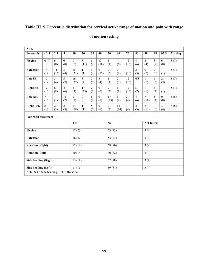
99
Table III. 5. Percentile distribution for cervical active range of motion and pain with range
of motion testing
N (%) Percentile <2.5 2.5 5 10 20 30 40 50 60 70 80 90 95 97.5 Missing
Flexion 0 (0) 6
(8) 0 (0)
0 (0)
8 (11)
6 (8)
13 (18)
1 (1)
4 (6)
12 (16)
4 (6)
3 (4)
5 (7)
6 (8)
5 (7)
Extension 14 (19)
11 (15)
3 (4)
15 (21)
1 (1)
3 (4)
9 (12)
2 (3)
0 (0)
7 (10)
2 (3)
0 (0)
0 (0)
1 (1)
5 (7)
Left SB 10 (14)
3 (4)
5 (7)
16 (22)
3 (4)
0 (0)
3 (4)
1 (1)
2 (3)
12 (16)
6(8) 1 (1)
4 (6)
2 (3)
5 (7)
Right SB 12 (16)
6 (8)
4 (6)
2 (3)
17 (23)
2 (3)
0 (0)
2 (3)
1 (1)
12 (16)
5 (7)
1 (1)
3 (4)
1 (1)
5 (7)
Left Rot. 7 (10)
1 (1)
15 (21)
1 (1)
0 (0)
6 (8)
0 (0)
17 (23)
3 (4)
3 (4)
4 (6)
7 (10)
3 (4)
0 (0)
6 (8)
Right Rot. 8 (11)
2 (3)
2 (3)
13 (18)
1 (1)
5 (7)
0 (0)
2 (3)
18 (24)
3 (4)
2 (3)
8 (11)
0 (0)
3 (4)
6 (8)
Pain with movement
Yes No Not tested
Flexion 17 (23) 53 (73) 3 (4)
Extension 16 (22) 54 (74) 3 (4)
Rotation (Right) 12 (16) 58 (80) 3 (4)
Rotation (Left) 10 (14) 60 (82) 3 (4)
Side bending (Right) 13 (18) 57 (78) 3 (4)
Side bending (Left) 11 (15) 59 (81) 3 (4) Note: SB = Side bending, Rot. = Rotation

100
Table III. 6. Segmental spine and rib mobility results
N (%)
Hypomobility Hypermobility Normal Not tested Mixed findings
All cervical segments 19 (26) 1 (1) 8 (11) 2 (3) 43 (59)
Upper cervical spine only (C0-C1 & C1-C2)
52 (71) 2 (3) 14 (19) 5 (7) NA
Lower cervical spine only (C3-C7)
3 (4) 3 (4) 34 (47) 4 (5) 29 (40)
More than two spinal segments
38 (52) 3 (4) NA 4 (5) 28 (39)
T1-T4 43 (60) 1 (1) 17 (23) 12 (16) NA
T5-T8 15 (21) 1 (1) 16 (22) 41 (56) NA
T9-T12 13 (18) 1 (1) 17 (23) 42 (58) NA
First rib 30 (41) 0 (0) 12 (17) 31 (42) NA
Second rib 10 (14) 0 (0) 16 (22) 47 (64) NA
Note: NA = Not applicable

101
Table III.7. Muscle strength and endurance results
N (%)
Strength
Muscle group Abnormal Normal Not tested Missing
Upper trapezius 2 (3) 24 (32) 47 (64) 1 (1)
Middle trapezius 22 (30) 7 (10) 44 (59) 1 (1)
Lower trapezius 23 (31) 5 (7) 45 (61) 1 (1)
Rhomboids 26 (35) 3 (4) 43 (58) 2 (3)
Longissimus colli 8 (11) 6 (8) 59 (80) 1 (1)
Sternocleidomastoid 9 (12) 5 (7) 29 (80) 1 (1)
Endurance
Neck flexor endurance test 29 (40) 12 (16) 32 (44) NA
Note: NA = Not applicable

102
CHAPTER IV
Measurement Properties of the Dizziness Handicap Inventory-Children and
Adolescent (DHI-CA) in children and adolescents post-concussion

103
Abstract
Background. Up to 80% of individuals report having dizziness post-concussion. Dizziness has
been identified as a predictor of prolonged recovery post-concussion and it impacts activities and
participation in post-concussed children and adolescents. Dizziness Handicap Inventory –
children and adolescents (DHI-CA) was recently developed to evaluate impact of dizziness on
activity and participation in children and adolescents with dizziness.
Purpose. The purpose of this study was to examine and report the psychometric properties of the
DHI-CA in post-concussion children and adolescents.
Study design. Retrospective study
Methods. A retrospective chart review was conducted for 132 consecutive children and
adolescents who received vestibular physical therapy post-concussion. Data was extracted on
various outcome measures including DHI-CA, sports concussion assessment tool -III (SCAT
III), vestibulo-ocular motor screen (VOMS) and patient-reported percent of recovery. The DHI-
CA was examined for validity, factor structure, responsiveness and internal consistency.
Results. The DHI-CA demonstrated convergent validity by demonstrating statistically
significant relationship with the SCAT-III symptoms related to dizziness (rs = 0.30-0.40). The
DHI-CA failed to demonstrate discriminant validity and showed limited diagnostic ability to
discriminate between individuals who did or did not show clinically meaningful improvements.
On factor analysis, structural inconsistencies were noted as the items demonstrated cross loading
and an overall poor model fit was indicated (RMSEA = 0.105).

104
Conclusion. Despite demonstrating convergent validity, DHI-CA demonstrated limited
discriminant validity and responsiveness along with significant structural limitations in the factor
structure. Hence, caution is recommended while making clinical decisions based on the DHI-CA
results.
Clinical relevance. This study indicates the need to revise the DHI-CA and reevaluate the
psychometric properties in post-concussion children and adolescents before it can be utilized in
clinical settings to evaluate the impact of dizziness following a concussion in this population.
Key words. Concussion, youth, dizziness adolescents

105
Introduction
Dizziness is a non-specific term that includes symptoms anywhere from disorientation
and lightheadedness to vertigo and imbalance1,2. Dizziness has been reported by up to 80% of the
individuals post-concussion.3 Both vestibular and cervical spine involvement contribute towards
lasting dizziness post-concussion.4 Dizziness has also been reported to be associated with
whiplash-associated disorders (WAD).5 This may be attributed to abnormal input from the
cervical spine secondary to damage in muscle and joint receptors.5 Post-concussion, the natural
course of recovery from dizziness is longer as compared to the non-dizziness oriented symptoms
and may last ranging from 6 months up to 5 years after the event.1,3
Dizziness at the time of injury has been identified as a predictor (Odd’s ratio = 6.34
(1.34-29.91)) for longer recovery times in post concussed athletes.6 Post-concussion dizziness
can negatively impact the life of an individual in regards to participation in sports, activities of
daily living, socializing and overall quality of life (QOL).7 There is emerging evidence that the
incidence of concussion among children and adolescents is rising. Despite this, currently
available self-reported assessment methods for dizziness are developed for adult population.
There is limited information on dizziness related outcome measures in children and adolescents.
Although several self-reported outcome measures to assess dizziness are currently available for
the adult population, information on assessment methods is still scarce in the children and
adolescents.8
Of the available outcome measures for dizziness, the Dizziness Handicap Inventory
(DHI) is the most widely used self-assessment inventory for evaluating precipitating physical,
emotional and functional factors that are associated with dizziness.9,10 The DHI was developed

106
from the hearing handicap inventory for the elderly.9 The DHI is an ordinal scale that consists of
25 items and a total score of 0-100 points with higher scores indicating more severe impairment.9
The DHI has been used by multiple rehabilitation professionals to evaluate activity limitations
and participation restriction, formulate therapeutic goals, plan of care and intervention strategies
in adults with dizziness.11 The DHI has been adapted in several languages and for different age
groups.11-14
Recently, DeSoussa and colleagues adapted the Brazilian Portuguese version of the adult
DHI to the children and adolescent population which was named DHI- children and adolescent
(DHI-CA).8 Internal consistency was reported for the overall DHI-CA (Cronbach’s α = 0.84) and
each subscale (Cronbach’s α = 0.70 for emotional, Cronbach’s α = 0.66 for functional and
Cronbach’s α = 0.62 for physical subscale) in 6-14 year old children with dizziness.8
Psychometric properties of the DHI-CA have not been studied in the subset of post-
concussion children and adolescents. Given the need for valid instruments to assess dizziness in
this population, the purpose of this study was to examine and report the psychometric properties
of the DHI-CA in post-concussion children and adolescents. The specific aims of this study were
1) to examine the construct validity (convergent and discriminant), 2) to examine the structure of
items within the DHI-CA to report the nature of their inter-relationships and 3) to examine
responsiveness of the DHI-CA and 4) to describe the internal consistency of the DHI-CA in post-
concussion children and adolescents.
107
Methods
Design, setting and participants
The data for this study was collected retrospectively from the electronic medical records
of a tertiary center specializing in comprehensive interdisciplinary management of patients with
concussion. The study was approved by the Institutional Review Board of the primary
investigator’s institution. A retrospective chart review was conducted for 132 children and
adolescents between the ages of 8 to 18 years who received vestibular physical therapy following
a concussion.
Data extraction
A data extraction sheet was developed by two investigators (DT and BA). The
investigators independently extracted data for 10 random patients and the extracted data was
compared to ensure consistency in data extraction. After ensuring quality of the extracted data,
the primary investigator (DT) completed the remaining data collection with random audits by the
secondary investigator (BA).
Data obtained from physician visits
Demographic, injury and care process data. Demographic and injury characteristics
were retrieved from electronic medical records. These characteristics included age, gender,
primary sport(s), prior history of migraine or prior learning disabilities, date of sustaining
concussion and mechanism of injury. In addition, the date of first medical visit, date of first
physical therapy visit, total number of physician visits and total number of vestibular physical
therapy visits were also collected to describe the care process.

108
The Sports Concussion Assessment Tool -3rd edition (SCAT-III). SCAT-III is a
concussion evaluation tool that was developed from the original SCAT to make decisions
regarding return to play.15 The data on symptom severity score was collected on 22 concussion
related symptoms including cognitive, physical, sleep and affect related symptoms using a Likert
scale (0 = none, 6 = severe), where higher scores indicated greater symptom severity (maximum
possible score = 132).16 For the purpose of this study, the SCAT-III data from physician initial
evaluation and discharge visit were utilized. Data on the individual symptoms such as balance
problems, dizziness, feeling like “in a fog”, headache, sensitivity to light and sensitivity to
sound, difficulty in concentrating, difficulty in remembering, nervousness or anxiety, irritability,
sadness and neck pain was extracted as well.
Data obtained during vestibular physical therapy visit
Assessment data on various outcome measures from the initial vestibular physical
therapy evaluation and discharge visit was extracted. In the event that a full assessment was not
completed due to an aggravation of patient’s symptoms, the subsequent two visits were screened
to extract additional assessment data.
Dizziness Handicap Inventory- Children & Adolescent (DHI-CA). The DHI-CA was
administered on the first and last vestibular physical therapy visits. The data on DHI-CA was
extracted from the first and final vestibular PT visits. The DHI-CA consists of 25 questions that
are organized into three subscales i.e. functional (9 items), emotional (9 items) and physical (7
items). Each item on the scale is scored on a 3-point Likert scale, where “no” is no dizziness, 2 is
“sometimes” and 4 is answered as “always” for dizziness (Appendix IV A).8 The DHI-CA was
administered on the first and second vestibular physical therapy visits and at discharge from

109
vestibular PT. Data on total score and individual items of DHI-CA was extracted from vestibular
physical therapy visit and the discharge visit.
Vestibulo-ocular motor screening (VOMS). The VOMS was designed to evaluate
vestibular and ocular motor impairments via patient-reported symptom provocation following
each assessment.17 The VOMS involves seven assessment categories which are smooth pursuit,
horizontal and vertical saccades, convergence, horizontal and vertical vestibular-ocular reflex
(VOR) and visual motion sensitivity (VMS).17 Following each assessment, the patient reports
changes in headache, dizziness, nausea and fogginess as compared to baseline values.17 In
addition to symptom provocation, the near point convergence (NPC) distance is measured (cm).
The VOMS has been reported to demonstrate high internal consistency (Cronbach’ α = 0.97)18
and acceptable construct validity (rs = 0.25-0.66, p ≤ 0.02)19 in children and adolescents. The
VOMS has been reported to be a predictor of recovery time in children and adolescents with
concussion.20 The change score of dizziness after performing maneuvers including smooth
pursuits, horizontal & vertical saccades, horizontal and vertical VOR, VMS and convergence
were used to examine convergent validity.
Percent of recovery. Patient-reported percentage of recovery values (out of 100) were
quantified at vestibular physical therapy visits. Change in percentage of recovery between first
and last vestibular PT visit was computed. An improvement by 15% was considered as clinically
meaningful. Global rating of change (GROC) has been utilized previously as an anchor-based
method for evaluating responsiveness. However, since GROC is scored using only 7 points, it is
challenging capture smaller but meaningful changes given the arbitrary choice of options such as
“little bit better or worse”.21 Since a point change in score in GROC is considered clinically
significant (considering a score of 7 representing 100% improvement), a 15% improvement was

110
used as the anchor as it may correspond to a one-point improvement in the global rating of
change scale assuming that the score of 7 represents 100% recovery.22,23

111
Statistical Analysis
The demographic, injury and process of care data was expressed using descriptive
statistics. The DHI-CA scores were evaluated for normality using Kolmogorov-Smirnov test.
Validity
Convergent and discriminant validity. Construct validity demonstrates the ability of an
instrument to measure an abstract concept.24 To examine the convergent validity of the DHI-CA,
it was validated against the total SCAT-III score and specific SCAT- III symptoms (balance
problems, dizziness, feeling like “in a fog”, headache, sensitivity to light and sensitivity to
sound), dizziness provocation in each of the seven VOMS items and the NPC distance was
collected at initial evaluation. For discriminant validity, associations between DHI-CA scores
and unrelated constructs identified from the SCAT- III (difficulty in concentrating, difficulty in
remembering, nervousness or anxiety, irritability, sadness and neck pain) were examined.
Spearman’s rho (rs) was used to examine convergent and discriminant validity.
Interpretation of correlation coefficients followed guidelines where coefficients exceeding 0.75
indicate good to excellent relationship, coefficients between 0.50-0.75 indicate moderate to good
relationship, coefficients between 0.25-0.50 indicate fair relationship and coefficients between
0.00-0.25 indicate little or no relationship.24
Factor analysis. An exploratory factor analysis (EFA) was performed to reveal the
underlying structures of the DHI-CA items. Also, a confirmatory factor analysis (CFA) was
performed to examine whether the factors of the DHI-CA that emerged fit with the previously

112
explained components (physical, emotional and functional). CFA is an accepted method to
evaluate and refine existing outcome measures.25
For this purpose, the DHI-CA data from initial vestibular physical therapy visit was
utilized. An EFA was performed on all 25 items of the DHI-CA using principal component
analysis (PCA) with oblique rotation to reveal patterns of relationship among the items. First, an
inter-item correlation matrix was created for all the DHI-CA items. The model was forced to
retain three factors. Factor loadings greater than 0.4 were considered indicative of some degree
of relationship and loadings ≥ 0.32 on two or more factors were considered as indicators of cross
loading.24,26 Items that highly loaded on one factor were grouped together. Factor loading values
were inspected to identify cross loading of items. Since, the factors were correlated, oblique
rotation was chosen as described by Portney & Watkins.24
A CFA using structural equation modeling was performed to examine whether the
relationships expected on theoretical constructs that were obtained from the EFA actually
appeared in the data.25 Based on the factors obtained from the EFA, all the items were entered in
the structural equation modeling. Model fit was evaluated using Chi-square test (non-significant
results i.e. p < 0.05 indicating a good model fit),27 and the root mean square error of
approximation (RMSEA). RMSEA is considered as the gold standard for testing goodness of fit
as unlike Chi-square test, RMSEA is not influenced by the sample size.28 The RMSEA values ≤
0.10 were considered acceptable with values between 0.0-0.05 indicating a close fit and values
between 0.05-0.08 indicating a reasonable fit.27
Responsiveness. Responsiveness is defined as the ability of an instrument to accurately
detect change when it has occurred.24 To determine responsiveness, the change in DHI-CA
scores from initial and final vestibular PT visits was measured against the difference in patient-

113
reported percent of recovery between initial and final vestibular PT visits. A clinically
meaningful change was operationally defined as a 15 % difference in the patient reported
recovery.
Responsiveness was determined by using both distribution and anchor-based methods.
Both effect size (ES) using Cohen’s D and standardized response mean (SRM) were used as
distribution-based methods. Cohen’s D was calculated using the formula ES = (M1-M2)/SD2 (M1
= Mean DHI- CA score post vestibular PT, M2 = Mean DHI- CA score pre vestibular PT, SD1 =
Standard deviation post vestibular PT, SD2 = Standard deviation pre vestibular PT) whereas
SRM was calculated using the formula SRM = (M1-M2)/SDchange (SDchange = Standard deviation of
change scores).24 Wilcoxon signed ranked test was used to determine pre to post physical therapy
change in the DHI-CA scores.24
Anchor-based method using patient reported percent of recovery values was used to
evaluate patient’s perception of their recovery status following physical therapy interventions.
Additionally, a receiver operating characteristics (ROC) curve was utilized to evaluate the ability
of the DHI-CA to identify patients who improved significantly based on the patient reported
percent of recovery values (improvement of 15% or more). The area under the curve (AUC) was
computed and the cutoff points for the minimally clinically important difference (MCID) were
determined from the point on the ROC curve that was closest to the top left corner. The MCID
was determined as the best cut-off point identified on the ROC curve to discriminate between the
participants who improved and those who did not improve. AUC values of > 0.70 were
considered as adequate for making this determination.29
Internal consistency. To determine reliability of the DHI-CA items, internal consistency
was examined. Internal consistency is a measure of the extent to which all the items in the

114
outcome measure address the same underlying concept.24 The DHI-CA data collected at the
initial vestibular physical therapy visit was utilized for assessment of internal consistency.
Cronbach’s alpha was used to examine internal consistency of the DHI-CA. Item to total
correlations were performed where each item was correlated with the total score, omitting that
individual item from the total score.24 Cronbach’s alpha values ranging from 0.70-0.90 were
considered indicative of strong internal consistency.24
Analyses were performed using Statistical Package for Social Sciences (SPSS) version
24.0 (SPSS Inc., Armonk, NY) and STATA version 15.0 (StataCorp LLC, College Station,
Texas). The level of significance was set at 0.05.

115
Results
Demographics, mechanism of injury and process of care
Data on 132 children and adolescents between the ages of 8 to 18 years received
vestibular physical therapy following a concussion between January 2017 and February 2018
was collected. Since, the missing data within the sample was less than 10% of the total data,
adjustments for missing data were not performed.30The average age of patients was 15.3±2.1
years (40.2% males). Nineteen percent of patients sustained concussion after contacting the
playing surface, 34% of the injuries were resulted from contact with another player whereas 22%
of patients sustained injury from coming into contact with sporting equipment. Mechanism of
injury was not sport-related for 23% of patients (Table IV.1).
Thirty-three percent of patients had a history of migraine, 9% had attention deficit, 13.6%
had learning disability whereas 9% of patients had attention deficit hyperactivity disorder. The
median time to first physician visit following injury was 16 days and the median time taken for
physical therapy evaluation following their first physician visit was 28 days (Table IV.1).
Validity
Convergent validity. Fair positive relationship was observed between the DHI-CA total
scores and SCAT-III total score (rs = 0.45, p < 0.001) and the SCAT-III items with the highest
value for dizziness (rs = 0.40, p < 0.001) and the lowest value for sensitivity to light (rs = 0.30, p
= 0.001) thereby demonstrating an increased perception of disability with an increase in
symptoms. Similarly, fair positive relationship was observed between the DHI-CA and dizziness
scores on all seven assessment categories of the VOMS (initial vestibular PT evaluation) with
highest value for NPC (rs = 0.48, p < 0.001) and the lowest value for visual motion sensitivity (rs

116
= 0.38, p < 0.001) indicating increased perceived disability with increased VOMS scores (Table
IV. 3). The DHI-CA did not demonstrate a statistically significant relationship with average NPC
distance scores (rs = 0.20, p = 0.134) (Table IV.2 & Table IV.3).
Discriminant validity. The DHI-CA lacked discriminant validity as it demonstrated
statistically significant positive relationship with all of the selected SCAT-III items that were
unrelated to the construct of dizziness (cognitive and affective symptoms). Difficulty in
remembering demonstrated the highest correlation value (rs = 0.33, p < 0.001) whereas
irritability demonstrated the lowest correlation values (rs = 0.21, p = 0.02) indicating possible
contribution of cognitive and affective symptoms towards perception of disability post-
concussion (Table IV.2).
Factor Analysis. The EFA revealed six factors with eigenvalues > 1 of which the top 3
factors explained 52% of the variance. The scree plot (Figure IV.1) also indicated three obvious
factor patterns to be retained for rotation. Based on this, a 3-factor solution was performed.
Bartlett’s test was significant (p< 0.001) and Kaiser Meyer Olkin measure of sampling adequacy
(KMO) value was 0.832 (values > 0.5 are considered acceptable) indicating that the items were
appropriate to be entered into factor analysis.31
Insert figure IV.1 here
Out of the three factors, factor 1 explained the most variance (32.3%). Factor 1
comprised of items that were originally included in the emotional subscale except item 18
(because of dizziness, do you have difficulty with concentrating on your school activities) which
demonstrated better loading on factor 2. Factor 2 consisted of items that were described in the
physical subscale except for item 17 (does walking on the sidewalk, passing or going over a
117
ground full of holes worsen the dizziness) which demonstrated better loading with factor 1.
Factor 3 consisted of only three items and explained the least variance in the model (8.26%).
Items that were included in this factor are described in the functional subscale of the DHI-CA
(item 3, item 6 and item 24) (Table IV.4).
Other items of the functional subscale demonstrated better loadings with either factor 1
(item 16: because of the dizziness it is too difficult for you to walk about alone and item 19:
because of the dizziness, are you unable to walk about in the dark) or factor 2 (item 5: because of
the dizziness, do you have difficulty with getting up from the bed, item 7: because of the
dizziness, do you have difficulty with reading, item12: because of the dizziness, do you stay
away from the high places and item 14: because of the dizziness, do you find it difficult to jump,
run, play ball games, ride a bicycle?). Cross loading was noted for four items (items 18,21,23 &
24). The EFA resulted in component matrices as shown in Table IV. 4.
In terms of the CFA, the goodness of fit statistics suggested an overall poor model fit.
The chi-square test was significant indicating a poor model fit.27 The RMSEA value of 0.105
was greater than the cutoff value for acceptable fit.27 Other indicators of model fit such as the
comparative fit index (CFI) = 0.733 (CFI > 0.95 indicate good model fit), Tucker Lewis index
(TLI) = 0.706 (TLI > 0.95 indicate good model fit) and size of residuals = 0.098, all suggested
that the model was a poor fit.27
Responsiveness. Significant decrease was observed in the DHI-CA scores following
physical therapy interventions (mean score pre = 26.6±17.7, mean score post = 10.7±15, Z = -
8.314, p < 0.001). The ES (Cohen’s D) and SRM for the DHI-CA were found to be 0.83 and 5.49
respectively indicating a large effect size. The MCID calculated from the ROC curve (AUC =
118
0.65, confidence interval = 0.53 – 0.76, p = 0.004) for the total DHI-CA score was 25 points
(sensitivity = 68%, specificity = 58%). (Figure IV.2)
Insert figure IV.2 here
Reliability analysis
Internal consistency. The overall internal consistency of the DHI-CA was found to be
strong (Cronbach’s alpha = 0.91). Strong internal consistency was also found across all the sub-
domains (Cronbach’s α = 0.82 (physical), 0.80 (emotional) and 0.81 (functional)). Corrected
item-total correlation ranged from 0.31 to 0.65 with item 3 demonstrating the lowest and item 10
demonstrating the highest correlation values. The internal consistency of individual subscales
and item to total correlations are reported in table IV.5.

119
Discussion
To the author’s knowledge, this is the first study to evaluate the validity and
responsiveness of the DHI-CA in this population subset of children and adolescent post-
concussion. The results from this study revealed that even though the DHI-CA demonstrated fair
convergent validity, it failed to demonstrate discriminant validity. Moreover, it was found that
the factor analysis revealed a different factor structure than originally described and a poor
model fit was observed. Additionally, the DHI-CA demonstrated inadequate AUC based on
previous recommendations29,32 indicating limited diagnostic abilities in identifying patients that
demonstrated clinically meaningful improvement. High Cronbach’s α values were observed for
all three subscales and overall scale.
Although the DHI-CA demonstrated fair relationship with dizziness items of the SCAT-
III (balance problems, dizziness, feeling like in a fog, sensitivity to light and sensitivity to sound)
and dizziness scores from all components of VOMS, it failed to demonstrate a significant
relationship with the average NPC distance. NPC distance has been identified as a measure of
vestibulo-ocular function and a diagnostic tool to discriminate between presence and absence of
concussion.17 An abnormality in the NPC distance has been reported to exist even in healthy
adolescent population and it is recommended that screening for NPC distance prior to injury
could provide more confidence in its diagnostic utility.19 Examining NPC does not involve head
movements whereas most of the items on the DHI-CA consist of activities that require some
degree of head movement. Additionally, the NPC utilizes double vision to evaluate convergence
insufficiency,33 and none of the DHI-CA items discuss double vision. These differences may
explain the lack of relationship between the NPC and the DHI-CA.

120
The DHI-CA demonstrated poor discriminant validity as significant relationship was
observed with cognitive and affective symptoms that are not theoretically related to the construct
of dizziness.34 On the other hand, it may be argued that the symptoms such as difficulty in
concentrating and remembering may be a sequalae of dizziness and since the DHI-CA items
have similar contents that include the ability to concentrate (item 7) and read (item 18), an
association was observed.
Since, the original version of the DHI-CA was adapted from the adult version retaining
all the original items within the pre-specified sub-scales i.e. physical, emotional and functional,
an EFA was first performed in this study to explore how well the items fit in each subscale
before examining its validity further. When the factor structure of a scale has not been
determined, performing an EFA is recommended.26 The results of the EFA indicated a different
factor structure as compared to the original version of the DHI-CA as well as to adult DHI
versions. Previous studies have reported of inconsistencies in terms of item loadings on to the
factors as originally described in the DHI for adults who experienced dizziness secondary either
to vestibular or non-vestibular causes.26,35 The findings from this study concur with the previous
literature. Considering the possible limitations in item construction, and/or inaccuracies in the
initial factor structure, restructuring of the DHI-CA for post-concussion population subset may
be considered.
The loading pattern of the items in this study was similar to a previous study by Perez
and colleagues on the original DHI where items under the emotional sub-scale demonstrated
best loadings together and items in functional subscale demonstrated a scattered loading
pattern.35 Related activities (item 5:getting up from bed and item 13: turning in bed; item 8:
jumping, running, riding a bicycle and item 14: riding a bicycle, games, sports) were placed in

121
different sub-scales in the DHI-CA. The results from the EFA in this study suggested grouping
these items together. All of these items measure functional activities and it is postulated that a
better structure could be obtained if these items were grouped under the same factor.
In addition, to examine if the newly identified item distribution actually conformed well to
the three factors, a CFA was performed. The results from the CFA demonstrated a poor model fit
warranting further exploration and revision of the item distribution in the scale. Several
explanations for this finding are possible. Multiple items demonstrated cross loadings across the
factors indicating item redundancy which may have resulted in a poor overall model fit.
The other possible explanation could be that the study was underpowered for the
assessment of CFA. A sample size of > 200 has been recommended to obtain adequate power for
the CFA.36 Although, a priori sample size calculation indicated the requirement of 250 participants
based on a participant to item ratio of 10:1 for the purpose of CFA,36,37 the retrospective chart
review revealed only 132 participants that met the inclusion criteria within the specified timeline
approved to review for the purpose of the study.
Additionally, the assumption of local independence for the CFA was not met in this study
as the items in different factors demonstrated statistically significant correlation with each
other.38Also, since the DHI-CA is scored on a Likert scale, transformation could not be performed
to obtain normal distribution of the data. Hence, the utility of CFA was limited in identifying an
appropriate model fit in this study. Previous studies that were done on original DHI and its adapted
versions also performed factor analysis and failed to completely support the original
structure.12,26,35 This finding is consistent with our study as the distribution of the items do not
completely fit in the originally described structure.

122
The DHI-CA demonstrated poor ability to accurately identify the individuals who
showed clinically meaningful improvement. The best cut-off point was found to be a score of 25
(sensitivity = 68%, specificity = 58%). The subjective nature of the DHI-CA may explain its
limited ability in accurately predicting those who would improve. Percentage of recovery is not a
specific question and is open to various interpretations. This is particularly true given the
multifaceted effects of concussion. Even though, the patient may get better with dizziness, their
perception of recovery can be hampered by other factors such as cognitive deficits, cervical spine
deficits, or restricted participation in sport/school.
Another explanation could be that although the scale is scored on a total of 100 points,
each item is scored on a Likert scale of 0,2 and 4. This scoring system may not be adequate to
capture small but meaningful changes in patient condition as the options the patients can chose
from are very limited. For e.g. in a 0-10 scale, a continuum of improvement/decline can be
captured. However, since the interval from 0 (no) to 2 (sometimes) to 4 (yes) is quite large, an
individual who started out at 4 and may have shown a small improvement, still might not
consider it an improvement big enough to be classified under 2. This inability of DHI-CA to
capture the continuum of improvement may have potentially created an ambiguity and affected
the responses.
The results of this study revealed a large effect size indicating a significant decline in
DHI-CA scores following physical therapy intervention. This was in contrast to the findings
noted by the anchor-based methods. Although distribution-based methods such as effect size
provide a statistical measure to identify change irrespective of the variations in sample size, it is
strongly influenced by the characteristics such as normal distribution and homogeneity at
baseline.39,40 The non-normal distribution of the sample in this study could have affected the
123
findings of the test. Another limitation of effect size lies in the lack of its ability to provide a
good sense of clinical relevance of change. The use of the cut points provided by effect size in
establishing clinically significant improvement are debatable in literature,39,41 further suggesting
that the effect size is questionable in determining clinically meaningful change.
The findings on internal consistency were similar to the results reported by DeSousa et
al.8 Previous literature recommends that a maximum value of 0.9 for Cronbach’s α is indicative
of strong internal consistency.24,42 Values greater than 0.9 have been reported to indicate that the
relationship among the items may be too high. This could suggest that the items may be
redundant and testing the same question in a different way.24,42 The high overall Cronbach’s α
(0.91) in this study may suggest reviewing the DHI-CA for possible item reduction.
Limitations. The authors recognize the limitations of this study. Although the DHI-CA
was assessed for validity using other subjective and clinical measures, objective measures
including instrumented measures of vestibular dysfunction was not utilized. Future studies may
consider validating this tool using instrumented measures such as video head impulse test,
vestibular-evoked myogenic potentials and caloric testing.
The retrospective design of the study limited the ability to ascertain if the patients
received assistance from the parents or therapists in completing the DHI-CA. Differences in the
level of assistance received in filling out the questionnaire may have influenced the responses.
Although the study was adequately powered for internal consistency and validity
analysis,29 it was underpowered for the CFA and future studies with larger sample sizes are
recommended.
Cross loading of items evidenced on EFA along with high overall Cronbach’s α indicate
a need for revision of the DHI-CA and further validation. Given that the ordinal nature of DHI-

124
CA and correlation between the items belonging to different subscales violate the assumptions of
local independence for CFA, an item response theory (IRT) using Rasch analysis is
recommended to conform and revise the scale.
125
Conclusion
The DHI-CA did demonstrate convergent validity but failed to demonstrate discriminant
validity. Structural inconsistencies on factor analysis and possible item redundancy as indicated
by the overall Cronbach’s α value warrant further exploration and possible re-structuring of the
DHI-CA using item response theory/Rasch analysis. Additionally, considering inadequate
diagnostic ability of the DHI-CA along with the aforementioned limitations, caution is
recommended when considering making clinical decisions based on DHI-CA in children and
adolescents post-concussion. Additionally, limitations of study design and sample limit the
generalizability of findings. Hence, further research to revise the scale and further explore its
psychometric properties is recommended.
126
References
1. Maskell F, Chiarelli P, Isles R. Dizziness after traumatic brain injury: overview and measurement in the clinical setting. Brain Inj 2006;20(3):293.
2. Heyer GL, Young JA, Fischer AN. Lightheadedness After Concussion: Not All Dizziness is Vertigo. Clin J Sport Med 2018;28(3):272-277.
3. Alsalaheen BA, Mucha A, Morris LO, et al. Vestibular Rehabilitation for Dizziness and Balance Disorders After Concussion. J Neurol Phys Ther 2010;34(2):87-93.
4. Schneider KJ, Meeuwisse WH, Nettel-Aguirre A, et al. Cervicovestibular rehabilitation in sport-related concussion: a randomised controlled trial. Br J Sports Med 2014;48(17):1294-1298.
5. Treleaven J, Gwndolen J, Sterling M. Dizziness and unsteadiness following whiplash injury: Characteristic features and relationship with cervical joint position error. J Rehabil Med 2003;35(1):36-43.
6. Lau BC, Kontos AP, Collins MW, Mucha A, Lovell MR. Which On-field Signs/Symptoms Predict Protracted Recovery From Sport-Related Concussion Among High School Football Players? Am J Sports Med 2011;39(11):2311-2318.
7. Yang CC, Tu, Y.K., Hua, M.S., Huang, S.J. The Association Between the Postconcussion Symptoms and Clinical Outcomes for Patients With Mild Traumatic Brain Injury. J Trauma 2007;62:657-663.
8. Maria da Gloria Canto de Sousa OC, Amanda Nery Santos, Cristina Gananca, Leandro Almeida, Eduardo Pondé de Sena. Brazilian adaptation of the dizziness handicap inventory for the pediatric population: reliability of the results. Audiol Commun Res 2015;20:327-335.
9. Jacobson GP, Newman CW. The Development of the Dizziness Handicap Inventory. Arch Otolaryngol Head Neck Surg 1990;116(4):424-427.
10. Mutlu B, Serbetcioglu B. Discussion of the dizziness handicap inventory. J Vestib Res 2013;23(6):271.
11. Kurre A, van Gool CJAW, Bastiaenen CHG, Gloor-Juzi T, Straumann D, de Bruin ED. Translation, cross-cultural adaptation and reliability of the german version of the dizziness handicap inventory. Otol Neurotol 2009;30(3):359-367.
12. Jafarzadeh S, Bahrami E, Pourbakht A, Jalaie S, Daneshi A. Validity and reliability of the Persian version of the dizziness handicap inventory. J Res Med Sci 2014;19(8):769-775.
13. Jarlsater S, Mattsson E. Test of reliability of the Dizziness Handicap Inventory and The Activities-specific Balance Confidence Scale for Use in Sweden. Adv Physiother 2003;5(3):137-144.
14. Nola G, Mostardini C, Salvi C, Ercolani AP, Ralli G. Validity of Italian adaptation of the Dizziness Handicap Inventory (DHI) and evaluation of the quality of life in patients with acute dizziness. Acta Otorhinolaryngol Ital 2010;30(4):190.
15. Graham R, Institute of M, National Research C, ProQuest. Sports-related concussions in youth: improving the science, changing the culture. Washington, District of Columbia: National Academies Press; 2014.
16. Herring S, Broglio S, J LJ, et al. Consensus statement on concussion in sport-the 5th international conference on concussion in sport held in Berlin, October 2016. Br J Sports Med 2017;51(11):838.
17. Mucha A, Collins MW, Elbin RJ, et al. A Brief Vestibular/Ocular Motor Screening (VOMS) Assessment to Evaluate Concussions: Preliminary Findings. Am J Sports Med 2014;42(10):2479-2486.
18. Moran RN, Covassin T, Elbin RJ, Gould D, Nogle S. Reliability and Normative Reference Values for the Vestibular/Ocular Motor Screening (VOMS) Tool in Youth Athletes. Am J Sports Med 2018;46(6):1475-1480.

127
19. Yorke AM, Smith L, Babcock M, Alsalaheen B. Validity and Reliability of the Vestibular/Ocular Motor Screening and Associations With Common Concussion Screening Tools. Sports Health 2017;9(2):174-180.
20. Anzalone AJ, Blueitt D, Case T, et al. A Positive Vestibular/Ocular Motor Screening (VOMS) Is Associated With Increased Recovery Time After Sports-Related Concussion in Youth and Adolescent Athletes. Am J Sports Med 2017;45(2):474-479.
21. Hajiro T, Nishimura K. Minimal clinically significant difference in health status: the thorny path of health status measures? Eur Respir J 2002;19(3):390-391.
22. Jaeschke R, Singer J, Guyatt GH. Measurement of health status: Ascertaining the minimal clinically important difference. Control Clin Trials 1989;10(4):407-415.
23. Kamper SJ, Ostelo RWJG, Knol DL, Maher CG, de Vet HCW, Hancock MJ. Global Perceived Effect scales provided reliable assessments of health transition in people with musculoskeletal disorders, but ratings are strongly influenced by current status. J Clin Epidemiol 2010;63(7):760-766.e761.
24. Portney L, Watkins M. Foundations of clinical research: applications to practice. 3rd Edition ed. . Vol 3rd. Upper Saddle River, NJ: Prentice-Hall,Inc; 2009.
25. Floyd FJ, Widaman KF. Factor Analysis in the Development and Refinement of Clinical Assessment Instruments. Psychol Assess 1995;7(3):286-299.
26. Tamber A-L, Wilhelmsen KT, Strand LI. Measurement properties of the Dizziness Handicap Inventory by cross-sectional and longitudinal designs. Health Qual Life Outcomes 2009;7(1):101-101.
27. Harrington D, ProQuest. Confirmatory factor analysis. New York;Oxford;: Oxford University Press; 2009.
28. Kelley K, Lai K. Accuracy in Parameter Estimation for the Root Mean Square Error of Approximation: Sample Size Planning for Narrow Confidence Intervals. Multivariate Behav Res 2011;46(1):1-32.
29. Terwee CB, Bot SDM, de Boer MR, et al. Quality criteria were proposed for measurement properties of health status questionnaires. J Clin Epidemiol 2006;60(1):34-42.
30. Newman DA. Missing Data: Five Practical Guidelines. Organ Res Methods 2014;17(4):372-411. 31. Kaiser-Meyer-Olkin value (KMO value). Dictionary of Psychological Testing, Assessment and
Treatment2007. 32. Šimundić A-M. Measures of Diagnostic Accuracy: Basic Definitions. EJIFCC 2009;19(4):203. 33. Pearce KL, Sufrinko A, Lau BC, Henry L, Collins MW, Kontos AP. Near Point of Convergence After
a Sport-Related Concussion: Measurement Reliability and Relationship to Neurocognitive Impairment and Symptoms. Am J Sports Med 2015;43(12):3055-3061.
34. McInnes K, Friesen C, MacKenzie D, Westwood D, Boe S. Mild Traumatic Brain Injury (mTBI) and chronic cognitive impairment: A scoping review. PLoS One 2017;12(4).
35. Nicolas P, Garmendia I, García-Granero M, Martin E, García-Tapia R. Factor Analysis and Correlation Between Dizziness Handicap Inventory and Dizziness Characteristics and Impact on Quality of Life Scales. Acta Otolaryngol 2001;121(545):145-154.
36. Kline RB, ProQuest. Principles and practice of structural equation modeling. Fourth ed. New York: The Guilford Press; 2016.
37. Osborne JW, Costello AB. Sample size and subject to item ratio in principal components analysis. Practical Assessment 2004;9(11):1-9.
38. Finbråten HS, Pettersen KS, Wilde-Larsson B, et al. Validating the European Health Literacy Survey Questionnaire in people with type 2 diabetes: Latent trait analyses applying multidimensional Rasch modelling and confirmatory factor analysis. J Adv Nurs 2017;73(11):2730-2744.

128
39. Crosby RD, Kolotkin RL, Williams GR. Defining clinically meaningful change in health-related quality of life. J Clin Epidemiol 2003;56(5):395-407.
40. Algina J, Keselman HJ, Penfield RD. An Alternative to Cohen's Standardized Mean Difference Effect Size: A Robust Parameter and Confidence Interval in the Two Independent Groups Case. Psychol Methods 2005;10(3):317-328.
41. Deyo RA, Patrick DL. The Significance of Treatment Effects: The Clinical Perspective. Med Care 1995;33(4):AS286-AS291.
42. Tavakol M, Dennick R. Making sense of Cronbach's alpha. Int J Med Educ 2011;2:53-55.
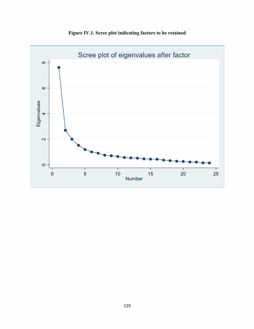
129
Figure IV.1. Scree plot indicating factors to be retained

130
Figure IV.2. Receiver operating characteristic (ROC) curve

131
Table IV.1 Demographic, injury and care characteristics of participants
N (% males) 53 (40.2) Age in years, mean (SD) 15.3 (2.1) Attention deficit, n (%) 12 (9.1) Attention deficit hyperactivity disorder, n (%) 12 (9.1) Learning disability, n (%) 18(13.6) History of migraine, n (%) 33 (25) Mechanism of injury N (%) Contact with another player 45 (34) Contact with playing surface 25 (19) Contact with sporting equipment 29 (22) Others (Non-sport related) 30 (23) Not specified 3 (2) Process of care, Median (Min – Max) Days to first physician visit following concussion 16 (1-479) Days to first PT visit following physician visit 28 (5-550) Number of physician visits 4 (1-12) Number of physical therapy visits 3 (1-11)

132
Table IV.2. Relationship between Dizziness Handicap Inventory-Children and Adolescents (DHI-CA) & SCAT III measures and Near point convergence (NPC)
DHI-CA symptoms (N) Spearman’s rho (p-value)
Balance problems (124) 0.37 (<0.001) Dizziness (124) 0.40 (<0.001) Feeling like in a fog (124) 0.39 (<0.001) Headache (124) 0.17 (0.056) Sensitivity to light (124) 0.30 (0.001) Sensitivity to sound (124) 0.38 (<0.001) SCAT-III total score 0.45 (<0.001) Difficulty in concentrating (124) 0.31 (<0.001) Difficulty in remembering (124) 0.33 (<0.001) Nervousness or anxiety (124) 0.25 (0.006) Neck pain (124) 0.26 (0.004) Irritability (124) 0.21 (0.020) Sadness (124) 0.28 (0.001) Average NPC scores (56) 0.20 (0.134)
SCAT III = sports concussion assessment tool III 3rd edition, NPC = Near point convergence, VOR = Vestibulo-ocular reflex

133
Table IV.3. Relationship between Dizziness Handicap Inventory-Children and Adolescents (DHI-CA) and dizziness scores VOMS at initial vestibular physical therapy evaluation
VOMS symptoms
Spearman’s rho (p-value); N
Smooth pursuit
Horizontal Saccads
Vertical Saccads
Convergence VOR horizontal
VOR vertical
Visual motion sensitivity
Dizziness 0.41 (p < 0.001); 125
0.39 (p < 0.001); 126
0.42 (p < 0.001); 126
0.48 (p < 0.001); 122
0.43 (p < 0.001); 125
0.39 (p < 0.001); 121
0.38 (p < 0.001); 122
VOMS = Vestibular-Ocular Motor screening, VOR = Vestibulo-ocular reflex

134
Table IV.4. Exploratory factor analysis of the DHI-CA
Factor 1 Factor 2 Factor 3 Item Loading value Item Loading value Item Loading value Item 9 0.82 Item 8 0.78 Item 3 0.71 Item 10 0.81 Item 11 0.78 Item 6 0.62 Item 15 0.80 Item 14 0.71 Item 24 0.47 Item 16 0.68 Item 7 0.70 Item 17 0.65 Item 1 0.64 Item19 0.64 Item 13 0.61 Item 20 0.62 Item 5 0.61 Item 22 0.61 Item 18 0.60 Item 23 0.54 Item 25 0.57 Item 2 0.43 Item 4 0.54 Item 21 0.40

135
Table IV.5. Internal consistency of the DHI-CA
Item number
Item description Median (Range)
Corrected item-total correlation
Cronbach’s α if item is deleted
Physical (Cronbach’s α = 0.82) 1 Does lifting your head up worsen dizziness?
2 (0-4) 0.64 0.78 4 Does walking around the supermarket looking at the shelves
worsen the dizziness?
0 (0-4) 0.54 0.79
8 Do games, sports, riding a bicycle, riding on roundabouts/merry-go-rounds worsen the dizziness?
2 (0-4) 0.57 0.79
11 Do fast movements of the head worsen your dizziness?
2 (0-4) 0.64 0.77 13 If you turn in bed while you are lying down (a) does it worsen
your dizziness?
0 (0-4) 0.58 0.79
17 Does walking on the sidewalk, passing or going over a ground full of holes worsen the dizziness?
0 (0-4) 0.41 0.81
25 If you lower your head or body, does the dizziness worsen?
2 (0-4) 0.54 0.79 Emotional (Cronbach’s α = 0.80) 2 Because of the dizziness, do you feel frustrated (a)? 2 (0-4) 0.51 0.78 9 Because of the dizziness, are you afraid to leave the house? 0 (0-4) 0.59 0.78 10 Because of the dizziness, do you feel ashamed (a) in front of
others?
0 (0-4) 0.65 0.76
15 Because of the dizziness, are you afraid that people will think you are not well?
2 (0-4) 0.60 0.77
18 Because of the dizziness, do you have difficulty with concentrating on your school activities?
0 (0-4) 0.39 0.81
20 Because of the dizziness, are you afraid to stay at home alone?
0 (0-4) 0.40 0.80 21 Because of the dizziness, do you feel harmed (a) in comparison
with your colleagues? / companions?
0 (0-4) 0.42 0.79
22 Because of the dizziness, do you quarrel with your friends, companions or persons in your family?
0 (0-4) 0.56 0.77
23 Because of the dizziness, do you feel sad, without wanting to do anything?
0 (0-4) 0.57 0.77
Functional (Cronbach’s α = 0.81) 3 Because of the dizziness, do you stay away from school?
0 (0-4) 0.31 0.81 5 Because of the dizziness, do you have difficulty with getting up
from the bed?
2 (0-4) 0.42 0.80
6 Do you stay away from birthdays, parties, movies, video game arcades because of the dizziness?
0 (0-4) 0.54 0.78
7 Because of the dizziness, do you have difficulty with reading?
2 (0-4) 0.59 0.77 12 Because of the dizziness, do you stay away high places?
0 (0-4) 0.47 0.79 14 Because of the dizziness, do you find it difficult to jump, run,
play ball games, ride a bicycle?
2 (0-4) 0.54 0.78
16 Because of the dizziness, it too difficult for you to walk about alone (a)?
0 (0-4) 0.52 0.79
19 Because of the dizziness, are you unable to walk about in the dark?
0 (0-4) 0.53 0.78
24 Does your dizziness hamper, interfere in your studies?
2 (0-4) 0.63 0.77

136
Appendix IVA. Dizziness Handicap Inventory – Children and adolescents (DHI-CA)
Subscales Questions Responses Yes Sometimes No
1- Physical Does lifting your head up worsen dizziness? 2- Emotional Because of the dizziness, do you feel frustrated (a)? 3- Functional Because of the dizziness do you stay away from school? 4- Physical Does walking around the supermarket looking at the shelves worsen
the dizziness?
5- Functional Because of the dizziness, do you have difficulty with getting up from the bed?
6- Functional Do you stay away from birthdays, parties, movies, video game arcades because of the dizziness.
7- Functional Because of the dizziness, do you have difficulty with reading?
8- Physical Do games, sports, riding a bicycle, riding on
roundabouts/merrygorounds worsen the dizziness?
9 – Emotional Because of the dizziness, are you afraid to leave the house?
10 – Emotional Because of the dizziness, do you feel ashamed (a) in front of others?
11- Physical Do fast movements of the head worsen your dizziness?
12- Functional Because of the dizziness, do you stay away high places?
13 – Physical If you turn in bed while you are lying down (a) does it worsen your
dizziness?
14 – Functional Because of the dizziness, do you find it difficult to jump, run, play ball games, ride a bicycle?
15 – Emotional Because of the dizziness, are you afraid that people will think you are not well?
16 – Functional Because of the dizziness, it to difficult for you to walk about alone (a)?
17 – Physical Does walking on the sidewalk, passing or going over a ground full of holes worsen the dizziness?
18 – Emotional Because of the dizziness, do you have difficulty with concentrating on your school activities?
19 – Functional Because of the dizziness, are you unable to walk about in the dark?
20 – Emotional Because of the dizziness, are you afraid to stay at home alone?
21 - Emotional Because of the dizziness, do you feel harmed (a) in comparison with
your colleagues?/ companions?
22 – Emotional Because of the dizziness, do you quarrel with your friends, companions or persons in your family?
23 – Emotional Because of the dizziness, do you feel sad, without wanting to do anything?
24 – Functional Does your dizziness hamper, interfere in your studies?
25 – Physical If you lower your head or body, does the dizziness worsen?

137
Chapter V
Overview
The focus of this dissertation was to explore cervical spine impairments and evaluate
measurement error of different assessments of postural control and to examine psychometric
properties of the Dizziness Handicap Inventory-Children and Adolescents (DHI-CA) in children
and adolescents post-concussion.
This dissertation utilized the three-paper method with three individual studies. Chapter I
provides a general background on the focus of this dissertation. Chapter II discusses the
measurement properties of postural control measures in typically developing children and
adolescents. This review will contribute towards directing future studies towards validating these
measures in children and adolescents post-concussion since typically developing population
bears close resemblance to post-concussion population. Chapter III provides a detailed
description on cervical spine impairments in children and adolescents post-concussion. Since
majority of the research on concussion has been focused on professional and college athletes, the
findings of this research will help to provide preliminary data to conduct further research for
development of structured assessment for this subset of population post-concussion.
Chapter IV describes the final study of this dissertation that includes an assessment of the
psychometric properties of the DHI-CA in children and adolescents post-concussion. This study
will help to further the development of valid and reliable assessment of dizziness in this
population subset. Since, dizziness has been reported to be negatively impacting the recovery
time and quality of life post-concussion, a valid and responsive tool is required to evaluate
dizziness post-concussion in this age range. DHI for adults has been validated in various
populations and languages. Recently, a version of DHI was developed for children and

138
adolescents from the Brazilian-Portuguese version and was translated to English. However, this
version has not yet been validated in post-concussion population.
This final chapter (Chapter V) summarizes the major findings of the three studies in this
dissertation and their implications for future research. It also discusses the limitations of the
research and how those limitations may have affected the results.
Summary of Research Design and Results
The following section describes a summary of the methods and results of the three
individual studies in this dissertation.
Overview
Study 1
The primary purposes of this study were to conduct a systematic review to 1) report the
test-retest, intra-rater and inter-rater reliability of postural control outcome measures, to 2) report
the minimal detectable change and standard error of measurement (SEM) of these outcome
measures and to 3) describe methodological and reporting qualities of the studies that examined
the reliability of postural control outcome measures in typically developing children with a mean
age of 8-18 years. The results of the electronic search identified 25 studies examining 22 (8
static, 14 dynamic) different postural control measures.
The findings of this review suggest that out of the the different static postural control
measures identified, the BESS was widely studied and demonstrated acceptable reliability across
all studies, indicating that this measure might be used for evaluation of static postural control in
typically developing children. Among the instrumented static postural control measures, the
CTSIB was found to demonstrate the highest reliability when used with the AccuGait force
platform. Among the dynamic postural control measures, TUG demonstrated good reliability and
can be used cautiously to evaluate dynamic postural control. Other measures including the

139
mFRT, one leg hop, BEST and mini BEST provide promising findings. However, observations
from single reliability studies and methodological inconsistencies limited the ability to draw
valid conclusions and make specific recommendations. Studies with stronger methodological
design in future are needed to draw meaningful conclusions.

140
Study 2
This study was conducted to characterize the type, frequency and severity of cervical
impairments in children and adolescents post-concussion. This study aimed at providing insights
into the extent and nature of cervical spine impairments post-concussion that may provide a
foundation to develop targeted physical therapy interventions. For this purpose, a retrospective
chart review was performed on 73 patient charts to extract the assessment data from the first
three physical therapy visits. Data was divided into five broad categories including posture,
movement quality and generalized joint hypermobility; joint mobility (ROM, segmental mobility
and pain with movement); myofascial tension to palpation; muscle strength & endurance and
proprioceptive testing. The results of this study revealed high prevalence of cervical spine
impairments with posture and myofascial impairments demonstrating highest impairment
frequency.
Data on joint mobility revealed that 71% of the patients demonstrated impairments in
upper cervical spine mobility. Additionally, out of all the muscles tested for strength, it was
found that the rhomboids demonstrated highest frequency in terms of muscle weakness (35%).
Poor neck flexor endurance was observed in 40% of the patients. It was also found that the joint
position error testing, neck flexor endurance test and cranial cervical flexion test were not
completed in majority of the patients. The results from this study contribute towards providing
preliminary data to support the framework of a cervical spine evaluation tool in children and
adolescents post-concussion. Further research is warranted in terms of developing population
specific screening tools for cervical spine impairments post-concussion.

141
Study 3
The purpose of this study was to examine validity, factor structure, responsiveness and
internal consistency of the DHI-CA in children and adolescents post-concussion. For this study,
a retrospective chart review of 132 patients (8-18 years) who received vestibular physical
therapy post-concussion was conducted and data was extracted. The results of this study
indicated that the DHI-CA demonstrated fair convergent validity as significant correlations were
observed with SCAT-III total scores (rs = 0.45, p < 0.001), with SCAT-III items related to the
construct of dizziness (rs = 0.30-0.40, p < 0.001) and with components of VOMS (rs = 0.38-0.48,
p < 0.001). Poor discriminant validity was observed as the DHI-CA demonstrated significant
relationship with the cognitive and affective symptoms of the SCAT-III. The DHI-CA
demonstrated 68% sensitivity and 58% specificity and a score of 25 was established as the cut
point. The DHI-CA demonstrated strong internal consistency for the subscales (Cronbach’s α ≥
0.80). However, the overall Cronbach’s α was > 0.90 indicating possible item redundancy.
The results from the factor analysis revealed a different factor structure from what was
described in the original scale and cross loading was observed in 4 items. Additionally, the
confirmatory factor analysis CFA indicated an overall poor model fit. Findings of this study
warrant further research in terms of subjecting the DHI-CA to item response theory and
conducting a Rasch analysis to improve the scale structure.

142
Discussion of results
This dissertation reviewed measurement errors of postural control measures in typically
developing children, cervical spine impairments post-concussion and examined psychometric
properties of the DHI-CA in children and adolescents post-concussion.
Study one was conducted to provide a comprehensive overview of measurement errors of
static and dynamic postural control measures in typically developing children. Since, typically
developing children and adolescents demonstrate close resemblance to post-concussion
population, it was important to evaluate measurement errors of postural control measures in
typically developing population before they could be tested on post-concussion population which
demonstrates higher variability in neuromotor function. Findings of this review will aid a
clinician in becoming a well-informed consumer in terms of selecting a postural control measure
to make informed clinical decision.
Findings from this review indicated that both instrumented and non-instrumented
versions of the BESS demonstrated comparable reliability and could be used with caution in
clinics.1 In terms of instrumented measures, high variability was observed across force platforms
in terms of reliability, which could be explained by the differences in foot placement and number
of trials used. Other factors including foot placement, number of trials, fatigue, motivation of the
participant and concentration may affect reliability.
Among the dynamic balance measures, TUG was the most studied measure and
demonstrated acceptable reliability across studies. The ease of administration, simplicity of
instructions and brevity of TUG could explain its higher reliability. Other measures such as
mFRT, TUDS, BEST, miniBEST although demonstrated comparable findings, were reported

143
only in single studies and demonstrated several methodological inconsistencies limiting the
ability to draw valid conclusions.
Overall, several inconsistencies in the quality of studies were noted across the studies that
were included in this review. Inconsistencies in sample size and power estimation, lack of
reporting of confidence intervals and inaccuracies in the type of the ICC model and form used
limit the generalizability of the findings of this review to a broader range of population.
Additionally, MDC was not calculated in 80% of the studies which further contribute towards
limiting the clinical applicability of the included studies.
Study 2 discussed the impairments of the cervical spine in children and adolescents post-
concussion. High prevalence of cervical spine impairments was observed in this study with over
90% of patients demonstrating impairments in 3 or more categories. Muscle tension, joint
mobility and muscle strength were the categories demonstrating highest frequency of
impairment. The categories of impairments examined in this cohort are consistent with the
impairments reported in the most recent clinical practice guidelines for neck pain. Previous
literature has affirmed that contribution of the cervical spine to various concussion symptoms
such as headaches and dizziness.2-4
Over 85% of the patients demonstrated impairments joint mobility with upper cervical
spine showing most restriction in mobility. Mobility impairments in the cervical spine and
increased muscle tension and impaired joint proprioception in the cervical spine have been
reported to contribute towards symptoms such as headache and dizziness post-concussion.2,3,5,6 It
is noteworthy that muscle strength and joint proprioception were not tested in more than 50% of
the patients thereby leading to potential under-representation of these impairments. Acute nature
of symptoms may explain under-utilization of these tests in this sample. Additionally, 40% of the
144
patients reported moderate to severe disability on the NDI. It is noteworthy that the NDI has not
been validated for population below 18 years of age thereby warranting further research. Despite
of the potential sample bias, this study provides a foundation on which further research can be
built upon to develop targeted assessment tools and intervention strategies.
The third study aimed at examining, validity, factor structure, responsiveness and internal
consistency of the DHI-CA in post-concussion population. Limited diagnostic abilities of this
scale were observed in this study. The DHI-CA demonstrated fair convergent validity but failed
to demonstrate discriminant validity. Very high values of the Cronbach’s α indicate a potential
for item redundancy. This could be explained by similarity in the item descriptions and activities
included in the individual items. Also, it is difficult to distinguish if the difficulty experienced by
the patients in activities of daily living are due to dizziness or from other symptoms related to
concussion. The factor structure and item distribution in the DHI-CA was kept same as that of
the adult version. Results from the EFA indicate that there is a possibility that the items should
be loaded under different factors as compared to the original version since the same factor
distribution may not be appropriate for the population that is considered in this study.

145
Limitations
Several limitations were associated with this dissertation. The systematic review revealed
a variety of outcome measures to examine postural control in typically developing children.
Although an extensive electronic and manual search was conducted, the search was limited to
studies that were published in the English language, and those that were published in peer
reviewed journals. The possibility of having missed good quality studies for these resources
cannot be ruled out. Additionally, heterogeneity in the sample size, limited number of studies
and methodological inconsistencies across the studies limited the generalizability of the results
from this review.
The sample for studies 2 and 3 were drawn from a single tertiary clinic, thereby creating
a potential for sample bias as this sample might not be an adequate representation of the entire
spectrum of children and adolescents post-concussion. Additionally, the retrospective study
design used in this dissertation limited the control on the way the data was collected and
recorded. There was missing data on several variables of interests that limited the availability of
the data thereby potentially affecting the generalizability of the results. In study 2, subjectivity of
the tests and measures, variability in selection of tests and potential discrepancies in
interpretation of theses tests could have impacted the impairment prevalence.
Additionally, inconsistencies in the assessment data were observed which could be
contributed to the acute nature of the injury and patient’s tolerance to testing. Hence, all tests and
measures were not conducted on all the patients, thereby introducing bias in the reported
prevalence of impairments. Also, the cross-sectional design of the study makes it difficult to
146
ascertain if the impairments were present prior to sustaining concussion or were the result of
concussion. Hence, a cause and effect relationship could not be established.
Owing to the retrospective design, it was not possible to evaluate if the patients received
any assistance from the parents or clinicians to complete the DHI-CA. This may have introduced
bias in the patient response if they received any assistance. Also, inadequate sample size for the
CFA and non-normal distribution of the data could have impacted the results.

147
Recommendations for future research
The limitations of this dissertation serve as a guiding point for further research. Study 1
identified various methodological inconsistencies in the included studies. Hence, future
reliability studies with adequate power and strong methodological designs are recommended to
evaluate reliability of postural control measures in typically developing children and adolescents.
Since, validity and responsiveness are other measurement properties that are essential for clinical
applicability of a measure, future systematic reviews should be conducted to describe validity
and responsiveness of the postural control measures in in this population.
Study 2 highlighted several limitations in terms of tests and measures used in terms of
sample bias, subjectivity of the tests and measures and limitation in their applicability. Future
studies with larger samples from multiple geographical locations is recommended to explore the
impairments further. It is recommended that future studies may focus on developing age
appropriate screening tools to examine cervical spine impairments in children and adolescents
post-concussion. Additionally, a comparative study must be done between individuals’ post-
concussion to the individuals who did not sustain a concussion to provide a direction towards
contribution of concussion to the impairments related to the cervical spine that were observed in
this study.
Study 3 revealed limitations in the current factor structure of the scale. Future research
using IRT with Rasch analysis on a larger sample should be performed to revise the scale, and
then to further explore its measurement properties.

148
Conclusion and clinical implication
The results of these studies provide us with the data on measurement properties of the
postural control measures in typically developing youth, description of impairment frequencies
related to cervical spine post-concussion and psychometric properties of the DHI-CA in post-
concussion children and adolescents.
The findings of this review suggest that among the static postural control measures,
BESS demonstrates acceptable reliability across studies and may be used for evaluation of static
postural control in typically developing children. The CTSIB may provide reliable data when
used with the AccuGait force platform. Among the dynamic postural control measures, TUG
demonstrated good reliability and can be used cautiously to evaluate dynamic postural control.
However, single study observations and methodological inconsistencies warrant cautious
interpretation of findings. Studies with stronger methodological design in future are needed to
draw meaningful conclusions.
This study has attempted to characterize specific cervical spine impairments in patients
referred for physical therapy after concussion. The findings of this study provide preliminary data
to support the framework of a cervical spine evaluation tool in children and adolescents following
concussion. Future studies should aim to develop screening tools to evaluate possible cervical
spine impairments in patients with concussion and to refer for a thorough cervical spine evaluation
if cervical spine impairments are suspected.
Finally, although DHI-CA demonstrated evidence of convergent validity, it failed to
demonstrate discriminant validity. Th DHI-CA demonstrated several limitations in terms of
variability in the underlying factor structure, item redundancy in the scale, poor model fit and poor
149
diagnostic accuracy Hence, caution is recommended while interpreting the results obtained from
the DHI-CA in clinical practice. Future studies should further explore the factor structure with
adequate sample size. Other psychometric properties such as test-retest reliability of the DHI-CA
should also be evaluated.

150
Future research agenda
Future research agenda will constitute of conducting a systematic review to examine
validity and responsiveness of postural control measures in typically developing children and
adolescents. A comparative study will be proposed to distinguish between children and
adolescents with and without concussion to gain further insight on the contribution of concussion
towards cervical spine impairments.
Additionally, refinement of the structure of the DHI-CA and examination its
psychometric properties of the newer version in post-concussion children and adolescents will be
considered. This will comprise of performing Rasch analysis and re exploring the factor structure
along with further examination of validity, responsiveness and test-retest reliability of the DHI-
CA with adequate sample size.

151
References
1. Alsalaheen B, Haines J, Yorke A, Broglio SP. Reliability and Construct Validity of Limits of Stability Test in Adolescents Using a Portable Forceplate System. Arch Phys Med Rehabil 2015;96(12):2194-2200.
2. Cheever K, Kawata K, Tierney R, Galgon A. Cervical Injury Assessments for Concussion Evaluation: A Review. J Athl Train 2016;51(12):1037-1044.
3. Leddy JJ, Baker JG, Merchant A, et al. Brain or Strain? Symptoms Alone Do Not Distinguish Physiologic Concussion From Cervical/Vestibular Injury. Clin J Sport Med 2015;25(3):237-242.
4. Leslie O, Craton N. Concussion: Purely a Brain Injury? Clin J Sport Med 2013;23(5):331-332. 5. Kristjansson E, Treleaven J. Sensorimotor function and dizziness in neck pain: implications for
assessment and management. J Orthop Sports Phys Ther 2009;39(5):364-377. 6. Armstrong B, McNair P, Taylor D. Head and Neck Position Sense. Sports Med 2008;38(2):101-
117.

152
Institutional Review Board Approval
Subject: Initial Study Approval for [HUM00126048]
SUBMISSION INFORMATION:
Study Title: Characterizing and treating post-concussion individuals with cervical and vestibular
physical therapy: A retrospective chart review
Full Study Title (if applicable): Characterizing and treating post-concussion individuals with
cervical and vestibular physical therapy: A retrospective chart review
Study eResearch ID: HUM00126048
Date of this Notification from IRB:6/7/2017
Review: Expedited
Initial IRB Approval Date: 6/7/2017
Current IRB Approval Period:6/7/2017 - 6/6/2018
Expiration Date: Approval for this expires at 11:59 p.m. on 6/6/2018
UM Federalwide Assurance (FWA): FWA00004969 (For the current FWA expiration date,
please visit the UM HRPP Webpage)
OHRP IRB Registration Number(s): IRB00001996
Supporting Documents: Data collection sheet. 0.01. This sheet list the variables that will be
collected for all patients in the study.
Approved Risk Level(s):
Name Risk Level
HUM00126048 No more than minimal risk

153
NOTICE OF IRB APPROVAL AND CONDITIONS:
The IRBMED has reviewed and approved the study referenced above. The IRB determined that
the proposed research conforms with applicable guidelines, State and federal regulations, and the
University of Michigan's Federalwide Assurance (FWA) with the Department of Health and
Human Services (HHS). You must conduct this study in accordance with the description and
information provided in the approved application and associated documents.
APPROVAL PERIOD AND EXPIRATION:
The approval period for this study is listed above. Please note the expiration date. If the approval
lapses, you may not conduct work on this study until appropriate approval has been re-
established, except as necessary to eliminate apparent immediate hazards to research subjects.
Should the latter occur, you must notify the IRB Office as soon as possible.
IMPORTANT REMINDERS AND ADDITIONAL INFORMATION FOR INVESTIGATORS
APPROVED STUDY DOCUMENTS:
You must use any date-stamped versions of recruitment materials and informed consent
documents available in the eResearch workspace (referenced above). Date-stamped materials are
available in the “Currently Approved Documents” section on the “Documents” tab.
RENEWAL/TERMINATION:
At least two months prior to the expiration date, you should submit a continuing review
application either to renew or terminate the study. Failure to allow sufficient time for IRB review
may result in a lapse of approval that may also affect any funding associated with the study.
AMENDMENTS:
All proposed changes to the study (e.g., personnel, procedures, or documents), must be approved

154
in advance by the IRB through the amendment process, except as necessary to eliminate apparent
immediate hazards to research subjects. Should the latter occur, you must notify the IRB Office
as soon as possible.
AEs/ORIOs:
You must inform the IRB of all unanticipated events, adverse events (AEs), and other reportable
information and occurrences (ORIOs). These include but are not limited to events and/or
information that may have physical, psychological, social, legal, or economic impact on the
research subjects or other.
Investigators and research staff are responsible for reporting information concerning the
approved research to the IRB in a timely fashion, understanding and adhering to the reporting
guidance (https://research.medicine.umich.edu/office-research/institutional-review-boards-
irbmed/guidance/adverse-events-aes-other-reportable-information-and-occurrences-orios-and-
other-required-reporting), and not implementing any changes to the research without IRB
approval of the change via an amendment submission. When changes are necessary to eliminate
apparent immediate hazards to the subject, implement the change and report via an ORIO and/or
amendment submission within 7 days after the action is taken. This includes all information with
the potential to impact the risk or benefit assessments of the research.
SUBMITTING VIA eRESEARCH:
You can access the online forms for continuing review, amendments, and AEs/ORIOs in the
eResearch workspace for this approved study (referenced above).
MORE INFORMATION:
You can find additional information about UM’s Human Research Protection Program (HRPP)

155
in the Operations Manual and other documents available at: http://research-
compliance.umich.edu/human-subjects.
Michael Geisser
Co-chair, IRBMED
Alan Sugar
Co-chair, IRBMED

156
Institutional Review Board Continuing Review Approval
Subject: Scheduled Continuing Review [CR00067763] Approved for [HUM00126048]
SUBMISSION INFORMATION:
Study Title: Characterizing and treating post-concussion individuals with cervical and vestibular
physical therapy: A retrospective chart review
Full Study Title (if applicable): Characterizing and treating post-concussion individuals with
cervical and vestibular physical therapy: A retrospective chart review
Study eResearch ID: HUM00126048
SCR eResearch ID: CR00067763
SCR Title: HUM00126048_Continuing Review - Wed Mar 14 13:17:48 EDT 2018
Date of this Notification from IRB:3/20/2018
Review: Expedited
Date Approval for this SCR: 3/20/2018
Current IRB Approval Period: 3/20/2018 - 3/19/2019
Expiration Date: Approval for this expires at 11:59 p.m. on 3/19/2019
UM Federalwide Assurance:FWA00004969 (For the current FWA expiration date, please visit
the UM HRPP Webpage)
OHRP IRB Registration Number(s): IRB00001996
Approved Risk Level(s) as of this Continuing Report:
Name Risk Level
157
HUM00126048 No more than minimal risk
NOTICE OF IRB APPROVAL AND CONDITIONS:
The IRBMED has reviewed and approved the scheduled continuing review (SCR) submitted for
the study referenced above. The IRB determined that the proposed research continues to
conform with applicable guidelines, State and federal regulations, and the University of
Michigan's Federalwide Assurance (FWA) with the Department of Health and Human Services
(HHS). You must conduct this study in accordance with the description and information
provided in the approved application and associated documents.
APPROVAL PERIOD AND EXPIRATION DATE:
The updated approval period for this study is listed above. Please note the expiration date. If the
approval lapses, you may not conduct work on this study until appropriate approval has been re-
established, except as necessary to eliminate apparent immediate hazards to research subjects or
others. Should the latter occur, you must notify the IRB Office as soon as possible.
IMPORTANT REMINDERS AND ADDITIONAL INFORMATION FOR INVESTIGATORS
APPROVED STUDY DOCUMENTS:
You must use any date-stamped versions of recruitment materials and informed consent
documents available in the eResearch workspace (referenced above). Date-stamped materials are
available in the “Currently Approved Documents” section on the “Documents” tab.
In accordance with 45 CFR 46.111 and IRB practice, consent document(s) and process are
considered as part of Continuing Review to ensure accuracy and completeness. The dates on the

158
consent documents, if applicable, have been updated to reflect the date of Continuing Review
approval.
RENEWAL/TERMINATION:
At least two months prior to the expiration date, you should submit a continuing review
application either to renew or terminate the study. Failure to allow sufficient time for IRB review
may result in a lapse of approval that may also affect any funding associated with the study.
AMENDMENTS:
All proposed changes to the study (e.g., personnel, procedures, or documents), must be approved
in advance by the IRB through the amendment process, except as necessary to eliminate apparent
immediate hazards to research subjects or others. Should the latter occur, you must notify the
IRB Office as soon as possible.
AEs/ORIOs:
You must continue to inform the IRB of all unanticipated events, adverse events (AEs), and
other reportable information and occurrences (ORIOs). These include but are not limited to
events and/or information that may have physical, psychological, social, legal, or economic
impact on the research subjects or others.
Investigators and research staff are responsible for reporting information concerning the
approved research to the IRB in a timely fashion, understanding and adhering to the reporting
guidance (https://research.medicine.umich.edu/office-research/institutional-review-boards-
irbmed/guidance/adverse-events-aes-other-reportable-information-and-occurrences-orios-and-
other-required-reporting), and not implementing any changes to the research without IRB
approval of the change via an amendment submission. When changes are necessary to eliminate

159
apparent immediate hazards to the subject, implement the change and report via an ORIO and/or
amendment submission within 7 days after the action is taken. This includes all information with
the potential to impact the risk or benefit assessments of the research.
SUBMITTING VIA eRESEARCH:
You can access the online forms for continuing review, amendments, and AE/ORIO reporting in
the eResearch workspace for this approved study, referenced above.
MORE INFORMATION:
You can find additional information about UM’s Human Research Protection Program (HRPP)
in the Operations Manual and other documents available at: http://research-
compliance.umich.edu/human-subjects.
Michael Geisser
Co-chair, IRBMED
Alan Sugar
Co-chair, IRBMED

University of Michigan recognizes
Devashish Tiwari
for the successful completion of
Authorship, Publication and Peer Review
Completed on: 5/18/2017

University of Michigan recognizes
Devashish Tiwari
for the successful completion of
Human Subjects Biomedical & Health Sciences Module &Certification Test
Completed on: 5/9/2017

University of Michigan recognizes
Devashish Tiwari
for the successful completion of
Conflict of Interest
Completed on: 5/7/2017

University of Michigan recognizes
Devashish Tiwari
for the successful completion of
Foundations of Good Research Practices Module & CertificationTest
Completed on: 5/7/2017

University of Michigan recognizes
Devashish Tiwari
for the successful completion of
Research Administration Module & Certification Test
Completed on: 5/8/2017

University of Michigan recognizes
Devashish Tiwari
for the successful completion of
Human Subjects Social & Behavioral Sciences Module &Certification Test
Completed on: 7/22/2018

University of Michigan recognizes
Devashish Tiwari
for the successful completion of
Authorship, Publication and Peer Review
Completed on: 5/18/2017


