
Assessment of Fecundity and Germ Line Transmission in Two ...
19
Genes 2012, 3, 615-633; doi:10.3390/genes3040615 genes ISSN 2073-4425 www.mdpi.com/journal/genes Article Assessment of Fecundity and Germ Line Transmission in Two Transgenic Pig Lines Produced by Sleeping Beauty Transposition Wiebke Garrels 1 , Stephanie Holler 1 , Nicole Cleve 1 , Heiner Niemann 1 , Zoltan Ivics 2 and Wilfried A. Kues 1, * 1. Friedrich-Loeffler-Institut, Institute of Farm Animal Genetics, Höltystraße 10, 31535 Neustadt, Germany; E-Mails: [email protected] (W.G.); [email protected] (S.H.); [email protected] (N.C.); [email protected] (H.N.) 2. Paul-Ehrlich-Institute, Paul-Ehrlich-Straße 51-59, 63225 Langen, Germany; E-Mail: [email protected] * Author to whom correspondence should be addressed; E-Mail: [email protected]; Tel.: +49-(0)5034 871 120; Fax: +49-(0)5034-871-101. Received: 9 August 2012; in revised form: 10 September 2012 / Accepted: 14 September 2012 / Published: 12 October 2012 Abstract: Recently, we described a simplified injection method for producing transgenic pigs using a non-autonomous Sleeping Beauty transposon system. The founder animals showed ubiquitous expression of the Venus fluorophore in almost all cell types. To assess, whether expression of the reporter fluorophore affects animal welfare or fecundity, we analyzed reproductive parameters of two founder boars, germ line transmission, and organ and cell specific transgene expression in animals of the F1 and F2 generation. Molecular analysis of ejaculated sperm cells suggested three monomeric integrations of the Venus transposon in both founders. To test germ line transmission of the three monomeric transposon integrations, wild-type sows were artificially inseminated. The offspring were nursed to sexual maturity and hemizygous lines were established. A clear segregation of the monomeric transposons following the Mendelian rules was observed in the F1 and F2 offspring. Apparently, almost all somatic cells, as well as oocytes and spermatozoa, expressed the Venus fluorophore at cell-type specific levels. No detrimental effects of Venus expression on animal health or fecundity were found. Importantly, all hemizygous lines expressed the fluorophore in comparable levels, and no case of transgene silencing or variegated expression was found after germ line transmission, suggesting that the insertions occurred at transcriptionally permissive loci. The results show that Sleeping Beauty OPEN ACCESS
Transcript of Assessment of Fecundity and Germ Line Transmission in Two ...

Genes 2012, 3, 615-633; doi:10.3390/genes3040615
genesISSN 2073-4425
www.mdpi.com/journal/genes Article
Assessment of Fecundity and Germ Line Transmission in Two Transgenic Pig Lines Produced by Sleeping Beauty Transposition
Wiebke Garrels 1, Stephanie Holler 1, Nicole Cleve 1, Heiner Niemann 1, Zoltan Ivics 2 and Wilfried A. Kues 1,*
1. Friedrich-Loeffler-Institut, Institute of Farm Animal Genetics, Höltystraße 10, 31535 Neustadt, Germany; E-Mails: [email protected] (W.G.); [email protected] (S.H.); [email protected] (N.C.); [email protected] (H.N.)
2. Paul-Ehrlich-Institute, Paul-Ehrlich-Straße 51-59, 63225 Langen, Germany; E-Mail: [email protected]
* Author to whom correspondence should be addressed; E-Mail: [email protected]; Tel.: +49-(0)5034 871 120; Fax: +49-(0)5034-871-101.
Received: 9 August 2012; in revised form: 10 September 2012 / Accepted: 14 September 2012 / Published: 12 October 2012
Abstract: Recently, we described a simplified injection method for producing transgenic pigs using a non-autonomous Sleeping Beauty transposon system. The founder animals showed ubiquitous expression of the Venus fluorophore in almost all cell types. To assess, whether expression of the reporter fluorophore affects animal welfare or fecundity, we analyzed reproductive parameters of two founder boars, germ line transmission, and organ and cell specific transgene expression in animals of the F1 and F2 generation. Molecular analysis of ejaculated sperm cells suggested three monomeric integrations of the Venus transposon in both founders. To test germ line transmission of the three monomeric transposon integrations, wild-type sows were artificially inseminated. The offspring were nursed to sexual maturity and hemizygous lines were established. A clear segregation of the monomeric transposons following the Mendelian rules was observed in the F1 and F2 offspring. Apparently, almost all somatic cells, as well as oocytes and spermatozoa, expressed the Venus fluorophore at cell-type specific levels. No detrimental effects of Venus expression on animal health or fecundity were found. Importantly, all hemizygous lines expressed the fluorophore in comparable levels, and no case of transgene silencing or variegated expression was found after germ line transmission, suggesting that the insertions occurred at transcriptionally permissive loci. The results show that Sleeping Beauty
OPEN ACCESS
Genes 2012, 3
616
transposase-catalyzed transposition is a promising approach for stable genetic modification of the pig genome.
Keywords: transgenic animal; germ line transmission; active transgenesis; hyperactive transposase; humanized pig model; livestock, cytoplasmic plasmid injection; gene silencing; permissive locus
1. Introduction
Transgenic animals are important disease models for biomedical research [1 5]. In laboratory mice the most commonly used techniques for transgenesis are pronuclear injection of foreign DNA into fertilized oocytes, and embryonic stem cell-mediated gene targeting [6,7]. With the ongoing genome projects of domestic species and the advances in genetic engineering, it is anticipated that genetically modified large mammals will increasingly complement the commonly used small animal models in biomedicine and pharmaceutical research, since several aspects of human diseases such as progression, physiology, metabolism and aging are not properly mirrored in small animal models [8,9]. At the moment, the generation of transgenic livestock is an expensive and inefficient process, mainly because of the lack of authentic embryonic stem cells in these species [10,11]. The first established method for stable transgenesis in livestock was the pronuclear injection of foreign DNA into a pronucleus of a zygote. The high content of lipids in porcine and bovine zygotes required high speed centrifugation to visualize the otherwise hidden pronuclei; with potentially compromising effects on their developmental competence [12]. During recent years alternative methods have been assessed, including sperm-mediated DNA transfer [13 15], injection of oocytes with retro- and lentiviruses [16,17] and somatic cell nuclear transfer (SCNT) [18 20]. The main problem associated with these techniques is, that only a small fraction of transgenic offspring show the expected
[21 23]. The term passive integration highlights that the site of integration cannot be controlled, but depends on sites of DNA double strand breaks randomly induced by physical or chemical mutagens [21 23]. It appears that these passive sites often do not permit faithful transgene expression following germ line transmission.
In contrast, recent developments of highly specific (exogenous) DNA modifying enzymes allow the performance of precise genetic modifications and to preferentially target transcriptional permissive genomic loci by employing non-viral transposase or integrase approaches [24 31]. In theory, the preference of DNA integrations into permissive loci may affect livestock welfare, and concerns due to potential insertional mutagenesis and effects of transgenesis in general have been raised [32 35]. Indeed only few studies have performed detailed health assessment of transgenic livestock [36,37] and provided long term results [36 38]. Pathomorphological phenotypes have been shown in transgenic swine and cattle derived from microinjected zygotes or reconstructed embryos derived by somatic cloning [34,36,39 44]. Long term studies on germ line transmission and general reproductive parameters have been followed in cattle, carrying a lentiviral phosphoglycerate kinase (PGK) promoter

Genes 2012, 3
617
driving an enhanced green fluorescent protein (EGFP) [38], and in lentivirus transgenic pigs with systemic expression of EGFP [37].
Recently, we produced transgenic pigs with a hyperactive Sleeping Beauty (SB) transposon system [25], which represents one example for active transgenesis by an exogenously delivered transposase enzyme [21]. Founder animals showed ubiquitous expression of the Venus reporter transposed into the porcine genome.
Integration of a transposon system occurs in two main steps: (i) precise excision of the transposon cassette from a donor plasmid at the ends of the flanking recognition sequences (inverted terminal repeats), and (ii) integration into the genome at a TA dinucleotide. In principle, the transposon plasmid can be supplemented with the required transposase activity by (i) a helper plasmid carrying an expression cassette of SB, (ii) in vitro-synthesized SB-transposase-mRNA or (iii) recombinant SB-transposase protein (Figure 1). The potential combinations will likely vary with respect to kinetics of integration, stability of the components and potential remobilization of primary integration events. After metabolization of the SB transposase, the integrated transposon will be fixed at a defined position in the genome.
Here, we assessed health and fecundity of two founder boars, both carrying three monomeric integrations of the transposon and expressing high levels of the transgene. This is the first assessment of animal welfare and reproductive aspects of transgenic large mammals generated by an active transgenesis approach with a hyperactive transposase.
Figure 1. Potential combinations of Sleeping Beauty components. (A) Cytoplasmic injection of an expression plasmid encoding Sleeping Beauty transposase and a plasmid carrying a SB-transposon; (B) Cytoplasmic injection of Sleeping Beauty-mRNA and a SB-transposon; (C) Injection of recombinant Sleeping Beauty protein and a SB-transposon; (D) In all possibilities stable transposition into a chromosome occurs, however, it is likely that A-C differ in the kinetics and potential remobilization events. CMV-SB100x, cytomegalovirus immediate early promoter driving a SB100x-cDNA; CAGGS, cytomegalovirus enhancer, chicken beta actin hybrid promoter driving a Venus-cDNA; ITR, SB specific-inverted terminal repeat; blue triangles, heterospecific loxP sites for a potential recombinase-mediated cassette exchange [25].

Genes 2012, 3
618
2. Results and Discussion
2.1. Generation and Analysis of Founder Animals
The founder animals were produced with a simplified DNA injection method of circular DNA plasmids into the cytoplasm of porcine zygotes [25,45]. We presume that the CPI techniques result in higher developmental rates of porcine zygotes, because the fractionation of cellular components by high speed centrifugation is omitted. To produce the transgenic founders, we combined the non-autonomous hyperactive Sleeping Beauty (SB) 100x-system [46], with CPI [45]. The components of a non-autonomous transposon system were separated on two different expression plasmids. The injected zygotes were surgically transferred to synchronized foster sows. The first litter was delivered at term (day 114) and had a size of 12 piglets, of which five were transgenic. All piglets had a normal birth weight (750 1,250 gram), were healthy and had normal behavior. A normal sex ratio was found, and no tendon problems or other malformations were detected [47,48]. Two Venus-transgenic boars (#503 and #505) were raised to sexual maturity. Figure 2 shows one of the F0 boars at the age of one month under specific Venus excitation. Southern blot analysis indicated that the transgenic founder boars carried three monomeric transposons [25]. Previously, we sequenced a total of 25 integration sites of the Venus transposon [25], however due to gaps and ambiguities in the current pig genome data, only five could be assigned to porcine chromosomes X, 3, 7, 8 and 13. Importantly, no random integrations of plasmid backbone sequences or of the SB transposase helper plasmid were found [25]. Here, we present a detailed report of reproductive parameters and germ line transmission of the founder boars, and cellular and molecular analyses of their offspring.
2.2. Analysis of Spermatozoa from Founder Boars
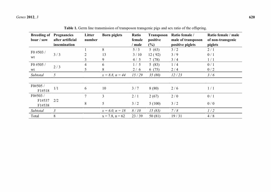
At the age of eight months, the founder boars (#503 and #505) were trained to use a dummy, both boars showed a good libido and ejaculated sperm could be recovered. Interestingly, all spermatozoa were uniformly Venus-positive. According to Mendelian rules, segregation of the three monomeric transposons should result in 12.5% triple-, 37.5% double-, 37.5% mono- and 12.5% non-transgenic sperm cells [49]. Apparently, the Venus protein was uniformly distributed between the developing sperm cells, which are connected via cytoplasmic bridges. This equal distribution of cytoplasmic components during spermatogenesis did not correlate with the genotype (Figure 3). A detailed analysis of ejaculated spermatozoa from the founder boar is given in reference [49]. Assessment of general parameters of sperm fertility showed that ejaculates from the transgenic boars fulfilled standard semen quality requirements, which included motility, velocity and morphological aspects [49].
2.3. Assessment of Germ Line Transmission and Derivation of F1-lines
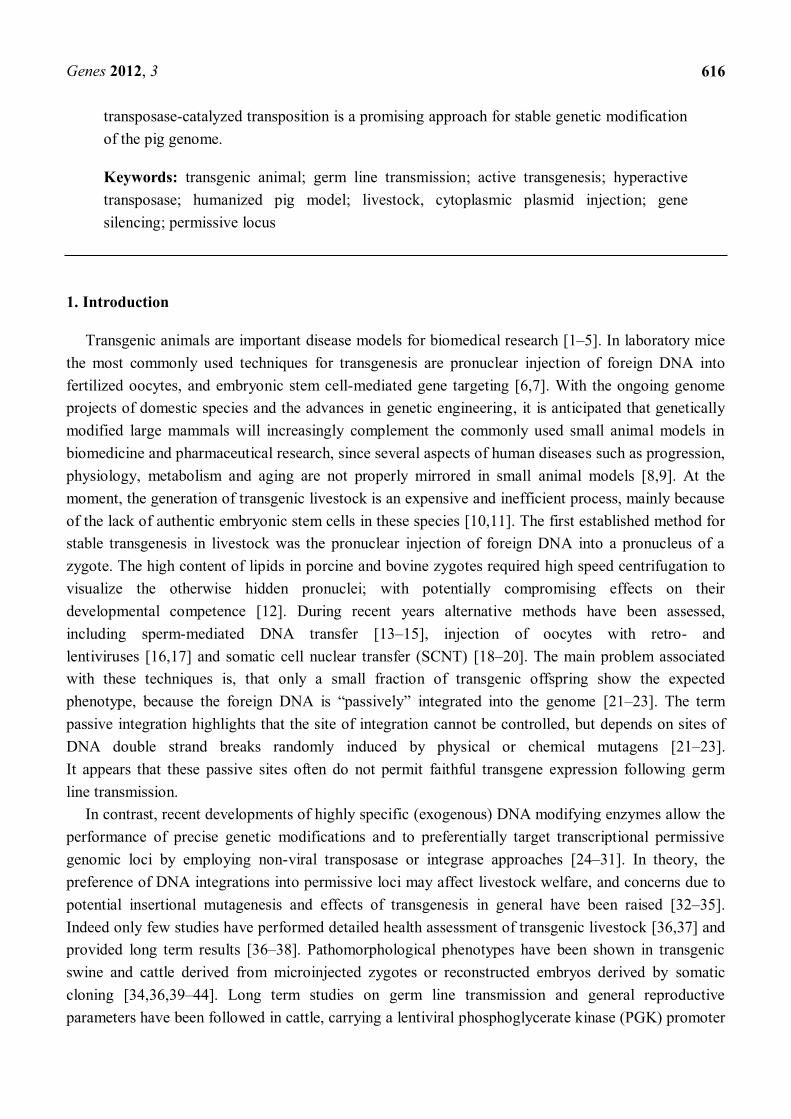
To test the fertility of transgenic sperm, wild type sows were artificially inseminated with semen from the two Venus transgenic boars (#503 and #505), respectively. Six wild type sows were inseminated at natural heat and five became pregnant. Subsequently, three transgenic F1 sows were inseminated with founder semen; all three became pregnant and delivered healthy piglets at term. In total, 62 piglets were born, of which 81% were transgenic (Table 1). Molecular analysis confirmed

Genes 2012, 3
619
segregation of the monomeric transposons in the offspring (see below). The piglets showed a normal birth weight, normal growth and social behaviour. The transgene expression did not affect health of the piglets. Previously, the most common clinical signs of disease associated with transgene expression in pigs included lethargy, lameness, uncoordinated gait, exophthalmos, and thickened skin [50]. Most likely, this phenotype was caused by ectopic expression of recombinant growth hormone. EGFP-transgenic piglets, produced with a lentiviral vector,were similar to wildtype animals with regard to reproductive parameters, behavior and general welfare criteria [37]. Here we extend these observations to transposon transgenic pigs, which showed an ubiquitous expression of the Venus fluorophore. Venus is an improved version of the yellow fluorescent protein (YFP) with stronger relative fluorescence, improved refolding after denaturation, and an increased pH resistance [51]. Compared to EGFP, the Venus protein carries several amino acids exchanges.
Figure 2. Transposon transgenic pig expressing Venus reporter. (A) Boar #505 under specific Venus excitation at the age of four weeks, Venus fluorescence is indicated by green color. Note the blue appearance of the hands holding the pig is due to reflected and scattered excitation light; (B) Scheme of CPI injection. After injection of the plasmid solution into the cytoplasm (I.), the plasmids translocate into the nucleus (II.). Expression of the SB100× helper plasmid results in the production of active SB protein and specific transposition of the transposon (without plasmid backbone) into the genome (III.).
Genes 2012, 3
620
Table 1. Germ line transmission of transposon transgenic pigs and sex ratio of the offspring.
Breeding of boar / sow
Pregnancies after artificial insemination
Litter number
Born piglets Ratio female / male
Transposon positive (%)
Ratio female / male of transposon positive piglets
Ratio female / male of non-transgenic piglets
F0 #503 / wt 3 / 3
1 8 5 / 3 5 (63) 3 / 2 2 / 1 2 13 3 / 10 12 ( 92) 3 / 9 0 / 1 3 9 4 / 5 7 (78) 3 / 4 1 / 1
F0 #505 / wt 2 / 3 4 6 1 / 5 5 (83) 1 / 4 0 / 1
5 8 2 / 6 6 (75) 2 / 4 0 / 2 Subtotal 5 x = 8.8, n = 44 15 / 29 35 (80) 12 / 23 3 / 6 F0#505 / F1#518 1/1 6 10 3 / 7 8 (80) 2 / 6 1 / 1
F0#503 / F1#537 F1#538
2/2 7 3 2 / 1 2 (67) 2 / 0 0 / 1
8 5 3 / 2 5 (100) 3 / 2 0 / 0
Subtotal 3 x = 6.0, n = 18 8 / 10 15 (83) 7 / 8 1 / 2 Total 8 x = 7.8, n = 62 23 / 39 50 (81) 19 / 31 4 / 8
Genes 2012, 3
621
Figure 3. Expression of Venus fluorescence in spermatozoa. (A) Specific Venus fluorescence of a spermatozoon from F0 boar #503; (B) Brightfield image of A; (C) Flow cytometric analysis of pig spermatozoa. The black line in the FACS analysis represents wild type sperm; blue line, spermatozoa from F1 boar with single integration; red line, spermatozoa from boar #503 with three integrations. Note that all spermatozoa from the transgenic pigs are uniformly Venus-positive.
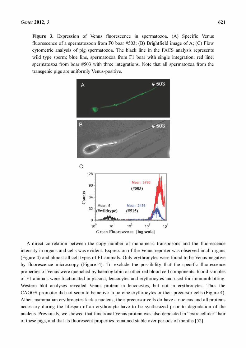
A direct correlation between the copy number of monomeric transposons and the fluorescence intensity in organs and cells was evident. Expression of the Venus reporter was observed in all organs (Figure 4) and almost all cell types of F1-animals. Only erythrocytes were found to be Venus-negative by fluorescence microscopy (Figure 4). To exclude the possibility that the specific fluorescence properties of Venus were quenched by haemoglobin or other red blood cell components, blood samples of F1-animals were fractionated in plasma, leucocytes and erythrocytes and used for immunoblotting. Western blot analyses revealed Venus protein in leucocytes, but not in erythrocytes. Thus the CAGGS-promoter did not seem to be active in porcine erythrocytes or their precursor cells (Figure 4). Albeit mammalian erythrocytes lack a nucleus, their precursor cells do have a nucleus and all proteins necessary during the lifespan of an erythrocyte have to be synthesized prior to degradation of the nucleus. Previously, we showed that functional Venus protein was also deposited in hair of these pigs, and that its fluorescent properties remained stable over periods of months [52].
Genes 2012, 3
622
Figure 4. Expression of Venus fluorophore in the pig heart and absence of Venus in erythrocytes. (A) Exemplarily, a transgenic (left) and a wild type (right) pig heart are shown under specific excitation of the Venus fluorophore. Inset, brightfield view; (B) Smear of blood cells. Brightfield view (left), specific Venus excitation (right). Note, red blood cells showed no Venus expression (some erythrocytes are indicated by an arrow); (C) Western blot detection of Venus in blood fractions. In blood, the Venus expression is restricted to leucocytes. Green bar indicates samples from Venus transgenic pig; black bar samples from wild type pig. M, molecular size marker; cb, complete blood; p, plasma; e, erythrocyte fraction; l, leucocyte fraction. Apparent molecular weight of Venus protein is ~30 kD.
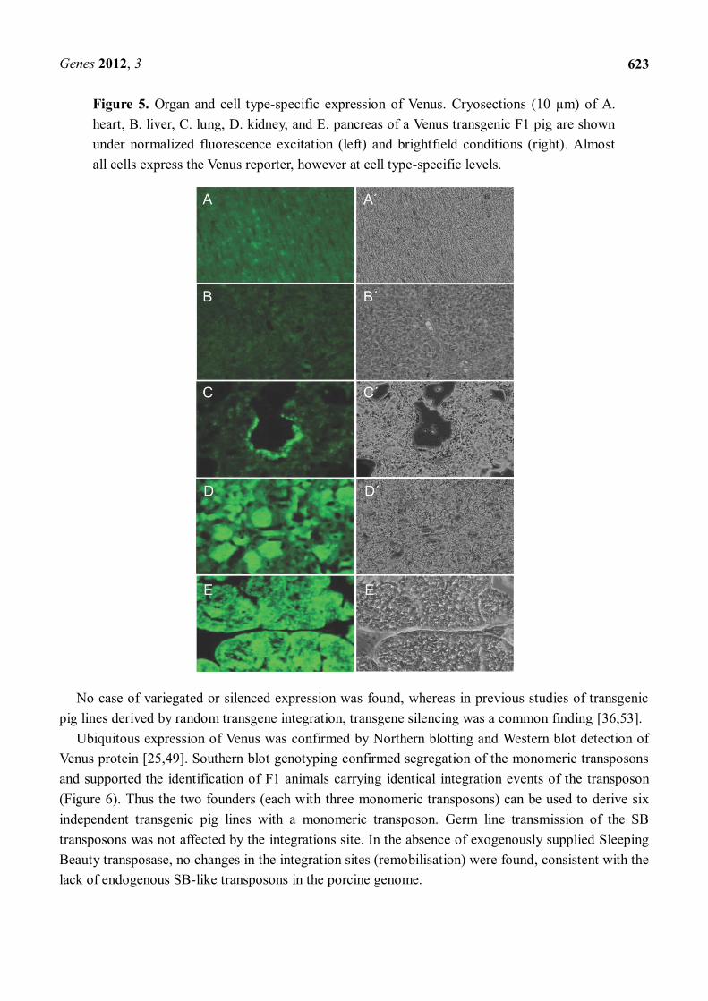
A histological examination showed expression in all solid organs of transgenic F1 animals containing different transposon integrations, and all cells expressed the transgene (Figures 4, 5). The level of expression varied between cell types, but specific cell types showed consistent expression levels. High expression levels were detected in kidney and pancreas, whereas heart, lung and liver showed moderate to low expression (Figure 5).
Genes 2012, 3
623
Figure 5. Organ and cell type-specific expression of Venus. Cryosections (10 µm) of A. heart, B. liver, C. lung, D. kidney, and E. pancreas of a Venus transgenic F1 pig are shown under normalized fluorescence excitation (left) and brightfield conditions (right). Almost all cells express the Venus reporter, however at cell type-specific levels.
No case of variegated or silenced expression was found, whereas in previous studies of transgenic pig lines derived by random transgene integration, transgene silencing was a common finding [36,53].
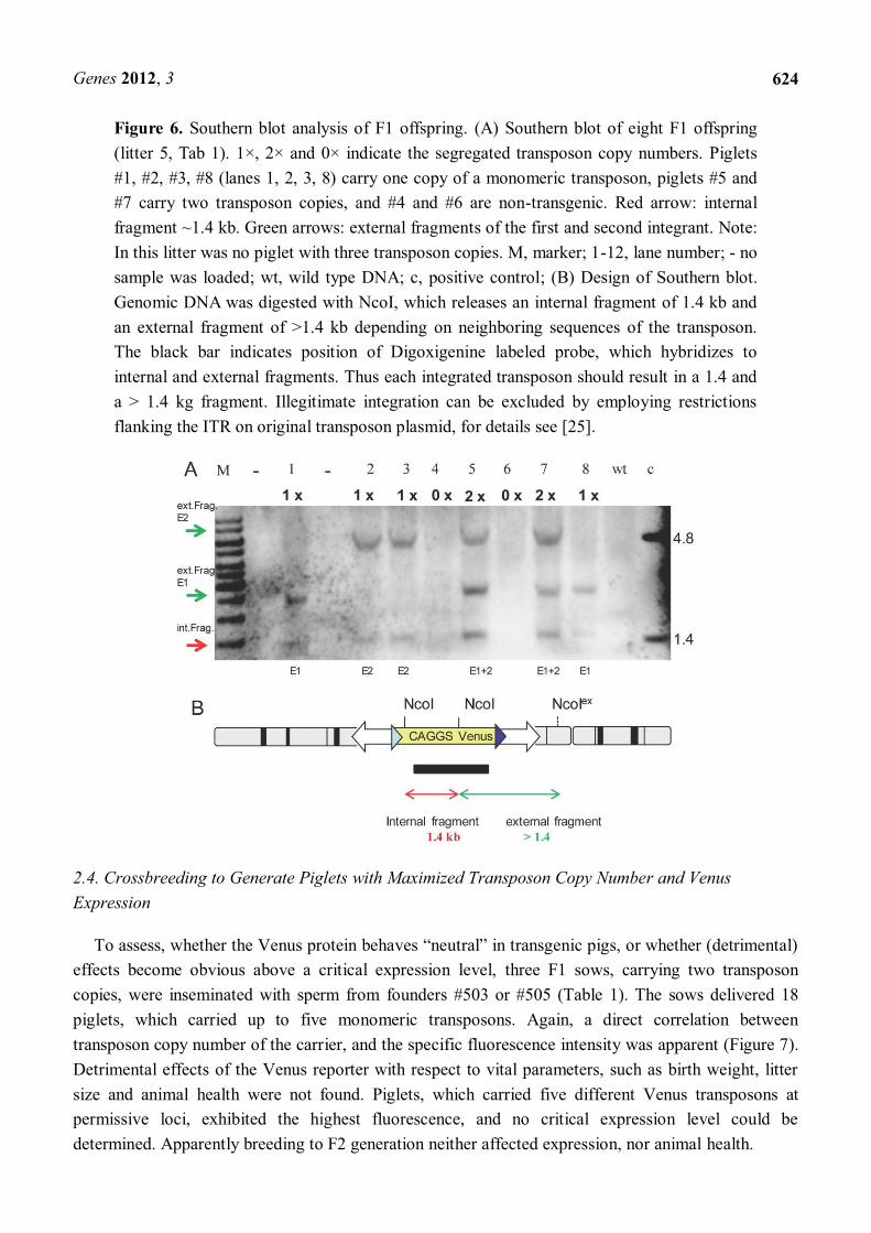
Ubiquitous expression of Venus was confirmed by Northern blotting and Western blot detection of Venus protein [25,49]. Southern blot genotyping confirmed segregation of the monomeric transposons and supported the identification of F1 animals carrying identical integration events of the transposon (Figure 6). Thus the two founders (each with three monomeric transposons) can be used to derive six independent transgenic pig lines with a monomeric transposon. Germ line transmission of the SB transposons was not affected by the integrations site. In the absence of exogenously supplied Sleeping Beauty transposase, no changes in the integration sites (remobilisation) were found, consistent with the lack of endogenous SB-like transposons in the porcine genome.
Genes 2012, 3
624
Figure 6. Southern blot analysis of F1 offspring. (A) Southern blot of eight F1 offspring (litter 5, Tab 1). 1×, 2× and 0× indicate the segregated transposon copy numbers. Piglets #1, #2, #3, #8 (lanes 1, 2, 3, 8) carry one copy of a monomeric transposon, piglets #5 and #7 carry two transposon copies, and #4 and #6 are non-transgenic. Red arrow: internal fragment ~1.4 kb. Green arrows: external fragments of the first and second integrant. Note: In this litter was no piglet with three transposon copies. M, marker; 1-12, lane number; - no sample was loaded; wt, wild type DNA; c, positive control; (B) Design of Southern blot. Genomic DNA was digested with NcoI, which releases an internal fragment of 1.4 kb and an external fragment of >1.4 kb depending on neighboring sequences of the transposon. The black bar indicates position of Digoxigenine labeled probe, which hybridizes to internal and external fragments. Thus each integrated transposon should result in a 1.4 and a > 1.4 kg fragment. Illegitimate integration can be excluded by employing restrictions flanking the ITR on original transposon plasmid, for details see [25].
2.4. Crossbreeding to Generate Piglets with Maximized Transposon Copy Number and Venus Expression
effects become obvious above a critical expression level, three F1 sows, carrying two transposon copies, were inseminated with sperm from founders #503 or #505 (Table 1). The sows delivered 18 piglets, which carried up to five monomeric transposons. Again, a direct correlation between transposon copy number of the carrier, and the specific fluorescence intensity was apparent (Figure 7). Detrimental effects of the Venus reporter with respect to vital parameters, such as birth weight, litter size and animal health were not found. Piglets, which carried five different Venus transposons at permissive loci, exhibited the highest fluorescence, and no critical expression level could be determined. Apparently breeding to F2 generation neither affected expression, nor animal health.

Genes 2012, 3
625
The mean littersize of 7.8 piglets for the germ line transmission experiments (Table 1) is smaller than the mean littersize of wild type pigs in the Institutes pig facility. However, this may be due to different selection criteria for these two populations of pigs. Whereas wild type animals are strongly selected for high fecundity, and animals which give rise to small litter sizes are slaughtered, the transposon founders were solely selected on the presence of the transgene. Also a higher ratio of male (62%) than female offspring (38%) was recorded (Table 1). This was found both in the transgenic and in the non-transgenic offspring of transposon transgenic parental animals, and did not correlate with the transgenic status. The smaller litter size and the altered sex ratio warrant further studies into the role of transposon transgenesis on porcine fertility.
Figure 7. Maximized Venus expression by crossbreeding. (A) Female F1 sow # 518 (descendant of boar # 503) was inseminated with semen of the F0 boar #505. Seven of the F2 offspring (Table 1, litter 6) are shown under specific Venus excitation. The piglets are labeled with the copy number of the transposon cassette. The number of integration sites of the Venus transposon shows a direct correlation to fluorescence intensity; (B) Calculated transposon segregation for parents with three and two independent integrants of the Venus transposon. Dot, Venus transposon integrant; line, wt allele.

Genes 2012, 3
626
2.5. Generation of Porcine Models for Biomedical and Pharmaceutical Research
Importantly, the active transposition in porcine zygotes did not require any antibiotic selection cassettes, which are un-wanted DNA elements according to guidelines provided by regulatory authorities like the European Medicines Agency (EMA) and the Food and Drug Administration (FDA) in the USA. Recent experimental data in transgenic mice and cattle support the notion that the presence of an antibiotic selection cassette may result in variegated expression of the gene of interest [28,54].
, are necessary to fully exploit transgenic animal models.
Methodological improvements in gene transfer have led to a rapidly increasing list of biomedical livestock models during recent years [55 62]. Transgenic pigs have been shown to mimic genetic and metabolic human diseases [1,5,55 58,60 63]. An important example is the porcine cystic fibrosis model, which shows a similar disease phenotype as for human patients [64], whereas transgenic mouse models failed to exhibit lung, pancreatic and intestinal obstructions. Huntington´s disease is a neurodegenerative disorder characterized by expression of a mutated huntingtin. A transgenic pig model expressing the N-terminal huntingtin with a polyglutamine tract seems to accumulate misfolded proteins in neurons, and initiated apoptosis in the affected neurons [56]. Photoreceptor topography in the pig retina is highly similar to that in humans, as it includes a cone rich, macula-like area centralis. Transgenic pigs expressing macular dystrophy-causing mutation in the ELOVL4 (elongation of very long chain fatty acids-4) gene showed photoreceptor loss, disorganised inner and outer segments, and diminished electro-retinography responses, suggesting that the transgenic pigs mirror macular degeneration and provide a unique model for therapeutic interventions [65]. Recently, the first immune deficient pigs were cloned [66 68], which is promising for large animal models for cell transplantation experiments. Our results suggest that Sleeping Beauty transposon-catalyzed stable transgene delivery is a useful addition in the toolkit for improved genetic engineering of the porcine genome for the generation of novel disease models [69].
3. Experimental Section
3.1. Animals and Samples
Animals were maintained and handled according to German laws according animal welfare and genetically modified organisms (GMO). All animal experiments were approved by an independent ethics committee. Sperm rich fractions were collected using a dummy and by the gloved hand technique. Sperm samples were extended with Androhep (1:1). For artificial insemination, semen was diluted to a final concentration of 108 sperm cells/mL. For genotyping, blood and ear biopsies were collected, and DNA was isolated with the proteinase K method.
3.2. Southern Blotting
Southern blot was done according to standard protocols. The genomic DNA was digested with NcoI. Hybridization with a CAGGS-Venus probe resulted in a constant internal fragment of around

Genes 2012, 3
627
1.4 kb, and a variable external fragment of >1.4 kb per integration [25]. Southern blotting was done according to standard protocols.
3.3. Sperm Analysis
Flow cytometry analysis of spermatozoa was performed using a FACScan (BD Bioscience) equipped with an argon laser (488 nm, 15 mW). Samples were diluted to 0.5 × 10⁶ cells/mL and measured in duplicates acquiring 10,000 cells per sample.
3.4. Western Blotting
Cells were extracted in RIPA buffer and 10 microgram of protein per slot was separated on a SDS-PAGE gel, blotted to a PVDF membrane, blocked and probed with a rabbit polyclonal anti-EGFP antibody (Santa Cruz) in 1:1000 dilution, followed by a secondary anti-rabbit antibody in 1:10 000 dilution (Sigma-Aldrich). For detection an ECL+ kit (GE Healthcare) and an image acquisition system (Vilber Lourmat, Fusion SL 3500) were used. For complete Western blotting protocol see references [49,52].
3.5. Macroscopic Imaging and Fluorescence Microscopy
Venus transgenic and wildtype animals were excited with a blue LED and images were recorded with a digital camera and an emission filter (Lee Filter). Isolated organs were recorded in an identical manner. Organ samples were fixed in neutralized 4% formaldehyde overnight, then transferred to a 20% sucrose solution, and frozen in embedding medium (Micom Laborgeräte, Walldorf, Germany) on dry ice. This treatment excellently preserved the Venus fluorescence. In a cryotome, 10 m sections were cut, mounted in Vectashield and viewed an an Olympus BX60 equipped with epifluorescence (excitation: 450 490 nm; emission: 515 550 nm; dichroic mirror: 505 nm). Normalized images were recorded with a scientific digital camera (Olympus DP71).
4. Conclusions
We have established the use of a Sleeping Beauty transposon system for the generation of germ line competent transgenic pigs by CPI. Generation of transgenic pigs with the transposon-system seems to be an efficient method for the production of large animal models and seems to avoid unwanted side effects of transgenesis, such as insertional mutagenesis, integration of concatemeres or integration into transcriptional non-permissive loci. In contrast to conventional random transgene integration, the transposase-catalyzed DNA integration preferentially resulted in monomeric integrations in transcriptionally permissive loci of the genome. Together with more accurate whole genome data and highly specific designed DNA modifying enzymes [59], precision genetic modifications became feasible in the pig genome [21,25,70]. It is anticipated that authentic pluripotent cells of the pig will be generated in the near future. Thus transgenesis in the pig will be significantly improved for the generation of humanized pig models [71]. The expected progress of pig transgenesis with increased success rates and decreased costs, will make the pig an attractive complementary model for advanced approaches in biomedical and pharmaceutical research.

Genes 2012, 3
628
Acknowledgments
The authors thank Peter Köhler for fluorescence photography, and Brigitte Barg-Kues and Maren Ziegler for excellent technical support. Rolf Poppenga, Edward Kufeld, Johann Kuhn and Toni Peker for animal caretaking and boar semen collection. Funding by the DFG is gratefully acknowledged (KU 1586/2-1 and IV 21/6-1).
Conflict of Interests
The authors declare no competing interests
References
1. Luo, Y.; Li, J.; Liu, Y.; Lin, L.; Du, Y.; Li, S.; Yang, H.; Vajta, G.; Callesen, H.; Bolund, L.; Sorensen, C.B. High efficiency of BRCA1 knockout using rAAV-mediated gene targeting: Developing a pig model for breast cancer. Transgenic. Res. 2011, 20, 975 988.
2. Muller, M.; Brem, G. Transgenic strategies to increase disease resistance in livestock. Reprod. Fertil. Dev. 1994, 6, 605 613.
3. Tanila, H. Wading pools, fading memories-place navigation in transgenic mouse models of Alzheimer's disease. Front. Aging. Neurosci. 2012, 4, 11.
4. Jacobsen, J.C.; Bawden, C.S.; Rudiger, S.R.; McLaughlan, C.J.; Reid, S.J.; Waldvogel, H.J.; MacDonald, M.E.; Gusella, J.F.; Walker, S.K.; Kelly, J.M.; Webb, G.C.; Faull, R.L.; Rees, M.I.; Snell, R.G. An ovine transgenic, Huntington's disease model. Hum. Mol. Genet. 2010, 19, 1873 1882.
5. Kragh, P.M.; Nielsen, A.L.; Li, J.; Du, Y.; Lin, L.; Schmidt, M.; Bogh, I.B.; Holm, I.E.; Jakobsen, J.E.; Johansen, M.G.; Purup, S.; Bolund, L.; Vajta, G.; Jorgensen, A.L. Hemizygous minipigs produced by random gene insertion and handmade cloning express the Alzheimer's disease-causing dominant mutation APPsw. Transgenic. Res. 2009, 18, 545 558.
6. Palmiter, R.D.; Brinster, R.L.; Hammer, R.E.; Trumbauer, M.E.; Rosenfeld, M.G.; Birnberg, N.C.; Evans, R.M. Dramatic growth of mice that develop from eggs microinjected with metallothionein-growth hormone fusion genes. Nature 1982, 300, 611 615.
7. Gama Sosa, M.A.; De Gasperi, R.; Elder, G.A. Animal transgenesis: An overview. Brain. Struct. Funct. 2010, 214, 91 109.
8. Wall, R.J.; Shani, M. Are animal models as good as we think? Theriogenology 2008, 69, 2 9. 9. Habermann, F.A.; Wuensch, A.; Sinowatz, F.; Wolf, E. Reporter genes for embryogenesis
research in livestock species. Theriogenology 2007, 68, S116 S124. 10. Laible, G.; Alonso-Gonzalez, L. Gene targeting from laboratory to livestock: Current status and
emerging concepts. Biotechnol. J. 2009, 4, 1278 1292. 11. Nowak-Imialek, M.; Kues, W.; Carnwath, J.W.; Niemann, H. Pluripotent stem cells and
reprogrammed cells in farm animals. Microsc. Microanal. 2011, 17, 474 497. 12. Hammer, R.E.; Pursel, V.G.; Rexroad, C.E., Jr.; Wall, R.J.; Bolt, D.J.; Ebert, K.M.;
Palmiter, R.D.; Brinster, R.L. Production of transgenic rabbits sheep and pigs by microinjection. Nature 1985, 315, 680 683.

Genes 2012, 3
629
13. Chang, K.; Qian, J.; Jiang, M.; Liu, Y.H.; Wu, M.C.; Chen, C.D.; Lai, C.K.; Lo, H.L.; Hsiao, C.T.; Brown, L.; Bolen, J., Jr.; Huang, H.I.; Ho, P.Y.; Shih, P.Y.; Yao, C.W.; Lin, W.J.; Chen, C.H.; Wu, F.Y.; Lin, Y.J.; Xu, J.; Wang, K. Effective generation of transgenic pigs and mice by linker based sperm-mediated gene transfer. BMC Biotechnol. 2002, 2, 5.
14. Watanabe, M.; Kurome, M.; Matsunari, H.; Nakano, K.; Umeyema, K.; Shiota, A.; Nakauchi, H.; Nagashima, H. The creation of transgenic pigs expressing human proteins using BAC-derived full-length genes and intracytoplasmic sperm injection-mediated gene transfer. Transgenic. Res. 2012, 21, 605 618.
15. Garcia-Vazquez, F.A.; Ruiz, S.; Matas, C.; Izquierdo-Rico, M.J.; Grullon, L.A.; De, O.A.; Vieira, L.; viles-Lopez, K.; Gutierrez-Adan, A.; Gadea, J. Production of transgenic piglets using ICSI-sperm-mediated gene transfer in combination with recombinase RecA. Reproduction. 2010, 140, 259 272.
16. Park, F. Lentiviral vectors: Are they the future of animal transgenesis? Physiol. Genomics. 2007, 31, 159 173.
17. Hofmann, A.; Kessler, B.; Ewerling, S.; Weppert, M.; Vogg, B.; Ludwig, H.; Stojkovic, M.; Boelhauve, M.; Brem, G.; Wolf, E.; Pfeifer, A. Efficient transgenesis in farm animals by lentiviral vectors. EMBO Rep. 2003, 4, 1054 1060.
18. Wilmut, I.; Schnieke, A.E.; McWhir, J.; Kind, A.J.; Campbell, K.H. Viable offspring derived from fetal and adult mammalian cells. Nature 1997, 385, 810 813.
19. Schnieke, A.E.; Kind, A.J.; Ritchie, W.A.; Mycock, K.; Scott, A.R.; Ritchie, M.; Wilmut, I.; Colman, A.; Campbell, KH. Human factor IX transgenic sheep produced by transfer of nuclei from transfected fetal fibroblasts. Science 1997, 278, 2130 2133.
20. Petersen, B.; Lucas-Hahn, A.; Oropeza, M.; Hornen, N.; Lemme, E.; Hassel, P.; Queisser, A.L.; Niemann, H. Development and validation of a highly efficient protocol of porcine somatic cloning using preovulatory embryo transfer in peripubertal gilts. Cloning. Stem. Cells. 2008, 10, 355 362.
21. Garrels, W.; Ivics, Z.; Kues, W.A. Precision genetic engineering in large mammals. Trends. Biotechnol. 2012, 7, 386 393.
22. Shinohara, E.T.; Kaminski, J.M.; Segal, D.J.; Pelczar, P.; Kolhe, R.; Ryan, T.; Coates, C.J.; Fraser, M.J.; Handler, A.M.; Yanagimachi, R.; Moisyadi, S. Active integration: New strategies for transgenesis. Transgenic. Res. 2007, 16, 333 339.
23. Fahrenkrug, S.C.; Blake, A.; Carlson, D.F.; Doran, T.; Van, E.A.; Faber, D.; Galli, C.; Gao, Q.; Hackett, P.B.; Li, N.; Maga, E.A.; Muir, W.M.; Murray, J.D.; Shi, D.; Stotish, R.; Sullivan, E.; Taylor, J.F.; Walton, M.; Wheeler, M.; Whitelaw, B.; Glenn, B.P. Precision genetics for complex objectives in animal agriculture. J Anim. Sci. 2010, 88, 2530 2539.
24. Staunstrup, N.H.; Madsen, J.; Primo, M.N.; Li, J.; Liu, Y.; Kragh, P.M.; Li, R.; Schmidt, M.; Purup, S.; Dagnaes-Hansen, F.; Svensson, L.; Petersen, T.K.; Callesen, H.; Bolund, L.; Mikkelsen, J.G. Development of transgenic cloned pig models of skin inflammation by DNA transposon-directed ectopic expression of human beta1 and alpha2 integrin. PLoS One 2012, 7, e36658.
25. Garrels, W.; Mates, L.; Holler, S.; Dalda, A.; Taylor, U.; Petersen, B.; Niemann, H.; Izsvak, Z.; Ivics, Z.; Kues, W.A. Germline transgenic pigs by, Sleeping, Beauty transposition in porcine zygotes and targeted integration in the pig genome. PLoS One 2011, 6, e23573.
Genes 2012, 3
630
26. Carlson, D.F.; Geurts, A.M.; Garbe, J.R.; Park, C.W.; Rangel-Filho, A.; O'Grady, S.M.; Jacob, H.J.; Steer, C.J.; Largaespada, D.A.; Fahrenkrug, S.C. Efficient mammalian germline transgenesis by cis-enhanced Sleeping Beauty transposition. Transgenic. Res. 2011, 20, 29 45.
27. McGrew, M.J.; Sherman, A.; Ellard, F.M.; Lillico, S.G.; Gilhooley, H.J.; Kingsman, A.J.; Mitrophanous, KA.; Sang, H. Efficient production of germline transgenic chickens using lentiviral vectors. EMBO Rep. 2004, 5, 728 733.
28. Tasic, B.; Hippenmeyer, S.; Wang, C.; Gamboa, M.; Zong, H.; Chen-Tsai, Y.; Luo, L. Site-specific integrase-mediated transgenesis in mice via pronuclear injection. Proc. Natl. Acad. Sci. USA 2011, 108, 7902 7907.
29. Macdonald, J.; Taylor, L.; Sherman, A.; Kawakami, K.; Takahashi, Y.; Sang, H.M.; McGrew, M.J. Efficient genetic modification and germ-line transmission of primordial germ cells using piggyBac and Tol2 transposons. Proc. Natl. Acad. Sci. USA 2012, 109, E1466 E1472.
30. Carlson, D.F.; Garbe, J.R.; Tan, W.; Martin, M.J.; Dobrinsky, J.R.; Hackett, P.B.; Clark, K.J.; Fahrenkrug, S.C. Strategies for selection marker-free swine transgenesis using the Sleeping Beauty transposon system. Transgenic. Res. 2011, 20, 1125 1137.
31. Jakobsen, J.E.; Li, J.; Kragh, P.M.; Moldt, B.; Lin, L.; Liu, Y.; Schmidt, M.; Winther, K.D.; Schyth, B.D.; Holm, I.E.; Vajta, G.; Bolund, L.; Callesen, H.; Jorgensen, A.L.; Nielsen, A.L.; Mikkelsen, J.G. Pig transgenesis by Sleeping Beauty DNA transposition. Transgenic. Res. 2011, 20, 533 545.
32. Van Reenen, C.G.; Meuwissen, T.H.; Hopster, H.; Oldenbroek, K.; Kruip, T.H.; Blokhuis, H.J. Transgenesis may affect farm animal welfare: A case for systematic risk assessment. J. Anim. Sci. 2001, 79, 1763 1779.
33. Clark, J.; Whitelaw, B. A future for transgenic livestock. Nat. Rev. Genet. 2003, 4, 825 833. 34. Carter, D.B.; Lai, L.; Park, K.W.; Samuel, M.; Lattimer, J.C.; Jordan, K.R.; Estes, D.M.;
Besch-Williford, C.; Prather, R.S. Phenotyping of transgenic cloned piglets. Cloning. Stem. Cells 2002, 4, 131 145.
35. Greger, M. Trait selection and welfare of genetically engineered animals in agriculture. J. Anim. Sci. 2010, 88, 811 814.
36. Deppenmeier, S.; Bock, O.; Mengel, M.; Niemann, H.; Kues, W.; Lemme, E.; Wirth, D.; Wonigeit, K.; Kreipe, H. Health status of transgenic pigs expressing the human complement regulatory protein CD59. Xenotransplantation. 2006, 13, 345 356.
37. Huber, R.; Remuge, L.; Carlisle, A.; Lillico, S.; Sandoe, P.; Sorensen, D.; Withelaw, B.; Olosson, A. Welfare assessment in transgenic pigs expressing green fluorescent protein GFP. Transgenic. Res. 2012, 21, 773 784.
38. Reichenbach, M.; Lim, T.; Reichenbach, H.D.; Guengoer, T.; Habermann, F.A.; Matthiesen, M.; Hofmann, A.; Weber, F.; Zerbe, H.; Grupp, T.; Sinowatz, F.; Pfeifer, A.; Wolf, E. Germ-line transmission of lentiviral, PGK-EGFP integrants in transgenic cattle: new perspectives for experimental embryology. Transgenic. Res. 2010, 19, 549 556.
39. Pursel, V.G.; Wall, R.J.; Solomon, M.B.; Bolt, D.J.; Murray, J.E.; Ward, K.A. Transfer of an ovine metallothionein-ovine growth hormone fusion gene into swine. J Anim. Sci. 1997, 75, 2208 2214.

Genes 2012, 3
631
40. Grupen, C.G.; Verma, P.J.; Du, Z.T.; McIlfatrick, S.M.; Ashman, R.J.; Nottle, M.B. Activation of in vivo- and in vitro-derived porcine oocytes by using multiple electrical pulses. Reprod. Fertil. Dev. 1999, 11, 457 462.
41. Hill, J.R.; Roussel, A.J.; Cibelli, J.B.; Edwards, J.F.; Hooper, N.L.; Miller, M.W.; Thompson, J.A.; Looney, C.R.; Westhusin, M.E.; Robl, J.M.; Stice, S.L. Clinical and pathologic features of cloned transgenic calves and fetuses (13 case studies). Theriogenology 1999, 51, 1451 1465.
42. Chavatte-Palmer, P.; Camous, S.; Jammes, H.; Le, C.N.; Guillomot, M.; Lee, R.S. Review: Placental perturbations induce the developmental abnormalities often observed in bovine somatic cell nuclear transfer. Placenta 2012, 33, S99 S104.
43. Watanabe, S.; Nagai, T. Health status and productive performance of somatic cell cloned cattle and their offspring produced in Japan. J. Reprod. Dev. 2008, 54, 6 17.
44. Cao, Z.; Li, Y.; Wen, X.; Li, Z.; Mi, C.; Zhang, Z.; Li, N.; Li, Q. Recloned transgenic pigs possess normal reproductive performance and stable genetic transmission capacity. Zygote 2012, 12, 1 7.
45. Iqbal, K.; Barg-Kues, B.; Broll, S.; Bode, J.; Niemann, H.; Kues, W.A. Cytoplasmic injection of circular plasmids allows targeted expression in mammalian embryos. Biotechniques 2009, 47, 959 968.
46. Mates, L.; Chuah, M.K.; Belay, E.; Jerchow, B.; Manoj, N.; Costa-Sanchez, A.; Grzela, D.P.; Schmitt, A.; Becker, K.; Matrai, J.; Ma, L.; Samara-Kuko, E.; Gysemans, C.; Pryputniewicz, D.; Miskey, C.; Fletcher, B.; Vandendriessche, T.; Ivics, Z.; Izsvak, Z. Molecular evolution of a novel hyperactive Sleeping Beauty transposase enables robust stable gene transfer in vertebrates. Nat. Genet. 2009, 41, 753 761.
47. Tamashiro, K.L.; Wakayama, T.; Blanchard, R.J.; Blanchard, D.C.; Yanagimachi, R. Postnatal growth and behavioral development of mice cloned from adult cumulus cells. Biol. Reprod. 2000, 63, 328 334.
48. Rhind, S.M.; King, T.J.; Harkness, L.M.; Bellamy, C.; Wallace, W.; DeSousa, P.; Wilmut, I. Cloned lambs-lessons from pathology. Nat. Biotechnol. 2003, 21, 744 745.
49. Garrels, W.; Holler, S.; Taylor, U.; Herrmann, D.; Struckmann, C.; Klein, S.; Barg-Kues, B.; Nowak-Imialek, M.; Ehling, C.; Rath, D.; Ivics, Z.; Niemann, H.; Kues, W.A. Genotype-independent transmission of transgenic fluorophore protein by boar spermatozoa. PLoS One 2011, 6, e27563.
50. Pursel, V.G.; Pinkert, C.A.; Miller, K.F.; Bolt, D.J.; Campbell, R.G.; Palmiter, R.D.; Brinster, R.L.; Hammer, R.E. Genetic engineering of livestock. Science 1989, 244, 1281 1288.
51. Nagai, T.; Ibata, K.; Park, E.S.; Kubota, M.; Mikoshiba, K.; Miyawaki, A. A variant of yellow fluorescent protein with fast and efficient maturation for cell-biological applications. Nat. Biotechnol. 2002, 20, 87 90.
52. Garrels, W.; Cleve, N., Niemann, H.; Kues, W.A. Rapid non-invasive genotyping of reporter transgenic mammals. Biotechniques 2012, 0, 1 4.
53. Kues, W.A.; Schwinzer, R.; Wirth, D.; Verhoeyen, E.; Lemme, E.; Herrmann, D.; Barg-Kues, B.; Hauser, H.; Wonigeit, K.; Niemann, H. Epigenetic silencing and tissue independent expression of a novel tetracycline inducible system in double-transgenic pigs. FASEB J. 2006, 20, 1200 1202.

Genes 2012, 3
632
54. Alonso-Gonzalez, L.; Couldrey, C.; Meinhardt, M.W.; Cole, S.A.; Wells, D.N.; Laible, G. Primary transgenic bovine cells and their rejuvenated cloned equivalents show transgene-specific epigenetic differences. PLoS One 2012, 7, e35619.
55. Luo, Y.; Bolund, L.; Sorensen, C.B. Pig gene knockout by rAAV-mediated homologous recombination: Comparison of BRCA1 gene knockout efficiency in Yucatan and Gottingen fibroblasts with slightly different target sequences. Transgenic. Res. 2012, 21, 671 676.
56. Yang, D.; Wang, C.E.; Zhao, B.; Li, W.; Ouyang, Z.; Liu, Z.; Yang, H.; Fan, P.; O'Neill, A.; Gu, W.; Yi, H.; Li, S.; Lai, L.; Li, X.J. Expression of Huntington's disease protein results in apoptotic neurons in the brains of cloned transgenic pigs. Hum. Mol. Genet. 2010, 19, 3983 3994.
57. Kues, W.A.; Niemann, H. The contribution of farm animals to human health. Trends. Biotechnol. 2004, 22, 286 294.
58. Yang, D.; Yang, H.; Li, W.; Zhao, B.; Ouyang, Z.; Liu, Z.; Zhao, Y.; Fan, N.; Song, J.; Tian, J.; Li, F.; Zhang, J.; Chang, L.; Pei, D.; Chen, Y.E.; Lai, L. Generation of PPAR gamma mono-allelic knockout pigs via zinc-finger nucleases and nuclear transfer cloning. Cell. Res. 2011, 21, 979 982.
59. Flisikowska, T.; Thorey, I.S.; Offner, S.; Ros, F.; Lifke, V.; Zeitler, B.; Rottmann, O.; Vincent, A.; Zhang, L.; Jenkins, S.; Niersbach, H.; Kind, A.J.; Gregory, P.D.; Schnieke, A.E.; Platzer, J. Efficient immunoglobulin gene disruption and targeted replacement in rabbit using zinc finger nucleases. PLoS One 2011, 6, e21045.
60. Renner, S.; Fehlings, C.; Herbach, N.; Hofmann, A.; Von Waldthausen, D.C.; Kessler, B.; Ulrichs, K.; Chodnevskaja, I.; Moskalenko, V.; Amselgruber, W.; Goke, B.; Pfeifer, A.; Wanke, R.; Wolf, E. Glucose intolerance and reduced proliferation of pancreatic beta-cells in transgenic pigs with impaired glucose-dependent insulinotropic polypeptide function. Diabetes 2010, 59, 1228 1238.
61. Klymiuk, N.; Mundhenk, L.; Kraehe, K.; Wuensch, A.; Plog, S.; Emrich, D.; Langenmayer, M.C.; Stehr, M.; Holzinger, A.; Kroner, C.; Richter, A.; Kessler, B.; Kurome, M.; Eddicks, M.; Nagashima, H.; Heinritzi, K.; Gruber, A.D.; Wolf, E. Sequential targeting of CFTR by BAC vectors generates a novel pig model of cystic fibrosis. J. Mol. Med. (Berl.) 2012, 90, 597 608.
62. Klymiuk, N.; Van Buerck, L.; Bahr, A.; Offers, M.; Kessler, B.; Wuensch, A.; Kurome, M.; Thormann, M.; Lochner, K.; Nagashima, H.; Herbach, N.; Wanke, R.; Seissler, J.; Wolf, E. Xenografted islet cell clusters from INSLEA29Y transgenic pigs rescue diabetes and prevent immune rejection in humanized mice. Diabetes 2012, 61, 1527 1532.
63. Wheeler, D.G.; Joseph, M.E.; Mahamud, S.D.; Aurand, W.L.; Mohler, P.J.; Pompili, V.J.; Dwyer, K.M.; Nottle, M.B.; Harrison, S.J.; d'Apice, A.J.; Robson, S.C.; Cowan, P.J.; Gumina, R.J. Transgenic swine: Expression of human CD39 protects against myocardial injury. J. Mol. Cell. Cardiol. 2012, 52, 958 961.
64. Rogers, C.S.; Stoltz, D.A.; Meyerholz, D.K.; Ostedgaard, L.S.; Rokhlina, T.; Taft, P.J.; Rogan, M.P.; Pezzulo, A.A.; Karp, P.H.; Itani, O.A.; Kabel, A.C.; Wohlford-Lenane, C.L.; Davis, G.J.; Hanfland, R.A.; Smith, T.L.; Samuel, M.; Wax, D.; Murphy, C.N.; Rieke, A.; Whitworth, K.; Uc, A.; Starner, T.D.; Brogden, K.A.; Shilyansky, J.; McCray, P.B., Jr.; Zabner, J.; Prather, R.S.; Welsh, M.J. Disruption of the CFTR gene produces a model of cystic fibrosis in newborn pigs. Science 2008, 321, 1837 1841.

Genes 2012, 3
633
65. Sommer, J.R.; Estrada, J.L.; Collins, E.B.; Bedell, M.; Alexander, C.A.; Yang, Z.; Hughes, G.; Mir, B.; Gilger, B.C.; Grob, S.; Wei, X.; Piedrahita, J.A.; Shaw, P.X.; Petters, R.M.; Zhang, K. Production of ELOVL4 transgenic pigs: a large animal model for Stargardt-like macular degeneration. Br. J. Ophthalmol. 2011, 95, 1749 1754.
66. Mendicino, M.; Ramsoondar, J.; Phelps, C.; Vaught, T.; Ball, S.; LeRoith, T.; Monahan, J.; Chen, S.; Dandro, A.; Boone, J.; Jobst, P.; Vance, A.; Wertz, N.; Bergman, Z.; Sun, X.Z.; Polejaeva, I.; Butler, J.; Dai, Y.; Ayares, D.; Wells, K. Generation of antibody- and B cell-deficient pigs by targeted disruption of the, J-region gene segment of the heavy chain locus. Transgenic. Res. 2011, 20, 625 641.
67. Ramsoondar, J.; Mendicino, M.; Phelps, C.; Vaught, T.; Ball, S.; Monahan, J.; Chen, S.; Dandro, A.; Boone, J.; Jobst, P.; Vance, A.; Wertz, N.; Polejaeva, I.; Butler, J.; Dai, Y.; Ayares, D.; Wells, K. Targeted disruption of the porcine immunoglobulin kappa light chain locus. Transgenic. Res. 2011, 20, 643 653.
68. Suzuki, S.; Iwamoto, M.; Saito, Y.; Fuchimoto, D.; Sembon, S.; Suzuki, M.; Mikawa, S.; Hashimoto, M.; Aoki, Y.; Najima, Y.; Takagi, S.; Suzuki, N.; Suzuki, E.; Kubo, M.; Mimuro, J.; Kashiwakura, Y.; Madoiwa, S.; Sakata, Y.; Perry, A.C.F.; Ishikawa, F.; Onishi, A. Il2rg gene-targeted severe combined immunodeficiency pigs. Cell. Stem. Cell 2012, 10, 753 758.
69. Hackett, P.B., Jr.; Aronovich, E.L.; Hunter, D.; Urness, M.; Bell, J.B.; Kass, S.J.; Cooper, L.J.; McIvor, S. Efficacy and safety of Sleeping Beauty transposon-mediated gene transfer in preclinical animal studies. Curr. Gene. Ther. 2011, 11, 341 349.
70. Luo, Y.; Kofod-Olsen, E.; Christensen, R.; Brandt-Sorensen, C.; Bolund, L. Targeted genome editing by recombinant adeno-associated virus (rAAV) vectors for generating genetically modified pigs. J. Genet. Genomics 2012, 39, 269 274.
71. Galli, C.; Lagutina, I.; Colleoni, S.; Duchi, R.; Luccini, F.; Lazzari, G. Somatic cell nuclear transfer and transgenesis in large animals: current and future insights. Reprod. Dom. Anim. 2012, 47, 2 11.
© 2012 by the authors; licensee MDPI, Basel, Switzerland. This article is an open access article distributed under the terms and conditions of the Creative Commons Attribution license (http://creativecommons.org/licenses/by/3.0/).


