
Assessment and diagnosis in stroke
34
Assessment and diagnosis in stroke Nick Ward DEPARTMENT OF HEADACHE, BRAIN INJURY, AND NEUROREHABILITATION NATIONAL HOSPITAL FOR NEUROLOGY AND NEUROSURGERY INSTITUTE OF NEUROLOGY UNIVERSITY COLLEGE LONDON
Transcript of Assessment and diagnosis in stroke

Assessment and diagnosis in stroke
Nick WardDEPARTMENT OF HEADACHE, BRAIN INJURY, AND NEUROREHABILITATION
NATIONAL HOSPITAL FOR NEUROLOGY AND NEUROSURGERY
INSTITUTE OF NEUROLOGYUNIVERSITY COLLEGE LONDON

Objectives
• You should know1. The essential clinical features to be elicited2. The essential investigations to be performed3. Understand some of the differential diagnosis4. Understand the basic subtypes of stroke
Pathology – what?Anatomy – where?Mechanism – why?
• You should be able to diagnose and assess a patient with suspected stroke

•65 year old man
•Found collapsed at home by wife
•Not moving right side very well
•Not speaking
•nicotine stained fingers
•bp 190/110
Positively diagnose stroke
CT normal

IMMEDIATE CLINICAL APPROACHABC
Check blood sugar
Glasgow Coma Scale <12 consider nasal airway
<8 consider intubation
Pyrexia, neck stiffness
Oxygen
IV access
RAPID neurological assessment motor
speech
visual
sensory

Clinical syndrome
• Syndrome of focal neurological symptoms
and signs
• Sudden onset
• Symptoms maximal within minutes to hours
• Predominantly negative symptoms
MAKE A POSITIVE DIAGNOSIS!
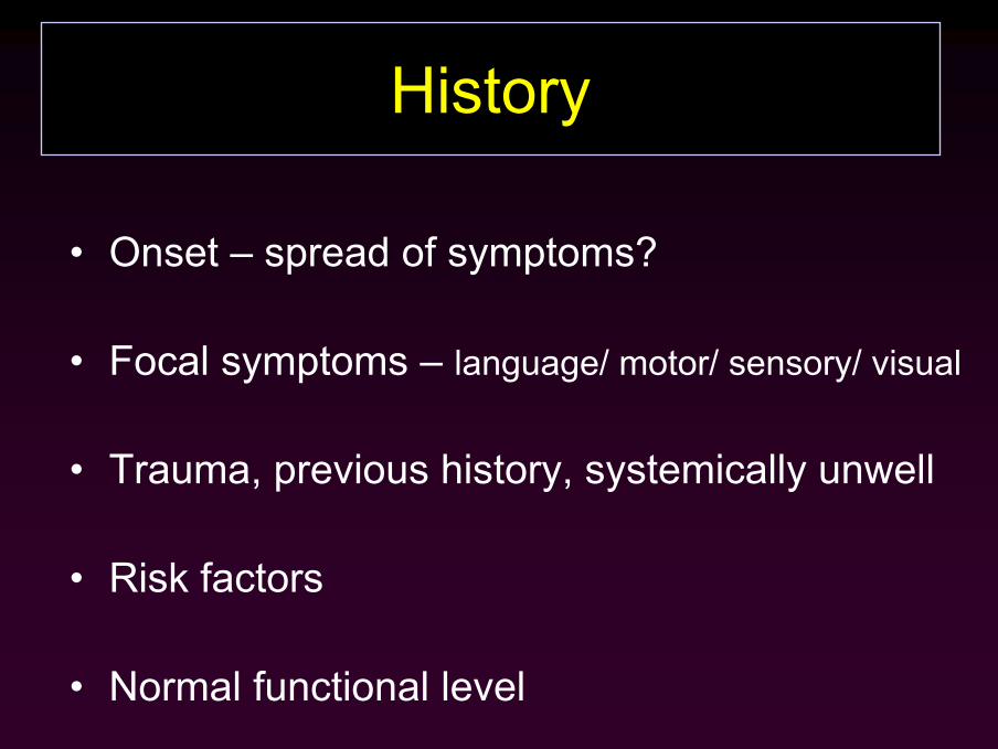
History
• Onset – spread of symptoms?
• Focal symptoms – language/ motor/ sensory/ visual
• Trauma, previous history, systemically unwell
• Risk factors
• Normal functional level

Examination
• Neurologic
– “standard” cranium and limbs
– status – degree of consciousness – GCS
– swallow

• General
– Cardiovascular
• Pulse / BP / Murmurs / Bruits
– Chest
• Pneumonia
Examination

Conditions that mimic acute stroke
411 patients initially diagnosed as having stroke
78 (19%) of these eventually diagnosed as some other condition
333 patients confirmed to have had stroke
Seizure (17%)
Systemic infection (17%)
Brain tumour (15%)
Toxic-metabolic (13%)
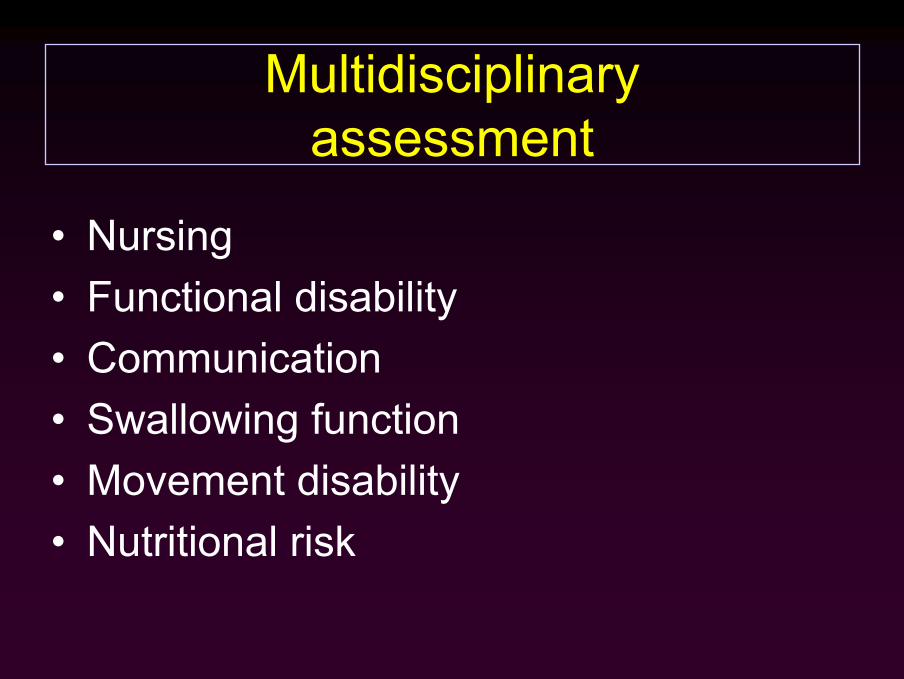
Multidisciplinaryassessment
• Nursing• Functional disability• Communication• Swallowing function• Movement disability• Nutritional risk

Objectives revisited
• You should know1. The essential clinical features to be elicited2. The essential investigations to be performed3. Understand some of the differential diagnosis4. Understand the basic subtypes of stroke
Pathology – what?Anatomy – where?Mechanism – why?
• You should be able to diagnose and assess a patient with suspected stroke

Diagnosis – PathologyWhat?
• 80% ischaemic vs 20% haemorrhagic
• No reliable clinical method– Haemorrhage:
• ? ↓ GCS• signs of ↑ ICP• headache?• on warfarin?
• Neuroimaging - only way to be sure

Infarction Haemorrhage
infarction or haemorrhage ?Answer……………….do scan

Diagnosis – AnatomyWhere?

Brain cross section showing the arteries after injection of contrast

Anatomy –Where?

Arterial territories and clinical presentations
• Anterior circulation – carotid + branches– Ophthalmic - amaurosis fugax
– MCA - Hemiparesis,hemisensory loss cortical signs
– ACA – Hemiparesis (Leg > Arm), no/mild sensory deficit, frontal lobe signs
• Posterior circulation – vertebrobasilar – PICA/AICA/PCA – Cranial nerve and long tract signs,
N+V, diplopia, Vertigo, ataxia, coma

Diagnosis – MechanismWhy?
• TOAST classification:– Lacunar (penetrating vessel occlusion)– Large vessel occlusion – Cardioembolic– Other (eg sickle cell disease)– Undetermined
• Haemorrhage

EMBOLIC SOURCES
Platelet clots
Fibrin clots
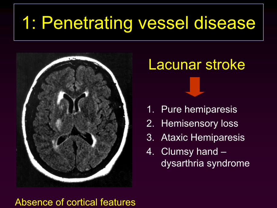
1: Penetrating vessel disease
Lacunar stroke
1. Pure hemiparesis2. Hemisensory loss3. Ataxic Hemiparesis4. Clumsy hand –
dysarthria syndrome
Absence of cortical features

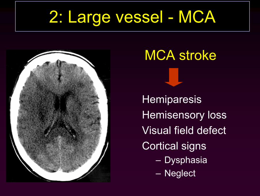
2: Large vessel - MCA
HemiparesisHemisensory lossVisual field defectCortical signs
– Dysphasia– Neglect
MCA stroke


3: Large vessel - PCA
Nausea + VomitingDiplopiaVertigoAtaxia‘Crossed’ signsVisual field defectComa


4. HaemorrhageConforms to this schema

Infarction Haemorrhage

Who to scan urgently
• Those with a depressed level of consciousness in whom neurosurgical intervention would be considered
• Patients on anticoagulants
• Patients who may be suitable for thrombolysis

Neuroimaging:CT or MRI?
CT • Readily available• Cheap• Better for blood• Can be used acutely• May be only choice eg
pacemaker• New techniques
MRI• Less availability• Expensive• Better anatomy• Better for posterior
fossa• Can be used acutely
(DWI)
Whichever is available urgently!

Other investigations
• FBC• U+E• Sugar• Cholesterol• ECG / Echo• CXR• Neuroimaging• Vascular imaging

Investigations
• Help to answer questions– Where? What? Why?
• e.g. which side/arterial territory?infarction or haemorrhage ?lacunar or large vessel?
Summary• Stroke is a clinical syndrome NOT a diagnosis
– Need then to answer• What is it?• Where is it?• Why did it happen?
• Urgent assessment should establish– Deficit – Risk factors + likely cause– Complications– Multidisciplinary team

HistoryStroke clerking proforma
Identify risk factors
Pre-stroke function
ExaminationNeurological assessment
Identify risk factors
MultidisciplinaryNursing
Functional disability
Communication
Swallowing function
Movement disability
Nutritional risk
Clinical InvestigationsHaemotology/biochemistry
Urinalysis
ECG
CXR
Investigations to considerCT scan
Carotid doppler
Echocardiography
MRI
ISCHAEMIC STROKE HAEMORRHAGIC STROKE
MANAGEMENT
ASSESSMENT OF STROKE PATIENTS: SUMMARY

Objectives Revisited
• You should know1. The essential clinical features to be elicited2. The essential investigations to be performed3. Understand some of the differential diagnosis4. Understand the basic subtypes of stroke
Pathology – what?Anatomy – where?Mechanism – why?
• You should be able to diagnose and assess a patient with suspected stroke