assay for diagnostic accuracy of cryptococcosis Evaluation ...
14
Page 1/14 Evaluation of lateral ow immunochromatographic assay for diagnostic accuracy of cryptococcosis Li-Min Xie The Third Aliated Hospital of Guangzhou Medical University Geng-ling Lin The Third Aliated Hospital of Guangzhou Medical University Hao-Neng Dong The Third Aliated Hospital of Guangzhou Medical University Ying-Xia Liao The Third Aliated Hospital of Guangzhou Medical University Ye-Ling Liu The Third Aliated Hospital of Guangzhou Medical University Jian-Feng Qin The Pharmic School of Guangzhou Medical University Xu-Guang Guo ( [email protected] ) Department of Clinical Laboratory Medicine, The Third Aliated Hospital of Guangzhou Medical University https://orcid.org/0000-0003-1302-5234 Research article Keywords: Lateral ow immunochromatographic assay, lateral ow assay, Cryptococcosis, Diagnostic Posted Date: August 27th, 2020 DOI: https://doi.org/10.21203/rs.2.19521/v3 License: This work is licensed under a Creative Commons Attribution 4.0 International License. Read Full License Version of Record: A version of this preprint was published at BMC Infectious Diseases on September 4th, 2020. See the published version at https://doi.org/10.1186/s12879-020-05368-x.
Transcript of assay for diagnostic accuracy of cryptococcosis Evaluation ...

Page 1/14
Evaluation of lateral �ow immunochromatographicassay for diagnostic accuracy of cryptococcosisLi-Min Xie
The Third A�liated Hospital of Guangzhou Medical UniversityGeng-ling Lin
The Third A�liated Hospital of Guangzhou Medical UniversityHao-Neng Dong
The Third A�liated Hospital of Guangzhou Medical UniversityYing-Xia Liao
The Third A�liated Hospital of Guangzhou Medical UniversityYe-Ling Liu
The Third A�liated Hospital of Guangzhou Medical UniversityJian-Feng Qin
The Pharmic School of Guangzhou Medical UniversityXu-Guang Guo ( [email protected] )
Department of Clinical Laboratory Medicine, The Third A�liated Hospital of Guangzhou MedicalUniversity https://orcid.org/0000-0003-1302-5234
Research article
Keywords: Lateral �ow immunochromatographic assay, lateral �ow assay, Cryptococcosis, Diagnostic
Posted Date: August 27th, 2020
DOI: https://doi.org/10.21203/rs.2.19521/v3
License: This work is licensed under a Creative Commons Attribution 4.0 International License. Read Full License
Version of Record: A version of this preprint was published at BMC Infectious Diseases on September 4th,2020. See the published version at https://doi.org/10.1186/s12879-020-05368-x.

Page 2/14
AbstractBackground: Cryptococcus is a conditional pathogenic fungus causing cryptococcosis, which is one ofthe most serious fungal diseases faced by humans. Lateral �ow immunochromatographic assay (LFA) issuccessfully applied to the rapid detection of cryptococcal antigens.
Methods: Studies were retrieved systematically from the Embase, PubMed, Web of Science, andCochrane Library before July 2019. The quality of the studies was assessed by Review Manager 5.0based on the Quality Assessment of Diagnostic Accuracy Study guidelines. The extracted data from theincluded studies were analyzed by Meta-DiSc 1.4. Stata 12.0 software was used to detect the publicationbias.
Results: A total of 15 articles with 31 fourfold tables were adopted by inclusion and exclusion criteria.The merged sensitivity and speci�city in serum were 0.98 and 0.98, respectively, and those in thecerebrospinal �uid were 0.99 and 0.99, respectively.
Conclusions: Compared to the urine and other samples, LFA in serum and cerebrospinal �uid is favorableevidence for the diagnosis of cryptococcosis with high speci�city and sensitivity.
BackgroundCryptococcosis is mainly caused by Cryptococcus, an opportunistic pathogen. Cryptococcus genus isbased on C. neoformans, C. deneoformans, C. gattii, and other non-pathogenic. Those strains of serotypeA or var. grubii are considered to be C. neoformans and serotype D or var. Neoformans are considered tobe C. deneoformans. The strains of C. gattii consist of �ve species: C. gattii, C. bacillisporus, C.deuterogattii, C. tetragattii and C. decagattii[1]. C. gattii and C. neoformans are responsible for almost allcryptococcal infections in humans[2]. Besides, people with low immunity have a high probability of beinginfected with Cryptococcus, for example, HIV patients and patients with long-term use of glucocorticoids,immunosuppressants, broad-spectrum antibiotics, and anti-tumor drugs[3-4]. All organs of humans can beinfected with Cryptococcus. Without complement and anti-Cryptococcus growth factors in cerebrospinal�uid (CSF), cryptococcal meningitis (CM) is the main clinical manifestation of the cryptococcal infectionin the central nervous system[5]. In 2014, the number of cryptococcal antigen-positive people worldwidewas 278 000, and the global incidence of cryptococcal meningitis was 223 100. Additionally, annualglobal deaths from cryptococcal meningitis were estimated at 181 100 and 135 900 deaths in sub-Saharan Africa and 15% of AIDS-related deaths are caused by cryptococcal meningitis worldwide[6].Thus, cryptococcosis has become a serious global public health problem.
However, the cryptococcal infection is short of speci�city with diverse clinical manifestations.Cryptococcosis is frequently misdiagnosed at the early stage[7]. The diagnosis of cryptococcosis relies onthe cultivation of conventional fungal and bacterial culture media from biological samples (CSF, sputumand skin biopsies, etc.), cytological examination of centrifuged CSF deposits and histopathological

Page 3/14
staining of other body �uids, or using of latex agglutination, enzyme immunoassay techniques to detectcryptococcal polysaccharide capsular antigen (CrAg) which has shed in serum and CSF duringinfection[8]. Molecular methods, although available and extensively used for research purposes, are notused currently in routine clinical practice[7]. These methods have the advantage of high speci�city, but thesensitivity is low. Moreover, these tests are time-consuming and require auxiliary equipment[9].
In 2009, Immuno-Mycologics (IMMY) invented a new cryptococcal antigen, lateral �ow immunoassay(LFA), for diagnosis of cryptococcal infection. LFA is a rapid diagnostic method for the quantitative orqualitative detection of analytes in complex mixtures providing results within 5-30 min[8]. LFA can detectsamples without special auxiliary equipment, which can also be used for the determination of singlesamples and preserve the results of the test. In addition to IMMY LFA, BIOSYNEX® CryptoPS is a rapidimmunochromatographic test for the semi-quantitative detection and titration of Cryptococcus capsularantigens in serum, plasma, whole blood and CFS to guide the diagnosis of cryptococcal infections,especially in cases of meningitis. Biosynex CryptoPS can detect the four serotypes of Cryptococcus, andprovides results within 10 min[10]. In July 2011, the U.S. Food and Drug Administration has approvedlateral �ow immunoassay (LFA) (Immy, Inc., Norman, OK, USA) as a semi-quantitative tool for the rapiddetection of cryptococcal capsular polysaccharide antigen in the serum or CSF[11]. The application of LFArapid detection of Cryptococcus greatly shortens the time for the diagnosis of the disease, and also has acertain positive effect on the subsequent early treatment. Therefore, we collected relevant articles for themeta-analysis to assess LFA for the diagnostic accuracy of cryptococcosis.
MethodsSearch strategy and source
Four investigators systematically searched all the articles about the Cryptococcus and LFA before July2019 in the Embase, PubMed, Web of Science, and Cochrane Library databases. We used the keywords“cryptococcus, torula, �lobasidiella” and “lateral �ow immunochromatographic assay, LFA, colloidal goldimmunochromatography: for advanced search. Geographical restrictions were not applied in thesearticles.
Study selection and screening criteria
Two investigators systematically screened all of the articles by pre-established screening criteria. Theinclusion criteria were as follows: (1) Studies published in English. (2) The purpose of the study wasrelated to LFA and cryptococcosis. (3) Studies are limited to original research. (4) Studies related todiagnostics. (5) Data can be extracted to construct fourfold tables. The exclusion criteria were as follows:(1) Duplicate studies, abstracts, conference abstracts, case reports, reviews, editorials. (2) Studies withouta reference standard or a detailed number of samples. (3) Samples not from humans. (4) LFA as thereference standard.

Page 4/14
Data extraction
In the process of carefully reading the included articles, the investigators simultaneously extracted relateddata from the studies, including the name of the �rst author, year of article, study design, geographicaldistribution of strains, patient population, reference standard, brand of LFA-test, sample type, true positive(TP), false positive (FP), true negative (TN), and false-negative (FN). The process of extracting data iscarried out independently by the investigators, and �nally, the synthesis results were compared.
Quality assessment standard
We used the Quality Assessment of Diagnostic Accuracy Study (QUADAS-2) guidelines[12] to assess thequality of included studies. Then, we analyzed the risk of bias and applicability concerns by ReviewManager 5.0, including patient selection, reference standard, index test, �ow, and timing. If theassessment results con�icted, the investigators reviewed the original studies, and a third investigatorwould intervene to achieve consensus.
Statistical analysis
We analyzed the extracted data, such as speci�city, sensitivity, negative likelihood ratio (NLR), positivelikelihood ratio (PLR), and diagnostic odds ratio (DOR), from the included studies using meta-DiSc 1.4software. Also, we analyzed the summary receiver operating characteristic (SROC) curve and calculatedthe area under the curve (AUC). According to the sample types, these studies were analyzed by differentmethods. Due to the lack of adequate data on urine and other samples in the included articles, thesesamples were analyzed by Review Manager 5.0 software for sensitivity and speci�city. Finally,publication bias was evaluated by Stata12.0 software.
ResultsSearch results
A total of 167 publications were retrieved, which decreased to 82 after excluding the duplicates. Also, 18studies were excluded after screening the abstracts. After full-text review, we excluded 49 articles. Thereasons for exclusion as shown in Figure 1. Finally, we included 15 quali�ed articles[9, 11, 13-25].
Characteristics of eligible studies
Fifteen studies were published between 2011 and 2019. 13/15 articles reported data from serumsamples, seven collected CSF samples, two contained urine samples, and one contained the samples of�ngerprick capillary blood and whole venous blood. A total of 9312 samples were included in the meta-analysis, with an average of 620 (range 59–3447) samples. The brands of LFA-tests of total includedstudies were IMMY. Table 1 summarizes the characteristics of these studies.
Quality assessment

Page 5/14
We assessed the quality of 15 articles using Review Manager 5.3. (Fig. 2)
Data analysis
We classi�ed the studies into different categories due to the different sample types.
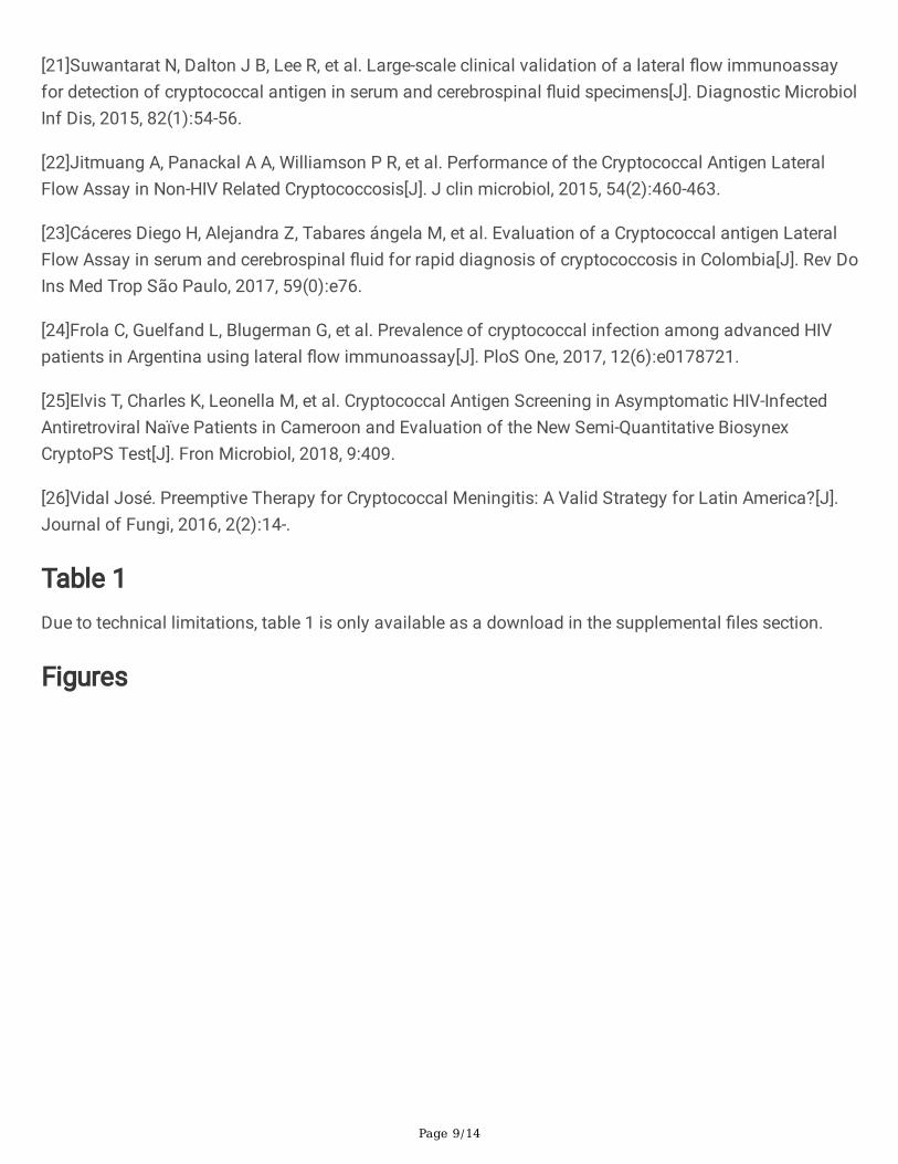
For serum specimens, the merged sensitivity and speci�city values were 0.98 (95% CI: 0.96–0.99) and0.98 (95% CI: 0.97–0.98), respectively. The average PLR of LFA in the serum was 45.05 (95% CI: 26.22–77.40) and the NLR was 0.04 (95% CI: 0.02–0.10). The merged DOR was 1574.65 (95% CI: 730.16–3395.87) and AUC was 0.9766. The results are shown in Figs. 3, 5A.
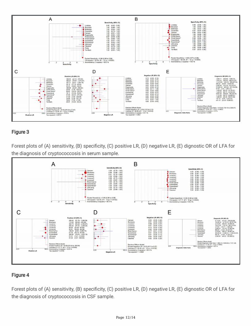
For CSF specimens, the merged sensitivity and speci�city values were 0.99 (95% CI: 0.98-0.99) and 0.99(95% CI: 0.99 to 0.99), respectively. The average PLR of LFA in CSF was 93.89 (95% CI: 53.54–164.64)and the NLR was 0.03 (95% CI: 0.01–0.07). The merged DOR was 3864.72 (95% CI: 1308.89–11411.28)and AUC was 0.9983. The results are shown in Figs. 4, 5B.
For other samples, the results of sensitivity and speci�city are shown in Figure 6.
Publication bias
In this meta-analysis, the data of serum and CSF samples were tested by Stata 12.0 for publication bias.Deek’s funnel plot asymmetry test was used to assess the potential published bias in the includedstudies. The results of serum and CSF samples indicated that there was no obvious publication bias (Fig.7).
DiscussionStudies have shown that cryptococcosis is a disease with a relatively high mortality rate. In low- andmiddle-income countries, especially in sub-Saharan Africa, the mortality rate is between 26% and 63%[26].Additionally, deaths related to cryptococcal meningitis can still reach hundreds of thousands every year[8].Therefore, a rapid diagnosis of cryptococcal infection is necessary for patients presenting appropriateclinical symptoms. A comprehensive search with stringent screening criteria retrieved 15 articles eligiblefor inclusion in the study. These 15 articles encompassed 3901 serum samples, 4403 CSF samples, 1125urine samples, 1163 venous whole blood samples, and 1163 �ngerprick capillary blood samples.Moreover, the brand of LFA-tests in the included studies were IMMY, which indicated that the data weextracted wound not cause great heterogeneity because of manufacturers of different brands.
The results in meta-analysis showed that the combined sensitivity of LFA in serum and CSF was 0.98(0.96–0.99) and 0.99 (0.98-0.99); speci�city was 0.98 (0.97–0.98) and 0.99 (0.99-0.99); DOR was1574.65 (730.16–3395.87) and 2509.29 (184.18–34187.48); SROC AUC was 0.9962 and 0.9983,respectively.
Page 6/14
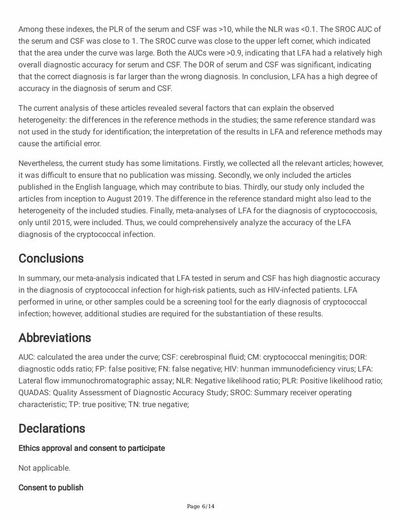
Among these indexes, the PLR of the serum and CSF was >10, while the NLR was <0.1. The SROC AUC ofthe serum and CSF was close to 1. The SROC curve was close to the upper left corner, which indicatedthat the area under the curve was large. Both the AUCs were >0.9, indicating that LFA had a relatively highoverall diagnostic accuracy for serum and CSF. The DOR of serum and CSF was signi�cant, indicatingthat the correct diagnosis is far larger than the wrong diagnosis. In conclusion, LFA has a high degree ofaccuracy in the diagnosis of serum and CSF.
The current analysis of these articles revealed several factors that can explain the observedheterogeneity: the differences in the reference methods in the studies; the same reference standard wasnot used in the study for identi�cation; the interpretation of the results in LFA and reference methods maycause the arti�cial error.
Nevertheless, the current study has some limitations. Firstly, we collected all the relevant articles; however,it was di�cult to ensure that no publication was missing. Secondly, we only included the articlespublished in the English language, which may contribute to bias. Thirdly, our study only included thearticles from inception to August 2019. The difference in the reference standard might also lead to theheterogeneity of the included studies. Finally, meta-analyses of LFA for the diagnosis of cryptococcosis,only until 2015, were included. Thus, we could comprehensively analyze the accuracy of the LFAdiagnosis of the cryptococcal infection.
ConclusionsIn summary, our meta-analysis indicated that LFA tested in serum and CSF has high diagnostic accuracyin the diagnosis of cryptococcal infection for high-risk patients, such as HIV-infected patients. LFAperformed in urine, or other samples could be a screening tool for the early diagnosis of cryptococcalinfection; however, additional studies are required for the substantiation of these results.
AbbreviationsAUC: calculated the area under the curve; CSF: cerebrospinal �uid; CM: cryptococcal meningitis; DOR:diagnostic odds ratio; FP: false positive; FN: false negative; HIV: hunman immunode�ciency virus; LFA:Lateral �ow immunochromatographic assay; NLR: Negative likelihood ratio; PLR: Positive likelihood ratio;QUADAS: Quality Assessment of Diagnostic Accuracy Study; SROC: Summary receiver operatingcharacteristic; TP: true positive; TN: true negative;
DeclarationsEthics approval and consent to participate
Not applicable.
Consent to publish
Page 7/14
Not applicable.
Availability of data and materials
All data generated or analyzed during this study are included in this published article.
Competing interests
The authors declare that they have no competing interests.
Funding
No funding was used to support this study.
Authors’ Contributions
XG conceived and designed the experiments. LX, GL, HD and Y-XL analyzed the data and made thetables. LX, Y-LL and JQ contributed to the production of �gures by the analysis tools. All authorsparticipated in the writing, reading, and revising of the manuscript and approved the �nal version of themanuscript.
Acknowledgements
Not applicable.
References[1] Idnurm A, Lin X. Rising to the challenge of multiple Cryptococcus species and the diseases theycause. Fungal Genet Biol. 2015;78:1-6.
[2]Springer DJ, Chaturvedi V. Projecting Global Occurrence of Cryptococcus gattii. Emer Inf Dis. 2010, 16:14-20.
[3]Gibson J F, Johnston S A. Immunity to Cryptococcus neoformansand C.gattii during cryptococcosis[J].Fungal Genet Biol, 2015, 78: 76-86.
[4]Xu J, Eastman A J, Flaczyk A, et al. Disruption of early tumor necrosis dactor alpha signaling preventsclassic alactivation of dendritic cells in lung-associated lymph nodes and development of protectiveimmunity against cryptococcal infection [J]. MBio, 2016, 7 (4): e00510-00516.
[5]Brown G D, Denning D W, Levitz S M. Tackling human fungal infections[J]. Sci, 2012, 336(6082):647.
[6]Rajasingham R, Smith RM, Park BJ, et al. Global burden of disease of HIV-associated cryptococcalmeningitis: an updated analysis. Lanc Infect Dis. 2017;17(8):873‐881.
[7]Maziarz EK, Perfect JR. Cryptococcosis. Inf Dis Clin North Am. 2016; 30(1): 179‐206.

Page 8/14
[8]Kozel T R, Bauman S K. CrAg lateral �ow assay for cryptococcosis[J]. Expert opinion on medicaldiagnostics, 2012, 6(3): 245-251.
[9]Drain P K, Hong T, Krows M, et al. Validation of clinic-based cryptococcal antigen lateral �ow assayscreening in HIV-infected adults in South Africa[J]. Sci. Rep, 2019, 9(1).
[10]Temfack E, Kouanfack C, Mossiang L, et al. Cryptococcal Antigen Screening in Asymptomatic HIV-Infected Antiretroviral Naïve Patients in Cameroon and Evaluation of the New Semi-Quantitative BiosynexCryptoPS Test. Fron Micr. 2018;9:409.
[11]Mcmullan B J, Catriona H , Sorrell T C, et al. Clinical Utility of the Cryptococcal Antigen Lateral FlowAssay in a Diagnostic Mycology Laboratory[J]. PLoS ONE, 2012, 7(11):e49541.
[12]Lan W, Yong Z, Xian-tao Z. The QUADAS-2 Tool for the Quality Assessment of Diagnostic AccuracyStudy:an Introduction[J]. J Hub Uni Med, 2013, 32(03):201-208.
[13]Lindsley M D, Mekha N, Baggett H C, et al. Evaluation of a Newly Developed Lateral FlowImmunoassay for the Diagnosis of Cryptococcosis[J]. Clin Inf Dis, 2011, 53(4):321-325.
[14]Binnicker M J, Jespersen D J, Bestrom J E, Rollins LO. Comparison of Four Assays for the Detection ofCryptococcal Antigen[J]. Clin Vac Immunol, 2012, 19(12): 1988-1990.
[15]Escandón, Patricia, Lizarazo J , Agudelo C I , et al. Evaluation of a rapid lateral �ow immunoassay forthe detection of cryptococcal antigen for the early diagnosis of cryptococcosis in HIV patients inColombia[J]. Med Mycol, 2013, 51(7): 765-768.
[16]Hansen J, Slechta E S, Gates-Hollingsworth M A, et al. Large-Scale Evaluation of the Immuno-Mycologics Lateral Flow and Enzyme-Linked Immunoassays for Detection of Cryptococcal Antigen inSerum and Cerebrospinal Fluid[J]. Clin Vac Immunol, 2013, 20(1):52-55.
[17]Rugemalila J, Maro V P, Kapanda G, et al. Cryptococcal antigen prevalence in HIV-infectedTanzanians: a cross-sectional study and evaluation of a point-of-care lateral �ow assay[J]. Trop Med IntHealth, 2013, 18(9): 1075-1079.
[18]Boulware D R, Rolfes M A, Rajasingham R, et al. Multisite Validation of Cryptococcal Antigen LateralFlow Assay and Quanti�cation by Laser Thermal Contrast[J]. Emerg Inf Dis, 2014, 20(1):45-53.
[19]Lourens A, Jarvis J N, Meintjes G, et al. Rapid Diagnosis of Cryptococcal Meningitis by Use of LateralFlow Assay on Cerebrospinal Fluid Samples: In�uence of the High-Dose "Hook" Effect[J]. J. Clin Microbiol,2014, 52(12): 4172-4175.
[20]Diane Rivet-Dañon, Guitard J, Grenouillet, Frédéric, et al. Rapid diagnosis of cryptococcosis using anantigen detection immunochromatographic test[J]. J Inf, 2015, 70(5):499-503.
Page 9/14
[21]Suwantarat N, Dalton J B, Lee R, et al. Large-scale clinical validation of a lateral �ow immunoassayfor detection of cryptococcal antigen in serum and cerebrospinal �uid specimens[J]. Diagnostic MicrobiolInf Dis, 2015, 82(1):54-56.
[22]Jitmuang A, Panackal A A, Williamson P R, et al. Performance of the Cryptococcal Antigen LateralFlow Assay in Non-HIV Related Cryptococcosis[J]. J clin microbiol, 2015, 54(2):460-463.
[23]Cáceres Diego H, Alejandra Z, Tabares ángela M, et al. Evaluation of a Cryptococcal antigen LateralFlow Assay in serum and cerebrospinal �uid for rapid diagnosis of cryptococcosis in Colombia[J]. Rev DoIns Med Trop São Paulo, 2017, 59(0):e76.
[24]Frola C, Guelfand L, Blugerman G, et al. Prevalence of cryptococcal infection among advanced HIVpatients in Argentina using lateral �ow immunoassay[J]. PloS One, 2017, 12(6):e0178721.
[25]Elvis T, Charles K, Leonella M, et al. Cryptococcal Antigen Screening in Asymptomatic HIV-InfectedAntiretroviral Naïve Patients in Cameroon and Evaluation of the New Semi-Quantitative BiosynexCryptoPS Test[J]. Fron Microbiol, 2018, 9:409.
[26]Vidal José. Preemptive Therapy for Cryptococcal Meningitis: A Valid Strategy for Latin America?[J].Journal of Fungi, 2016, 2(2):14-.
Table 1Due to technical limitations, table 1 is only available as a download in the supplemental �les section.
Figures

Page 10/14
Figure 1
Flow diagram of study identi�cation and inclusion.

Page 11/14
Figure 2
Quality evaluation of the included studies.
Page 12/14
Figure 3
Forest plots of (A) sensitivity, (B) speci�city, (C) positive LR, (D) negative LR, (E) dignostic OR of LFA forthe diagnosis of cryptococcosis in serum sample.
Figure 4
Forest plots of (A) sensitivity, (B) speci�city, (C) positive LR, (D) negative LR, (E) dignostic OR of LFA forthe diagnosis of cryptococcosis in CSF sample.

Page 13/14
Figure 5
Forest plots of SROC curve of the sample in (A) serum sample and (B) CSF sample.
Figure 6
Forest plots of the sensitivity and speci�city of LFA for the diagnosis of cryptococcal infection in urineand blood.

Page 14/14
Figure 7
Deeks’ funnel plot asymmetry test to assess publication bias in estimates of diagnostic odds ratio forLFA detection of cryptococcal infections.
Supplementary Files
This is a list of supplementary �les associated with this preprint. Click to download.
Table1.pdf