Aspen: Ecology and management in the western … · ground, release basidiospores that infect the...
20
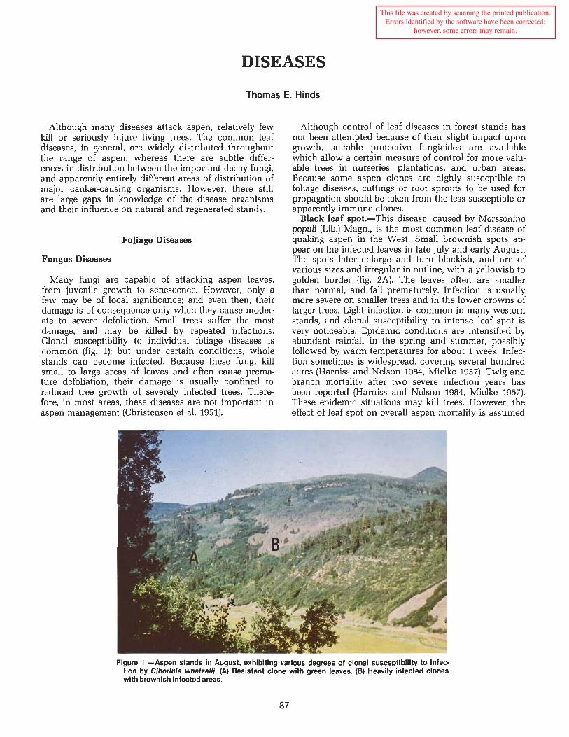
DISEASES Thomas E. Hinds Although many diseases attack aspen, relatively few kill or seriously injure living trees. The common leaf diseases, in general, are widely distributed throughout the range of aspen, whereas there are subtle differ- ences in distribution between the important decay fungi, and apparently entirely different areas of distribution of major cankercausing organisms. However, there still are large gaps in knowledge of the disease organisms and their influence on natural and regenerated stands. Foliage Diseases Fungus Diseases Many fungi are capable of attacking aspen leaves, from juvenile growth to senescence. However, only a few may be of local significance; and even then, their damage is of consequence only when they cause moder- ate to severe defoliation. Small trees suffer the most damage, and may be killed by repeated infections. Clonal susceptibility to individual foliage diseases is common (fig. 1); but under certain conditions, whole stands can become infected. Because these fungi kill small to large areas of leaves and often cause prema- ture defoliation, their damage is usually confined to reduced tree growth of severely infected trees. There- fore, in most areas, these diseases are not important in aspen management (Christensen et al. 1951). Although control of leaf diseases in forest stands has not been attempted because of their slight impact upon growth, suitable protective fungicides are available which allow a certain measure of control for more valu- able trees in nurseries, plantations, and urban areas. Because some aspen clones are highly susceptible to foliage diseases, cuttings or root sprouts to be used for propagation should be taken from the less susceptible or apparently immune clones. Black leaf spot.-This disease, caused by Marssonina populi (Lib.) Magn., is the most common leaf disease of quaking aspen in the West. Small brownish spots ap- pear on the infected leaves in late July and early August. The spots later enlarge and turn blackish, and are of various sizes and irregular in outline, with a yellowish to golden border (fig. 2A). The leaves often are smaller than normal, and fall prematurely. Infection is usually more severe on smaller trees and in the lower crowns of larger trees. Light infection is common in many western stands, and clonal susceptibility to intense leaf spot is very noticeable. Epidemic conditions are intensified by abundant rainfall in the spring and summer, possibly followed by warm temperatures for about 1 week. Infec- tion sometimes is widespread, covering several hundred acres (Harniss and Nelson 1984, Mielke 1957). Twig and branch mortality after two severe infection years has been reported (Harniss and Nelson 1984, Mielke 1957). These epidemic situations may kill trees. However, the effect of leaf spot on overall aspen mortality is assumed Figure 1.-Aspen stands in August, exhibiting various degrees of clonal susceptibility to infec- tion by Ciborinia whetzelii. (A) Resistant clone with green leaves. (B) Heavily infected clones with brownish infectedareas. This file was created by scanning the printed publication. Errors identified by the software have been corrected; however, some errors may remain.
Transcript of Aspen: Ecology and management in the western … · ground, release basidiospores that infect the...
DISEASES
Thomas E. Hinds
Although many diseases attack aspen, relatively few kill or seriously injure living trees. The common leaf diseases, in general, are widely distributed throughout the range of aspen, whereas there are subtle differ- ences in distribution between the important decay fungi, and apparently entirely different areas of distribution of major cankercausing organisms. However, there still are large gaps in knowledge of the disease organisms and their influence on natural and regenerated stands.
Foliage Diseases
Fungus Diseases
Many fungi are capable of attacking aspen leaves, from juvenile growth to senescence. However, only a few may be of local significance; and even then, their damage is of consequence only when they cause moder- ate to severe defoliation. Small trees suffer the most damage, and may be killed by repeated infections. Clonal susceptibility to individual foliage diseases is common (fig. 1); but under certain conditions, whole stands can become infected. Because these fungi kill small to large areas of leaves and often cause prema- ture defoliation, their damage is usually confined to reduced tree growth of severely infected trees. There- fore, in most areas, these diseases are not important in aspen management (Christensen et al. 1951).
Although control of leaf diseases in forest stands has not been attempted because of their slight impact upon growth, suitable protective fungicides are available which allow a certain measure of control for more valu- able trees in nurseries, plantations, and urban areas. Because some aspen clones are highly susceptible to foliage diseases, cuttings or root sprouts to be used for propagation should be taken from the less susceptible or apparently immune clones.
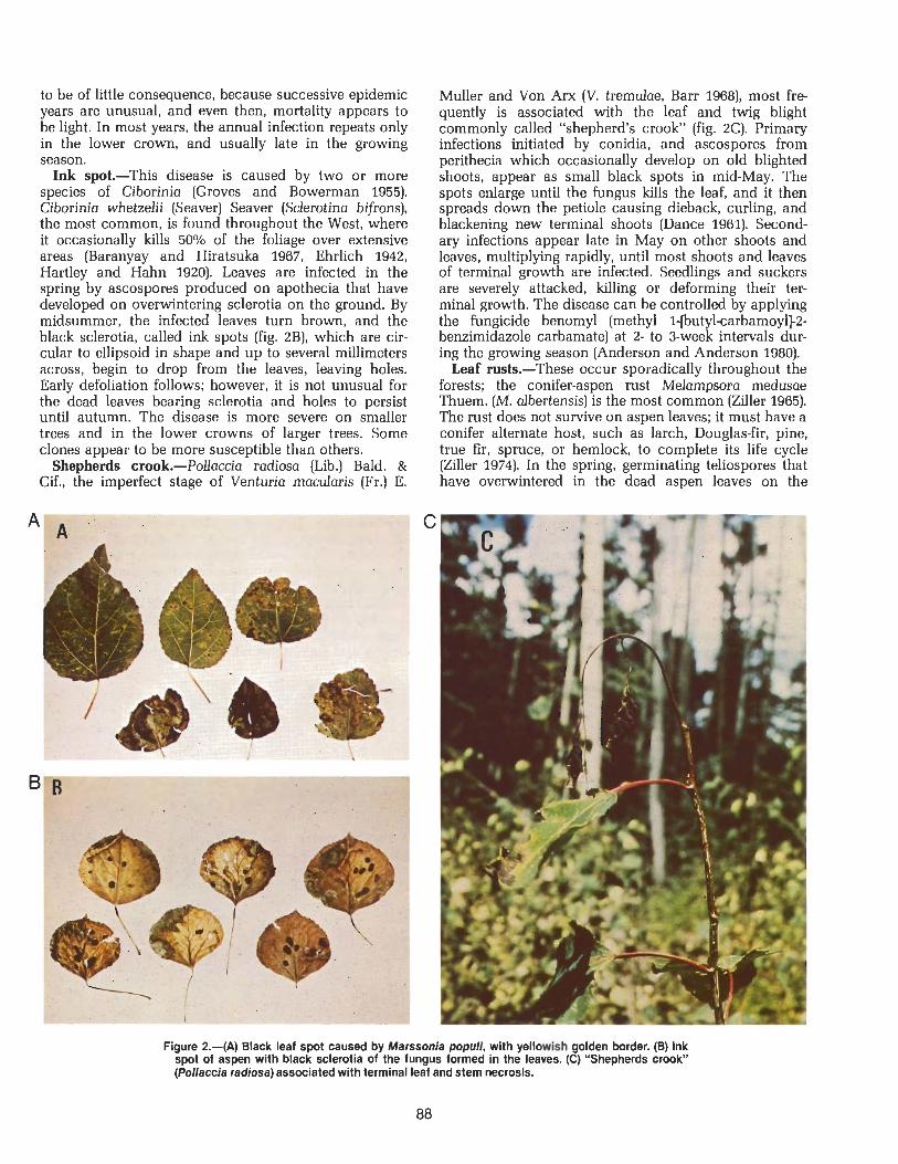
Black leaf spot.-This disease, caused by Marssonina populi (Lib.) Magn., is the most common leaf disease of quaking aspen in the West. Small brownish spots ap- pear on the infected leaves in late July and early August. The spots later enlarge and turn blackish, and are of various sizes and irregular in outline, with a yellowish to golden border (fig. 2A). The leaves often are smaller than normal, and fall prematurely. Infection is usually more severe on smaller trees and in the lower crowns of larger trees. Light infection is common in many western stands, and clonal susceptibility to intense leaf spot is very noticeable. Epidemic conditions are intensified by abundant rainfall in the spring and summer, possibly followed by warm temperatures for about 1 week. Infec- tion sometimes is widespread, covering several hundred acres (Harniss and Nelson 1984, Mielke 1957). Twig and branch mortality after two severe infection years has been reported (Harniss and Nelson 1984, Mielke 1957). These epidemic situations may kill trees. However, the effect of leaf spot on overall aspen mortality is assumed
Figure 1.-Aspen stands in August, exhibiting various degrees of clonal susceptibility to infec- tion by Ciborinia whetzelii. (A) Resistant clone with green leaves. (B) Heavily infected clones with brownish infected areas.
This file was created by scanning the printed publication.Errors identified by the software have been corrected;
however, some errors may remain.
to be of little consequence, because successive epidemic years are unusual, and even then, mortality appears to be light. In most years, the annual infection repeats only in the lower crown, and usually late in the growing season.
Ink spot.-This disease is caused by two or more species of Ciborinia (Groves and Bowerman 1955). Ciborinia whetzelii (Seaver) Seaver (Sclerotina bifrons), the most common, is found throughout the West, where it occasionally kills 50•‹/o of the foliage over extensive areas (Baranyay and Hiratsuka 1967, Ehrlich 1942, Hartley and Hahn 1920). Leaves are infected in the spring by ascospores produced on apothecia that have developed on overwintering sclerotia on the ground. By midsummer, the infected leaves turn brown, and the black sclerotia, called ink spots (fig. 2B), which are cir- cular to ellipsoid in shape and up to several millimeters across, begin to drop from the leaves, leaving holes. Early defoliation follows; however, it is not unusual for the dead leaves bearing sclerotia and holes to persist until autumn. The disease is more severe on smaller trees and in the lower crowns of larger trees. Some clones appear to be more susceptible than others.
Shepherds crook.-Pollaccia radiosa (Lib.) Bald. & Cif., the imperfect stage of Venturia macdaris (Fr.) E.
Muller and Von Am (V. tremulae, Barr 1968), most fre- quently is associated with the leaf and twig blight commonly called "shepherd's crook" (fig. 2C). Primary infections initiated by conidia, and ascospores from perithecia which occasionally develop on old blighted shoots, appear as small black spots in mid-May. The spots enlarge until the fungus kills the leaf, and it then spreads down the petiole causing dieback, curling, and blackening new terminal shoots (Dance 1961). Second- ary infections appear late in May on other shoots and leaves, multiplying rapidly, until most shoots and leaves of terminal growth are infected. Seedlings and suckers are severely attacked, killing or deforming their ter- minal growth. The disease can be controlled by applying the fungicide benomyl (methyl l-[butylcarbamoyl]-2- benzimidazole carbamate) at 2- to 3-week intervals dur- ing the growing season (Anderson and Anderson 1980).
Leaf rusts.-These occur sporadically throughout the forests; the conifer-aspen rust Melampsora medusae Thuem. (M. albertensis) is the most common (Ziller 1965). The rust does not survive on aspen leaves; it must have a conifer alternate host, such as larch, Douglas-fir, pine, true fir, spruce, or hemlock, to complete its life cycle (Ziller 1974). In the spring, germinating teliospores that have overwintered in the dead aspen leaves on the
Figure 2.-(A) Black leaf spot caused by Marssonia populi, with yellowish golden border. (B) Ink spot of aspen with black sclerotia of the fungus formed in the leaves. (C) "Shepherds crook" (Pollaccia radiosa) associated with terminal leaf and stem necrosis.

ground, release basidiospores that infect the alternate host. Winddisseminated aeciospores produced on the alternate host then infect aspen leaves in the summer, causing yellow spots and the formation of orangeyellow urediospores, which, in turn, reinfect more leaves. Late in the summer, masses of teliospores are produced on the underside of the leaf beneath the yellow spots. Premature defoliation may or may not occur (Hartley and Hahn 1920, Ziller 1974), and, although the leaf tissue dies, damage in aspen stands is not considered serious.
Powdery mildew.-This disease, caused by Erysiphe cichoracearum DC. ex Merat and Uncinula salicis (DC. ex Merat) Wint., is often found on lower leaves of small trees and sprouts after periods of high moisture. Although the fungi are widespread (Meinecke 1929, Shaw 1973, USDA 1960), they appear to act as sapro- phytes on debilitated leaves and are normally consid- ered to be of minor importance.
Roadside Salt Damage
Although foliar damage to aspen caused by winter road salting is not considered a disease in the strictest sense, it is included here, because the symptoms of chloride toxicity may appear to be caused by disease. Symptoms of salt damage to aspen along roads begin to show up in late August. By midSeptember, the leaves in the lower crowns of the larger trees appear smaller than normal, with their margins somewhat curled and discolored reddish-brown (fig. 3A). The discoloration may encompass up to two-thirds of the leaf area. Smaller trees are more affected, and their entire crowns appear reddish. Tree decline or mortality associated with aspen salt damage has not been studied.
Shortle and Rich (1970) considered quaking aspen in southeastern New Hampshire to be relatively salt- tolerant. There, uninjured roadside trees had leaves containing a chloride content (dry weight) of 0.78% in comparison to 0.12% in healthy woodlot trees. In New Mexico, Gosz found that aspen trees along a roadside showed symptoms in September, when the chloride con- tent of some trees reached a maximum of 2.g0/0 (dry weight).' However, values as low as 0.6% were found in some trees exhibiting stress symptoms. Leaves of trees alongside an unsalted road contained only 0.14% chloride. Damage differences were found between in- dividual locations and within groups of trees in a single area, which indicated a possible genetic difference in susceptibility. Various road and site characteristics in- fluence the road salt distribution into forested areas and the accumulation of chloride by leaves (Gosz,' Piatt and Krause 1974).
~Gosz, James R. 1974. Effects of road surtacing and salting on roadside vegetation in New Mexico mountain areas. 32 p. Research Agreement 16-361-CA, Eisenhower Consortium for Western Envi- ronmental Forestry Research. Rocky Mountain Forest and Range Experiment Station, Fort Collins, Colo.
Figure 3.-(A) Bronze foliage damage of roadside aspen caused by chloride toxiclty resulting from using a sand-salt mixture on the road during the winter. (B) "Droopy aspen." The symptoms of pendulous branches and lack of lateral growth existed before road construction.
Virus and Virus-like Diseases
Viruses and systemic pathogens (mycoplasms, rickett- sia, flagellates) are the least understood in aspen pathology. Because they are far more difficult to recover from trees than from herbaceous plants and the research on them is more difficult and timeconsuming than is research on ordinary pathogens, very few tree

pathologists have studied these diseases and their ef- stand age, genotype, insects and diseases, browsing, fects upon forest trees. However, because the impor- and apical dominance, all contribute to clonal deteriora- tance of virus and virus-like diseases in the intensive tion (Hibben et al. 1979) and overshadow the role of culture of hybrid aspen and other poplars is becoming viruses. more significant in plantations established for the pro- duction of wood fiber, more research on their recogni- tion and diagnosis might be expected in the future. Droopy Aspen
Boyer (1962) reported a necrotic leaf spot disease of hybrid and native aspen in Ontario, transmitted by grafting and by insects under conditions that suggested it may be caused by a virus. Although a further study did not determine the infectious agent (Boyer and Navratil 1970), it has been speculated that the disease very likely is present and widespread in the United States (Berbee et al. 1976). Navratil (1979) later observed virus and virus-like diseases of poplar in Ontario and Saskat- chewan between 1972 and 1976. The necrotic leaf spot was not found; however, he reported poplar mosaic virus (PMV) on various hybrids, and, although it was suspicious on aspen, it was not confirmed. A vein mot- tling of aspen leaves also recognized as a virus-like disorder was found in locations associated with human activities and believed to have been introduced into those areas.
Although the Canadian virus and virus-like disorders have not been reported in the United States, virus-like decline symptoms in aspen clones and in Aigeiros (cot- tonwoods) are being investigated. An apparently new poplar virus belonging to the potato virus Y group was recovered from five different Aigeiros clones in Wiscon- sin, and an isolate that may be identical, was recovered from a deteriorating aspen clone (Berbee et al. 1976).
Martin et al. (1982) isolated a virus in the potyvirus group from Popdus spp. and four declining, native aspen clones in Wisconsin. The decline symptoms in- cluded necrotic leaf spots early in the growing season, with leaf bronzing symptoms scattered throughout the crown in late July and August. Branches with bronzed leaves died the next year. The symptoms were observed throughout Wisconsin. Transmission trials established that the virus was a pathogen of poplars, including P, tremuloides. Similar leaf bronzing symptoms have been observed on aspen at Fallen Leaf Lake, south of Lake Tahoe, California.
The role of viruses in deterioration of aspen clones in the West, characterized by trees with low vigor, poor form, increased mortality, and scarce regeneration, has received some attention (Schier 1975a). Foliar symptoms of infection include chlorotic spots, line patterns, and abnormalities in size, color, and shape. Hibben et al. (1979) isolated a tobacco necrosis virus (TNV-A) from 5 of 33 clones with symptoms indicative of virus infection. Two additional isolates of TNV autigenically dissimilar to TNV-A and to each other also were recovered. The low rate of TNV recovery from the deteriorating clones was insufficient to implicate the virus as a cause of deterioration. The importance of virus or virus-like diseases in natural stands of aspen in the West is unknown. Other causal factors, such as site conditions,
"Droopy aspen" is a fairly descriptive term for the symptoms of this disorder. Affected trees are character- ized by flexuous-rubbery, pendulous branches through- out the crowns of small trees; in larger trees, the second- ary branches are symptomatic (fig. 3B). The affected branches have shortened internodes and enlarged nodes, a lack of lateral twig growth and foliage for the preceding 5 to 20 or more years, and larger than usual terminal leaves. After 20 or more years, the pendant branches die, and, depending upon the severity of infec- tion and tree size, the entire tree succumbs. Although these abnormal trees usually are seen along roadsides, in campgrounds, and as transplants in urban areas and mountain communities, single trees and small groups are found in forest areas not associated with human activities.
The symptoms do not appear to be clonal in nature. Droopy aspen have been observed in Colorado, New Mexico, and Utah, but not in Alaska or Wisconsin, suggesting that this malady may be unique to the southern Rocky Mountain region (Hinds and Laurent 1978, Livingston et al. 1979). Preliminary studies failed to reveal any virus particles or mycoplasma-like bodies associated with the symptoms; the cause or causal agents of droopy aspen remain unknown (Livingston et al. 1979).
Aspen Decay
Tree decay has long been recognized as important to aspen management (Baker 1925, Weigle and Frothing- ham 1911). Essentially, merchantable volume lost to decay increases with age; but this age factor varies be- tween the Northeast and the West.
While trees grow faster in the Great Lakes area, they also deteriorate and decay earlier. The mean annual growth of aspen stands in northern Minnesota, on aver- age sites, culminates in about 50 years, which indicates a pathological rotation of from 40 to 50 years for produc- tion of mass products (Schmitz and Jackson 1927). Volume lost to decay amounted to 4.8% at 30 years, 7.8% at 40 years, 11.4% at 50 years, and 15.7% at 60 years. To minimize losses to insects and diseases, recom- mended rotations for aspen stands there now range from about 30 years on poor sites to 50 or 60 years on good sites (Brinkman and Roe 1975).
In the Upper Pic region of Ontario, Basham (1958) found decay in 69% of the trees on 47 plots. Merchant- able volume loss was 13.1%. Trees with heart rot in the merchantable portion of the bole increased steadily from 26.7% in stands at age class 41-60 to 100% in

stand age class 161-180. Two types of stain were re- corded: a red-mottled stain, which occupied approxi- mately 2% of the total tree volume at all ages, and a brown stain, which increased from about 10% in stands 41-60 years old to more than 20% in stands older than 120 years. A later comparison between gross and net volumes per acre showed that, whereas the gross mer- chantable volume per acre was greater at 100 years, the net merchantable volume was at a maximum at 90 years (Basham 1960). The mean annual increment reached a peak value at 60 years for both gross and net volumes.
The results of a more comprehensive cull study cover- ing a larger area in Ontario showed there was a marked uniformity in the percentages of the total merchantable volume defective at similar age classes in the two studies, although there was a lower rotlstain ratio in the Upper Pic sample. Variations in the extent of decay on four sites, based mainly on the availability of soil moisture, were not pronounced, although stands on deep, sandy silts or loams, or on shallow, sandy loams over impervious material, generally were less defective than stands on drier or wetter sites. Similar conclusions were made earlier regarding aspen stands in Wisconsin and Minnesota (Stoeckeler 1948).
In Alberta, slightly more decay is present on wet than dry sites because of the increased activity of Fomes ig- niarius on the wet areas (Thomas et al. 1960). During a study of 835 living aspen (Thomas et al. 19601, an overall volume loss of 25% was found in 73% of the trees with decay. Butt infections accounted for 31.5% of the infec- tions (10.8% of the rot volume), and trunk infections for 68.5% (89.2% of the rot volume).
In the West, Baker (1925) recommended a patholog- ical rotation age of about 110 years for aspen growing on the better sites in central Utah, based on the net maximum volume, Meinecke (1929), however, from the same study data, recommended a rotation age of about 80-90 years, on the basis of net volume production and net increment. In Meinecke's study, decay accounted for 6% of the gross volume in the age class 61-70, 18% in the class 101-110 age and from 10% to 41% in the older age classes. Decay amounted to 18% of the gross mer- chantable volume.
In a broader study of decay in typical commercial aspen forests, Davidson et al. (1959) found decay in 53% of the trees (8.4% of the gross volume) dissected on 35 plots, in five national forests, in Colorado. Although there was little relationship between decay and site class for the younger stands, the differences were marked in stands more than 100 years old. In 100-year- old stands, cubic foot decay averaged 4% on site 1 (the better site), 8% on site 2, and 13% on site 3. The in- cidence of decay was lower than that reported by Meinecke (1929) for Utah. Decay volumes in the older age classes varied from 7% to 27%.
The merchantability of aspen on a board foot basis was analyzed later from the Colorado study (Hinds and Wengert 1977). Incidence of decay and cull, based on Baker's (1925) site quality classes, plotted as a function of 10-year age classes, showed linear relationships.
Tree infection increased with age, and the percentage of cull at tree age 100 amounted to 21% and 25% on sites 1 and 2, respectively. The variation of cull in trees on site 3 was too large to obtain a significant relationship. It was concluded that, before decay data can be applied to stands, the age distribution of the merchantable trees must be known.
Aspen is extremely susceptible to attacks by fungi; however, most wooddestroying fungi are only capable of infecting a wound to the wood. Because it is often dif- ficult to determine the exact mode of entrance of a fungus causing heart rot, the association of external in- dicators with decay frequently is based upon general observations. Although some are reasonably accurate, definite figures for the frequency of infection often are questionable.
As early as the 1920s, infection was associated with fire scars, branch scars, insect injuries, and grazing (Hofer 1920, Schmitz and Jackson 1927). Basham (1958) suggested that most of the fungi responsible for butt rots probably enter through roots, and that only a minority originate from basal wounds, such as fire scars, frost cracks, and branch stubs. Approximately 90% of the trunk rots in his study were traced to dead, broken branch stubs; a few entered in forked crowns, frost cracks, and mechanical injuries. Extensive heart rot was associated with 84% of the pronounced trunk wounds, indicating that they were fairly reliable in- dicators of heart rot. Basham (1958) also suggested that "preliminary fungi," not generally associated with ad- vanced decay, invade and colonize the heartwood before the "principal fungi" causing advanced decay become established.
Meinecke (1929) analyzed 255 open and closed wounds and found 126 decay infections. Incidence of in- fection was fire scars, 88%; bruises, 33%; dead and broken tops, 19%; ingrown stubs, 60%; frost cracks, 17%; and undetermined wounds, 20%.
Etheridge (1961) studied the cause of infection in liv- ing and dead branches of aspen to obtain information regarding the time and conditions under which dead branches might serve as entrance points for heart rot fungi. He found that a higher incidence of branch in- fections were on wet sites; young branches were more prone to infection than old branches; and there were at least three successive stages of infection by different organisms before heart rot fungi became established (8 to 1 2 years after branch mortality). Because F. igniarius appeared only rarely in the succession, and then as lateral extensions of heartwood infections after 19 years, it was suggested that bark wounding constituted its main avenue of infection into the heartwood of aspen.
The most reliable external indication of decay in aspen is the appearance of Phellinus tremulae (Fomes igniarius) fruiting bodies (fig. 4A), often called sporo- phores or conks, which usually project from branch stubs or old wounds. Basham (1958) found conks on 86% of the infected trees. Hinds and Wengert (1977) reported 75% of the merchantable size trees with scalable cull attributed to the fungus had these external indicators of decay. Cull averaged 82% of the gross tree volume when

conks were present, whereas infected trees without conks averaged 40•‹h cull. The extent of decay as in- dicated by the presence of conks has been reported for Ontario (Riley and Bier 1936), Minnesota (Horton and Hendee 1934), and Colorado, where the average length of decay above and below the highest and lowest conk was 12.0 f 0.7 feet (3.7 m + 21 cm) (Hinds 1963). A system for predicting the amount of P. tremulae trunk rot in 45- to 50-year-old stands in the Lake States has been developed. Aspen stands older than about 40 years there are subject to breakup because of the decay (Anderson and Schipper 1978). This early stand breakup has not been reported in the West.
Cull resulting from decay varies greatly in unman- aged aspen stands in the West. The tree age difference in many uneven-aged stands accounts for much of the cull variation. Decay is usually more prevalent in the older trees; the greater the proportion is of older trees in a stand, the greater are the decay losses.
The fungi causing cull in the older stands are likely to be found in the younger stands, also. However, their im- pact on volume losses should not be as great in the regenerated stands, and their relative importance may change when even-aged stands become more prevalent.
Decay Fungi
More than 250 species of wooddecaying basidio- mycetes have been recorded on aspen in North America (Lindsey and Gilbertson 1978). However, only about 25 species are considered important in the decay of dead
standing or fallen trees, and a dozen or more in the decay in living trees. Much of the following information was derived from the only broadly based, quantitative decay study of live aspen in the West (Davidson et al. 1959).
Trunk Rots
Since 1909, Phellinus tremulae (Bond.) Bond. et Borris (Fomes igniarius var. popdinus] has been recognized as the predominant aspen trunk rot fungus in North America (Schrenk and Spaulding 1909). Although the decay is usually considered a white trunk rot or white heart rot (fig. 4B), it frequently occurs in the basal por- tion of a tree but seldom, if ever, in the root system (Schmitz and Jackson 1927, Ross 1976a). The false tinder fungus is essentially a wound parasite; infection takes place through wounds to the sapwood and heartwood (Etheridge 1961, Manion and French 1968, Riley 1952). Numerous other fungi are associated with the decay, and many are assumed to be precursors of P. tremulae (Good and Nelson 1962, Shigo 1963).
In the West, P. tremulae is also the major cause of volume loss. Meinecke (1929) considered it the most im- portant individual factor causing the 18% decay cull in his Utah study; however, he did not give specific infor- mation on the decay fungi. Although the incidence of trunk infection by Peniophora polygonia (Pers. ex Fr.) Bourd. et Galz. was greater (28%) than that of Phellinus tremulae (26%) in a Colorado decay study (Davidson et al. 1959), P. tremulae was responsible for 59.1% of the
Figure 4.-(A) Conks of the false tinder fungus on the trunk indicate extensive trunk rot. (B) Cross section of a live 7-inch (17-cm) diameter aspen with Phellinus tremulae trunk rot.
92
cubic foot decay volume, compared to 9.6% for Penie phora polygonia. On a linear volume basis, P. tremulae loss amounted to 10.2% of the gross volume (33.8% of the rot volume) and was found in 15% of the trees larger than 8 inches (20 cm) d.b.h. (Hinds and Wengert 1977). Trees infected with the fungus had an average of 70% cull.
There is no apparent relationship between site and cubic foot volume of P. tremulae decay, although the in- cidence and amount of decay increases with stand age (Riley 1952) and may vary among clones (Wall 1969). In one area of Ontario, 2889% of the trees in stands 60-70 years old were infected (Riley 19521, whereas 42% of the trees 41-180 years old in the Upper Pic region were infected (Basham 1958). Decay attributed to this fungus has amounted to 75% of the decay volume in Ontario (Basham 1958) and 35% in Alberta (Thomas et al. 1960), in contrast to 59% in Colorado. Total cubic foot volume losses attributed to P, tremulae in stands 41-180 years old range from 6.4% in Ontario to 3.6% in Colorado.
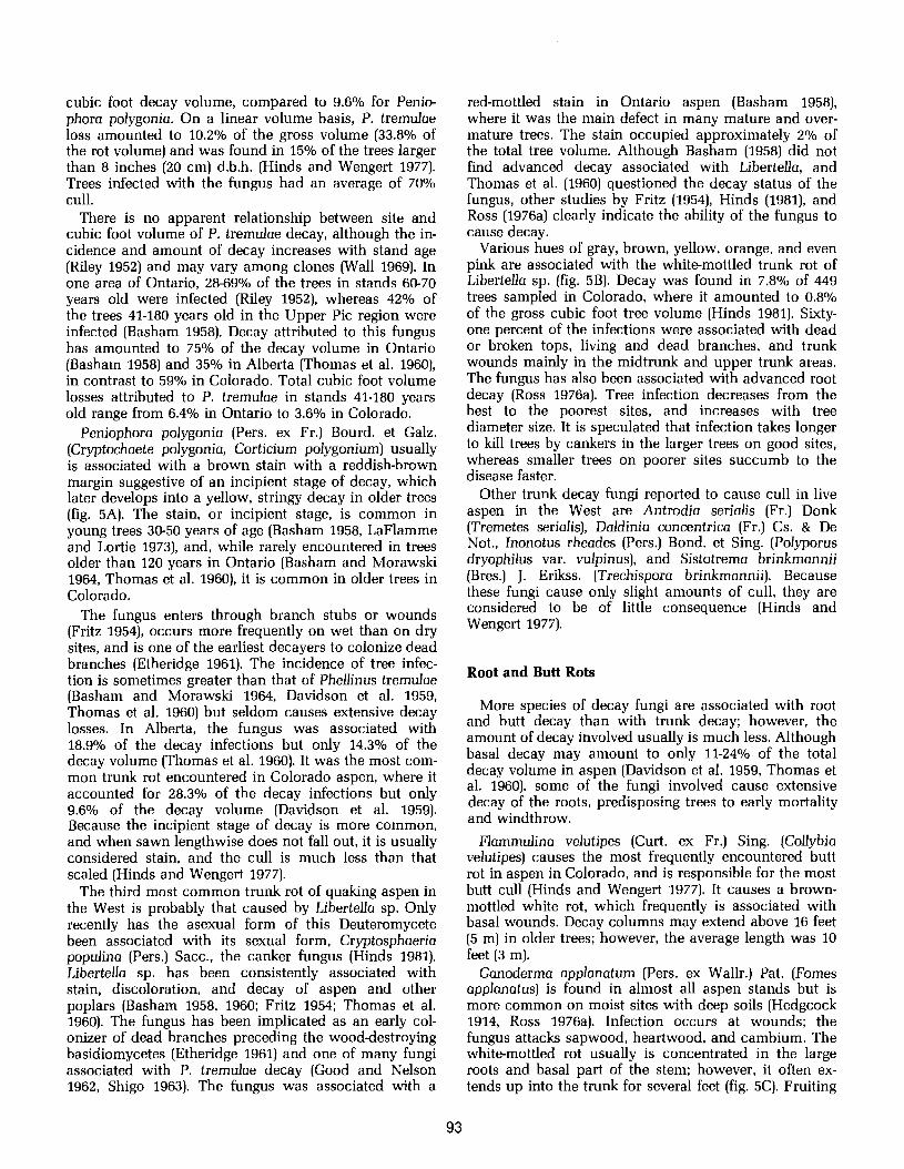
Peniophora polygonia (Pers. ex Fr.) Bourd. et Galz. (Cryptochaete polygonia, Corticium polygonium) usually is associated with a brown stain with a reddish-brown margin suggestive of an incipient stage of decay, which later develops into a yellow, stringy decay in older trees (fig. 5A). The stain, or incipient stage, is common in young trees 30-50 years of age (Basham 1958, LaFlamme and Lortie 19731, and, while rarely encountered in trees older than 120 years in Ontario (Basham and Morawski 1964, Thomas et al. 1960), it is common in older trees in Colorado.
The fungus enters through branch stubs or wounds (Fritz 1954), occurs more frequently on wet than on dry sites, and is one of the earliest decayers to colonize dead branches (Etheridge 1961). The incidence of tree infec- tion is sometimes greater than that of Phellinus tremulae (Basham and Morawski 1964, Davidson et al. 1959, Thomas et al. 1960) but seldom causes extensive decay losses. In Alberta, the fungus was associated with 18.9% of the decay infections but only 14.3% of the decay volume (Thomas et al. 1960). It was the most com- mon trunk rot encountered in Colorado aspen, where it accounted for 28.3% of the decay infections but only 9.6% of the decay volume (Davidson et al. 1959). Because the incipient stage of decay is more common, and when sawn lengthwise does not fall out, it is usually considered stain, and the cull is much less than that scaled (Hinds and Wengert 1977).
The third most common trunk rot of quaking aspen in the West is probably that caused by Libertella sp. Only recently has the asexual form of this Deuteromycete been associated with its sexual form, Cryptosphaeria populina (Pers.) Sacc., the canker fungus (Hinds 1981). Libertella sp. has been consistently associated with stain, discoloration, and decay of aspen and other poplars (Basham 1958, 1960; Fritz 1954; Thomas et al. 1960). The fungus has been implicated as an early col- onizer of dead branches preceding the wooddestroying basidiomycetes (Etheridge 1961) and one of many fungi associated with P. tremulae decay (Good and Nelson 1962, Shigo 1963). The fungus was associated with a
red-mottled stain in Ontario aspen (Basham 1958), where it was the main defect in many mature and over- mature trees. The stain occupied approximately 2% of the total tree volume. Although Basham (1958) did not find advanced decay associated with Libertella, and Thomas et al. (1960) questioned the decay status of the fungus, other studies by Fritz (19541, Hinds (19811, and Ross (1976a) clearly indicate the ability of the fungus to cause decay.
Various hues of gray, brown, yellow, orange, and even pink are associated with the whitemottled trunk rot of Libertella sp. (fig. 5B). Decay was found in 7.8% of 449 trees sampled in Colorado, where it amounted to 0.8% of the gross cubic foot tree volume (Hinds 1981). Sixty- one percent of the infections were associated with dead or broken tops, living and dead branches, and trunk wounds mainly in the midtrunk and upper trunk areas. The fungus has also been associated with advanced root decay (Ross 1976a). Tree infection decreases from the best to the poorest sites, and increases with tree diameter size. It is speculated that infection takes longer to kill trees by cankers in the larger trees on good sites, whereas smaller trees on poorer sites succumb to the disease faster.
Other trunk decay fungi reported to cause cull in live aspen in the West are Antrodia serialis (Fr.) Donk (Tremetes serialis), Daldinia concentrica (Fr.) Cs. & De Not., Inonotus rheades (Pers.) Bond. et Sing. (Polyporus dryophilus var. vulpinus), and Sistotrema brinkmannii (Bres.) J. Erikss. (Trechispora brinkmannii). Because these fungi cause only slight amounts of cull, they are considered to be of little consequence (Hinds and Wengert 1977).
Root and Butt Rots
More species of decay fungi are associated with root and butt decay than with trunk decay; however, the amount of decay involved usually is much less. Although basal decay may amount to only 11-24% of the total decay volume in aspen (Davidson et al. 1959, Thomas et al. 19601, some of the fungi involved cause extensive decay of the roots, predisposing trees to early mortality and windthrow.
Flammulina velutipes (Curt. ex Fr.) Sing. (Collybia velutipes) causes the most frequently encountered butt rot in aspen in Colorado, and is responsible for the most butt cull (Hinds and Wengert 1977). It causes a brown- mottled white rot, which frequently is associated with basal wounds. Decay columns may extend above 16 feet (5 m) in older trees; however, the average length was 10 feet (3 m).
Ganoderma applanatum (Pers. ex Wallr.) Pat. (Fomes applmatus) is found in almost all aspen stands but is more common on moist sites with deep soils (Hedgcock 1914, Ross 1976a). Infection occurs at wounds; the fungus attacks sapwood, heartwood, and cambium. The whitemottled rot usually is concentrated in the large roots and basal part of the stem; however, it often ex- tends up into the trunk for several feet (fig. 5C). Fruiting
Figure 5.-(A) Peniophora polygonia trunk rot in cross section. (B) Libertella discoloration and decay associated with large branch stub (scale is cm). (C) Ganoderma applanatum causing extensive butt rot with the artist conk fruiting at the base of the live tree (scale is 1 foot (30 cm)). (D) Windthrown aspen with broken roots decayed by G. applanatum.
bodies of the fungus, frequently found at the base of an infected tree, indicate extensive butt rot. Root rot is restricted to roots larger than 2.5 inches (6 cm) in diameter, indicating that only large roots might act as avenues of spread to new hosts (Ross 1976b). Because of this, rot centers may occur more frequently on good sites, because large roots there are further from in- fected trees.
Ganoderma applanatum eventually rots entire cross sections of larger roots, and windthrow is common in
mature aspen stands in the Rocky Mountains (fig. 5D). Trees in infection centers on good sites often blow down in groups, whereas single, isolated trees go down on medium and poor sites (Ross 1976a). Although the loss caused by decay may be small-it amounted to 6.3% of the cubic foot decay volume in Colorado (Davidson et al. 1959)-windthrow losses may be considerably greater. Windthrow in overstory aspen 10@120 years old, caused by a windstorm in the San Juan Mountains of southwestern Colorado, resulted in a loss of 2.3O/0 of the

bolewood biomass of the stand (Landis and Evans 1974). Other fungi that have been associated with root Sporophores of the fungus were found on 86% of the diseases of aspen in Wyoming include an Ascocoryne downed trees that were larger than 6 inches (15 cm) sp., Phidophora sp., Tdnrornyces verrniculatus (Dang.) d.b.h., but were only on 5.2% of the remaining standing C. R. Benjamin, and Satorya fumigata Vuill. (Ross trees, indicating a relationship between windthrow and 1976a). occurrence of the fungus.
Stain or Discoloration While of secondary importance, Pholiota squarrosa
(Fr.) Kumm., leur rot us ostreatus Fr., and ~istotrerna raduloides (Karst.) Donk. (Trechispora raduloides) cause basal white rots that often extend into the larger roots (Davidson et al. 1959). Although Pholiota squarrosa ap- pears to be more common, the amount of decay they all cause is about equal, and none appear to be as parasitic as G. applanatum. Other white rot fungi associated with minor amounts of butt rot in living trees include Arrnilla- riella rnellea (Vahl ex Fr.) Karst. (Armillaria rnellea), Bjerkandera adusta (Willd. ex Fr.) Karst. (Polyporus adustus), Hirschioporus pargamenus (Fr.) Bond. et Sing. (Polyporus pargamenus), Radulodon arnericanus Ryv. (Radulum caseariurn), and Pleurotus elongatipes Pk. (Tricholoma unifacturn). One or more species of Con- iophora are associated with brown butt rots, which are fairly common, and Coprinus atramentarius (Fr.) Fr. with brown cubical root and butt rot (Ross 1976a). Sporophores of the various root and butt fungi, although not numerous, often are found at the base or on the ground at the base of an infected tree, indicating butt rot.
kmillariella rnellea, the "shoe-string" or "honey mushroom" fungus, is one of the most consistently reported root and butt decays of aspen in North America. Although numerous infections are usually associated with only minor amounts of decay (Davidson et al. 1959, LaFlamme and Lortie 1973, Ross 1976a, Thomas et al. 1960), Basham (1958) considered it one of the two principal causes of butt rot in northern Ontario aspen. The fungus is widespread on many species of forest trees, usually as a saprophyte; but it is capable of killing trees of subnormal vigor by destroying the roots. Diseased trees may appear in groups that increase in size as more trees are attacked, or as individuals scat- tered throughout a stand. Its effect in aspen stands has not been studied; but observations indicate that its ef- fects may be similar. Ives et al. (1974) reported that ap- proximately 50% of the mature aspen at the campsite in Crimson Lake Provincial Park, in the Prairies Region of Canada, had been killed by the fungus; and Hinds and Laurent (1978) noted that A, mellea and insect borers were associated with the extensive mortality of saplings covering several acres on a poor site, at the Bonanza Creek Experimental Forest, in interior Alaska. Observa- tions by Hinds in the southern Rocky Mountains indicate that the fact that some stand openings have no repro- duction may be attributed to this root disease. Large dead and live trees surrounding such openings are in- fected with A. rnellea. As the disease spreads outward, somewhat in a circular manner, the root systems are killed; sprouts are not formed or are too weak to grow; and peripheral trees eventually die or are windthrown.
Estimates of the amount of stain, or discoloration, in quaking aspen in the West are not available; however, studies in Ontario indicate it could be considerable, and of more importance in regenerated stands than present- ly acknowledged (Basham and Navratil 1975). In On- tario, 76% of the stems on a 5-yearald cutover were af- fected by a light-todark brown stain (Smith 1973); and the incidence of stain in 23-year-ld cutover stands amounted to 84% for a defect of 1.4% (Kemperman et al. 1976). The volume of stain within the merchantable portion of trees in a 41- to 6Gyear age class, in uncut stands in the same general area, was earlier found to amount to 12.6% of the volume, and increased to 24.6% in trees in a 161- to 18Gyear age class (Basham 1958).
Stain discolorations include hues of black, brown, red, yellow, and green in both heartwood and sapwood. Because stain normally affects lumber quality rather than quantity, cull usually is not deducted when the stain is firm and light in color .2 Many hymenomycetes (decay fungi), ascomycetes, fungi imperfecti, bacteria, and veast are associated with the various discolorations ash am 1958, Etheridge 1961, Good and Nelson 1962, Kemperman et al. 1976, LaFlamme and Lortie 1973, Shigo 1963); yet, trees can discolor at wounds even without microorganisms (Sucoff et al. 1967).
The role of microorganisms in discolored aspen is not completely understood. A succession of organisms takes place in the discoloration and decay in living trees (Shigo 1967). Etheridge (1961) provided a good account of the succession in branch infections in aspen. Shigo and Larson (1969) expanded the concept to other hard- woods. Basically, a tree reacts to wounding by chemical changes taking place in the wounded tissues, resulting in discoloration; bacteria and nondecay fungi then become active before the decay fungi.
Wetwood
The term "wetwood" usually is applied to a water- soaked condition of wood in living trees. It is found in many tree species and is common in aspen and other Populus species (Hartley et al. 1961, Ward and Pong 1980). Wetwood zones in aspen can be in the heartwood or sapwood, or extend into both, but usually are limited to the inner growth rings between heartwood and sap- wood. Wetwood also is found in roots (Sachs et al. 1974). Wetwood areas usually are somewhat darker than the surrounding tissues; they often have a fermentation odor; and the high moisture content makes it easier to
'US. Department of Agriculture, Forest Service. 1970. National Forest log scaling handbook. Forest Service Handbook FSH-2409.11. 193 p. Washington, D.C.
detect them on freshly cut cross sections. Trees with wetwood are common in some aspen stands, while they are rare in others. While the discolored zones usually are not associated with decay columns, they have been associated with wood borer tunnels, frost cracks, and wounds in which decay was present (Davidson et al. 1959).
The moisture content, pH, and mineral content of wet- wood is considerably higher than that of normal heart- wood and sapwood (Clausen et al. 1949, Hartley et al. 1961). Phycomycetes, yeasts, and numerous bacterial species consistently have been isolated from wetwood; however, because bacteria are also found in the wood of normal aspen, their role in the formation of wetwood is not clear (Bacon and Mead 1971, Etheridge 1961, Knut- son 1973, Sachs et al. 1974, Seliskar 1952).
More recently, Sachs et al. (1974), using a scanning electron microscope (SEM), compared observations with cultures of wetwood from bigtooth aspen (Populus gran- didentata), white poplar (Populus alba), and cottonwood (Populus spp.). Although bacterial populations were isolated from the inner sapwood, they were not as numerous or as diverse as those from wetwood. The SEM supplied information not easily obtained by the culture techniques, and showed that the bacteria in- vaded the vessel lumina of aging sapwood and selec- tively attacked the vessel-to-ray pit membranes. Their observations suggested that wetwood occurs after inva- sion of sapwood by bacteria, presumably from initial root infections, and can be characterized under the SEM as a bacterial degradation of the pit membranes.
The mechanical properties of wetwood differ from that of normal wood. In addition to having a higher moisture content, the wood is lower in specific gravity, in toughness, and in compression strength, and is imper- vious to the passage of air and water (Clausen and Kaufert 1952, Clausen et al. 1949, Haygreen and Wong 1966). Because the wetwood zones are weaker than nor- mal sapwood, collapse at the zone between heartwood and sapwood in aspen lumber during kilndrying can cause serious defect. Collapse in air-seasoned lumber is not as serious.
Disease symptoms are associated with wetwood in Lombardy poplar (Populus nigra var. italica) and cotton- woods (Hartley et al. 1961). They include branch dieback and crown wilting, usually in August and par- ticularly during dry summers, often resulting in premature death. Wounds and dead bark with underly- ing wetwood also bleed. In such cases, wetwood is found in large branches and in most of the lower part of the trunk, including portions of the current year's growth. These symptoms have not been associated with aspen decline or mortality.
Cankers
Trunk canker is the most obvious disease problem of aspen in the West. Because the bark is soft and living, the tree is extremely susceptible to damage and subse quent attacks by cankercausing fungi. Perennial
cankers are the most important, because they gradually enlarge until they girdle and kill the tree. Although some cankers may never girdle the infected trunk, it becomes so deformed that it is useless for commercial purposes. As early as 1920, Hartley and Hahn considered trunk lesions and cankers to be the most serious damage to aspen in the Pike's Peak area, even though they were unable to identify them (Hartley and Hahn 1920).
Two studies in Colorado (Hinds 1964, Juzwik et al. 1978) were made to determine the distribution and abundance of the different aspen cankers in western stands. Based on 30 sites (two 0.04-ha plots each) within nine national forests, canker frequency on a site basis was sooty-bark, 93%; Cryptosphaeria, 83%; and Ceratocystis, 80%. Canker incidence on 2,873 live trees was sooty-bark, 1.1%; Cryptosphaeria, 1.1%; and Ceratocystis, 4.4%. More than onehalf (55%) of the 13% tree mortality found during the survey was at- tributed to sooty-bark canker and onefourth (26%) to Cryptosphaeria canker. Ceratocystis canker was found on only 8.9% of the dead trees, but was not considered responsible for tree mortality in every case. Hypoxylon canker was not on the sites examined. However, it was observed in one forest. This is not too surprising, because it was found only on 13O/0 of the plots, on 0.2% of the living trees, and on 2% of the dead trees (Hinds 1964). Information on Cytospora canker was included by Hinds (1964); but, because it is so commonly associated with wounds, other cankers, and trees weakened by other causes, it was eliminated as a serious canker disease from the later survey by Juzwik et al. (1978).
Host records, observations, and collections made throughout the western United States indicate a general distribution of these aspen cankers, with the exception of Hypoxylon canker.
Canker infection resulting from wounding of live trees, and subsequent tree mortality can increase dramatically in managed stands. Walters et al. (1982) found a 19% mortality of residual live trees in partially cut stands, in New Mexico and Colorado, 5 to 7 years after harvest. Trunk cankers infecting logging wounds were one of the major causes of tree death. Forty per- cent of the remaining residual trees were infected by the various cankers, indicating that tree mortality would continue to increase.
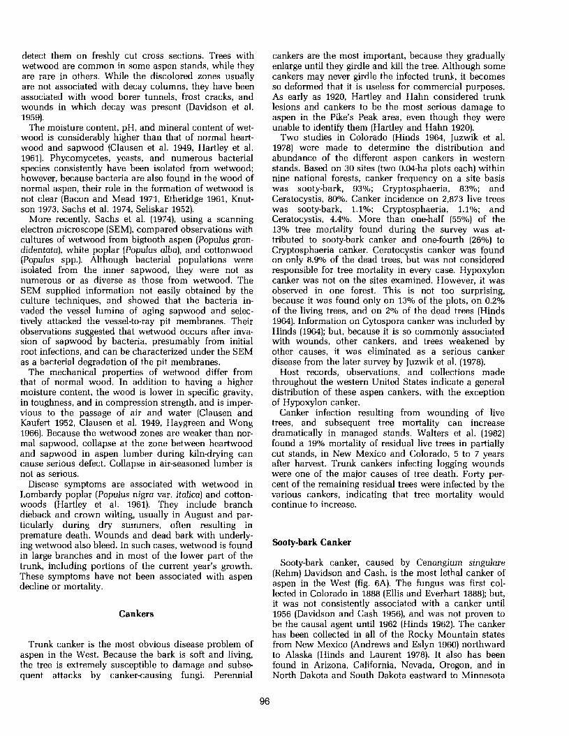
Sooty-bark Canker
Sooty-bark canker, caused by Cenangium singulare (Rehm) Davidson and Cash, is the most lethal canker of aspen in the West (fig. 6A). The fungus was first col- lected in Colorado in 1888 (Ellis and Everhart 1888); but, it was not consistently associated with a canker until 1956 (Davidson and Cash 1956), and was not proven to be the causal agent until 1962 (Hinds 1962). The canker has been collected in all of the Rocky Mountain states from New Mexico (Andrews and Eslyn 1960) northward to Alaska (Hinds and Laurent 1978). It also has been found in Arizona, California, Nevada, Oregon, and in North Dakota and South Dakota eastward to Minnesota

Figure 6.-Sooty-bark canker. (A) Large aspen in center with typical elliptical shaped, 6-year-old canker, and smaller trees girdled within 3 years without typical canker symptoms. (B) Black, stringy, dead bark. (C) Black, net-like patterns where the fungus mats retain the dead bark to the sapwood for several years. (D) Apothecia (fruiting structures) of the fungus found on dead bark (scale Is mm).

(Hinds and Anderson 1970), Michigan, and New Hamp- shire (Davidson and Cash 1956). In western Canada, it has been collected on aspen in Alberta, British Columbia (Tripp et al. 1975), and the Yukon Territory (Hinds and Laurent 1978).
Although the cankers sometimes start at points where there is no apparent injury (Davidson and Cash 1956), the fungus infects trunk wounds (Hinds 1976, Krebill 1972), penetrates the inner bark and cambium, and spreads rapidly. Cankers can extend to 40 inches (1 m) in length in 1 year, and 12 feet (4 m) in length by 29 inches (74 cm) in width in 4 years (Hinds 1962). Trees of all sizes are killed, usually within 3 to 10 years.
Young cankers first appear on aspen bark as slightly sunken oval areas with blackened inner bark. The fungus invades bark tissue so rapidly that a prominent callus formation is unusual. The bark area killed by the fungus can be seen each succeeding year by the expan- sion of the original sunken area. The dead bark epider- mis begins to slough off after 2 or 3 years, exposing the blackened inner bark, which has become a uniform sooty black (fig. 6B). Because the epidermis sloughs off quicker in the central portions, the cankers assume a somewhat concentric zoned pattern. The thicker inner bark remains tightly attached to the wood for several years, even after the tree dies and falls. It eventually sloughs off in long stringy strips, revealing black netlike patterns on the trunk where fungus mats held the dead bark to the wood (fig. 6C).
The canker has been termed "sooty-bark canker," because the dead bark easily crumbles to a sooty-like residue when handled. The wood behind the canker tends to dry out and, consequently, usually is not decayed; however, wind breakage at the canker point is not unusual. The wood is light gray and exhibits various patterns of yellow fluorescence under ultraviolet light.
A phytotoxin has been implicated in causing canker. While working with a phytotoxin produced by the Hypoxylon canker fungus, Schipper (1978) also found a toxin produced by C. singdare. The toxin reactions of both fungi were similar, as measured by an aspen leaf bioassy; and the two toxins migrated in almost identical manner on thin-layer chromatography plates.
Apothecia (fruiting bodies) of the fungus usually appear on bark that has been dead for at least 1 year (fig. 6D). The light gray apothecia are about 1-2 mm in diameter, angular to hysteroid in shape, and open when they become wet. The spores formed on the surface are forcibly ejected and wind disseminated when moisture and temperature conditions are favorable.
Numerous fruiting primodia (pycnidia) are found pen- etrating through the epidermis around the perimeter of infection, in the spring, before apothecia form. The epi- dermis sloughs off during the summer, and the pycnidia disintegrate. Whether or not this form is the asexual stage of the fungus remains to be determined. Small in- sects often are present and may feed on these fruiting bodies (Davidson and Cash 1956).
Nematodes are common in the necrotic tissues of sooty-bark canker. Most are closely related to nema- todes known to be insect associates. The nematodes
probably are carried to the diseased trees by various species of Epurea, and may be a factor in the etiology or pathogenesis of the cankers (Massey and Hinds 1970).
Sooty-bark canker is found mainly on the larger domi- nant and codominant trees older than 60 years, in the middle elevational limits of aspen (Davidson and Cash 1956, Hinds 1964, Juzwik et al. 1978). Although it occurs on trees as small as 2 inches (5 cm) in diameter, the in- fection is atypical in that it girdles the stem in 1 or 2 years and the canker is not obvious; only the sooty-bark is present. Apothecia production is rare, possibly because of the thinner bark on the smaller trees. Cankers are more common in stands disturbed by partial cutting (Walters et al. 1982), construction (Hinds 1976), or animal damage (Krebill 1972).
Black Canker
Black canker, caused by Ceratocystis fimbriata Ell. & Halst., is the common name given to this canker (Boyce 1948), which was described by Long (1918), although he realized that it was not caused by Cytospora chryse sperma (Long 1918). Baker (1925) found similar cankers of unknown origin plentiful in areas throughout the Rocky Mountain region; Meinecke (1929) published the first photographs of them in 1929. Wood and French (1963) first reported that Ceratocystis fimbriata was associated with a similar canker on aspen in Minnesota and that the fungus was capable of attacking aspen sprouts and causing canker. Soon thereafter, the association was reported in Pennsylvania (Wood 1964), Colorado (Hinds 1964), and the Provinces of Quebec (Ouellette 1965), Manitoba, and Saskatchewan in Canada (Laut and Hildahl 1965).
With the exception of Cytospora infection, this is the most common canker of aspen throughout its range in the western United States. It is not uncommon to find that 5@75O/0 of the trees in small areas have numerous cankers (figs 7A, 7B). Cankers are common in stands in Arizona, Colorado, New Mexico, Utah, and Wyoming (Hinds 1972a). Specimens also have been collected from California, Idaho, Nevada, Oregon, Montana, South Dakota, and from the Turtle Mountains in North Dakota eastward to Minnesota (Hinds and Anderson 1970) and north to British Columbia, the Yukon Territory, and Alaska (Hinds and Laurent 1978).
Ceratocystis fimbriata can infect through the epider- mis of leaf blades, petioles, and young stems (Zalasky 1965); but trunk wounds are considered to be the primary places of infection (Hinds 1972a). Infection first appears as a circular necrotic area on the trunk around a fresh wound or branch junction. During cambial growth in the spring, the tree forms a callus at the margins of the canker, which temporarily walls off the infection. The fungus invades the new cambium and in- ner bark during the tree's next dormant season, and kills a new zone of tissue. This process is repeated each year until the canker, consisting of successive rings of dead bark and wood, is formed.

Figure 7.-Black canker. (A) A young stand heavlly infected with canker. (B) An older stand with numerous older trunk cankers. (C) Cankers initiated at 12-year-old trunk injuries. (D) A young canker of about 24 years (bottom) and an old canker of about 59 years (top) on an 83-year-old aspen. (E) Old canker which originated along the sides of a basal wound.

Small cankers are young, are typically oval or ellip- tical, and appear "target shaped." The dead bark usual- ly adheres to the wood for several years; then it begins to slough off, exposing successive rings of dead woody tissue (fig. 7C). The canker grows faster vertically than horizontally; and the height-width ratio increases with age. Because the tree generally grows in circumference faster than the canker enlarges, cankers seldom kill large trees, unless one or more coalesce. Infection is f re quently callused off at various places on the canker margin. Older cankers typically have a central area of dead wood surrounded by a series of bark calluses. These callused areas may be concentric in outline, but usually are irregularly shaped and ragged in appear- ance because of the massive callus folds and flaring dead bark (fig. 7D). The canker face and dead bark tissue adjacent to the canker is usually black- therefore, the name "black canker."
Black perithecia of the fungus often are hard to find. They form along the canker perimeter in the spring, on wood or bark that has been dead for at least 1 year. Ascospores are forced out of the perithecia necks in a sticky mass and can be accidentally picked up by insects inhabiting the canker. Although perithecia frequently are consumed by insects, and some are disintegrated by sap flow and rain, remnants of perithecial bases can r e main for several years. Perithecia of C. alba DeVay, Davidson, & Moller, C, crassivaginata Griffin, C. populina Hinds & Davidson, and C. trernulo-aurea David- son & Hinds often coexist with those of C. firnbriata (Hinds 1972a). Past, unreported inoculation studies utilizing these other species of Ceratocystis revealed that they were incapable of causing perennial canker.
Insects burrow along the canker edges. Some pupate in the bark crevices, while others overwinter in cankers of all ages. The disease is thus transmitted by insects that visit new wounds. Nitidulid beetles (sap-feeding beetles) are considered to be the principal vectors (Hinds 1972b). However, C. firnbriata also can over- winter with beetle pupae in the soil, with the adults capable of spreading infection to new wounds in the spring, when they emerge.
The major impact of black canker is trunk deformity and cull (Meinecke 1929) not mortality, because infected trees survive for a long time (fig. 7E). Cankers 78 years old and older have been found on trees that were 103 years old (Hinds 1972a). A brown stain and wetwood extending into the heartwood above and below the canker's limit usually is present in the tree trunk. On older trees infected with wood decay fungi, sporophores are produced in the dead portion of the canker, and wood beneath such cankers is decayed.
Cryptosphaeria Canker
This canker is a relative newcomer to the list of aspen cankers. Although the fungus Cryptosphaeria populina was collected on dead aspen bark near Golden, Colo., by E. Bethel in 1897, it was not associated with a canker un-
til 1969 (Hinds 1981). In northwestern Wyoming, Krebill (1972) found the canker on 2% of the sampled trees which had been damaged by big game in the Gros Ven- tre elk winter range. Examining trees wounded by campers, Hinds (1976) later found the canker on 2% of the live and 8% of the dead trees surrounding aspen campsites in Colorado. This incidence of infection in wounded trees was nearly double that found by Juzwik et al. (1978) in natural stands.
The canker has been found in the northern states of Coahuila and Chihuahua in Mexico (Hawksworth and Tovar 1983), northward in the Rocky Mountains from Arizona and New Mexico to Idaho, British Columbia, the Yukon Territory, and Alaska (Hinds and Laurent 1978). The fungus also has been reported in the Lake States area and on other poplar hosts (Hinds 1981).
The cankers, usually associated with trunk wounds, are long and narrow (fig. 8A). They may be only 2 to 4 inches (5 cm to 10 cm) wide, yet up to 10 feet (3 m) or more long, following the grain of the underlying wood. Annual lateral extension of the canker margin may be only several millimeters per year, but it may be several centimeters or more in the vertical direction. Small trees die several years after infection and before the trunk is girdled. Branch cankers often are found on large trees, where they girdle the branch and enlarge onto the trunk. Cytospora chrysosperrna frequently is found along the canker perimeter, and is quick to colonize the remaining bark after tree death.
The infected bark around the perimeter of a canker is discolored light brown to orange. Annual callus forma- tion by the host in an attempt to limit bark infection is obvious after 2 or more years. The dead bark adheres tightly to the sapwood. Bark that has been dead for more than 1 year is black, stringy, and sooty-like, similar to sooty-bark canker. However, they are easy to distin- guish, because the dead bark contains small, lenticular, lightcolored areas, varying from 0.5 to 2.0 mm in size (fig. 8B). Perithecia of the fungus are formed within an effused pseudostroma, beneath the dead bark periderm, in the central portion of the bark that has been dead for at least 1 year (fig. 8C). Light orange acervuli of the imperfect Libertella stage occasionally are found in the advanced portion of the canker.
The fungus infects fresh wounds in the inner bark and wood, colonizing sapwood and heartwood, and causing discoloration and decay before it penetrates the bark, causing canker (fig. 8D). Inoculation studies have shown that Cryptosphaeria populina is capable of killing branches and sprouts within 1 year; saplings in 2 years; and causing cankers ranging up to 3 inches by 13 inches (7 cm by 33 cm), with sapwood discoloration extending up to 13 feet (4 m) beneath the canker, after 4 years, in larger trees. This discoloration, in hues of gray, brown, yellow, orange, and even pink, usually extends up to 3 feet (1 m) or more beyond the vertical extent of the canker, and is associated with the Libertella stage of decay.

Flgure 8.-Cryptosphaeria canker. (A) Elongate cankers at Cyear-old trunk wounds on 12-inch (30.cm) diameter aspens. (B) Lenticular light-colored areas in the dead, black bark. (C) Current year's perithecia formation under dead bark epidermis (left); epidermis removed (right) to show perithecia formed the previous year (scale is cm). (D) Sapwood discoloration with cambium and bark necrosis preceding canker formation. (E) Cross section through canker (right side) with discoloration and brown mottle trunk decay (scale is cm). (F) Fluorescence of cross section (B) under ultraviolet light.
Cytospora Canker
This canker, caused by Valsa sordida Nit., usually is referred to as Cytospora canker, because the imperfect stage of the fungus Cytospora chrysosperrna (Pers.) Fr. is more commonly encountered (Christensen 1940). Long (1918) described the canker on poplar, and also found it on aspen in Arizona and New Mexico. It is the most com- mon fungus found on aspen throughout its range. The fungus is considered a normal inhabitant of aspen bark microflora. It readily enters and parasitizes bark that has been injured or weakened by any cause (Hubert 1920, Long 1918, Povah 1921).
Infection takes place through bark wounds or dying twigs. The bark invaded by the fungus may be either regular or very irregular in outline. Infection can be so general on small branches, twigs, sprouts, and small trees that a definite canker is not formed (fig. 9A). Trunk cankers are formed by a gradual killing of the bark in a more or less circular area, over a period of several
years (fig. 9B). Annual canker growth can be seen by the slight annual callus formation around the perimeter of infection, which usually is sunken. The diseased inner bark turns dark brown to black, and the sapwood beneath is stained light brown. The dead bark remains cattached to the tree for 2 or 3 years. It then turns lighter brown in color and falls off in large pieces.
The fungus fruits readily in the dead outer bark, even when typical canker symptoms fail to develop. Pycnidia-small, black, fruiting bodies of the Cytospora stage--are the most common. Sticky pycniospores ooze out of the pycnidia in long, coiled, orange to dark red masses called spore tendrils, spore horns, or cirri (fig. 9C). During rains, the spores are partially washed away, leaving sticky masses of spores about the open- ings. They then dry to hard, hemispherical, colored masses. The Valsa stage also is common on aspen (fig. 9D). Flask-shaped perithecia are formed beneath and in a circle around the old pycnidia (Christensen 1940). Some ascospores are forcibly discharged; others

(scales are mm). collect around the ostioles of the perithecia in sticky white masses on the dead bark.
Inoculation experiments indicate that the fungus is a facultative wound parasite, and that the degree of parasitism is more severe during the tree's dormant period, and is usually greatest on poorly growing trees and branches (Schreiner 1931). Large trees in healthy condition may successfully callus out an infection or severely limit canker growth. Although the fungus often is not responsible for the injury with which it is asso- ciated, Cytospora infection is associated with frost cracks and sunscald (Hinds 1964), elk feeding wounds (Krebill 1972, Packard 1942), partially cut stands (Mar- tin 1965), tree vigor and slash fires (Hubert 1920), drought (Riley and Hildahl 1963, Schreiner 1931, Wright 1957), and as a secondary parasite with other cankers (Hinds 1981).
Factors influencing the development of the disease on poplars, such as bark and soil moisture content, temper- ature and humidity, and anatomy, have been studied to help understand the role of the host factors in the disease with a view toward selecting poplar varieties for disease resistance (Bloomberg 1962a, 1962b; Bloomberg and Farris 1963).
Hypoxylon Canker
Although Hypoxylon, caused by Hypoxylon mam- matum (Wahl.) Miller, is the most important canker disease of aspen in the Lake States region (Anderson 1964, Bier 1940), it is less important in the West. Hypoxylon pruinatum (Klotz.) Cke. (a synonym of H. mammatum) was first reported in New York as a canker disease of aspen in 1924 (Povah 1924). It occurs throughout much of the range of aspen in the eastern United States (Anderson and Anderson 1969) and Canada (Conners 1967). In the West, the canker was first found in the Rocky Mountains in the interior of British Columbia in 1953 (Molnar 1954) and in Colorado in 1955 (Davidson and Hinds 1956). Since then, it also has been reported in Arizona, New Mexico, and Wyo- ming (Riffle and Hinds 1969).
Young infections appear as slightly sunken, irregular, yellowisharange areas around wounds, branch stubs, or insect injury and galls (Anderson et al. 1979, Bier 1940, Manion 1975, Nord and Knight 1972). Although callus tissue develops at the margin of infection, the fungus invades new tissue each year, and the cankers

elongate (fig. 10A). The diseased bark appears lami- nated or mottled black and yellowish white, and white mycelia fans are formed near the canker margin under the bark. About a year after infection, the fungus pro- duces pillarlike structures between the bark cortex and periderm, causing blistered areas in the central portion of the canker. The periderm ruptures, exposing the grayish layer of hyphal pegs (fig. lOB), which are solidly covered by a layer of conidiophores and conidia of the asexual stage. The spores are wind disseminated and the conidial fructifications eventually disintegrate.
Cankers are easier to identify after 2 or 3 years, when perithecia are formed in small crustlike stroma up to several millimeters in diameter (fig. 10C). The young stroma are covered with a grayish bloom and are then formed annually on the dead black bark. They persist for several years. Ascospores are forcibly discharged from the perithecia; the most active discharge occurs immediately after rainfall (Bier 1940). The faded, dead bark in the center of older cankers begins to crack in a
checkerboard fashion, and it sloughs off in small patches, revealing a checkering of the wood beneath (fig. 10D).
The fungus invades the sapwood (Bier 1940, Hubbs 1964) and trees often die before they are completely girdled. Sapwood decay beneath a trunk canker predis- poses the tree to wind breakage, often before girdling is complete. Decay is more rapid in the Lake States area, where broken stems are common (Anderson and Ander- son 1969), in contrast to the arid conditions in the South- west, where breakage is not common. Cankers are found on saplings, which may be girdled in 4 or 5 years; yet, cankers on large trees in the Southwest may attain ages of 20 to 50 years before tree death. A live, 33-inch (84cm) d.b.h. aspen in Arizona has been observed with a Hypoxylon canker extending from the ground to a height of about 39 feet (12 m), only half-girdling the tree.
Hypoxylon canker annually kills an estimated 1-2% of the standing aspen volume in the Lake States area (Anderson 1964). Because of this, most research on the
Figure 10.-Hypoxylon canker. (A) Canker approximately 20 years old originating at a dead leader. (B) Hyphal pegs formed beneath the blistered bark periderm (scale is mm). (C) Perithecia bearing stroma of the fungus as they appear on dead bark (scale is mm). (D) Checkebard pat- tern of dead bark as it begins to slough off old cankers.

disease has been conducted in that area. While the disease causes serious mortality in localized areas in the Southwest, its overall importance there remains to be determined (Hinds and Krebill 1975); and only generalizations based on eastern studies can be made concerning the prevalence of infection and mortality.
Infection varies from one geographic area to another (Anderson 1964). There does appear to be a genetic rela- tionship, because some clones are more infected than others (Copony and Barnes 1974). Lowdensity stands, mixed stands, and thinned stands appear to have more infection (Anderson 1953, Anderson and Anderson 1968, Day and Strong 1959), as do trees on the edges of, rather than within, stands (Anderson 1964). In the Lake States area, juvenile trees are more susceptible to infec- tion (Anderson and Anderson 1969), with less infection found in older stands (Anderson 1964, Bier 1940, Gruenhagen 1945); new cankers usually occur on the up- per bole or in the crown of older trees (Day and Strong 1959); severity apparently is greater on poorer sites (Anderson 1953, Gruenhagen 1945); and infection fluc- tuates substantially from year to year (Schmiege and Anderson 1960).
Observations in the Southwest do not confirm, nor dispute, these findings. As elsewhere, the disease is not uniformly distributed over the range of aspen in the West; and there does not appear to be a relationship between canker and site index (Anderson 1964).
Other Cankers
The occurrence and importance of the littleknown fungus Dothiora polyspora Shear and Davidson in aspen regeneration should not be overlooked. It was described from the dead tips of living twigs of aspen and willow, and annual stem cankers of young aspen on Grand Mesa, Colorado (Shear and Davidson 1940). Shear and Davidson (1940) implied it might be a weak parasite capable of infecting frost-injured tissue. Although pathogenicity studies with this fungus have not been made, observations indicate that it is associated with a perennial canker and mortality of aspen regeneration throughout the West and Alaska. Stem wounds caused by browsing and trampling by domestic livestock, deer, and elk appear to be particularly susceptible to infec- tion. These wounds usually are near the ground. The stem is girdled by the fungus in 2 or 3 years, and Cytospora colonizes the remaining live bark. Because the small canker near the ground is often overlooked, Cytospora or the wound is blamed for the mortality. Its frequency of occurrence has not been determined; but the amount of infection and mortality appears to be related to the amount of animal damage within a regen- erating stand.
There have been reports of two other canker and dieback diseases of poplars occurring on aspen. Dothichiza canker, caused by Dothichiza populae Sacc. & Br., attacks numerous species of poplars and poplar hybrids in Europe, Canada, and the United States
(Waterman 1957). Although it has been in this country since 1915 (Hedgcock and Hunt 1916), the only report of it on aspen was in Wisconsin, when Honey (1944) ob- served it on Populus tremuloides and P. grandidentata nursery stock.
Neofabraea canker, caused by Neofabraea pop& Thom., was found mainly at the base of 3- to 6-yearald Populus grandidentata, P, tremuloides, and P. balsamifera Mill. trees smaller than 1.5 inches (4 cm) d.b.h., on Bear Island, Ontario (Thompson 1939). Six of seven inoculations on aspen sprouts proved it to be the causal agent. The canker has not been reported in the United States. Like Dothichiza canker, it probably is an aberration on aspen.
Canker Formation
How these fungi induce canker formation is not en- tirely clear; but the concept that they produce a toxin which results in cell death, bark collapse, and necrosis has been shown for Hypoxylon canker, and has been strongly suggested for others. Schreiner (1931) found a few hyphae among cells which apparently were not en- tirely dead, on the edge of V. sordida cankers. The wood was stained brown, and the mycelium was found in the vessels, the fibers, to some extent in the wood paren- chyma, and also between the medullary ray cells; but they apparently penetrated into the medullary ray cells only when the cells were dead. A black line of demarca- tion always formed when different clones of the fungus were grown in culture. Schreiner (1931) suggested that an enzyme action preceded the advance of the mycelium, and that the nature or amount of the toxic substance was specific for the individual clone.
The mode of H. mammatum infection under natural conditions is unknown, because the bark of aspen con- tains fungitoxic compounds that strongly inhibit mycelia growth (Hubbes 1966). The fungus is a wound parasite of sapwood tissue that invades the bark from within (Bier 1940, Hubbes 1964, Schipper and Anderson 1971). The fungus produces a toxin, mammatoxin, that causes bark necrosis and collapse in advance of the fungus; consequently, the fungus is well established in the sap- wood before canker symptoms appear in the bark (Schipper 1978). A mammatoxin assay has been devel- oped to determine genetic and environmental predispo- sition of aspen to cankering (Bruck and Manion 1980).
Results of recent work with Cryptosphaeria populina indicate it is similar to H. mammatum (Hinds 1981). The fungus more readily infects sapwood wounds and pene trates the sapwood before the canker forms in the bark (fig. 8E). A waterextractive material produced by the fungus in culture is similar to that found in infected sap- wood. The material is fluorescent under ultraviolet light (fig. 8F), like material produced by H. mammatum, which suggests that this material may be toxic to living cells and may be a precursor to the eventual discoloration and decay caused by the fungus.

Figure 11.-Aspen rough bark. (A) Extensive trunk infection by Diplodia tumefaciens. (B) Con. fined oval trunk infection typical of Curcurbitaha staphula. (C) Angular trunk infection by Rhytidiella baranyayi. (D) Old damage by rodents at the base of aspen. (E) Sunscald over many years on the south side of the trunk. (F) Common bark wounding by campers.
Canker Control
No control measures are known for these aspen cankers; and, as yet, they cannot be prevented, except by preventing wounds. If aspen stands are opened too quickly, the residuals will suffer from sunscald and canker, and the stand might deteriorate rapidly. Wound cankers on high-value trees sometimes can be excised by cutting away the infected bark and adjacent healthy tissue (Hinds and Krebill 1975).
Certain silvicultural techniques can minimize canker impact. Because canker diseases frequently increase with stand age, managing aspen in small even-aged groups on a short rotation of 80-100 years may be effec- tive. Clearcutting, prescribed burning, and managed wildfires often are effective techniques (Hinds and Krebill 1975). Because Hypoxylon canker is favored by stand openings and poor stocking, maintaining fully stocked stands and a closed canopy without openings or poorly stocked patches will reduce its occurrence (Anderson and Anderson 1969, Schipper and Anderson 1976).

Figure 12.-Aspen bark abnormalities of unknown origin. (A) Small nodules. (B) Globose trunk gall presumed to be insect related. (C) Rough clinker-like trunk gall.
Aspen Rough Bark and Branch Galls
Branch galls and rough bark on the smooth stems of aspen in the West are widespread. Although the natural healing of wounds is often responsible for this condition, various fungi are associated with the rough oval spots and fissured bands of grayish-black, corky bark that often extends all or part way around the trunk or branches. The damage caused to the tree by rough bark is unknown but presumed to be unimportant, although some of the fungi affect the bark periderm, cortex, and phloem. Lichens and one or more fungi often are found fruiting on the corky ridges; consequently, a microscopic examination is necessary to identify them. The number of trees affected with this type of hypertrophy at any one location can vary from a few to 100 or more. In a r e cent survey of aspen in Colorado, rough bark attributed to fungi infection was found on 23.8% of the live trees (Juzwik et al. 1978).
Diplodia turnefaciens (Shear) Zalasky (Macrophorna turnefaciens) has been proven capable of producing galls on aspen (Kaufert 1937). Branch galls do not appear to be as numerous as the trunk rough bark (fig. 11A). The fungus gains entrance into the bark through lenticles but does not penetrate deeper than the outer layer of the cortex, because it apparently stimulates the formation of a protective periderm. As the fungus invades the layer of periderm, a new phellogen develops and a new layer of periderm forms. This fungus invasion and formation of a protective bar- rier against the pathogen continues for years, resulting in the formation of rough bark and branch galls. Zalasky (1964) reported fungus penetration of intact or broken cuticle and epidermis of aspen and black poplar (Populus nigra), and its occurrence on other poplars.
Curcurbitaria staphula Dearness often is associated with D. turnefaciens galls on aspen and balsam poplar
(Populus balsamifera), and is speculated to be a second- ary invader in the tissue of the galls initiated by D. turnefaciens (Arnold and Russell 1960). C. staphula f r e quently is the only fungus found fruiting on aspen rough bark (fig. 11B); however, its exact relation to the disorder of rough bark is unknown.
Rhytidiella baranyayi Funk and Zalasky is consistent- ly associated with and considered the probable cause of another cork-bark disease of aspen in western Canada (Funk and Zalasky 1975). Observations in the West in- dicate it is widespread. The rough bark is more angular in shape without forming a band around the trunk (fig. 11C); and it frequently is initiated around branch stubs. Parkerella populi Funk has been found fruiting within the bark fissures produced by R. baranyayi; but it is suspected to be a secondary invader (Funk 1976).
Seirnatosporiurn etheridgei Funk is associated with cushion-like swellings, more circular in outline and smaller than the other rough barks. The fungus affects only the cortical area of the bark. The central portion of the swelling later assumes a cork-bark appearance, with the fruiting bodies of the fungus near the surface, sometimes forming in roughly concentric rings (Funk 1978). Leciographa gallicola Funk is considered to be a putative parasite on S. etheridgei and D. turnefaciens galls (Funk 1979).
Other agents are responsible for aspen rough bark. Rodent damage, mainly voles, gnawing at the base dur- ing the winter months is very common (fig. l lD), sun- scald on the south and southwest side of trees suddenly exposed to the sun (fig. 11E), and even mechanical wounds caused by various means (fig. 11F). (See the ANIMAL IMPACTS and the OTHER PHYSICAL FAC- TORS chapters.) There also are other bark abnor- malities of unknown origin, such as bark nodules (fig. 12A) and trunk galls (fig. 12B, 12C), which may cause rough bark.