Artificial Ventilation Theory into practice - Vetronic · Artificial Ventilation Theory into...
58
Artificial Ventilation Theory into practice Keith Simpson BVSc MRCVS MIET(Electronics) www.vetronic.co.uk [email protected] June 13 th 2014 Today we will discuss the administration of IPPV to anaesthetised patients
Transcript of Artificial Ventilation Theory into practice - Vetronic · Artificial Ventilation Theory into...
Artificial VentilationTheory into practice
Keith Simpson BVSc MRCVS MIET(Electronics)
www.vetronic.co.uk
June 13th 2014
Today we will discuss the administration of IPPV
to anaesthetised patients

Artificial VentilationTheory into practice

Artificial VentilationTheory into practice
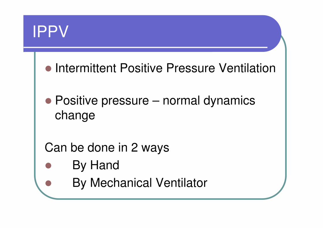
IPPV
� Intermittent Positive Pressure Ventilation
� Positive pressure – normal dynamics change
Can be done in 2 ways
� By Hand
� By Mechanical Ventilator
IPPV – by hand
� Consists of squeezing the bag and control of the exhaust valve (if there is one)
Before we go too far we need to understand the circuit we are using before we give IPPV
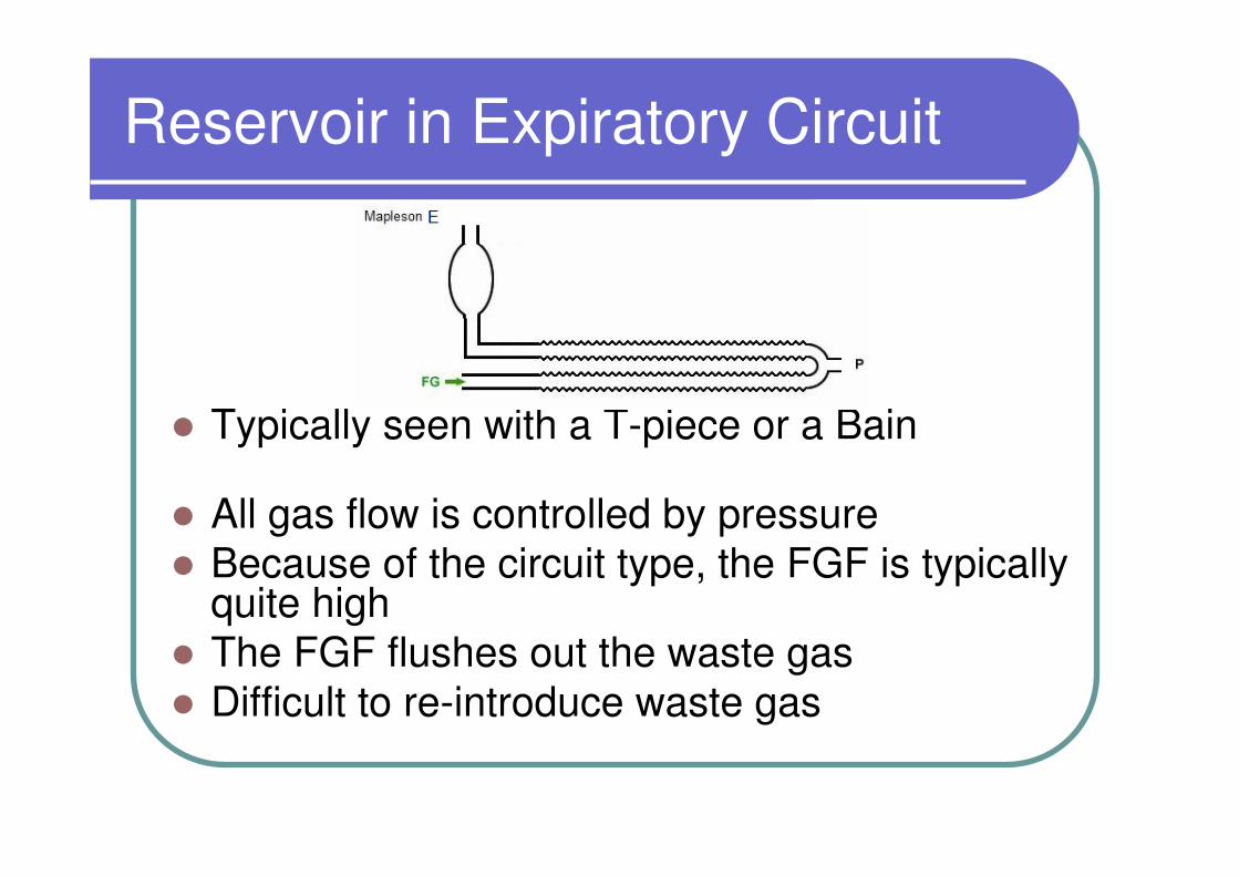
Reservoir in Expiratory Circuit
� Typically seen with a T-piece or a Bain
� All gas flow is controlled by pressure
� Because of the circuit type, the FGF is typically quite high
� The FGF flushes out the waste gas
� Difficult to re-introduce waste gas

Reservoir in Expiratory Circuit
� When you occlude the bag it fills up with fresh gas
� Squeezing it forces fresh gas into the patient –both from the bag/exp limb and incoming fresh gas
� When you release the bag the patient exhales and incoming FGF ensures it goes out of expiratory limb
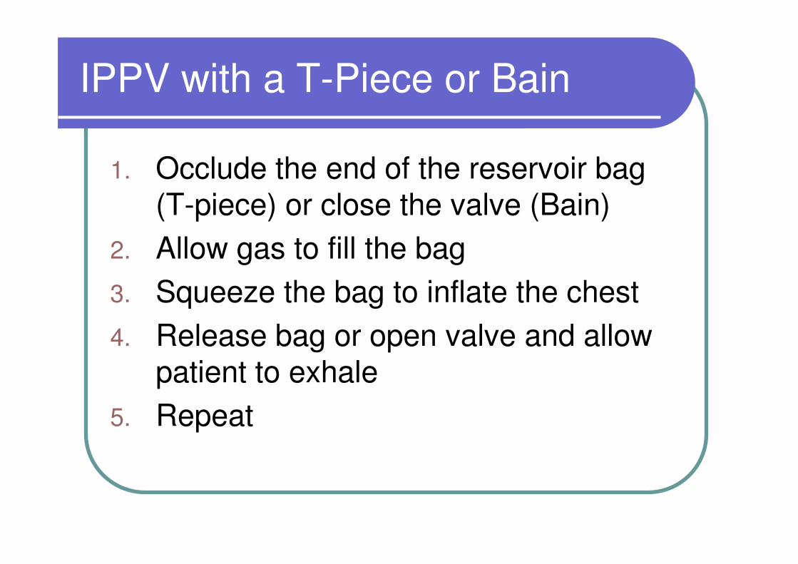
IPPV with a T-Piece or Bain
1. Occlude the end of the reservoir bag (T-piece) or close the valve (Bain)
2. Allow gas to fill the bag
3. Squeeze the bag to inflate the chest
4. Release bag or open valve and allow patient to exhale
5. Repeat
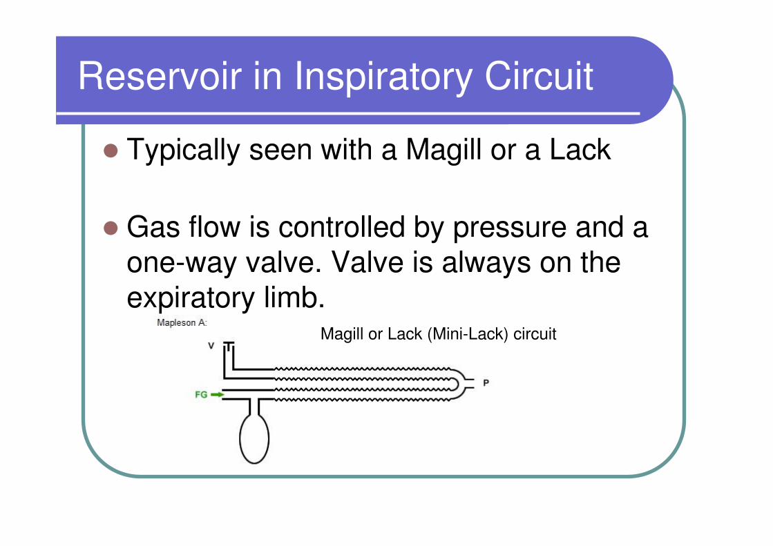
Reservoir in Inspiratory Circuit
� Typically seen with a Magill or a Lack
� Gas flow is controlled by pressure and a one-way valve. Valve is always on the expiratory limb.
Magill or Lack (Mini-Lack) circuit

Reservoir in Inspiratory Circuit
Magill or Lack (Mini-Lack) circuit
� FGF is much less than T-piece circuit
� Circuit is efficient for spontaneous breathing but bad for IPPV
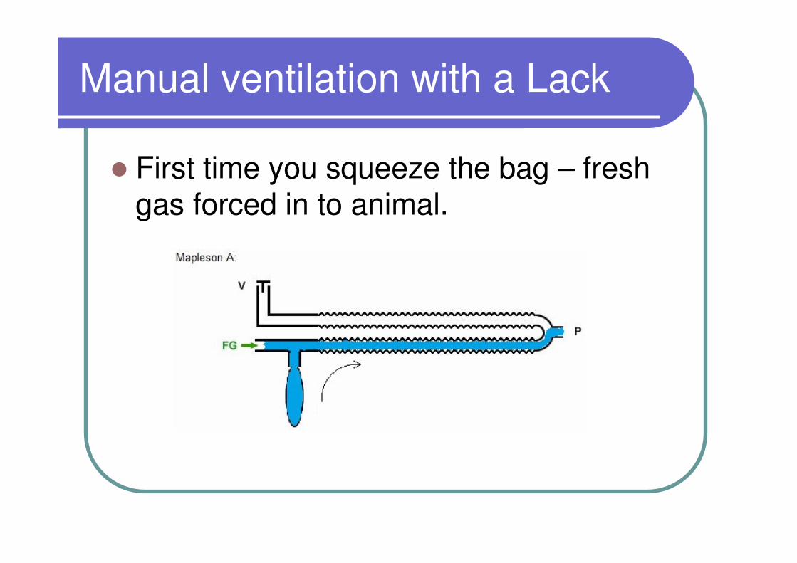
Manual ventilation with a Lack
� First time you squeeze the bag – fresh gas forced in to animal.

Manual ventilation with a Lack
� Then when you release the bag, waste gas preferentially goes back into bag
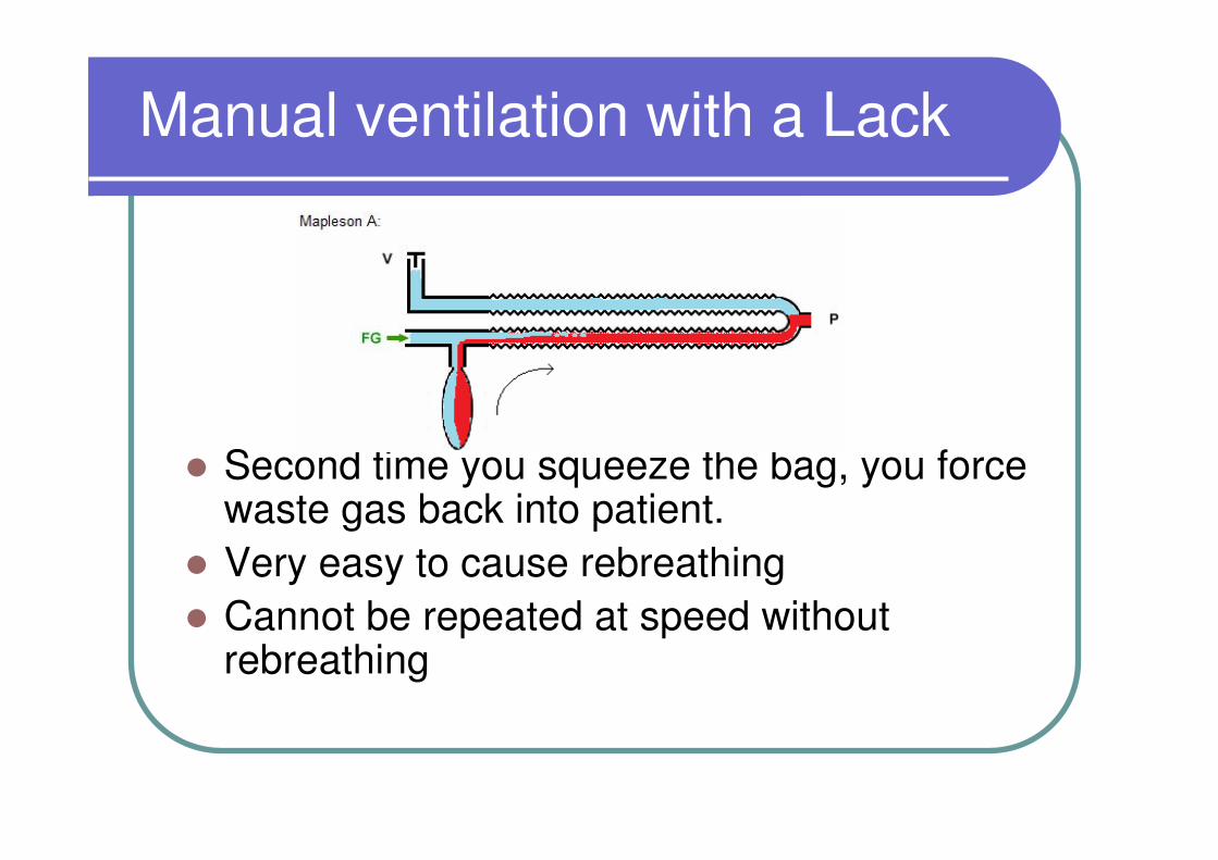
Manual ventilation with a Lack
� Second time you squeeze the bag, you force waste gas back into patient.
� Very easy to cause rebreathing
� Cannot be repeated at speed without rebreathing
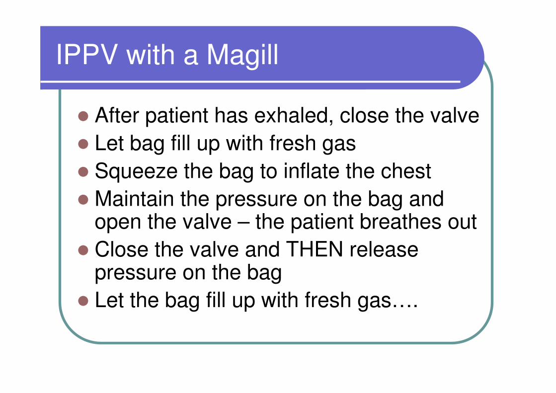
IPPV with a Magill
� After patient has exhaled, close the valve
� Let bag fill up with fresh gas
� Squeeze the bag to inflate the chest
� Maintain the pressure on the bag and open the valve – the patient breathes out
� Close the valve and THEN release pressure on the bag
� Let the bag fill up with fresh gas….
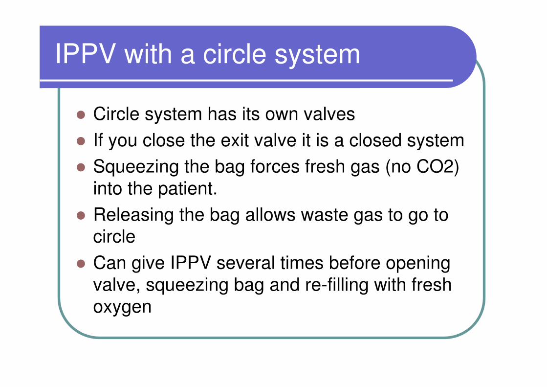
IPPV with a circle system
� Circle system has its own valves
� If you close the exit valve it is a closed system
� Squeezing the bag forces fresh gas (no CO2)
into the patient.
� Releasing the bag allows waste gas to go to
circle
� Can give IPPV several times before opening
valve, squeezing bag and re-filling with fresh
oxygen
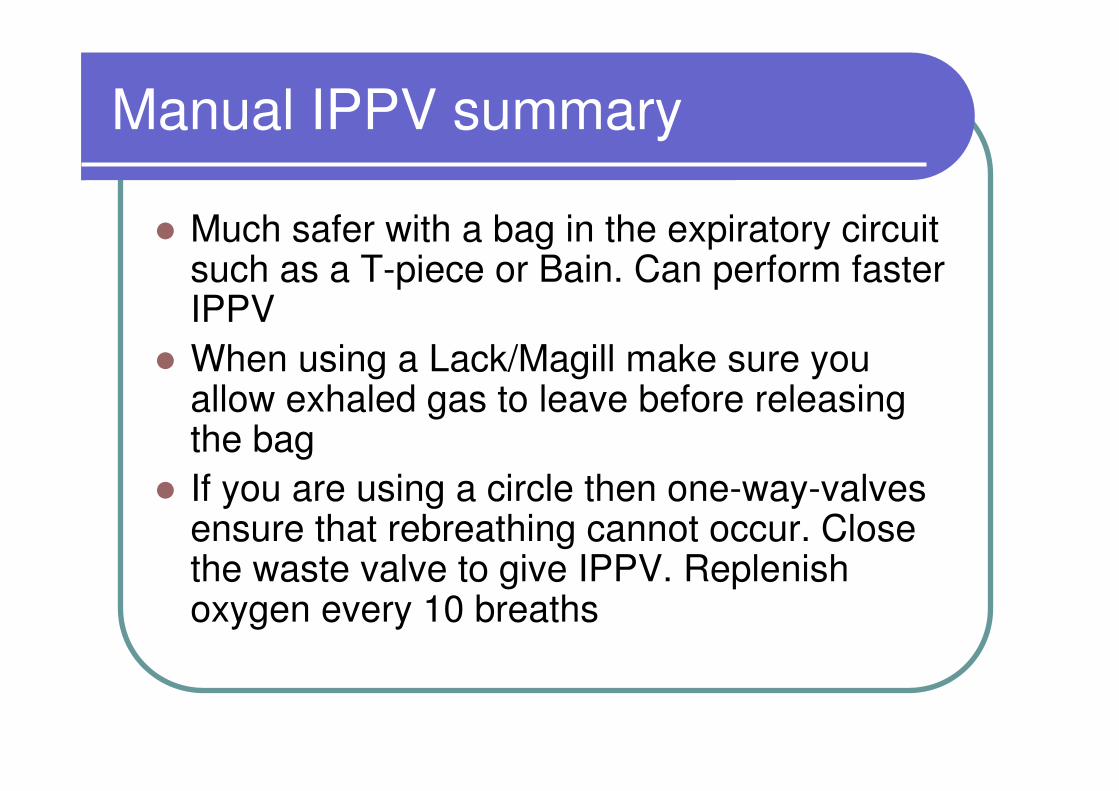
Manual IPPV summary
� Much safer with a bag in the expiratory circuit such as a T-piece or Bain. Can perform faster IPPV
� When using a Lack/Magill make sure you allow exhaled gas to leave before releasing the bag
� If you are using a circle then one-way-valves ensure that rebreathing cannot occur. Close the waste valve to give IPPV. Replenish oxygen every 10 breaths
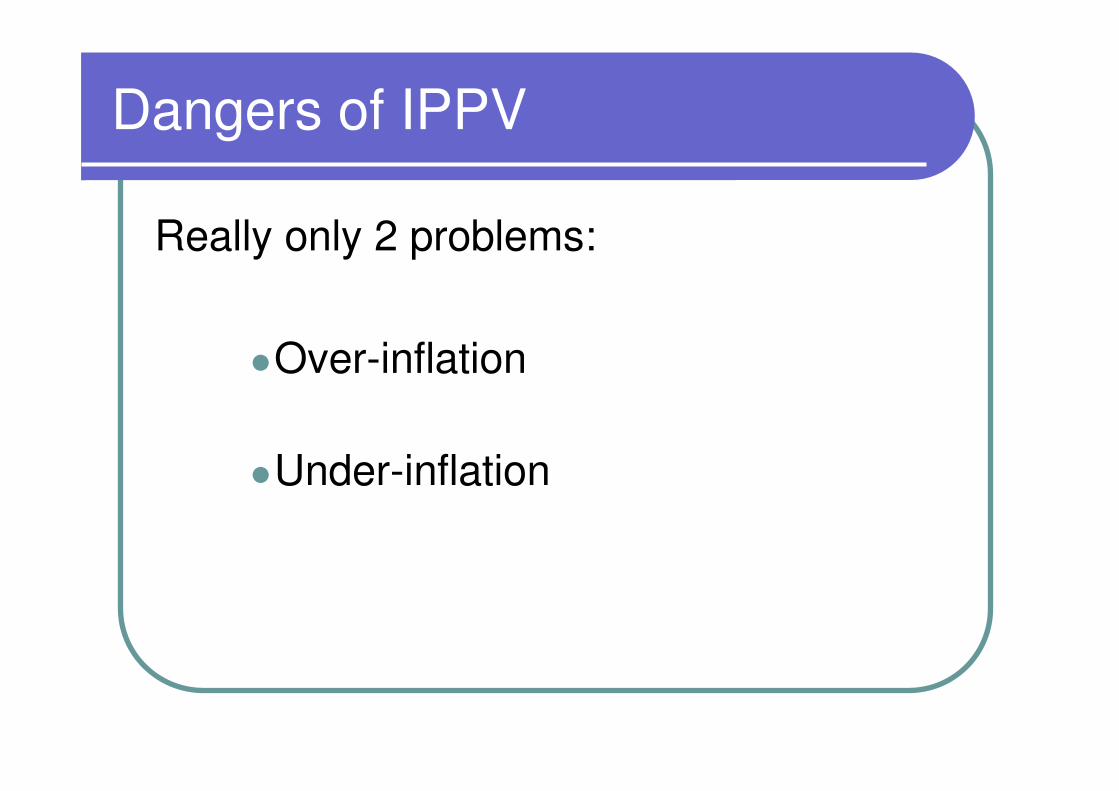
Dangers of IPPV
Really only 2 problems:
�Over-inflation
�Under-inflation

Dangers of IPPV
� Over-inflation
� In mammals safety is conferred by a
diaphragm which seals the chest cavity limiting
expansion
� Therefore care needed in birds and reptiles
and all non-mammals
� In mammals Volutrauma/Barotrauma does not
occur below 30cm pressure
Dangers of IPPV
� Without a diaphragm there is less to limit expansion of lung tissue, so over distension can occur.
� The only measure we have as a guide to safety is to monitor the airway pressure.
� In non-mammals ventilation pressures of over 15 cm H20 are rarely required
Dangers of IPPV
� When giving manual IPPV how can you be sure that you are not exceeding 15cm pressure?
� This is very difficult to achieve
� It is also difficult to be consistent
Dangers of IPPV
Demonstration of hand ventilation pressures using a T-piece circuit and an open-ended bag
Dangers of IPPV
� Under-inflation
� Failure to deliver anaesthetic agent –
patient wakes up
� Failure to remove CO2 - patient becomes
hypercapnic
� Failure to deliver oxygen – patient becomes
hypoxaemic
IPPV
What are the aims of IPPV?
� Maintain anaesthesia by delivery of gaseous anaesthetic
� Deliver Oxygen to the patient
� Remove Carbon Dioxide from the patient

Dead Space
� What is dead space?
The volume in the common gas circuit
that does not contribute to gas
exchange
The common gas circuit is the area where both inspired and expired gases pass (two-way flow).
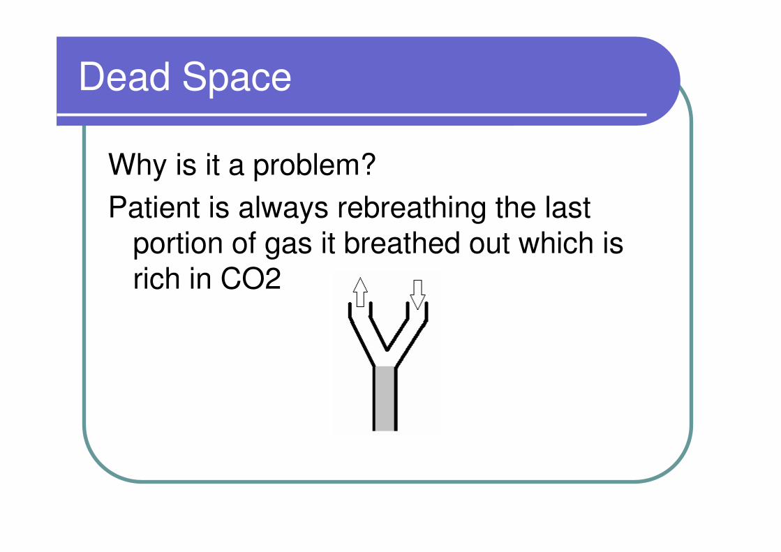
Dead Space
Why is it a problem?
Patient is always rebreathing the last portion of gas it breathed out which is rich in CO2
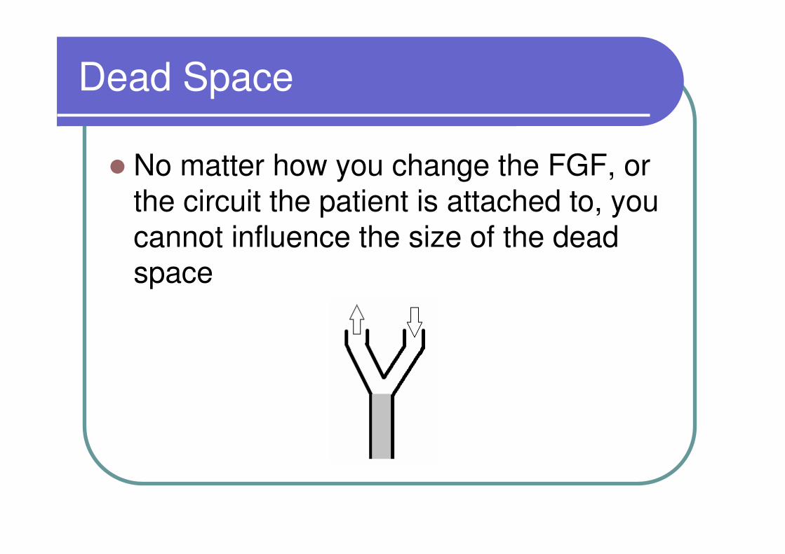
Dead Space
� No matter how you change the FGF, or the circuit the patient is attached to, you cannot influence the size of the dead space
Dead Space
� More of a problem with our smaller patients
� Wish to keep dead space as small as possible but at least less than 10% of tidal volume
Dead Space
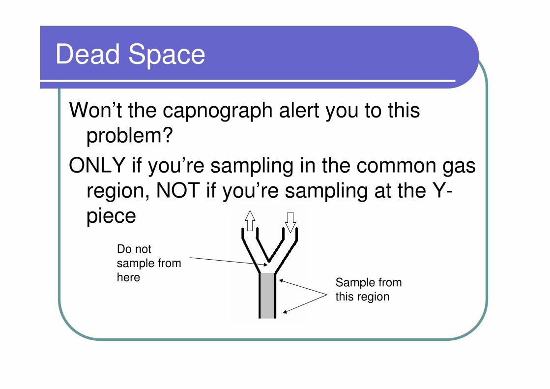
Won’t the capnograph alert you to this problem?
ONLY if you’re sampling in the common gas region, NOT if you’re sampling at the Y-piece
Sample from
this region
Do not
sample from
here
Mechanical IPPV
� Now we know all about manual IPPV we can look at mechanical IPPV and why it can help
� We will be looking at pressure-cycled ventilators with particular reference to the SAV03
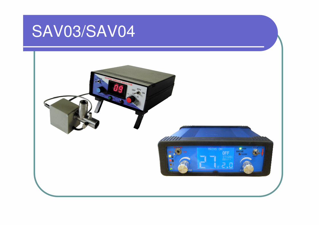
SAV03/SAV04
SAV03
How does it work?
Very simple.
It is best described as a mechanical thumb.
SAV03 Ventilator
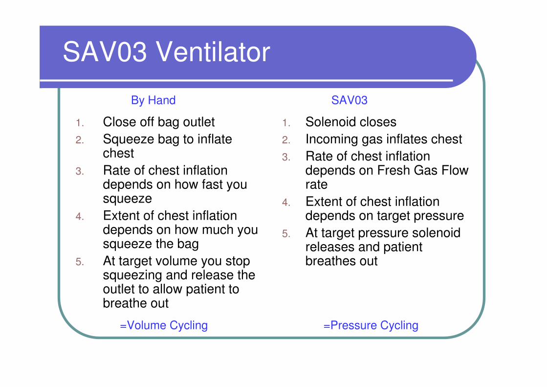
1. Close off bag outlet
2. Squeeze bag to inflate chest
3. Rate of chest inflation depends on how fast you squeeze
4. Extent of chest inflation depends on how much you squeeze the bag
5. At target volume you stop squeezing and release the outlet to allow patient to breathe out
1. Solenoid closes
2. Incoming gas inflates chest
3. Rate of chest inflation depends on Fresh Gas Flow rate
4. Extent of chest inflation depends on target pressure
5. At target pressure solenoid releases and patient breathes out
By Hand SAV03
=Volume Cycling =Pressure Cycling
SAV03 Ventilator
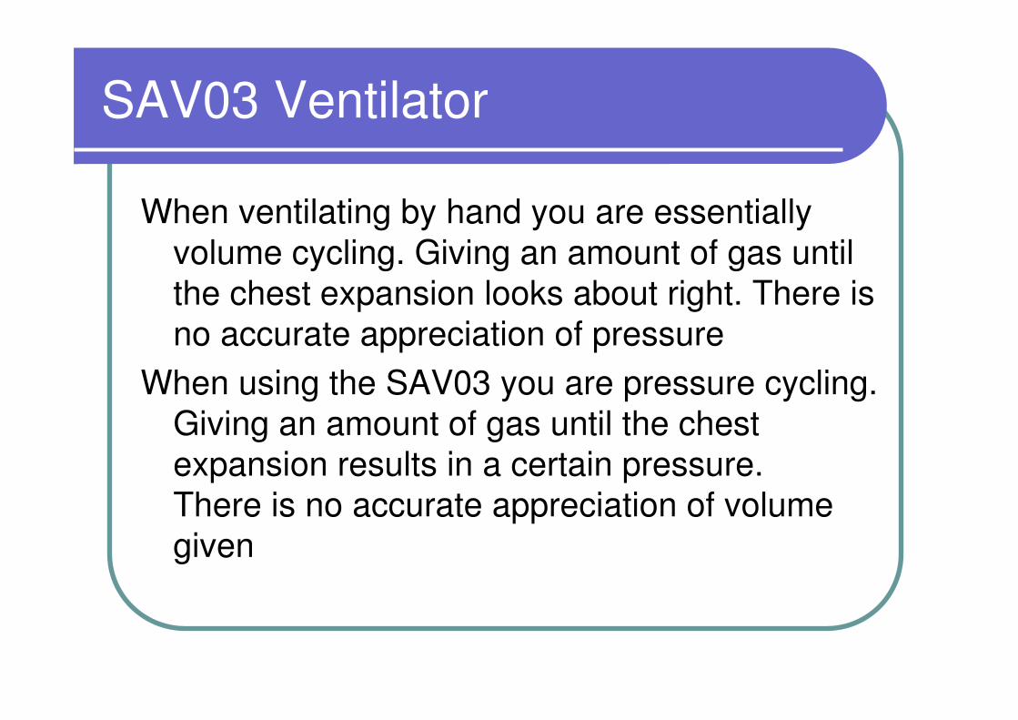
When ventilating by hand you are essentially
volume cycling. Giving an amount of gas until
the chest expansion looks about right. There is
no accurate appreciation of pressure
When using the SAV03 you are pressure cycling.
Giving an amount of gas until the chest
expansion results in a certain pressure.
There is no accurate appreciation of volume
given
SAV03 Ventilator
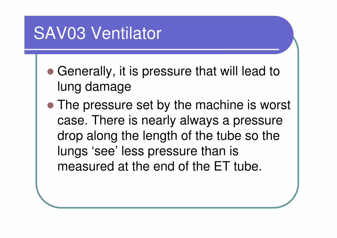
� Generally, it is pressure that will lead to lung damage
� The pressure set by the machine is worst case. There is nearly always a pressure drop along the length of the tube so the lungs ‘see’ less pressure than is measured at the end of the ET tube.

SAV03 Operation
� When the valve is closed the fresh gas (blue) can only go down the ET tube to the patient. This is inspiratory phase
SAV03 Operation
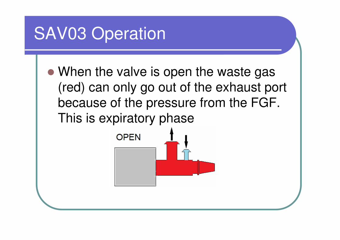
� When the valve is open the waste gas (red) can only go out of the exhaust port because of the pressure from the FGF. This is expiratory phase
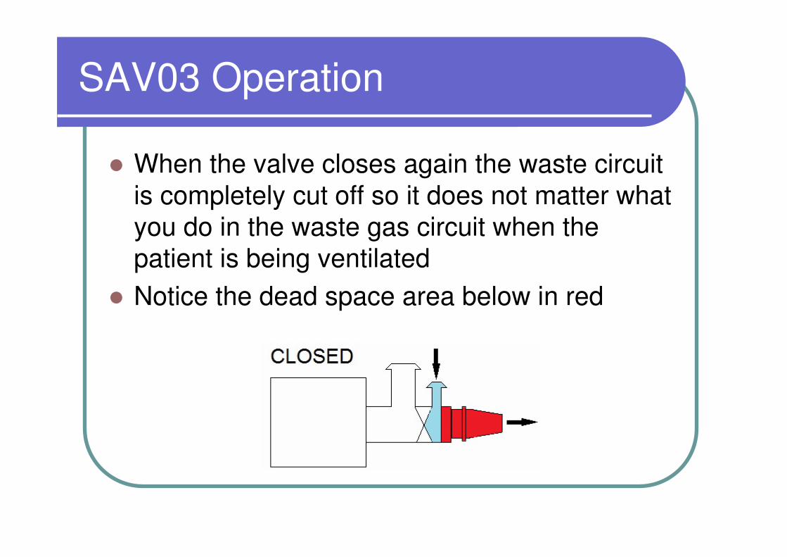
SAV03 Operation
� When the valve closes again the waste circuit
is completely cut off so it does not matter what
you do in the waste gas circuit when the
patient is being ventilated
� Notice the dead space area below in red
SAV03 Operation
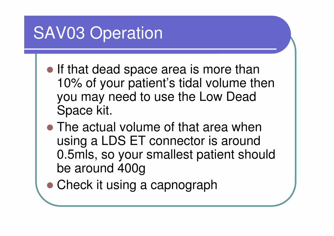
� If that dead space area is more than 10% of your patient’s tidal volume then you may need to use the Low Dead Space kit.
� The actual volume of that area when using a LDS ET connector is around 0.5mls, so your smallest patient should be around 400g
� Check it using a capnograph
Primary aims of ventilation
� Keep animal asleep
� Deliver oxygen/agent into the patient's chest to inflate it to a level consistent with normal oxygen exchange
� Ensure adequate removal of CO2 with little or no expiratory resistance
Sounds easy. So where are the problems?
Ventilating a patient
� How much do you inflate?
� How fast do you inflate?
� What is too much?
� What happens when you give too much?
Ventilating a patient
The answers to all of the above relate to pressure.
It is pressure that regulates the efficiency of gas exchange. It is pressure that can potentially cause some harm
So how much is enough and how much is too much?

How much do you inflate?
Target pressures
Mouse : 6-7cm H20 weight 30g
Horse : 20cm H20 weight 500kg
Target ventilation pressures are not related to
weight.
Cannot weigh an animal, look at a chart and
determine the ventilation pressure required.
But! You can safely assume that any animal will
fall in the range of 6-20cm H20

How much do you inflate?
So start low and increase your target pressure to obtain ‘normal’ chest movements and if possible normo-capnia
You will need a ‘snug’ fitting ET tube for ventilation

How fast do you inflate?
Remember the inflation rate is controlled by the FGF rate from the machineKeep it as close to 3 x Minute Volume as possible.
Under 3 x MV and will have excessive Insp Time
Over 3 x MV and may lose pressure along the tube –much worse with very small patients
MV = Resp Rate x Tidal Volume
It is better to err on the side of a lower flow rate than a high one – will get better oxygen delivery at slower inflation rates

How fast do you inflate?
Example
500g Patient
TV = 6 mls (12mls/kg)
RR = 30 bpm
MV = TV x RR = 180mls/minute
3 x MV = 540mls/minute
Set FGF to 0.4 - 0.5L/Min.

What is too much?
Human studies would suggest that 30cm is the upper safe limit for mammals because Ventilator Induced Lung Trauma is not seen at levels below this.
Mammals have a restraining chest wall & diaphragm. Other species do not.
In those other species, pressures less than 30 cm may cause shearing/tearing of lung tissue, but there is rarely any need to exceed 15cm pressure.
What is too much? - Birds
� Birds have a unique one-way flow pattern of air through their lungs.
� Not known if this pattern is fully maintained in birds during IPPV
� Their through-flow system means that flow is much more important than pressure
� So keep pressure low (<15) and RR high (at least at normal Resp Rates)
What is too much? - reptiles
� Have a more mammalian-like lung structure
� But no restraining diaphragm
� Two-way flow pattern, so good alveolar distension is important
� At risk of shear-damage if over-distend
� Try to keep pressures < 15 cm H2O and do not exceed 20 cm H2O
What is too much? - Mammals
� With a closed chest pressures of up to 60cm can be tolerated in large animals such as the horse.
� In normal practice however, it is unusual to need to ventilate at target pressures of more than 25 cm H2O
� Most patients adequately ventilated at 10-15 cm H2O
What happens when you give too much?
� There may be no obvious outward signs initially
� Over-distension & shear damage in the lungs can lead to an inflammatory response that will produce an interstitial oedema
� Over time this inflammatory response will reduce lung function and it may appear that the patient needs more ventilation – resist the urge to increase the TV by increasing the Target Pressure.
� In birds, air sacs may rupture, particularly if diseased. Unless this results in a leak of gas (i.e. into the room), air flow through the lungs is likely to remain sufficient
Ventilating a patient
Now we know the theory behind performing IPPV, we can ventilate a patient
Ventilating a patient
1. Induce and intubate your patient usingthe best-fitting tube that you can for minimal loss of gas around the tube
2. Connect to your circuit and start IPPV
3. Control can be accomplished in nearly all animals by initially over-ventilating
4. Use a short expiratory time and a slightly higher than normal Target Pressure.

Ventilating a patient
Example:
Bird with a normal RR of 40 breaths per minute.
� Aim for a ventilation rate of 50 breaths per minute to overcome the respiratory drive
� Use a peak pressure of 10-12 cm H2O to deliver big breaths until the bird is clearly under ventilator control and then return it to 8-9.
� Keep the ventilation rate at around 40 and if at all possible monitor with a capnograph to assess expired CO2 levels.
� Adjust the peak pressure to give normal chest movements
Ventilating a patient
How to respond
Patient becomes light
– Increase frequency of ventilation to increase Minute Volume
- Increase Target Pressure by 2cm H20 to increase Minute Volume
- Change anaesthetic concentration
Ventilating a patient
How to respond
Rising end-tidal CO2
- Increase frequency of ventilation to increase Minute Volume
- Increase Target Pressure by 2cm H20 to increase Minute Volume
Ventilating a patient
Stopping Ventilation/Weaning off
Have spent all the time suppressing
spontaneous respiratory drive
Now need to re-establish respiratory drive
Reduce feedback by stretch receptors -
reduce Target Pressure by 2cm H2O
Increase resp drive by increasing CO2 -
reduce resp rate
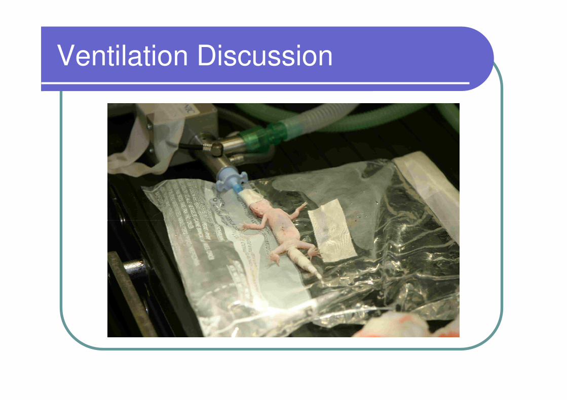
Ventilation Discussion
Ventilation
� Summary
� When using manual ventilation be aware of
the circuit configuration you are using
� When using mechanical ventilation be
aware of the pressures being delivered
� Well-controlled IPPV is a life-saver