Articulations (Joints) Martini Chapter 9 Bio 103 Feb 11, 2008.
92
Articulations (Joints) Martini Chapter 9 Bio 103 Feb 11, 2008
-
Upload
todd-barrs -
Category
Documents
-
view
217 -
download
0
Transcript of Articulations (Joints) Martini Chapter 9 Bio 103 Feb 11, 2008.
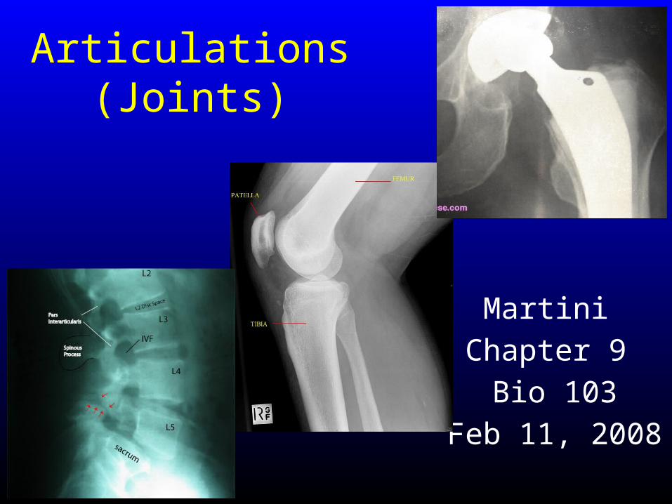
Articulations (Joints)
Martini
Chapter 9
Bio 103
Feb 11, 2008
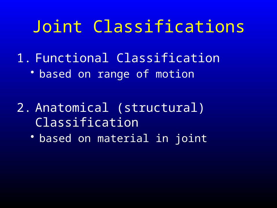
Joint Classifications
1. Functional Classification• based on range of motion
2. Anatomical (structural) Classification• based on material in joint
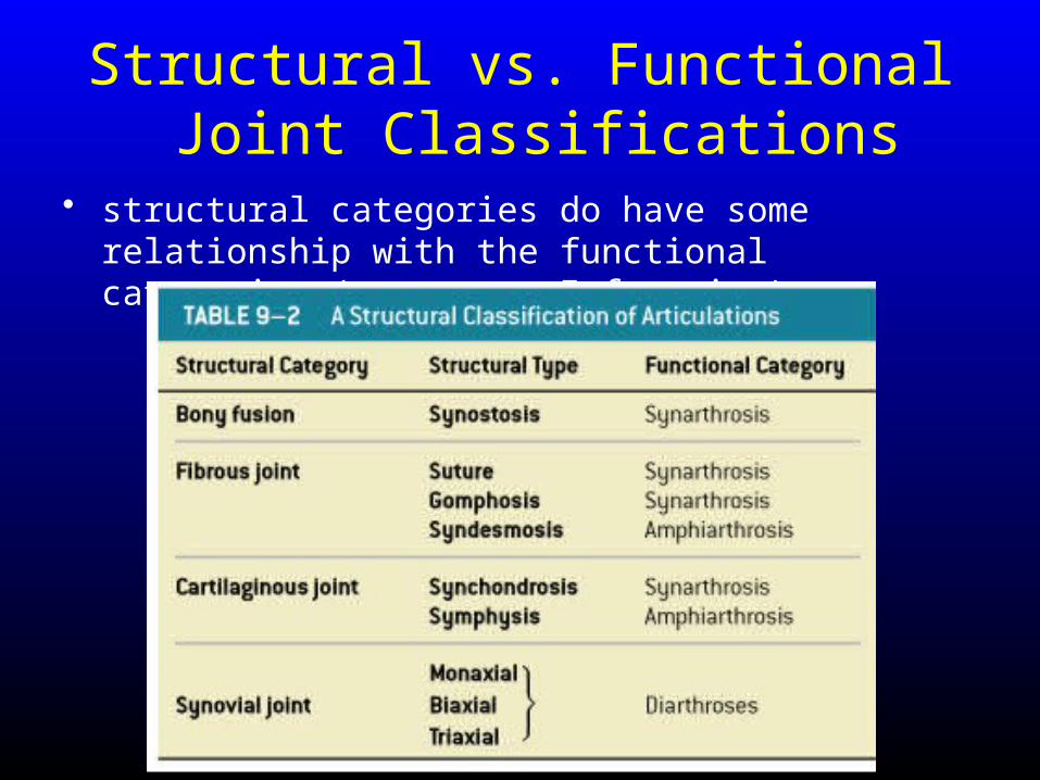
Structural vs. Functional Joint Classifications
• structural categories do have some relationship with the functional categories (structure function)
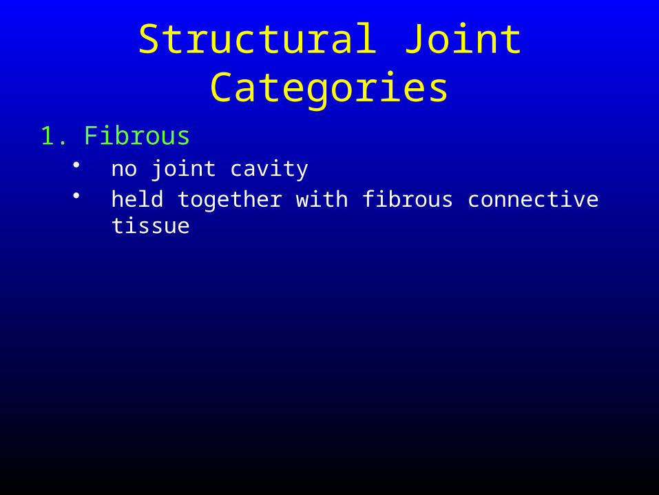
Structural Joint Categories
1. Fibrous• no joint cavity• held together with fibrous connective tissue

Structural Joint Categories
1. Fibrous• no joint cavity• held together with fibrous connective tissue
2. Cartilaginous• no joint cavity• held together with cartilage
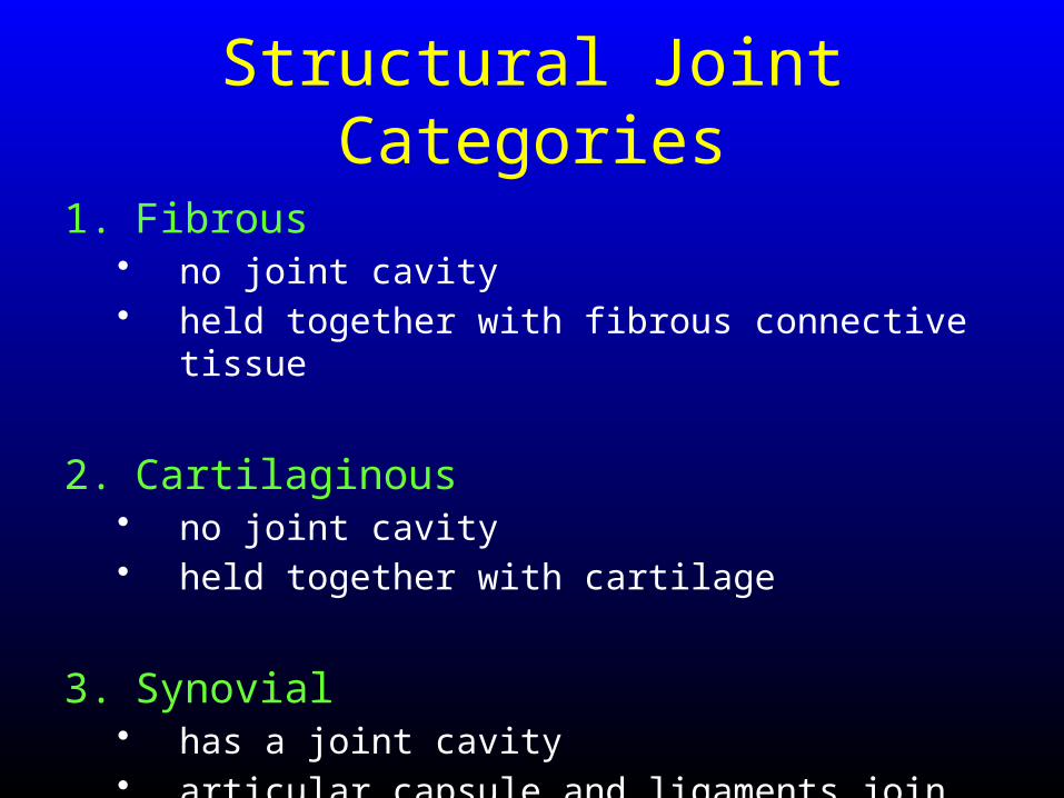
Structural Joint Categories
1. Fibrous• no joint cavity• held together with fibrous connective tissue
2. Cartilaginous• no joint cavity• held together with cartilage
3. Synovial• has a joint cavity• articular capsule and ligaments join bones
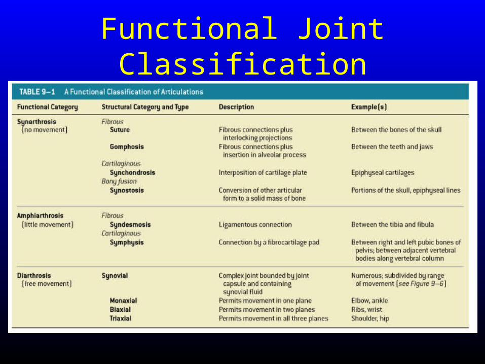
Functional Joint Classification
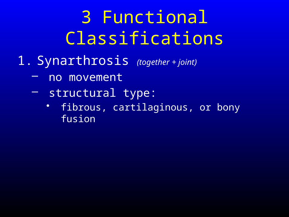
3 Functional Classifications
1. Synarthrosis (together + joint)
– no movement– structural type:
• fibrous, cartilaginous, or bony fusion

4 Types of Synarthrotic Joints
1. Suture (sewn together)
• fibrous• bound by dense fibrous connective tissue• found only in skull
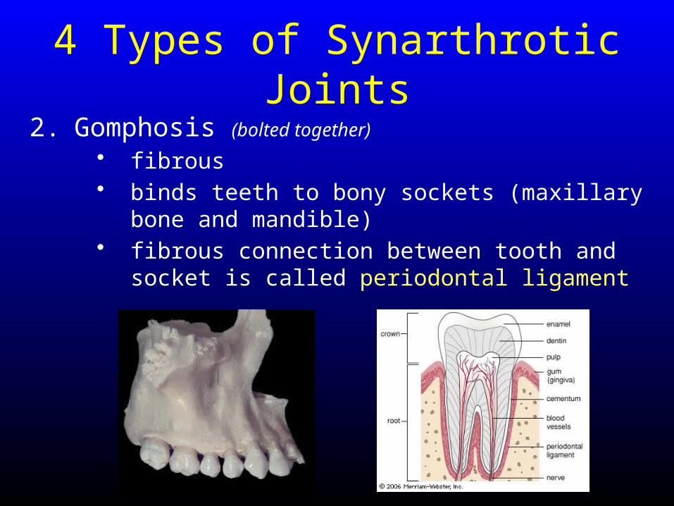
4 Types of Synarthrotic Joints
2. Gomphosis (bolted together)
• fibrous• binds teeth to bony sockets (maxillary bone and
mandible) • fibrous connection between tooth and socket is
called periodontal ligament
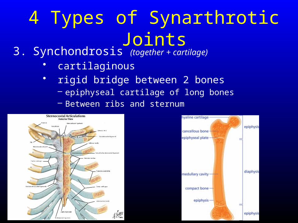
4 Types of Synarthrotic Joints3. Synchondrosis (together + cartilage)
• cartilaginous• rigid bridge between 2 bones
– epiphyseal cartilage of long bones – Between ribs and sternum
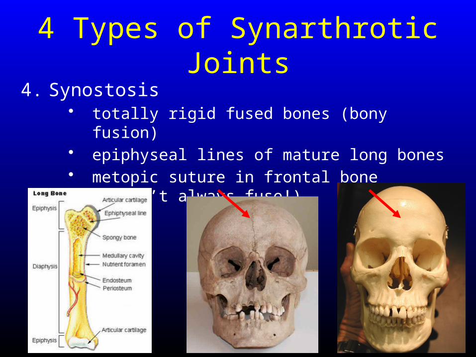
4 Types of Synarthrotic Joints
4. Synostosis • totally rigid fused bones (bony fusion)• epiphyseal lines of mature long bones• metopic suture in frontal bone (doesn’t always fuse!)
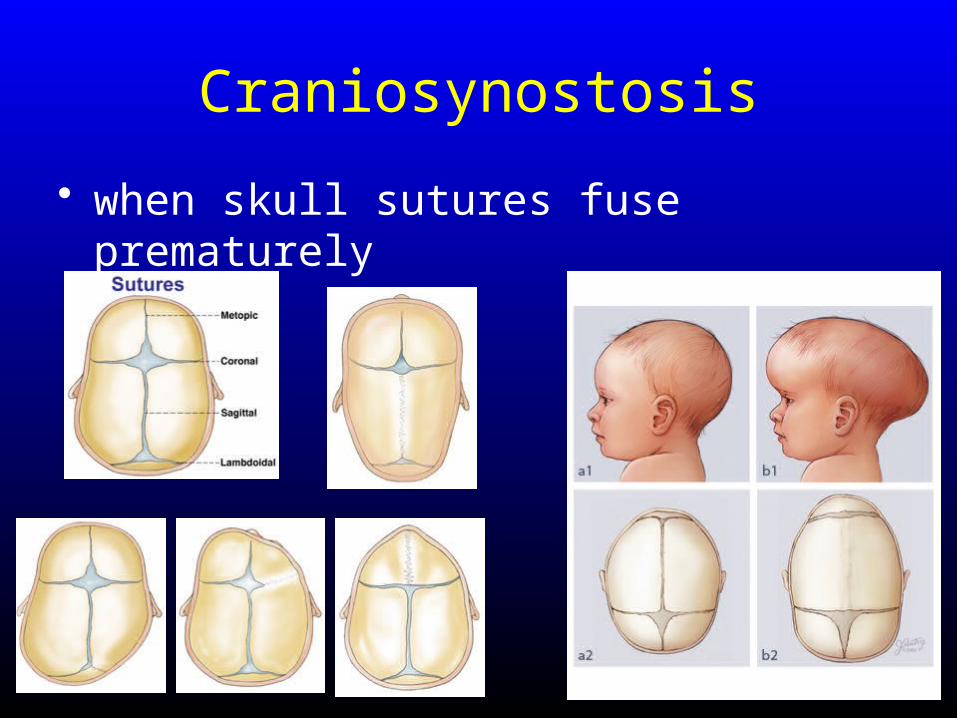
Craniosynostosis
• when skull sutures fuse prematurely
3 Functional Classifications
2. Amphiarthrosis (both sides + joint)
– little movement– structural type:
• fibrous or cartilaginous
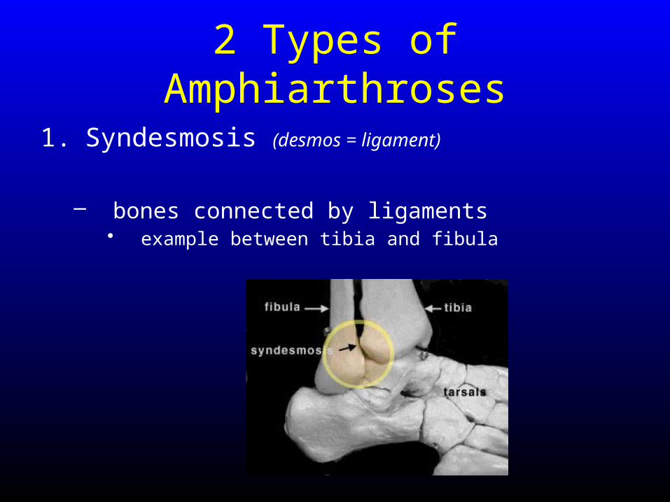
2 Types of Amphiarthroses
1. Syndesmosis (desmos = ligament)
– bones connected by ligaments• example between tibia and fibula
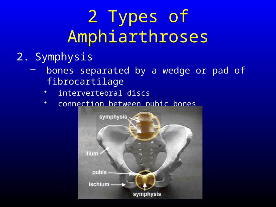
2 Types of Amphiarthroses
2. Symphysis– bones separated by a wedge or pad of fibrocartilage
• intervertebral discs• connection between pubic bones
3 Functional Classifications
3. Diarthrosis (through + joint)
– more movement– articular cartilage + synovial fluid +
accessory structures
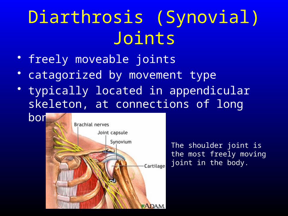
Diarthrosis (Synovial) Joints
• freely moveable joints• catagorized by movement type• typically located in appendicular skeleton, at
connections of long bones
The shoulder joint is the most freely moving joint in the body.
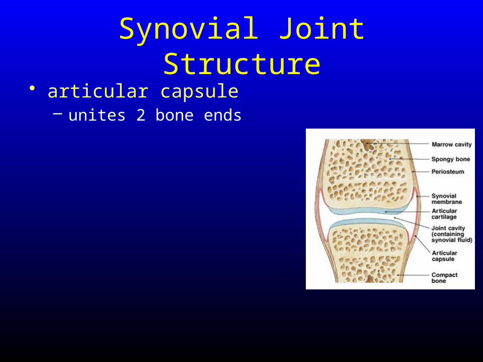
Synovial Joint Structure• articular capsule
– unites 2 bone ends
Synovial Joint Structure• articular capsule
– unites 2 bone ends
– outer layer • fibrous, dense irregular connective
tissue that blends with the periostea of the two bones to form ligaments
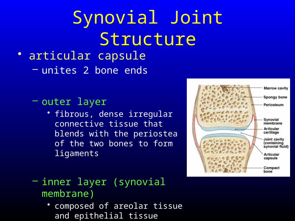
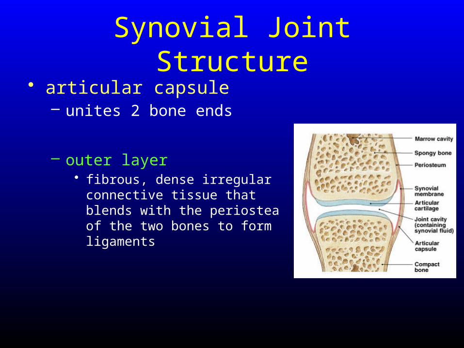
Synovial Joint Structure• articular capsule
– unites 2 bone ends
– outer layer • fibrous, dense irregular connective
tissue that blends with the periostea of the two bones to form ligaments
– inner layer (synovial membrane) • composed of areolar tissue and
epithelial tissue
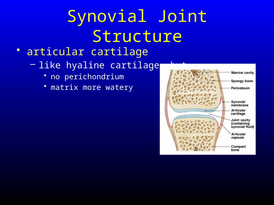
Synovial Joint Structure• articular cartilage
– like hyaline cartilage, but• no perichondrium• matrix more watery
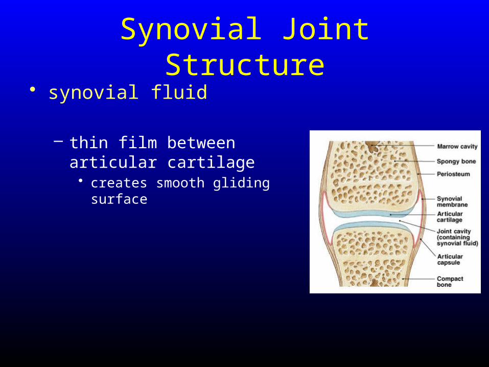
Synovial Joint Structure• synovial fluid
– thin film between articular cartilage• creates smooth gliding surface
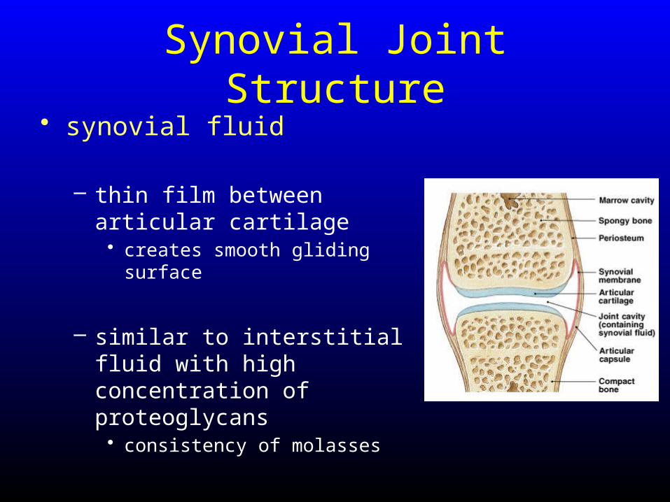
Synovial Joint Structure• synovial fluid
– thin film between articular cartilage• creates smooth gliding surface
– similar to interstitial fluid with high concentration of proteoglycans• consistency of molasses
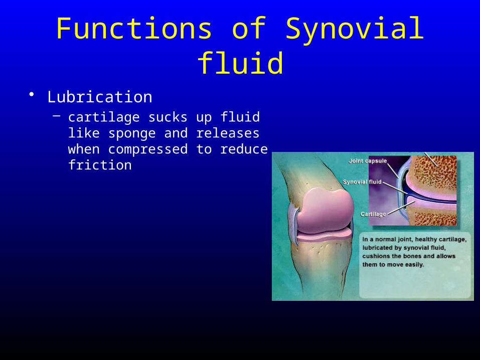
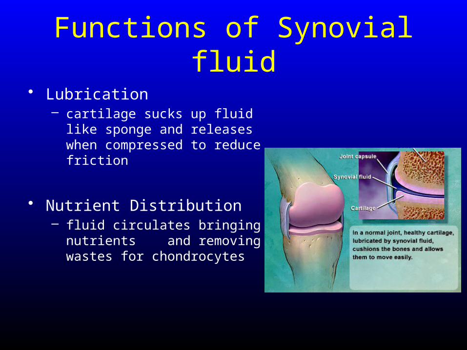
Functions of Synovial fluid
• Lubrication– cartilage sucks up fluid like sponge
and releases when compressed to reduce friction
Functions of Synovial fluid
• Lubrication– cartilage sucks up fluid like sponge
and releases when compressed to reduce friction
• Nutrient Distribution– fluid circulates bringing nutrients
and removing wastes for chondrocytes
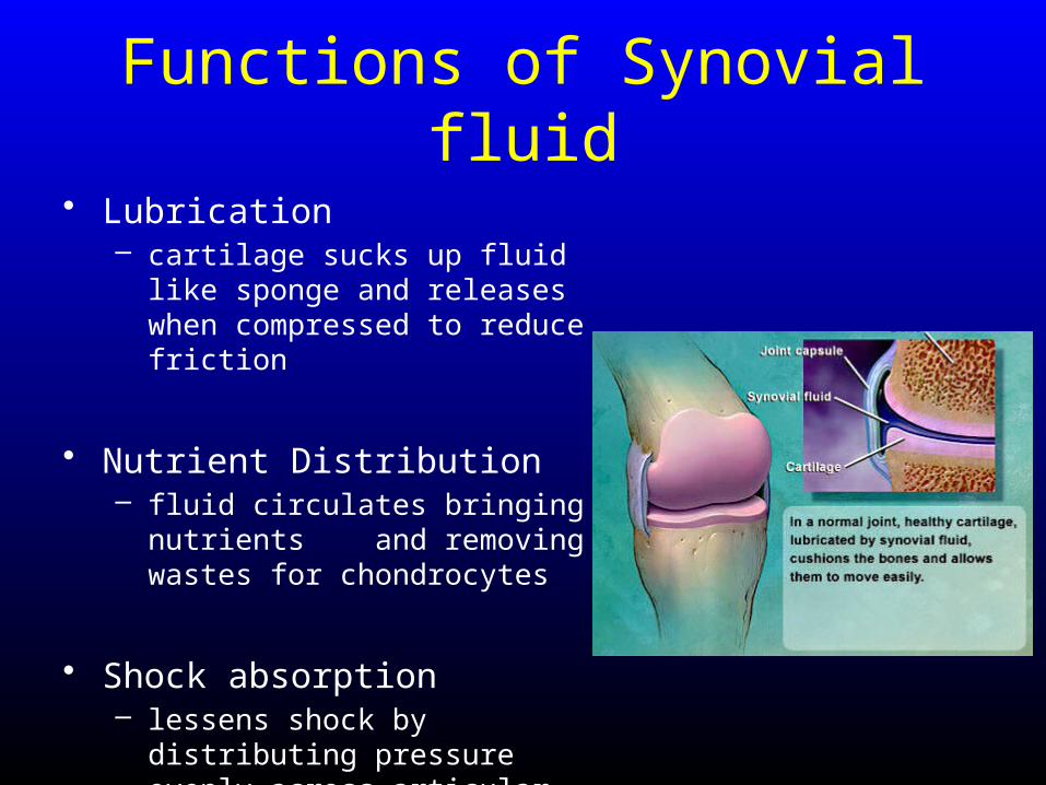
Functions of Synovial fluid
• Lubrication– cartilage sucks up fluid like sponge
and releases when compressed to reduce friction
• Nutrient Distribution– fluid circulates bringing nutrients
and removing wastes for chondrocytes
• Shock absorption– lessens shock by distributing
pressure evenly across articular surface
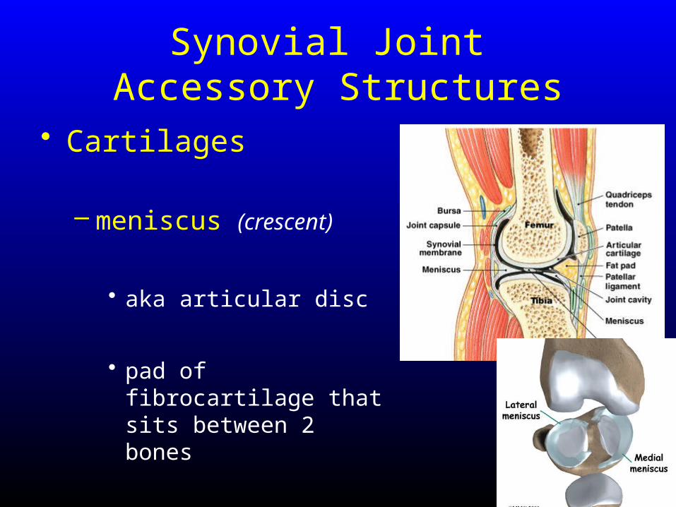
Synovial Joint Accessory Structures
• Cartilages
– meniscus (crescent)
• aka articular disc
• pad of fibrocartilage that sits between 2 bones
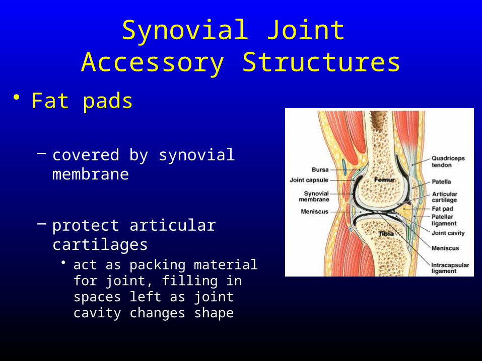
Synovial Joint Accessory Structures
• Fat pads
– covered by synovial membrane
– protect articular cartilages • act as packing material for joint,
filling in spaces left as joint cavity changes shape
Synovial Joint Accessory Structures
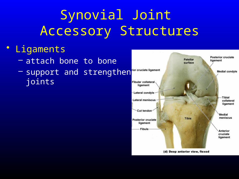
• Ligaments– attach bone to bone– support and strengthen joints
Synovial Joint Accessory Structures
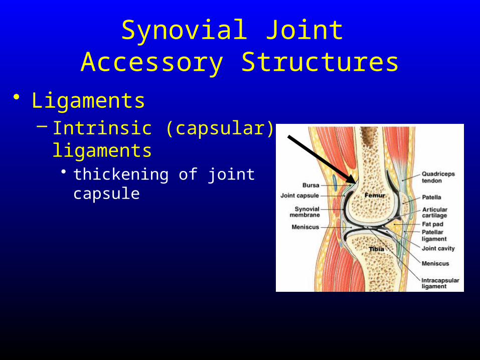
• Ligaments– Intrinsic (capsular)
ligaments• thickening of joint capsule
Synovial Joint Accessory Structures
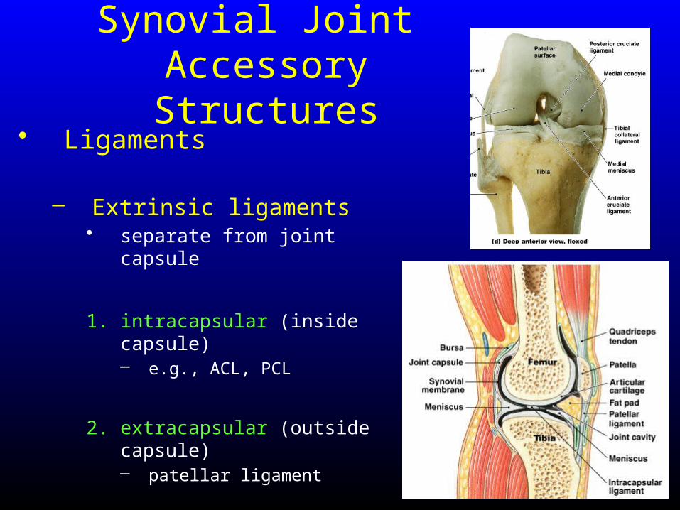
• Ligaments
– Extrinsic ligaments• separate from joint capsule
1. intracapsular (inside capsule)– e.g., ACL, PCL
2. extracapsular (outside capsule)– patellar ligament
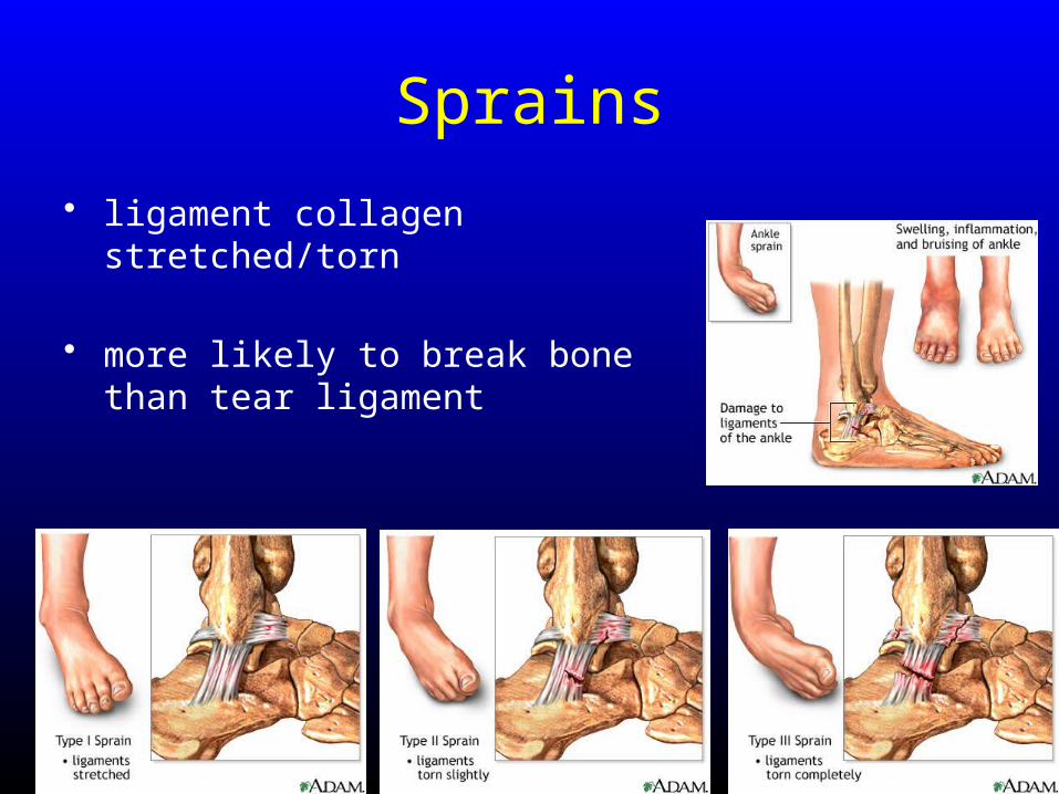
Sprains
• ligament collagen stretched/torn
• more likely to break bone than tear ligament
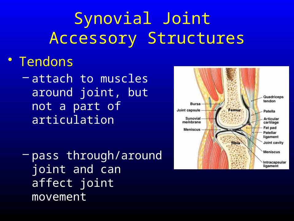
Synovial Joint Accessory Structures
• Tendons– attach to muscles around
joint, but not a part of articulation
– pass through/around joint and can affect joint movement
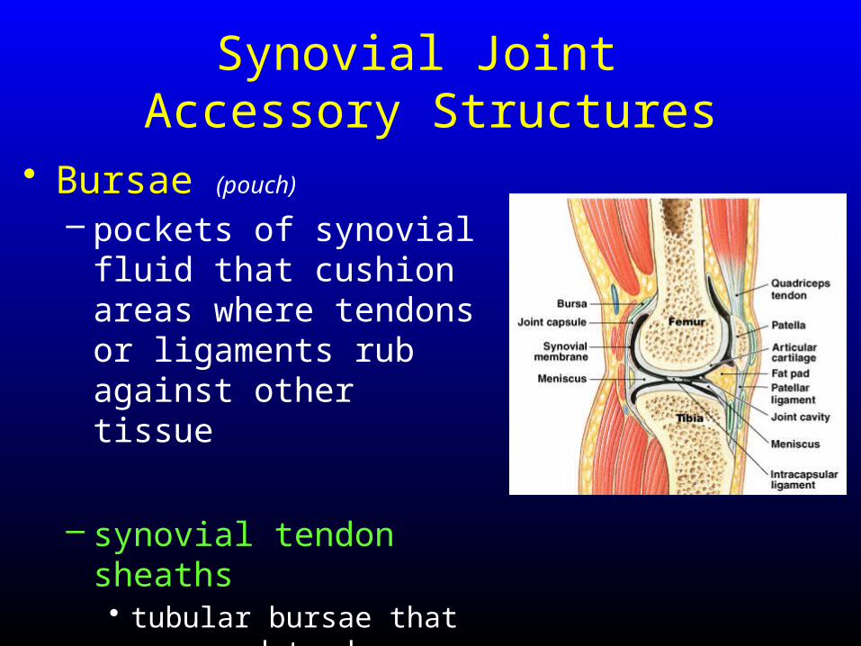
Synovial Joint Accessory Structures
• Bursae (pouch) – pockets of synovial fluid
that cushion areas where tendons or ligaments rub against other tissue
– synovial tendon sheaths• tubular bursae that surround
tendons where they cross bone
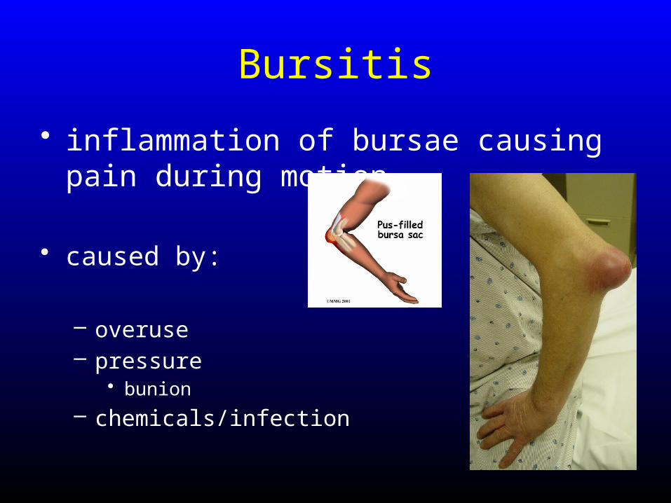
Bursitis
• inflammation of bursae causing pain during motion
• caused by:
– overuse– pressure
• bunion
– chemicals/infection

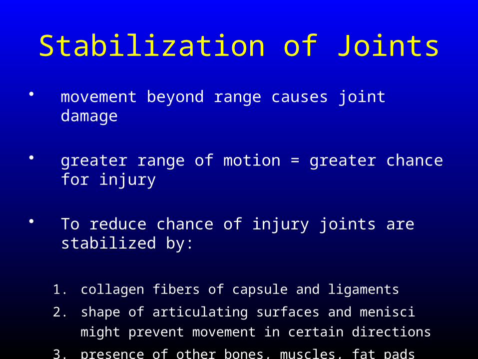
Stabilization of Joints
• movement beyond range causes joint damage

Stabilization of Joints
• movement beyond range causes joint damage
• greater range of motion = greater chance for injury
Stabilization of Joints
• movement beyond range causes joint damage
• greater range of motion = greater chance for injury
• To reduce chance of injury joints are stabilized by:
Stabilization of Joints
• movement beyond range causes joint damage
• greater range of motion = greater chance for injury
• To reduce chance of injury joints are stabilized by:
1. collagen fibers of capsule and ligaments
2. shape of articulating surfaces and menisci might prevent
movement in certain directions
3. presence of other bones, muscles, fat pads around joint
4. tension in tendons encourages movement in specific direction
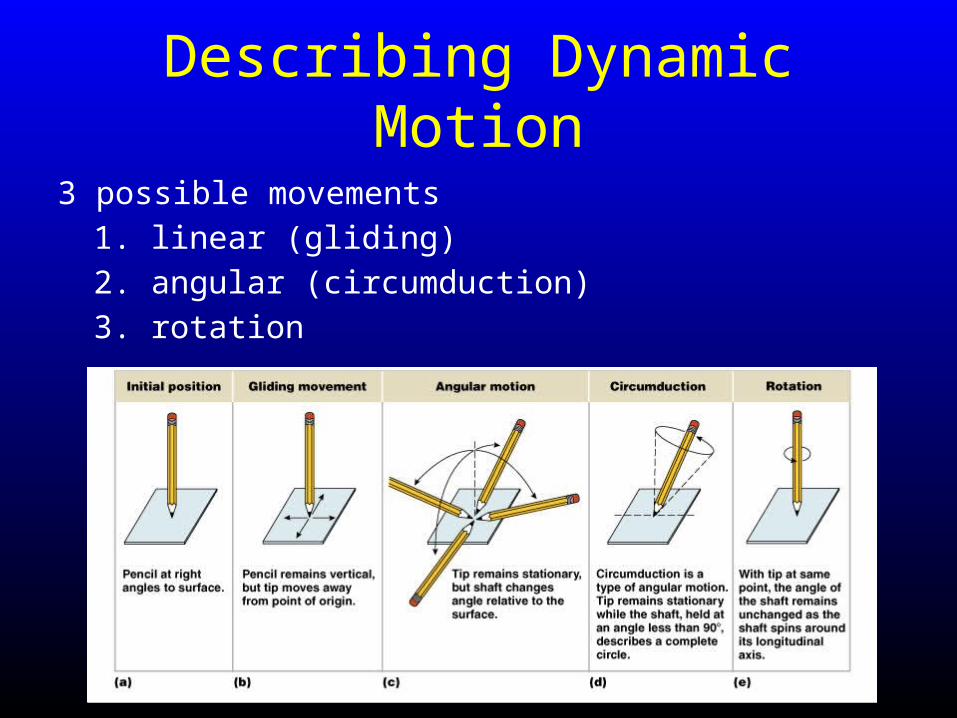
Describing Dynamic Motion
3 possible movements
1. linear (gliding)
2. angular (circumduction)
3. rotation
Types of Movements at Synovial Joints
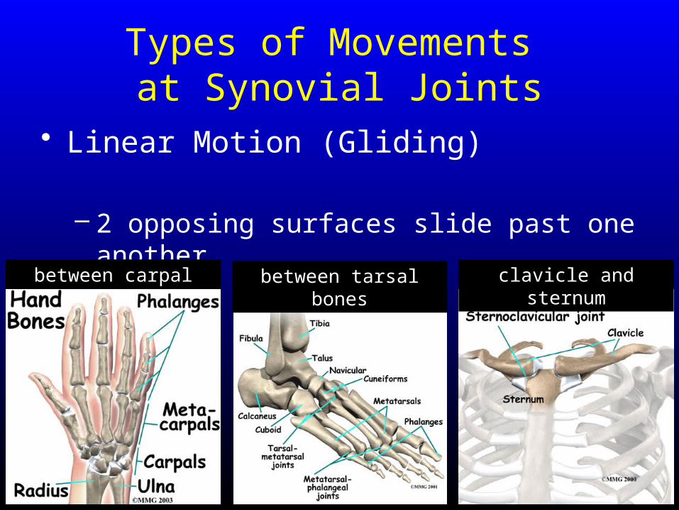
Types of Movements at Synovial Joints
• Linear Motion (Gliding)
– 2 opposing surfaces slide past one another
between tarsal bonesbetween carpal bones clavicle and sternum
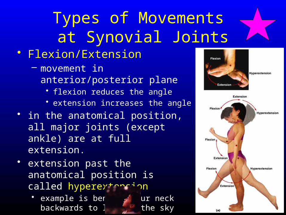
Types of Movements at Synovial Joints
• Flexion/Extension– movement in anterior/posterior
plane• flexion reduces the angle• extension increases the angle
• in the anatomical position, all major joints (except ankle) are at full extension.
• extension past the anatomical position is called hyperextension• example is bending your neck backwards
to look at the sky
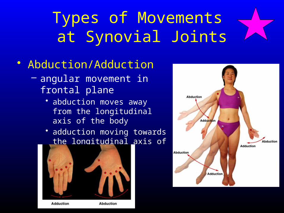
Types of Movements at Synovial Joints
• Abduction/Adduction– angular movement in frontal
plane• abduction moves away from the
longitudinal axis of the body• adduction moving towards the
longitudinal axis of the body
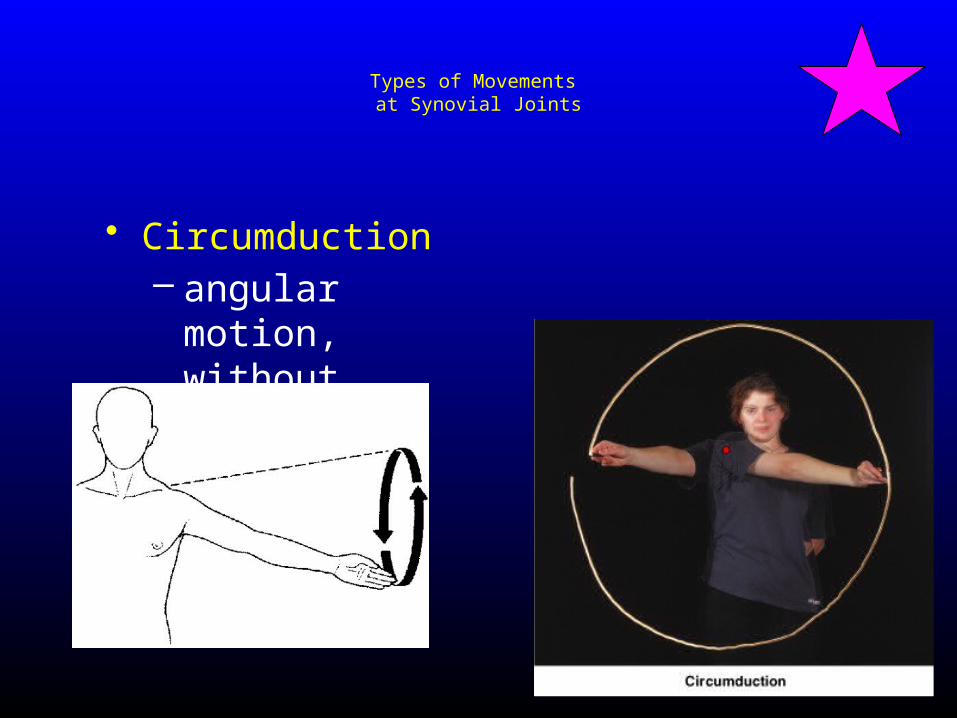
Types of Movements at Synovial Joints
• Circumduction– angular motion,
without rotation
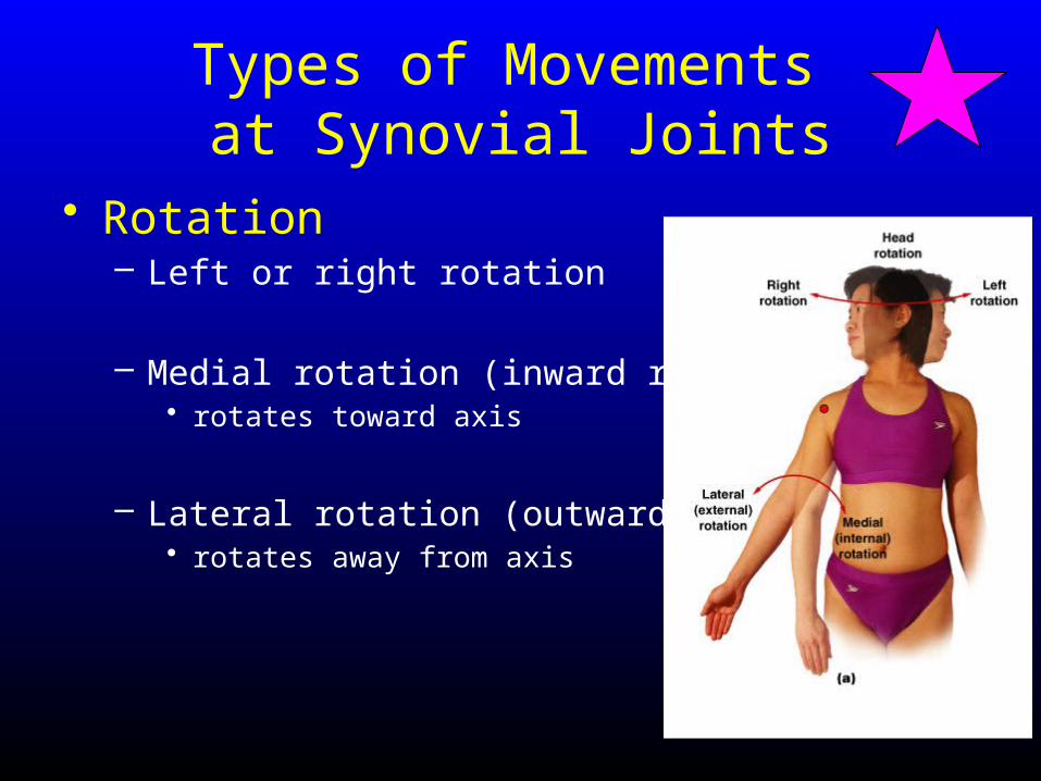
Types of Movements at Synovial Joints
• Rotation– Left or right rotation
– Medial rotation (inward rotation) • rotates toward axis
– Lateral rotation (outward rotation) • rotates away from axis
Types of Movements at Synovial Joints
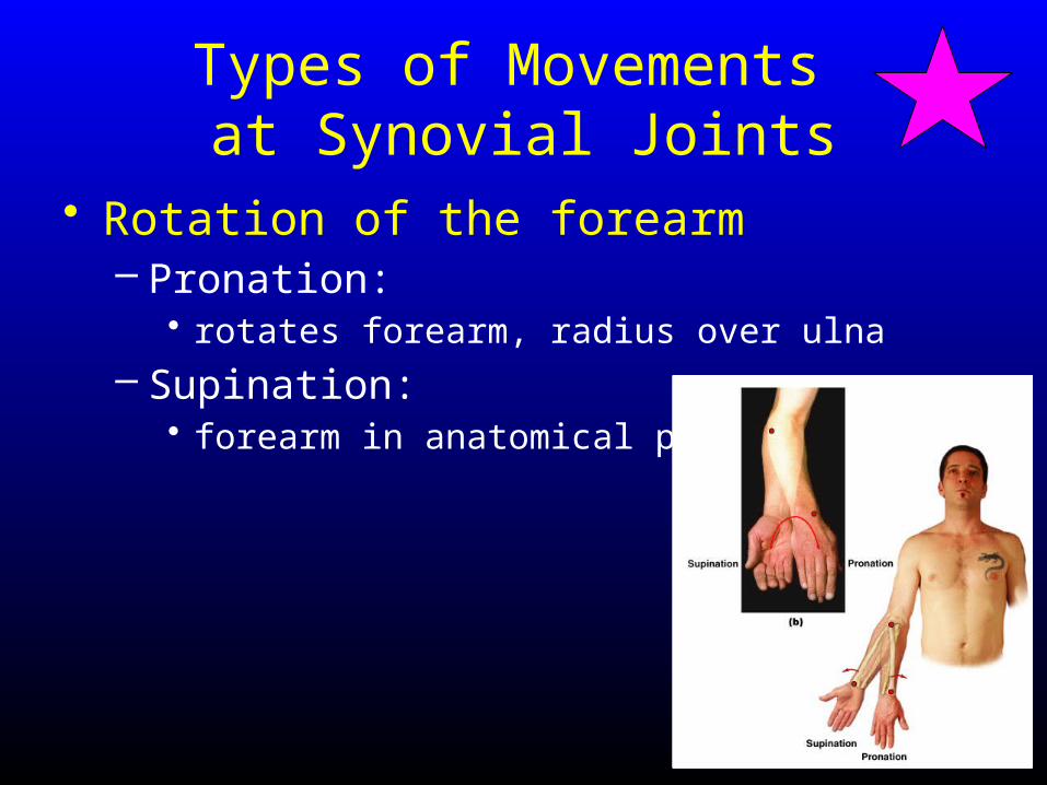
• Rotation of the forearm– Pronation:
• rotates forearm, radius over ulna
– Supination:• forearm in anatomical position
Special Movements
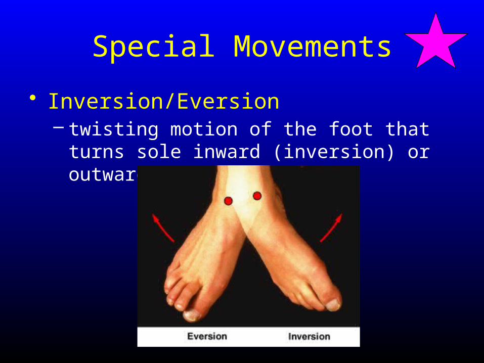
• Inversion/Eversion– twisting motion of the foot that turns sole
inward (inversion) or outward (eversion)
Special Movements
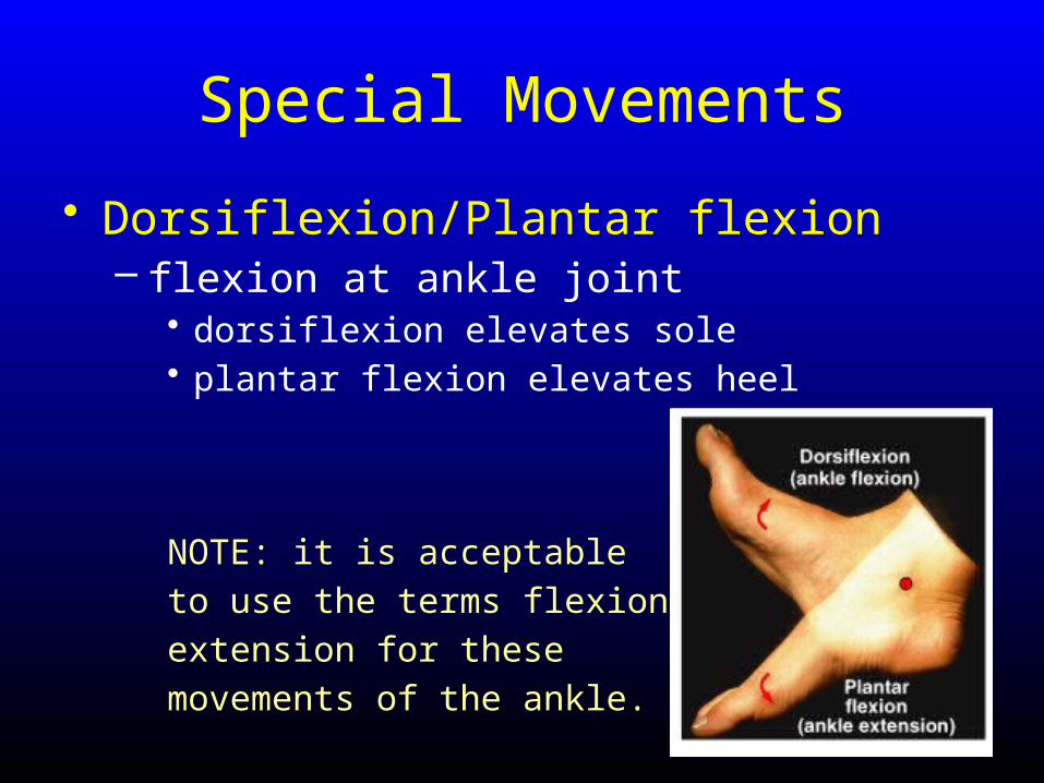
• Dorsiflexion/Plantar flexion– flexion at ankle joint
• dorsiflexion elevates sole• plantar flexion elevates heel
NOTE: it is acceptable
to use the terms flexion and
extension for these
movements of the ankle.
Special Movements
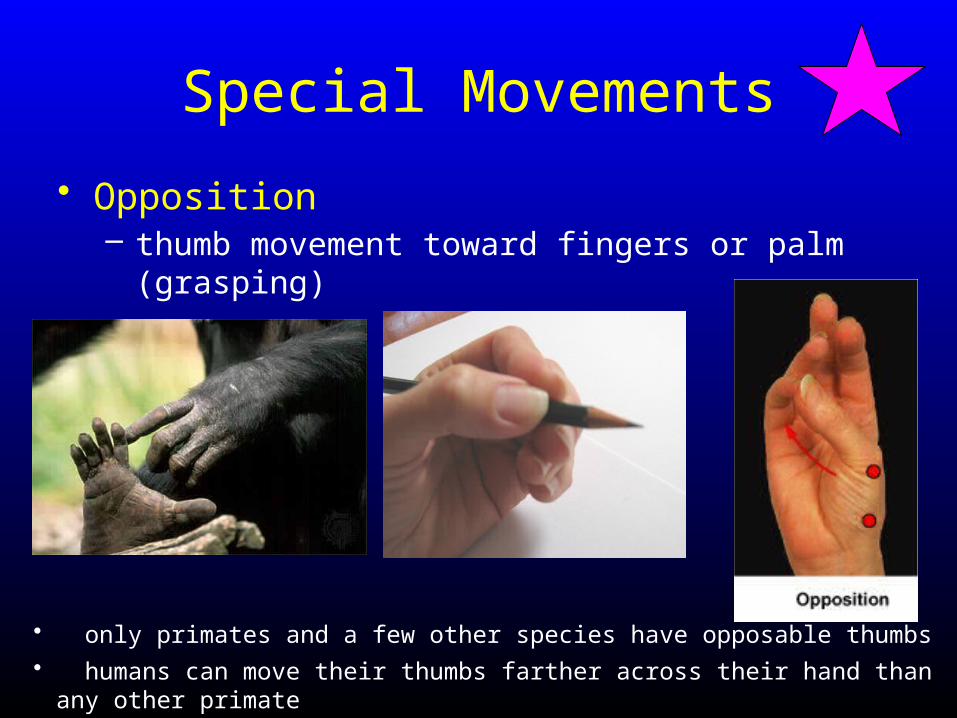
• Opposition– thumb movement toward fingers or palm (grasping)
• only primates and a few other species have opposable thumbs• humans can move their thumbs farther across their hand than any other primate
Special Movements
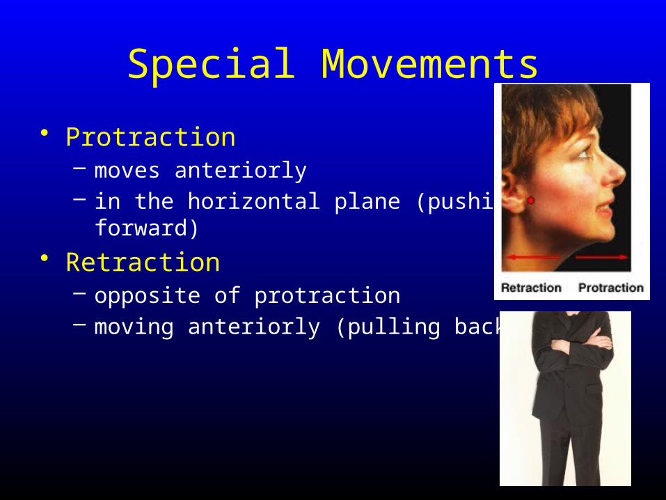
• Protraction– moves anteriorly– in the horizontal plane (pushing forward)
• Retraction– opposite of protraction– moving anteriorly (pulling back)
Special Movements
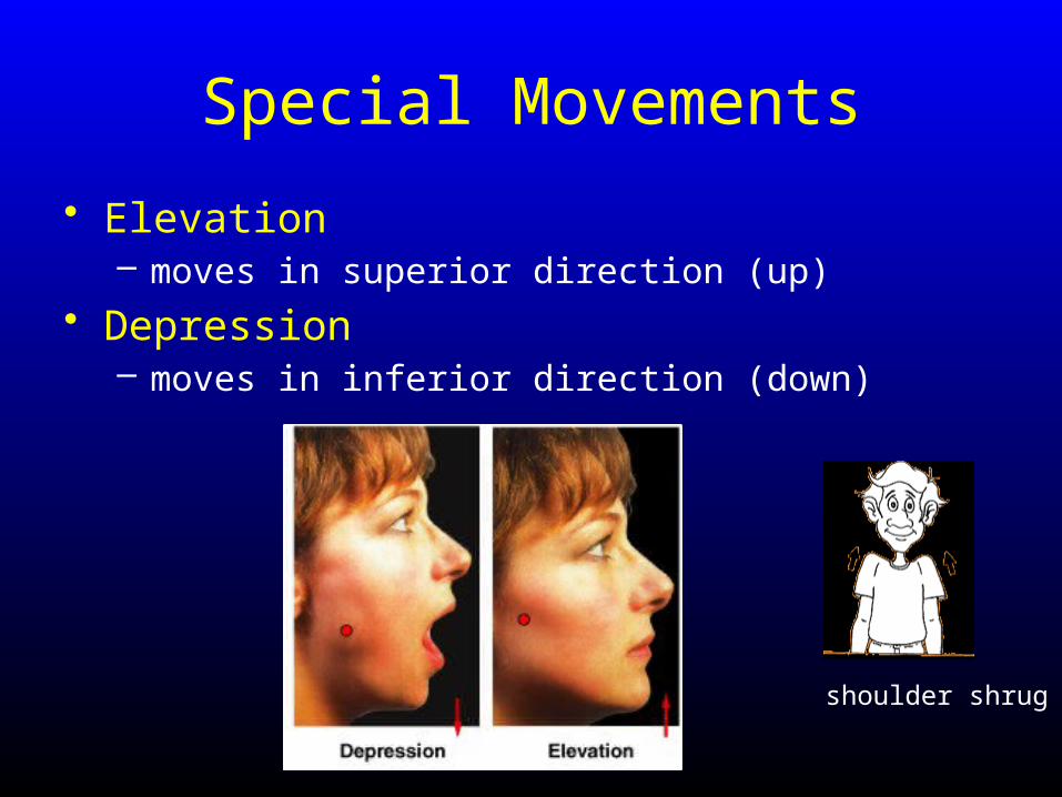
• Elevation– moves in superior direction (up)
• Depression– moves in inferior direction (down)
shoulder shrug
Special Movements
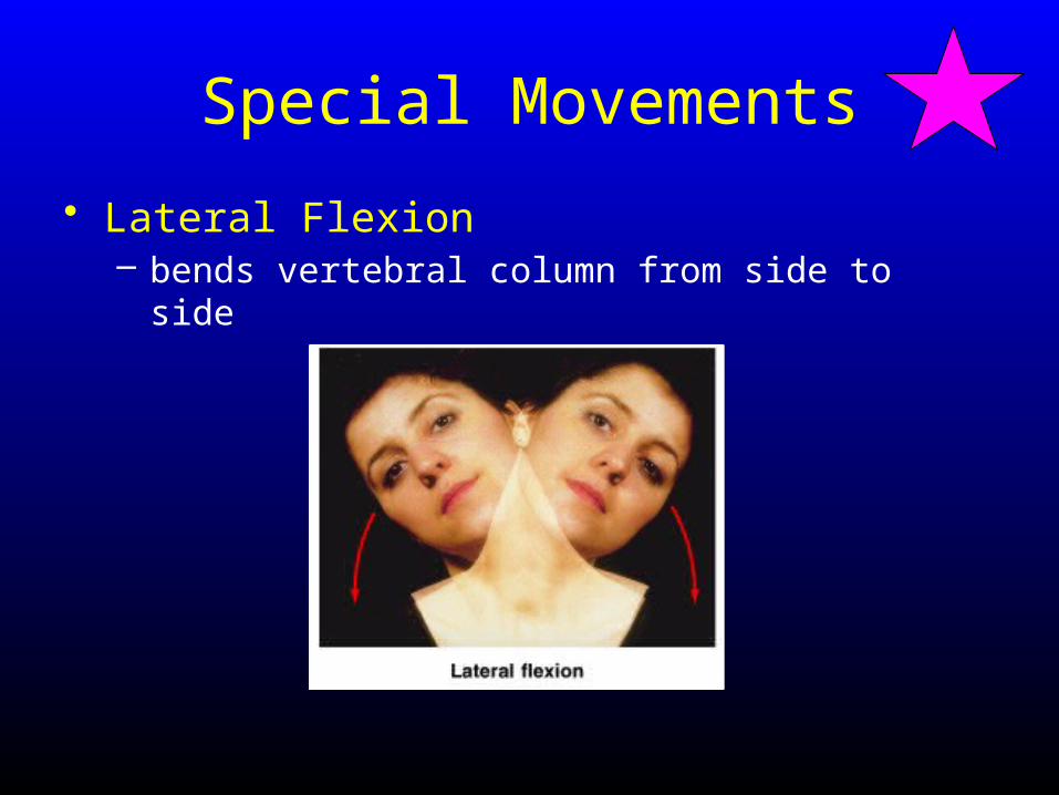
• Lateral Flexion– bends vertebral column from side to side

Synovial Joint Types
Classified by:1. Movement Type
2. Structure Type
Synovial Joint Classification Based on Movement
Synovial Joint Classification Based on Movement
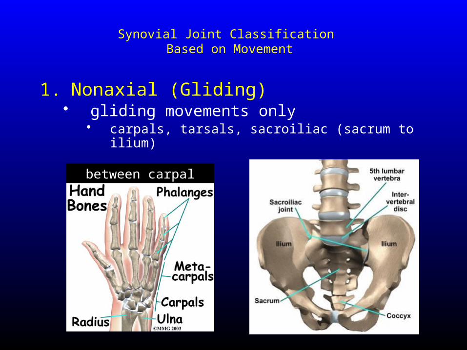
1. Nonaxial (Gliding) • gliding movements only
• carpals, tarsals, sacroiliac (sacrum to ilium)
between carpal bones

Synovial Joint Classification Based on Movement
2. Uniaxial (monoaxial)• angular movement in one plane
• hinge joints (temporomandibular joint)• pivot joints (atlas to axis and proximal radioulnar)
Synovial Joint Classification Based on Movement
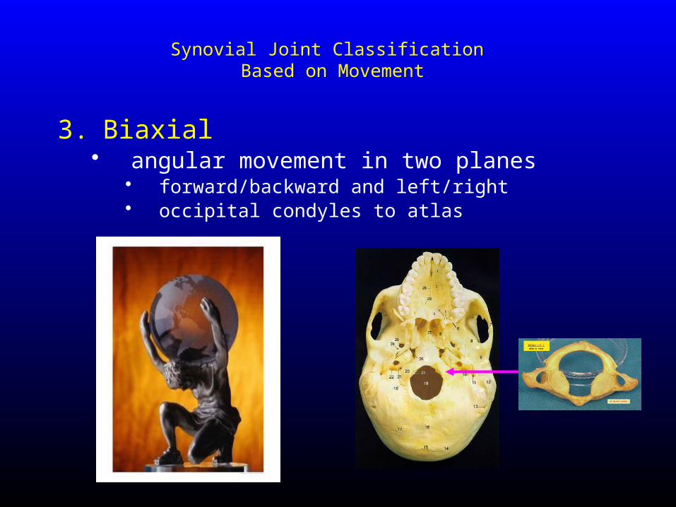
3. Biaxial • angular movement in two planes
• forward/backward and left/right• occipital condyles to atlas
Synovial Joint Classification Based on Movement
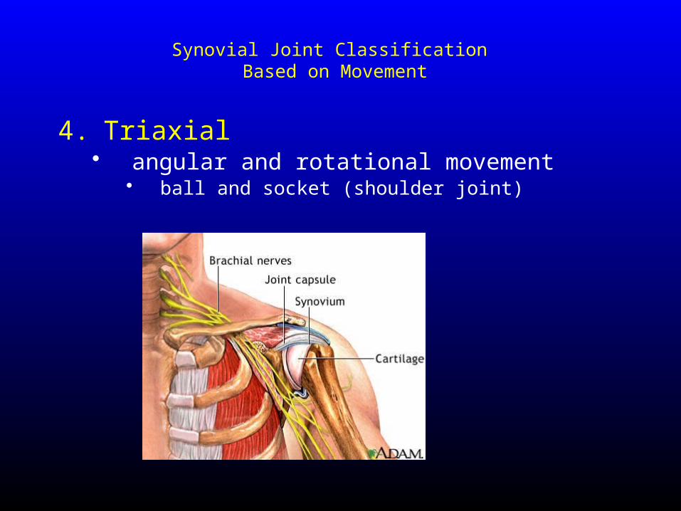
4. Triaxial • angular and rotational movement
• ball and socket (shoulder joint)
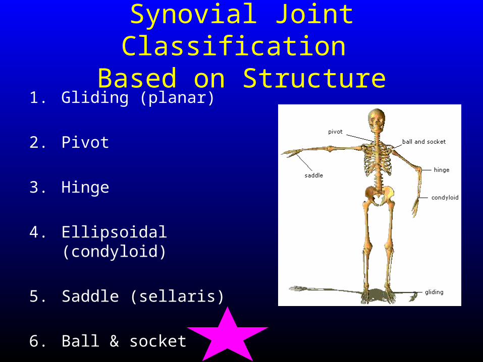
Synovial Joint Classification Based on Structure
Synovial Joint Classification Based on Structure
1. Gliding (planar)
2. Pivot
3. Hinge
4. Ellipsoidal (condyloid)
5. Saddle (sellaris)
6. Ball & socket
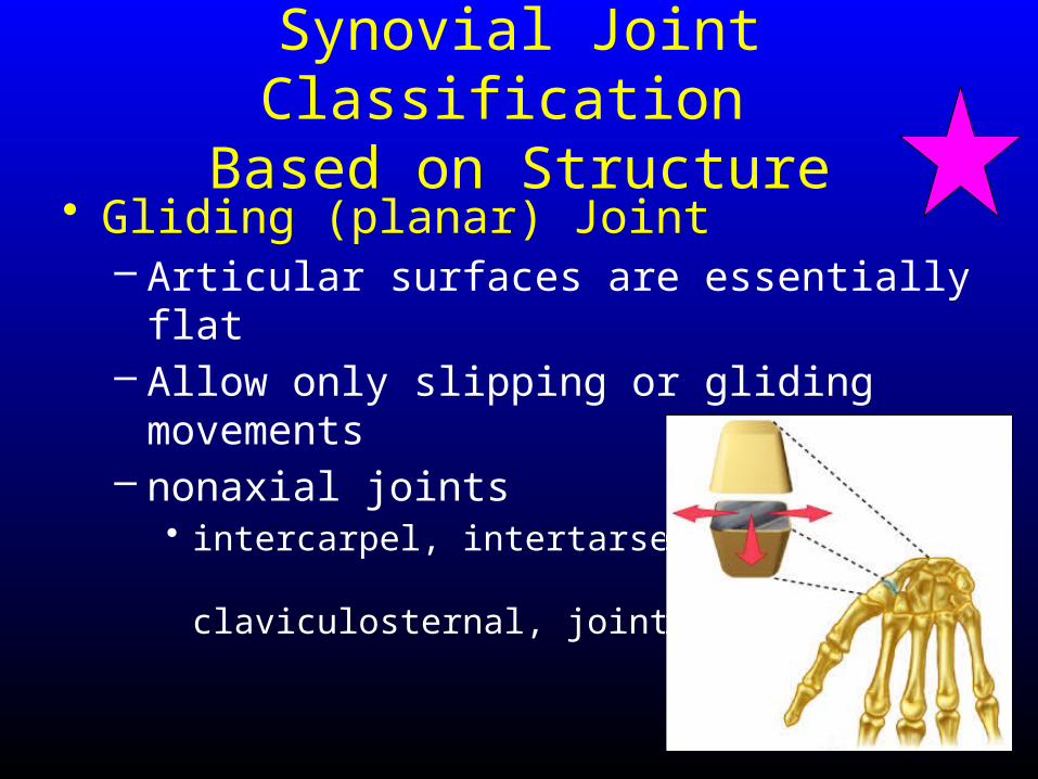
Synovial Joint Classification Based on Structure
• Gliding (planar) Joint – Articular surfaces are essentially flat– Allow only slipping or gliding movements– nonaxial joints
• intercarpel, intertarsel, claviculosternal, joints
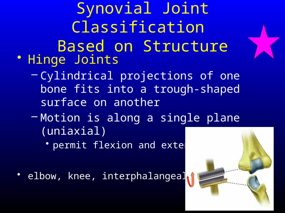
Synovial Joint Classification Based on Structure
• Hinge Joints – Cylindrical projections of one bone fits into a
trough-shaped surface on another– Motion is along a single plane (uniaxial)
• permit flexion and extension only
• elbow, knee, interphalangeal joints
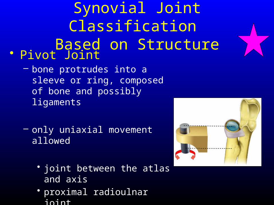
Synovial Joint Classification Based on Structure
• Pivot Joint– bone protrudes into a sleeve or
ring, composed of bone and possibly ligaments
– only uniaxial movement allowed
• joint between the atlas and axis• proximal radioulnar joint
Synovial Joint Classification Based on Structure
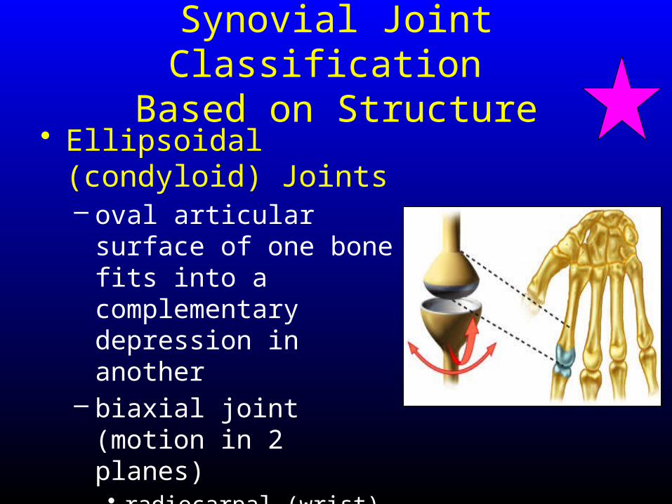
• Ellipsoidal (condyloid) Joints – oval articular surface of
one bone fits into a complementary depression in another
– biaxial joint (motion in 2 planes)• radiocarpal (wrist) joints, • metacarpophalangeal
(knuckle) joints

Synovial Joint Classification Based on Structure
• Saddle Joints– Similar to ellipsoidal joints but allow greater
movement– articular surface has both a concave and a
convex surface
• carpometacarpal joint of the thumb
Synovial Joint Classification Based on Structure
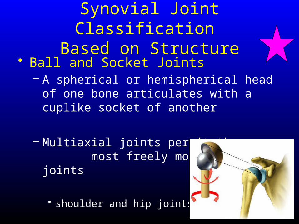
• Ball and Socket Joints– A spherical or hemispherical head of one
bone articulates with a cuplike socket of another
– Multiaxial joints permit the most freely moving synovial joints
• shoulder and hip joints
Specific Articulations
1. Intervertebral Articulations
2. Knee Joint
3. Shoulder Joint (maybe)
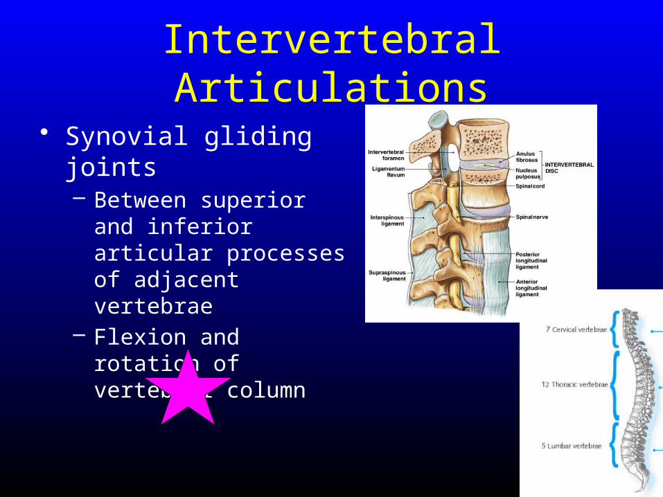
Intervertebral Articulations
• Synovial gliding joints– Between superior and
inferior articular processes of adjacent vertebrae
– Flexion and rotation of vertebral column
Intervertebral Articulations
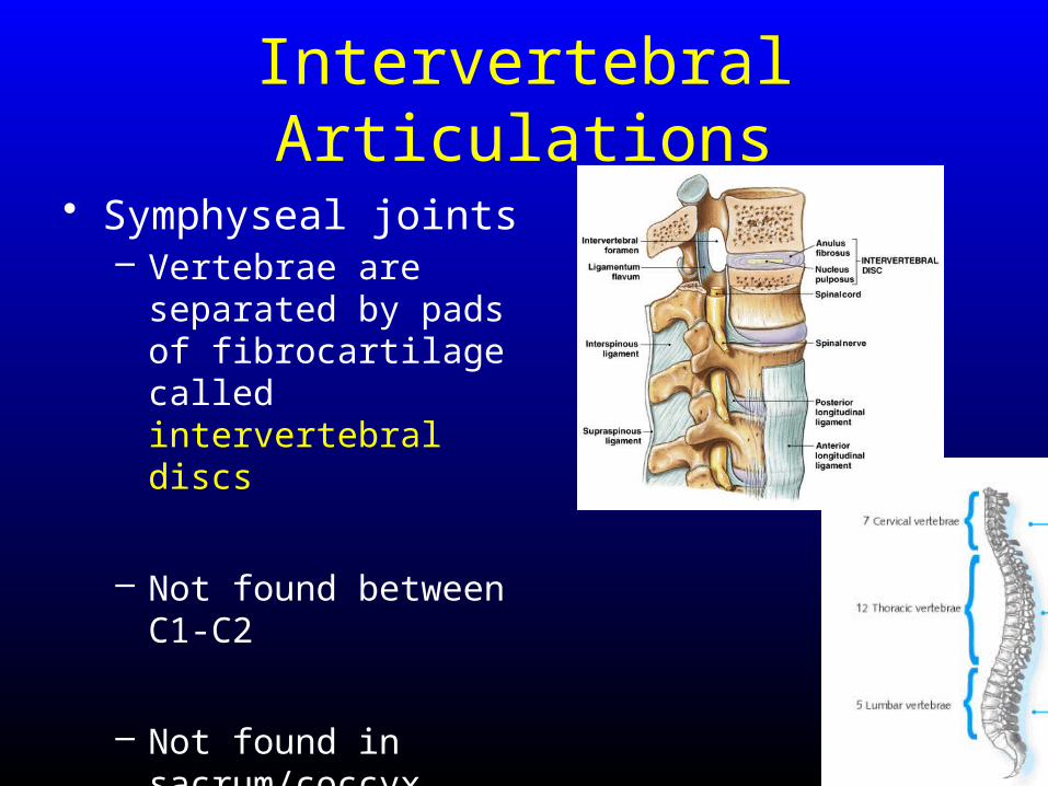
• Symphyseal joints– Vertebrae are separated
by pads of fibrocartilage called intervertebral discs
– Not found between C1-C2
– Not found in sacrum/coccyx where bones are fused
The Intervertebral Discs
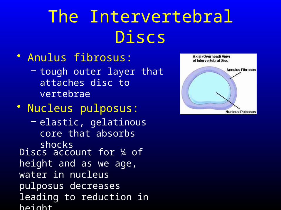
• Anulus fibrosus:– tough outer layer that attaches
disc to vertebrae
• Nucleus pulposus:– elastic, gelatinous core that
absorbs shocks
Discs account for ¼ of height and as we age, water in nucleus pulposus decreases leading to reduction in height
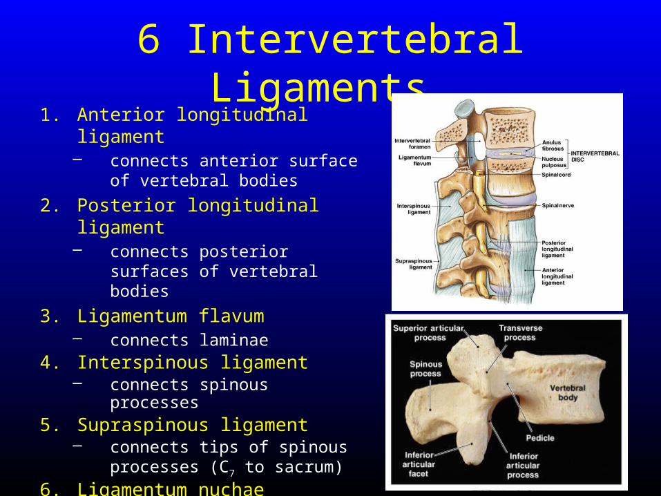
6 Intervertebral Ligaments 1. Anterior longitudinal ligament
– connects anterior surface of vertebral bodies
2. Posterior longitudinal ligament– connects posterior surfaces of
vertebral bodies
3. Ligamentum flavum– connects laminae
4. Interspinous ligament – connects spinous processes
5. Supraspinous ligament– connects tips of spinous
processes (C7 to sacrum)
6. Ligamentum nuchae– continues supraspinous ligament
(C7 to skull)
Damage to Intervertebral Discs
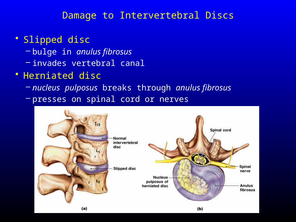
• Slipped disc– bulge in anulus fibrosus – invades vertebral canal
• Herniated disc– nucleus pulposus breaks through anulus fibrosus– presses on spinal cord or nerves
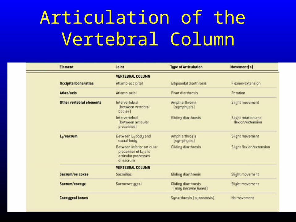
Articulation of the Vertebral Column
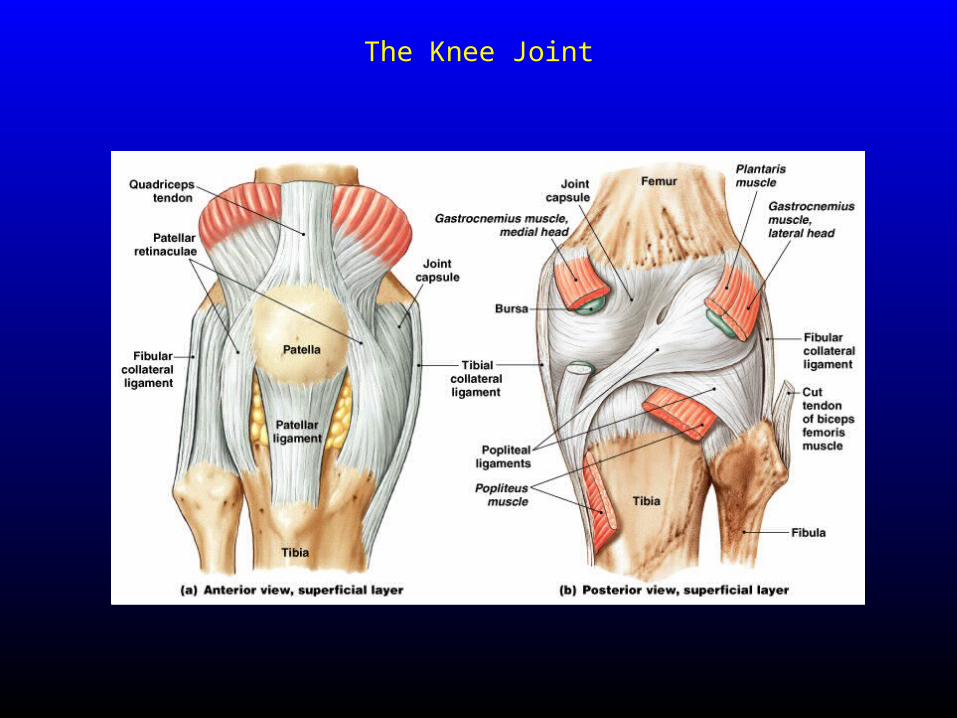
The Knee Joint
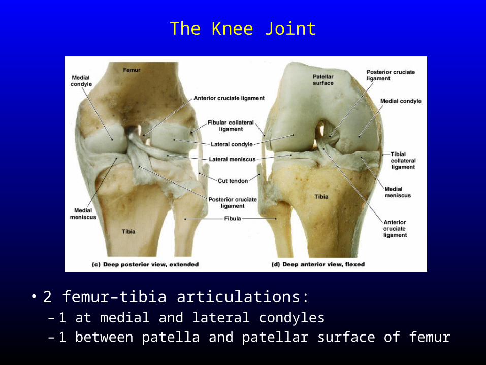
The Knee Joint
• 2 femur–tibia articulations:– 1 at medial and lateral condyles– 1 between patella and patellar surface of femur
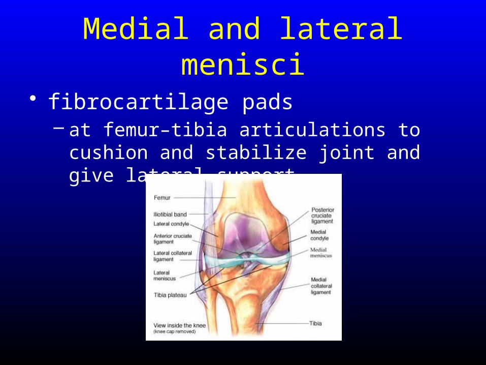
Medial and lateral menisci
• fibrocartilage pads – at femur–tibia articulations to cushion and
stabilize joint and give lateral support
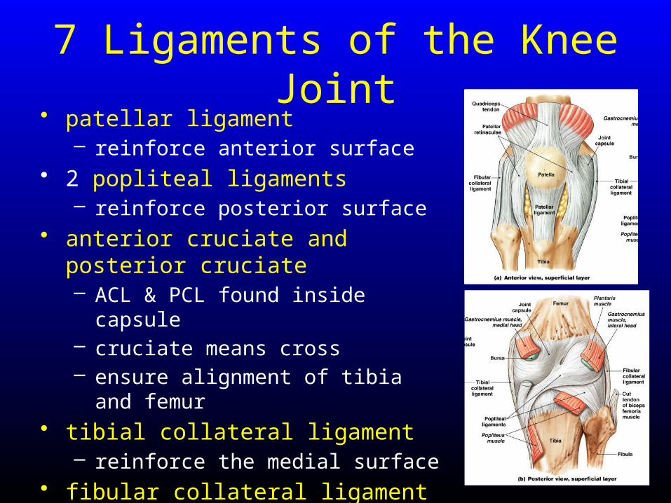
7 Ligaments of the Knee Joint• patellar ligament
– reinforce anterior surface• 2 popliteal ligaments
– reinforce posterior surface• anterior cruciate and posterior
cruciate– ACL & PCL found inside capsule– cruciate means cross– ensure alignment of tibia and femur
• tibial collateral ligament – reinforce the medial surface
• fibular collateral ligament– reinforce the lateral surface
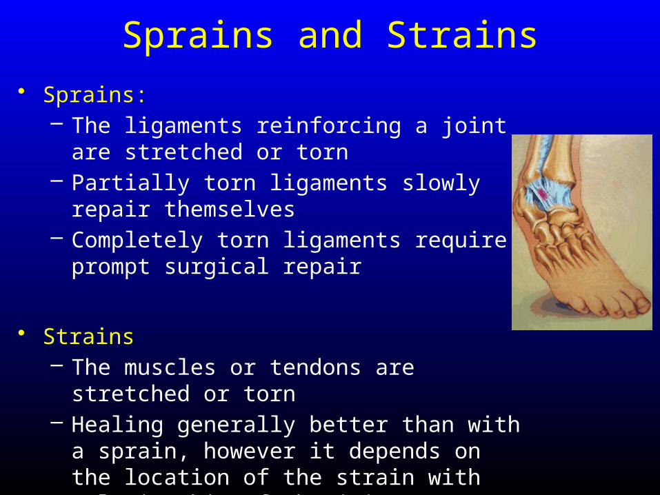
Sprains and Strains• Sprains:
– The ligaments reinforcing a joint are stretched or torn
– Partially torn ligaments slowly repair themselves
– Completely torn ligaments require prompt surgical repair
• Strains– The muscles or tendons are stretched or torn– Healing generally better than with a sprain,
however it depends on the location of the strain with relationship of the joint
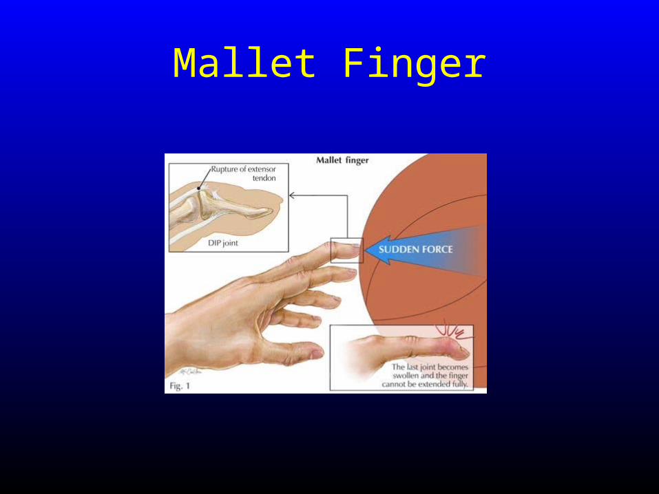
Mallet Finger
Bursitis and Tendonitis
• Bursitis– An inflammation of a bursa, usually caused by a blow
or friction– Symptoms are pain and swelling– Treated with anti-inflammatory drugs; excessive fluid
may be aspirated
• Tendonitis– Inflammation of tendon sheaths typically caused by
overuse– Symptoms and treatment are similar to bursitis
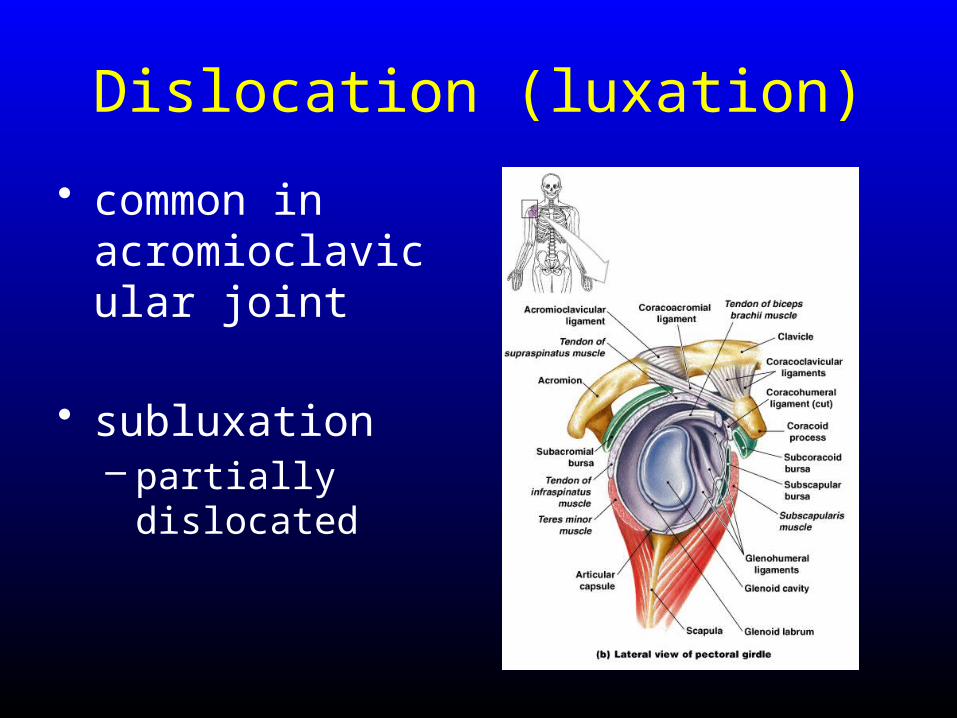
Dislocation (luxation)
• common in acromioclavicular joint
• subluxation– partially
dislocated
Rheumatism
• general term to describe pain/stiffness of the skeletal and/or muscular systems
• There are more than 100 different types of inflammatory or degenerative diseases that damage the joints
– Arthritis is a rheumatism of the synovial joints
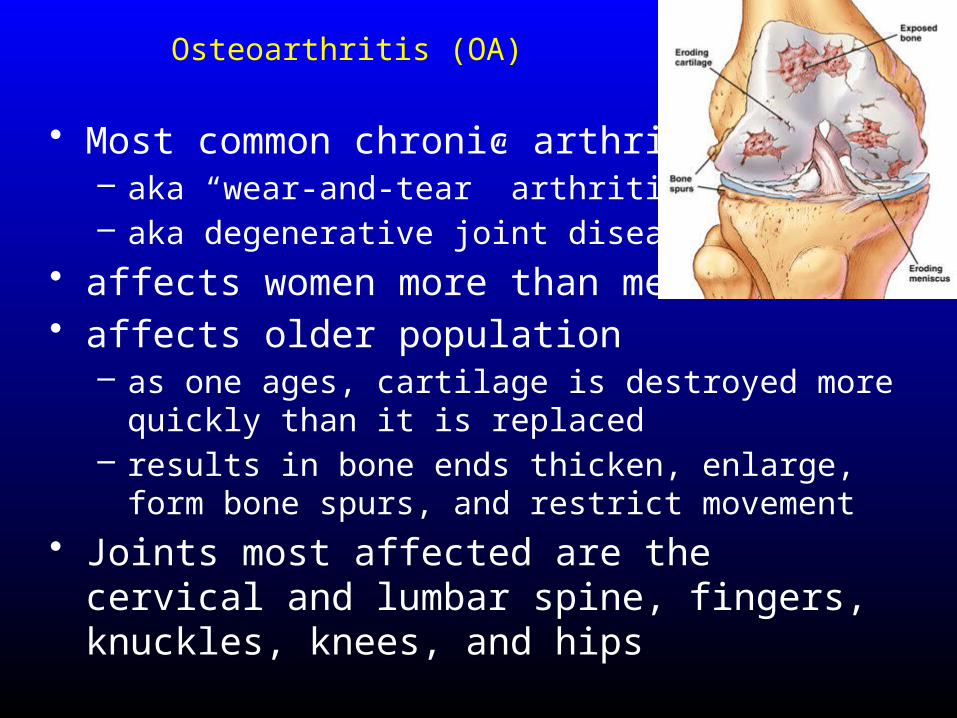
Osteoarthritis (OA)
• Most common chronic arthritis– aka “wear-and-tear” arthritis– aka degenerative joint disease (DJD)
• affects women more than men• affects older population
– as one ages, cartilage is destroyed more quickly than it is replaced
– results in bone ends thicken, enlarge, form bone spurs, and restrict movement
• Joints most affected are the cervical and lumbar spine, fingers, knuckles, knees, and hips
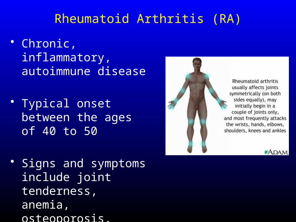
Rheumatoid Arthritis (RA)
• Chronic, inflammatory, autoimmune disease
• Typical onset between the ages of 40 to 50
• Signs and symptoms include joint tenderness, anemia, osteoporosis, muscle atrophy, and cardiovascular problems
Gouty Arthritis (Gout)
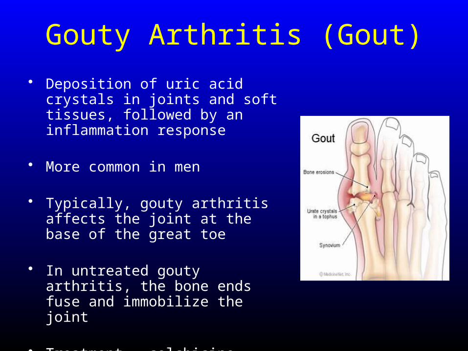
• Deposition of uric acid crystals in joints and soft tissues, followed by an inflammation response
• More common in men
• Typically, gouty arthritis affects the joint at the base of the great toe
• In untreated gouty arthritis, the bone ends fuse and immobilize the joint
• Treatment – colchicine, nonsteroidal anti-inflammatory drugs, and glucocorticoids

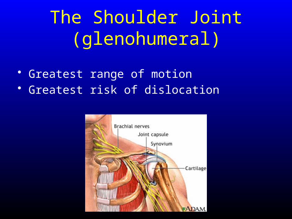
The Shoulder Joint(glenohumeral)
• Greatest range of motion• Greatest risk of dislocation
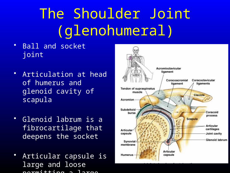
The Shoulder Joint(glenohumeral)
• Ball and socket joint
• Articulation at head of humerus and glenoid cavity of scapula
• Glenoid labrum is a fibrocartilage that deepens the socket
• Articular capsule is large and loose permitting a large range of motion
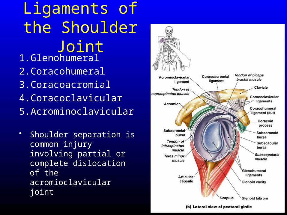
Ligaments of the Shoulder Joint
1. Glenohumeral
2. Coracohumeral
3. Coracoacromial
4. Coracoclavicular
5. Acrominoclavicular
• Shoulder separation is common injury involving partial or complete dislocation of the acromioclavicular joint
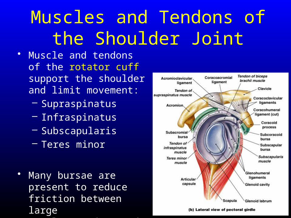
Muscles and Tendons of the Shoulder Joint
• Muscle and tendons of the rotator cuff support the shoulder and limit movement:– Supraspinatus– Infraspinatus– Subscapularis– Teres minor
• Many bursae are present to reduce friction between large muscles/tendons and shoulder capsule