Joints or Articulations. A joint, or articulation, is the place of union between two or more bones.
Upload
norma-popeCategory
view
227download
0
Articulations
Chapter 9

Introduction
• Skeleton composition– Many bones joined together (articulated)
• Articulation– Joints
• Advantages and disadvantages of strong vs. weak joints
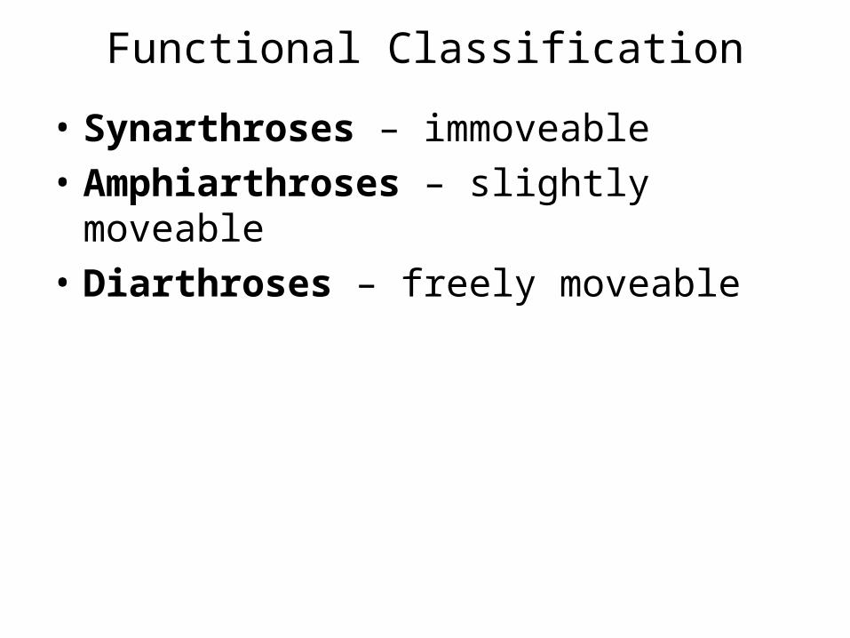
Functional Classification
• Synarthroses – immoveable
• Amphiarthroses – slightly moveable
• Diarthroses – freely moveable

Structural Classification
• Fibrous joints– Lack joint cavity– Held together by fibrous connective tissue– Synarthritic or amphiarthritic
• Cartilaginous joints– Lack joint cavity– Held together by cartilage– Synarthritic or amphiarthritic
• Synovial joints– Joint cavity present– Held together by ligaments– Diarthritic

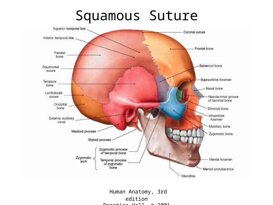
Synarthroses
• Sutures – found between skull bones– Held together by dense fibrous connective
tissue
• Types– Serrate– Squamous– Plane– Synostoses

Human Anatomy, 3rd editionPrentice Hall, © 2001
Serrate Suture
Human Anatomy, 3rd editionPrentice Hall, © 2001
Squamous Suture

Human Anatomy, 3rd editionPrentice Hall, © 2001
Plane Suture

Human Anatomy, 3rd editionPrentice Hall, © 2001
Synostoses
Human Anatomy, 3rd editionPrentice Hall, © 2001
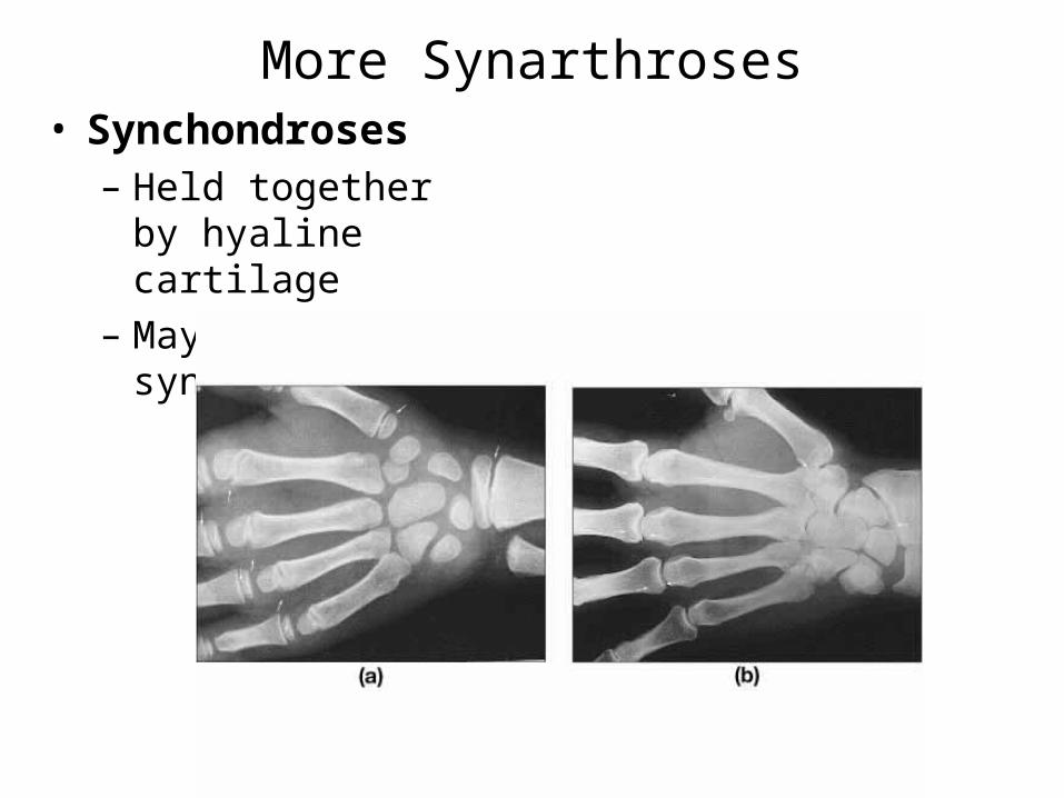
More Synarthroses• Synchondroses
– Held together by hyaline cartilage
– May form synostoses

More Synarthroses
• Gomphosis– Held together by
ligaments
– Cone-shaped pet fits into a socket
Human Anatomy, 3rd editionPrentice Hall, © 2001
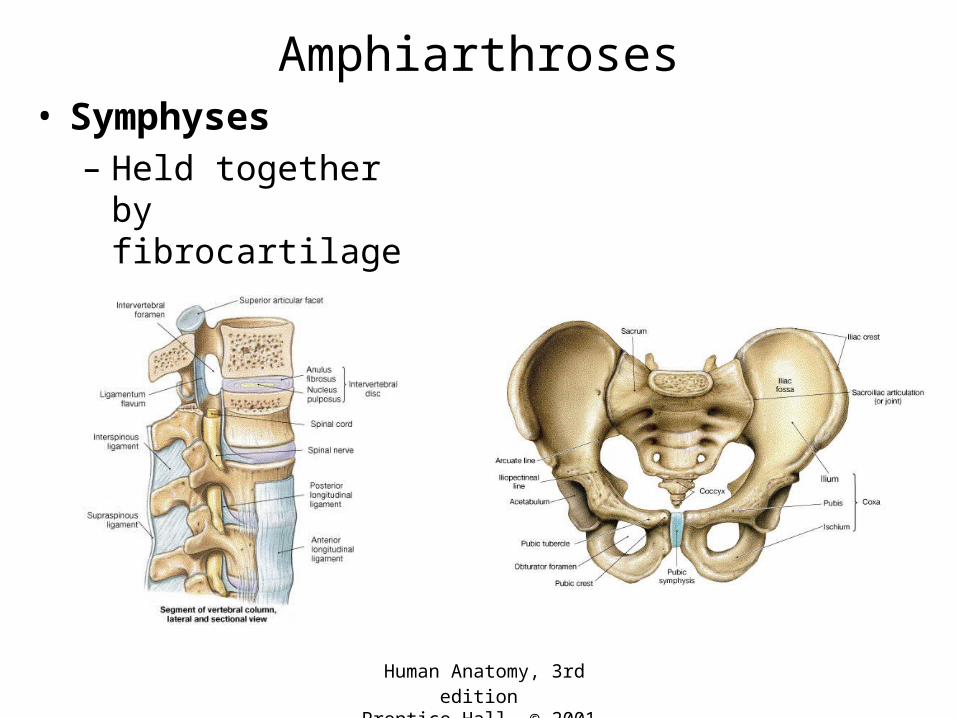
Amphiarthroses• Symphyses
– Held together by fibrocartilage

Human Anatomy, 3rd editionPrentice Hall, © 2001
More Amphiarthroses• Syndesmoses
– Held together by collagen fibers
Human Anatomy, 3rd editionPrentice Hall, © 2001
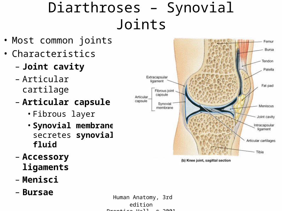
Diarthroses – Synovial Joints
• Most common joints• Characteristics
– Joint cavity
– Articular cartilage
– Articular capsule• Fibrous layer
• Synovial membrane secretes synovial fluid
– Accessory ligaments
– Menisci
– Bursae
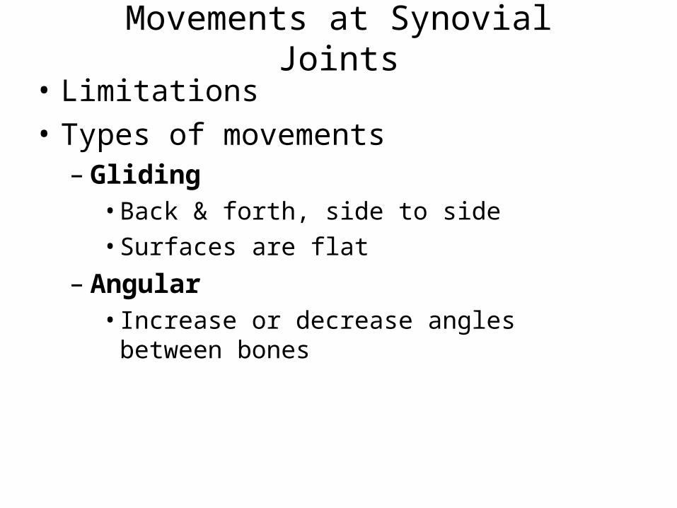
Movements at Synovial Joints
• Limitations
• Types of movements– Gliding
• Back & forth, side to side
• Surfaces are flat
– Angular• Increase or decrease angles between bones

Types of Angular Movements
Human Anatomy, 3rd editionPrentice Hall, © 2001
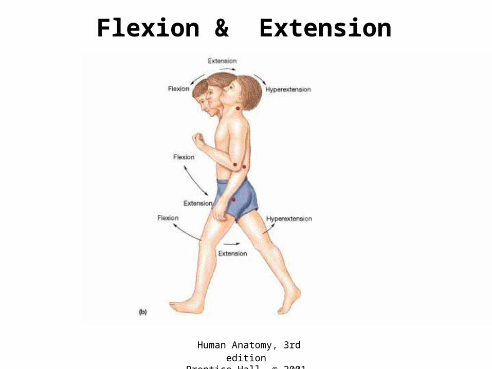
Flexion & Extension
Human Anatomy, 3rd editionPrentice Hall, © 2001
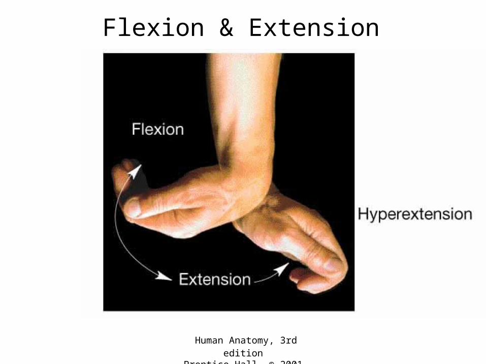
Flexion & Extension

Human Anatomy, 3rd editionPrentice Hall, © 2001
Dorsiflexion & Plantar Flexion

Human Anatomy, 3rd editionPrentice Hall, © 2001
Abduction & Adduction
Human Anatomy, 3rd editionPrentice Hall, © 2001
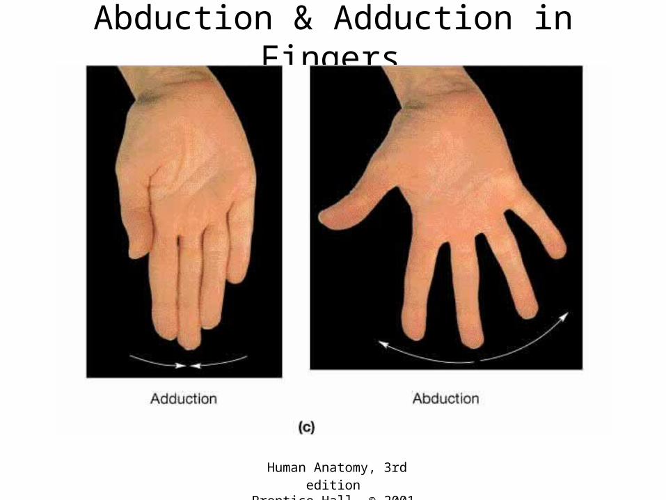
Abduction & Adduction in Fingers
Human Anatomy, 3rd editionPrentice Hall, © 2001
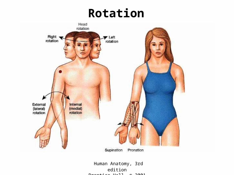
Rotation
Human Anatomy, 3rd editionPrentice Hall, © 2001
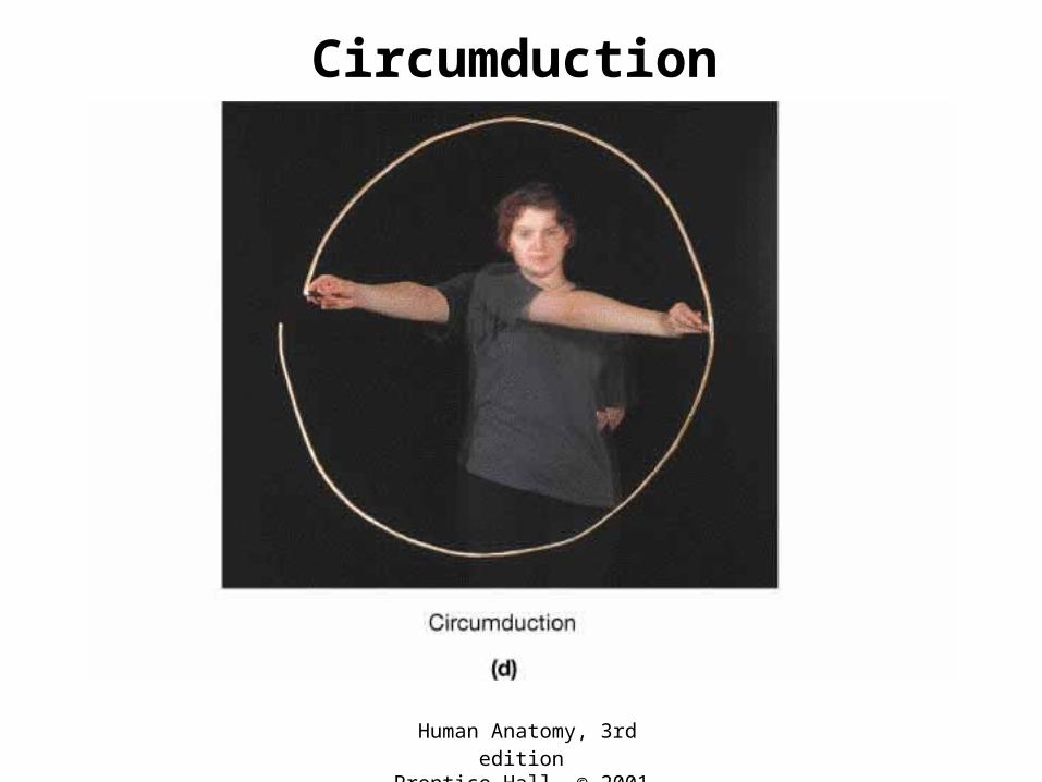
Circumduction

Special Types of Angular Movements
Human Anatomy, 3rd editionPrentice Hall, © 2001
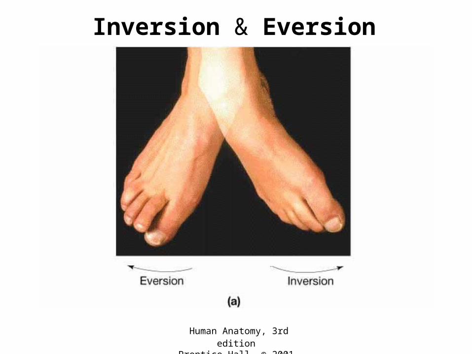
Inversion & Eversion

Human Anatomy, 3rd editionPrentice Hall, © 2001
Protraction & Retraction
Human Anatomy, 3rd editionPrentice Hall, © 2001
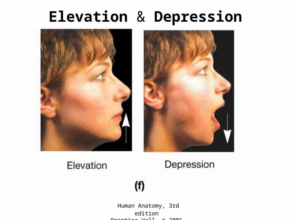
Elevation & Depression

Subtypes of Synovial Joints

Human Anatomy, 3rd editionPrentice Hall, © 2001
Gliding Joint• Flat surfaces• Biaxial movement only• eg. Between carpals
Human Anatomy, 3rd editionPrentice Hall, © 2001
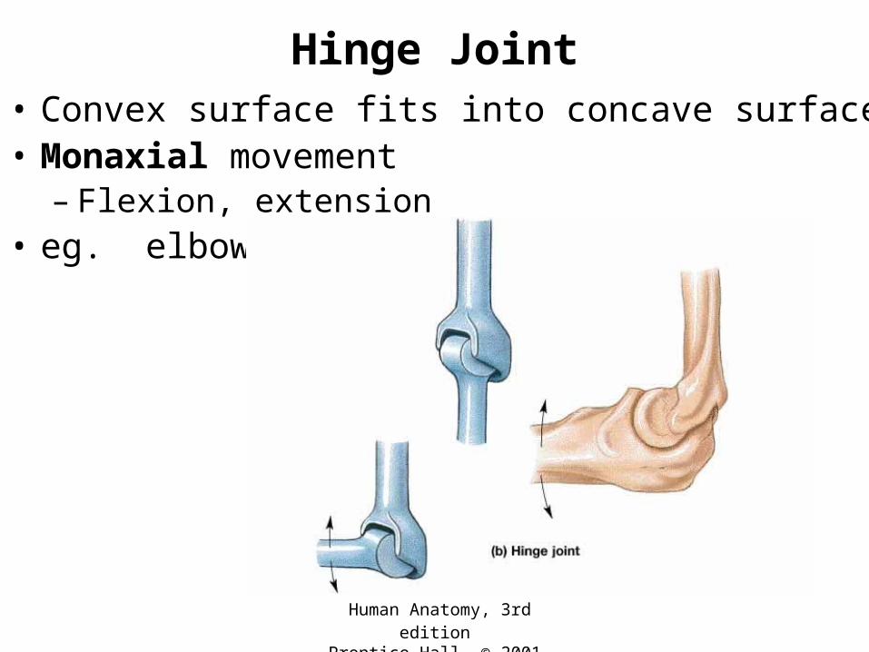
Hinge Joint• Convex surface fits into concave surface• Monaxial movement
– Flexion, extension• eg. elbow

Human Anatomy, 3rd editionPrentice Hall, © 2001
Pivot Joint• Rounded surface articulates within a ring• Monaxial movement
– Rotation• eg. between atlas & axis

Human Anatomy, 3rd editionPrentice Hall, © 2001
Ellipsoidal Joint• Oval-shaped condyle fits into elliptical cavity• Biaxial movement• eg. between radius
& carpals

Human Anatomy, 3rd editionPrentice Hall, © 2001
Saddle Joint
• Articular surfaces of both bones are concave in one direction and convex in the other
• biaxial movement• eg. Between 1st
metacarpal of thumb & trapezium

Human Anatomy, 3rd editionPrentice Hall, © 2001
Ball and Socket Joint
• Ball-like surface fits into cuplike depression
• Triaxial movement– Flexion, extension
– Abduction, adduction
– Rotation
• eg. shoulder
Human Anatomy, 3rd editionPrentice Hall, © 2001
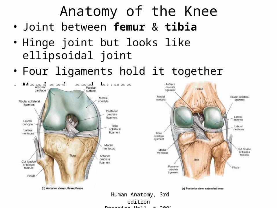
Anatomy of the Knee• Joint between femur & tibia• Hinge joint but looks like ellipsoidal joint• Four ligaments hold it together• Menisci and bursae are present

Disorders

Dislocation
http://www.sportsmed.buffalo.edu/info/fingerdis.html

Bursitis
http://www.healthopedia.com/pictures/bursitis.html
Human Anatomy, 3rd editionPrentice Hall, © 2001
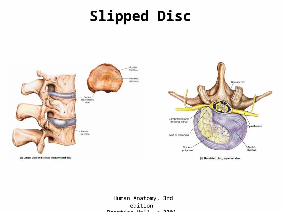
Slipped Disc

Human Anatomy, 3rd editionPrentice Hall, © 2001
Arthritis
• Arthritis – inflammation of joints– Osteoarthritis– Rheumatoid arthritis– Gouty arthritis

Gouty Arthritis
http://healthgate.partners.org/browsing/LearningCenter.asp?fileName=11825.xml&title=Gout

Osteoarthritis
http://www.abc.net.au/health/library/osteoarthritis_ff.htm

Rheumatoid Arthritis
http://arthritis.upmc.com/RheumatoidArthritis/Overview.htm