Articulations and Movement -...
43
Articulations and Movement
Transcript of Articulations and Movement -...

Articulations and Movement

Articulations or Joints
• Articulation or Joint – Place where two bones come together
– Freely movable to limited to no apparent movement
– Structure correlated with movement
• Named – According to bones or parts united at joint
– According to only one of articulating bones
– By Latin equivalent of common name

Classification of Joints
• Structural: Based on major connective tissue type that binds bones – Fibrous
– Cartilaginous
– Synovial
• Functional: Based on degree of motion – Synarthrosis: Nonmovable
– Amphiarthrosis: Slightly movable
– Diarthrosis: Freely movable

Fibrous joints
• Suture
– Bones tightly bound by minimal fiber
– Only found in skull
• Syndemoses – Bones connected by
ligaments
– E.g. tibiofibular ligament, interosseous membrane of radius/ulna
• Gomphoses – Peg in socket joint
– Only found in teeth/alveoli
Fig. 9.1 a, M&M

Fibrous joints • Suture
– Bones tightly bound by minimal fiber
– Only found in skull
• Syndemoses – Bones connected by
ligaments
– E.g. tibiofibular ligament, interosseous membrane of radius/ulna
• Gomphoses – Peg in socket joint
– Only found in teeth/alveoli
Fig. 8.4, M&M
Fig. 9.1 b, M&M

Fibrous joints
• Suture – Bones tightly bound by
minimal fiber
– Only found in skull
• Syndemosis – Bones connected by
ligaments
– E.g. tibiofibular ligament, interosseous membrane of radius/ulna
• Gomphosis
– Peg in socket joint
– Only found in teeth/alveoli
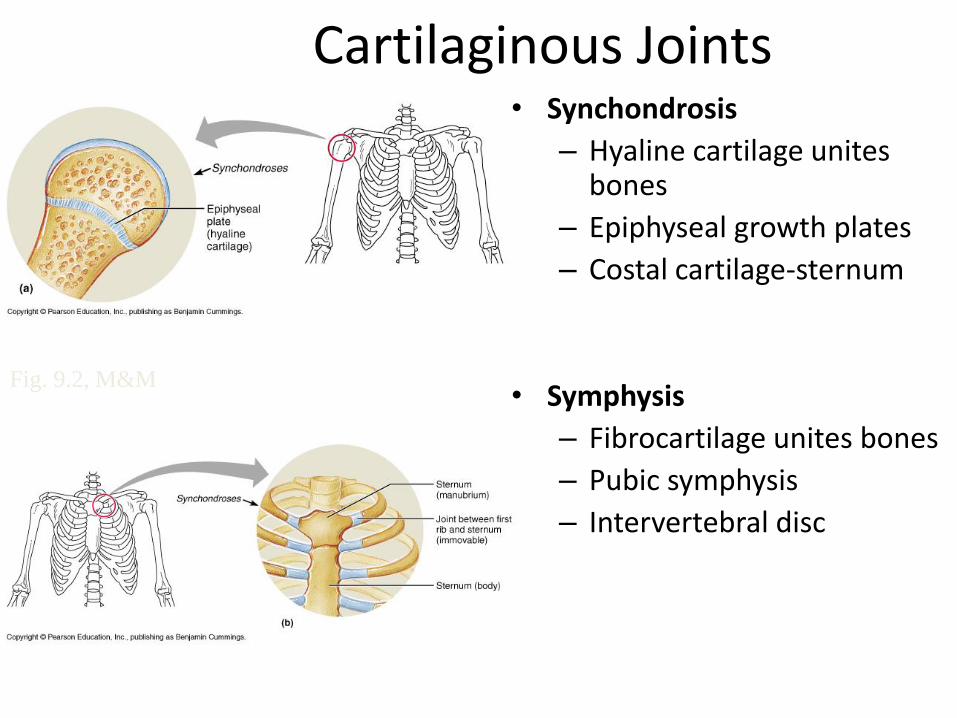
Cartilaginous Joints • Synchondrosis
– Hyaline cartilage unites bones
– Epiphyseal growth plates
– Costal cartilage-sternum
• Symphysis
– Fibrocartilage unites bones
– Pubic symphysis
– Intervertebral disc
Fig. 9.2, M&M
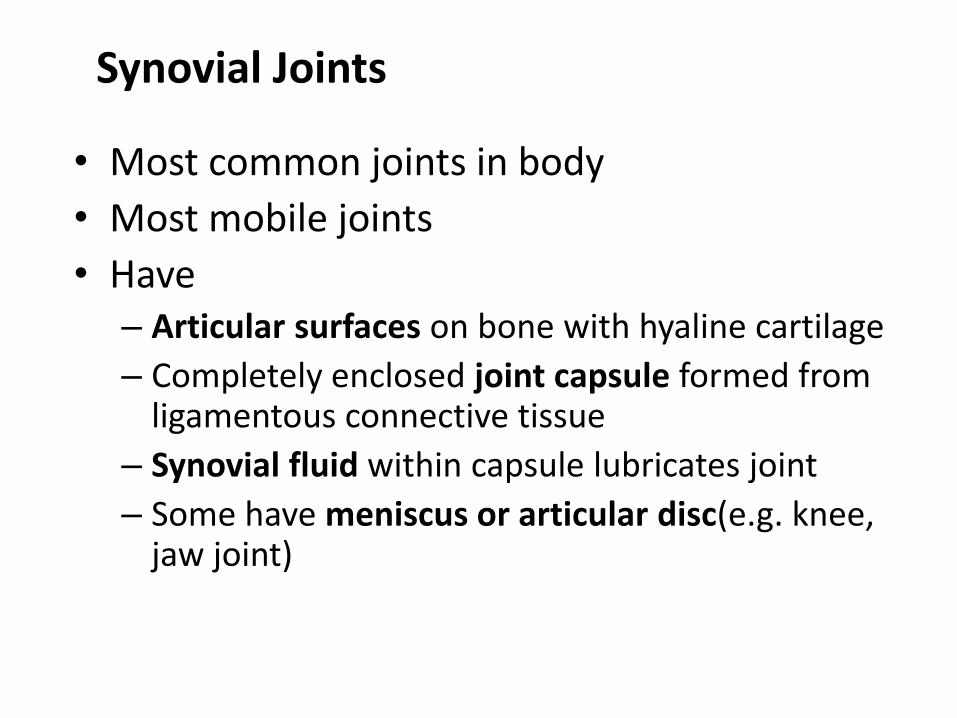
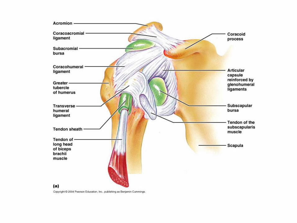
Synovial Joints
• Most common joints in body
• Most mobile joints
• Have – Articular surfaces on bone with hyaline cartilage
– Completely enclosed joint capsule formed from ligamentous connective tissue
– Synovial fluid within capsule lubricates joint
– Some have meniscus or articular disc(e.g. knee, jaw joint)
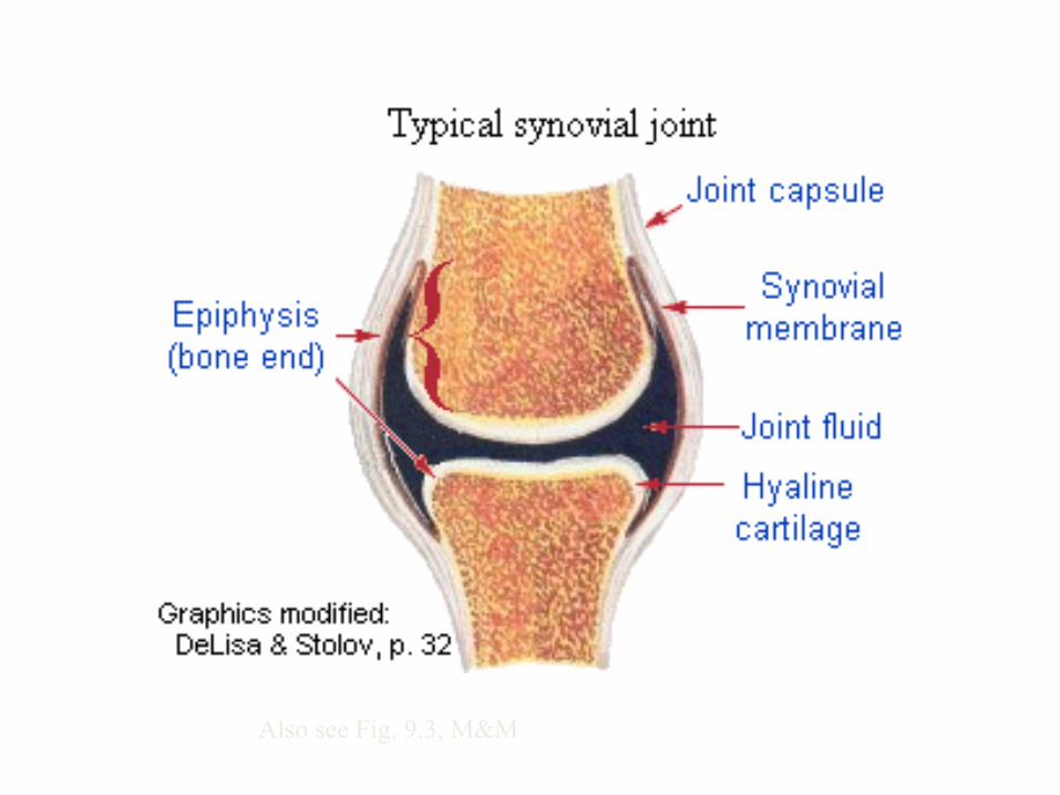
Also see Fig. 9.3, M&M

Synovial joints
• Components of synovial joints
– Articular cartilage • Resemble hyaline cartilage
– Matrix contains more water comparatively
• Has no perichondrium
• Slick and smooth, so reduce friction
• Separated by thin film of synovial fluid
Articular
Cartilage
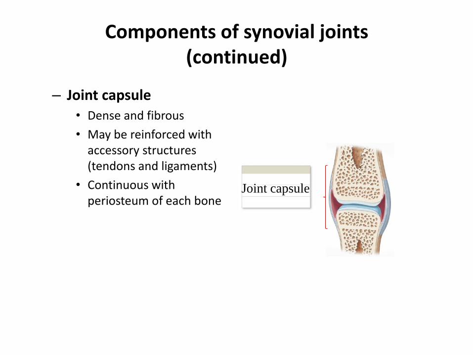
Components of synovial joints (continued)
– Joint capsule • Dense and fibrous
• May be reinforced with accessory structures (tendons and ligaments)
• Continuous with periosteum of each bone
Joint capsule
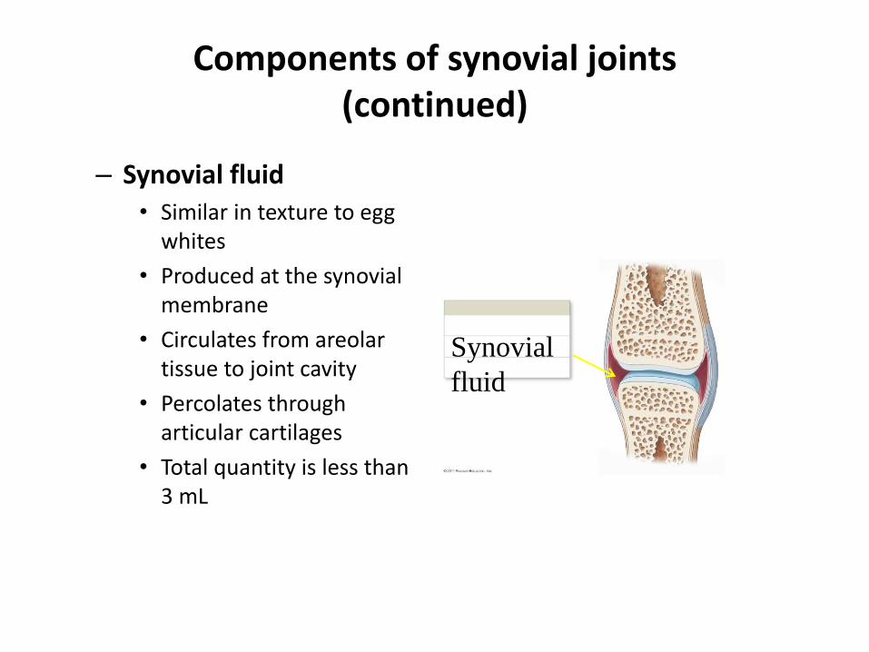
Components of synovial joints (continued)
– Synovial fluid • Similar in texture to egg
whites
• Produced at the synovial membrane
• Circulates from areolar tissue to joint cavity
• Percolates through articular cartilages
• Total quantity is less than 3 mL
Synovial
fluid
Functions of synovial fluid
– Lubrication • With articular cartilage compression, synovial fluid is squeezed out
and reduces friction between moving surfaces
– Synovial fluid distribution • Provide nutrients and oxygen, as well as waste disposal for the
chondrocytes of articular cartilages
• Compression and reexpansion of articular cartilages pump synovial fluid in and out of cartilage matrix
– Synovial fluid absorption • Distributes compression forces across articular surfaces and
outward to joint capsule
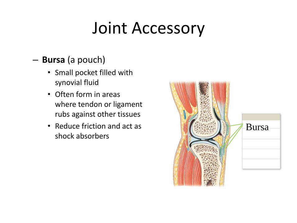
Joint Accessory
– Bursa (a pouch) • Small pocket filled with
synovial fluid
• Often form in areas where tendon or ligament rubs against other tissues
• Reduce friction and act as shock absorbers
Bursa


Accessory structures in knee (continued)
– Fat pads • Adipose tissue covered by
synovial membrane
• Protect articular cartilages
• Act as packing material for joint
– Meniscus (a crescent) • Pad of fibrous cartilage
between bones of synovial joint
• May subdivide joint cavity and affect fluid flow or allow variations in shapes of articular surfaces
Meniscus
Fat pad

Frolich, Human Anatomy,
Mechanics of Movement


• Accessory structures in knee – Tendons of quadriceps
• Pass across joint – Limit movement – Provide
mechanical support
• Accessory ligaments • __________________,
strengthen, and reinforce joint • Intrinsic ligaments
– Localized thickening of joint capsule
– Example: cruciate liagments of knee
• ___________________ ligaments – Separate from joint capsule – May pass inside (intracapsular) or
outside (extracapsular) the joint capsule
– Intracapsular example: cruciate ligaments
– Extracapsular example: patellar ligament

Synovial joints
• Motion vs. strength in joints
– Greater range of motion = ______________ joint • Examples:
– Synarthrosis (strongest type of joint, no movement)
– Diarthrosis (far weaker but broad range of motion)
– Displacement (luxation) • Movement beyond normal range of motion
• Articulating surfaces forced out of position
• Can damage joint structures
• No pain from inside joint but from nerves or surrounding structures

Types of Synovial Joints
• Plane or gliding
• Saddle
• Hinge
• Pivot
• Ball-and-socket
• Ellipsoid
Plane and Pivot Joints
• Plane or Gliding joints
– Monoaxial
– Example:Articular processes between vertebrae
• Pivot joints
– Monoaxial
– Example: Articulation between dens of axis and atlas
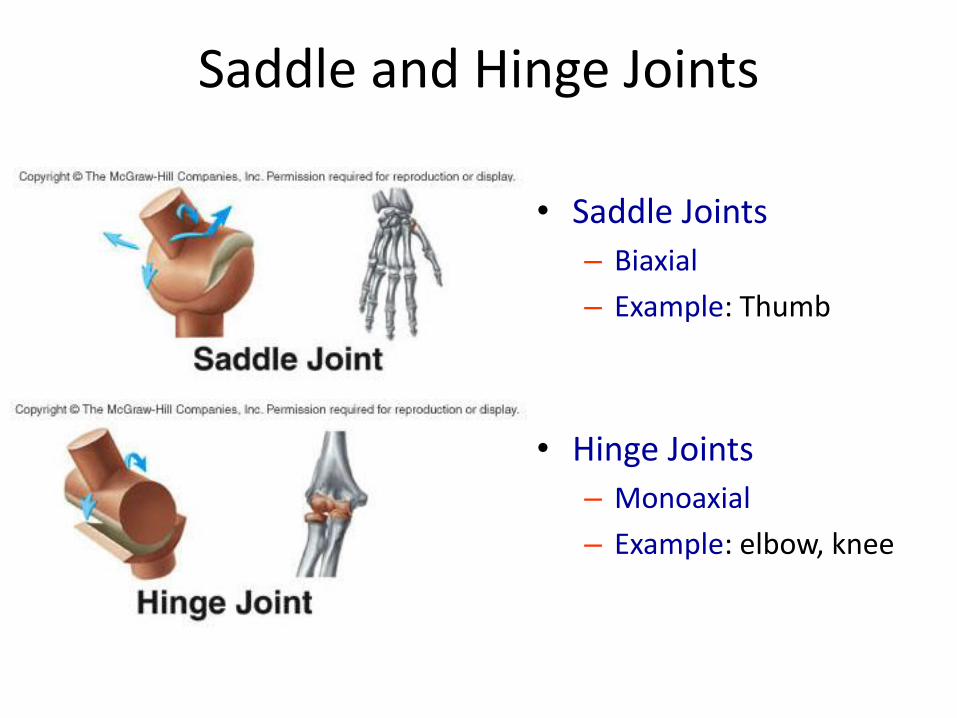
Saddle and Hinge Joints
• Saddle Joints
– Biaxial
– Example: Thumb
• Hinge Joints
– Monoaxial
– Example: elbow, knee
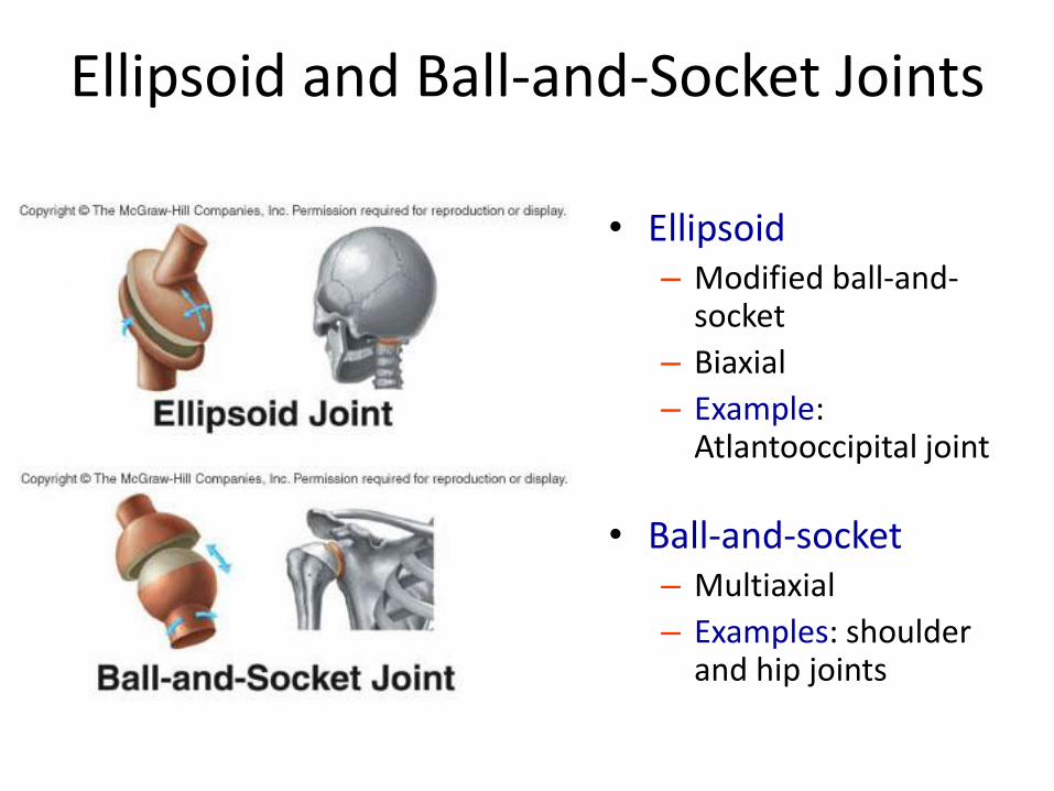
Ellipsoid and Ball-and-Socket Joints
• Ellipsoid – Modified ball-and-
socket
– Biaxial
– Example: Atlantooccipital joint
• Ball-and-socket – Multiaxial
– Examples: shoulder and hip joints
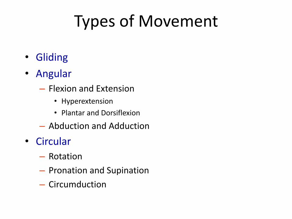
Types of Movement
• Gliding
• Angular
– Flexion and Extension • Hyperextension
• Plantar and Dorsiflexion
– Abduction and Adduction
• Circular
– Rotation
– Pronation and Supination
– Circumduction

Flexion and Extension

Dorsiflexion and Plantar Flexion
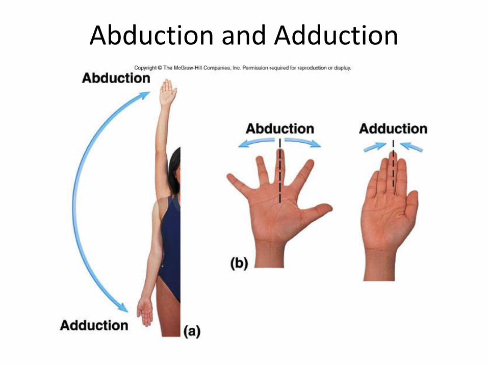
Abduction and Adduction
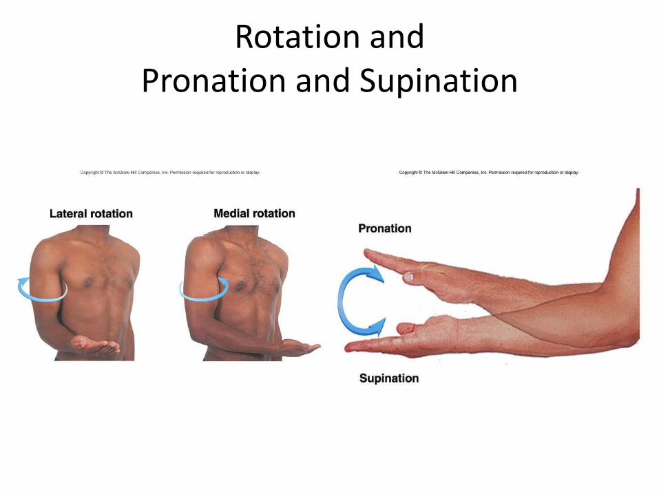
Rotation and Pronation and Supination
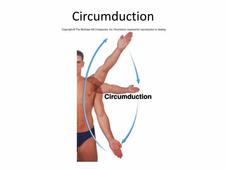
Circumduction
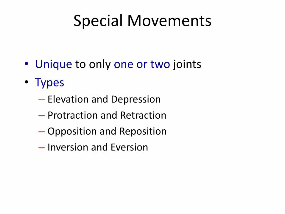
Special Movements
• Unique to only one or two joints
• Types
– Elevation and Depression
– Protraction and Retraction
– Opposition and Reposition
– Inversion and Eversion

Elevation and Depression

Protraction and Retraction

Excursion

Opposition and Reposition
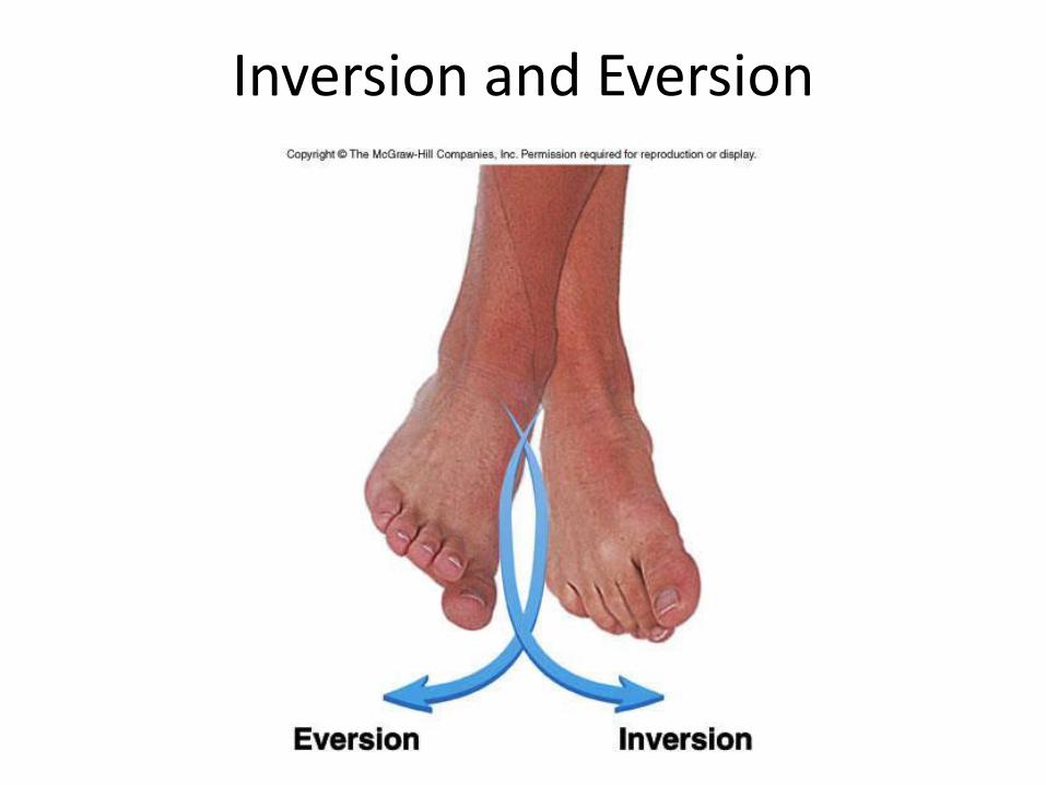
Inversion and Eversion

Range of Motion
• Amount of mobility demonstrated at a given joint
• Types – Active
– Passive
• Influenced by – Shape of articular surfaces forming joint
– Amount and shape of cartilage covering surfaces
– Strength and location of ligaments and tendons
– Location of muscles associated with joint
– Amount of fluid in and around joint
– Amount of use/disuse of joint
– Amount of pain in and around joint

Effects of Aging on Joints
• Tissue repair slows
• Production of synovial fluid declines
• Ligaments and tendons become less flexible
• Decrease in ROM
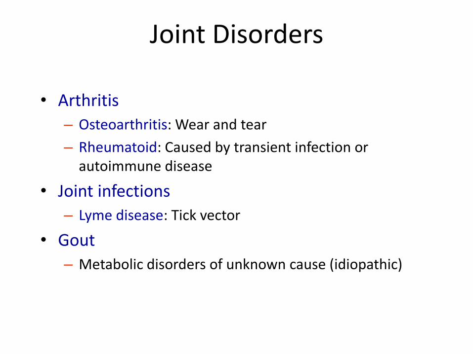
Joint Disorders
• Arthritis
– Osteoarthritis: Wear and tear
– Rheumatoid: Caused by transient infection or autoimmune disease
• Joint infections
– Lyme disease: Tick vector
• Gout
– Metabolic disorders of unknown cause (idiopathic)
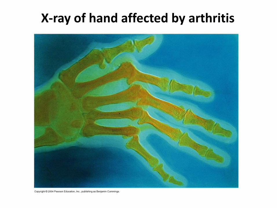
X-ray of hand affected by arthritis

Artificial Hip Joint