Articular involvement in hematological diseases and cancer
56
Articular involvement in hematological diseases and cancer Jorge López-Benítez, M.D. Chief of Pediatrics and Director, Pediatric Rheumatology Program La Costa Medical Center Asuncion, Paraguay PReS Latin America Basic Rheumatology Course June 22 to 24, 2015 Aguas de Sao Pedro, Sao Paulo - Brazil
Transcript of Articular involvement in hematological diseases and cancer

Articular involvement in hematological diseases and cancer
Jorge López-Benítez, M.D. Chief of Pediatrics and Director, Pediatric Rheumatology Program La Costa Medical Center Asuncion, Paraguay
PReS Latin America Basic Rheumatology Course June 22 to 24, 2015
Aguas de Sao Pedro, Sao Paulo - Brazil

Musculoskeletal manifestations of malignancy in childhood may take one of four forms: 1. Primary benign or malignant tumors of bone, cartilage,
fibrous or soft tissue, or miscellaneous origin
2. Metastatic bone tumors
3. Malignant infiltration of bone marrow: leukemia
4. Secondary effects of malignancy
PReS Latin America Basic Rheumatology Course

Bone tumors
PReS Latin America Basic Rheumatology Course
EPIDEMIOLOGY
Approximately one-half of bone tumors occurring in the first two decades of life are malignant
Malignant bone tumors occur more frequently in boys, with a ratio of approximately 1.5:1; except for parosteogenic sarcoma, which is more frequent in girls The male-to-female ratio is approximately 3:1 for osteoid osteomas and osteoblastomas

Bone tumors
PReS Latin America Basic Rheumatology Course
CLINICAL MANIFESTATIONS
Bone tumors may be completely asymptomatic until a mass is detected by the patient or parent.
They usually present with the insidious onset of tenderness, swelling, and localized pain accentuated at night or by weight-bearing.
Local tenderness or bony swelling in the absence of trauma suggests the diagnosis.
Systemic symptoms such as fever and weight loss are nonspecific but support the diagnosis of a malignant rather than a benign tumor.

Bone tumors
PReS Latin America Basic Rheumatology Course
RADIOGRAPHICAL EVALUATIONS
A plain radiograph of the affected area is the best initial diagnostic evaluation Most tumors of a long bone arise in the metaphysis. Malignant tumors of the epiphysis are rare; Ewing sarcoma is the only arising in the diaphysis. For estimate the extent of the lesion, a CT is more useful MRI delineates the soft tissue extent of early lesions, including the bone marrow. Ultrasonography is valuable in assessing extraosseous extension. Radionuclide scanning is useful in localizing a tumor or tumors, rather than in assisting with a specific diagnosis.

Most common types of tumors in specific bones (Data from Dahlin DC, Unni
KK: Bone tumors: general aspects and data on 8,542 cases, ed 4, Springfield, IL, 1986, Charles C Thomas.cal)
PReS Latin America Basic Rheumatology Course
Bone Benign Malignant
Femur Osteochondroma Osteosarcoma
Tibia Osteoid osteoma Osteosarcoma
Osteochondroma —
Giant cell tumor —
Innominate Osteochondroma Osteosarcoma
Humerus Osteochondroma Osteosarcoma
Vertebra Osteoid osteoma Reticulum cell sarcoma
Rib Osteochondroma Chondrosarcoma
Hand Chondroma Chondrosarcoma
Radius Giant cell tumor Osteosarcoma

Bone tumors
PReS Latin America Basic Rheumatology Course
LABORATORY EVALUATION
The laboratory provides little assistance because anemia, leukocytosis, elevation of the erythrocyte sedimentation rate (ESR), and other indicators of inflammation are nonspecific and may be absent even in the presence of osseous malignancy.
Serum alkaline phosphatase levels may be elevated beyond those associated with growth of the child.

PReS Latin America Basic Rheumatology Course

Benign tumors of the bone
PReS Latin America Basic Rheumatology Course
• Osteiod osteomas • Osteoblastoma

PReS Latin America Basic Rheumatology Course
Characteristic Description – OSTEOID OSTEOMA
Age at onset Childhood to young adulthood
Sex ratio Boys > girls
Symptoms Long history, localized aching or boring pain, mild at first, increasing in severity, worse at night, with rest, or with elevation. Limpness, stiffness, or weakness is frequent
Most common sites are the proximal femur, neck and greater trochanter; the proximal tibia; and the pedicles, facets, and spinous processes of the vertebrae
Signs Joint effusion occasionally
Localized swelling or tenderness
X-rays Solitary, lucent nucleus surrounded by sclerosis on radiographs – Positive bone scan
Treatment Pain is dramatically relieved by aspirin or nonsteroidal antiinflammatory drugs
Excision

PReS Latin America Basic Rheumatology Course
Osteoid Osteoma
PReS Latin America Basic Rheumatology Course
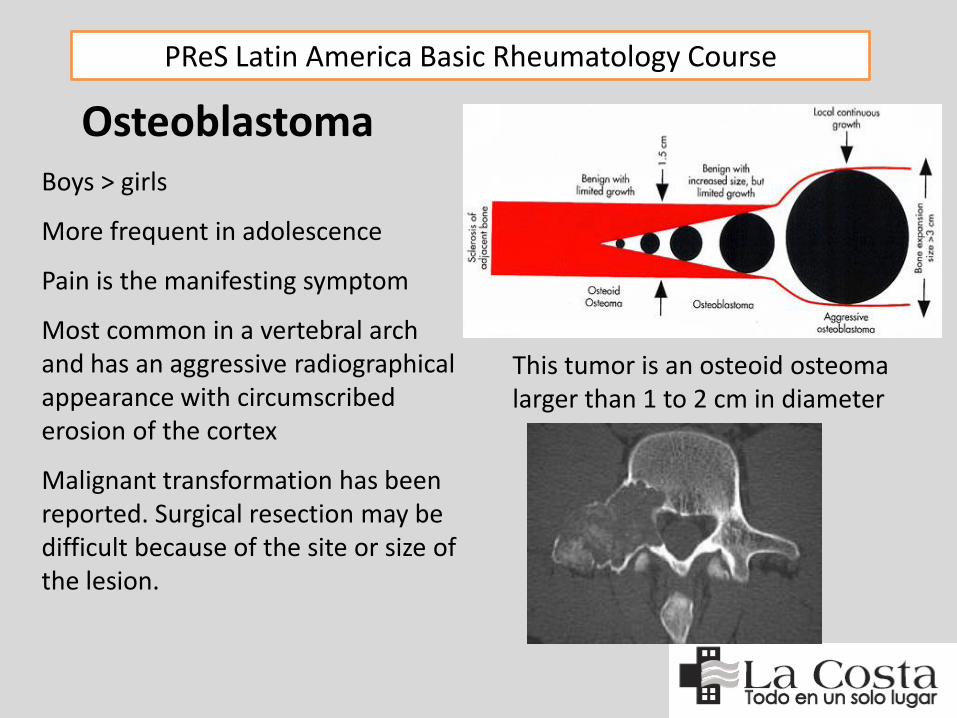
Boys > girls
More frequent in adolescence
Pain is the manifesting symptom
Most common in a vertebral arch and has an aggressive radiographical appearance with circumscribed erosion of the cortex
Malignant transformation has been reported. Surgical resection may be difficult because of the site or size of the lesion.
Osteoblastoma
This tumor is an osteoid osteoma larger than 1 to 2 cm in diameter

Benign tumors of Cartilaginous Origin
PReS Latin America Basic Rheumatology Course
• Osteochondroma • Chondromas • Periosteal Chondroma • Chondroblastoma • Chondromyxoid Fibroma

PReS Latin America Basic Rheumatology Course
Most common benign bone tumor Between the ages of 5- and 15-years-old Equal frequency in boys and girls. Painless exostotic mass Affects the metaphysis of a long bone and arises at the site of a tendon insertion (most commonly around the distal femur or proximal tibia) or in the distal humerus Extends away from the epiphysis as a bony outgrowth capped with cartilage up to 1 cm thick Some children with multiple lesions exhibit an autosomal dominant pattern of inheritance (i.e., multiple hereditary exostoses) Treatment consists of surgical excision. Malignant change may occur in solitary or multiple osteochondromas.
Osteochondroma

PReS Latin America Basic Rheumatology Course
Osteochondroma

PReS Latin America Basic Rheumatology Course
Chondromas (i.e., enchondromas) make up about 10% of benign bone tumors. Boys = girls. Affects young children and those in the second decade Occurs in the small tubular bones of the hand and foot, although other sites may be involved It often presents as a solitary, asymptomatic mass with a pathological fracture or is discovered as an incidental radiological finding Prophylactic resection after biopsy confirmation is not usually considered necessary, but careful follow-up is warranted, because there is a low rate of malignant transformation to chondrosarcoma Cytogenetic abnormalities are common and of value in tumor typing.
Chondromas

PReS Latin America Basic Rheumatology Course
Chondromas The radiographical appearance of a chondroma is that of a well-demarcated metaphyseal lesion of central destruction that may protrude from the surface of the bone or be confined within the medullary canal (i.e., enchondroma), usually with linear or speckled calcification

PReS Latin America Basic Rheumatology Course
Multiple enchondromatosis, or Ollier disease, commonly affects the hands and feet. Joint range of motion may be impaired, and the clinical presentation may be mistaken for arthritis. Growth deformities are common and require surgical management. Maffucci syndrome is endochondromatosis in the presence of multiple cavernous hemangiomas. The frequency of malignant sarcomatous change is high (50%) in patients with Ollier or Maffucci syndrome
Chondromas

PReS Latin America Basic Rheumatology Course
PERIOSTEAL CHONDROMA: Arises from the cortical surface, proximal humerus and other long bones. Pain is often the presenting symptom. Wide excision is the treatment of choice
CHONDROBLASTOMA: Uncommon epiphyseal cartilaginous tumor of childhood (20 – 30 years). Hip, shoulder, or knee. The histopathology consists of polyhedral and giant cells with areas of fine calcifications. Foci of osteoid and bone may resemble a chondromyxoid fibroma. Most lesions are cured by excision and bone graft, but recurrences are a major concern.
CHONDROMYXOID FIBROMA: Arises in the metaphyseal area with pain and tenderness. Occur in children at about the age of 10 years-old. The radiographical appearance is one of an eccentric, sharply circumscribed zone of rarefaction, often with expansion of surrounding bone
Chondromas

Benign tumors of Fibrous Tissue
PReS Latin America Basic Rheumatology Course
• Fibrous Cortical Defect (Nonossifying Fibroma) • Juvenile Fibromatosis • Fibrous Dysplasia • Ossifying Fibroma

PReS Latin America Basic Rheumatology Course
Fibrous Cortical Defect (Nonossifying fibroma)
Ages of 4 and 8 More frequent in boys, 40% of children There may be two or more fibromas, and they are usually an incidental finding (most often in the distal femur and proximal tibia) on x-rays The radiological appearance is virtually diagnostic, with a sharply marginated eccentric lucency in the metaphyseal cortex Occasionally, pathological fractures occur.

PReS Latin America Basic Rheumatology Course
The juvenile fibromatoses, are rare, and include: juvenile aponeurotic fibroma, extraabdominal desmoid tumor and diffuse infantile fibromatosis.
Infantile systemic hyalinosis is a rare inherited disorder characterized by deposition of hyaline material in skin with the formation of nodules, in the musculoskeletal system with development of progressive joint contractures, and in viscera. Death occurs by the age of 2 years. The basic defect is in mutations in the capillary morphogenesis protein 2
Another subtype of the disorder is juvenile hyaline fibromatosis. Other names have included molluscum fibrosum and mesenchymal dysplasia. The early onset of this disorder is in some ways similar to that of neonatal-onset multisystem inflammatory disorder.
Juvenile Fibromatosis

PReS Latin America Basic Rheumatology Course
Difficult to classify and probably represents a developmental abnormality or a benign neoplastic fibrous tissue lesion It is a relatively common disorder with a variable presentation mimicking that of almost any bone lesion
Monostotic disease, most commonly in a rib, occurs in approximately 85% of patients
Polyostotic fibrous dysplasia may be limited to two or three sites or result in extensive skeletal abnormalities. It is also part of the McCune-Albright syndrome in association with multiple endocrine abnormalities.
Fibrous dysplasia

PReS Latin America Basic Rheumatology Course
The classic radiological appearance of fibrous dysplasia is an intramedullary diaphyseal lesion with a thinned, sometimes bulging cortex. An angular deformity of the bone is often present at the site of the lesion and may require surgical intervention, depending on its severity and the potential for pathological fracture.
Osteofibrous dysplasia is a rare lesion of childhood that resembles monostotic disease.40
Fibrous dysplasia

PReS Latin America Basic Rheumatology Course
Ossifying fibroma, or osteofibrous dysplasia, involves the mandible or tibia Usually benign, it may be locally aggressive. Symptoms are often absent, and bony deformity prompts the consultation. Curettage is often unsuccessful, and observation alone is usually recommended after careful histopathological review.
Ossifying fibroma

Benign tumors of Soft Tissue
PReS Latin America Basic Rheumatology Course
• Pigmented Villonodular Synovitis • Synovial hemangioma • Synovial Chondromatosis

PReS Latin America Basic Rheumatology Course
PVS may represent a benign neoplasia, may be caused by an infectious process, or may be associated with repeated episodes of intraarticular hemorrhage
This condition is rare in childhood and is most common between the ages of 20 and 40 years. PVS affects males and females equally It may manifest as recurrent swelling in a knee, ankle, or tendon sheath, and may be nodular or diffuse
Nodular disease affects joints, bursae, or tendon sheaths; in diffuse PVS, a monarthritis of the knee, ankle, or less frequently, the hip is most common. Only rarely is the upper extremity affected.
Pigmented Villonodular Synovitis

PReS Latin America Basic Rheumatology Course
Recurrent painless effusions with progressive destruction of cartilage and erosion of bone. A boggy fullness is present on examination. The most striking feature is the presence of blood-stained, dark brown synovial fluid on joint aspiration. The synovium is dark and characterized by nodular areas of hypertrophy and hemosiderin-laden macrophages
Pigmented Villonodular Synovitis

PReS Latin America Basic Rheumatology Course
MRI has a characteristic finding with low signal density on T1- and T2-weighted images
Treatment requires surgical excision
NSAIDs are useful in suppressing the inflammatory disease, and intraarticular glucocorticoids may have a role in management.
Malignant PVS is rare, and its precise classification is controversial.
Pigmented Villonodular Synovitis
PReS Latin America Basic Rheumatology Course
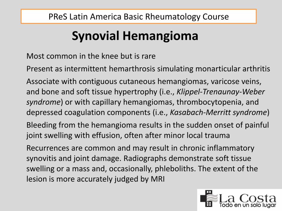
Most common in the knee but is rare
Present as intermittent hemarthrosis simulating monarticular arthritis
Associate with contiguous cutaneous hemangiomas, varicose veins, and bone and soft tissue hypertrophy (i.e., Klippel-Trenaunay-Weber syndrome) or with capillary hemangiomas, thrombocytopenia, and depressed coagulation components (i.e., Kasabach-Merritt syndrome)
Bleeding from the hemangioma results in the sudden onset of painful joint swelling with effusion, often after minor local trauma
Recurrences are common and may result in chronic inflammatory synovitis and joint damage. Radiographs demonstrate soft tissue swelling or a mass and, occasionally, phleboliths. The extent of the lesion is more accurately judged by MRI
Synovial Hemangioma

PReS Latin America Basic Rheumatology Course
Unusual condition where synovial cartilaginous and osteocartilaginous bodies develop in the synovium and then are released free into the synovial fluid
Synovial chondromatosis is more common in boys and usually affects the knee
Manifesting symptoms are usually those of a loose body with intermittent pain, swelling, locking, and giving way
In early disease, radiographical studies are normal, but as the lesions calcify, they may be identified as discrete areas of stippled calcification. Treatment requires surgical excision. Malignant transformation is rare
Synovial Chondromatosis

Benign tumors of Miscellaneous Origin
PReS Latin America Basic Rheumatology Course
• Unicameral (Solitary) Bone Cyst • Aneurysmal Bone Cyst • Giant Cell Tumor • Eosiniphilic Granuloma

Unicameral (Solitary) Bone Cyst
PReS Latin America Basic Rheumatology Course
Rare before 3, most common between 6 and 10. More frequent in boys
Arises in the metaphysis of a long bone and is asymptomatic or causes localized pain, swelling, a pathological fracture, or growth disturbance of the limb
Radiography shows a lucent lesion that does not cross the physis or a fusiform widening of the bone
Curettage and insertion of bone chips, injection with glucocorticoid has been effective
Aneurysmal Bone Cyst
PReS Latin America Basic Rheumatology Course
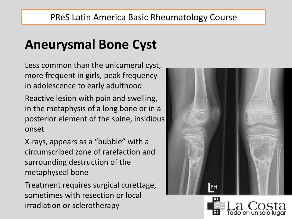
Less common than the unicameral cyst, more frequent in girls, peak frequency in adolescence to early adulthood
Reactive lesion with pain and swelling, in the metaphysis of a long bone or in a posterior element of the spine, insidious onset
X-rays, appears as a “bubble” with a circumscribed zone of rarefaction and surrounding destruction of the metaphyseal bone
Treatment requires surgical curettage, sometimes with resection or local irradiation or sclerotherapy

PReS Latin America Basic Rheumatology Course
Pain is the usual manifesting symptom, is common in the 20’s and 30’s and occurs more frequently in girls On radiographs, an expanding zone of eccentric radiolucency in the epiphysis of a long bone is characteristic
Giant Cell Tumor
Eosinophilic Granuloma Localized Langerhans cell histiocytosis (histiocytosis X) occurs more in boys between 5 and 10 years of age. Localized pain and swelling over a solitary mass (skull, spine, and pelvis and the diaphysis of the femur) X-rays: discrete lytic lesion with cortical erosion and a periosteal reaction Percutaneous needle biopsy for diagnosis Surgical curettage, sometimes with low-dose radiatio and intralesional glucocorticoid may be a useful adjunct to therapy Other types of histiocytosis include Hand-Schüller-Christian disease andLetterer-Siwe disease.

PReS Latin America Basic Rheumatology Course
Malignant musculoskeletal tumors of childhood account for 5% to 10% of malignant neoplasms
The most common of these are osteogenic sarcoma, rhabdomyosarcoma, and Ewing sarcoma
Osteogenic sarcoma is rare in the first decade of life but is the most common malignant bone tumor in the second decade
The mean age of children with Ewing sarcoma is younger than that for any other primary tumor of bone
Survival rates (60% to 70%) have continued to improve modestly for these tumors
There has also been progress in understanding their biology and genetic predisposition, especially in Ewing sarcoma and rhabdomyosarcoma
Malignant tumors of the bone

PReS Latin America Basic Rheumatology Course
60% of all bone tumors in children
Most common at the time of maximal growth velocity in the second decade of life with 75% of cases occurring between the ages of 8 and 25, especially in taller children, occasionally occurs in siblings
Annual incidence is approximately 0.6 to 0.7 per 100,000 children
Approximately 3% of these tumors develop in a field that has received previous irradiation
Osteosarcomas are also associated with retinoblastoma, enchondromatosis, hereditary multiple exostoses, and fibrous dysplasia
There is an increased risk of osteosarcoma in patients with Rothmund-Thomson syndrome (short stature, telangiectases, small hands and feet, and hypoplastic thumbs)
Osteosarcoma
PReS Latin America Basic Rheumatology Course
Arises in the medullary canal of the bone, has a metaphyseal location, and occurs most often (60%) around the knee (e.g., distal femur, proximal tibia) and in the proximal humerus
Are highly malignant and metastasize early by hematogenous spread to many organs, especially the lungs, where they are a significant cause of secondary hypertrophic osteoarthropathy.
Pain is the most common manifesting symptom
Swelling over the involved bone occurs later , secondary signs include local inflammation, involvement of regional lymph nodes, and loss of function of the limb
The presence of systemic signs such as weight loss, fever, or secondary hypertrophic osteoarthropathy suggests that skeletal and pulmonary metastases have already occurred
Osteosarcoma

PReS Latin America Basic Rheumatology Course
On x-rays has a moth-eaten appearance with cortical destruction, periosteal elevation (i.e., Codman triangle), and a soft tissue mass
Bone scan, CT, or MRI may be indicated to delineate the extent of the lesion
Diagnosis is confirmed by biopsy (osteoblastic, chondroblastic, fibroblastic, telangiectatic, and a small-cell type that has features overlapping those of Ewing sarcoma).
Osteosarcoma

PReS Latin America Basic Rheumatology Course
Modification of the surgical approach and adjuvant chemotherapy, has led to a disease-free survival rate of more than 50% at five years Prognostic features: histological grade, size of the initial lesion, and its response to preoperative treatment. Metastases at diagnosis are associated with a poor outcome (less than 20% survival). The rare multifocal sclerosing osteosarcoma has a poor prognosis
Surface osteosarcomas do not involve the medullary cavity and often encircle the entire shaft of the bone, they behave different: Parosteal (or juxtacortical) and periosteal
The parosteal variety is more common in girls and occurs on the posterior surface of the distal femur. Malignant potential is low, it tends to metastasize late, and it has a much better prognosis. The periosteal type occurs in the tibia and is associated with an abundant proliferation of cartilage.
Osteosarcoma

PReS Latin America Basic Rheumatology Course
Is a rare tumor (less than 5%) that develops from malignant transformation of a preexisting enchondroma or, later in life, in a patient with multiple heritable exostoses
The initial symptoms are localized swelling or pain
X-rays are usually diagnostic with destruction of bone combined with mottled densities of calcification and ossification (i.e., “popcorn” appearance)
Treatment consists of wide resection, adjuvant chemotherapy, and irradiation
Malignant tumors of the Cartilage Chondrosarcoma

PReS Latin America Basic Rheumatology Course
Tumor on a distal extremity presenting as a soft, infiltrative mass with areas of hemorrhage or necrosis, histopathology confirms fibroblastic or myofibroblastic differentiation of all degrees
Two patterns: congenital fibrosarcoma in boys younger than 2; and postpubertal fibrosarcoma is more aggressive
No radiographical features distinguish this tumor from osteosarcoma
Congenital fibrosarcomas undergo rapid growth and extensive local invasion, metastatic rate 7%. In children older than 10, this rate is 50%
Treatment involves surgical resection; and of the postpubertal type combination of radical excision, postoperative irradiation, and adjuvant chemotherapy
The overall survival rate is 40% at five years and 30% at 10 years
Malignant tumors of the Fibrous Tissue Fibrosarcoma

PReS Latin America Basic Rheumatology Course
RHABDOMYOSARCOMA Most common soft tissue sarcomas in children and account for one half of all soft tissue neoplasms in patients younger than 15-years
Most common in children between the ages of 2 and 6 years or during adolescence, with an annual incidence of 0.4 to 0.9 per 100,000 children
The tumor presents as a localized, painless, soft tissue mass, in the head and neck but can arise in any striated muscle. Approximately 20% occur in the extremities, especially during adolescence
Metastasizes early to the lung, bone, and bone marrow, can erode into adjacent bone and produce a radiographical appearance of a soft tissue mass with an underlying periosteal reaction. The five year survival rate is approximately 70%
Malignant tumors of the Soft Tissue

PReS Latin America Basic Rheumatology Course
SYNOVIAL CELL SARCOMA Rare in childhood, synovial cell sarcoma is the most common soft tissue sarcoma (6% to 10%) after the rhabdomyosarcomas
This tumor rarely occurs within a joint; but in the periarticular soft tissue and tendon sheaths may be involved. Lower extremity involvement is most common, especially around the knee, foot or in the hand. The tumor may be associated with calcifications that suggest the diagnosis on radiographs
Surgical excision, irradiation, and chemotherapy result in a seven-year survival rate of 60% to 70%. Prognosis is related to tumor size and ease of resection
Most synovial sarcomas express a specific chromosomal abnormality: t(X;18) (p11.2;q11.2)
Malignant tumors of the Soft Tissue

PReS Latin America Basic Rheumatology Course
From primitive multipotential mesenchymal or neural crest cell in the medulla of bone or occasionally in soft tissue
The most malignant of bone tumors and the second most common in children, it accounts for 7% to 15% of all malignant bone tumors in childhood, with an annual incidence of 0.2 to 0.3 per 100,000 children Is most common in white boys in the second decade of life (male to female ratio of 1.5:1), it is uncommon in Asian or African descent
Occurs in the diaphysis of the long bones (i.e., femur, humerus, or tibia) or in the innominate, but it can develop in any bone, including those of the axial skeleton, or even as an extra-skeletal lesion
The tumor presents with pain, and local swelling; systemic signs, including fever, are common, often occurring with abnormalities of laboratory indices of inflammation
Malignant tumors of Miscellaneous origin: Ewing Sarcoma

PReS Latin America Basic Rheumatology Course
Neural markers, chimeric fusion products, or specific chromosomal translocation (t[11;22] [q24;q12]) in more than 95% of cases differentiate this tumor from lymphoma, rhandomyosarcoma, or neuroblastoma
Treatment combines surgical resection with irradiation and adjuvant chemotherapy. 5-year survival rate 50 to 80%. Metastases to the lungs or skeleton reduce this to 20 or 30%
Malignant tumors of Miscellaneous origin: Ewing Sarcoma
X-rays shows an aggressive, elongated, lytic lesion filling the medullary cavity, disrupting the cortex, and causing a roughening of the periosteum described as having an onion-skin or sunburst appearance

PReS Latin America Basic Rheumatology Course
Lesson et al, over 38 years, described 39 patients from 18 months to 20 years • Neuroblastoma (41%) • Rhabdomyosarcoma (18%) • Teratoma-carcinoma (10%) • Wilms Tumors (8%) • Retinoblastoma (5%)
Metastatic Bone Tumors
M. C. Lesson, et al. Metastatic skeletal disease in the pediatric population, J. Pediatr. Orthop. 5 (1985) 261-267

PReS Latin America Basic Rheumatology Course
Of all childhood malignancies, excluding musculoskeletal tumors, ALL is the most frequently associated with musculoskeletal symptoms at presentation
Symptoms are cause by infiltration of the synovia, the periosteum, or the bone marrow by malignant cells is one explanation, but a secondary uric acid or immune complex deposits may also cause pain and inflammation in a joint. A less common factor may be an intraarticular bleeding resulting from thrombocytopenia
Pain disproportionate to physical findings, accompanied by haematologic alterations and not alleviated by NSAIDs, strongly suggest the presence of a haematologic malignancy
Malignant infiltration of bone marrow: Leukemias

PReS Latin America Basic Rheumatology Course
Leukemias Characteristic Description
Presentation Low-grade fever, fatigue, pallor,
weight loss
Pain Often disproportionate to objective
findings
Diffuse musculoskeletal aching or
pain and tenderness of
metaphyses of long bones
May be migratory joint pain or
periarticular swelling or joint
effusion
Hematological parameters
May be normal or with increased or
decreased WBC count or platelet
count, sometimes with blast cells in
peripheral smear
Increased ESR and lactate
dehydrogenase and urate levels
Radiographs Metaphyseal rarefaction, periosteal
new-bone apposition

PReS Latin America Basic Rheumatology Course
Pain is diffusely localized to one area of the body, particularly over the metaphyses of the long bones, joint effusion occurs. Large joints, especially the knees, are most commonly affected, although small joints of the hands may be involved. Associated periarticular disease is common
X-rays: localized metaphyseal rarefaction and subperiosteal elevation or an elongated osteolytic reaction
Malignant infiltration of bone marrow: Leukemias

PReS Latin America Basic Rheumatology Course
SECONDARY HYPERTROPHIC OSTEOARTHROPATHY (SHO) Terminal clubbing, painful swelling of distal joints and soft tissues, profuse sweating, and radiographical evidence of periosteal new-bone formation affecting the hands, feet, and distal limbs
Related to chronic pulmonary disease, congenital heart disease, or, occasionally, the antiphospholipid antibody syndrome. Also biliary atresia and regional enteritis and POEMS syndrome (i.e., polyneuropathy, organomegaly, endocrinopathy, M protein, and skin changes) in adults
An important cause of SHO in childhood is pulmonary malignancy, most often caused by metastases from osteogenic sarcoma
Secondary effects of malignancy

PReS Latin America Basic Rheumatology Course
ACANTHOSIS NIGRICANS
Associated with malignancy, diabetes, obesity, various genetic syndromes, connective tissue diseases such as dermatomyositis, and glucocorticoid administration
Also occur in children with lipodystrophy or may be familial
In juvenile dermatomyositis, it is associated with premature thelarche, lipodystrophy, and insulin resistance
Secondary effects of malignancy

PReS Latin America Basic Rheumatology Course
HOMOZYGOUS SICKLE CELL DISEASE: cause severe musculoskeletal manifestations as a result of hematopoietic expansion of the bone marrow and repeated episodes of avascular necrosis, is autosomal dominant and occurs almost exclusively in black children Acute arthritis and long bone pain may be severe and incapacitating during sickle cell crises. In the infant, dactylitis and periostitis of the small bones of the hands may cause painful swollen extremities and the hand-foot syndrome. Each acute episode lasts 1 to 3 weeks and is characterized by diffuse, symmetric, painful swelling of the hands or feet.
Others: Hemoglobinopathies

PReS Latin America Basic Rheumatology Course
Β-THALASSEMIA : group of heterogeneous syndromes of inherited hypochromic anemias of differing severity. Thalassemia minor is characterized by anemia, hepatosplenomegaly, and recurrent brief episodes of joint pain, swelling, and effusion, especially in the ankles.Bone pain was frequently reported in with βthalassemia Deferiprone, an iron chelating agent used to treat patients with thalassemia has been associated with arthritis.
Others: Hemoglobinopathies

PReS Latin America Basic Rheumatology Course
CLASSIC HEMOPHILIA A (FACTOR VIII DEF)
Recurrent intra-articular hemorrhage, frequency of episodes related to plasma concentration of factor VIII; hemarthroses almost invariably occur in children with levels below 5% of normal X-linked recessive coagulopathy.
Hemarthrosis can occur even before the child starts walking, and the frequency of episodes increases during the early childhood years
The joints most commonly affected are the knees, elbows, and ankles
Others: Hemophilia

La Costa Medical Center
Pediatric Rheumatology Program
Asunción, Paraguay
Thank you!