ARTICLE IN PRESS - University of British · PDF...
27
www.elsevier.de/ejop European Journal of PROTISTOLOGY European Journal of Protistology 40 (2004) 85–111 Molecular data and the evolutionary history of dinoflagellates Juan F. Saldarriaga a, *, F.J.R. ‘‘Max’’ Taylor a,b , Thomas Cavalier-Smith c , Susanne Menden-Deuer d , Patrick J. Keeling a a Department of Botany, University of British Columbia, 6270 University Boulevard, Vancouver, BC, Canada V6T 1Z4 b Department of Earth and Ocean Sciences, University of British Columbia, 6270 University Boulevard, Vancouver, BC, Canada V6T 1Z4 c Department of Zoology, University of Oxford, South Parks Road, Oxford OX1 3PS, UK d School of Oceanography, University of Washington, Box 357940, Seattle, WA 98195, USA Received 16 July 2003; accepted 28 November 2003 Abstract We have sequenced small-subunit (SSU) ribosomal RNA (rRNA) genes from 16 dinoflagellates, produced phylogenetic trees of the group containing 105 taxa, and combined small- and partial large-subunit (LSU) rRNA data to produce new phylogenetic trees. We compare phylogenetic trees based on dinoflagellate rRNA and protein genes with established hypotheses of dinoflagellate evolution based on morphological data. Protein-gene trees have too few species for meaningful in-group phylogenetic analyses, but provide important insights on the phylogenetic position of dinoflagellates as a whole, on the identity of their close relatives, and on specific questions of evolutionary history. Phylogenetic trees obtained from dinoflagellate SSU rRNA genes are generally poorly resolved, but include by far the most species and some well-supported clades. Combined analyses of SSU and LSU somewhat improve support for several nodes, but are still weakly resolved. All analyses agree on the placement of dinoflagellates with ciliates and apicomplexans (=Sporozoa) in a well-supported clade, the alveolates. The closest relatives to dinokaryotic dinoflagellates appear to be apicomplexans, Perkinsus, Parvilucifera, syndinians and Oxyrrhis. The position of Noctiluca scintillans is unstable, while Blastodiniales as currently circumscribed seems polyphyletic. The same is true for Gymnodiniales: all phylogenetic trees examined (SSU and LSU-based) suggest that thecal plates have been lost repeatedly during dinoflagellate evolution. It is unclear whether any gymnodinialean clades originated before the theca. Peridiniales appear to be a paraphyletic group from which other dinoflagellate orders like Prorocentrales, Dinophysiales, most Gymnodiniales, and possibly also Gonyaulacales originated. Dinophysiales and Suessiales are strongly supported holophyletic groups, as is Gonyaulacales, although with more modest support. Prorocentrales is a monophyletic group only in some LSU-based trees. Within Gonyaulacales, molecular data broadly agree with classificatory schemes based on morphology. Implications of this taxonomic scheme for the evolution of selected dinoflagellate features (the nucleus, mitosis, flagella and photosynthesis) are discussed. r 2004 Elsevier GmbH. All rights reserved. Keywords: Dinoflagellates; Ribosomal RNA; Chloroplast evolution; Molecular phylogeny; Theca; Photosynthesis Introduction The importance of dinoflagellates in aquatic commu- nities is hard to overestimate. They are ubiquitous in marine and freshwater environments, where they con- stitute a large percentage of both the phytoplankton and ARTICLE IN PRESS *Corresponding author. Fax: (604)-822-6089. E-mail address: [email protected] (J.F. Saldarriaga). 0932-4739/$ - see front matter r 2004 Elsevier GmbH. All rights reserved. doi:10.1016/j.ejop.2003.11.003
-
Upload
vuongduong -
Category
Documents
-
view
217 -
download
3
Transcript of ARTICLE IN PRESS - University of British · PDF...

European Journal of
PROTISTOLOGYEuropean Journal of Protistology 40 (2004) 85–111
ARTICLE IN PRESS
*Correspondin
E-mail addres
0932-4739/$ - see
doi:10.1016/j.ejo
www.elsevier.de/ejop
Molecular data and the evolutionary history of dinoflagellates
Juan F. Saldarriagaa,*, F.J.R. ‘‘Max’’ Taylora,b, Thomas Cavalier-Smithc,Susanne Menden-Deuerd, Patrick J. Keelinga
aDepartment of Botany, University of British Columbia, 6270 University Boulevard, Vancouver, BC, Canada V6T 1Z4bDepartment of Earth and Ocean Sciences, University of British Columbia, 6270 University Boulevard, Vancouver, BC,
Canada V6T 1Z4cDepartment of Zoology, University of Oxford, South Parks Road, Oxford OX1 3PS, UKdSchool of Oceanography, University of Washington, Box 357940, Seattle, WA 98195, USA
Received 16 July 2003; accepted 28 November 2003
Abstract
We have sequenced small-subunit (SSU) ribosomal RNA (rRNA) genes from 16 dinoflagellates, producedphylogenetic trees of the group containing 105 taxa, and combined small- and partial large-subunit (LSU) rRNA datato produce new phylogenetic trees. We compare phylogenetic trees based on dinoflagellate rRNA and protein geneswith established hypotheses of dinoflagellate evolution based on morphological data. Protein-gene trees have too fewspecies for meaningful in-group phylogenetic analyses, but provide important insights on the phylogenetic position ofdinoflagellates as a whole, on the identity of their close relatives, and on specific questions of evolutionary history.Phylogenetic trees obtained from dinoflagellate SSU rRNA genes are generally poorly resolved, but include by far themost species and some well-supported clades. Combined analyses of SSU and LSU somewhat improve support forseveral nodes, but are still weakly resolved. All analyses agree on the placement of dinoflagellates with ciliates andapicomplexans (=Sporozoa) in a well-supported clade, the alveolates. The closest relatives to dinokaryoticdinoflagellates appear to be apicomplexans, Perkinsus, Parvilucifera, syndinians and Oxyrrhis. The position ofNoctiluca scintillans is unstable, while Blastodiniales as currently circumscribed seems polyphyletic. The same is truefor Gymnodiniales: all phylogenetic trees examined (SSU and LSU-based) suggest that thecal plates have been lostrepeatedly during dinoflagellate evolution. It is unclear whether any gymnodinialean clades originated before the theca.Peridiniales appear to be a paraphyletic group from which other dinoflagellate orders like Prorocentrales,Dinophysiales, most Gymnodiniales, and possibly also Gonyaulacales originated. Dinophysiales and Suessiales arestrongly supported holophyletic groups, as is Gonyaulacales, although with more modest support. Prorocentrales is amonophyletic group only in some LSU-based trees. Within Gonyaulacales, molecular data broadly agree withclassificatory schemes based on morphology. Implications of this taxonomic scheme for the evolution of selecteddinoflagellate features (the nucleus, mitosis, flagella and photosynthesis) are discussed.r 2004 Elsevier GmbH. All rights reserved.
Keywords: Dinoflagellates; Ribosomal RNA; Chloroplast evolution; Molecular phylogeny; Theca; Photosynthesis
g author. Fax: (604)-822-6089.
s: [email protected] (J.F. Saldarriaga).
front matter r 2004 Elsevier GmbH. All rights reserved.
p.2003.11.003
Introduction
The importance of dinoflagellates in aquatic commu-nities is hard to overestimate. They are ubiquitous inmarine and freshwater environments, where they con-stitute a large percentage of both the phytoplankton and
ARTICLE IN PRESSJ.F. Saldarriaga et al. / European Journal of Protistology 40 (2004) 85–11186
the microzooplankton, and in benthic communities asinterstitial flora and fauna or as symbionts in reef-building corals, other invertebrates and unicellularorganisms (Taylor, 1987). Both ecto- and endoparasiticdinoflagellate species are also common, infecting hostsranging from other protists like ciliates, radiolarians oreven other dinoflagellates, to crustaceans, cnidarians,appendicularians, polychaetes, fish and many others(Cachon and Cachon, 1987). Many species of dino-flagellates are notorious for producing toxins that cancause human illness through shellfish or fish poisoning(Steidinger, 1993); dinoflagellates are the ultimate causeof diseases like diarrheic shellfish poisoning (DSP),neurotoxic shellfish poisoning (NSP), paralytic shellfishpoisoning (PSP) and ciguatera. Some toxic dinoflagel-lates (as well as other protists) can also cause fish killsand mortality of other marine fauna (Steidinger, 1993).
One recent definition of dinoflagellates is found inFensome et al. (1993, p. 3): they are ‘‘eukaryotic,primarily single-celled organisms in which the motile cellpossesses two dissimilar flagella: a ribbon-like flagellumwith multiple waves which beats to the cell’s left, and amore conventional flagellum with one or a few waveswhich beats posteriorly’’. Taxonomic treatments of thegroup have traditionally been based on two sets ofcytological characters. One is the presence of adinokaryon, a uniquely modified nucleus that lacksnucleosomal histones and contains fibrillar chromo-somes with a typical ultrastructure that remain con-densed throughout the cell cycle, and that dividesthrough a special type of closed mitosis with anextranuclear spindle (review in Dodge, 1987). Dinokar-ya are present in most dinoflagellates, but not in theparasitic order Syndiniales or in particular life stages ofthe Blastodiniales (also parasitic) and Noctilucales(Fensome et al., 1993). The other character, applied todinokaryotic dinoflagellates, is the arrangement ofcortical alveoli, flattened vesicles immediately under-neath the plasma membrane that often contain cellulosethecal plates (in dinoflagellate literature cortical alveoliare generally referred to as amphiesmal vesicles, reviewin Netzel and D .urr (1984)). In thecate orders (Gonyau-lacales, Peridiniales, Dinophysiales, Prorocentrales), thetheca is contained in relatively few alveoli with a patternthat can be determined relatively easily (thecal platetabulation). Athecate taxa, however, (notably the orderGymnodiniales, but also Syndiniales, Noctilucales, etc.)often contain hundreds of alveoli, making it difficult todetermine homologies and locational relationships. As aconsequence, thecate taxa are much easier to classifythan athecate ones.
Thecal plate patterns are also easier to determine inspecies that are easily found as motile stages, the celltype that typically displays this feature. However, thesemotile stages are often very short phases of dinoflagel-late life cycles; some species are most often found as
cysts (Suessiales, Thoracosphaerales, Phytodiniales, afew Gonyaulacales), plasmodia (many Syndiniales) or asstrongly modified trophonts that are not easily compar-able to the typical dinoflagellate motile stages (Noctilu-cales or Blastodiniales). The tabulation of the motilestages is often reflected in cysts, a feature that has beenused extensively to detect relationships between extantand fossil genera (Fensome et al., 1993; Fensome et al.,1999; most fossil dinoflagellates are cysts). Within somethecate orders, a (putative) radiation of forms can befollowed remarkably well using extant species (e.g. inDinophysiales, Gonyaulacales and Peridiniales, Taylor,1980), even if the direction of the changes cannot.Nevertheless, this cannot be done between orders, forthere appear to be few intermediate forms. As aconsequence, except for some cases where informativeintermediate fossil taxa have been found (Fensome et al.,1993), the mutual relationship of many dinoflagellateorders is still unclear. Also unclear is which groups ofdinoflagellates are early or late diverging; different setsof characters support different hypotheses (discussion inTaylor, 1980; Fensome et al., 1993).
Early phylogenetic studies showed the monophyly ofdinoflagellates (Maroteaux et al., 1985; Herzog andMaroteaux, 1986) and disproved notions that dinofla-gellates are early branches of the eukaryote tree (themesokaryotic theory, Dodge, 1965, 1966). A relation-ship between dinoflagellates and ciliates that had beenpostulated earlier (Corliss, 1975; Taylor, 1976) was alsocorroborated by these sequences, as was a newlydiscovered one to apicomplexans (Wolters, 1991;Gajadhar et al., 1991). In 1991 a new taxon, theAlveolata, was created encompasing ciliates, dinoflagel-lates, apicomplexans and their close relatives, theprotalveolates (Cavalier-Smith, 1991), and numerousstudies have repeatedly supported its validity (e.g.Cavalier-Smith, 1993; Van de Peer et al., 1996; Fastet al., 2002). The relationship of alveolates to othergroups has been more difficult to resolve, but recentstudies based on phylogenies of concatenated proteinsand chloroplast-targeted genes (Baldauf et al., 2000;Fast et al., 2001) have supported the relationshipbetween this group and chromists as predicted by thechromalveolate hypothesis (Cavalier-Smith, 1999, 2003)and by earlier taxonomic schemes (e.g. Taylor, 1976).
Within alveolates, dinoflagellates are more closelyrelated to the apicomplexans than to the ciliates (Fastet al., 2002). Other close relatives of dinoflagellatesinclude forms that share a number of features typical ofall alveolates (e.g. cortical alveoli, mitochondria withtubular cristae, presence of trichocysts in diverse forms),but lack the synapomorphies that define ciliates,dinoflagellates or apicomplexans, the so-called protal-veolates (Cavalier-Smith, 1991, 1993). The genusPerkinsus, for example, a parasite of oysters andother bivalves, and Parvilucifera, a parasite infecting
ARTICLE IN PRESSJ.F. Saldarriaga et al. / European Journal of Protistology 40 (2004) 85–111 87
dinoflagellates, often form a clade closely related todinoflagellates (Siddall et al., 1997; Nor!en et al., 1999;Saldarriaga et al., 2003a); the genus Rastrimonas
(formerly Cryptophagus, Brugerolle, 2003), a parasiteof cryptomonads, could be a third member of this group(Brugerolle, 2002b). Other protalveolate taxa that seemto have close links to the dinoflagellates are the free-living genus Oxyrrhis, recently excluded from the group(Fensome et al., 1993), and the ellobiopsids, a group ofparasites of crustaceans that are either derived from orvery closely related to dinoflagellates (J. Silbermann,personal communication). The genus Colpodella, how-ever, appears to be a basal branch to the apicomplexans(Cavalier-Smith, 2000; Brugerolle, 2002a; Kuvardinaet al., 2002; Leander and Keeling, 2003). Otherprotalveolates have not been characterized at themolecular level, and so it remains to be seen where thephylogenetic affiliation of Colponema, Acrocoelus, andothers may lie.
Nearly all molecular phylogenetic studies of the in-group relationships of dinoflagellates have used rRNA,either partial sequences of the large-subunit (LSU)ribosomal RNA (rRNA) gene (LSU, e.g. Lenaers et al.,1991; Zardoya et al., 1995; Daugbjerg et al., 2000), orthe small-subunit (SSU) rRNA gene (SSU, e.g. Saunderset al., 1997; Grzebyk et al., 1998; Gunderson et al., 1999;Saldarriaga et al., 2001). Phylogenies based on SSU arethe only ones with data for phylogenetically importantgroups like the Syndiniales, Noctilucales or Blastodi-niales. Relationships of orders to one-another aremostly unresolved (e.g. Saunders et al., 1997; Saldarria-ga et al., 2001), but those at the base of the lineage areoften well supported, as are some late-branching groups.Phylogenies based on the first two or three domains(D1–D3) of the LSU contain fewer taxa than SSU-basedones, but since the two molecules appear to evolve atdifferent rates (Ben Ali et al., 2001; John et al., 2003)they have also proven very valuable since bootstrapsupport for certain groupings is greater. Protein-genebased phylogenies are still scarce, e.g. HSP90, actin,alpha- and beta-tubulin genes (Saldarriaga et al., 2003a;B. Leander, unpublished data), and plastid-encodedgenes (e.g. psbA in Takishita and Uchida (1999), psaAin Yoon et al. (2002), Zhang et al. (2000)). None of theseyet contain many taxa, and support for their in-groupclades tends to be weak. Nevertheless, they have provenvaluable for determining the position of some basal taxa(e.g. Saldarriaga et al., 2003a).
The objective of the present work is to clarify somekey events in the evolutionary history of the dinofla-gellates and their close relatives. We re-examine allavailable data, molecular, morphological, paleontologi-cal and biochemical, to produce a phylogenetic frame-work for the group, with special consideration given torRNA trees. We explore the origin of the dinokaryonand of dinokaryotic dinoflagellates, the development of
cortical alveoli in the group and the history of dino-flagellate photosynthesis. We also examine some smallerscale questions, e.g. the relationship of Perkinsus,Oxyrrhis, Noctiluca, Syndiniales and Blastodiniales toother dinoflagellates and the circumscription of thedifferent groups of Gymnodiniales. Lastly, we considerwhether the phylogeny of Gonyaulacales, generally bettersupported than other parts of the tree, is congruent withthe proposals for dinoflagellate classification based onmorphology put forward by Fensome et al. (1993).
Materials and methods
Organisms, DNA extraction, amplification and
sequencing
Photosynthetic dinoflagellate species were obtainedfrom non-axenic culture collections (Table 1) andcultured according to established protocols (e.g. Harri-son et al., 1980). The heterotrophic Protoperidinium
species were fed the diatom Ditylum brightwellii andmaintained at 12�C in F/2 medium at 30 mmols photonsm�2 s�1 on a plankton wheel at 1 rpm. Cells wereharvested by centrifugation. DNA was extracted usingthe DNeasy Plant DNA Purification Kit (Qiagen).Whenever possible, the 18S (nuclear SSU) rRNA genewas amplified as a single fragment using a polymerasechain reaction with two eukaryotic universal SSUprimers (50-CGAATTCAACCTGGTTGATCCTGC-CAGT-30 and 50-CCGGATCCTGATCCTTCTGCAGGTTCACCTAC-30). However, in many cases twooverlapping fragments had to be produced usinginternal primers designed to match existing eukaryoticSSU sequences (4F: 50-CGGAATTCCAGTC-30 and11R: 50-GGATCACAGCTG-30). PCR products wereeither sequenced directly or cloned into pCR-2.1 vectorusing the TOPO TA cloning kit (Invitrogen). Sequen-cing reactions were completed with both the originalPCR primers as well as 2–3 additional primers in eachdirection. When using cloned fragments, 2–4 clones weresequenced to detect and clarify possible ambiguities.
Phylogenetic analysis
New sequences were added to the SSU alignment ofSaldarriaga et al. (2001); they are now available fromGenBank. The final multiple alignment contained 98dinoflagellate species, plus Perkinsus, Parvilucifera andseveral ciliate and apicomplexan sequences that wereused as outgroups. The sequence for Oxyrrhis marina
was excluded from the analyses: previous experienceshowed that this species has an extremely derived SSUsequence that distorts the topologies of SSU trees(Saldarriaga et al., 2003a). We also included seven

ARTICLE IN PRESS
Table 1. List of strains examined in this study and GenBank Accession Numbers for their nuclear SSU rRNA sequences
Species name Strain Genbank accession
number
Amphidinium britannicum (Herdman) Lebour (as Amphidinium
asymmetricum var. compactum)aCCCM 081 AY443010
Amphidinium operculatum Clapar"ede & Lachmanna CCMP 1342 AY443011
Amphidinium rhynchocephalum Anissimowaa UTEX LB 1946 AY443012
Amylax diacantha Meunier None AY443013
Ceratium hirundinella (O. F. M .uller) Dujardin None AY443014
Gyrodinium instriatum Freudenthal & Lee CCMP 431 AY443015
Hemidinium nasutum Stein NIES 471 AY443016
Peridinium polonicum Woloszynska NIES 500 AY443017
Peridinium wierzejskii Woloszynska NIES 502 AY443018
Prorocentrum gracile Sch .utt CCCM 765 AY443019
Protoperidinium conicum (Gran) Balech None AY443020
Protoperidinium excentricum (Paulsen) Balech None AY443021
Protoperidinium pellucidum Bergh None AY443022
Pyrophacus steinii (Schiller) Wall & Dale NIES 321 AY443024
Symbiodinium sp. (symbiont of Aiptasia pallida)
(=‘‘Symbiodinium bermudense’’)
None AY443023
Woloszynskia leopoliensis (Woloszynska) Thompson NIES 619 AY443025
Abbreviations: CCCM: Canadian Centre for the Culture of Microorganisms; CCMP: Provasoli-Guillard National Center for Culture of Marine
Phytoplankton; NIES: National Institute for Environmental Studies, Japan; UTEX: Culture Collection of Algae at the University of Texas, Austin.aA major revision of the genus Amphidinium is underway (N. Daugbjerg, personal communication). The names of all three Amphidinium species
examined here are likely to change soon.
J.F. Saldarriaga et al. / European Journal of Protistology 40 (2004) 85–11188
sequences obtained from environmental samples ofmarine picoplankton by L !opez-Garc!ıa et al. (2001)(GenBank accessions AF290066, AF290068, AF290077and AF290078) and Moon-van der Staay et al. (2001)(GenBank accessions AJ402326, AJ402330 andAJ402354). Only unambiguously aligned sections of themolecule (1479 characters) were used in the phylogeneticanalyses. A second set of analyses of SSU data wasperformed excluding all ciliate and apicomplexan taxa(Perkinsus was used as the outgroup); by doing this wewere able to align confidently a significantly largerportion of the SSU molecule (1649 sites).
SSU sequences were also concatenated with publishedsequences for sections of the LSU rRNA gene (LSU).Concatenated alignments that included SSU and do-mains D1–D3 of the LSU included 25 alveolate species(22 of them dinoflagellates) and 2418 nucleotides, whilealignments with SSU and domains D1–D2 of the LSUincluded 34 species (31 of them dinoflagellates) and 2100nucleotides. Phylogenetic trees based on LSU only werealso calculated for comparison; in them the choice ofsites was extremely conservative, only 447 sites foralignments of domains D1–D2, 718 sites for those ofdomains D1–D3.
Distances were calculated with PUZZLE 5.0. (Strim-mer and von Haeseler, 1996) using the HKY substitu-tion frequency matrix. Nucleotide frequencies andtransition/transversion ratios were estimated from thedata, and site-to-site variation was modeled by a gammadistribution with invariable sites plus 8 variable rate
categories and the shape parameter alpha estimatedfrom the data. Distance trees were constructed usingBioNJ (Gascuel, 1997), Weighbor (Bruno et al., 2000)and Fitch-Margoliash (Felsenstein, 1993). One hundredbootstrap data sets were made using SEQBOOT andtrees inferred as described for parsimony and correcteddistances, where distances were calculated using puzzle-boot (by M. Holder and A. Roger) with the alpha shapeparameter, nucleotide frequencies and transition/trans-version ratio from the initial tree enforced on the 100replicates. Maximum likelihood trees were calculated forthe concatenated SSU/LSU (D1–D2) datasets and for aheavily reduced alignment of SSU sequences (40 species;35 dinoflagellates). They were inferred under an HKYmodel incorporating a discrete gamma distribution(invariable sites and 8 variable rate categories; shapeparameter, nucleotide frequencies and transition/trans-version ratio estimated from the data, 5 jumbles, PAUP4.0, Swofford, 1999). Maximum likelihood trees werealso calculated from the 100 bootstrap data sets in thecase of the concatenated data.
Results
SSU rRNA phylogeny
It is unknown whether the sequences from theenvironmental samples from L !opez-Garc!ıa et al. (2001)
ARTICLE IN PRESSJ.F. Saldarriaga et al. / European Journal of Protistology 40 (2004) 85–111 89
and Moon-van der Staay et al. (2001) come fromorganisms that would be called dinoflagellates based onmorphology, and for that reason it is very difficult to saywhether the dinoflagellate clade as a whole wassupported in our trees or not. Those environmentalsequences, however, always grouped in two clades. Oneof them (group II in L !opez-Garc!ıa et al., 2001) generallyalso included all known sequences of Syndiniales(Hematodinium and 3 species of Amoebophrya; in theFitch tree Hematodinium was outside of the group). Theother clade (group I in L !opez-Garc!ıa et al., 2001)included only environmental sequences, and in theBioNJ (Fig. 1) and weighbour trees branched basal toall other dinoflagellates but not to the Perkinsus/Parvilucifera grouping (in the Fitch tree this cladebranched after the Syndinians and Noctiluca). If oneassumes that all these environmental sequences comefrom true dinoflagellates, then the dinoflagellate clade issupported in all trees by bootstraps of 60–65%.
Placement of Noctiluca in all trees was very unstable.In BioNJ and Weighbor trees, it branched withnegligible support at the base of all established dino-flagellates (including syndinians but not the members ofthe group I clade). Interestingly, SSU trees includingonly dinoflagellates and Perkinsus that utilized moresites invariably placed Noctiluca scintillans within theGPP complex (Fig. 2). All other dinoflagellates, includ-ing two species considered members of the Blastodi-niales in Fensome et al. (1993) (Amyloodinium sp. andHaplozoon axiothellae), form a single clade in all treesexamined. A large part of that clade is composed of veryshort-branched members of the orders Gymnodiniales,Peridiniales, Prorocentrales and Dinophysiales, the so-called GPP complex (Saunders et al., 1997), along withThoracosphaera (Thoracosphaerales), Hemidinium (Phy-todiniales), Amyloodinium, Haplozoon and the parasiticgenus Pfiesteria. Only the order Dinophysiales, repre-sented only by the genus Dinophysis, groups strongly asa distinct clade within the GPP complex (Edvardsenet al., 2003). The Prorocentrales break into two groups,one containing benthic species (Prorocentrum lima,
P. concavum), the other more planktonic species(P. micans, P. gracile, P. minimum, Grzebyk et al.,1998). The Gymnodiniales scatter throughout the tree,forming at least five major subgroups. One, composedof several (but not all) species of the genus Amphidinium,lacks the characteristic short branches of the GPPcomplex and generally groups close to Gonyaulacales. Asecond group of Gymnodiniales always groups stronglywith the only two extant genera of the order Suessiales,Symbiodinium and Polarella (bootstrap supports97–99%). The last three strongly supported gymnodi-nialean clades are bona fide members of the GPPcomplex: one includes the type species of Gymnodinium
(G. fuscum) and close relatives (including Lepidodinium
viride); the second, members of Karenia and Karlodinium
but also Amphidinium herdmanii; and the third, threeputative members of the genus Gyrodinium (G. instria-
tum and G. dorsum have identical SSU sequences thatdiffer from that of G. uncatenum by only 3 nucleotidesout of 1755). The sequences for Amphidinium cf.operculatum, Amphidinium massartii and ‘‘Amphidinium
rhynchocephalum’’ are also identical, differing by 8nucleotides (from a total of 1752) from that ofA. carterae.
In some trees (e.g. Fitch), the majority of Peridinialesform a clade, albeit very weakly supported andinterrupted by Haplozoon axiothellae. It includes allmembers of Heterocapsa, Scrippsiella and Pentapharso-
dinium, plus Lessardia, Roscoffia, Peridiniopsis, and twospecies of Peridinium, P. umbonatum and P. wierzejskii
(in Weighbor and BioNJ trees this clade is interruptedby gymnodinialean and/or prorocentralean groups;Thecadinium dragescoi is probably a misnamed memberof the peridinialean genus Amphidiniopsis, M. Hoppen-rath, pers. comm.). Nevertheless, several peridinialeantaxa never group with the bulk of the order. Theseinclude a well-supported clade of the three Protoper-
idinium species and a well-supported grouping of threePeridinium species (Peridinium sp., P. willei and P. bipes)that sometimes includes Glenodiniopsis steinii. Thediatom-bearing genera Kryptoperidinium and Durinskia
form a weakly supported clade in BioNJ trees, as doPfiesteria and the putatively blastodinialean Amyloodi-
nium in the Fitch and Weighbor trees. None of thesegroupings ever branch with the bulk of the Peridiniales.
The Gonyaulacales generally have longer branchesthan other dinoflagellates (only Syndiniales, Haplozoon,
Protoperidinium and the A. carterae clade have compar-ably long branches). They tend to form a clade to theexclusion of almost all other dinoflagellates (e.g. in theFitch and BioNJ trees), although it is never wellsupported. The phytodinialean genus Halostylodinium
consistently branches within the clade. Within theGonyaulacales (Table 2), several groupings appearconsistently, e.g. one containing Ostreopsis, Alexan-
drium, Fragilidium, Pyrophacus, Pyrodinium and Pyro-
cystis (suborder Goniodominae, 50–60% bootstrapsupport) and another containing all Ceratium species(Ceratiineae, 75–90% bootstrap support). Members ofthe Gonyaulacineae (Protoceratium, Lingulodinium,Gonyaulax, Amylax and Ceratocorys) consistentlybranch at the base of the Gonyaulacales, as aparaphyletic group that gives rise to the Ceratiineaeand Goniodominae and that also contains Crypthecodi-
nium and Halostylodinium.
LSU rRNA phylogeny
Phylogenetic trees based on LSU data were similar tothose based on SSU. As LSU sequences for Perkinsus,

ARTICLE IN PRESS
0.1
Pyrocystis lunulaPyrocystis noctiluca
Fragilidium subglobosumPyrophacus steinii
Ostreopsis cf. ovataAlexandrium minutum
Alexandrium tamarensePyrodinium bahamense
Ceratium furcaCeratium tenueCeratium fusus
Ceratium hirundinellaThecadinium mucosum
Ceratocorys horridaProtoceratium reticulatum
Crypthecodinium cohniiGonyaulax spinifera
Halostylodinium arenarium PHYTODINIALESAmylax diacanthaLingulodinium polyedricum
Protoperidinium excentricumProtoperidinium conicum
Protoperidinium pellucidumWoloszynskia leopoliensis
Amphidinium corpulentumAmphidinium britannicum
Amphidinium asymmetricumAmphidinium cf. operculatumAmphidinium massartii“Amphidinium rhynchocephalum”Amphidinium carteraeAmphidinium belauense
Hemidinium nasutum PHYTODINIALESProrocentrum concavum
Prorocentrum limaPeridinium volziiPeridinium willei
Peridinium sp.Peridinium bipes
Glenodiniopsis steiniiSymbiodinium sp. in Aiptasia pallida (“S. bermudense”)
Symbiodinium sp. CCMP 421Symbiodinium microadriaticum
Gymnodinium beiiPolarella glacialisGymnodinium simplex
Gyrodinium instriatumGyrodinium uncatenum
Gyrodinium dorsumGloeodinium viscum PHYTODINIALESDinophysis norvegica 1Dinophysis acuminata
Dinophysis fortiiDinophysis norvegica (AF239261)
Durinskia balticaKryptoperidinium foliaceum
Thecadinium kofoidii GONYAULACALESKarenia mikimotoiKarenia brevis
Amphidinium herdmaniiKarlodinium micrum
Prorocentrum gracileProrocentrum minimumProrocentrum micansAmphidinium longum GYMNODINIALES
Heterocapsa halliiHeterocapsa nieiHeterocapsa pygmaeaHeterocapsa rotundata
Heterocapsa triquetraLessardia elongata
Haplozoon axiothellae BLASTODINIALESRoscoffia capitata
Scrippsiella sweeneyaeScrippsiella trochoidea
Peridinium polonicumPeridinium wierzejskii
Thoracosphaera heimii THORACOSPHAERALESThecadinium dragescoi
Zooxanthella nutricolaAkashiwo sanguinea
Pentapharsodinium tyrrhenicumPentapharsodinium sp.
Peridinium umbonatumAdenoides eludens
Amphidinium semilunatum GYMNODINIALES Gymnodinium impudicum
Gymnodinium catenatumLepidodinium viride
Gymnodinium fuscumPfiesteria shumweyae
Pfiesteria piscicidaAmyloodinium ocellatum BLASTODINIALES
AJ402326Amoebophrya in Dinophysis norvegica
Amoebophrya in Akashiwo sanguineaAF290077
Amoebophrya in Karlodinium micrumAF290068
AJ402330Hematodinium sp.
Noctiluca scintillans NOCTILUCALESAJ402354
AF290078AF290066
Perkinsus marinusParvilucifera infectans
Babesia bigeminaEimeria tenella
Monocystis agilisCryptosporidium parvum
Colpodella sp. COLPODELLIDAColpoda inflata
Paramecium tetraureliaOxytricha nova
Blepharisma americanumTracheloraphis sp.
10083
89
10088
6060
60
97
7598
100
100100
83
10070
100
79
97
72
95
97
100
100100
10069
6896
93
968675
72
6872
92
100
100
91100
8598
60
99
81
65
80
70
81
100
9689
82
GONYAULACALES
PERIDINIALES
GYMNODINIALES
PROROCENTRALES
PERIDINIALES
SUESSIALES(and related Gymnodiniales)
GYMNODINIALES
DINOPHYSIALES
PERIDINIALES
GYMNODINIALES
PROROCENTRALES
PERIDINIALES
GYMNODINIALES
PERIDINIALES
GYMNODINIALES
PERIDINIALES
SYNDINIALES
?PERKINSIDA
APICOMPLEXA
CILIOPHORA
Fig. 1. Phylogenetic tree constructed with BioNJ from a gamma-corrected distance matrix of SSU rRNA sequences (1479
nucleotides) from 117 species of alveolates, including 98 dinoflagellates and 7 undescribed species from environmental samples
identified by their GenBank accession numbers (L !opez-Garc!ıa et al., 2001; Moon-van der Stay et al., 2001). Bootstrap values are
shown when larger than 60%. Problematic names of taxa are given in quotation marks.
J.F. Saldarriaga et al. / European Journal of Protistology 40 (2004) 85–11190

ARTICLE IN PRESS
0.1
Pyrocystis lunulaPyrocystis noctiluca
Fragilidium subglobosumPyrophacus steinii
Alexandrium minutumAlexandrium tamarense
Ostreopsis cf. ovataPyrodinium bahamense
Ceratium furcaCeratium fusus
Ceratium tenueCeratium hirundinella
Thecadinium mucosumCeratocorys horrida
Protoceratium reticulatumHalostylodinium arenarium PHYTODINIALES
Gonyaulax spiniferaAmylax diacantha
Lingulodinium polyedrumCrypthecodinium cohnii
Thecadinium kofoidiiProtoperidinium excentricum
Protoperidinium conicumProtoperidinium pellucidum
Amphidinium corpulentumAmphidinium britannicum
Amphidinium cf. operculatumAmphidinium massartii“Amphidinium rhynchocephalum”Amphidinium carterae
Amphidinium belauenseHemidinium nasutum PHYTODINIALESAmphidinium asymmetricum
Noctiluca scintillans NOCTILUCALESWoloszynskia leopoliensis
Symbiodinium sp. CCMP 421Symbiodinium microadriaticum
Symbiodinium sp. in Aiptasia pallida (“S. bermudense”)Gymnodinium simplexGymnodinium beiiPolarella glacialisGyrodinium instriatumGyrodinium uncatenum
Gyrodinium dorsumProrocentrum concavum
Prorocentrum limaGloeodinium viscum PHYTODINIALES
Glenodiniopsis steinii PERIDINIALESGymnodinium catenatum
Gymnodinium impudicumLepidodinium viride
Gymnodinium fuscumPfiesteria shumwayae
Pfiesteria piscicidaAmyloodinium ocellatum BLASTODINIALES
Durinskia balticaKryptoperidinium foliaceum
Dinophysis acuminataDinophysis norvegica 1Dinophysis fortii
Dinophysis norvegica 2 (AF239261)Akashiwo sanguinea
Zooxanthella nutricolaScrippsiella trochoideaScrippsiella sweeneyae
Peridinium polonicumPeridinium wierzejskii
Thoracosphaera heimii THORACOSPHAERALESPeridinium umbonatum
Pentapharsodinium tyrrhenicumPentapharsodinium sp.
Thecadinium dragescoiHeterocapsa nieiHeterocapsa halliiHeterocapsa pygmaeaHeterocapsa rotundata
Heterocapsa triquetraAmphidinium longum GYMNODINIALESLessardia elongata
Haplozoon axiothellae BLASTODINIALESRoscoffia capitata
Peridinium willei var. volziiPeridinium willei
Peridinium sp.Peridinium bipes
Adenoides eludensProrocentrum gracileProrocentrum minimumProrocentrum micansAmphidinium semilunatum
Karenia mikimotoiKarenia brevis
Amphidinium herdmaniiKarlodinium micrum
AJ402326Amoebophrya in Dinophysis
Amoebophrya in AkashiwoAF290077
Amoebophrya in KarlodiniumAF290068
AJ402330Hematodinium sp.
AJ402354AF290078
AF290066Perkinsus marinus PERKINSIDA
100
95
100
62
100
8099
9 1
10085
64
10086
88
64
93
100
97
88100
100
62100
100
100
6998
100
88
90
100
100100
82
100
7 1
83
84
85
10073
100
64
GONYAULACALES
PERIDINIALES
GYMNODINIALES
SUESSIALES(and related Gymnodiniales)
GYMNODINIALES
PROROCENTRALES
GYMNODINIALES
PERIDINIALES
DINOPHYSIALES
GYMNODINIALES
PERIDINIALES
PROROCENTRALES
GYMNODINIALES
SYNDINIALES
?
Fig. 2. Phylogenetic tree constructed with weighted neighbour joining from a gamma-corrected distance matrix of SSU rRNA
sequences (1649 nucleotides) from 98 dinoflagellates, 7 undescribed species from environmental samples identified by their GenBank
accession numbers, and Perkinsus marinus, used as the outgroup. Bootstrap values are shown when larger than 60%. Problematic
names of taxa are given in quotation marks.
J.F. Saldarriaga et al. / European Journal of Protistology 40 (2004) 85–111 91

ARTICLE IN PRESS
Table 2. Classification of the order Gonyaulacales according to Fensome et al. (1993), including only species for which SSU
sequence data are available
Suborder Family Subfamily Genus
Gonyaulacinae Gonyaulacaceae Cribroperidinioideae Protoceratium
Lingulodinium
Gonyaulacoideae Gonyaulax
Amylax
Ceratocoryaceae Ceratocorys
Ceratiineae Ceratiaceae Ceratium
Goniodomineae Goniodomaceae Gambierdiscoideae Ostreopsis
Coolia
Helgolandinioideae Alexandrium
Fragilidium
Pyrophacus
Pyrodinioideae Pyrodinium
Pyrocystaceae Pyrocystis
Uncertain Crypthecodiniaceae Crypthecodinium
Uncertain Thecadinium
Order Phytodiniales in Horiguchi et al. (2000) Halostylodinium
J.F. Saldarriaga et al. / European Journal of Protistology 40 (2004) 85–11192
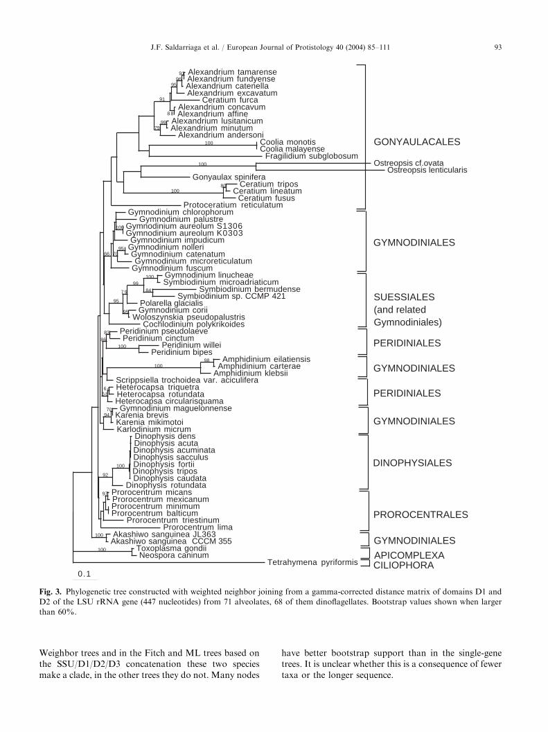
Oxyrrhis, Syndiniales, Noctilucales or Blastodiniales areunavailable, the trees consisted of a large, badly resolvedgroup of very short-branched taxa (the GPP complex,Gymnodiniales, Peridiniales, Prorocentrales and Dino-physiales) and a monophyletic grouping of longer-branched members of the order Gonyaulacales (Fig. 3).Within the GPP complex, groupings well supported inSSU trees are also well supported here, e.g. theGymnodinium fuscum group (henceforth Gymnodinium
sensu stricto, Daugbjerg et al. (2000)), the Karenia/
Karlodinium group, Suessiales (including several Gym-
nodinium species), and Dinophysiales. There are, how-ever, several differences from SSU trees. In at least someLSU trees, all Prorocentrales do group together (e.g. inthe Weighbor tree, Fig. 3), and while the A. carterae
group still holds together with good support and arelatively long branch, it is not at the base of theGonyaulacales (in Weighbor and Fitch trees it interruptsa badly supported clade of Peridiniales). The positionof Woloszynskia is also different in SSU and LSUtrees: while in LSU it branches with the Suessiales with95–97% bootstrap support, in SSU its position is veryunstable (the two alignments contain different speciesof the genus: W. pseudopalustris in LSU, W. leopoliensis
in SSU).Gonyaulacales also form a clade in most LSU trees,
albeit with modest bootstrap support (in the ML tree,the genus Ceratium branches together with theApicomplexan outgroup). The majority of the gonyau-lacalean species with LSU data are members ofthe Goniodominae (Alexandrium, Fragilidium, Coolia
and Ostreopsis), and they form a clade excludingall other taxa, with low bootstrap support (the sequencefor Ceratium furca interrupts a very strongly supported
clade of many Alexandrium species in all trees;we suspect this to be an error). The other Ceratium
sequences, as well as those for Protoceratium andGonyaulax, often make a paraphyletic group at thebase of the Gonyaulacales that gives rise to theGoniodominae (not in the Fitch trees, where Goniodo-minae appear to give rise to Gonyaulacinae andCeratium). Protoceratium and Gonyaulax were neversisters.
Combined rRNA phylogeny
Phylogenetic trees based on combined datasets (Fig. 4)generally show the basic structure discussed above: abadly supported backbone of short-branched taxa (theGPP complex) that includes some very well-supportedsubgroups, and the Gonyaulacales, longer-branchedtaxa that invariably form a clade, here very wellsupported (80–100% bootstrap support). The well-supported groups in the GPP complex are identical tothose discussed above, but their relative order is vari-able. Prorocentrales never group together, forming thesame two clades as in SSU trees. Within Gonyaulacales,the Gonyaulacinae (Gonyaulax and Protoceratium)generally branch as sisters to a group that containsCeratium and the Goniodominae (in the Fitch andWeighbor trees based on SSU/D1/D2/D3 concatena-tions the Gonyaulax/Protoceratium clade is not re-tained). One major difference between theconcatenated and single gene trees is that in allconcatenated trees the two Heterocapsa species includedare sisters to the bulk of the GPP complex withbootstrap support between 43% and 71%. In the
ARTICLE IN PRESS
0.1
Alexandrium tamarenseAlexandrium fundyenseAlexandrium catenellaAlexandrium excavatum
Ceratium furcaAlexandrium concavumAlexandrium affine
Alexandrium lusitanicumAlexandrium minutum
Alexandrium andersoniCoolia monotisCoolia malayense
Fragilidium subglobosumOstreopsis cf.ovata
Ostreopsis lenticularisGonyaulax spinifera
Ceratium triposCeratium lineatum
Ceratium fususProtoceratium reticulatum
Gymnodinium chlorophorumGymnodinium palustre
Gymnodinium aureolum S1306Gymnodinium aureolum K0303
Gymnodinium impudicumGymnodinium nolleriGymnodinium catenatumGymnodinium microreticulatum
Gymnodinium fuscumGymnodinium linucheaeSymbiodinium microadriaticum
Symbiodinium bermudenseSymbiodinium sp. CCMP 421
Polarella glacialisGymnodinium corii
Woloszynskia pseudopalustrisCochlodinium polykrikoides
Peridinium pseudolaevePeridinium cinctum
Peridinium willeiPeridinium bipes
Amphidinium eilatiensisAmphidinium carterae
Amphidinium klebsiiScrippsiella trochoidea var. aciculiferaHeterocapsa triquetraHeterocapsa rotundataHeterocapsa circularisquamaGymnodinium maguelonnense
Karenia brevisKarenia mikimotoiKarlodinium micrum
Dinophysis densDinophysis acutaDinophysis acuminataDinophysis sacculusDinophysis fortiiDinophysis triposDinophysis caudata
Dinophysis rotundataProrocentrum micansProrocentrum mexicanumProrocentrum minimumProrocentrum balticum
Prorocentrum triestinumProrocentrum lima
Akashiwo sanguinea JL363Akashiwo sanguinea CCCM 355
Toxoplasma gondiiNeospora caninum
Tetrahymena pyriformis
9196
95
8 8
91
9979
100
100
80100
100
957066
100
8499
7 9
95
95
8386
100
98100
6 46 8
7094
100
92
97
100
100
GONYAULACALES
GYMNODINIALES
SUESSIALES(and related Gymnodiniales)
PERIDINIALES
GYMNODINIALES
PERIDINIALES
GYMNODINIALES
DINOPHYSIALES
PROROCENTRALES
GYMNODINIALES
APICOMPLEXACILIOPHORA
Fig. 3. Phylogenetic tree constructed with weighted neighbor joining from a gamma-corrected distance matrix of domains D1 and
D2 of the LSU rRNA gene (447 nucleotides) from 71 alveolates, 68 of them dinoflagellates. Bootstrap values shown when larger
than 60%.
J.F. Saldarriaga et al. / European Journal of Protistology 40 (2004) 85–111 93
Weighbor trees and in the Fitch and ML trees based onthe SSU/D1/D2/D3 concatenation these two speciesmake a clade, in the other trees they do not. Many nodes
have better bootstrap support than in the single-genetrees. It is unclear whether this is a consequence of fewertaxa or the longer sequence.

ARTICLE IN PRESS
Alexandrium tamarense
Alexandrium fundyense
Alexandrium cohorticula
Ostreopsis cf. ovata
Alexandrium minutum
Fragilidium subglobosum
Ceratium fusus
Ceratium furca
Gonyaulax spinifera
Protoceratium reticulatum
Symbiodinium sp. in Aiptasia pallida (“S. bermudense”)
Symbiodinium sp. CCMP 421
Symbiodinium microadriaticum
Polarella glacialis
Prorocentrum lima
Gymnodinium catenatum
Gymnodinium impudicum
Gymnodinium fuscum
Dinophysis norvegica
Peridinium bipes
Peridinium willei
Amphidinium carterae
Akashiwo sanguinea
Karenia brevis
Karenia mikimotoi
Karlodinium micrum
Prorocentrum micans
Prorocentrum mexicanum
Prorocentrum minimum
Heterocapsa triquetra
Heterocapsa rotundata
Toxoplasma gondii
Neospora caninum
Tetrahymena pyriformis
0.05substitutions/site
97
100
68
86
96
10081
76
100
100
96
100
100
45
78
81
100
100
GONYAULACALES
SUESSIALES
PROROCENTRALES
GYMNODINIALES
DINOPHYSIALES
PERIDINIALES
GYMNODINIALES
PROROCENTRALES
PERIDINIALES
APICOMPLEXA
CILIOPHORA
Fig. 4. Maximum likelihood phylogenetic tree constructed from concatenated LSU (domains D1 and D2) and SSU rRNA
sequences (2100 nucleotides) from 34 alveolates, 31 of them dinoflagellates. Transition/transversion ratio 2.13; other trees found
with slightly lower log likelihoods differed only in minor details. Bootstrap values are shown when higher than 60%.
J.F. Saldarriaga et al. / European Journal of Protistology 40 (2004) 85–11194

ARTICLE IN PRESSJ.F. Saldarriaga et al. / European Journal of Protistology 40 (2004) 85–111 95
Discussion
Rates of evolution, the structure of dinoflagellate
phylogenetic trees and the mesozoic radiation of
dinoflagellates
There is a striking asymmetry of evolutionary rates inthe ribosomal genes of dinoflagellates, more pronouncedin the SSU genes but also present in the domains of theLSU investigated here. As a consequence, both SSU-and LSU-based phylogenetic trees present a verycharacteristic structure: a large group of very short-branched GPP species, and a clade with medium- to verylong branches. As far as these two groupings areconcerned, the differences in evolutionary rate arecertainly correlated with the phylogenetic history ofthe group: one clade of medium to long-branchedspecies is composed exclusively of taxa classified in theorder Gonyaulacales, and there are typically noGonyaulacales elsewhere. Nevertheless, other speciesalso have divergent sequences, including Oxyrrhis,Haplozoon, Protoperidinium and Amoebophrya in SSUtrees and the A. carterae clade in both SSU and LSU.The fact that, with the exception of Oxyrrhis (Salda-rriaga et al., 2003a) and in a few trees the A. carterae
clade these long-branched taxa do not generally intrudeinto the Gonyaulacales is a sign that the grouping mayreflect a real phylogenetic signal rather than long-branchattraction. It is interesting that no protein sequencesknown from the gonyaulacalean Crypthecodinium cohnii
are particularly divergent; the asymmetry of evolution-ary rates in ribosomal genes of dinoflagellates may notextend to protein genes.
The ‘‘backbone’’ of all dinoflagellate rRNA trees isvery weakly supported. This is consistent with a rapiddinoflagellate radiation into the major forms we seetoday. The palaeontological record gives a very similarpicture (Fensome et al., 1999): although putativedinoflagellate fossils in the form of acritarchs andbiogeochemical traces exist from as early as theCambrian (e.g. Moldowan and Talyzina, 1998, discus-sion in Fensome et al., 1999), undisputed dinoflagellatesappear for the first time in the early Mesozoic, and bythe mid-Jurassic practically all variations of at leastgonyaulacalean and peridinialean forms were alreadypresent. Nevertheless, the fossil record consists almostexclusively of groups that produce fossilizable cysts (ca.15% of extant species of dinoflagellates, Head, 1996),other groups are very badly represented, making itunclear whether the rapid increase in gonyaulacaleanand peridinialean morphological types in the earlyJurassic was caused by a radiation of the whole group.The congruence between the patterns suggested by thefossil record and the rRNA trees, which include non-cyst-formers, implies a general radiation that includedathecate forms.
A phylogenetic framework for understanding
dinoflagellate evolution
Fig. 5 shows a hypothesis on the evolutionary historyof dinoflagellates and their close relatives. It is based onfeatures of molecular trees that are well supported and/or congruent with one-another and on morphologicaland palaeontological information.
Perkinsus; Oxyrrhis and the SyndinialesThe relative positions of Perkinsus, the dinokaryotic
dinoflagellates and the apicomplexans derived frommolecular data are well supported by many differentgenes coding both for rRNAs and for proteins (e.g.Reece et al., 1997; Fast et al., 2001; Saldarriaga et al.,2003a). The relationship between Colpodella and theapicomplexans is based entirely on SSU rRNA phylo-genies, it is recovered consistently and correlates wellwith morphological data (Kuvardina et al., 2002;Leander et al., 2003).
The phylogenetic position of Oxyrrhis has been moreproblematic. Phylogenies based on the SSU gene place itwithin the dinokaryotic dinoflagellates, with 76–81%bootstrap support for a clade of Oxyrrhis and Gonyau-
lax spinifera (Saldarriaga et al., 2003a). Protein-genedata give a very different result: all protein-genephylogenies investigated to date (actin, alpha- andbeta-tubulin in Saldarriaga et al., 2003a, also HSP90,B. Leander, personal communication) place Oxyrrhis atthe base of the dinoflagellates. Because of the extremedivergence of the Oxyrrhis SSU sequence and thecongruence amongst the protein-gene phylogenies (andalso a short LSU fragment, Lenaers et al., 1991), it islikely that Oxyrrhis is a sister to the dinokaryoticdinoflagellates, but this will only be established byimproved sampling of protein-coding genes from dino-flagellates. In any case, Oxyrrhis seems to have divergedlater than Perkinsus; like dinokaryotes it lacks a curvedribbon at the apical end (regarded by some authors as ahomologue to the apicomplexan conoid) that is typicalof several protalveolates (e.g. Colpodella, Perkinsus,
Parvilucifera and Rastrimonas) and the micronemesshared by them and apicomplexans.
Mitosis in Perkinsus is apparently similar to that insyndinians and dinokaryotic dinoflagellates in thatchannels containing an extranuclear spindle traversethe nucleus (Perkins, 1996), while Oxyrrhis has anintranuclear spindle (Triemer, 1982). Postulating thatOxyrrhis originated later than Perkinsus assumes areversal in the organisation of the mitotic apparatus ofOxyrrhis (ciliates, like Oxyrrhis, have a closed mitosiswith an internal spindle, apicomplexans a semiopenone). Nevertheless, other features strongly ally Oxyrrhis
to the dinoflagellates, notably the lack of most realhistones (Li, 1984) and several flagellar features (see

ARTICLE IN PRESS
Fig. 5. Hypothesis on the evolutionary history of dinoflagellates and their close relatives based on the features of molecular trees
that are well supported and/or congruent with one-another and on morphological and palaeontological information.
J.F. Saldarriaga et al. / European Journal of Protistology 40 (2004) 85–11196
discussion below). It is not known whether Perkinsus
has histones, but electron microscopy shows nuclei withdecondensed chromatin during the majority of their lifecycle (Perkins, 1996), very different from those of eitherOxyrrhis or the dinokaryotic dinoflagellates and similarto those of most other eukaryotes. All in all, because ofthe strength of the molecular data and the unlikelihoodthat histones were lost more than once, it seems clearthat Oxyrrhis branches between Perkinsus and the
dinokaryotic dinoflagellates, but that assessment couldchange if Perkinsus unexpectedly proved to lackhistones.
The order Syndiniales is very heterogeneous (e.g.Fensome et al., 1993). The only molecular data availablefor the group are SSU sequences from Hematodinium
and Amoebophrya (different families), but no data are yetavailable for the morphologically most aberrant familyof the order, the Duboscquellaceae. Hematodinium and
ARTICLE IN PRESSJ.F. Saldarriaga et al. / European Journal of Protistology 40 (2004) 85–111 97
Amoebophrya generally form a clade in phylogenetictrees (always with weak bootstrap), but in a few analysesthey separate. Interestingly, the diversity of the ordermight be underestimated: many SSU sequences obtainedfrom picoplanctonic environmental samples cluster withvery high bootstrap support (up to 99%) aroundAmoebophrya. When these sequences were first pre-sented (Moon-van der Staay et al., 2001; L !opez-Garc!ıaet al., 2001), it could not be stated categorically thatthose sequences were actually from syndinians. Theaddition of Hematodinium to the data set greatlystrengthens that assumption, as this syndinian branchesat the base of the clade that contains several strains ofAmoebophrya (another syndinian) and the picoplanc-tonic taxa. The monophyly of the order Syndiniales iscontroversial, and so it will be interesting to see whetherSyndinium and the Dubosquellaceae also branch in thisclade. It is also interesting to speculate whether thosesequences from picoplanktonic cells represent free-livingorganisms (there are no named free-living syndinians) orthe infective stages of parasitic forms. A second group ofenvironmental marine sequences forms a well-resolvedclade that is not closely related to any named alveolates.Since there is no morphological information for it, it isimpossible to say whether the sequences are syndinian(or indeed dinoflagellate) or not. They always branchafter Perkinsus, and so we consider them members of thedinoflagellate lineage, but little can be inferred aboutdinoflagellate evolution from them until their morpho-logy is characterized.
The relative positions of the syndinians and Oxyrrhis
cannot be determined from the available molecular dataalone: only SSU sequences are known for syndinians,and the Oxyrrhis sequence for that gene is misleading(Saldarriaga et al., 2003a). However, syndinians andPerkinsus share an invagination of the nuclear mem-brane in interphase that houses centrioles (Ris andKubai, 1974; Perkins, 1996) that does not occur indinokaryotic dinoflagellates or in Oxyrrhis. For thisreason, we weakly favour a topology where syndiniansare sisters to a clade comprising Oxyrrhis and thedinokaryotic dinoflagellates. Nevertheless, more data isneeded to confirm this.
Noctilucales and Blastodiniales
In the most recent general classification of dinofla-gellates (Fensome et al., 1993) Noctilucales and Blas-todiniales are basal classes of their own within thesubdivision of dinokaryotic dinoflagellates. This isbecause members of both orders have non-dinokaryoticlife stages: the trophonts of Noctiluca, Blastodinium,Amyloodinium and many others have nuclei that lack thetypical fibrillar chromosomes of dinokaryotic dinofla-gellates and stain brightly with alkali fast green, achemical reagent that colors basic proteins (histones oftypical eukaryotic nuclei are easily stained by it,
dinokarya are not). Nevertheless, these species have lifestages with real dinokarya: at certain phases of their lifecycle trophonts start a series of divisions that produceever smaller nuclei with chromosomes that graduallycondense to produce the typical dinokarya (Soyer, 1969,1971, 1972). The dinokaryotic cells thus produced havethe typical appearance of biflagellate dinoflagellates.
Molecular sequences (only SSU) exist for three taxaof either Noctilucales or Blastodiniales: Noctiluca,Amyloodinium and Haplozoon. Noctiluca branches basalto the dinokaryotic dinoflagellates (and usually also thesyndinians) in many phylogenetic trees, but never withhigh bootstrap support (e.g. Fig. 1). However, in analyseswith few outgroups and more aligned sites Noctiluca
branches from within the GPP complex (Fig. 2).Moreover, Noctiluca chromatin may be more similarto that of dinokaryotes than to typical eukaryotes:electrophoretic gels of nuclear basic proteins extractedfrom the Noctiluca trophont produce a band patternconsistent with that of completely dinokaryotic dino-flagellates, not with eukaryotic, histone-containingnuclei (Li, 1984). In other words, Noctiluca may welllack typical core histones throughout its life cycle,suggesting that the alkali fast green stains other non-core-histone proteins in the trophont nucleus. As aconsequence, the basal position of Noctiluca within thedinokaryotic dinoflagellates should be reexamined: thetwo main arguments for proposing such a basal positionhave been shown to be either very weak (SSU-basedphylogenetic analyses), or probably wrong (the osten-sible presence of histones in the nuclei of feeding stages).The fact that the vegetative stages (trophonts) of otherNoctilucales, genera like Leptodiscus, Craspedotella,Petalodinium, Kofoidinium and Spatulodinium, appearto have typical dinokarya (M. Elbr.achter, personalcommunication) further strengthens this view. Threemorphological features of Noctiluca and other Noctilu-cales argue for a relationship of the order to at leastsome groups of gymnodinialean dinoflagellates: youngtrophonts and/or dinospores of several of theless morphologically derived noctilucalean taxa (e.g.Kofoidinium and Spatulodinium) are practically indis-tinguishable from a number of athecate dinoflagellategenera, especially Amphidinium (Cachon and Cachon,1968). More importantly, Noctiluca shares with mem-bers of the genus Gymnodinium senso stricto (Daugbjerget al., 2000) two rare morphological features. First,Gymnodinium, like the gametes of Noctiluca, lacks atransverse striated flagellar root, a feature typical ofmost dinoflagellates (Hansen et al., 2000). Furthermore,the nuclear envelope of many (but not all) Gymnodinium
(as well as Polykrikos) and the trophont of Noctiluca
have peculiar chambers (ampullae) in which the nuclearpores are situated (Afzelius, 1963; Soyer, 1969; Dodgeand Crawford, 1969; Daugbjerg et al., 2000). Thesechambers disappear in Noctiluca as the dinospores are
ARTICLE IN PRESSJ.F. Saldarriaga et al. / European Journal of Protistology 40 (2004) 85–11198
formed (Soyer, 1972), and so they may not behomologous to the ones in Gymnodinium, but if theyare homologues they would provide an importantmorphological connection between the two groups. Itis unknown whether other Noctilucales have ampullaearound the nucleus.
The order Blastodiniales is almost certainly polyphy-letic (e.g. Chatton, 1920; Fensome et al., 1993).Amyloodinium and Haplozoon never branch together inour trees, although both are typically members of theGPP complex (in some trees Amyloodinium may branchat the base of the dinokaryotic dinoflagellates as awhole, e.g. Fig. 1, but see also Fig. 2). Furthermore,although several members of the order have, likeNoctiluca, non-dinokaryotic nuclei in some life stages(e.g. Blastodinium, Amyloodinium, Oodinium, Caryotoma
and Crepidoodinium: Soyer, 1971; Lom and Lawler,1973; Cachon and Cachon, 1977; Hollande and Corbel,1982; Lom et al., 1993), others do not: Dissodinium andProtoodinium are purely dinokaryotic (Cachon andCachon, 1971; Drebes, 1981), as are probably Piscinoo-
dinium and Haplozoon (trophonts in these two generaare dinokaryotic (Lom and Schubert, 1983; Siebert andWest, 1974), and dinospores have never been shown tohave anything other than a dinokaryon in dinokaryoticdinoflagellates). Other genera are understudied, e.g.Apodinium, Cachonella, Sphaeripara; the true phyloge-netic affinities of these taxa are unclear. The derivedposition of Amyloodinium in SSU trees is stronglysupported by morphology: Amyloodinium (and alsoPfiesteria, its sister taxon in most trees) has dinosporeswith a thecal plate pattern like that of Peridiniales(Landsberg et al., 1994; Steidinger et al., 1996; Fensomeet al., 1999). Could other Blastodiniales also bePeridiniales? The trophont of Oodinium fritillariae hasthecal plates like Amyloodinium, but also has ampullaearound the nucleus (Cachon and Cachon, 1977), theProtoodinium trophont even has peridinialean tabula-tion (Cachon and Cachon, 1971). Other Blastodinialesshare more similarities with athecate dinoflagellates:Haplozoon, Crepidoodinium and probably also Piscinoo-
dinium have many polygonal alveoli in surface view(Lom, 1981; Lom and Schubert, 1983; Lom et al., 1993;Leander et al., 2002). It is important to keep in mind,however, that many features known for Blastodiniales(e.g. the small, polygonal alveoli) have been observed intheir trophonts, an often heavily modified life stage. Themorphology of their dinospores will surely be muchmore helpful in determining their true phylogeneticaffinities, as shown by the example of Amyloodinium
ocellatum.
Gymnodiniales, Suessiales and the search for the first
dinokaryotic dinoflagellates
The branching order of extant groups at the base ofthe dinokaryotic dinoflagellates is proving to be very
difficult to determine using molecular methods: phylo-genetic trees calculated through different algorithms andbased on different genes place very different taxa atthose basal positions, and bootstrap supports are neverstrong. Nevertheless, there are tendencies that warrantcomparison with the morphological and paleontologicaldata available.
Yoon et al. (2002), for example, propose Karenia andKarlodinium as sister taxa to the rest of the dinokaryoticdinoflagellates. They used three plastid-encoded genes(psaA, psbA and rbcL) to test the phylogenetic relation-ships between haptophytes and the plastids of peridinin-and 19-hexanoyloxyfucoxanthin-containing dinoflagel-lates (photosynthetic dinoflagellates contain differenttypes of chloroplasts, the type that contains peridinin isby far most common, see below). They found, asexpected, a strong phylogenetic relationship betweenhaptophytes and the plastids of Karenia and Karlodi-
nium, the two genera with 19-hexanoyloxyfucoxanthin-containing plastids (see also Tengs et al., 2000; Ishidaand Green, 2002). Surprisingly, they also found that inpsaA and psbA-based trees peridinin-containing dino-flagellates group either as a sister-taxon to Karenia andKarlodinium, or embedded within a clade with 19-hexanoyloxyfucoxanthin-containing ancestors. Theyproposed an early tertiary endosymbiosis event for thedinoflagellate lineage, and a later transformation of thatplastid into the peridinin-type after the divergence ofKarenia and Karlodinium.
The Karenia/Karlodinium clade is one of the groupingsthat does sometimes branch at the base of thedinokaryotic dinoflagellates in SSU-based phylogenetictrees (e.g. Fig. 2). Both of these genera are athecate taxacurrently classified in the order Gymnodiniales, thegrouping proposed by Fensome et al. (1993) as the mostbasal of the wholly dinokaryotic dinoflagellates. How-ever, there are reasons to question this model for theorigins of peridinin-containing plastids and the phylo-genetic position of the 19-hexanoyloxyfucoxanthin-containing dinoflagellates. First, as Yoon et al. (2002)point out, the divergence rate of the dinoflagellate genesthey examined is noticeably accelerated. Consequently,the dinoflagellate sequences may be attracted to one-another because they share long branches. The authorsattempted to correct for this attraction, but neverthelessthe concern remains. Second, an analogous study of therelationships between Karenia, the haptophytes and theperidinin-containing dinoflagellates using a nuclear-encoded but plastid-targeted gene (psbO, Ishida andGreen, 2002) produced different results: the onesequence for a peridinin-containing dinoflagellate (Het-
erocapsa triquetra) was strongly excluded from aKarenia/haptophyte grouping. This finding is probablymore reliable because the divergence rates in thenuclear-encoded dinoflagellate psbO genes appear tobe much slower than the plastid-encoded genes.

ARTICLE IN PRESSJ.F. Saldarriaga et al. / European Journal of Protistology 40 (2004) 85–111 99
The genus Heterocapsa occupies a basal positionwithin Dinokaryota surprisingly often in phylogenetictrees, especially those based on LSU (maximum like-lihood) and alpha-tubulin; in combined SSU/LSU treesHeterocapsa consistently occupied such a position,although with a low bootstrap support (40–50%).Heterocapsa also has a somewhat atypical sulcaltabulation that could be interpreted as primitive withrespect to that of the rest of the Peridiniales and theGonyaulacales (discussion in Fensome et al., 1993). It isthus not unreasonable that this group may havediverged before the split between those two orders.Paleontological data also agree with this hypothesis: theearliest fossils from the family Heterocapsaceae are earlyJurassic (Wille, 1982), prior to the radiative explosion ofall other peridininialean and gonyaulacalean forms inthe Mesozoic (Fensome et al., 1999).
The phylogenetic history of gymnodinialean dino-flagellates is particularly difficult to discern for severalreasons. First, although the order is well defined as agroup in which the cellular cortex contains relativelynumerous amphiesmal vesicles arranged non-serially(Fensome et al., 1993), several of the species that havehistorically been classified here have been shown tocontain tabulations that make them obvious members ofother orders (e.g. Hansen, 1995 for Katodinium rotun-
datum/Heterocapsa rotundata, Montresor et al., 1999 forPolarella glacialis, Saldarriaga et al., 2003b for Gymno-
dinium elongatum/Lessardia elongata). These tabulationsare very difficult to discover using light microscopyalone, so it is a virtual certainty that several (perhapsmany) taxa currently classified in Gymnodiniales arereally members of other orders. This makes theevaluation of phylogenetic trees where putativelygymnodinialean clades intrude into thecate orders verydifficult; a stringent evaluation of the tabulationalpatterns of putatively gymnodinialean taxa is neededbefore strong statements can be made about theirphylogenetic history. Furthermore, small, non-seriallyarranged amphiesmal vesicles do not necessarily implythe absence of a theca (Netzel and D .urr, 1984), manygymnodinialean taxa have either a full-fledged theca (e.g.the genus Woloszynskia: Crawford et al., 1970; Crawfordand Dodge, 1971), or an incipient one (several membersof Gymnodinium, e.g. G. fuscum and G. cryophilum:Hansen et al., 2000; Wilcox et al., 1982); others haveflocculent material or just liquid and no signs of a theca(e.g. Karlodinium micrum, Leadbeater and Dodge, 1966;A. carterae, Dodge and Crawford, 1968). These featurescan only be studied by electron microscopy, and becauserelatively few species have been investigated in suchdetail, the degree to which presence and type ofintraalveolar material in Gymnodiniales is phylogeneti-cally informative remains unknown.
Molecular data only exists for some gymnodinialeanfamilies, there are still no data available for Polykrika-
ceae, Warnowiaceae, Actiniscaceae, Ptychodiscaceae andothers. Even so, molecular phylogenies always show anumber of separate gymnodinialean clades originatingfrom within the GPP complex, generally separated fromthecate forms by very weak bootstrap supports and notnecessarily basal to them. But are Gymnodiniales sensustricto (i.e. dinoflagellates with numerous small alveoliarranged non-serially) polyphyletic or not? Or is thereason for the polyphyly of the Gymnodiniales sensulato only the fact that it contains species withunrecognized non-gymnodinialean tabulations? Mole-cular data seem to suggest that even Gymnodinialessensu stricto are polyphyletic: well-studied taxa withsmall alveoli (e.g. A. carterae, Karenia brevis, G. fuscum)never group together in phylogenetic trees.
The fact that in virtually all molecular trees gymno-dinialean species arise from within the GPP complex,separated from thecate taxa by very weak bootstrapvalues, suggests that most, if not all, groups ofGymnodiniales had thecate ancestors; the differenttypes of alveolar inclusions in the group, from thecaeto flocculent material to nothing at all would thereforerepresent intermediate stages of thecal loss. The alter-native would be that the Gymnodiniales (or at leastsome of their subgroups) are the sister group to theother dinokaryotic dinoflagellates. This view is sup-ported by the fact that the small alveoli of Gymnodi-niales are shared with more basal members of thedinoflagellate lineage, e.g. the syndinians (plasmodiallife stage), Oxyrrhis and even Colpodella. Moleculardata cannot distinguish between these possibilities atpresent; it cannot determine whether some Gymnodi-niales are ancestral and others derived. Palaeontology isnot very helpful in this regard either: gymnodinialeancysts are often very difficult to ally to identifiable motilestages, so fossil cysts of this type are particularly likelyto be considered acritarchs, microfossils without knowntaxonomical affinities (Fensome et al., 1993); the earliestcertain gymnodinialean fossils (skeletal elements fromActiniscaceae and Dicroerismaceae) are relatively re-cent, from Tertiary formations.
Palaeontological data suggest an early origin foranother order of dinokaryotic dinoflagellates, theSuessiales. They comprise organisms with amphiesmalvesicles arranged in 7–10 latitudinal series, fewer than intypical athecate dinoflagellates and more than in thecateones, a feature that suggests an interesting position forthe order between thecate and athecate forms. Muchmore interesting, however, is the fact that suessialeanfossils are known from the mid-Triassic, prior to theemergence of most (if not all) peridinialean andgonyaulacalean forms (the gonyaulacalean Shublikodi-niaceae, like the earliest suessialean fossils, are from themid-Triassic, Fensome et al., 1999; the identity of anyPalaeozoic fossils as true dinoflagellates is still con-troversial). Molecular trees presently do not support any

ARTICLE IN PRESSJ.F. Saldarriaga et al. / European Journal of Protistology 40 (2004) 85–111100
particular position of the Suessiales: although the grouprarely appears at the base of the dinokaryotic dino-flagellates (e.g. in Edvardsen et al., 2003), its position inother parts of the tree is never supported either. Oneadditional feature of the Suessiales is becoming cleareras the SSU gene of more species of dinoflagellates issequenced: the group is likely to be larger thanpreviously assumed. Montresor et al. (1999) describedthe first extant member of the family Suessiaceae, agroup until then known only from fossils, and since thenmany species of putatively athecate dinoflagellates havebeen shown to group in the same clade in both SSU- andLSU-based trees (e.g. G. beii, G. simplex, G. corii andWoloszynskia pseudopalustris). It will be interesting tosee whether close morphological examination of more ofthese species will agree with the molecular data.
In summary, the problem of the earliest-branchingdinokaryotic dinoflagellates is very far from beingresolved: morphological and palaeontological datacould be interpreted as pointing towards gymnodinia-lean and suessialean taxa for these positions, moleculardata towards Heterocapsa. What is clear is thatGymnodiniales are a polyphyletic group; many gymno-dinialean taxa are more closely related to thecate formsthan to other gymnodinialeans and thecal loss indinoflagellates seems to be common. Suessiales mayform an intermediate stage between the primitivelyathecate gymnodiniales (if they exist) and thecate forms,but this is particularly uncertain as phylogenetic trees donot usually put them in a basal position. If dinokaryoticdinoflagellates indeed underwent an event of rapidevolutionary radiation early in their history, it will bevery difficult to determine the phylogenetic order of thegroups that originated in that explosion.
Phytodiniales
The order Phytodiniales (also Dinococcales, Dino-capsales or Dinamoebales, see Fensome et al. (1993) fora nomenclatural discussion) contains dinoflagellates inwhich the principal life stage is either a non-calcareouscoccoid cell or a continuous-walled multicellular stage.It is a polyphyletic grouping of convenience used tocontain species that are poorly understood; the onlycriterion for determining whether a species belongs tothis order is a shift in life cycle that has also been seen inmany dinoflagellate genera with well-known tabula-tions, e.g. in Symbiodinium (Suessiales), Pyrocystis
(Gonyaulacales) and Thoracosphaera (probably Peridi-niales). SSU rRNA sequences exist for three dinofla-gellate species formally classified in the Phytodiniales:Halostylodinium arenarium, Hemidinium nasutum, andGloeodinium viscum. H. nasutum and the type species ofGloeodinium, G. montanum, have extremely similarcoccoid stages, the two species have even been proposedto be identical (Popovsky, 1971). A fourth species in ourtrees, Glenodiniopsis steinii, is currently classified in the
Peridiniales, but has a coccoid life stage reminiscent ofthe Gloeodinium-like stage of Hemidinium nasutum
(Popovsky and Pfiester, 1990).Halostylodinium arenarium groups with gonyaulaca-
lean taxa in all phylogenetic trees examined, a placementthat is congruent with most (but not all) tabulationalfeatures of the species as interpreted by Horiguchi et al.(2000). Glenodiniopsis, Hemidinium and Gloeodinium onthe other hand, consistently branch within the GPPcomplex, although only in the Weighbor trees do thethree species weakly branch close to one-another (cladesconsisting of two of the three species are common inmany trees). This placement is congruent with theperidinialean tabulation of the motile stage of Glenodi-
niopsis, and suggests that once the tabulations ofHemidinium and Gloeodinium are fully determined (onlya partial tabulation is known for Hemidinium nasutum,no tabulational data exists for G. viscum) they will showperidinialean affinities. Molecular data do not stronglysupport a phylogenetic relationship between Hemidi-
nium nasutum and Gloeodinium viscum, but do notdisprove it either.
Peridiniales, Gonyaulacales, Dinophysiales and
Prorocentrales
Members of the Peridiniales, Gonyaulacales, Dino-physiales and Prorocentrales are likely to have acommon ancestor. The relative positions of the thecalplates in Peridiniales and Gonyaulacales are so similarthat a close relationship between the two orders hasnever been doubted, and paleontological and morpho-logical evidence points to a close relationship betweenPeridiniales, Dinophysiales and Prorocentrales. Pa-laeontological data yielded very strong evidence linkingDinophysiales to peridinialean ancestors: the fossilgenus Nannoceratopsis, found as dinosporin cystsin marine strata of Jurassic origin, has distinctlydinophysialean features in its lateral compressed shape,hyposomal sagittal features and hyposomal pseudota-bulation, but its epitheca has distinct peridinialeantraits, very different from those of other Dinophysiales(Piel and Evitt, 1980; Fensome et al., 1993). WithinPeridiniales, the groups with the most similarity toNannoceratopsis are the fossil Comparodiniaceae andthe extant Oxytoxaceae (Fensome et al., 1993). Nomolecular data exist for Oxytoxum, but a close relation-ship between Peridiniales and Dinophysiales is weaklyapparent in molecular phylogenetic trees: in our treesDinophysiales are always embedded in the GPP com-plex (alternative placings for the group also exist, e.g.Edvardsen et al., 2003). Prorocentrales also branchwithin the GPP complex, even if in SSU trees the ordersplits into 2 groups (or more: Grzebyk et al., 1998).Despite this split, we consider that this morphologicallyvery cohesive order is monophyletic, as tabulationpatterns within it are both radically derived and

ARTICLE IN PRESSJ.F. Saldarriaga et al. / European Journal of Protistology 40 (2004) 85–111 101
homogenous. Interestingly, at least some LSU trees(notably Weighbor) show Prorocentrales as monophy-letic. The phylogenetic origins of the group are moredifficult to discern. The fact that Prorocentrales aremembers of the GPP complex in molecular trees weaklyargues for a relationship to Peridiniales and Dinophy-siales, as well as to many Gymnodiniales. Two largelateral plates (valves) that contact each other along asagittal suture are a common feature of Prorocentralesand Dinophysiales; they may be closely related to eachother (Taylor, 1980). Nevertheless, no intermediatefossil forms exist to shed light on this.
Thus, a relationship between Peridiniales and theDinophysiales/Prorocentrales on the one hand, andGonyaulacales on the other, seems likely. But whichappeared first? The earliest dinoflagellate fossils (exceptfor the controversial Palaeozoic forms already men-tioned) are, in addition to the Suessiales, members of thegonyaulacalean Shublikodiniaceae (Fensome et al.,1999). This is an exclusively fossil family with verycharacteristic tabulation: more than five climactal platesand at least three fundital plates. Fensome et al. (1993)points out tabulational resemblances between thisfamily and two other groups: early cladopyxiineansand living members of the genus Glenodinium. What isinteresting is that whereas Shublikodiniaceae andCladopyxiineae are early lineages within the Gonyaula-cales (as classified by Fensome et al., 1993), Glenodi-niaceae are undoubtedly peridinialean forms. Thus, thelines between the two orders blur at this level. Moleculardata tend to give trees where a paraphyletic Peridinialesis ancestral to the holophyletic Gonyaulacales, althoughbootstrap support for this branching order is generallylow. Nevertheless, molecular data do not exist for manyputatively basal groups of either Gonyaulacales orPeridiniales, genera like Cladopyxis, Acanthodinium,
Amphidoma or Palaeophalacroma (Gonyaulacales), orGlenodinium (Peridiniales). Heterocapsa (Peridiniales) isan exception to this, and as discussed above, it tends totake a basal position to other thecate dinoflagellates inmany phylogenetic trees, particularly those based oncombined data sets of small- and large subunit rRNAgenes. The implication of this position would be thatPeridiniales are indeed ancestral to Gonyaulacaleans, athesis that runs contrary to palaeontological data: notrue peridinialean fossils are known from before theappearance of the earliest gonyaulacaleans, the Shubli-kodiniaceae. Nevertheless, just as it is dangerous to givetoo much credence to the branching order of Hetero-
capsa in phylogenetic trees because of mediocre supportsof the relevant nodes, the present lack of peridinialeanfossils from the Triassic does not necessarily mean thatthe group was completely absent then.
One more dinoflagellate ‘‘order’’ appears to be veryclosely related to Peridiniales: the Thoracosphaerales.The principal life-stage of Thoracosphaera, the only
genus in the order, is a coccoid cell surrounded by acalcareous wall, very similar to calcareous cysts of asubgroup (subfamily Calciodinelloideae of the Peridini-aceae) within the Peridiniales that includes Scrippsiella,
Ensiculifera, etc. Nevertheless, the motile stage ofThoracosphaera is apparently athecate and the arche-opyle of the cyst quite atypical, so a separate order wascreated for it (Tangen et al., 1982). Molecular data tendsto support a relationship between Thoracosphaera andseveral genera of Peridiniales, including Peridiniopsis
and, interestingly, Scrippsiella. This position in mole-cular trees (as well as the calcareous cyst wall) wouldpredict a peridinialean tabulation of the motile stage. Ifthis turns out to be the case, the order Thoraco-sphaerales should be abolished and Thoracosphaera
made a member of the Peridiniales and of theCalciodinelloideae.
If the scenario presented above is correct, thePeridiniales would occupy a very important place forthe phylogeny of the dinoflagellates as a whole. Theywould be a complexly paraphyletic group that gave risenot only to the other obviously thecate taxa (Dinophy-siales and Prorocentrales, and perhaps also Gonyaula-cales), but to many athecate and putatively athecateforms as well (many lineages of Gymnodiniales,Thoracosphaerales, as well as, perhaps, also Noctilu-cales and Blastodiniales).
Whereas branching orders within Peridiniales are notresolved in any of our trees, within Gonyaulacales therate of evolution of both the large- and the SSU rRNAgenes is faster, and as a consequence branches of theresulting phylogenetic trees are longer; lineages are alsogenerally separated by better bootstrap supports.Felicitously, the Gonyaulacales are also a group with avery good fossil record, and the tabulational patterns ofextant and fossil members are well known. For thisreason, the group provides a good model to contrasttaxonomic schemes based on morphology (i.e. tabula-tion) with molecular data.
The two sets of data correlate well within this group.Two of the three gonyaulacalean suborders for whichthere are SSU sequences are normally recovered inphylogenetic trees with decent bootstrap support:Goniodomineae (50–60%) and Ceratiineae (75–90%).The third suborder, Gonyaulacineae, usually forms aparaphyletic group that gives rise to both Goniodomi-neae and Ceratiineae, as well as to taxa of uncertaintaxonomic position like Thecadinium and Crypthecodi-
nium (and the formally phytodinialean genus Halosty-
lodinium). Large subunit gene trees also generallysupport the monophyly of Goniodominae, althoughwith weak bootstrap support and one important caveat:it is always intruded by the sequence from Ceratium
furca (SSU and morphological data agree that thisspecies is a Ceratiineae, the veracity of the identity ofthis LSU sequence should be reexamined). The other
ARTICLE IN PRESSJ.F. Saldarriaga et al. / European Journal of Protistology 40 (2004) 85–111102
Ceratiineae (i.e. the genus Ceratium) group strongly, andProtoceratium and Gonyaulax, the only Gonyaulacineaein the trees, make a paraphyletic group at the base of theorder. One difference between molecular trees andtaxonomic schemes based on morphology (i.e. Fensomeet al., 1993) is the position of Protoceratium: SSU-basedphylogenies never place it with Lingulodinium, Gonyau-
lax and Amylax in the family Gonyaulacaceae, butrather with Ceratocorys (Ceratocoryaceae, 100% boot-strap support).
Tracing morphological characters onto molecular
trees: from protalveolates to dinoflagellates
Having proposed a putative framework for thephylogenetic history of dinoflagellates, we now examinethe evolutionary history of certain morphologicalfeatures in its light.
The nucleus
Although undoubtedly eukaryotic (e.g. M!ınguez et al.,1994; Salamin Michel et al., 1996), dinokaryotic nucleishow important biochemical differences with respectto the nuclei of other eukaryotes: they lack histones(e.g. Rizzo, 1991), can contain very large amounts ofDNA (2–200 pg DNA per haploid nucleus, nuclei ofhuman cells have ca. 5.6 pg DNA per cell, Sigee, 1986)and up to 70% of the thymine in their DNA is replacedby 5-hydroxymethyluracil (Rae, 1976). They also dividethrough a type of mitosis characteristic for the group:the nuclear membrane remains intact, chromosomesremain attached to the nuclear membrane, and, duringmitosis, channels are formed that contain the micro-tubules of the mitotic spindle; microtubules attach to thechromosomes only where they touch the nuclearmembrane (references in Dodge, 1987). The scale ofthe ultrastructural and biochemical reorganization thatoccurred in the nuclei of the alveolates that becamedinoflagellates is unparalleled in any other group ofeukaryotes and the process that led to it is completelyunknown. It is thus of interest to trace some of thefeatures of this change down the dinoflagellate lineage,to determine when exactly the different characters of thedinokaryon originated.
The question of the presence or absence of histones inthe dinoflagellate lineage has interested many research-ers over the years. The paradigm on the absence oftypical histones in the group is based on several facts:nucleosomes have not been detected in dinoflagellatesusing any method (e.g. electron-microscopical observa-tion of chromatin spreads, digestion of internucleosomalDNA followed by electrophoresis, etc., Rizzo, 1991), theratio of basic chromatin to DNA is much lower indinokaryotic dinoflagellates than in any other eukaryote(Rizzo and Nood!en, 1973), and electrophoresis of
dinoflagellate nuclear basic proteins has consistentlyproduced banding patterns that do not correspond tothe ones formed by eukaryotic histones (e.g. Rizzo,1981). Only recently have some of the nuclear basicproteins from dinoflagellates started to be sequenced(Sala-Rovira et al., 1991; Taroncher-Oldenburg andAnderson, 2000; Chudnovsky et al., 2002), and to datethere are three sequences available, from Crypthecodi-
nium cohnii, Alexandrium fundyense and Lingulodinium
polyedrum (all Gonyaulacales). Homologies of thesehistone-like proteins (HLPs) of dinoflagellates to otherproteins are not obvious, but Kasinsky et al. (2001)reported a 31% similarity in amino acid compositionbetween the complete HCc2 of Crypthecodinium cohnii
(a histone-like protein) and the C-terminus of the linkerhistone H1b of the sea urchin. Nucleosomal histoneshave never been detected in dinoflagellate nuclei.
The presence or absence of histone proteins in thenuclei of protalveolates and dinoflagellates is obviouslya very important feature in the study of the phylogeneticquestions that interest us. It is highly unlikely thatnucleosomal histones in dinoflagellates were lost morethan once. Historically, the determination of just whichtaxa (or in some cases life stages) of dinoflagellates havehistones and which do not has been done by chemicalstaining of the basic proteins in their nuclei: dinokaryado not stain with alkaline fast green, while the nuclei ofmost eukaryotes, including syndinians, Oxyrrhis and thetrophonts of taxa like Noctiluca, Blastodinium andOodinium do (references in Table 3). The ultrastructureof those nuclei is also quite different from that ofdinokarya, and so it was thought that they wereprofoundly different from them. Preliminary biochem-ical analyses of the nuclear basic proteins of Oxyrrhis
and the Noctiluca trophont (Li, 1984) have shownhowever that the electrophoretic pattern of thoseproteins in SDS- and acidic urea gels resembles theones of dinokaryotic HLPs, not the patterns of histone-containing organisms. If the basic proteins in the nucleiof Noctiluca and Oxyrrhis are not normal histones, thenthe change from histone-containing to histone-lackingnuclei in the dinoflagellate lineage occurred earlier thanpreviously assumed. Where exactly is not easy todetermine. No biochemical studies on syndinian nucleiexist, but Hollande (1974) did stain the nuclei of fourspecies with alkali fast green. The nuclei of differentsyndinians stain differently: in Syndinium and Solenodi-
nium the chromosomes are stained, while in Amoebo-
phrya and Duboscquella only the nucleoli are.Unfortunately only Amoebophrya is represented inmolecular based phylogenetic trees, so it is uncertainwhether the order is really monophyletic; nevertheless,the staining pattern in Amoebophrya and Duboscquella ismore consistent with the presence of histone-likeproteins in those organisms rather than real histones.Ciliates and apicomplexans clearly have histones
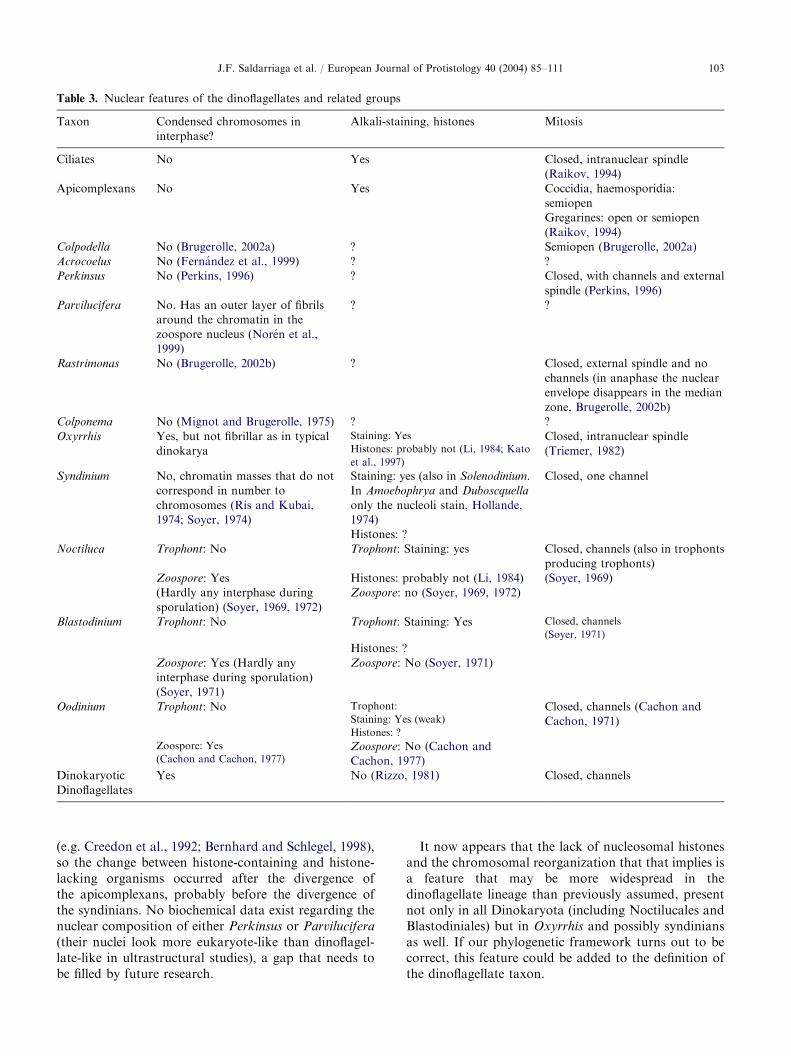
ARTICLE IN PRESS
Table 3. Nuclear features of the dinoflagellates and related groups
Taxon Condensed chromosomes in
interphase?
Alkali-staining, histones Mitosis
Ciliates No Yes Closed, intranuclear spindle
(Raikov, 1994)
Apicomplexans No Yes Coccidia, haemosporidia:
semiopen
Gregarines: open or semiopen
(Raikov, 1994)
Colpodella No (Brugerolle, 2002a) ? Semiopen (Brugerolle, 2002a)
Acrocoelus No (Fern!andez et al., 1999) ? ?
Perkinsus No (Perkins, 1996) ? Closed, with channels and external
spindle (Perkins, 1996)
Parvilucifera No. Has an outer layer of fibrils
around the chromatin in the
zoospore nucleus (Nor!en et al.,
1999)
? ?
Rastrimonas No (Brugerolle, 2002b) ? Closed, external spindle and no
channels (in anaphase the nuclear
envelope disappears in the median
zone, Brugerolle, 2002b)
Colponema No (Mignot and Brugerolle, 1975) ? ?
Oxyrrhis Yes, but not fibrillar as in typical
dinokarya
Staining: Yes
Histones: probably not (Li, 1984; Kato
et al., 1997)
Closed, intranuclear spindle
(Triemer, 1982)
Syndinium No, chromatin masses that do not
correspond in number to
chromosomes (Ris and Kubai,
1974; Soyer, 1974)
Staining: yes (also in Solenodinium.
In Amoebophrya and Duboscquella
only the nucleoli stain, Hollande,
1974)
Closed, one channel
Histones: ?
Noctiluca Trophont: No Trophont: Staining: yes Closed, channels (also in trophonts
producing trophonts)
Zoospore: Yes Histones: probably not (Li, 1984) (Soyer, 1969)
(Hardly any interphase during
sporulation) (Soyer, 1969, 1972)
Zoospore: no (Soyer, 1969, 1972)
Blastodinium Trophont: No Trophont: Staining: Yes Closed, channels
(Soyer, 1971)
Histones: ?
Zoospore: Yes (Hardly any
interphase during sporulation)
(Soyer, 1971)
Zoospore: No (Soyer, 1971)
Oodinium Trophont: No Trophont:
Staining: Yes (weak)
Histones: ?
Closed, channels (Cachon and
Cachon, 1971)
Zoospore: Yes
(Cachon and Cachon, 1977)Zoospore: No (Cachon and
Cachon, 1977)
Dinokaryotic
Dinoflagellates
Yes No (Rizzo, 1981) Closed, channels
J.F. Saldarriaga et al. / European Journal of Protistology 40 (2004) 85–111 103
(e.g. Creedon et al., 1992; Bernhard and Schlegel, 1998),so the change between histone-containing and histone-lacking organisms occurred after the divergence ofthe apicomplexans, probably before the divergence ofthe syndinians. No biochemical data exist regarding thenuclear composition of either Perkinsus or Parvilucifera
(their nuclei look more eukaryote-like than dinoflagel-late-like in ultrastructural studies), a gap that needs tobe filled by future research.
It now appears that the lack of nucleosomal histonesand the chromosomal reorganization that that implies isa feature that may be more widespread in thedinoflagellate lineage than previously assumed, presentnot only in all Dinokaryota (including Noctilucales andBlastodiniales) but in Oxyrrhis and possibly syndiniansas well. If our phylogenetic framework turns out to becorrect, this feature could be added to the definition ofthe dinoflagellate taxon.

ARTICLE IN PRESSJ.F. Saldarriaga et al. / European Journal of Protistology 40 (2004) 85–111104
Mitosis
Most chromists have an open (sometimes semi-open)mitosis, ciliates have a closed mitosis with an intra-nuclear spindle, and the majority of apicomplexans (andColpodella, Brugerolle, 2002a) have a semi-open one (ina number of gregarines it is open). The dinoflagellatelineage is very consistent in this regard (Table 3):Perkinsus, the syndinians, Oxyrrhis and the dinokar-yotic dinoflagellates all have a closed mitosis, and inmost of the members of the group the spindle is external(Perkins, 1996; Triemer and Fritz, 1984). Oxyrrhis,
however, has an internal spindle (Triemer, 1982). Withthe exception of Oxyrrhis, all members of the lineageform channels during mitosis, syndinians only one,dinokaryotic dinoflagellates more (the number ofchannels in Perkinsus is unclear, and mitosis inParvilucifera is entirely unknown). The mitotic channelsthus probably originated at the base of the dinoflagellatelineage, roughly at the same time as the histones werelost (a detailed study of these features in Perkinsus isneeded to confirm this). The external spindle possiblyoriginated before this, prior to the differentiation of theapicomplexan lineage. The only way to explain the stateof these characters in Oxyrrhis while taking into accountthe molecular data on its position in the phylogenetictree is to postulate an internalization of the mitoticspindle and the loss of all mitotic channels (deep, narrownuclear membrane invaginations are common in Oxy-
rrhis during interphase (Triemer, 1982); they may ormay not have any relationship to mitotic channels).Interestingly, Rastrimonas divides through a modifiedclosed mitosis (in anaphase the nuclear envelopedisappears in the median zone) with an external spindle,but it does not seem to form channels (Brugerolle,2002b). It will be interesting to see where this genus fallsin phylogenetic trees.
One other feature significant for understanding theevolution of these organisms is the nature of theircentrosomes, the cell regions that act as microtubuleorganizing centers (MTOCs). In dinokaryotic dinofla-gellates spindle microtubules originate in centriole-lacking centrosomes (also called archeoplasmic spheres)located outside the nucleus and connected to the basalbodies by a microtubular fibre (Perret et al., 1993;Ausseil et al., 2000). Centrosomes in Perkinsus and insyndinians, however, do contain centrioles (references inTable 3), while in Oxyrrhis the mitotic spindle originatesin electron-dense plaques embedded in the nuclearenvelope (Triemer, 1982). Similar electron-dense zonesalso exist in the nuclear envelope of syndinians (and inOodinium, Cachon and Cachon, 1977), but whereas inOxyrrhis the plaques act as MTOCs for microtubulesthat either cross the nucleus or attach to chromosomes(Triemer, 1982), in syndinians these are kinetochores,with chromosomes attached on the inner side of themembrane and microtubules on the outer side. Whether
these structures in syndinians and Oxyrrhis are homo-logous structures is unknown. Interestingly, Oxyrrhis
centrioles may also be involved in mitosis: they migratetowards the nuclear poles early in division, and remainthere throughout mitosis (Triemer, 1982). However,microtubules were never observed between these cen-trioles and the nucleus, so their role is unclear.
Plastids and photosynthesis
Only roughly half of the species of dinoflagellates areknown to be photosynthetic (Taylor, 1987), and the typeof plastids that they contain can be extremely differentfrom one-another (Schnepf and Elbr.achter, 1999;Saldarriaga et al., 2001): although most photosyntheticdinoflagellates harbour peridinin-containing plastidssurrounded by two to three membranes (here calledperidinin plastids), other forms probably arose fromhaptophyte, prasinophyte, cryptomonad or diatomendosymbionts (Watanabe and Sasa, 1991; Chesnicket al., 1997; Tengs et al., 2000; Hackett et al., 2003). Thispromiscuity in the incorporation of endosymbionts is afeature unique to dinoflagellates; no other group ofeukaryotes contains a comparable variety of plastidtypes.
Mixotrophy is unusually common in photosyntheticdinoflagellates (Schnepf and Elbr.achter, 1992; Stoecker,1999), so in the absence of other data it was originallypostulated that the peridinin plastid was incorporatedby a full-fledged dinoflagellate (e.g. Whatley et al., 1979;Gibbs, 1981), just as the other types of plastids in thelineage are still believed to have been. However, theincorporation of the ancestor of the peridinin plastidprobably occurred much earlier (Cavalier-Smith, 1999,2003; Fast et al., 2001; Harper and Keeling, 2003). Thefirst clues to this arose when a plastid remnantwas found in apicomplexans, the closest relatives todinoflagellates (Wilson et al., 1991). Phylogenetic treeswere at first produced based on genes from the plastidgenomes of both dinoflagellates and apicomplexans, andthese trees tended to cluster the two together. However,the plastid genomes of both groups are extremelyderived, so long branch attractions could be excluded(Takishita and Uchida, 1999; Zhang et al., 2000). Fastet al. (2001) used nuclear encoded, plastid-targeted geneswith more conclusive results: there appears to have beena gene duplication event in an ancestor of not onlydinoflagellates and apicomplexans but also the restof the alveolates and chromists. The product of thatgene duplication (a plastid-targetted GAPDH of cyto-solic ancestry) appears to exist in plastid-bearing alveo-lates as well as in cryptomonads, heterokonts andhaptophytes (Fast et al., 2001; Harper and Keeling,2003), implying that the ancestor of all of these groupscontained a plastid.
Within the dinoflagellate lineage, photosyntheticforms only appear relatively late in the evolutionary
ARTICLE IN PRESSJ.F. Saldarriaga et al. / European Journal of Protistology 40 (2004) 85–111 105
history of the group, early branching taxa (i.e.Perkinsus, Parvilucifera, the syndinians and Oxyrrhis)are all non-photosynthetic. Interestingly, however, twoiron superoxide dismutases have been described inPerkinsus (Wright et al., 2002), and one of these encodesa substantial amino-terminal leader that is predicted tobegin with a signal peptide. This is preliminary butsuggestive evidence that Perkinsus might harbour a relictplastid, something that should become clear when moredata from that organism are available.
That all peridinin-containing dinoflagellates musthave a common ancestor is beyond doubt: peridininprobably originated only once (e.g. Saunders et al.,1997; Saldarriaga et al., 2001). The implication of this isthat all the non-photosynthetic lineages that appearafter the latest possible common ancestor of peridinin-containing dinoflagellates must represent instances ofloss of plastids or at least of photosynthetic ability (non-photosynthetic plastids can be difficult to identify).Using the same logic, all lineages branching after thatlatest possible peridinin-containing ancestor that con-tain plastids different from the peridinin type, must beinstances of plastid replacement (Saldarriaga et al.,2001). Plastid replacement differs fundamentally fromsecondary symbiogenesis in that it probably occurs bythe recruitment of at least some of the pre-existingplastid-targeting machinery rather than the evolution ofentirely novel systems (Cavalier-Smith, 2003). It seemsto have been able to occur multiple times in dino-flagellates, but never in other chromalveolates, perhapsbecause they retained the ability to phagocytose(necessary to acquire foreign algae) and were able toeffect such recruitment because they also retained theancestral chromalveolate ability to target endomem-brane vesicles to the outermost smooth (epiplastid)membrane surrounding the plastid (for details on theorigins of chromalveolate plastid protein targeting, seeCavalier-Smith, 2003). Interestingly, the same charac-teristics are found in the chlorarachniophyte algae,which have recently been shown to have replaced manyof their plastid genes with homologues from other algae(Archibald et al., 2003), but have not been demonstratedto have replaced their plastid.
The flagella and the definition of Dinoflagellates
The definition used by Fensome et al. (1993) in theirtreatment of dinoflagellates is based on flagellarcharacters. The transverse flagellum of dinoflagellatesis very distinct in its ultrastructure: the flagellar axonemeis accompanied by a striated strand throughout its entirelength and both structures are contained by a commonplasmalemma that produces a ribbon-like structure.Simple mastigonemes arise in a row along the outer edgeof the axoneme (Gaines and Taylor, 1985). The striatedstrand is always shorter than the axoneme, so theflagellum takes a ‘‘wavy’’ appearance. In addition to
these ultrastructural features, the fact that both flagellainsert laterally is characteristic of the group (the‘‘apical’’ flagellar insertion in the Prorocentrales is nottopologically different from that of the rest of thedinoflagellates, Taylor, 1980).
The flagella of apicomplexans and cilia of ciliates aregenerally smooth (in apicomplexans only the micro-gametes of some groups are flagellated), but most taxain the dinoflagellate lineage, including Perkinsus, Parvi-
lucifera, Oxyrrhis and at least some syndinians (e.g.Amoebophrya, W. Coats, personal communication)appear to have at least one flagellum that carriesmastigonemes (Table 4, the syndinian genus Hematodi-
nium apparently does not, Appleton and Vickerman,1998). The same is true for at least some species ofColpodella, a sister taxon to the apicomplexans (Leanderet al., 2003, but see also Brugerolle, 2002a). This factcombined with the presence of more complex mastigo-nemes in heterokonts and cryptomonads suggests thatsimple non-tubular mastigonemes may have been anearly feature of alveolates, and that ciliates, apicomplexaand some syndinians lost them secondarily. This wouldonly be true, however, if the mastigonemes in thedinoflagellate lineage are related to those of heterokonts;the two structures are not ultrastructurally identical, butthe possibility remains that at least some proteins ofwhich they are made are homologues and that their verymarked ultrastructural differences arose by divergencefrom a common ancestral mastigoneme rather than denovo (see also Cavalier-Smith, 2004).
A paraxial rod in the transverse flagellum (heredefined as the flagellum that carries mastigonemes in alateral row) is, on the other hand, only present inOxyrrhis, in the dinokaryotic dinoflagellates and in atleast one syndinian (Amoebophrya); it is not present inthe apicomplexan lineage, Perkinsus or Parvilucifera.The ultrastructures of the paraxial rod/striated strand ofOxyrrhis and the dinokaryotic dinoflagellates aredifferent, however, but the exact nature of thosedifferences is not understood (Gaines and Taylor,1985; Dodge and Crawford, 1971).
The longitudinal flagellum of dinoflagellates rarelycarries mastigonemes (never in a lateral row) andparaflagellar material, sometimes in the form of aparaxial rod (e.g. Leadbeater and Dodge, 1967;Maruyama, 1982) that can cause a characteristic‘‘ribbon-like’’ appearance of the flagellum. This is foundin several members of the dinoflagellate lineage, e.g. inParvilucifera and in many dinokaryotic dinoflagellates,especially Gonyaulacales (Leadbeater and Dodge, 1967;Maruyama, 1982; Nor!en et al., 1999). Additionalfeatures of the flagellar apparatuses of dinoflagellatesand their relatives (ultrastructure and arrangement ofbasal bodies, microtubular assemblages, fibrous roots,etc.) have been shown to be phylogenetically informative(Roberts, 1991; Perret et al., 1993), but data are still

ARTICLE IN PRESS
Table 4. Flagella in the dinoflagellates and related lineages
Taxon Flagellar insertion Anterior/transversal
flagellum
Posterior/longitudinal
flagellum
Reference
Apicomplexans Essentially apical,
when present
Only in microgametes of
some groups.
Only in microgametes of
some groups.
Perkins et al. (2002) (see
references)
No mastigonemes. No mastigonemes.
Colpodella Subapical C. vorax: Paraxonemal
structure in the proximal
portion.
Unremarkable. Brugerolle (2002a),
Leander et al. (2003)
No mastigonemes. No mastigonemes.
C. edax: Mastigonemes
present
Acrocoelus Ventral Both flagella are posteriorly
directed.
Unremarkable. Fern!andez et al. (1999)
No mastigonemes. No mastigonemes.
Perkinsus Ventral Only in zoospores. Only in zoospores. Perkins (1996)
Filamentous mastigonemes
along one side, in groups.
Unremarkable, much
shorter than anterior
flagellum.
Spur at the base of each
group.
No mastigonemes.
Parvilucifera Subapical Only in zoospore. Only in zoospore. Nor!en et al. (1999)
Short mastigonemes on one
side, long, thin hairs on the
other.
Much shorter than anterior
flagellum
Proximal part with a wing,
distal part lacks the peripheral
doublets of the axoneme on the
side opposite to the wing after it
terminates.
Rastrimonas Subapical Anterior flagellum shorter.
Mastigonemes ‘‘have not
been satisfactorily
demonstrated’’
Longer. Terminates in a thin
filament.
Brugerolle (2002b)
Colponema Subapical Anterior flagellum shorter.
Filamentous mastigonemes
along one side
Longer. With a very high
wing in the median and
distal sections.
Mignot and Brugerolle
(1975)
Oxyrrhis Ventral, emerge from
the base of the
tentacle
Single row of fine hairs.
Paraxial rod present.
Mostly smooth, but has a
paintbrush-like structure at
the end
Dodge and Crawford
(1971)
Amoebophrya Ventral Single row of mastigonemes.
Paraxial rod present.
Unremarkable. No
mastigonemes
W. Coats, personal
communication
Dinokaryotes Ventral Single row of fine hairs,
contains a striated strand
Smooth. A paraxial rod
and/or diffuse paraflagellar
material can be present
Leadbeater and Dodge
(1967), Maruyama (1982)
J.F. Saldarriaga et al. / European Journal of Protistology 40 (2004) 85–111106
scarce and comprehensive analyses of their evolutionaryhistory seem premature.
Conclusions
The degree to which the current classification of thedinoflagellates corresponds with the phylogenetic historyof the group is very unclear. On the one hand, themolecular data shown here does support many groups,notably Gonyaulacales, Dinophysiales and an extendedSuessiales, as well as most gonyaulacalean suborders and
families and many genera (Alexandrium, Pyrocystis,
Ceratium, Gonyaulax, Heterocapsa, Protoperidinium, Pen-
tapharsodinium, Scrippsiella, Karenia, Pfiesteria, Dinophy-
sis and Amoebophrya). Data on key taxa of othergroupings (notably Noctilucales and Syndiniales) are stillmissing, so their monophyly cannot be ascertained. Butmany taxa (Gymnodiniales, Blastodiniales, Phytodiniales,Gymnodinium, Amphidinium, Gyrodinium and Peridinium)appear polyphyletic, probably because of real polyphyleticorigins of the subgroups contained in them. Peridinialesare apparently paraphyletic because of intruding crypti-cally thecate forms and the (putative) position of the order

ARTICLE IN PRESSJ.F. Saldarriaga et al. / European Journal of Protistology 40 (2004) 85–111 107
at the base of the diversification of dinokaryoticdinoflagellates. Incorrect topologies caused by the un-avoidably low resolution of the backbone of currentphylogenetic trees, however, may be the cause of otherapparent polyphylies, e.g. that of Prorocentrum and theorder Prorocentrales. Whether this effect applies to othergroups is both unknown and very important fordetermining the true phylogeny of dinoflagellates.
Acknowledgements
This work was supported by a grant (MOP 42517)from the CIHR to PJK and from NSERC to FJRT. TomWilcox (University of Texas at Austin) provided DNAfor Symbiodinium bermudense. We also thank MalteElbr.achter (Wattenmeerstation Sylt, Germany) for read-ing an early version of the manuscript and providingimportant insights, as well as Brian S. Leander, NaomiM. Fast, John M. Archibald and James T. Harper foruseful discussions and practical help.
References
Afzelius, B.A., 1963. The nucleus of Noctiluca scintillans;
aspects of nucleocytoplasmic exchanges and the formation
of nuclear membranes. J. Cell Biol. 19, 229–238.
Appleton, P.L., Vickerman, K., 1998. In vitro cultivation and
developmental cycle in culture of a parasitic dinoflagellate
(Hematodinium sp.) associated with mortality of the Nor-
way lobster (Nephrops norvegicus) in British waters.
Parasitology 116, 115–130.
Archibald, J.M., Rogers, M.B., Toop, M., Ishida, K.-I.,
Keeling, P.J., 2003. Lateral gene transfer and the evolution
of plastid-targeted proteins in the secondary plastid-
containing alga Bigelowiella natans. Proc. Natl. Acad. Sci.
USA 100, 7678–7683.
Ausseil, J., Soyer-Gobillard, M.-O., G!eraud, M.-L., Bhaud,
Y., Perret, E., Barbier, M., Albert, M., Plaisance, L.,
Moreau, H., 2000. Dinoflagellate centrosome: associated
proteins old and new. Eur. J. Protistol. 36, 1–19.
Baldauf, S.L., Roger, A.J., Wenk-Siefert, I., Doolittle, W.F.,
2000. A kingdom-level phylogeny of eukaryotes based on
combined protein data. Science 290, 972–977.
Ben Ali, A., De Baere, R., Van der Auwera, G., De Wachter,
R., Van de Peer, Y., 2001. Phylogenetic relationships
among algae based on complete large-subunit rRNA
sequences. Int. J. Syst. Evol. Microbiol. 51, 737–749.
Bernhard, D., Schlegel, M., 1998. Evolution of histone H4 and
H3 genes in different ciliate lineages. J. Mol. Evol. 46,
344–354.
Brugerolle, G., 2002a. Colpodella vorax: ultrastructure, preda-
tion, life cycle, mitosis and phylogenetic relationships. Eur.
J. Protistol. 38, 113–125.
Brugerolle, G., 2002b. Cryptophagus subtilis: a new parasite of
cryptophytes affiliated with the perkinsozoa lineage. Eur.
J. Protistol. 37, 379–390.
Brugerolle, G., 2003. Apicomplexan parasite Cryptophagus
renamed Rastrimonas gen. nov. Eur. J. Protistol. 39, 101.
Bruno, W.J., Socci, N.D., Halpern, A.L., 2000. Weighted
neighbor joining: a likelihood-based approach to distance-
based phylogeny reconstruction. Mol. Biol. Evol. 17,
189–197.
Cachon, J., Cachon, M., 1968. Contribution a l’!etude des
Noctilucidae Saville-Kent. I: Les Kofoidininae Cachon J. et
M. !Evolution morphologique et syst!ematique. Protistolo-
gica 3, 427–443.
Cachon, J., Cachon, M., 1971. Protoodinium chattoni Hovasse:
manifestations ultrastructurales des rapports entre le
p!eridinien et la m!eduse h #ote: fixation, phagocytose. Arch.
Protistenk. 113, 293–305.
Cachon, J., Cachon, M., 1977. Observations on the mitosis
and on the chromosome evolution during the life cycle of
Oodinium, a parasitic dinoflagellate. Chromosoma (Berlin)
60, 237–251.
Cachon, J., Cachon, M., 1987. Parasitic dinoflagellates. In:
Taylor, F.J.R. (Ed.), The Biology of Dinoflagellates.
Botanical Monographs, Vol. 21. Blackwell Scientific Pub-
lications, Oxford, pp. 571–610.
Cavalier-Smith, T., 1991. Cell diversification in heterotrophic
flagellates. In: Patterson, D.J., Larsen, J. (Eds.), The Biology
of Free-Living Heterotrophic Flagellates. Syst. Assoc.
Special, Vol. 45. Clarendon Press, Oxford, pp. 113–131.
Cavalier-Smith, T., 1993. Kingdom Protozoa and its 18 phyla.
Microbiol. Rev. 57, 953–994.
Cavalier-Smith, T., 1999. Principles of protein and lipid targeting
in secondary symbiogenesis: euglenoid, dinoflagellate, and
sporozoan plastid origins and the eukaryote family tree.
J. Eukaryot. Microbiol. 46, 347–366.
Cavalier-Smith, T., 2000. Membrane heredity and early plastid
evolution. Trends Plant Sci. 5, 174–182.
Cavalier-Smith, T., 2003. Genomic reduction and evolution of
novel genetic membranes and protein-targeting machinery
in eukaryote-eukaryote chimaeras (meta-algae). Philos.
Trans. R. Soc. B. 358, 109–134.
Cavalier-Smith, T., 2004. Chromalveolate diversity and cell
megaevolution: interplay of membranes, genomes and
cytoskeleton. In: Hirt, R., Horner, D. (Eds.), Organelles,
Genomes and Eukaryote Evolution. Syst. Assoc. Symp.
Taylor & Francis, London, pp. 71–103.
Chatton, E., 1920. Les p!eridiniens parasites: morphologie,
reproduction, ethologie. Archives de zoologie exp-!erimentale et g!en!erale, Tome 59. Reprint 1975 Otto Koeltz
Science Publishers, Koenigstein, Germany, pp. 1–475.
Chesnick, J.M., Kooistra, W.H.C.F., Wellbrock, U., Medlin,
L.K., 1997. Ribosomal RNA analysis indicates a benthic
pennate diatom ancestry for the endosymbionts of the
dinoflagellates Peridinium foliaceum and Peridinium balti-
cum (Pyrrophyta). J. Eukaryot. Microbiol. 44, 314–320.
Chudnovsky, Y., Li, J.F., Rizzo, P.J., Woodland-Hastings, J.,
Fagan, T.F., 2002. Cloning, expression and characteriza-
tion of a histone-like protein from the marine dinoflagellate
Lingulodinium polyedrum (Dinophyceae). J. Phycol. 38,
543–550.
Corliss, J.O., 1975. Nuclear characteristics and phylogeny
in the protistan phylum Ciliophora. Biosystems 7,
338–349.

ARTICLE IN PRESSJ.F. Saldarriaga et al. / European Journal of Protistology 40 (2004) 85–111108
Crawford, R.M., Dodge, J.D., 1971. The dinoflagellate genus
Woloszynskia. II. The fine structure of W. coronata. Nova
Hedwigia 22, 699–719.
Crawford, R.M., Dodge, J.D., Happey, C.M., 1970. The
dinoflagellate genus Woloszynskia. I. Fine structure and
ecology of W. tenuissima from Abbot’s Pond, Somerset.
Nova Hedwigia 19, 825–840.
Creedon, K.A., Kaslow, D.C., Rathod, P.K., Wellems, T.E.,
1992. Identification of a Plasmodium falciparum histone 2A
gene. Mol. Biochem. Parasitol. 54, 113–115.
Daugbjerg, N., Hansen, G., Larsen, J., Moestrup, Ø., 2000.
Phylogeny of some of the major genera of dinoflagellates
based on ultrastructure and partial LSU rDNA sequence
data, including the erection of three new genera of
unarmoured dinoflagellates. Phycologia 39, 302–317.
Dodge, J.D., 1965. Chromosome structure in the dinoflagel-
lates and the problem of the mesokaryotic cell. Excerpta
Med. Int. Congr. Ser. 91, 339–341.
Dodge, J.D., 1966. The Dinophyceae. In: Godward, M.B.E.
(Ed.), The Chromosomes of the Algae. Arnold, London,
pp. 96–115.
Dodge, J.D., 1987. Dinoflagellate ultrastructure. In: Taylor,
F.J.R. (Ed.), The Biology of Dinoflagellates. Botanical
Monographs, Vol. 21. Blackwell Scientific Publications,
Oxford, pp. 93–119.
Dodge, J.D., Crawford, R.M., 1968. Fine structure of the
dinoflagellate Amphidinium carteri Hulbert. Protistologica
4, 231–242.
Dodge, J.D., Crawford, R.M., 1969. The fine structure of
Gymnodinium fuscum (Dinophyceae). New Phytol. 68,
613–618.
Dodge, J.D., Crawford, R.M., 1971. Fine structure of the
dinoflagellate Oxyrrhis marina. II: the flagellar system.
Protistologica 7, 399–409.
Drebes, G., 1981. Possible resting spores of Dissodinium
pseudolunula (Dinophyta) and their relation to other taxa.
Br. Phycol. J. 16, 207–215.
Edvardsen, B., Shalchian-Tabrizi, K., Jakobsen, K.S., Medlin,
L.K., Dahl, E., Brubak, S., Paasche, E., 2003. Genetic
variability and molecular phylogeny of Dinophysis species
(Dinophyceae) from Norwegian waters inferred from single
cell analyses of rDNA. J. Phycol. 39, 395–408.
Fast, N.M., Kissinger, J.C., Roos, D.S., Keeling, P.J., 2001.
Nuclear-encoded, plastid-targeted genes suggest a single
common origin for apicomplexan and dinoflagellate plas-
tids. Mol. Biol. Evol. 18, 418–426.
Fast, N.M., Xue, L., Bingham, S., Keeling, P.J., 2002. Re-
examining alveolate evolution using multiple protein
molecular phylogenies. J. Eukaryot. Microbiol. 49, 30–37.
Felsenstein, J., 1993. Phylip (Phylogeny Inference Package)
3.57c. Distributed by the author, Seattle, Washington,
USA.
Fensome, R.A., Taylor, F.J.R., Norris, G., Sarjeant, W.A.S.,
Wharton, D.I., Williams, G.L., 1993. A classification of
living and fossil dinoflagellates. Micropaleontology Special
Publication, Vol. 7. Sheridan Press, Hanover, PA, USA.
Fensome, R.A., Saldarriaga, J.F., Taylor, F.J.R., 1999.
Dinoflagellate phylogeny revisited: reconciling mor-
phological and molecular based phylogenies. Grana 38,
66–80.
Fern!andez, I., Pardos, F., Benito, J., Arroyo, N.L., 1999.
Acrocoelus glossobalani gen. nov. et sp. nov., a protistan
flagellate from the gut of the enteropneust Glossobalanus
minutus. Eur. J. Protistol. 35, 55–65.
Gaines, G., Taylor, F.J.R., 1985. Form and function of t
he dinoflagellate transverse flagellum. J. Protozool. 32,
290–296.
Gajadhar, A.A., Marquardt, W.C., Hall, R., Gunderson, J.,
Ariztia-Carmona, E.V., Sogin, M.L., 1991. Ribosomal
RNA sequences of Sarcocystis muris, Theileria annulata
and Crypthecodinium cohnii reveal evolutionary relation-
ships among apicomplexans, dinoflagellates, and ciliates.
Mol. Biochem. Parasitol. 45, 147–154.
Gascuel, O., 1997. BioNJ: an improved version of the NJ
algorithm based on a simple model of sequence data. Mol.
Biol. Evol. 14, 685–695.
Gibbs, S.P., 1981. The chloroplasts of some algal groups may
have evolved from endosymbiotic eukaryotic algae. Ann.
N. Y. Acad. Sci. 361, 193–208.
Grzebyk, D., Sako, Y., Berland, B., 1998. Phylogenetic
analysis of nine species of Prorocentrum (Dinophyceae)
inferred from 18S ribosomal DNA sequences, morphologi-
cal comparisons and description of Prorocentrum pana-
mensis sp. nov. J. Phycol. 34, 1055–1068.
Gunderson, J.H., Goss, S.H., Coats, D.W., 1999. The
phylogenetic position of Amoebophrya sp. infecting
Gymnodinium sanguineum. J. Eukaryot. Microbiol. 46,
194–197.
Hackett, J.D., Maranda, L., Yoon, H.S., Bhattacharya, D.,
2003. Phylogenetic evidence for the cryptophyte origin of
the plastid of Dinophysis (Dinophysiales, Dinophyceae).
J. Phycol. 39, 440–448.
Hansen, G., 1995. Analysis of the thecal plate pattern in the
dinoflagellate Heterocapsa rotundata (Lohmann) comb.
nov. (=Katodinium rotundatum (Lohmann) Loeblich).
Phycologia 34, 166–170.
Hansen, G., Moestrup, Ø., Roberts, K.R., 2000. Light and
electron-microscopical observations on the type species of
Gymnodinium, G. fuscum (Dinophyceae). Phycologia 39,
365–376.
Harper, J.T., Keeling, P.J., 2003. Nucleus-encoded, plastid-
targeted glyceraldehyde-3-phosphate dehydrogenase
(GAPDH) indicates a single origin for chromalveolate
plastids. Mol. Biol. Evol. 20, 1730–1735.
Harrison, P.J., Waters, R.E., Taylor, F.J.R., 1980. A broad-
spectrum artificial medium for coastal and open ocean
phytoplankton. J. Phycol. 16, 28–35.
Head, M.J., 1996. Modern dinoflagellate cysts and their
biological affinities. In: Jansonius, J., McGregor, D.C.
(Eds.), Palinology: Principles and Applications, Vol. 3.
American Association of Stratigraphic Palynologists Foun-
dation, Dallas, pp. 1197–1248.
Herzog, M., Maroteaux, L., 1986. Dinoflagellate 17S
rRNA sequence inferred from the gene sequence: evolu-
tionary implications. Proc. Natl. Acad. Sci. USA 83,
8644–8648.
Hollande, A., 1974. !Etude compar!ee de la mitose syndinienne,
de celle des p!eridiniens libres et des hypermastigines;
infrastructure et cycle !evolutif des syndinides parasites de
radiolaires. Protistologica 10, 413–451.

ARTICLE IN PRESSJ.F. Saldarriaga et al. / European Journal of Protistology 40 (2004) 85–111 109
Hollande, A., Corbel, J.C., 1982. Ultrastructure, cycle!evolutif et position syst!ematique de Caryotoma bernardi
Holl. et Enj. (Dinoflagell!es, Oodinides), parasite endocap-
sulaire des Thalassicolles (Radiolaires). Protistologica 18,
123–133.
Horiguchi, T., Yoshizawa-Ebata, J., Nakayama, T., 2000.
Halostylodinium arenarium gen. et sp. nov. (Dinophyceae),
a coccoid, sand-dwelling dinoflagellate from subtropical
Japan. J. Phycobiol. 36, 960–971.
Ishida, K.I., Green, B., 2002. Second- and third-hand
chloroplasts in dinoflagellates: Phylogeny of oxygen-evol-
ving enhancer 1 (psbO) protein reveals replacement of a
nuclear-encoded plastid gene by that of a haptophyte
tertiary endosymbiont. Proc. Natl. Acad. Sci. USA 99,
9294–9299.
John, U., Fensome, R.A., Medlin, L.K., 2003. The application
of a molecular clock based on molecular sequences and the
fossil record to explain biogeographic distributions within
the Alexandrium tamarense ‘‘species complex’’ (Dinophy-
ceae). Mol. Biol. Evol. 20, 1015–1027.
Kasinsky, H.E., Lewis, J.D., Dacks, J.B., Ausi !o, J., 2001.
Origin of H1 linker histones. FASEB J. 15, 34–42.
Kato, K.H., Moriyama, A., Huitorel, P., Cosson, J., Cachon,
M., Sato, H., 1997. Isolation of the major basic nuclear
protein and its localization on chromosomes of the
dinoflagellate Oxyrrhis marina. Biol. Cell 89, 43–52.
Kuvardina, O.N., Leander, B.S., Aleshin, V.V., Mylnikov,
A.P., Keeling, P.J., Simdyanov, T.G., 2002. The phylogeny
of Colpodellids (Alveolata) using small-subunit rRNA gene
sequences suggests they are the free-living sister group to
Apicomplexans. J. Eukaryot. Microbiol. 49, 498–504.
Landsberg, J.H., Steidinger, K.A., Blakesley, B.A., Zonder-
van, R.L., 1994. Scanning electron microscope study of
dinospores of Amyloodinium cf. ocellatum, a pathogenic
dinoflagellate parasite of marine fish, and comments on its
relationship to the peridiniales. Dis. Aquat. Org. 20,
23–32.
Leadbeater, B., Dodge, J.D., 1966. The fine structure of
Woloszynskia micra sp. nov., a new marine dinoflagellate.
Br. Phycol. Bull. 3, 1–17.
Leadbeater, B., Dodge, J.D., 1967. An electron microscope
study of dinoflagellate flagella. J. Gen. Microbiol. 46,
305–314.
Leander, B.S., Keeling, P.J., 2003. Rebuilding alveolate
ancestry with ‘‘living microfossils’’. Trends Ecol. Evol. 18,
395–402.
Lenaers, G., Scholin, C., Bhaud, Y., Saint-Hilaire, D., Herzog,
M., 1991. A molecular phylogeny of dinoflagellate protists
(Pyrrhophyta) inferred from the sequence of 24S rRNA
divergent domains D1and D8. J. Mol. Evol. 32, 53–63.
Leander, B.S., Saldarriaga, J.F., Keeling, P.J., 2002. Surface
morphology of the marine parasite Haplozoon axiothellae
Siebert (Dinoflagellata). Eur. J. Protistol. 38, 287–297.
Leander, B.S., Kuvardina, O.N., Aleshin, V.V., Mylnikov,
A.P., Keeling, P.J., 2003. Molecular phylogeny and surface
morphology of Colpodella edax (Alveolata). insights into
the phagotrophic ancestry of apicomplexans. J. Eukaryot.
Microbiol. 50, 334–340.
Li, J.Y., 1984. Studies on dinoflagellate chromosomal basic
protein. BioSystems 16, 217–225.
Lom, J., 1981. Fish-invading dinoflagellates: A synopsis of
existing and newly proposed genera. Folia Parasitol.
(Praha) 28, 3–11.
Lom, J., Lawler, A.R., 1973. An ultrastructural study on the
mode of attachment in dinoflagellates invading gills of
cyprinodontidae. Protistologica 9, 293–309.
Lom, J., Schubert, G., 1983. Ultrastructural study of
Piscinoodinium pillulare (Sch.aperclaus, 1954) Lom, 1981
with special emphasis on its attachment to the fish host.
J. Fish Dis. 6, 411–418.
Lom, J., Rohde, K., Dykov!a, I., 1993. Crepidoodinium australe
n. sp., an ectocommensal dinoflagellate from the gills of
Sillago ciliata, an estuarine fish from the New South Wales
coast of Australia. Dis. Aquat. Org. 15, 63–72.
L !opez-Garc!ıa, P., Rodr!ıguez-Valera, F., Pedr !os-Ali !o, C.,
Moreira, D., 2001. Unexpected diversity of small eukar-
yotes in deep-sea Antarctic phytoplankton. Nature 409,
603–607.
Maroteaux, L., Herzog, M., Soyer-Gobillard, M.O., 1985.
Molecular organization of dinoflagellate ribosomal DNA:
molecular implications of the deduced 5.8S rRNA second-
ary structure. BioSystems 18, 307–319.
Maruyama, T., 1982. Fine structure of the longitudinal
flagellum in Ceratium tripos. J. Cell Sci. 58, 109–123.
Mignot, J.P., Brugerolle, G., 1975. !Etude ultrastructurale du
flagell!e phagotrophe Colponema loxodes Stein. Protistolo-
gica 11, 429–444.
M!ınguez, A., Franca, S., Moreno D!ıaz de la Espina, S., 1994.
Dinoflagellates have a eukaryotic nuclear matrix with
lamin-like proteins and topoisomerase II. J. Cell Sci. 107,
2861–2873.
Moldowan, J.M., Talyzina, N.M., 1998. Biogeochemical
evidence for dinoflagellate ancestors in the early Cambrian.
Science 281, 1168–1170.
Montresor, M., Procaccini, G., Stoecker, D.K., 1999. Polarella
glacialis gen. nov., sp. nov. (Dinophyceae): Suessiaceae are
still alive! J. Phycol. 35, 186–197.
Moon-van der Staay, S.Y., De Wachter, R., Vaulot, D.,
2001. Oceanic 18S rDNA sequences from picoplankton
reveal unsuspected eukaryotic diversity. Nature 409,
607–610.
Netzel, H., D .urr, G., 1984. Dinoflagellate cell cortex. In:
Spector, D.L. (Ed.), Dinoflagellates. Academic Press,
Orlando, FL, pp. 43–105.
Nor!en, F., Moestrup, Ø., Rehnstam-Holm, A.S., 1999.
Parvilucifera infectans Nor!en and Moestrup gen. et sp.
nov. (Perkinsozoa phylum nov.): a parasitic flagellate
capable of killing toxic microalgae. Eur. J. Protistol. 35,
233–254.
Perkins, F.O., 1996. The structure of Perkinsus marinus
(Mackin, Owen and Collier, 1950) Levine, 1978 with
comments on taxonomy and phylogeny of Perkinsus spp.
J. Shellfish Res. 15, 67–87.
Perkins, F.O., Barta, J.R., Clopton, R.E., Peirce, M.A.,
Upton, S.J., 2002, dated 2000. Apicomplexa. In: Lee, J.J.,
Leedale, G.F., Bradbury, P. (Eds.), An Illustrated Guide to
the Protozoa, 2nd Edition. Society of Protozoologists,
Allen Press Inc., Lawrence, KS, USA.
Perret, E., Davoust, J., Albert, M., Besseau, L., Soyer-
Gobillard, M.-O., 1993. Microtubule organization during
ARTICLE IN PRESSJ.F. Saldarriaga et al. / European Journal of Protistology 40 (2004) 85–111110
the cell cycle of the primitive eukaryote dinoflagellate
Crypthecodinium cohnii. J. Cell Sci. 104, 639–651.
Piel, K.M., Evitt, W.R., 1980. Paratabulation in the Jurassic
dinoflagellate genus Nannoceratopsis and a comparison
with modern taxa. Palynology 4, 79–104.
Popovsky, J., 1971. Some remarks to the life cycle of
Gloeodinium montanum Klebs and Hemidinium nasutum
Stein (Dinophyceae). Arch. Protistenk. 113, 131–136.
Popovsky, J., Pfiester, L.A., 1990. Dinophyceae (Dinoflagelli-
da). Suesswasserflora von Mitteleuropa, Band 6. Gustav
Fischer Verlag, Jena, Stuttgart.
Rae, P.M.M., 1976. Hydroxymethiluracil in eukaryote DNA:
A natural feature of the Pyrrhophyta (dinoflagellates).
Science 194, 1062–1064.
Raikov, I.B., 1994. The diversity of forms of mitosis in protozoa:
a comparative review. Eur. J. Protistol. 30, 253–269.
Reece, K.S., Siddall, M.E., Burreson, E.M., Graves, J.E.,
1997. Phylogenetic analysis of Perkinsus based on actin
gene sequences. J. Parasitol. 83, 417–423.
Ris, H., Kubai, D.F., 1974. An unusual mitotic mechanism
in the parasitic protozoan Syndinium sp. J. Cell Biol. 60,
702–720.
Rizzo, P.J., 1981. Comparative aspects of basic chromatin
proteins in dinoflagellates. BioSystems 14, 433–443.
Rizzo, P.J., 1991. The enigma of the dinoflagellate chromo-
some. J. Protozool. 38, 246–252.
Rizzo, P.J., Nood!en, L.D., 1973. Isolation and chemical
composition of dinoflagellate nuclei. J. Protozool. 20,
666–673.
Roberts, K.R., 1991. The flagellar apparatus and cytoskeleton
of dinoflagellates: organization and use in systematics. In:
Patterson, D.J., Larsen, J. (Eds.), The Biology of Free-
living Heterotrophic Flagellates, Systematics Association
Special Vol. 45. Clarendon Press, Oxford, pp. 285–302.
Salamin Michel, L., Soyer-Gobillard, M.-O., G!eraud, M.-L.,
Gautier, A., 1996. Argyrophilic proteins in dinoflagellates:
an ultrastructural, cytochemical and immunocytochemical
study. Biol. Cell 87, 149–170.
Sala-Rovira, M., G!eraud, M.-L., Caput, D., Jacques, F.,
Soyer-Gobillard, M.-O., Vernet, G., Herzog, M., 1991.
Molecular cloning and immunolocalization of two variants
of the major basic nuclear protein (HCc) from the histone-
less eukaryote Crypthecodinium cohnii (Pyrrhophyta).
Chromosoma 100, 510–518.
Saldarriaga, J.F., Taylor, F.J.R., Keeling, P.J., Cavalier-
Smith, T., 2001. Dinoflagellate nuclear SSU rRNA
phylogeny suggests multiple plastid losses and replace-
ments. J. Mol. Evol. 53, 204–213.
Saldarriaga, J.F., McEwan, M.L., Fast, N.M., Taylor, F.J.R.,
Keeling, P.J., 2003a. Multiple protein phylogenies show
that Oxyrrhis marina and Perkinsus marinus are early
branches of the dinoflagellate lineage. Int. J. Syst. Evol.
Microbiol. 53, 355–365.
Saldarriaga, J.F., Leander, B.S., Taylor, F.J.R., Keeling, P.J.,
2003b. Lessardia elongata gen. et sp. nov. (Dinoflagellata,
Peridiniales, Podolampaceae) and the taxonomic position
of the genus Roscoffia. J. Phycol. 39, 1–12.
Saunders, G.W., Hill, D.R.A., Sexton, J.P., Andersen, R.A.,
1997. Small-subunit ribosomal rna sequences from selected
dinoflagellates: testing classical evolutionary hypotheses
with molecular systematic methods. Pl. Syst. Evol. 11
(Suppl.), 237–259.
Schnepf, E., Elbr.achter, M., 1992. Nutritional strategies in
dinoflagellates: a review with emphasis on cell biological
aspects. Eur. J. Protistol. 28, 3–24.
Schnepf, E., Elbr.achter, M., 1999. Dinophyte plastids and
phylogeny: a review. Grana 38, 81–97.
Siddall, M.E., Reece, K.S., Graves, J.E., Burreson, E.M.,
1997. Total evidence refutes the inclusion of Perkinsus
species in the phylum Apicomplexa. Parasitology 115,
165–176.
Siebert, A.E., West, J.A., 1974. The fine structure of the
parasitic dinoflagellate Haplozoon axiothellae. Protoplasma
81, 17–35.
Sigee, D.C., 1986. The dinoflagellate chromosome. Adv. Bot.
Res. 12, 205–264.
Soyer, M.-O., 1969. L’enveloppe nucl!eaire chez Noctiluca
miliaris Suriray (Dinoflagellata). I: Quelques donn!ees sur
son ultrastructure et son !evolution au cours de la sporogen-"ese. J. Microscopie 8, 569–580.
Soyer, M.-O., 1971. Structure du noyeau des Blastodinium
(dinoflagell!es parasites): division et condensation chroma-
tique. Chromosoma (Berl.) 33, 70–114.
Soyer, M.-O., 1972. Les ultrastructures nucl!eaires de la
noctiluque (dinoflagell!e libre) au cours de la sporogen"ese.
Chromosoma (Berl.) 39, 419–441.
Soyer, M.-O., 1974. !Etude ultrastructurale de Syndinium sp.
Chatton, parasite coelomique de cop!epodes p!elagiques. Vie
Milieu 24, 191–212.
Steidinger, K., 1993. Some taxonomic and biologic aspects of
toxic dinoflagellates. In: Falconer, I.R. (Ed.), Algal
Toxins in Seafood and Drinking Water. Academic Press,
New York.
Steidinger, K.A., Burkholder, J.M., Glasgow Jr., H.B., Hobbs,
C.W., Garrett, J.K., Truby, E.W., Noga, E.J., Smith, S.A.,
1996. Pfiesteria piscicida gen. et sp. nov. (Pfiesteriaceae fam.
nov.), a new toxic dinoflagellate with a complex life cycle
and behavior. J. Phycol. 32, 157–164.
Stoecker, D.K., 1999. Mixotrophy among dinoflagellates.
J. Eukaryot. Microbiol. 46, 397–401.
Strimmer, K., von Haeseler, A., 1996. Quartet puzzling: a
quartet maximum-likelihood method for reconstructing
tree topologies. Mol. Biol. Evol. 13, 964–969.
Swofford, D.L., 1999. Phylogenetic analysis using parsimony
(and other methods) PAUP� 4.0 (test version). Sinauer,
Sunderland, MA.
Takishita, K., Uchida, A., 1999. Molecular cloning and
nucleotide sequence analysis of psbA from the dinoflagel-
lates: origin of the dinoflagellate plastid. Phycol. Res. 47,
207–216.
Tangen, K., Brandt, L.E., Blackwelder, P.L., Guillard, R.R.L.,
1982. Thoracosphaera heimii (Lohmann) Kamptner is a
dinophyte: observations on its morphology and life cycle.
Marine Micropaleontol. 7, 193–212.
Taroncher-Oldenburg, G., Anderson, D.M., 2000. Identifica-
tion and characterization of three differentially expressed
genes, encoding S-adenosylhomocysteine hydrolase,
methionine aminopeptidase and a histone-like protein in
the toxic dinoflagellate Alexandrium fundyense. Appl.
Environ. Microbiol. 66, 2105–2112.

ARTICLE IN PRESSJ.F. Saldarriaga et al. / European Journal of Protistology 40 (2004) 85–111 111
Taylor, F.J.R., 1976. Flagellate phylogeny: a study of conflicts.
J. Protozool. 23, 28–40.
Taylor, F.J.R., 1980. On dinoflagellate evolution. BioSystems
13, 65–108.
Taylor, F.J.R., 1987. General group characteristics, special
features of interest, short history of dinoflagellate study. In:
Taylor, F.J.R. (Ed.), The Biology of Dinoflagellates.
Botanical Monographs, Vol. 21. Blackwell Scientific Pub-
lications, Oxford, pp. 1–23.
Tengs, T., Dahlberg, O.J., Shalchian-Tabrizi, K., Klaveness,
D., Rudi, K., Delwiche, C.F., Jakobsen, K.S., 2000.
Phylogenetic analyses indicate that the 19’ hexanoyloxyfu-
coxanthin-containing dinoflagellates have tertiary plastids
of haptophyte origin. Mol. Biol. Evol. 17, 718–729.
Triemer, R.E., 1982. A unique mitotic variation in the marine
dinoflagellate Oxyrrhis marina (Pyrrhophyta). J. Phycol. 18,
399–411.
Triemer, R.E., Fritz, L., 1984. Cell cycle and mitosis. In:
Spector, D.L. (Ed.), Dinoflagellates. Academic Press,
Orlando, FL, pp. 149–179.
Van de Peer, Y., Van der Auwera, G., De Wachter, R., 1996.
The evolution of stramenopiles and alveolates as derived by
substitution rate calibration of small ribosomal subunit
RNA. J. Mol. Evol. 42, 201–210.
Watanabe, M.M., Sasa, T., 1991. Major carotenoid composi-
tion of an endosymbiont in a green dinoflagellate,
Lepidodinium viride. J. Phycol. 27 (Suppl.), 75.
Whatley, J.M., John, P., Whatley, F.R., 1979. From extra-
cellular to intracellular: the establishment of mitochondria
and chloroplasts. Proc. R. Soc. London B 204, 165–187.
Wilcox, L.W., Wedemayer, G.J., Graham, L.E., 1982. Amphi-
dinium cryophilum sp. nov. (Dinophyceae), a new freshwater
dinoflagellate. II. Ultrastructure. J. Phycol. 18, 18–30.
Wille, W., 1982. Evolution and ecology of Upper Liassic
dinoflagellates from SW Germany. Neues Handbuch f .ur
Geologie und Pal.aontologie, Abhandlungen 164, 74–82.
Wilson, R.J.M., Gardner, M.J., Feagin, J.E., Williamson,
D.H., 1991. Have malaria parasites three genomes?
Parasitol. Today 7, 134–136.
Wolters, J., 1991. The troublesome parasites—molecular and
morphological evidence that Apicomplexa belong to the
dinoflagellate-ciliate clade. BioSystems 25, 75–83.
Wright, A.C., Ahmed, H., Gauthier, J.D., Silva, A.M., Vasta,
G.R., 2002. cDNA cloning and characterization of two iron
superoxide dismutases from the oyster parasite Perkinsus
marinus. Mol. Biochem. Paras. 123, 73–77.
Yoon, H.S., Hackett, J.D., Bhattacharya, D., 2002. A single
origin of the peridinin- and fucoxanthin-containing plastids
in dinoflagellates through tertiary endosymbiosis. Proc.
Natl. Acad. Sci. USA 99, 11724–11729.
Zardoya, R., Costas, E., L !opez-Rodas, V., Garrido-Pertierra,
A., Bautista, J.M., 1995. Revised dinoflagellate phylogeny
inferred from molecular analysis of large-subunit ribosomal
RNA gene sequences. J. Mol. Evol. 41, 637–645.
Zhang, Z., Green, B.R., Cavalier-Smith, T., 2000. Phylogeny
of ultra-rapidly evolving dinoflagellate chloroplast genes: a
possible common origin for sporozoan and dinoflagellate
plastids. J. Mol. Evol. 51, 26–40.