ARRO Case Resected Intrahepatic Cholangiocarcinoma · ARRO Case Resected Intrahepatic...
35
ARRO Case Resected Intrahepatic Cholangiocarcinoma Stephen Rosenberg, MD (PGY-4) Adam Burr, MD, PhD (PGY-2) Faculty Advisor: Michael Bassetti, MD, PhD Department of Human Oncology, University of Wisconsin
Transcript of ARRO Case Resected Intrahepatic Cholangiocarcinoma · ARRO Case Resected Intrahepatic...
ARRO Case
Resected Intrahepatic
Cholangiocarcinoma
Stephen Rosenberg, MD (PGY-4)
Adam Burr, MD, PhD (PGY-2)
Faculty Advisor: Michael Bassetti, MD, PhD
Department of Human Oncology, University of Wisconsin
History
• 67-year-old woman noted acute onset of right upper
quadrant abdominal pain associated with nausea and
non-bilious and non-bloody emesis. This was
associated with 30 lb weight loss, fatigue, and loss of
appetite over a few months.
• She was sent for an abdominal ultrasound that showed a
4-5 cm hypoechoic lobulated mass in the liver adjacent
to the gallbladder. There was no definitive biliary
dilatation. CT of the abdomen and pelvis and an MRI
abdomen was ordered.
CT Abdomen and Pelvis
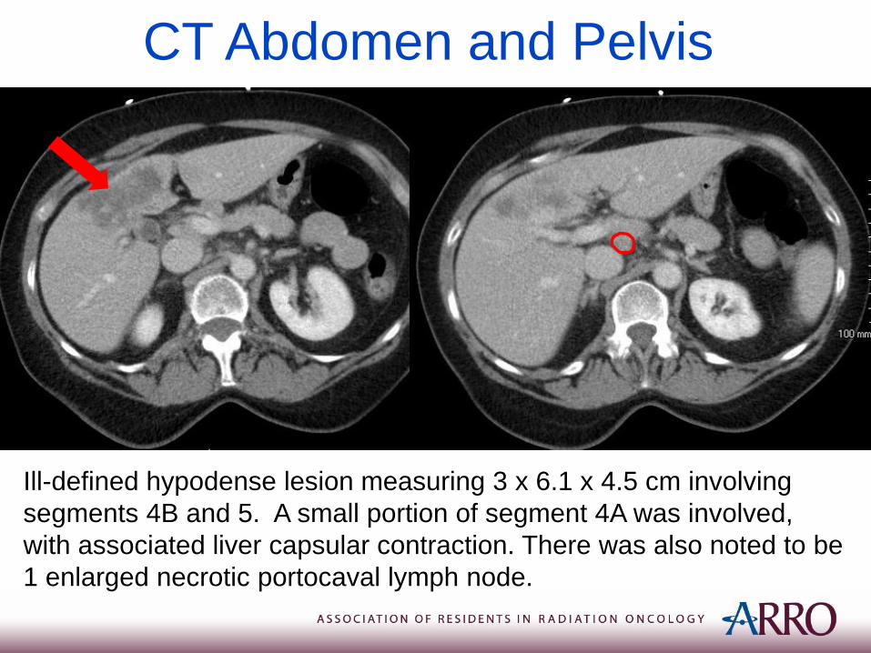
Ill-defined hypodense lesion measuring 3 x 6.1 x 4.5 cm involving
segments 4B and 5. A small portion of segment 4A was involved,
with associated liver capsular contraction. There was also noted to be
1 enlarged necrotic portocaval lymph node.
MRI Imaging
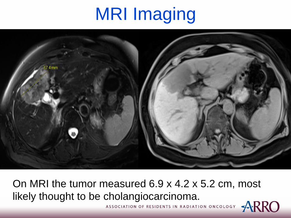
On MRI the tumor measured 6.9 x 4.2 x 5.2 cm, most
likely thought to be cholangiocarcinoma.
Differential Diagnosis
• Hepatocellular Carcinoma (HCC)
• Intrahepatic Cholangiocarcinoma
• Metastatic Disease
• Gallbladder Carcinoma
Imaging Key Point: On multiphase CT scan HCC often enhances on the arterial phase while the intrahepatic cholangiocarcinoma has a delayed enhancement.
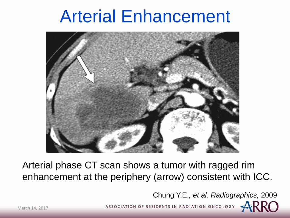
Arterial Enhancement
March 14, 2017
Chung Y.E., et al. Radiographics, 2009
Arterial phase CT scan shows a tumor with ragged rim
enhancement at the periphery (arrow) consistent with ICC.
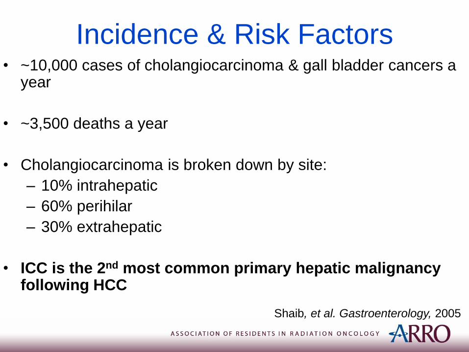
Incidence & Risk Factors• ~10,000 cases of cholangiocarcinoma & gall bladder cancers a
year
• ~3,500 deaths a year
• Cholangiocarcinoma is broken down by site:
– 10% intrahepatic
– 60% perihilar
– 30% extrahepatic
• ICC is the 2nd most common primary hepatic malignancy following HCC
Shaib, et al. Gastroenterology, 2005
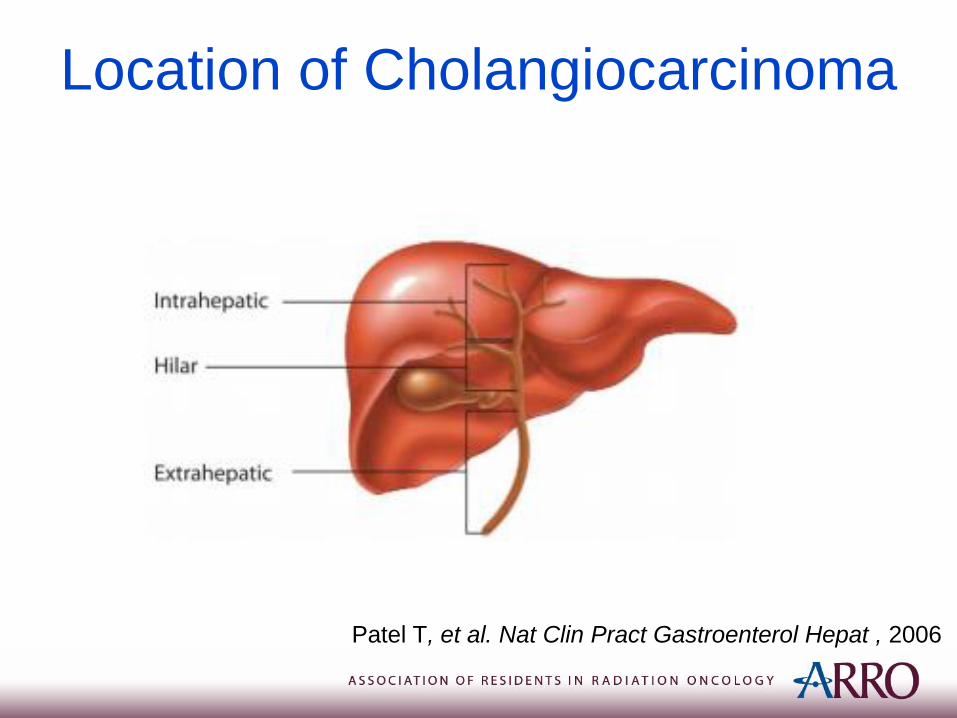
Location of Cholangiocarcinoma
Patel T, et al. Nat Clin Pract Gastroenterol Hepat , 2006
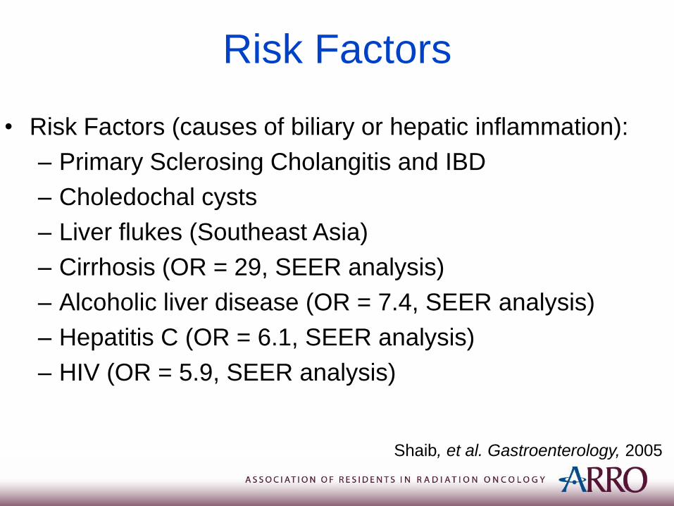
Risk Factors
• Risk Factors (causes of biliary or hepatic inflammation):
– Primary Sclerosing Cholangitis and IBD
– Choledochal cysts
– Liver flukes (Southeast Asia)
– Cirrhosis (OR = 29, SEER analysis)
– Alcoholic liver disease (OR = 7.4, SEER analysis)
– Hepatitis C (OR = 6.1, SEER analysis)
– HIV (OR = 5.9, SEER analysis)
Shaib, et al. Gastroenterology, 2005
Presentation & Natural History
• Patients present with malaise, nausea, abdominal
pain, and jaundice.
• Intrahepatic CC has 20-30% risk of LN metastases
(less than extrahepatic or hilar)
• Lymph node drainage: pericholedochal, portal
vein, common hepatic artery,
pancreaticoduodenal, celiac/SMA
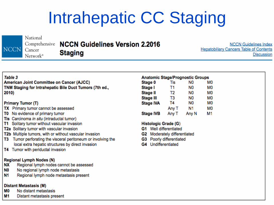
Intrahepatic CC Staging
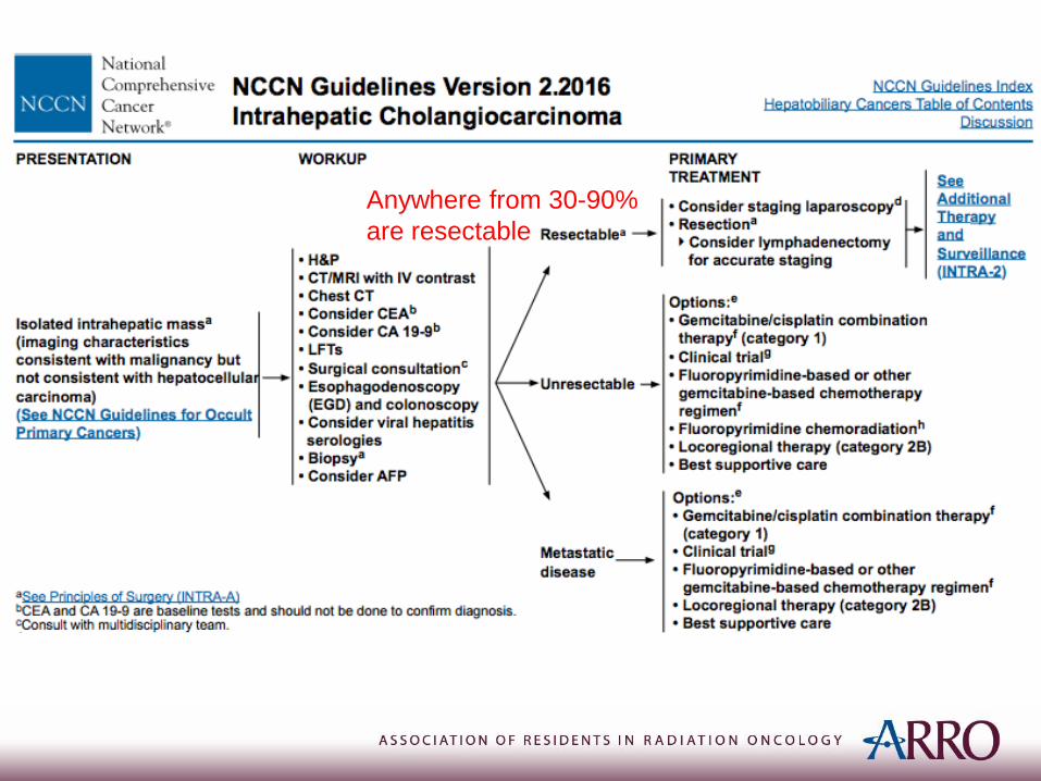
Anywhere from 30-90%
are resectable
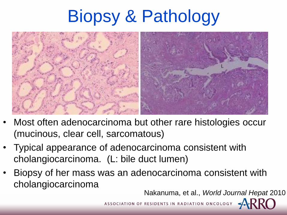
Biopsy & Pathology
• Most often adenocarcinoma but other rare histologies occur
(mucinous, clear cell, sarcomatous)
• Typical appearance of adenocarcinoma consistent with
cholangiocarcinoma. (L: bile duct lumen)
• Biopsy of her mass was an adenocarcinoma consistent with
cholangiocarcinomaNakanuma, et al., World Journal Hepat 2010
Surgical resection• She underwent an exploratory laparotomy and
extended right hepatectomy with celiac and portal
lymphadenectomy.
• In recent large series, only 50% of surgeries included
LN dissection.
• Most common surgery was hemihepatectomy (42.1%)
followed by extended hemihepatectomy (31%).
• 81% R0 resection rate in large multi-institutional series.
Jong, et al., JCO 2011
Surgical Pathology
• 6.5 x 6.3 x 3.8 cm, moderately differentiated
adenocarcinoma
• Negative margins
• 1/1 common hepatic LNs involved and 2/2 portal
lymph nodes involved
• Final diagnosis is Stage IVA (pT2a pN1c M0)
intrahepatic cholangiocarcinoma.
Prognostic factors
• 449 patients analyzed in recent surgical series.
• Tumor size NOT associated with prognosis (mean =6.5cm)
• 5 year OS was 30-35%.
• Vascular invasion, tumor number, positive margin and
LN involvement were all associated with worse OS.
• Estimated 20-30% risk of LN involvement.
Jong, et al., JCO 2011
Would this patient benefit from
adjuvant treatment?
• No adequate prospective randomized Phase III trials for
recommendations on adjuvant therapy.
• No known benefit of adjuvant therapy in margin and node
negative patients but with high risk features (positive LVSI,
multi-centric tumors, large tumors) should enroll on clinical trial
• Margin-positive or LN positive, systemic therapy with
gemcitabine, 5FU, or chemoXRT should be considered
• No direct prospective Phase II/III data to guide adjuvant
treatment.
• Our institution extrapolates SWOG 0809 to guide treatment of
ICCs.
• N=79 patients, 2 year survival 65% and median OS 35
months (Well tolerated and better than historical controls)
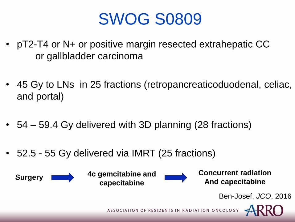
SWOG S0809
• pT2-T4 or N+ or positive margin resected extrahepatic CC
or gallbladder carcinoma
• 45 Gy to LNs in 25 fractions (retropancreaticoduodenal, celiac,
and portal)
• 54 – 59.4 Gy delivered with 3D planning (28 fractions)
• 52.5 - 55 Gy delivered via IMRT (25 fractions)
Ben-Josef, JCO, 2016
Surgery 4c gemcitabine and
capecitabine
Concurrent radiation
And capecitabine
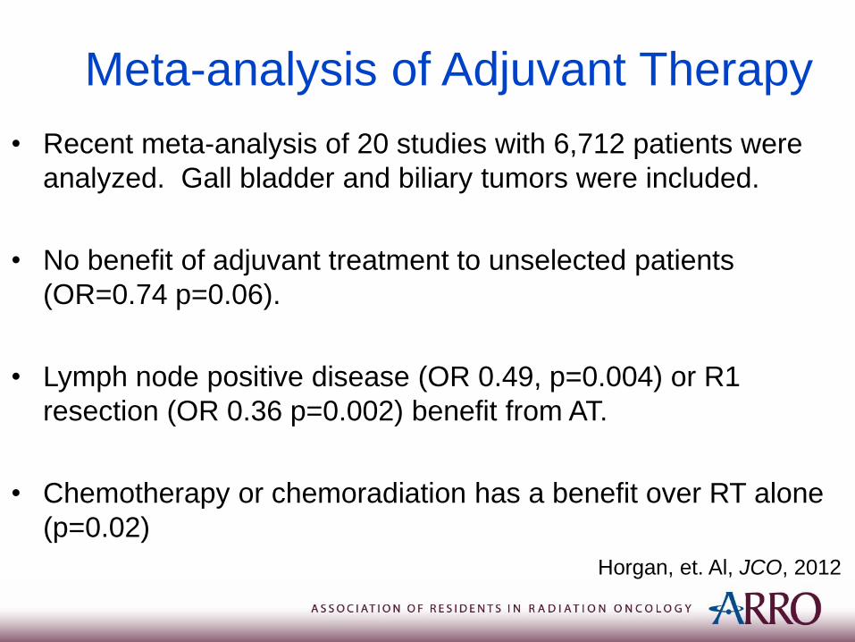
Meta-analysis of Adjuvant Therapy
• Recent meta-analysis of 20 studies with 6,712 patients were
analyzed. Gall bladder and biliary tumors were included.
• No benefit of adjuvant treatment to unselected patients
(OR=0.74 p=0.06).
• Lymph node positive disease (OR 0.49, p=0.004) or R1
resection (OR 0.36 p=0.002) benefit from AT.
• Chemotherapy or chemoradiation has a benefit over RT alone
(p=0.02)
Horgan, et. Al, JCO, 2012
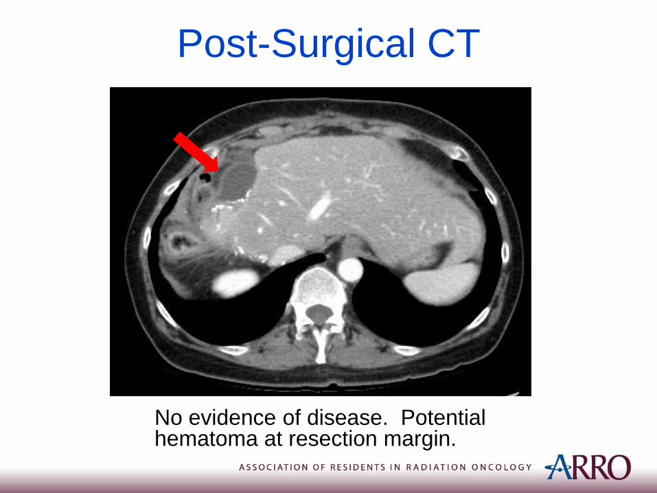
Post-Surgical CT
No evidence of disease. Potential hematoma at resection margin.
Adjuvant Chemotherapy
As a result of LN involvement, she
underwent 4 cycles of adjuvant gemcitabine
and capecitabine.
She tolerated chemotherapy well and is now
presenting to the Radiation Oncology Clinic
to discuss the need for radiation.
March 14, 2017
History ContinuedPMH/PSH: Hypothyroidism, Osteopenia, Laparoscopy and
Partial Hepatectomy
Medications: Capecitabine, Gemcitabine, Colace,
Levothyroxine
Allergies: Hydromorphone, Naproxen
Family History: Her mother had a history of malignancy not
known by patient. She reports she had an aunt with
questionable bone cancer.
Social History: She is a nonsmoker and reports social alcohol
use in the past. She is married with 4 children.
ROS: Fatigue throughout the past few months. She is down 30-
40 lbs over a 6-7 month period. She has mild right upper
quadrant discomfort. She denies nausea/vomiting, melena,
hematochezia, fevers/chills, chest pain, shortness of breath,
or other changes.
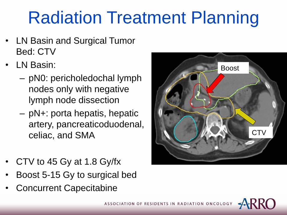
Radiation Treatment Planning• LN Basin and Surgical Tumor
Bed: CTV
• LN Basin:
– pN0: pericholedochal lymph
nodes only with negative
lymph node dissection
– pN+: porta hepatis, hepatic
artery, pancreaticoduodenal,
celiac, and SMA
• CTV to 45 Gy at 1.8 Gy/fx
• Boost 5-15 Gy to surgical bed
• Concurrent Capecitabine
CTV
Boost
Simulation
• Arms up with body-fix
• 2 mm slices
• Free breathing CT and 4-D CT to assess motion
• Contrast optional to help delineate vessels
depending on coverage needed
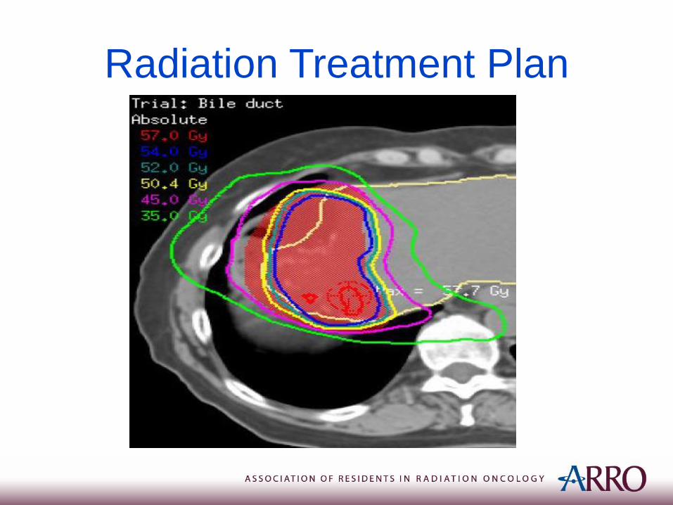
Radiation Treatment Plan
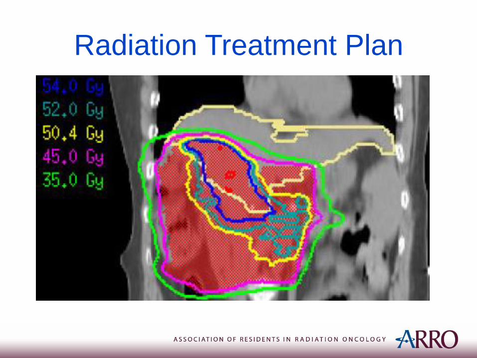
Radiation Treatment Plan
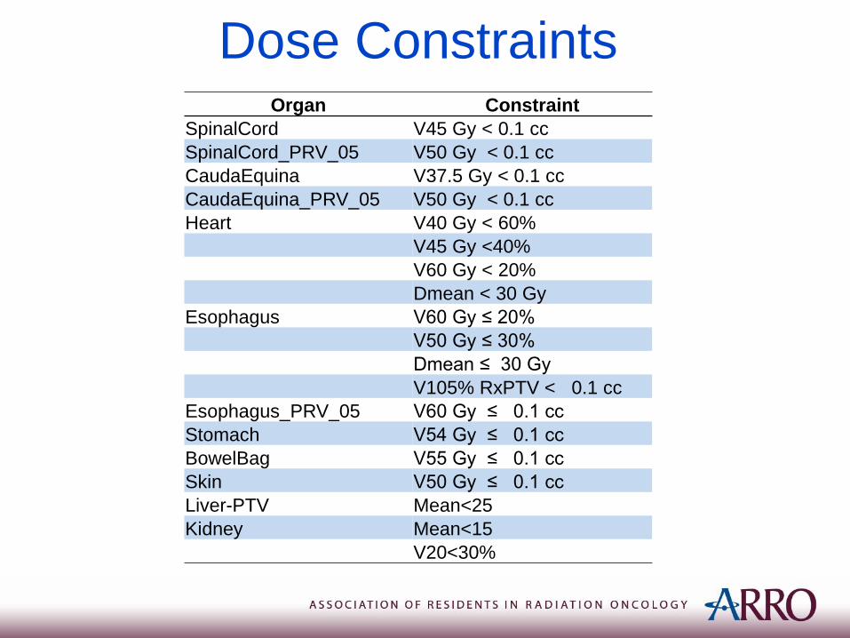
Dose ConstraintsOrgan Constraint
SpinalCord V45 Gy < 0.1 cc
SpinalCord_PRV_05 V50 Gy < 0.1 cc
CaudaEquina V37.5 Gy < 0.1 cc
CaudaEquina_PRV_05 V50 Gy < 0.1 cc
Heart V40 Gy < 60%
V45 Gy <40%
V60 Gy < 20%
Dmean < 30 Gy
Esophagus V60 Gy ≤ 20%
V50 Gy ≤ 30%
Dmean ≤ 30 Gy
V105% RxPTV < 0.1 cc
Esophagus_PRV_05 V60 Gy ≤ 0.1 cc
Stomach V54 Gy ≤ 0.1 cc
BowelBag V55 Gy ≤ 0.1 cc
Skin V50 Gy ≤ 0.1 cc
Liver-PTV Mean<25
Kidney Mean<15
V20<30%
Follow-up After Treatment
• Physical exam and imaging every 6
months for the first two years
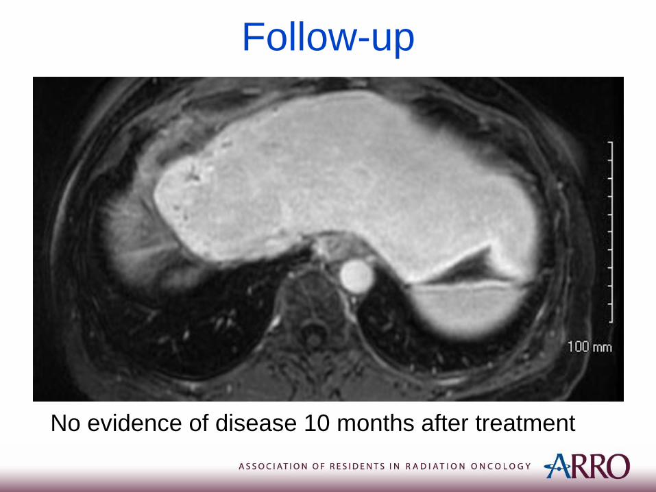
Follow-up
No evidence of disease 10 months after treatment
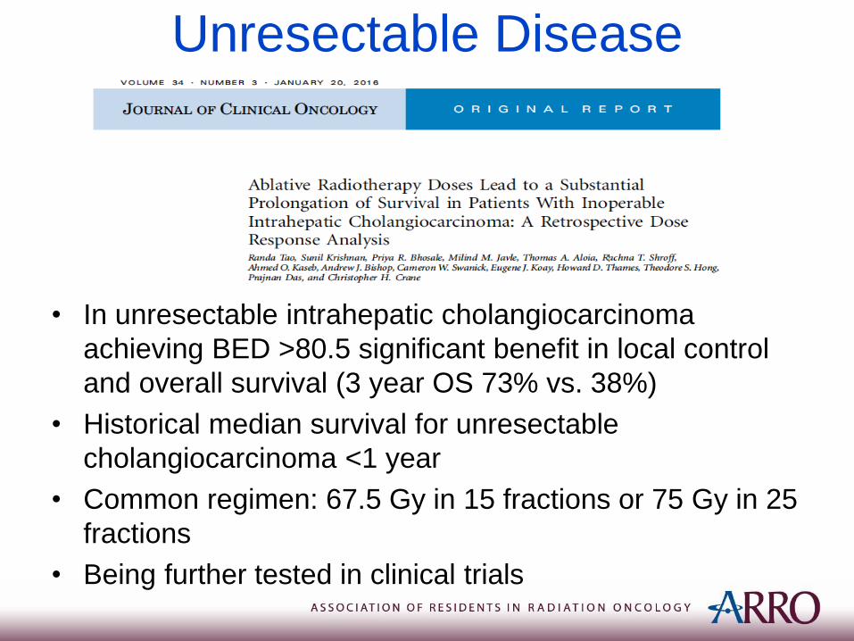
Unresectable Disease
• In unresectable intrahepatic cholangiocarcinoma
achieving BED >80.5 significant benefit in local control
and overall survival (3 year OS 73% vs. 38%)
• Historical median survival for unresectable
cholangiocarcinoma <1 year
• Common regimen: 67.5 Gy in 15 fractions or 75 Gy in 25
fractions
• Being further tested in clinical trials
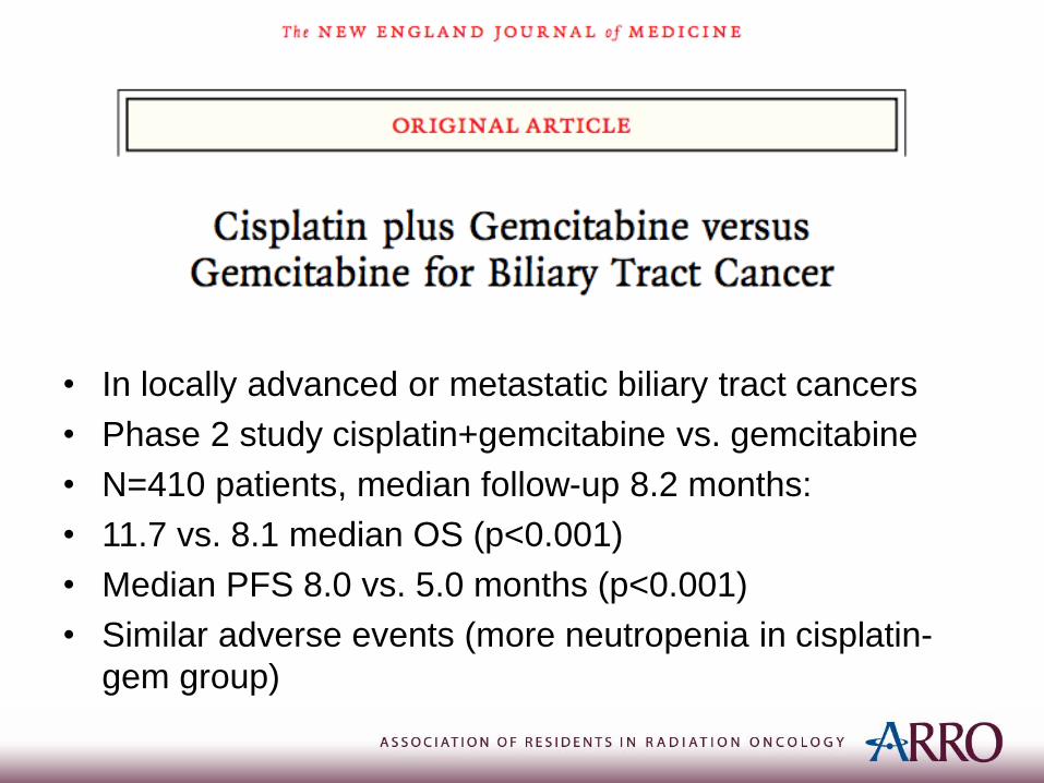
• In locally advanced or metastatic biliary tract cancers
• Phase 2 study cisplatin+gemcitabine vs. gemcitabine
• N=410 patients, median follow-up 8.2 months:
• 11.7 vs. 8.1 median OS (p<0.001)
• Median PFS 8.0 vs. 5.0 months (p<0.001)
• Similar adverse events (more neutropenia in cisplatin-
gem group)
Summary• Intrahepatic CC Rare
• Anywhere from 30-90% are resectable
• Limited prospective data
• Potential indications for adjuvant therapy include R1
resections or positive lymph nodes
• Extrapolation of SWOG0809 for post-operative treatment
• Hypofractionation (3-4.5 Gy/fx) for unresectable disease