ARE YOU NUMB YET?
34
1 ARE YOU NUMB YET? PROBLEM-SOLVING THE DELIVERY OF LOCAL ANESTHESIA Alan W. Budenz, MS, DDS, MBA Professor, Dept. of Biomedical Sciences and Interim Chair, Dept. of Diagnostic Sciences University of the Pacific, Arthur A. Dugoni School of Dentistry San Francisco, California [email protected] 90% of all patients report being anxious about going to the dentist or dental hygienist and receiving a shot . Patients rate “painless injections” as the most important criteria in evaluating their dentist or dental hygienist de St Georges J, How dentists are judged by patients, Dentistry Today, Vol. 23, August 2004 Friedman & Krochak, Using a precision-metered injection system to minimize dental injection anxiety, Compend Contin Educ Dent, Vol. 19(2), Feb 1998 Reasons for Anesthetic Failures 1. Anatomical/physiological variations 2. Technical errors of administration 3. Patient anxiety 4. Inflammation and infection 5. Defective/expired solutions Wong MKS & Jacobsen PL, Reasons for local anesthesia failures, J Am Dent Assoc, Vol 123, Jan 1992 Reasons for Anesthetic Failures 1. Anatomical/physiological variations Wide flaring mandible Wide flaring ramus Long (A - P) ramus Bulky musculature Large buccal fat pad Class III occlusion Missing teeth Children Accessory or anomalous nerve pathways Reasons for Anesthetic Failures 2. Technical errors of administration Too high Too low Too anterior Too posterior Too medial Too lateral Intravascular Reasons for Anesthetic Failures 1. Anatomical/physiological variations 2. Technical errors of administration These two are closely related: We will solve by reviewing the anatomy and landmarks McMinn, Hutchings & Logan, Color Atlas of Head & Neck Anatomy, 2 nd Ed, Mosby, 1994
Transcript of ARE YOU NUMB YET?

1
ARE YOU NUMB YET?PROBLEM-SOLVING THE DELIVERY
OF LOCAL ANESTHESIA
Alan W. Budenz, MS, DDS, MBA
Professor, Dept. of Biomedical Sciences and Interim Chair, Dept. of Diagnostic Sciences
University of the Pacific, Arthur A. Dugoni School of Dentistry
San Francisco, California
90% of all patients report being anxious
about going to the dentist or dental hygienist
and receiving a shot.
Patients rate “painless injections” as
the most important criteria in evaluating
their dentist or dental hygienistde St Georges J, How dentists are judged by patients, Dentistry Today, Vol. 23, August 2004
Friedman & Krochak, Using a precision-metered injection
system to minimize dental injection anxiety, Compend Contin
Educ Dent, Vol. 19(2), Feb 1998
Reasons for Anesthetic Failures
1. Anatomical/physiological variations
2. Technical errors of administration
3. Patient anxiety
4. Inflammation and infection
5. Defective/expired solutions
Wong MKS & Jacobsen PL, Reasons for local anesthesia failures, J Am Dent Assoc, Vol 123, Jan 1992
Reasons for Anesthetic Failures
1. Anatomical/physiological
variations
Wide flaring mandible
Wide flaring ramus
Long (A - P) ramus
Bulky musculature
Large buccal fat pad
Class III occlusion
Missing teeth
Children
Accessory or anomalous nerve pathways
Reasons for Anesthetic Failures
2. Technical errors of administration
Too high
Too low
Too anterior
Too posterior
Too medial
Too lateral
Intravascular
Reasons for Anesthetic Failures1. Anatomical/physiological
variations
2. Technical errors of
administration
These two are
closely related:
We will solve by
reviewing the anatomy
and landmarksMcMinn, Hutchings & Logan, Color Atlas of
Head & Neck Anatomy, 2nd Ed, Mosby, 1994

2
REVIEW OF ANATOMY
General Anatomy and Landmarks for
Mandibular Anesthesia
The Masticator Space
Includes the Temporal and Infratemporal Fossae
Liebgott, The Anatomical Basis of Dentistry, 2nd Ed, Mosby, 2001
The Masticator Space
The Infratemporal Fossa Boundaries:
A = Maxillary tuberosity
P = Styloid process
M = Lateral pterygoid
plate
L = Ramus of mandible
Agur & Lee, Grant’s Atlas of Anatomy, 10th Ed,
Lippincott Williams & Wilkins, 1999 Liebgott, The Anatomical Basis
of Dentistry, 2nd Ed, Mosby, 2001
Infratemporal Fossa
Contents
Muscles of mastication
Mandibular division of
Trigeminal nerve, V3
Chorda tympani
branch of Facial nerve
Maxillary artery and
vein
Agur & Lee, Grant’s Atlas of Anatomy, 10th Ed,
Lippincott Williams & Wilkins, 1999
The Masticator Space
A Fascial Compartment:
Derived from investing
layer of deep cervical
fascia
Envelopes mandible and
muscles of mastication
Medial Lateral
Hollinshead, Anatomy for Surgeons, Vol 1, The Head & Neck, 3rd Ed, Harper & Row, 1982
The Muscles of Mastication
Four total: 2 superficial
1. Temporalis
Liebgott, The Anatomical Basis of Dentistry, 2nd Ed, Mosby, 2001

3
The Muscles of Mastication
Four total: 2 superficial
1. Temporalis
2. Masseter
Liebgott, The Anatomical Basis of Dentistry, 2nd Ed, Mosby, 2001
The Muscles of Mastication
Four total: 2 superficial
1. Temporalis
2. Masseter
Netter, Atlas of Human Anatomy, 2nd Ed, Novartis, 1997
The Muscles of Mastication
Four total: 2 superficial; 2 deep
1. Temporalis
2. Masseter
3. Medial
pterygoid
Liebgott, The Anatomical Basis of Dentistry, 2nd Ed, Mosby, 2001
The Muscles of Mastication
Four total: 2 superficial; 2 deep
1. Temporalis
2. Masseter
3. Medial
pterygoid
4. Lateral
pterygoid
Liebgott, The Anatomical Basis of Dentistry, 2nd Ed, Mosby, 2001
The Muscles of Mastication
Four total: 2 superficial; 2 deep
1. Temporalis
2. Masseter
3. Medial
pterygoid
4. Lateral
pterygoid
Netter, Atlas of Human Anatomy, 2nd Ed, Novartis, 1997
Accessory Muscles of Mastication:
Muscles of Facial Expression
Oral musculature
Levator labii superioris
Levator anguli oris
Zygomaticus major
Buccinator
Risorius
Mentalis
Depressor anguli oris
Depressor labii inferioris
Orbicularis oris
Platysma
Netter, Atlas of Human Anatomy, 2nd Ed, Novartis, 1997
4
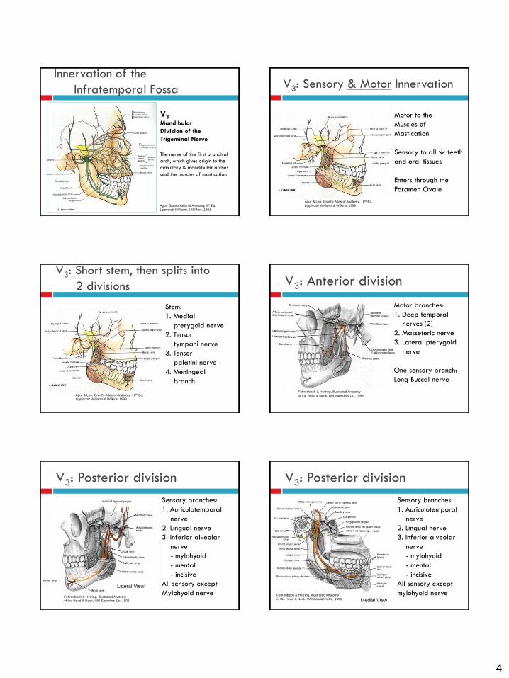
Innervation of the
Infratemporal Fossa
V3Mandibular
Division of the
Trigeminal Nerve
The nerve of the first branchial
arch, which gives origin to the
maxillary & mandibular arches
and the muscles of mastication
Agur, Grant’s Atlas of Anatomy, 9th Ed,
Lippincott Williams & Wilkins, 1991
V3: Sensory & Motor Innervation
Motor to the
Muscles of
Mastication
Sensory to all teeth
and oral tissues
Enters through the
Foramen Ovale
Agur & Lee, Grant’s Atlas of Anatomy, 10th Ed,
Lippincott Williams & Wilkins, 1999
V3: Short stem, then splits into
2 divisions
Stem:
1. Medial
pterygoid nerve
2. Tensor
tympani nerve
3. Tensor
palatini nerve
4. Meningeal
branch
Agur & Lee, Grant’s Atlas of Anatomy, 10th Ed,
Lippincott Williams & Wilkins, 1999
V3: Anterior division
Motor branches:
1. Deep temporal
nerves (2)
2. Masseteric nerve
3. Lateral pterygoid
nerve
One sensory branch:
Long Buccal nerve
Fehrenbach & Herring, Illustrated Anatomy
of the Head & Neck, WB Saunders Co, 1996
V3: Posterior division
Sensory branches:
1. Auriculotemporal
nerve
2. Lingual nerve
3. Inferior alveolar
nerve
- mylohyoid
- mental
- incisive
All sensory except
Mylohyoid nerveLateral View
Fehrenbach & Herring, Illustrated Anatomy
of the Head & Neck, WB Saunders Co, 1996
V3: Posterior division
Sensory branches:
1. Auriculotemporal
nerve
2. Lingual nerve
3. Inferior alveolar
nerve
- mylohyoid
- mental
- incisive
All sensory except
mylohyoid nerveMedial View
Fehrenbach & Herring, Illustrated Anatomy
of the Head & Neck, WB Saunders Co, 1996

5
Additional Innervation in the
Infratemporal Fossa
Chorda tympani:
• Branch of CN VII
• Carries taste fibers
from anterior tongue
• Secretomotor fibers to
salivary glands
Joins lingual nerve of
V3 in ITF
Medial ViewFehrenbach & Herring, Illustrated Anatomy
of the Head & Neck, WB Saunders Co, 1996
Blood Supply to the
Infratemporal Fossa
Maxillary artery:
3 parts
1. Mandibular
2. Pterygoid
3. Pterygopalatine
Agur & Lee, Grant’s Atlas of Anatomy, 10th Ed,
Lippincott Williams & Wilkins, 1999
1.
2.3.
Blood Supply to the
Infratemporal Fossa
Maxillary artery
Part 1: Mandibular
1. Deep auricular
2. Anterior tympanic
3. Middle meningeal
4. Accessory middle
meningeal
5. Inferior alveolar
- mylohyoid, mental,
& incisive branchesAl-Faraje L, Surgical and Radiologic Anatomy for Oral
Implantology, Quintessence Publishing Co, 2013
Blood Supply to the
Infratemporal Fossa
Maxillary artery
Part 2: Pterygoid
1. Deep temporal (2)
2. Medial pterygoid
3. Lateral pterygoid
4. Masseteric
5. Buccal
6. Lingual
Liebgott, The Anatomical Basis of Dentistry, 2nd Ed, Mosby, 2001
Blood Supply to the
Infratemporal Fossa
Maxillary artery
Part 3: Pterygopalatine
1. Posterior superior
alveolar
2. Infraorbital
3. Artery of pterygoid
canal
4. Pharyngeal branch
5. Descending palatine
6. Sphenopalatine
Liebgott, The Anatomical Basis of Dentistry, 2nd Ed, Mosby, 2001
Blood Supply to the
Infratemporal Fossa
Pterygoid Venous
Plexus
Primary drainage to
Maxillary vein
Liebgott, The Anatomical Basis of Dentistry, 2nd Ed, Mosby, 2001

6
Blood Supply to the
Infratemporal Fossa
Pterygoid Venous
Plexus
Connections to:
1. Cavernous sinus
2. Facial vein
3. Inferior ophthalmic
vein
4. Pharyngeal plexus
Netter’s Atlas, 4th Ed, Saunders/Elsevier, 2006
View of infratemporal fossa
with mandible resected
Agur & Lee, Grant’s Atlas of Anatomy, 10th Ed,
Lippincott Williams & Wilkins, 1999
View of infratemporal fossa
fully dissected
Agur & Lee, Grant’s Atlas of Anatomy, 10th Ed,
Lippincott Williams & Wilkins, 1999
MANDIBULAR ANESTHESIA
Conventional and Alternative Techniques
Mandibular Infiltration Anesthesia Works well for the maxilla, but for the mandible…
Works fairly well for anteriors and bicuspids
More variable predictability for molars
Greater success using articaine
Lidocaine 45 – 67%; articaine 75 – 92%
Lidocaine 6.1 – 11.1 minutes; articaine 4.2 – 4.7 minutes
Facial Lingual
Robertson et al, The anesthetic efficacy of articaine in buccal infiltration of mandibular posterior teeth, JADA, Vol 138 No 8, 2007
& faster onset
Meechan, Practical Dental Local Anesthesia, Quintessence, 2002
Why not infiltrate
both buccally and
lingually?Use ½ - 1 cartridge of
articaine for each
Pharmacology of Anesthetic Agents
A Practical Armamentarium:
From a meta-analysis of 13 clinical trials:
Evidence strongly supported articaine’s superiority over lidocaine for infiltration anesthesia
Evidence was weak for any significant difference between lidocaine and articaine for block anesthesia
Brandt RG et al, The pulpal anesthetic efficacy of articaine versus
lidocaine in dentistry: A meta-analysis, J Am Dent Assoc, Vol 142(5), May 2011
Articaine was 4 times more effective, with greater duration, than lidocaine as an infiltration injection when used for teeth diagnosed with irreversible pulpitisAshraf H et al, Efficacy of articaine versus lidocaine in block and infiltation anesthesia administered in
teeth with irreversible pulpitis: A prospective, randomized, double-blind study, JOE, Vol 39(1), Jan 2013

7
Mandibular Anesthesia
Mandible: Nerve blocks
Inferior alveolar nerve block
Lingual nerve block
Long buccal nerve block
Mental (& incisive) nerve block
Mylohyoid nerve block
Complete mandibular division
nerve block
Gow-Gates mandibular division
block
Vazirani – Akinosi mandibular
division blockJastak, Yagiela & Donaldson, Local Anesthesia of
the Oral Cavity, WB Saunders Co, 1995
Mandibular Anesthesia
Mandible: Landmarks
Mandibular notch
Neck of condyle
Coronoid process
Coronoid notch
External oblique ridge
Internal oblique ridge/
mylohyoid line
Mandibular foramen & lingula
Mental foramen
Coronoid notch
Agur & Lee, Grant’s Atlas of Anatomy, 10th Ed,
Lippincott Williams & Wilkins, 1999
Mandibular Anesthesia
Mandible: Nerve blocks
Inferior alveolar nerve block
Bisection approach
McMinn, Hutchings & Logan, Color Atlas of
Head & Neck Anatomy, 2nd Ed, Mosby, 1994Jastak, Yagiela & Donaldson, Local Anesthesia of
the Oral Cavity, WB Saunders Co, 1995
Mandibular Anesthesia
Mandible: Nerve blocks
Inferior alveolar nerve block
Bisection approach
Position of mandibular foramen
Below mandibular occlusal
plane in 75%
Even with occlusal plane
in 22.5%
Nicholson ML, A study of the position of the
mandibular foramen in the adult human mandible,
Anat Rec, Vol 212, 1985
Evers & Haegerstam, Introduction to Dental
Local Anaesthesia, Mediglobe, 1990
Mandibular Anesthesia
Mandible: Nerve blocks
Inferior alveolar nerve block
Bisection approach
Position of mandibular foramen
Variable from infancy to adulthood
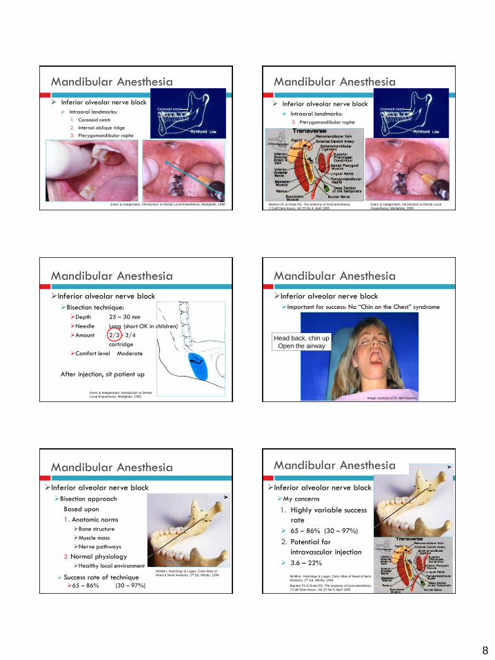
Mandibular Anesthesia
Inferior alveolar nerve block
Intraoral landmarks:
1. Coronoid notch
2. Internal oblique ridge
3. Pterygomandibular raphe
Coronoid notch
Meechan, Practical Dental Local Anesthesia, Quintessence, 2002
Pterygomandibular triangle
8
Mandibular Anesthesia
Inferior alveolar nerve block
Intraoral landmarks:
1. Coronoid notch
2. Internal oblique ridge
3. Pterygomandibular raphe
Coronoid notch
Evers & Haegerstam, Introduction to Dental Local Anaesthesia, Mediglobe, 1990
Mandibular Anesthesia
Inferior alveolar nerve block
Intraoral landmarks:
3. Pterygomandibular raphe
Coronoid notch
Evers & Haegerstam, Introduction to Dental Local
Anaesthesia, Mediglobe, 1990
Blanton PL & Roda RS, The anatomy of local anesthesia,
J Calif Dent Assoc, Vol 23 No 4, April 1995
Mandibular Anesthesia
Inferior alveolar nerve block
Bisection technique:
Depth 25 – 30 mm
Needle Long
Amount 2/3 - 3/4
cartridge
Comfort level Moderate
After injection, sit patient up
Evers & Haegerstam, Introduction to Dental
Local Anaesthesia, Mediglobe, 1990
(short OK in children)
Mandibular Anesthesia
Inferior alveolar nerve block
Important for success: No “Chin on the Chest” syndrome
Head back, chin up
Open the airway
Image courtesy of Dr. Mel Hawkins
Mandibular Anesthesia
Inferior alveolar nerve block
Bisection approach
Based upon
1. Anatomic norms
Bone structure
Muscle mass
Nerve pathways
2. Normal physiology
Healthy local environmentMcMinn, Hutchings & Logan, Color Atlas of
Head & Neck Anatomy, 2nd Ed, Mosby, 1994 Success rate of technique
65 – 86% (30 – 97%)
Mandibular Anesthesia
Inferior alveolar nerve block
My concerns
1. Highly variable success
rate
65 – 86% (30 – 97%)
2. Potential for
intravascular injection
3.6 – 22%
McMinn, Hutchings & Logan, Color Atlas of Head & Neck
Anatomy, 2nd Ed, Mosby, 1994
Blanton PL & Roda RS, The anatomy of local anesthesia,
J Calif Dent Assoc, Vol 23 No 4, April 1995

9
Mandibular Anesthesia Inferior alveolar nerve blockMy concerns
3. Potential injury: Nerve, vasculature
McMinn, Hutchings & Logan, Color Atlas of
Head & Neck Anatomy, 2nd Ed, Mosby, 1994Blanton PL & Roda RS, The anatomy of local anesthesia,
J Calif Dent Assoc Vol 23 No 4, April 1995
Mandibular Anesthesia
Inferior alveolar nerve block
Bisection technique:
Unfortunately, most of the mandibular anatomy varies widely
Wide flaring mandible
Wide flaring ramus
Long (A – P) ramus
Bulky muscles or buccal fat pad
Class III occlusion
Missing molars/edentulous
Age/children
Except one feature, not so much Prado FB et al, Morphological changes in the
position of the mandibular foramen in dentate
and edentate Brazilian subjects, Clinical
Anatomy, Vol 23, 2010
Mandibular Anesthesia
Inferior alveolar nerve block
Alternative techniques:
IA “Walk-In” technique
1. Deliberately contact bone
anterior to mandibular
foramen, feel depth
Meechan, Practical Dental Local Anesthesia, Quintessence, 2002
Mandibular Anesthesia
Inferior alveolar nerve block
IA “Walk-In” technique:
1. Penetrate tissue, then put posterior pressure on syringe to produce strong needle deflection
2. Deliberately contact bone
anterior to mandibular
foramen, feel depth
3. Withdraw 2 – 3 mm, reduce posterior pressure on syringe
4. Insert 2 – 3 mm posteriorly,
contact bone again, feel depth
5. Repeat 1 – 2 times
Mandibular Anesthesia
Inferior alveolar nerve block
IA “Walk-In” technique:
When you reach the same injection depth withoutcontacting bone,
Stop
Aspirate
Inject
Mandibular Anesthesia
Inferior alveolar nerve block
IA “Walk-in” technique
One more structure:
sphenomandibular ligament
Meechan, Practical Dental Local Anesthesia, Quintessence, 2002
Sabotta, Atlas of Human Anatomy, 12th Ed, Williams & Wilkins, 1997

10
Mandibular Anesthesia
Inferior alveolar nerve block
Indirect IA technique: bisection technique = Direct technique
1. Contact bone anterior to mandibular foramen
2. Redirect to medial
3. “Hook” around lingula, insert slighty
Stop
Aspirate
Inject
Meechan, Practical Dental Local Anesthesia, Quintessence, 2002
Mandibular Anesthesia
Inferior alveolar nerve block
My concerns
4. This is NOT a complete mandibular division nerve block!
1. Lingual nerve block given
in combination with IA
2. No long buccal
nerve blockade
Requires separate
injection
Common accessory
innervation to molars
NO
lingual
anesthesia
NO
LB
anes
Meechan, Practical Dental Local Anesthesia, Quintessence, 2002
Inferior alveolar regional nerve block
Mandibular Anesthesia
Lower lip and chin is numb
Tongue is numb
But the molar tooth is only
partially numb!
Give the long buccal regional nerve block
The long buccal injection should
be given to complement the IA &
lingual regional blocks
Inferior alveolar
and lingual
Long buccal
Meechan, Practical Dental Local Anesthesia, Quintessence, 2002
Mandibular Anesthesia
Long buccal regional nerve block
Accessory innervation to mandibular molars
Average of 27 foramina in the retromolar area or in
the superior medial region of the ramus above and
anterior to the mandibular foramenHaveman & Tebo, Posterior accessory foramina of the human mandible, J Prosth Dent, Vol 35, 1976
Przystanska A, Bruska M, Accessory mandibular foramina: histological and immunohistochemical studies
of their contents, Arch Oral Biol, 55(1), 2010
Mandibular Anesthesia
Long buccal regional nerve block
In cadaver dissections, 37.5% of nerves entering the
superior medial and retromolar regions of the mandible
had direct connections with branches of the inferior
alveolar nerve to the molars
Meechan, Practical Dental Local
Anesthesia, Quintessence, 2002
Carter RB & Keen EN, The intramandibular course of the inferior alveolar nerve, J Anat Vol 108, 1971
Przystanska A, Bruska M, Accessory mandibular foramina: histological and immunohistochemical studies
of their contents, Arch Oral Biol 55(1), 2010
Jastak, Yagiela & Donaldson, Local
Anesthesia of the Oral Cavity, WB
Saunders Co, 1995
Mandibular Anesthesia
Long buccal regional nerve block
Accessory innervation to mandibular molars
Meechan, Practical Dental Local Anesthesia, Quintessence, 2002

11
Mandibular Anesthesia
Long buccal regional nerve block
Accessory innervation to mandibular molars
Depth 2 – 4 mm
Needle Short
Amount ½ - ¾ cartridge (~⅓ for children)
Comfort level Moderate to high
Troubleshooting Mandibular Anesthesia
You’ve given the IA and lingual block, and the long
buccal block
But the tooth is still only partially numb!
What can the problem
be?
What solutions should
we try?
Jastak, Yagiela & Donaldson, Local Anesthesia of
the Oral Cavity, WB Saunders Co, 1995
Troubleshooting Mandibular Anesthesia
Lower lip and chin is numb
Tongue is numb
But the tooth is only partially numb!
Or the tooth is numb, but duration is short and/or
anesthesia is not profound
Give a second injection
at the same site?
Go higher and deeper for
a second injection?
Troubleshooting Mandibular Anesthesia
The tooth is only partially numb!
Or the tooth is numb, but duration is short and/or
anesthesia is not profound
Go higher and deeper for
a second injection?
Risk higher incidence of
positive aspiration
Blanton PL & Roda RS, The anatomy of local anesthesia,
J Calif Dent Assoc, Vol 23 No 4, April 1995
Troubleshooting Mandibular Anesthesia
You’ve given the IA and lingual regional blocks, and the long buccal regional block
But the tooth is still only partially numb!
Solutions
For one tooth, buccal &
lingual infiltrations, PDL, or
intraosseous injections
work well
For a quadrant, a
mylohyoid nerve block
may be best
Jastak, Yagiela & Donaldson, Local Anesthesia of
the Oral Cavity, WB Saunders Co, 1995
Mandibular Anesthesia
Mylohyoid regional nerve block
Accessory innervation to any mandibular tooth
Medial ViewFehrenbach & Herring, Illustrated Anatomy
of the Head & Neck, WB Saunders Co, 1996

12
Mandibular Anesthesia
Mylohyoid regional nerve block
Accessory innervation to any mandibular tooth
53% of mandibles had accessory foramina near the mylohyoid line, particularly in the premolar area.*
*Haveman & Tebo, Posterior accessory foramina of the human mandible, J Prosth Dent, Vol 35, 1976
Katakami K et al, Characteristics of accessory mental foramina on limited cone-beam computed tomography images, J Endod, Vol 34(12), 2008
In cadaver dissections, 50% exhibited branches of the mylohyoidnerve entering foramina in the lingual surface of the mandible.
These nerves ended directly in mandibular teeth or joined the incisive branch of the inferior alveolar nerve.
Madeira et al, Clinical significance of supplementary innervation of the lower incisor teeth: A dissection study of the mylohyoid nerve, O Surg O Med O Pathol, Vol 46, 1978
Mandibular Anesthesia
Mylohyoid regional nerve block
Accessory innervation to any mandibular tooth
Upon histological examination of the mylohyoid nerve from its origin to its termination, the loss of small diameter pain and temperature fibers was detected along its entire length.
Frommer et al, The possible role of the mylohyoid nerve in mandibular posterior tooth sensation, J Am Dent Assoc, Vol 85, 1972
Przystanska A, Bruska M, Accessory mandibular foramina: histological and immunohistochemical studies of their contents, Arch Oral Biol, Vol 55(1), 2010
Evers & Haegerstam, Introduction to Dental
Local Anaesthesia, Mediglobe, 1990
Mandibular Anesthesia
Mylohyoid regional nerve block
Accessory innervation to any mandibular tooth
The point at which the mylohyoid nerve branched from the
inferior alveolar nerve ranged from 5 to 23 mm above the
mandibular foramen, with a mean distance of 14.7 mm.**Wilson et al, The inferior alveolar and mylohyoid nerves: An anatomic study and relationship
to local anesthesia of the anterior mandiblular teeth, J Am Dent Assoc, Vol 108 No 3, 1984
Bennett S & Townsend G, Distribution of the mylohyoid nerve: Anatomical variability and
clinical implications, Austral Endo J, Vol 27(3), 2001
Jastak, Yagiela & Donaldson,
Local Anesthesia of the Oral
Cavity, WB Saunders Co, 1995
Mandibular Anesthesia
Mylohyoid nerve block
Between mandible and
sublingual fold
Just distal to last tooth
to be worked on
Approximate apices of
roots
Easiest for anterior teeth
Access to molars may be difficult
Evers & Haegerstam, Introduction to Dental
Local Anaesthesia, Mediglobe, 1990
Mandibular Anesthesia
Mylohyoid nerve block
Between mandible and
sublingual fold
Just distal to last tooth
to be worked on
Approximate apices of roots
Easiest for anterior teeth
Access to molars may be difficult
It’s a lingual
infiltration
injection!
Mandibular Anesthesia
Mylohyoid nerve block
Depth 2 – 4 mm
Needle Short
Amount 1/3 – 1/2
cartridge
Comfort level High
Good for any
mandibular tooth
Evers & Haegerstam, Introduction to Dental
Local Anaesthesia, Mediglobe, 1990

13
Troubleshooting Mandibular Anesthesia
You’ve given the IA and lingual block, and the long
buccal and mylohyoid regional blocks
But the tooth is
still not completely numb!
Give complete mandibular
division nerve block for molars
Evers & Haegerstam, Introduction to Dental
Local Anaesthesia, Mediglobe, 1990
Mandibular Anesthesia
Mandible: Nerve blocks
Inferior alveolar nerve block
Lingual nerve block
Long buccal nerve block
Mental (& incisive) nerve block
Mylohyoid nerve block
Complete mandibulardivision nerve block
Gow-Gates mandibulardivision block
Vazirani – Akinosimandibular division block
NO
lingual
anesthesia
NO
LB
anes
Meechan, Practical Dental Local
Anesthesia, Quintessence, 2002
Mandibular Anesthesia
Mandible: Nerve blocks
Inferior alveolar regional
“mandibular” block
Gow-Gates complete
mandibular division block
Agur & Lee, Grant’s Atlas of Anatomy, 10th Ed,
Lippincott Williams & Wilkins, 1999
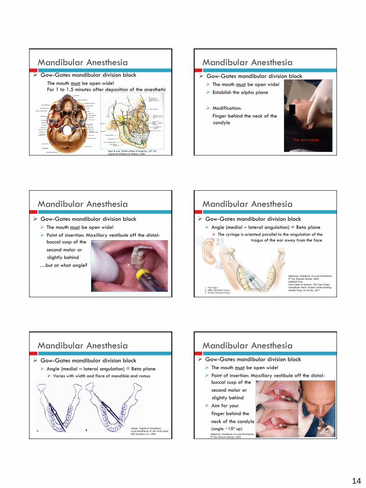
Mandibular Anesthesia
Gow-Gates mandibulardivision block
Landmarks
1. Alpha plane: from intertragic notch of the ear to corner of the mouth, and across to the opposite corner of the mouth
Anterior – posterior orientation
Malamed, Handbook of Local Anesthesia,
5th Ed, Elsevier Mosby, 2004
Mandibular Anesthesia
Gow-Gates mandibular division block
Target: Contact bone at the neck of the condyle
Alpha
plane
Mandibular Anesthesia
Gow-Gates mandibular division block
The mouth must be open wide!
Agur & Lee, Grant’s Atlas of Anatomy, 10th Ed,
Lippincott Williams & Wilkins, 1999
14
Mandibular Anesthesia
Gow-Gates mandibular division block
The mouth must be open wide!
Agur & Lee, Grant’s Atlas of Anatomy, 10th Ed,
Lippincott Williams & Wilkins, 1999
For 1 to 1.5 minutes after deposition of the anesthetic
Mandibular Anesthesia
Gow-Gates mandibular division block
The mouth must be open wide!
Establish the alpha plane
Modification:
Finger behind the neck of the
condyle
The alpha plane
Mandibular Anesthesia
Gow-Gates mandibular division block
The mouth must be open wide!
Point of insertion: Maxillary vestibule off the distal-
buccal cusp of the
second molar or
slightly behind
…but at what angle?
Mandibular Anesthesia
Gow-Gates mandibular division block
Angle (medial – lateral angulation) = Beta plane
The syringe is oriented parallel to the angulation of the
tragus of the ear away from the face
Malamed, Handbook of Local Anesthesia,
5th Ed, Elsevier Mosby, 2004,
adapted from:
Gow-Gates & Watson, The Gow-Gates
mandibular block: Further understanding,
Anesth Prog, Vol 25 (6), 1977
Mandibular Anesthesia
Gow-Gates mandibular division block
Angle (medial – lateral angulation) = Beta plane
Varies with width and flare of mandible and ramus
Aim for your extraoral finger behind
the neck of the condyle
About 10o upward angle from the
maxillary occlusal plane
Jastak, Yagiela & Donaldson,
Local Anesthesia of the Oral Cavity,
WB Saunders Co, 1995
Mandibular Anesthesia
Gow-Gates mandibular division block
The mouth must be open wide!
Point of insertion: Maxillary vestibule off the distal-
buccal cusp of the
second molar or
slightly behind
Aim for your
finger behind the
neck of the condyle
(angle ~10o up)Malamed, Handbook of Local Anesthesia,
5th Ed, Elsevier Mosby, 2004

15
Mandibular Anesthesia
Gow-Gates mandibular division block
Depth 25 – 28 mm (contact bone)
Needle Long
Amount 1 – 2 cartridges (⅔ - 1⅓ for child)
Comfort level Moderate to high
Keep mouth open for
1 to 1.5 minutes after
deposition of the anesthetic
Meechan, Practical Dental Local Anesthesia, Quintessence, 2002
Mandibular Anesthesia
Complete mandibular
division nerve block
Vazirani – Akinosi
mandibular division block
A closed mouth technique
Wolfe SH, The Wolfe nerve block: A modified high mandibular
nerve block, Dentistry Today, June/July 1992
Vazirani - Akinosi
Complete mandibular
division nerve block
A closed mouth technique
delivered at a higher level
than the conventional IA
block
10 – 14 mm higher
Image courtesy of Dr. Mel Hawkins
Mandibular Anesthesia
Vazirani – Akinosi mandibular
division block
A closed mouth technique
Meechan, Practical Dental Local Anesthesia, Quintessence, 2002
Malamed, Handbook of Local Anesthesia, 5th Ed, Elsevier Mosby, 2004
Ramus Tuberosity
Vazirani – Akinosi Quadrant Block
Mandibular Anesthesia
Image courtesy of Dr. Mel Hawkins
Hawkins JM, Local Anesthetic Techniques and Adjuncts, Chapter 13: Pain & Anxiety in the Dental Office, WB Saunders, 2002

16
Have the patient slide their lower jaw towards the
injection side
Vazirani – Akinosi Quadrant Block
Hawkins JM, Local Anesthetic Techniques and Adjuncts, Chapter 13: Pain & Anxiety in the Dental Office, WB Saunders, 2002
Hawkins JM, Local Anesthetic Techniques and Adjuncts, Chapter 13: Pain & Anxiety in the Dental Office, WB Saunders, 2002 Hawkins JM, Local Anesthetic Techniques and Adjuncts, Chapter 13: Pain & Anxiety in the Dental Office, WB Saunders, 2002
Hawkins JM, Local Anesthetic Techniques and Adjuncts, Chapter 13: Pain & Anxiety in the Dental Office, WB Saunders, 2002
Malamed, Handbook of Local Anesthesia, 6th Ed, Elsevier Mosby, 2013
Mandibular Anesthesia
Vazirani – Akinosi mandibular division block
Depth 25 – 30 mm (no bone contact)
Needle Long
Amount 1 – 2 cartridges (⅔ - 1⅓ for child)
Comfort level Moderate
Injection site visibility difficult
with mouth closed

17
Mandibular Anesthesia
Vazirani – Akinosi mandibular division block
Modifications
1. Mouth slightly open
2. Use bent needle
Area of anesthesia
Meechan, Practical Dental Local Anesthesia, Quintessence, 2002
Wolfe SH, The Wolfe nerve block: A modified high mandibular
nerve block, Dentistry Today, June/July 1992
Mandibular Anesthesia
Comparison of mandibular division nerve
block techniques
Conventional (Halsted) technique
Gow-Gates technique
Vazirani – Akinosi
technique
Jastak, Yagiela & Donaldson, Local Anesthesia of
the Oral Cavity, WB Saunders Co, 1995
Mandibular Anesthesia
Success rate of techniques
Conventional 65 – 86% (30 – 97%)
Gow-Gates 90 – 100%
Vazirani – Akinosi 76 – 93%
But how is success defined?
Mandibular Anesthesia
Success rate of techniques
Conventional* 65 – 86%
Gow-Gates *† 90 – 100%
Vazirani – Akinosi* 76 – 93%
* What volume of anesthetic is being used?
†An additional cartridge may increase profundity & decrease onset time
† Kohler BR et al, Gow-Gates technique: A pilot study for extraction
procedures with clinical evaluation and review, Anesth Prog, Vol. 55, 2008
Mandibular Anesthesia
Success rate of techniques
Conventional* 65 – 86%
Gow-Gates * 90 – 100%
Vazirani – Akinosi* 76 – 93%
* Using 1 – 2 cartridges
(⅔ - 1⅓ for child)
to flood masticator space
Hollinshead, Anatomy for Surgeons, Vol 1, The Head & Neck, 3rd Ed, Harper & Row, 1982
Medial Lateral
Kohler BR et al, Gow-Gates technique: A pilot study for extraction
procedures with clinical evaluation and review, Anesth Prog, Vol. 55, 2008
Mandibular Anesthesia
Success rate of techniques
Conventional 65 – 86%
Gow-Gates * 90 – 100%
Vazirani – Akinosi 76 – 93%
* Reliably anesthetizes the
most nerve branches
with a single injection
18
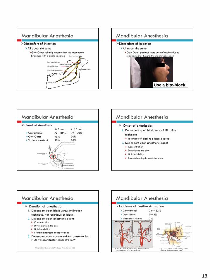
Mandibular Anesthesia
Discomfort of injection
All about the same
Gow-Gates reliably anesthetizes the most nerve
branches with a single injection
Mandibular Anesthesia
Discomfort of injection
All about the same
Gow-Gates perhaps more uncomfortable due to
requirement of having the mouth wide open
Use a bite-block!
Mandibular Anesthesia
Onset of AnesthesiaAt 5 min. At 10 min.
Conventional 72 – 85% 79 – 90%
Gow-Gates 45% 90%
Vazirani – Akinosi 90% 90%
Mandibular Anesthesia
Onset of anesthesia:
1. Dependent upon block versus infiltration
technique
Technique of block to a lesser degree
2. Dependent upon anesthetic agent
Concentration
Diffusion to the site
Lipid solubility
Protein binding to receptor sites
Mandibular Anesthesia
Duration of anesthesia:
1. Dependent upon block versus infiltration
technique, not technique of block
2. Dependent upon anesthetic agent
Concentration
Diffusion from the site
Lipid solubility
Protein binding to receptor sites
3. Dependent upon vasoconstrictor presence, but NOT vasoconstrictor concentration*
*Malamed, Handbook of Local Anesthesia, 5th Ed, Elsevier, 2004
Mandibular Anesthesia
Incidence of Positive Aspiration
Conventional 3.6 – 22%
Gow-Gates 0 – 2%
Vazirani – Akinosi 2%
Blanton PL & Roda RS, The anatomy of local anesthesia,
J Calif Dent Assoc, Vol 23 No 4, April 1995
Agur & Lee, Grant’s Atlas of Anatomy, 10th Ed,
Lippincott Williams & Wilkins, 1999

19
Mandibular Anesthesia
Incidence of Positive Aspiration
Conventional 3.6 – 22%
Gow-Gates 0 – 2%
Vazirani – Akinosi 2%
Al-Faraje L, Surgical and Radiologic Anatomy for Oral
Implantology, Quintessence Publishing Co, 2013 Netter’s Atlas, 4th Ed, Saunders/Elsevier, 2006
Mandibular Anesthesia
Incidence of Other Undesirable Side Effects
1. Hitting a nerve
2. Piercing a muscle
3. Injecting the parotid gland
Most common with IA block
Jastak, Yagiela & Donaldson, Local Anesthesia of
the Oral Cavity, WB Saunders Co, 1995
Mandibular Anesthesia
Incidence of Other Undesirable Side Effects
2. Piercing a muscle = Trismus
Evers & Haegerstam, Introduction to Dental
Local Anaesthesia, Mediglobe, 1990
Mandibular Anesthesia
Incidence of Other Undesirable Side Effects
2. Piercing a muscle = Trismus
Possible causes include insertion of the needle into a muscle, bleeding into a muscle, or injection of anesthetic into a muscle
All of these may produce
muscle spasms
Result is limited ability to
open and possible pain on
opening
McMinn, Hutchings & Logan, Color Atlas of Head &
Neck Anatomy, 2nd Ed, Mosby, 1994
Mandibular Anesthesia
Incidence of Other Undesirable Side Effects
2. Piercing a muscle = Trismus
Trismus symptoms may appear within 1 to 6 days post-injection
If there is no improvement within 2 to 3 days, or if the condition worsens, the patient may have an infection
Infection from an injection is rare
If an infection does occur, it will usually manifest itself initially as pain and trismus 1 day post-injection
Mandibular Anesthesia
Incidence of Other Undesirable Side Effects
2. Piercing a muscle = Trismus
Treatment
1. Apply heat
2. Recommend muscle relaxants (ibuprofen)
3. Analgesics/anti-inflammatories if needed
4. Exercises
Symptoms commonly last a few days

20
Mandibular Anesthesia
Incidence of Other Undesirable Side Effects
3. Injecting the parotid gland
Liebgott, The Anatomical Basis of Dentistry, 2nd Ed, Mosby, 2001
Mandibular Anesthesia
Injecting the parotid gland
Temporary facial paralysis: anesthesia of CN VII,
the facial nerve, to the muscles of facial expression
Liebgott, The Anatomical Basis
of Dentistry, 2nd Ed, Mosby, 2001Netter, Atlas of Human Anatomy, 2nd Ed, Novartis, 1997
Mandibular Anesthesia
Incidence of Other Undesirable Side Effects
1. Hitting a nerve
2. Piercing a muscle
3. Injecting the parotid gland
Most common with IA block
4. Anesthesia in the opposite
arch
5. Other unusual events
Most common with Vazirani –
Akinosi blockJastak, Yagiela & Donaldson, Local Anesthesia of
the Oral Cavity, WB Saunders Co, 1995
Mandibular Anesthesia
Comparison of mandibular division nerve block techniques
Conventional (Halsted) technique
Advantages:
Most familiar and most widely used
Good success rate (65 – 86%+)
Disadvantages:
Higher success rates associated with increased incidence of positive aspiration
Moderate incidence of trismus and/or paresthesia
Multiple injections required for anesthesia of inferior alveolar, lingual, long buccal, and mylohyoid nerves
Mandibular Anesthesia
Comparison of mandibular division nerve block techniquesGow-Gates technique
Advantages:
Very high success rate (90 – 100%)
Extremely low incidence of positive aspirations
Significantly reduced incidence of trismus and/or paresthesia
Single injection for anesthesia of inferior alveolar, lingual, long buccal, and mylohyoid nerves
Disadvantages:
Technically a more difficult technique to master
Slower onset of anesthesia
Possible increased patient discomfort
Mandibular Anesthesia
Comparison of mandibular division nerve block techniques
Vazirani – Akinosi technique
Advantages:
Moderate to high success rate (76 – 93%)
Extremely low incidence of positive aspirations
Significantly reduced incidence of trismus and/or paresthesia
Potential single injection for anesthesia of inferior alveolar, lingual, long buccal, and mylohyoid nerves
Less threatening to apprehensive patients (closed mouth)
Ability to anesthetize both sensory and motor nerve branches uniquely useful for patients with severe trismus

21
Mandibular Anesthesia
Comparison of mandibular division nerve block
techniques
Vazirani – Akinosi technique
Disadvantages:
Increased potential for operator error due to no bone
contact
Higher incidence of unexpected and unusual side effects
Not as reliable a technique to achieve anesthesia of the
long buccal nerve
Mandibular Anesthesia
Comparison of mandibular
division nerve block
techniques
Conventional (Halsted)
technique
Gow-Gates technique
Vazirani – Akinosi technique
So which technique is the best?
Depends mostly on patient
characteristics
The “Hot” Tooth / “Hot” Gum
Includes:
1. Infected teeth with irreversible pulpitis
2. Severe periodontal infections
3. Hypoplastic teeth with severe sensitivity
4. Teeth with hypersensitivity due to recession, occlusal
trauma/bruxing, etc.
All of these may be highly problematic to anesthetize
Troubleshooting Mandibular Anesthesia
The “Hot” Tooth / “Hot” Gum
First, give a block injection
Well away from the site of any local inflammation or
infection
The low pH will prevent the disassociation of the
anesthetic agent
A needle should not be inserted into an area of active
infection, such as a periodontal or periapical abcess
The volume of anesthetic is likely to increase pain
There is the potential for spreading the infection
Haas DA, Localized complications from local anesthesia, J Calif Dent Assoc, Vol 26 No 9, 1998
Troubleshooting Mandibular Anesthesia
The “Hot” Tooth / “Hot” Gum
First, give a block injection
The Gow-Gates mandibular division block has a significantly
higher success rate than all other techniques
Gow-Gates 52%
Vazirani-Akinosi 41%
Conventional IA 36%
Buccal-plus-lingual infiltration 27%
No technique was fully acceptable by itself
Aggarwal V et al, Comparative evaluation of anesthetic efficacy of Gow-Gates mandibular conduction anesthesia,
Vazirani-Akinosi technique, buccal-plus-lingual infiltrations, and conventional inferior alveolar nerve anesthesia in
patients with irreversible pulpitis, O Surg O Med O Path O Radio Endo, Vol. 109 No 2, Feb. 2010
All with 4% articaine with 1:100,000 epinephrine
Patients who took 600mg of ibuprofen 1 hour before IANB for endodontic
treatment of mandibular posterior teeth with irreversible pulpitis were 2x
more likely to have “little or no pain during endodontic treatment.”Lapidus D et al, Effect of premedication to provide analgesia as a supplement to inferior alveolar
nerve block in patients with irreversible pulpitis, J Amer Dent Assoc 147(6), June 2016
Troubleshooting Mandibular Anesthesia
The “Hot” Tooth / “Hot” Gum
First, give a block injection
Well away from the site of any local inflammation or infection
Second, is topical/Oraqix around the tooth adequate?
If not, give a periodontal ligament (PDL) or intraosseous
injection
Periodontal ligament injections are quick and easy to give,
but offer only short duration
Intraosseous injections are more reliable and have better
duration
Troubleshooting Mandibular Anesthesia

22
The “Hot” Tooth / “Hot” Gum
First, give a block injection
Well away from the site of any local inflammation or infection
Second, is topical/Oraqix around the tooth adequate?
If not, give a periodontal ligament (PDL) or intraosseous
injection
Or, give a buccal &/or lingual infiltration with articaine
(or prilocaine) Hasse et al, Comparing anesthetic efficacy of articaine versus lidocaine as a
supplemental buccal infiltration of the mandibular first molar after an inferior
alveolar nerve block, J Am Dent Assoc, Vol 139 No 9, Sept 2008
Kanaa et al, Articaine buccal infiltration enhances the effectiveness of lidocaine
inferior alveolar nerve block, Int Endo J, Vol 42, 2009
Troubleshooting Mandibular Anesthesia Infiltration Anesthesia Works well for the maxilla, but for the mandible…
Works fairly well for anteriors and bicuspids
More variable predictability for molars
Greater success using articaine
Lidocaine 45 – 67%; articaine 75 – 92%
Lidocaine 6.1 – 11.1 minutes; articaine 4.2 – 4.7 minutes
Facial Lingual
Robertson et al, The anesthetic efficacy of articaine in buccal infiltration of mandibular posterior teeth, JADA, Vol 138 No 8, 2007
& faster onset
Meechan, Practical Dental Local Anesthesia, Quintessence, 2002
Why not infiltrate
both buccally and
lingually?Use ½ - 1 cartridge
of articaine for each
Mandibular Anesthesia
The risk of nerve injury with administration of
prilocaine (Citanest) or articaine (Septocaine)
may be reduced by using “high” mandibular division
block techniques
Gow-Gates technique
Vazirani – Akinosi technique
Wolfe SH, The Wolfe nerve block: A modified high mandibular
nerve block, Dentistry Today, June/July 1992
Troubleshooting Mandibular Anesthesia
Repeated failure to achieve adequate anesthesia
Take a panoramic radiograph
Troubleshooting Mandibular Anesthesia
Repeated failure to achieve adequate anesthesia
Take a panoramic radiograph
Incidence of bifid IA nerve: 4 patients in 5,000 films
Grover PS & Lorton L, Bifid
mandibular nerve as a possible
cause of inadequate
anesthesia in the mandible,
Journ O Maxillofac Surg
Vol 179, 1983
With Cone Beam Computed Tomography (CBCT), the incidence of bifid mandibular
canals/inferior alveolar nerves has been found to be at least 15.6%, and may be as
high as 30%.
Kuribayashi A et al, Bifid mandibular canals: Cone beam computed tomography evaluation, Dentomaxillfac Radiol 39(4), 2010
Fukami K et al, Bifid mandibular canal: Confirmation of limited cone beam CT findings by gross anatomical and histological
investigations, Dentomaxillofac Radiol Vol 41, 2012

23
Troubleshooting Mandibular Anesthesia
Repeated failure to achieve adequate anesthesia
Take a panoramic radiograph
Mental foramina
Mandibular Anesthesia
Mandible: Nerve blocks
Mental (& incisive) nerve block
Fehrenbach & Herring, Illustrated Anatomy
of the Head & Neck, WB Saunders Co, 1996
Jastak, Yagiela & Donaldson, Local Anesthesia of
the Oral Cavity, WB Saunders Co, 1995
Mandibular Anesthesia
Mental (& incisive) nerve
block
Meechan, Practical Dental Local Anesthesia, Quintessence, 2002
For children, anesthetizes the
five primary mandibular teeth
in a quadrant
Mandibular Anesthesia
Mental (& incisive) nerve
block
Meechan, Practical Dental Local Anesthesia, Quintessence, 2002
Evers & Haegerstam, Introduction to Dental Local Anaesthesia, Mediglobe, 1990
Mandibular Anesthesia
Mental (& incisive) nerve block
Malamed, Handbook of Local Anesthesia,
5th Ed, Elsevier Mosby, 2004
Mandibular Anesthesia
Mental (& incisive) nerve block
Depth 3 – 6 mm
Needle Short
Amount 1/3 -1/2
cartridge
Comfort level High
After injection, massage site
Jastak, Yagiela & Donaldson, Local Anesthesia of
the Oral Cavity, WB Saunders Co, 1995

24
REVIEW OF MAXILLARY
ANATOMY
General Anatomy and Landmarks for
Maxillary Anesthesia
Maxillary Anesthesia
Trigeminal nerve, CN V
Maxillary division, CN V2
Sensory only
To all maxillary teeth and
gingiva
Mandibular division, CN V3
Both motor and sensory
Sensory to all mandibular
teeth and gingiva
Motor to primary muscles of
mastication
Fehrenbach & Herring, Illustrated Anatomy
of the Head & Neck, WB Saunders Co, 1996
The Masticator Space
Infratemporal FossaPterygopalatine fossa
opens into medial wallBoundaries:A gap between the
maxilla anteriorly and the lateral pterygoid plate of the sphenoid bone posteriorly
Leaves an opening, thepterygomaxillary fissure,into the infratemporalfossa
Medial wall: the palatine bone & sphenopalatineforamenAgur & Lee, Grant’s Atlas of Anatomy, 10th Ed,
Lippincott Williams & Wilkins, 1999
Pterygopalatine Fossa
Liebgott, The Anatomical Basis
of Dentistry, 2nd Ed, Mosby, 2001
Pterygopalatine Fossa
Contents
Maxillary division of
Trigeminal nerve, V2
Pterygopalatine
ganglion
Terminus of maxillary
artery
Fehrenbach & Herring, Illustrated Anatomy of the Head & Neck,
WB Saunders Co, 1996
Agur & Lee, Grant’s Atlas of Anatomy, 10th Ed,
Lippincott Williams & Wilkins, 1999
Blood Supply to the
Infratemporal Fossa
Maxillary artery:
3 parts
1. Mandibular
2. Pterygoid
3. Pterygopalatine
Agur & Lee, Grant’s Atlas of Anatomy, 10th Ed,
Lippincott Williams & Wilkins, 1999
1.
2.3.

25
Blood Supply to the
Infratemporal Fossa
Maxillary artery
Part 3:
Pterygopalatine1. Posterior superior
alveolar
2. Infraorbital
3. Artery of pterygoid
canal
4. Pharyngeal branch
5. Descending palatine
6. Sphenopalatine
Liebgott, The Anatomical Basis of Dentistry, 2nd Ed, Mosby, 2001
Blood Supply to the
Infratemporal Fossa
Pterygoid Venous
Plexus
Primary drainage to
Maxillary vein
Liebgott, The Anatomical Basis of Dentistry, 2nd Ed, Mosby, 2001
Maxillary Anesthesia
Maxilla: Nerves
Infraorbital nerve
Anterior superior
alveolar nerve
Middle superior
alveolar nerve
Posterior superior
alveolar nerve
Meechan, Practical Dental Local Anesthesia, Quintessence, 2002
Maxilla: Nerves
Infraorbital nerve
Anterior superior alveolar nerve
Middle superior alveolar nerve
Posterior superior alveolar
nerve
Greater palatine nerve
Lesser palatine nerve
Nasopalatine nerve
Maxillary Anesthesia
Meechan, Practical Dental Local
Anesthesia, Quintessence, 2002
MAXILLARY ANESTHESIA
Conventional and Alternative Techniques
Maxillary Anesthesia
Two basic types of injections
1. Infiltrations
2. Blocks
Infiltrations
Work well throughout maxilla
Greater success using articaine
Faster onset and longer duration
Frequent palatal anesthesia with buccal infiltration
Costa DG et al, Onset and duration periods of articaine and lidocaine
on maxillary infiltration, Quintessence Int, Vol 36 No 3, 2005

26
Maxillary Anesthesia
Infiltrations
* zygomatic buttress
McMinn, Hutchings & Logan, Color Atlas of
Head & Neck Anatomy, 2nd Ed, Mosby, 1994
Evers & Haegerstam, Introduction to Dental
Local Anaesthesia, Mediglobe, 1990
Maxillary Anesthesia
Additional alternatives for the maxilla:
PDL injection
Intraosseous injection
Interdental injection
Intraseptal injection
Palatal sulcular injection
For primary teeth when palatal
anesthesia is needed in addition
to buccal infiltration and
interdental injections
Good for gingiva and pulpal tissue
Good for gingiva only
Maxillary Anesthesia
Maxillary blocks:Anterior & middle superior
alveolar nerve block
Infraorbital nerve block
AMSA palatal block
ASA palatal block
Posterior superior alveolar
nerve block
Nasopalatine nerve block
Greater palatine nerve block
Complete maxillary division block
Meechan, Practical Dental Local Anesthesia, Quintessence, 2002
Maxillary Anesthesia
Maxilla: Nerve blocks
Anterior & middle superior alveolar nerve block
Infraorbital nerve block approach
Meechan, Practical Dental Local Anesthesia,
Quintessence, 2002
Maxillary Anesthesia
Anterior & middle superior alveolar nerve block
Infraorbital nerve block approach
Delivered at the infraorbitalforamen
Evers & Haegerstam, Introduction to Dental
Local Anaesthesia, Mediglobe, 1990
Maxillary Anesthesia
Anterior & middle superior alveolar nerve block
Infraorbital nerve block approach
Delivered at the infraorbital foramen
Palpate the inferior orbital rim
Agur & Lee, Grant’s Atlas of Anatomy, 10th Ed,
Lippincott Williams & Wilkins, 1999

27
Maxillary Anesthesia
Anterior & middle superior alveolar nerve block
Infraorbital nerve block approach
Delivered at the infraorbital foramen
Palpate the inferior orbital rim
Drop 10 mm below lowest point
Maxillary Anesthesia
Anterior & middle superior alveolar nerve block
Infraorbital nerve block approach
Depth 3 – 15 mm
Needle Short
Amount 1/3 - 1/2 cartridge
Comfort level Moderate to high (technique dependent)
Maxillary Anesthesia
Anterior & middle superior alveolar nerve blocks
Infraorbital approach
Comfort level Moderate to high (technique dependent)
Jastak, Yagiela & Donaldson,
Local Anesthesia of the Oral
Cavity, WB Saunders Co, 1995
Note: You do NOT need
to get the needle tip into
the foramen
Maxillary Anesthesia
Anterior & middle superior alveolar nerve block
Infraorbital nerve block approach
This can’t really happen! Keep finger over inferior rim
Maxillary Anesthesia
Anterior & middle superior alveolar nerve block
Infraorbital approach
MSA absent in ~28% of patients
Meechan, Practical Dental Local Anesthesia, Quintessence, 2002
Evers & Haegerstam, Introduction to Dental Local Anaesthesia, Mediglobe, 1990
Maxillary Anesthesia
Anterior & middle superior
alveolar nerve block
The AMSA palatal approach
(P-AMSA injection)
Meechan, Practical Dental Local Anesthesia, Quintessence, 2002
X

28
Maxillary Anesthesia
Anterior & middle superior alveolar nerve blocks
The AMSA palatal approach (P-AMSA injection)
Depth 2 – 4 mm
Needle Short
Amount ≤1/4 cartridge of articaine
Comfort level Moderate
Meechan, Practical Dental Local Anesthesia, Quintessence, 2002
Maxillary Anesthesia
Anterior & middle superior alveolar nerve block The AMSA palatal approach vs. infraorbital
approach Advantages
1. Buccal and palatal anesthesia
of bicuspids and incisors
2. No lip anesthesia
3. More reliable anesthesia of
middle superior alveolar
nerve/bicuspids
Disadvantages
1. Shorter duration
2. A palatal injection
X
X
Y
Meechan, Practical Dental Local Anesthesia, Quintessence, 2002
Y
Y
Maxillary Anesthesia
Techniques to minimize the discomfort of all
injections
1. Topical anesthesia
2. Pressure distraction/analgesia
3. Slow injection with small volumes
4. Buccal infiltrations
5. Explain all that you do to minimize the
discomfort
Learn to give comfortable palatal injections!
Maxillary Anesthesia
Maxilla: Nerve blocks
The ASA palatal approach
(P-ASA injection)
To bilaterally anesthetize:
Incisor pulps
Buccal gingiva
Anterior palatal tissue
© Milestone Scientific, Inc. 2007
Maxillary Anesthesia
Bilateral anterior superior alveolar nerve block
The ASA palatal approach (P-ASA injection)
1. Inject from side of incisive papilla initially, then gently
shift to vertical orientation as enter incisive canal
2. SLOWLY inject 1/4 – 1/3 cartridge of articaine
© Milestone Scientific, Inc. 2007
Maxillary Anesthesia
Maxilla: Nerve blocks
Nasopalatine nerve block: Replaced with the P-ASA
Liebgott, The Anatomical Basis of Dentistry, 2nd Ed, Mosby, 2001
Meechan, Practical Dental Local Anesthesia, Quintessence, 2002

29
Maxillary Anesthesia
Nasopalatine nerve block
The Three-Step technique
1. Buccal infiltration over either central incisor
Maxillary Anesthesia
Nasopalatine nerve block
The Three-Step technique
1. Buccal infiltration over either central incisor
2. Infiltrate central papilla
Maxillary Anesthesia
Nasopalatine nerve block
The Three-Step technique
1. Buccal infiltration over either central incisor
2. Infiltrate central papilla
3. Inject nasopalatine (incisive) papilla
Maxillary Anesthesia
Nasopalatine nerve block
Depth 2 – 4 mm
Needle Short
Amount ½ cartridge total, or less,
for all three injections
Comfort level Moderate to high
Maxillary Anesthesia
Posterior superior alveolar nerve block
Meechan, Practical Dental Local
Anesthesia, Quintessence, 2002Fehrenbach & Herring, Illustrated Anatomy
of the Head & Neck, WB Saunders Co, 1996
Maxillary Anesthesia
Posterior superior alveolar nerve block
McMinn, Hutchings & Logan, Color Atlas of
Head & Neck Anatomy, 2nd Ed, Mosby, 1994

30
Maxillary Anesthesia
Posterior superior alveolar nerve block
Evers & Haegerstam, Introduction to Dental
Local Anaesthesia, Mediglobe, 1990
Maxillary Anesthesia
Posterior superior alveolar nerve block
Depth 12 – 18 mm
Needle Long
Amount 3/4 cartridge
Comfort level High
High risk of positive
aspiration and hematoma
Maxillary Anesthesia
Hematoma
A hematoma may form independently of aspiration results.
Aspiration results merely report the contents at the needle tip at the time of aspirating
Haas DA, Localized complications from local anesthesia,
J Calif Dent Assoc, Vol 26 No 9, 1998
Courtesy Dr. H. Shirazi
Aspiration During Injections
Hematoma The vessels most
commonly associated with hematomas are
1. Pterygoid venous plexus
2. Posterior superior alveolar vessels
3. Inferior alveolar vessels
4. Mental vessels
Haas DA, Localized complications from local anesthesia, J Calif Dent Assoc, Vol 26 No 9, 1998
Liebgott, The Anatomical Basis of Dentistry, 2nd Ed, Mosby, 2001
Agur & Lee, Grant’s Atlas of Anatomy, 10th Ed, Lippincott Williams & Wilkins, 1999
Aspiration During Injections
Hematoma
Arterial vs. Venous
Fast Slow
Red Blue
Warm Normal
Management
1. Initial ice pack and pressure
2. Analgesics/anti-inflammatories (usually not needed)
3. Rest
Maxillary Anesthesia
Greater palatine nerve block
Meechan, Practical Dental Local Anesthesia, Quintessence, 2002
Fehrenbach & Herring, Illustrated Anatomy
of the Head & Neck, WB Saunders Co, 1996

31
Maxillary Anesthesia
Greater palatine nerve block
Meechan, Practical Dental Local Anesthesia, Quintessence, 2002
Maxillary Anesthesia
Greater palatine nerve block
Meechan, Practical Dental Local Anesthesia, Quintessence, 2002
Maxillary Anesthesia
Greater palatine nerve block
Depth 2 – 4 mm
Needle Short
Amount 1/4 - 1/3 cartridge
Comfort level Moderate to high
McMinn, Hutchings & Logan, Color Atlas of Head & Neck Anatomy, 2nd Ed, Mosby, 1994
Maxillary Anesthesia
Maxilla: Nerve blocks
Complete maxillary division block
With 2 injections
With 1cartridge
Two approaches
PSA (lateral)
approach
Greater palatine
canal approach
Meechan, Practical Dental Local Anesthesia, Quintessence, 2002
Pterygopalatine Fossa
Contents
Maxillary division of
Trigeminal nerve, V2
Passes across the top
of the fossa
Fehrenbach & Herring, Illustrated Anatomy of the
Head & Neck, WB Saunders Co, 1996
Maxillary Anesthesia
Complete maxillary division block
PSA (lateral) approach
PSA approach
Meechan, Practical Dental Local Anesthesia, Quintessence, 2002
Agur & Lee, Grant’s Atlas of Anatomy, 10th Ed, Lippincott Williams & Wilkins, 1999

32
Maxillary Anesthesia
Complete maxillary division block
PSA (lateral) approach
High risk of hematoma
PSA approachMcMinn, Hutchings & Logan, Color Atlas of
Head & Neck Anatomy, 2nd Ed, Mosby, 1994Agur & Lee, Grant’s Atlas of Anatomy,
10th Ed, Lippincott Williams & Wilkins, 1999
Maxillary Anesthesia
Complete maxillary division block
Greater palatine canal approach
Meechan, Practical Dental Local
Anesthesia, Quintessence, 2002
Fehrenbach & Herring, Illustrated Anatomy
of the Head & Neck, WB Saunders Co, 1996
Maxillary Anesthesia
Greater palatine canal approach
1. Give greater palatine block injection
2. Re-palpate the greater palatine foramen
3. With a single penetration, gently probe
for the foramen
Maxillary Anesthesia
Greater palatine canal approach
With a single penetration, gently probe for the foramen
Passively insert needle up the canal
Deposit the cartridge of anesthetic
Maxillary Anesthesia
Complete maxillary division block
Greater palatine canal approach
Depth Varies, ~15 mm
Needle Long
Amount 1 cartridge
Comfort level Moderate
Meechan, Practical Dental Local Anesthesia, Quintessence, 2002
Maxillary Anesthesia
Complete maxillary division block
With either approach, may anesthetize zygomatic
branch of V2
Innervation to lacrimal (tear) gland
Agur & Lee, Grant’s Atlas of Anatomy, 10th Ed, Lippincott Williams & Wilkins, 1999Liebgott, The Anatomical Basis of Dentistry,
Mosby, 1986

33
Troubleshooting Maxillary Anesthesia
Give buccal infiltration in anterior region*
Tissue under eye blanches
and/or
There is a facial twitch/spasm
Stay calm
1. Stimulated facial nerve
2. Contact with blood vessel
3. Muscle contact/spasm
4. Localized vasoconstriction
*May occur with PSA and inferior alveolar blocks as well
Troubleshooting Maxillary Anesthesia
Buccal tissue is numb
Tooth is still sensitive!
Give palatal injection
Use articaine for both buccal &
palatal infiltrations
Often produces more profound
anesthesia
Meechan, Practical Dental Local Anesthesia, Quintessence, 2002
Reasons for Anesthetic Failures
1. Anatomical/physiological
variations
2. Technical errors of
administration
3. Patient anxiety
4. Inflammation and
infection
5. Defective/expired
solutions
What defines success?
“Adequate anesthesia to
insure patient comfort for
the duration of the
procedure”
Different for each
procedure
Different for each
patient
Keys to Success
Anesthetic failures happen
The “Three Strikes Rule”
3 attempts at anesthesia, then stop
It’s not about “fault”
It’s not the patient’s fault
It’s not your fault
Failures happen
Reschedule the patient!
Reasons for Anesthetic Failures
3. Patient anxiety
Anxiety lowers the
threshold of pain.
Therefore, even non-
painful stimuli are
likely to be
perceived as painful.

34
Keys to Success
3. Patient anxiety
When patients sense that
the dentist or dental
hygienist is sincere in
doing everything possible
to insure the patient’s
comfort,
they will relax!
Keys to Success
The No Fault Theory
It is important to note that complications with oral injections are not always preventable, and their occurrence does not necessarily imply poor technique by the dentist or dental hygienist.
Haas DA, Localized complications from local anesthesia, J Calif Dent Assoc, Vol 26 No 9, Sept 1998
Keys to Success
It’s the thought that counts