Spine injuries in baseball players | cervical spine injuries | Colorado Spine Surgeon
Upload
hattie-webbCategory
view
217download
0description

In this issue...
Damien SmithCervical Headaches
Jason HarveyRunning & Spinal Problems
Tom EmberEvidence Base for Spinal Surgery
SPINE EVENING The Fortius Clinic Lecture Series - April 2016 Issue
2
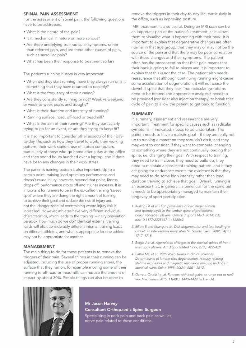
This month’s lecture evening was in our new City venue and it was great to see some new faces from City physio practices joining us for the occasion, as well as plenty of regulars making the trip to the new location. Only a couple of minutes’ walk from our new City clinic on King William Street, it was a great opportunity to venture into our new surroundings and spread the word on the upcoming open evenings.
The topic for the evening was the Spine, and the session was led by three Fortius consultants; Dr Damien Smith, Mr Tom Ember and Mr Jason Harvey. It was a fascinating insight into various research studies, typical spine injuries and their treatments, and it triggered plenty of discussion from the audience.
Dr Smith, Consultant in Pain Medicine and Anaesthesia, introduced the talks on the topic of cervical headaches. He provided analysis into the signs to look for when making the diagnosis, together with the anatomy and treatment plans that you can advise to tackle these headaches.
Next to take the stage was Consultant Spinal Surgeon Mr Harvey who spoke about spinal problems connected to running. He discussed whether sport or
genetic predisposition played a greater part in spine degeneration and gave a detailed explanation of spine pain assessment and the management of the pain.
Last to speak was Mr Ember who is the newest member of our Spine Team at the Fortius Clinic. His presentation reviewed the evidence base for spinal surgery; a crucial element as the occurrence of lower back pain continues to increase as does the number of operations being undertaken.
In this newsletter we have included access instructions to all spine sessions from FISIC’15, please see page 3 for more information. In addition you will find information on the final countdown to our new City clinic opening, the beginning stages of the creation of our day surgery centre and a FISIC blog on the 'Spondylolysis: Diagnosis & Follow-up' session.
Don’t forget that the May lecture will be at the Marylebone Hotel, hosted by our Foot and Ankle Team. If you have any questions that you would like to be covered in the question and answer section or have a particular topic in mind, please contact the marketing department.
APRIL TOPIC:
SPINE EVENING
Videos and Newsletters available onlinewww.fortiusclinic.com
> news-events > education
THE FORTIUS LECTURE EVENINGEvery month the Fortius Clinic hosts a lecture evening for Physiotherapists and Sports & Exercise Medicine professionals, led by a different team of specialists. The evening event is held in central London and the City. If you would like to be added to our invitation list, please email [email protected].
Sophie Whitby Marketing Executive
e: [email protected] t: 0203 853 3043
Don’t forget to follow us on Twitter @FortiusClinicUK. You will also find us on Facebook and LinkedIn

3
Conference videos have been made available to you through the Co-Kinetic website (co-kinetic.com) until midnight on Thursday 26th May.
• Register for a free account at this page https://co-kinetic.com/register
• Click this link to access the presentations https://co-kinetic.com/category/spine-fisic-conference-2015
• Co-Kinetic is an online content resource for sports medicine practitioners. It features articles, videos, podcasts and conference presentations on a wide variety of topics relevant to practitioners involved in the delivery
For a limited time only you are entitled to complimentary
access to spine lecture videos via the Co-Kinetic site
FREE ACCESS TO FISIC SPINE LECTURES
Session details:
CERVICAL SPINE
Increasing prevalence of degenerative changes in young cervical spines Richard Nelson
Screening and classification/ assessment for return to playPeter Hamlyn
Role of cervical proprioceptionLesley McBride
Brachial plexus injuries in rugbyTim Hems
Surgical decision making challenges Peter Hamlyn
Cervical discectomy in the sportsmanJason Harvey
DISC PATHOLOGY & SPONDYLOLYSIS
Disc Pathology - minimal access surgeryMichael Mayer
Spondylolsis - surgical managementDavid Harrison
Spondylolsis: conservative treatmentDamian Fahy
Spondylolsis: diagnosis & follow-up: how does imaging inform management?Ranju Dhawan
Disc Pathology - conservative treatment Jonathan Rees
SPINE - IMAGING & TREATMENT STRATEGY
Axial/ dynamic MRI in sportsFrancis Smith
A physio's perspectiveNick Allen
A surgeon's perspectiveDamian Fahy
Pain management in sportsGlyn Towlerton
MRI findings in the spine in asymptomatic young athletesGajan Rajeswaran

4
FEATURE | CERVICAL HEADACHESLecture by Dr Damien Smith Consultant in Pain Medicine and Anaesthesia
Headaches can be split into two groups: primary headache and secondary headache. Primary headache accounts for 90% of all headaches and are very common, benign and with no underlying disease – the problem is a functional one rather than a structural one. Secondary headache, which accounts for the other 10% of headaches, is headache due to underlying disease, and so is caused by things such as infection, malformations and space-occupying lesions. Hence, with secondary headache, the underlying cause may well be visible on imaging. The source of primary headache is in the head, for example: cluster headaches, migraines and tension. However, the source of secondary headaches is elsewhere, such as the sinuses, nose, mouth, ears and neck, etc. Cervicogenic headache (CGH) is, therefore, a secondary headache specifically due to pathology in and around the cervical spine (C spine); primarily, from a musculoskeletal dysfunction of C1, C2 and C3.
ANATOMYWith CGH, the message is relayed from C spine to the pain sensors via the trigeminocervical nucleus. The name ‘trigeminocervical nucleus’ indicates where the messages come from: afferents come from the trigeminal nerve and from C1, 2 and 3. The afferents join together in the nucleus and the signal then goes through the ascending tract to the thalamus and then on to the somatosensory cortex. Messages also go on to the limbic system, which is why patients can feel a bit down when they have these headaches.
In 1944, Douglas Campbell and Clare Parsons, in the Department of Anatomy and Psychiatry at the California Medical School, USA, investigated noxious referral patterns by intramuscular injection of healthy volunteers with 6% saline [1]. It was found that injections at different levels of the C spine caused referred pain in specific places: C1 at the frontal lobe, C1/2 at the temporal lobe, C2/3 caused auricular pain, C3/4 caused occipital pain and C4/5 at the trapezius. The study was repeated with injections into the facets and the referral pattern was very similar.
A similar referral pattern is seem in patients with cervical arthropathy, the higher the facet joints that are injected, the more likely benefit to the incidence of headache, the lower the facets the more likely benefit to pain down into the trapezius, rhomboids and the beta scapulae. Hence, for CGH patients, performing a cervical facet joint block at the C1/2/3 level will result in an improvement in the headache, but blocks at the C4/5/6 level will be ineffective, as the referral pattern is much lower down the body.

5
Professor Gwendolen Jull (University of Queensland, Brisbane, Australia) investigated measures of sensitivity and specificity for CGH [2–4] and found that CGH patients have: (1) reduced range of movement (ROM) in the C spine, particularly extension; (2) painful joint dysfunction over level C1, 2 and 3; and (3) impaired muscle function, which was identified by a failed neck flexor endurance test (39 seconds for males, 29 seconds for females). In the neck flexor endurance test, the patient lies down with a towel under their neck, the towel is removed and the patient has to flex their neck and hold the position for as long as possible.
Other considerations in patients with headaches include the accessory nerve, the 11th cranial nerve, which provides motor function to the sternocleidomastoid and trapezius, which can often be trigger points for headaches also. In patients with these headaches, up to 44% had some temporomandibular joint (TMJ) dysfunction. It has been shown that patients treated for CGH and TMJ, where TMJ dysfunction was recognised, had a better outcome than those treated for CGH alone.
In sports, weight lifters are particularly prone to this condition, which can also occur in whiplash and concussion. In the case of concussion injury, these headaches can even develop more than 3 months after the actual injury itself.
DIAGNOSISSigns for the diagnosis of CGH include a headache with associated neck pain and stiffness, it is unilateral, and cervical dysfunction on manual examination. Pain can often occur due to a trigger point palpation, and the condition can be aggravated by sustained neck positions.
MANAGEMENT PLANTo create a management plan, C spine ROM, C spine facets, neck flexors, as well as sternocleidomastoid, trapezius and TMJ have to be assessed.
TREATMENT PLANTreatment can be through a manual therapy treatment plan as well as a more interventional treatment plan. The manual therapy treatment plan involves improving C spine mobilisation, strengthening the neck flexors and the upper quarter muscles (including the neck flexors, trapezius, sternocleidomastoid, levator scapulae and pectoralis), and treating the TMJ if it is also involved. The more interventional treatment plan depends on the source of the pain. For example, trigger point injections can be used if the source is a muscle trigger point, and cervical facet medial branch blocks can be used if the source is a cervical facet. A cervical epidural is indicated if the cause is at the C2/3 discs, and a greater occipital nerve block can also be tried as this carries sensory fibres from C2 and 3. However, usually a full multimodal approach is the best option.
1. Campbell DG and Parsons CM. Referred head pain and its concomitants: report of preliminary experimental investigation with implications for the post-traumatic "head" syndrome. J Nervous & Mental Dis. 1944; 99(5): 544–551.
2. J ull G et al. Cervical musculoskeletal impairment in frequent intermittent headache. Part 1:sSubjects with single headaches. Cephalalgia 2007; 27: 793–802.
3. Amiri M et al. Cervical musculoskeltal impairment in frequent intermittent headache. Part 2: subjects with multiple headaches. Cephalagia 2007; 27: 891–898.
4. Jull G et al. A Randomised controlled trial of exercise and manipulative therapy for cervicogenic headaches. Spine 2002; 27: 1835–1843.
Dr Damien Smith Consultant in Pain Medicine & AnaesthesiaDr Smith specialises in assessing and treating patients with lower back pain, cervical pain, radiculopathy, headaches and complex regional pain syndrome.

Many patients seen by Mr Harvey have lumbar spine problems related to running. With regard to these patients, the important questions are: Should this person be running? and What can we do to make it more comfortable for them? Often, the patient is a middle-aged office worker, a desk-based person, who wants to run but is experiencing back pain that is either acute exacerbations or chronic relapsing in nature. The patients’ reasons for running include, “Running is what I enjoy”, “I’ve always run”, “I want to get fit, so I’ve returned to running”, “It fits in with my busy life, work and family balance”, “I want to compete”, and “I want to complete a marathon”. However, they are experiencing pain and they are often also worried that they are going to develop significant degeneration of their spine.
There is some evidence to suggest that sport can cause spine degeneration. In a study of 29 male beach volleyball players, average age 29 years, 79% had at least one degenerate disc, although there was no correlation with back pain [1]. Disc degeneration was classified by the Pfirmann grading system of 1 to 5, where 3 and above is deemed as degenerate. In cricket, a study found that specific positions of the bowler were causing specific changes with the disc patterns [2], and also unilateral facet joint degeneration or hypertrophy particularly related to their dominant (serving) hand has been seen with tennis players. Degeneration is not only isolated to the lumbar spine, but is also seen in the cervical spine. In an MRI study of the cervical spines of 47 front row rugby players with matched controls, 83% of players had osteophytes compared with 33% in the controls, 48% had disc bulges compared to 7% in the control group, and other findings included changes within the bone marrow and thickening of the endplates [3]. Non-professional rugby players can also be affected, and this was exemplified by scans of the spine of a 25-year-old tighthead prop, who had multiple level degenerative changes and significant disc protrusions. However, in the general population, a classic study by Battié and Videman of 115 pairs of monozygotic twins showed that the vast majority of the degenerative changes were related to genetic predisposition and other aspects of their lifestyles accounted for a much smaller percentage [4]. A study of 77 half-marathon runners found that 54% had experienced back pain, 49% said that their back pain improved with running, and 27% said that running made it worse [5].
FEATURE | RUNNING & SPINE PROBLEMS
Lecture by Mr Jason HarveyConsultant Orthopaedic Spine Surgeon
7
Mr Jason HarveyConsultant Orthopaedic Spine SurgeonSpecialising in neck pain and back pain,as well as nerve pain related to these conditions.
SPINAL PAIN ASSESSMENTFor the assessment of spinal pain, the following questions have to be addressed:
• What is the nature of the pain?
• Is it mechanical in nature or more serious?
• Are there underlying true radicular symptoms, rather than referred pain, and are there other causes of pain, such as sacroiliac pain?
• What has been their response to treatment so far?
The patient’s running history is very important:
• When did they start running, have they always run or is it something that they have returned to recently?
• What is the frequency of their running?
• Are they consistently running or not? Week vs weekend, or week-to-week peaks and troughs?
• What is their duration and intensity of running?
• Running surface: road, off-road or treadmill?
• What is the aim of their running? Are they particularly trying to go for an event, or are they trying to keep fit?
It is also important to consider other aspects of their day-to-day life, such as how they travel to work, their working pattern, their work station, use of laptop computers, particularly of those who go home after a day at the office and then spend hours hunched over a laptop, and if there have been any changes in their work stress.
The patient’s training pattern is also important. Up to a certain point, training load optimises performance and doesn’t cause injury; however, beyond that point, fitness drops off, performance drops off and injuries increase. It is important for runners to be in the so-called training ‘sweet spot’ where they are doing the right amount of training to achieve their goal and reduce the risk of injury and not the ‘danger zone’ of overtraining where injury risk is increased. However, athletes have very different individual characteristics, which leads to the training¬–injury prevention paradox: how much do we do? Identical external training loads will elicit considerably different internal training loads on different athletes, and what is appropriate for one athlete may not be appropriate for another.
MANAGEMENTThe main thing to do for these patients is to remove the triggers of their pain. Several things in their running can be adjusted, including the use of proper running shoes, the surface that they run on, for example moving some of their running to off-road or treadmills can reduce the amount of impact by about 30%. Simple things can also be done to
remove the triggers in their day-to-day life, particularly in the office, such as improving posture.
‘MRI treatment’ is also useful. Doing an MRI scan can be an important part of the patient’s treatment, as it allows them to visualise what is happening with their back. It is important to explain that degenerative changes are often normal in that age group, that they may or may not be the source of the pain and that there may be poor correlation with those changes and their symptoms. The patient often has the preconception that their pain means that their back is going to fall to pieces and it is important to explain that this is not the case. The patient also needs reassurance that although continuing running might cause some acceleration of degeneration, it will not cause the downhill spiral that they fear. True radicular symptoms need to be treated and appropriate analgesia needs to be provided (consider also injection therapy) to break that cycle of pain to allow the patient to get back to function.
SUMMARYIn summary, assessment and reassurance are very important. Treatment for specific causes such as radicular symptoms, if indicated, needs to be undertaken. The patient needs to have a realistic goal – if they are really not up to running a marathon they shouldn’t do it, and they may want to consider, if they want to compete, changing to something where they are not continually loading their spine, i.e. changing their goal. With respect to training, they need to train clever, they need to build up, they need to maintain a consistent training pattern, and if they are going for endurance events the evidence is that they may need to do some high intensity rather than long duration training to achieve that goal. Overall, running is an exercise that, in general, is beneficial for the spine but it needs to be appropriately managed to maintain their longevity of sport participation.
1. Külling FA et al. High prevalence of disc degeneration and spondylolysis in the lumbar spine of professional beach volleyball players. Orthop J Sports Med. 2014; 2(4): doi:10.1177/2325967114528862.
2. Elliott B and Khangure M. Disk degeneration and fast bowling in cricket: an intervention study. Med Sci Sports Exerc. 2002; 34(11): 1717–1718.
3. Berge J et al. Age-related changes in the cervical spines of front-line rugby players. Am J Sports Med 1999; 27(4): 422–429.
4. Battié MC et al. 1995 Volvo Award in clinical sciences. Determinants of lumbar disc degeneration. A study relating lifetime exposures and magnetic resonance imaging findings in identical twins. Spine 1995; 20(24): 2601–2612.
5. Garreta-Català I et al. Runners with back pain: to run or not to run? Rev Med Suisse 2015; 11(481): 1440–1444 (in French).

8
FEATURE |EVIDENCE-BASED SPINAL SURGERY
Lecture by Mr Tom EmberConsultant Orthopaedic Spine Surgeon
This presentation reviewed the evidence base for spinal surgery. Having an evidence base for spinal surgery is crucial as the incidence of low back pain is rising, as is the number of spinal operations performed.
Research has shown that low back pain is the number one cause of disability worldwide; ahead even of conditions such as chronic anaemias, tuberculosis and major depression, which was second. Disability from musculoskeletal problems has increased by 45% in the last 20-year period and is still rising, in part due to the natural aging population, but also because of increasing obesity and reduction in physical activity. Importantly, the evidence now exists to show that obesity, smoking, decreased activity levels all impact on back pain, and that as body mass index (BMI) increases so does the incidence of back pain. Spinal surgery is a growth industry; the number of spinal operations performed has risen by 77% between 1996 and 2001. This is not simply the reflection of an aging population as the number of hip and knee joint replacements rose only by 13–14% in the same time period. The financial impact is also important as back pain now rivals cardiac pathologies in terms of cost to the NHS.
Evidence-based medicine is not new and it is part of everyday life for doctors in fields such as cardiology and respiratory medicine. Also, increasingly, purchasers require evidence before treatments are provided to ensure that they are worthwhile and cost-effective. However, there has been a lack of evidence-based medicine in spinal surgery, but there have been attempts to address this.
THE EVIDENCE BASE FOR LUMBAR SURGERYLumbar discectomy is the most commonly performed spinal operation and there has been one Cochrane Review by the National Institute for Health and Care Excellence (NICE) for it. Five randomised controlled trials (RCTs) were analysed for the effect of surgical treatment for individuals with a slipped lumbar disc. However, only three of these trials directly compared discectomy with non-surgical approaches, so only three were relevant to the review, and they provide suggestive rather than conclusive results. Overall, the results suggested that
surgical discectomy appears to provide faster relief from an acute attack than non-surgical management, when used for carefully selected patients with sciatica due to a prolapsed disc. However, the surgery does not address the natural history of the underlying disease, so although the patient gets better faster, the end result is probably much the same as the starting point. So to emphasise, the evidence base for lumbar discectomy, the most commonly performed spinal surgery, consists of five RCTs, only three of which directly compare discectomy with non-surgical treatment.
Although there has been a 77% increase in the number of spinal fusions performed in a five-year period, the evidence to support this procedure is largely lacking. There was only moderate evidence that instrumentation (putting metal work in) can increase the fusion rate, but any improvement in clinical outcomes was probably marginal. Therefore, the conclusion was that surgery has no meaningful benefit over a structured rehabilitation programme with a cognitive behavioural component. Again, to emphasise, there is only a very weak evidence base for an operation that is being done at an exponentially increasing rate.
For completeness, the evidence base for total disc replacement in the lumbar spine is completely lacking and this procedure is very rarely performed in the UK.
There is RCT evidence for short-term benefit of micro discectomy, but not for the medium and longer term. There is also level 2/3 evidence (but no level 1 RCT evidence) to support lumbar spinal stenosis surgery in the older patient with claudicant-type leg pain. Interspinous distractors were commonly used but there is no proof of any benefit for any condition.
Stratifying the procedures by NICE approval and proven benefit by RCT demonstrates that: micro discectomy and stenosis surgery for people with claudicant leg symptoms satisfy both those criteria; interspinous distraction, spinal fusion and disc replacement have NICE approval but no proven benefit; adult spinal deformity is in the process of being reviewed; and XLIF (extreme lateral lumbar interbody fusion) and flexible stabilisation are new technologies.

9
DIFFICULTIES WITH THE EVIDENCE BASE IN SPINAL SURGERYOne of the main reasons for the lack of an evidence base for spinal surgery is the difficulty of performing RCTs in this field, and a number of factors contribute to this.
The natural history of the condition is in itself difficult: it fluctuates, it can improve and deteriorate, unlike other medical conditions where the natural history is often much more linear and the effect of an intervention is clearer.
RCTs also need outcome measures. Measuring outcomes in a field such as hip replacement is straightforward: if the hip needs to be revised that can be classed as failure. However, outcome measures in spinal surgery are exceptionally difficult and there is no agreement on outcome scoring systems. There are also problems with pain scoring systems. For example, the visual analogue scale is a useful measure in part but doesn’t tell the whole story.
There has also been a rapid pace of evolution in spinal surgery. Technology and technique advances in the last 20 years make it very difficult to compare ‘historic’ papers from as little as 10 years ago. For example, not that long ago discectomy and fusion were done through a large scar with significant collateral damage and are now being done through very, very minimal access approaches or less access approaches so the amount of collateral damage is reduced. There has also sometimes been a lack of common sense applied to the use of spinal surgery by spinal surgeons, meaning that unusual procedures have been carried out for unusual reasons, again making comparisons difficult.
One example that highlights the problems of RCTs in spinal surgery was a £50 million spine patient outcomes research trial in the USA. The 5-year sports study involved 11 states and approximately 500 patients with a disc prolapse with leg pain that was radicular in nature randomised into two groups, of surgical and non-surgical intervention. However, the results were still very unclear and the problem is largely due to the natural history of a disc prolapse. For example, only 60% of the patients in the surgical group had surgery because a number of the patients got better while on the waiting list, and 45% of the non-surgical intervention group had surgery because they did not get better. This cross-over means that the results are then very difficult to interpret statistically. One of the most interesting parts was the complication rates: dural tear 4%, womb infection 6%, recurrent disc requiring revision 5%; these are very real complications with statistically reasonably high rates indicating that it is not a benign operation. The conclusion was that surgery allows a more rapid relief from pain and an earlier
return to work, reduced analgesia usage and reduced psychosocial complications. However, at the 2-year time point, the conclusion was that there was very little difference between the two groups.
One example that demonstrated how disc prolapses can often just get better was a patient with an extremely large L4/5 disc prolapse, which was large enough to be almost a radiological cauda equina. However, the patient did not have cauda equina syndrome, had normal bladder function and did not want surgery. He had an injection for severe leg pain and had a repeat MRI scan 6 months later, which showed that he had complete restoration of his spinal canal, the disc was completely reabsorbed and the nerve routes were all completely clear. However, there is a definite group of patients who continually get low level symptoms whenever they try to get back to their normal activities. With these patients, the disc is often partially reabsorbed, but then walls off and calcifies and surgery is then often beneficial.
TECHNIQUES AND TECHNOLOGYTechniques and technology are advancing and spinal surgery is improving. Standard open surgery causes a large amount of collateral damage, which is one of the reasons why the results from fusion operations were so poor. In the 1990s and early 2000s many surgeons changed to minimally invasive surgery, which is technically very difficult. The small surgical field often results in a compromised operation and higher complication rates. Now there is the concept of ‘less invasive’ surgery, which is a compromise between the between minimal access and standard open surgery, and it is made possible by advances in retractor systems and light sources and techniques. This allows the surgeon to achieve everything that would be achieved through standard open surgery, but with a smaller incision and lower retractor times and so minimises muscle injury, and has very encouraging results.
OUTLYING BEHAVIOUR AND COMMON SENSEOutlying behaviour exists in every profession but can be particularly hard to identify in surgery, as innovation often looks like outlying behaviour. One example of this is John Harrington who designed the Harrington rod for scoliosis. He was initially booed and heckled when he presented his work in the 1950s, but it subsequently became the gold standard for a generation of spinal surgeons working on scoliosis.
However, outlying behaviour can simply be a lack of common sense, which can result in a far more complicated surgical intervention than was necessary. Common sense is also needed to recognise what is normal and what is pathological. ➠ continued
10
For example, for a patient in their early thirties, some early disc degeneration, disc dehydration and a small bulge can be normal. The pathological issue is that the cross-sectional core muscles are of tiny volume and the fat layer is far too high. Hence, the instability is caused by the muscles around the spine, rather than the disc dehydration that that was seen with no modic changes.
However, there are patients for whom surgery is indicated. They have a disc that is giving them severe leg pain, they do not respond to anti-inflammatories, they are in too much pain for physiotherapy, injection does not help, but early micro discectomy is often beneficial. Also there are patients with degeneration who have severe back and leg pain where spinal fusion can play a role. In this situation, in order to address the leg pain (which is the primary reason for surgery), there may be an indication to perform fusion to try to address elements of the back pain. Also, for patients with instability-type problems (e.g. spondylolisthesis and associated leg pain), simple decompression surgery is not going to suffice and fusion becomes indicated. Hence, there are indications for fusion, but exceptionally careful choice of patients is required.
Regarding instability, a Swiss scientist showed that a simple model of load demonstrated that column stiffness reflects how much load can be borne by the column before deflection of the column occurs. However, applying guy ropes to a very thin column causes a large increase in column stiffness, so that a much higher load can be applied to it. In vivo, the guy wires around the lumbar spine are the lumbar muscles – the erector spinae muscles at the back of the spine, but more importantly the front group, the anterior and obliques, the rectae, etc., which provide stability around early disc degeneration.
CURRENT CONCEPTSNICE is drafting new guidance for low back pain and sciatica, with anticipated publication at the end of 2016. The guidance is separated into non-specific back pain and sciatic leg pain, and the advice for sciatic leg pain does not seem to be changing much: continue with physiotherapy, anti-inflammatories, epidurals and surgery for those whose conditions do not settle. For non-specific back pain, the advice is changing considerably: reduce imaging, no TENs, acupuncture, spinal or facet injections, but denervation is advised, along with physiotherapy and combined psychological modalities. Spinal fusions are to be done only as part of a trial. The reduced amount of imaging is of concern as it can show unexpected conditions, such as tumours, and it is very reassuring for patients to see that what they have is normal wear and tear.
CONCLUSIONSThere are different evidence bases for different spinal operations, but it does have its limitations. Informing the patient is paramount and consent has to be tailored to the patient. Multidisciplinary teams (MDTs) are spreading in spinal care and should be the standard, rather than the gold standard of care. MDTs provide the opportunity to discuss complex patients with colleagues from across different professions and providers and so take away outlying behaviour and help with common sense decisions.
Mr Tom Ember Consultant Spine SurgeonHis interests and areas of expertise include lumbar and cervical degenerative disc disease (including low back pain and sciatica), spinal tumours and infection.
11
FISIC FEATUREOn the 13th and 14th October 2015, we held the Fortius International Sports Injury Conference. Below is a summary of the "Spondylolysis: Diagnosis & Follow-up" session. The speakers in this session were Michael Mayer, Jonathan Rees, Ranju Dhawan, Damian Fahy and David Harrison.
The afternoon session in the Westminster room on day two was focused on spondylolysis, an overuse injury seen often in young sports people who perform activity that involves significant hyperextension and rotation of the lumbar spine. As David Harrison stated in the start of his presentation, the fractures observed in this condition are normally presenting in the contra-lateral side to the performing side.
It was discussed that occasionally stress fractures of the pars interarticularis are asymptomatic and may present as a referred issue such as unexplained hamstring tightness. However, in most cases, the individual will complain of unilateral back ache and pain aggravated with hyperextension activity. From my experience, objectively individuals may have a lordotic posture, specific pain on palpation of the lumbar process unilaterally as well as pain being recreated on extension with rotation whilst standing on the affecting leg.
Discussing the radiological approach to diagnosing spondylolysis Ranju Dhawan stated that X-rays aren’t particularly useful in individuals with recent onset of symptoms, however fractures can be observed normally on 45 degree oblique views in those with longstanding issues. It was discussed that more and more emphasis is now placed upon SPECT scans (single photon emission computed tomography) to further investigate cases where subjective and objective findings suggest a spondylolysis, yet the X-rays is negative. If positive on SPECT, then a reverse gantry CT scan should be performed to image the fracture further. This ideally is then repeated throughout the healing process.
As with any orthopaedic pathology the management of this condition was debated with the conservative vs surgical approaches weighed up. The prognosis of such injury is dependent on the site and stage of injury with early recognition of pathology key in decreasing return to play times. From a conservative perspective, early intervention is crucial - naturally the key is to offload from the aggravating activity, strengthening the trunk and stretching both the gluteal and hamstring muscles when possible. From my own experience, a controlled, graded rehabilitation plan over a six-week period is necessary once the original aggravating factors have become pain free. It was discussed that if the conservative approach fails, surgical options were dependant on the site and stages of injury with decompression or removal of the affected facet joint, lamina and foraminal space showing good results. In bilateral cases of stress fracture, a stabilisation procedure may be required in order to prevent the issue becoming a spondylolisthesis which was stated to have the potential of being a retiring injury.
The session underlined the importance of early diagnosis in the management of spondylolysis. As with most pathologies, the importance of initial clinical subjective and objective findings is crucial in the condition’s prognosis.
Gruff ParsonsHead of Performance at London Scottish Football (Rugby) Club.
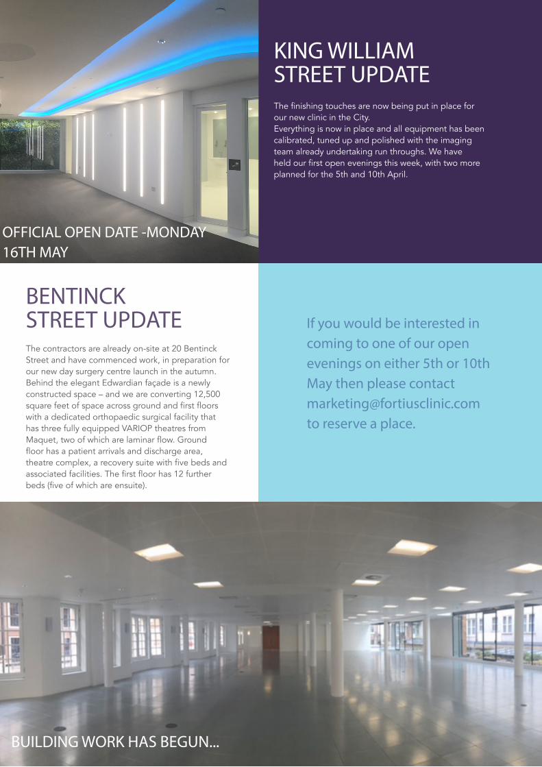
KING WILLIAM STREET UPDATE
BENTINCK STREET UPDATE
The finishing touches are now being put in place for our new clinic in the City. Everything is now in place and all equipment has been calibrated, tuned up and polished with the imaging team already undertaking run throughs. We have held our first open evenings this week, with two more planned for the 5th and 10th April.
If you would be interested in coming to one of our open evenings on either 5th or 10th May then please contact [email protected] to reserve a place.
The contractors are already on-site at 20 Bentinck Street and have commenced work, in preparation for our new day surgery centre launch in the autumn. Behind the elegant Edwardian façade is a newly constructed space – and we are converting 12,500 square feet of space across ground and first floors with a dedicated orthopaedic surgical facility that has three fully equipped VARIOP theatres from Maquet, two of which are laminar flow. Ground floor has a patient arrivals and discharge area, theatre complex, a recovery suite with five beds and associated facilities. The first floor has 12 further beds (five of which are ensuite).
BUILDING WORK HAS BEGUN...
OFFICIAL OPEN DATE -MONDAY 16TH MAY

OPEN EVENINGS AT OUR NEW OUTPATIENT AND DIAGNOSTIC CITY CLINIC
5TH MAY AND 10TH MAY
75 King William Street, London EC4N 7BE
MAY LECTURE | THE DIFFICULT FOOTOur May lecture evening will be held at the Marylebone hotel:7 Welbeck Street, London W1G 8DN
Mr Callum ClarkDifficult Forefoot
Mr Andy RocheDifficult Midfoot
Ms Anne-Marie O'Connor Podiatrist's Perspective

14
How to find us in the City: Our Outpatient and Diagnostic Clinic is located within the historic and financial heart of London. Close to Monument Tube station and Cannon St Rail.
King William St London EC4
For further information or to book an appointment, please contact us:
t: +44 (0) 203 195 2442 f: 0203 070 0106 e: [email protected] www.fortiusclinic.com
Don’t forget to follow us on Twitter @FortiusClinicUK You will also find us on Facebook and LinkedIn
How to find us in central London: Fortius Clinic is close to Selfridges, and just off Manchester Square.
17 Fitzhardinge Street London W1H 6EQ