Approaches to Improve the Clinical Performance of ...
54
Approaches to Improve the Clinical Performance of Genetically Engineered T Cells CAR-TCR Annual Summit Pete DeMuth, PhD Vice President of Research Cambridge, Massachusetts
Transcript of Approaches to Improve the Clinical Performance of ...
Approaches to Improve the Clinical Performance of Genetically Engineered T Cells
CAR-TCR Annual Summit
Pete DeMuth, PhDVice President of ResearchCambridge, Massachusetts
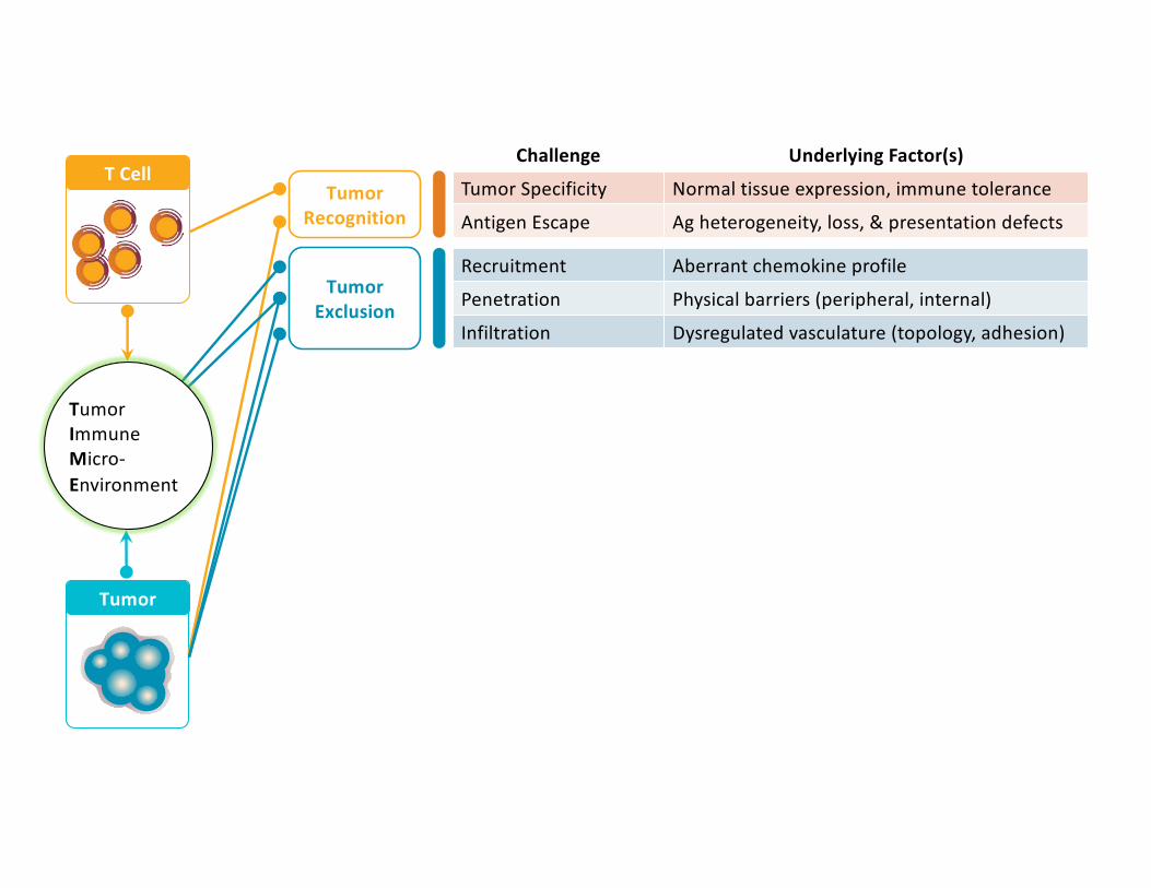
What are the major challenges to improving engineered T cell therapeutic efficacy?

Tumor Specificity Normal tissue expression, immune tolerance
Antigen Escape Ag heterogeneity, loss, & presentation defects
Challenge Underlying Factor(s)
Tumor Recognition
T Cell
Tumor
Tumor Immune Micro-Environment
Tumor Specificity Normal tissue expression, immune tolerance
Antigen Escape Ag heterogeneity, loss, & presentation defects
Challenge Underlying Factor(s)
Tumor Recognition
T Cell
Tumor
Recruitment Aberrant chemokine profile
Penetration Physical barriers (peripheral, internal)
Infiltration Dysregulated vasculature (topology, adhesion)
Tumor Exclusion
Tumor Immune Micro-Environment

Tumor Specificity Normal tissue expression, immune tolerance
Antigen Escape Ag heterogeneity, loss, & presentation defects
Challenge Underlying Factor(s)
Tumor Recognition
T Cell
Tumor
Recruitment Aberrant chemokine profile
Penetration Physical barriers (peripheral, internal)
Infiltration Dysregulated vasculature (topology, adhesion)
Tumor Exclusion
Exhaustion TME-induced chronic activation
Exhaustion Tonic signalingT Cell
FitnessTumor Immune Micro-Environment

Tumor Specificity Normal tissue expression, immune tolerance
Antigen Escape Ag heterogeneity, loss, & presentation defects
Challenge Underlying Factor(s)
Tumor Recognition
T Cell
Tumor
Recruitment Aberrant chemokine profile
Penetration Physical barriers (peripheral, internal)
Infiltration Dysregulated vasculature (topology, adhesion)
Tumor Exclusion
Exhaustion TME-induced chronic activation
Exhaustion Tonic signalingT Cell
Fitness
Limited Proliferation Terminal phenotype, lack of APC & co-stim
Pro-apoptotic Factors TME-derived FasLT Cell
Proliferation
Tumor Immune Micro-Environment

Recruitment Aberrant chemokine profile
Penetration Physical barriers (peripheral, internal)
Infiltration Dysregulated vasculature (topology, adhesion)
Functional Inhibition Suppressor Cells (Treg, MDSC, TAMs)
Functional Inhibition Inhibitory Cytokines (TGFβ, IL-10)
Functional Inhibition Tumor-intrinsic Factors (PD-L1)
Functional Inhibition Hypoxia, acidosis
Metabolic Suppression Glucose limitation
Metabolic Suppression Amino acid shortage (tryptophan, arginine)
Oxidative Stress Elevated ROS production
Exhaustion TME-induced chronic activation
Exhaustion Tonic signaling
Tumor Specificity Normal tissue expression, immune tolerance
Antigen Escape Ag heterogeneity, loss, & presentation defects
Limited Proliferation Terminal phenotype, lack of APC & co-stim
Pro-apoptotic Factors TME-derived FasL
Challenge Underlying Factor(s)
Tumor Recognition
Tumor Exclusion
T Cell Fitness
Tumor Immuno-
suppression
T Cell Proliferation
T Cell
Tumor
Tumor Immune Micro-Environment

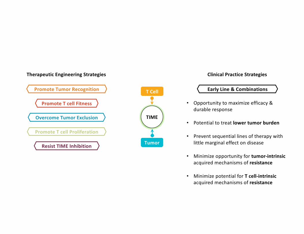
What solutions are most promising to address these challenges?
T Cell
Tumor
TIME

Promote T cell Fitness
Overcome Tumor Exclusion
Promote Tumor Recognition
Promote T cell Proliferation
Therapeutic Engineering Strategies Clinical Practice Strategies
Early Line & Combinations
• Opportunity to maximize efficacy & durable response
• Potential to treat lower tumor burden
• Prevent sequential lines of therapy with little marginal effect on disease
• Minimize opportunity for tumor-intrinsicacquired mechanisms of resistance
• Minimize potential for T cell-intrinsic acquired mechanisms of resistance
T Cell
Tumor
TIME
Resist TIME Inhibition
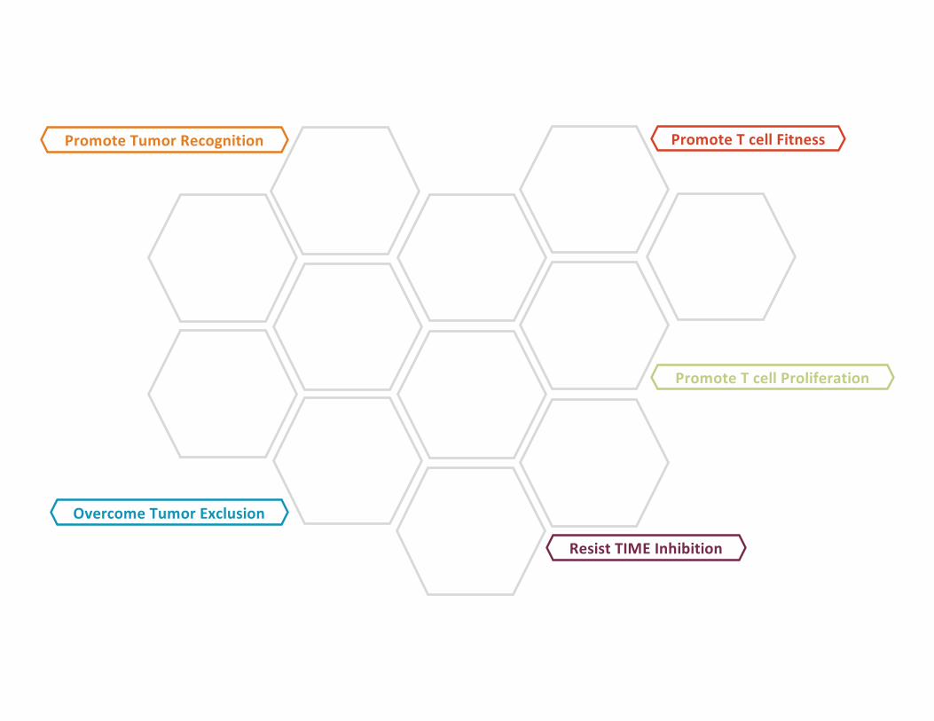
Promote T cell Fitness
Overcome Tumor Exclusion
Promote Tumor Recognition
Promote T cell Proliferation
Resist TIME Inhibition

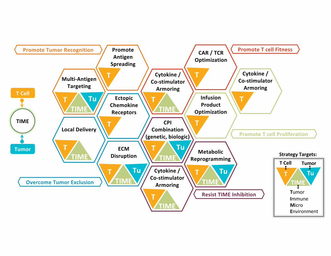
Promote Antigen
Spreading
Multi-Antigen targeting Strategies
Ectopic Chemokine
Receptor Expression
ECM Disruption
Local Delivery
Cytokine / Co-stimulator Armoring
CAR / TCR Optimization
Cytokine / Co-stimulator Armoring
Infusion Product
OptimizationCPI
Combination (genetic, biologic)
Cytokine / Co-stimulator Armoring
Metabolic Reprogramming
Promote T cell Fitness
Overcome Tumor Exclusion
Promote Tumor Recognition
Promote T cell Proliferation
Resist TIME Inhibition
T Cell
Tumor
TIME
Promote Antigen
Spreading
T
Ectopic Chemokine Receptors
T
Cytokine / Co-stimulator
Armoring
TInfusion Product
Optimization
TCPI Combination
(genetic, biologic)
T TuTIME
T TuTIME
ECM Disruption
T
CAR / TCR Optimization
Promote T cell Fitness
Overcome Tumor Exclusion
Promote Tumor Recognition
Promote T cell Proliferation
T Tu
Multi-Antigen Targeting
TIME
Cytokine / Co-stimulator
Armoring
TTIME
Cytokine / Co-stimulator
Armoring
TTIME
Local Delivery
TTIME
T TuTIME
Metabolic Reprogramming
T TuTIME
T Cell
TumorImmuneMicroEnvironment
Tumor
Strategy Targets:
Resist TIME Inhibition

What role can the lymphatics play in promoting T cell activity and therapeutic
efficacy?
T Cell
Tumor
TIME Lymph Nodes
1) Designing a system to target immune agents to lymph nodes
2) Boosting TCR-T Cell therapeutic responses
3) Boosting CAR-T Cell therapeutic responses
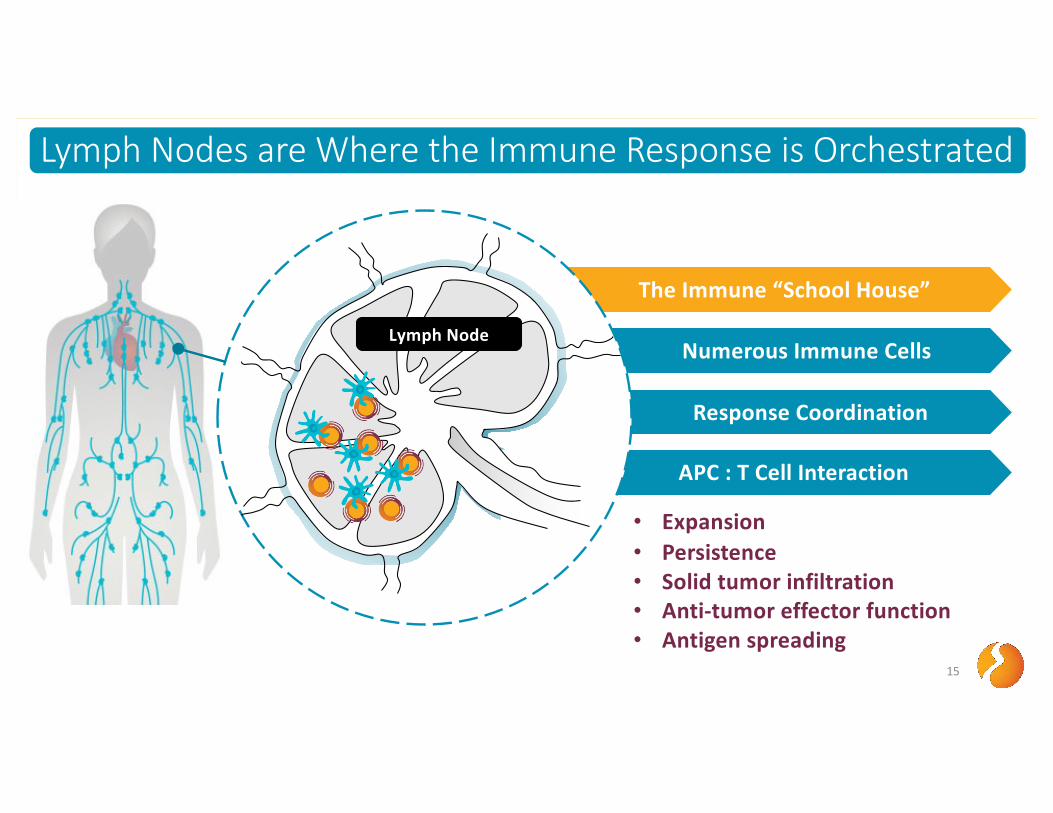
Lymph Nodes are Where the Immune Response is Orchestrated
15
Lymph Nodes are Where the Immune Response is Orchestrated
Response Coordination
The Immune “School House”
Numerous Immune Cells
APC : T Cell Interaction
Lymph Node
• Expansion• Persistence• Solid tumor infiltration• Anti-tumor effector function• Antigen spreading
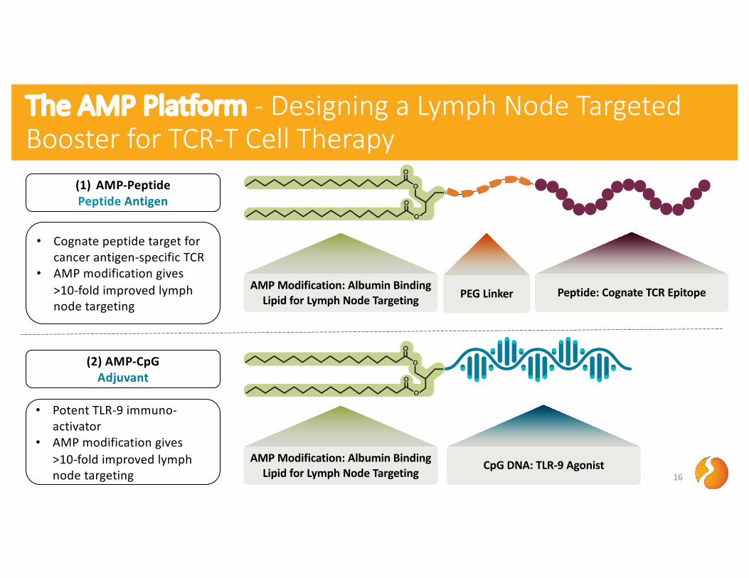
The AMP Platform - Designing a Lymph Node Targeted Booster for TCR-T Cell Therapy
16
(1) AMP-Peptide Peptide Antigen
• Cognate peptide target for cancer antigen-specific TCR
• AMP modification gives >10-fold improved lymph node targeting
(2) AMP-CpG Adjuvant
AMP Modification: Albumin Binding Lipid for Lymph Node Targeting
CpG DNA: TLR-9 Agonist
• Potent TLR-9 immuno-activator
• AMP modification gives >10-fold improved lymph node targeting
AMP Modification: Albumin Binding Lipid for Lymph Node Targeting
Peptide: Cognate TCR EpitopePEG Linker

The AMP Platform - Designing a Lymph Node Targeted Booster for TCR-T Cell Therapy
17
1 Subcutaneous Injection
Lymph Node
3 Lymph Node Targeting
Albumin Binding
Albumin
AMP-CpG
A
B
Tissue Injection Site
2
AMP-Peptide
• Uptake in resident APCs• Potent APC activation • TCR T cell activation
4 Immune Activation

Tumor
18
1T Cell Collection
2TCR Engineering
3Adoptive Cell Transfer
4 TCR T Cell Migration to Lymph Node
6TCR T Cell Infiltration and
Elimination of Tumor
The AMP Platform - Designing a Lymph Node Targeted Booster for TCR-T Cell Therapy
Lymph Node
• Interaction with activated APCs presenting cognate peptide
• TCR T cell activation, expansion
5 TCR-T Activation / Expansion

0 20 40 600
20
40
60
80
100
Days after Tumor
Perc
ent s
urvi
val
0 20 40 600
200
400
600
800
1000
Days after Tumor
Tum
or V
olum
e (m
m3 )
AMP-Boosting Potently Enhances TCR-T Therapy to Eliminate Established Solid Tumors
19
day 0
day 7
day -1 AMP-Boosting
AMP-Boosting
TCR-T Cells
day -10 Tumor Injection
day 10 AMP-Boostingday 14 AMP-Boosting
5x10⁶ gp100-specific TCR-T CellsmCherry Transduced
AMP- Boost:10 µg gp100 Peptide
1 nmol CpG
day 3 AMP-Boosting
Dylan Drakes, PhD, Abdul Abbas, MS
TCR-T Cells TCR-T Cells + AMP Boost
n = 24n = 10
TCR-T Cells: 0/10 cured
TCR-T Cells + AMP Boost: 7/24 cured
0 20 40 60Days after Tumor
B16F10 Melanoma
0 10 20 30 40 500
20
40
60
80
100
Days after Rechallenge
Perc
ent s
urvi
val
0 10 20 30 40 50Days after Rechallenge
0 10 20 30 40 500
200
400
600
800
1000
Days after Tumor
Tum
or V
olum
e (m
m3 )
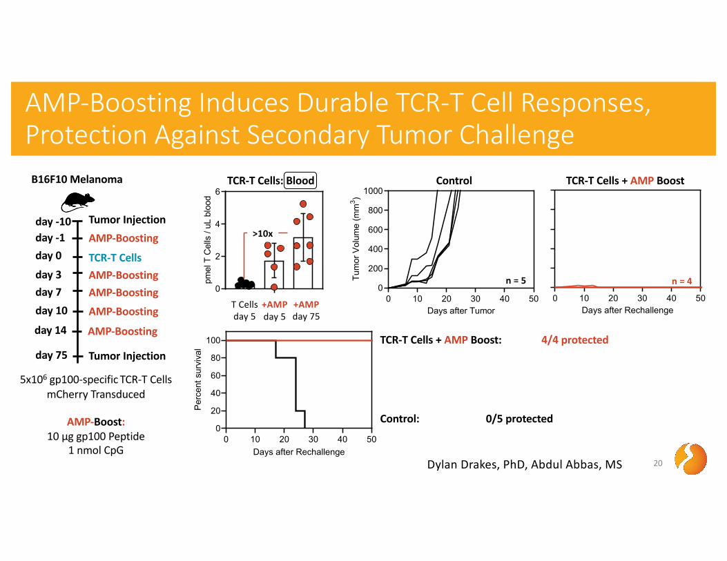
AMP-Boosting Induces Durable TCR-T Cell Responses, Protection Against Secondary Tumor Challenge
20
5x10⁶ gp100-specific TCR-T CellsmCherry Transduced
AMP-Boost:10 µg gp100 Peptide
1 nmol CpG
Control
Control: 0/5 protected
TCR-T Cells + AMP Boost: 4/4 protected
n = 4n = 5
day 75 Tumor Injection
TCR-T Cells + AMP Boost
0
2
4
6pm
el T
Cel
ls /
uL b
lood
TCR-T Cells: Blood
T Cellsday 5
+AMPday 5
+AMPday 75
Dylan Drakes, PhD, Abdul Abbas, MS
>10x
day 0
day 7
day -1 AMP-Boosting
AMP-Boosting
TCR-T Cells
day -10 Tumor Injection
day 10 AMP-Boostingday 14 AMP-Boosting
day 3 AMP-Boosting
B16F10 Melanoma
0
50
100
150
pmel
T C
ells
in L
N (x
103 )
0
100
200
300
pmel
T C
ells
/mg
of tu
mor
0
1
2
3
4pm
el T
Cel
ls/u
L bl
ood
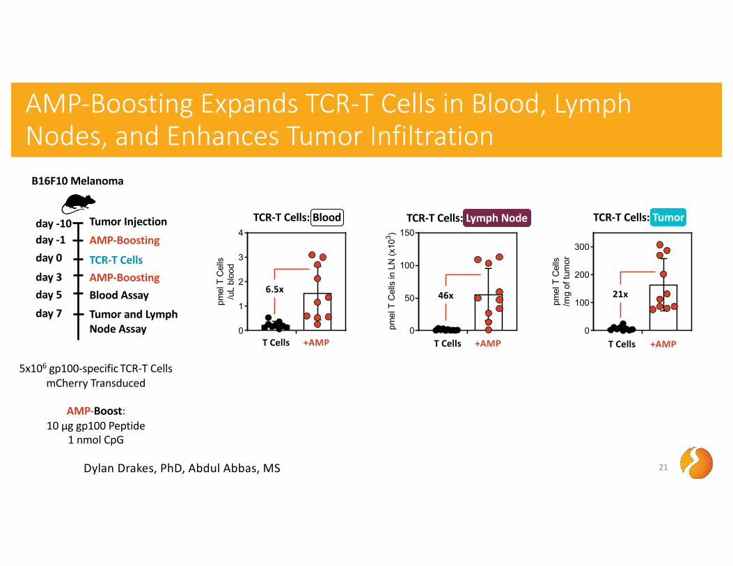
AMP-Boosting Expands TCR-T Cells in Blood, Lymph Nodes, and Enhances Tumor Infiltration
21
5x10⁶ gp100-specific TCR-T CellsmCherry Transduced
AMP-Boost:10 µg gp100 Peptide
1 nmol CpG
TCR-T Cells: Blood TCR-T Cells: TumorTCR-T Cells: Lymph Node
T Cells +AMP
6.5x 46x
Dylan Drakes, PhD, Abdul Abbas, MS
T Cells +AMP
day 0
day 5
day -1 AMP-Boosting
Blood Assay
TCR-T Cells
day -10 Tumor Injection
day 7 Tumor and Lymph Node Assay
day 3 AMP-Boosting
T Cells +AMP
21x
B16F10 Melanoma

0
100
200
300
pmel
T C
ells
/mg
of tu
mor
AMP-Boosting Enhances TCR-T Cell Tumor Infiltration, Functionality, and Proliferation
22
5x10⁶ gp100-specific TCR-T CellsmCherry Transduced
AMP-Boost:10 µg gp100 Peptide
1 nmol CpG
TCR-T Cells: Tumor T Cell Activation: Tumor
gp100 T Cell Cytokines: Tumor T Cell Proliferation: Tumor
Dylan Drakes, PhD, Abdul Abbas, MS
T Cells +AMP
day 0
day 5
day -1 AMP-Boosting
Blood Assay
TCR-T Cells
day -10 Tumor Injection
day 7 Tumor and Lymph Node Assay
day 3 AMP-Boosting
21x
T Cells +AMP0
100
200
300
400
# of
CD
25+ C
D8+ T
Cel
ls/m
g tu
mor
6.8x
0
20
40
60
80
100
# of
Ki6
7+ , CD
8+ T C
ells
/m
g tu
mor
T Cells +AMP
3.5x
IFNγ+ TNFα+
TNFα+
IFNγ+
0
100
200
300
# of
CD
8 T
Cel
ls/m
g tu
mor
T Cells +AMP
3x
B16F10 Melanoma
0
50
100
150
# of
CD
8 T
Cel
ls/m
g tu
mor
0
100
200
300
# of
CD
8 T
Cel
ls/m
g tu
mor
0
50
100
150
# of
CD
8 T
Cel
ls/m
g tu
mor
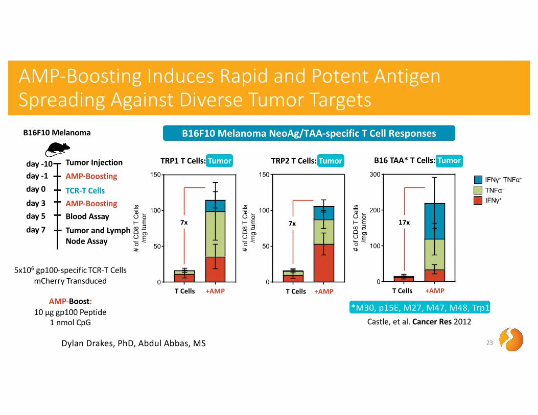
AMP-Boosting Induces Rapid and Potent Antigen Spreading Against Diverse Tumor Targets
23
day 0
day 5
day -1 AMP-Boosting
Blood Assay
TCR-T Cells
day -10 Tumor Injection
day 7 Tumor and Lymph Node Assay
day 3 AMP-Boosting
5x10⁶ gp100-specific TCR-T CellsmCherry Transduced
AMP-Boost:10 µg gp100 Peptide
1 nmol CpG
TRP1 T Cells: Tumor
T Cells +AMPT Cells +AMP
TRP2 T Cells: Tumor
IFNγ+ TNFα+
TNFα+
IFNγ+
7x 7x
Dylan Drakes, PhD, Abdul Abbas, MS
T Cells +AMP
B16 TAA* T Cells: Tumor
17x
B16F10 Melanoma B16F10 Melanoma NeoAg/TAA-specific T Cell Responses
*M30, p15E, M27, M47, M48, Trp1Castle, et al. Cancer Res 2012
0
50
100
150
pmel
T C
ells
in L
N (x
103 )
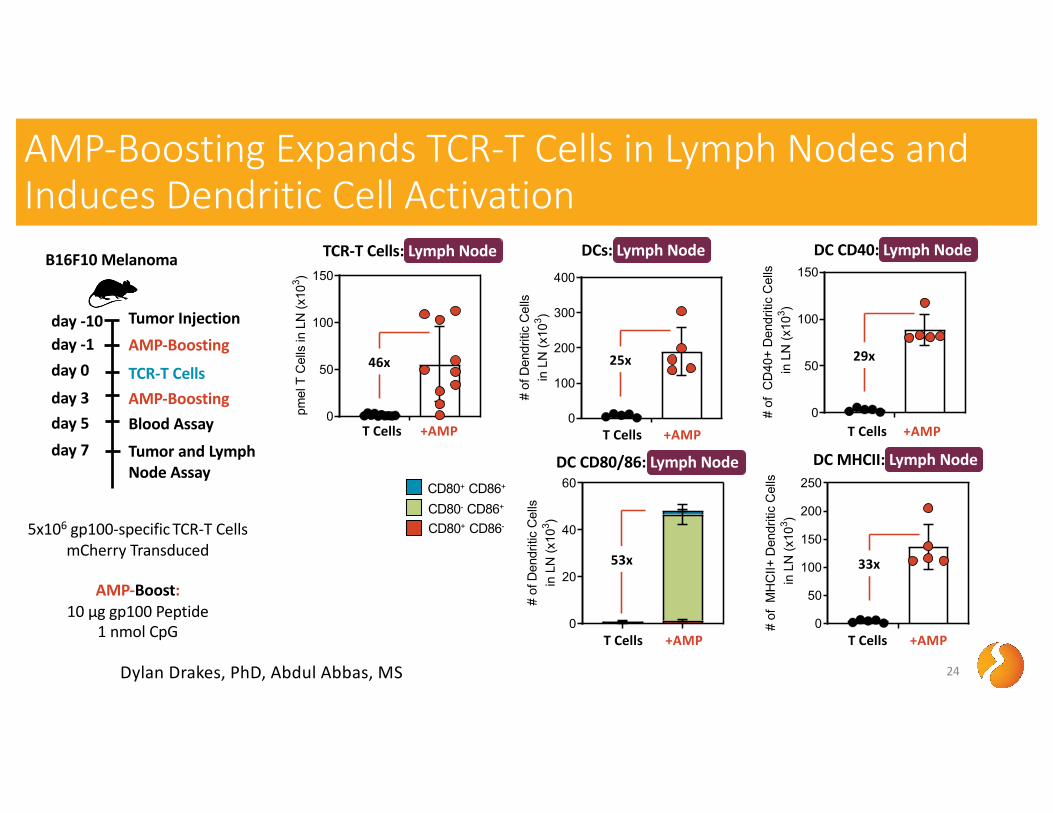
AMP-Boosting Expands TCR-T Cells in Lymph Nodes and Induces Dendritic Cell Activation
24
day 0
day 5
day -1 AMP-Boosting
Blood Assay
TCR-T Cells
day -10 Tumor Injection
day 7 Tumor and Lymph Node Assay
day 3 AMP-Boosting
5x10⁶ gp100-specific TCR-T CellsmCherry Transduced
AMP-Boost:10 µg gp100 Peptide
1 nmol CpG
DCs: Lymph Node DC CD40: Lymph Node
Dylan Drakes, PhD, Abdul Abbas, MS
TCR-T Cells: Lymph Node
46x
T Cells +AMP0
100
200
300
400
# of
Den
driti
c C
ells
in L
N (x
103 )
T Cells +AMP
25x
DC MHCII: Lymph Node
0
50
100
150
200
250
# of
MH
CII+
Den
driti
c C
ells
in L
N (x
103 )
T Cells +AMP
33x
0
50
100
150
# of
CD
40+
Den
driti
c C
ells
in L
N (x
103 )
29x
T Cells +AMP
DC CD80/86: Lymph NodeCD80+ CD86+
CD80- CD86+
CD80+ CD86-
0
20
40
60
# of
Den
driti
c C
ells
in L
N (x
103 )
T Cells +AMP
53x
B16F10 Melanoma
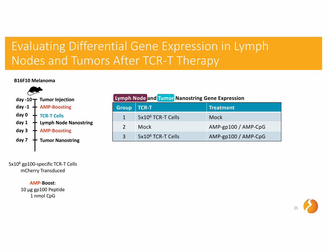
Evaluating Differential Gene Expression in Lymph Nodes and Tumors After TCR-T Therapy
25
day 0
day 3
day -1 AMP-Boosting
AMP-Boosting
TCR-T Cells
day -10 Tumor Injection
day 7 Tumor Nanostring
day 1 Lymph Node Nanostring
5x10⁶ gp100-specific TCR-T CellsmCherry Transduced
AMP-Boost:10 µg gp100 Peptide
1 nmol CpG
Group TCR-T Treatment
1 5x10⁶ TCR-T Cells Mock
2 Mock AMP-gp100 / AMP-CpG
3 5x10⁶ TCR-T Cells AMP-gp100 / AMP-CpG
Lymph Node and Tumor Nanostring Gene Expression
B16F10 Melanoma
-2
-1
0
1
2
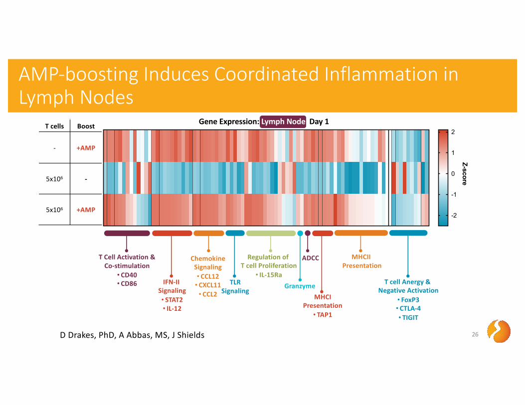
AMP-boosting Induces Coordinated Inflammation in Lymph Nodes
26
Gene Expression: Lymph Node Day 1
Z-score
T Cell Activation &Co-stimulation
• CD40• CD86 TLR
SignalingIFN-II
Signaling• STAT2• IL-12
ChemokineSignaling• CCL12• CXCL11• CCL2
Granzyme
Regulation of T cell Proliferation
• IL-15Ra
ADCC
MHCIPresentation• TAP1
MHCIIPresentation
T cell Anergy & Negative Activation
• FoxP3• CTLA-4• TIGIT
D Drakes, PhD, A Abbas, MS, J Shields
T cells Boost
- +AMP
5x10⁶ -
5x10⁶ +AMP
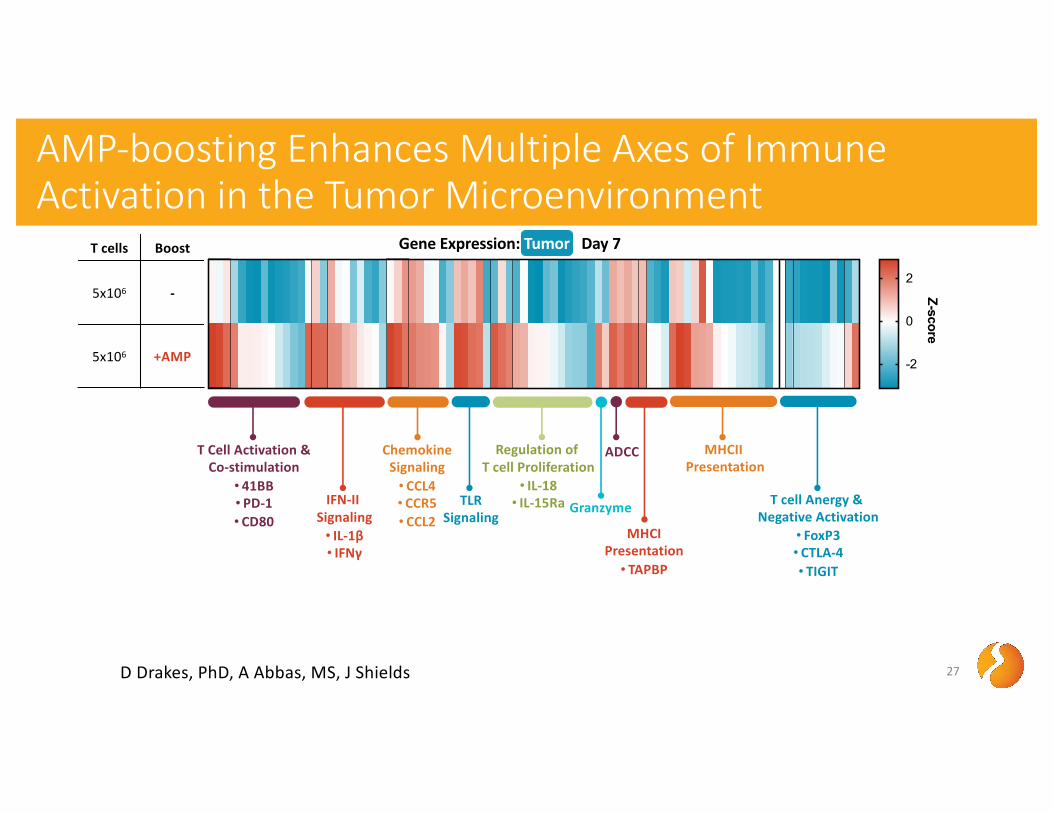
AMP-boosting Enhances Multiple Axes of Immune Activation in the Tumor Microenvironment
27
T cells Boost
5x10⁶ -
5x10⁶ +AMP
Gene Expression: Tumor Day 7
T Cell Activation &Co-stimulation
• 41BB• PD-1• CD80
TLRSignaling
IFN-II Signaling• IL-1β• IFNγ
ChemokineSignaling• CCL4• CCR5• CCL2
Granzyme
Regulation of T cell Proliferation
• IL-18• IL-15Ra
ADCC
MHCIPresentation• TAPBP
MHCIIPresentation
T cell Anergy & Negative Activation
• FoxP3• CTLA-4• TIGIT
-2
0
2
Z-score
D Drakes, PhD, A Abbas, MS, J Shields
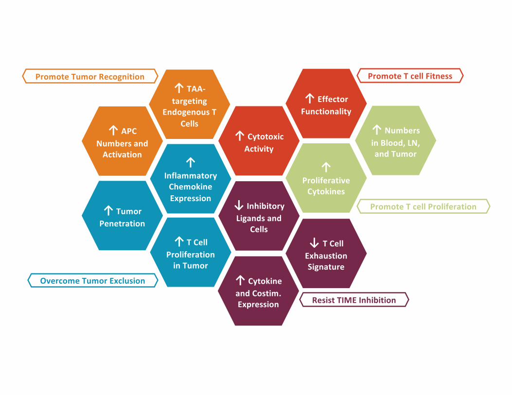
↑ Cytotoxic Activity
Promote T cell Fitness
↑Inflammatory
Chemokine Expression
Overcome Tumor Exclusion
↑ TAA-targeting
Endogenous T Cells
Promote Tumor Recognition
↑ APC Numbers and
Activation
↑ T Cell Proliferation
in Tumor
↑ Tumor Penetration
↑ Effector Functionality
Promote T cell Proliferation
↑ Numbers in Blood, LN, and Tumor
↑Proliferative
Cytokines
↓ Inhibitory Ligands and
Cells
↑ Cytokine and Costim. Expression
↓ T Cell Exhaustion Signature
Resist TIME Inhibition
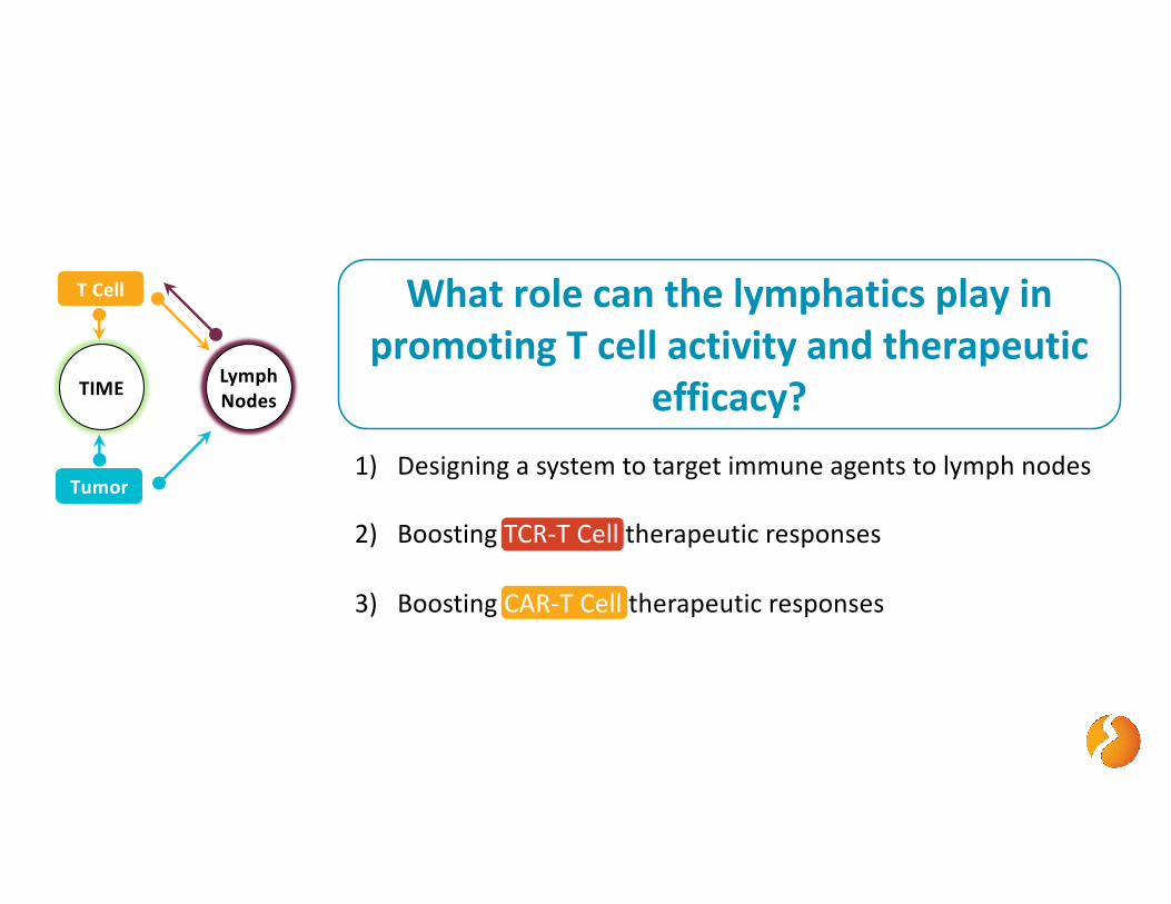
What role can the lymphatics play in promoting T cell activity and therapeutic
efficacy?
T Cell
Tumor
TIME Lymph Nodes
1) Designing a system to target immune agents to lymph nodes
2) Boosting TCR-T Cell therapeutic responses
3) Boosting CAR-T Cell therapeutic responses

The AMP Platform - Designing a Lymph Node Targeted AMPlifier for CAR-T Cell Therapy
30
(1) AMP-Peptide Peptide Surrogate Ligand
• Surrogate peptide ligand (mimotope) for CAR antigen binding domain
• AMP modification gives >10-fold improved lymph node targeting
(2) AMP-CpG Adjuvant
AMP Modification: Albumin Binding Lipid for Lymph Node Targeting
CpG DNA: TLR-9 Agonist
• Potent TLR-9 immuno-activator
• AMP modification gives >10-fold improved lymph node targeting
AMP Modification: Albumin Binding Lipid for Lymph Node Targeting
Peptide: Surrogate CAR LigandPEG Linker

The AMP Platform - Designing a Lymph Node Targeted Booster for CAR-T Cell Therapy
31
Surrogate Peptide as CAR Ligands
Ma, Irvine, et al. Science 2019
AMP-Peptide on Albumin
CAR T Cells
CARAMP / CAR
Binding
AMP-Decorated APC
CAR T Cell
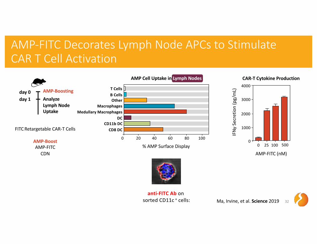
T CellsB CellsOther
Macrophages
DCCD11b DC
CD8 DC
0 20 40 60 80 100
% AMP Surface Display
Medullary Macrophages
AMP-FITC Decorates Lymph Node APCs to Stimulate CAR T Cell Activation
32
AMP Cell Uptake in Lymph Nodes
day 1 AnalyzeLymph NodeUptake
day 0 AMP-Boosting
anti-FITC Ab on sorted CD11c + cells: Ma, Irvine, et al. Science 2019
FITC Retargetable CAR-T Cells
AMP-BoostAMP-FITC
CDN
IFNγ
Secr
etio
n (p
g/m
L)
2000
3000
4000
1000
00 25 100 500
AMP-FITC (nM)
CAR-T Cytokine Production

AMP-Boosting Significantly Expands CAR-T Cells In Vivo
33
day 0day -1 CD45.1 CAR-T transfer
AMP-Boosting
day -2 Lymphodeplete
CD45.2 Recipient Mice
Blood Assay
FITC Retargetable CAR-T Cells
AMP-BoostAMP-FITC
CDN
day 7 AMP-Boosting
0
20
80
40
60
0 7 14 21 28Days post vaccination
10x106 CAR T50x103 CAR T + AMP-Boosting50x103 CAR T
Expansion of CAR-T Cells: Blood
Ma, Irvine, et al. Science 2019
% o
f CD3
+CD
8+T
cells
in
perip
hera
l blo
od
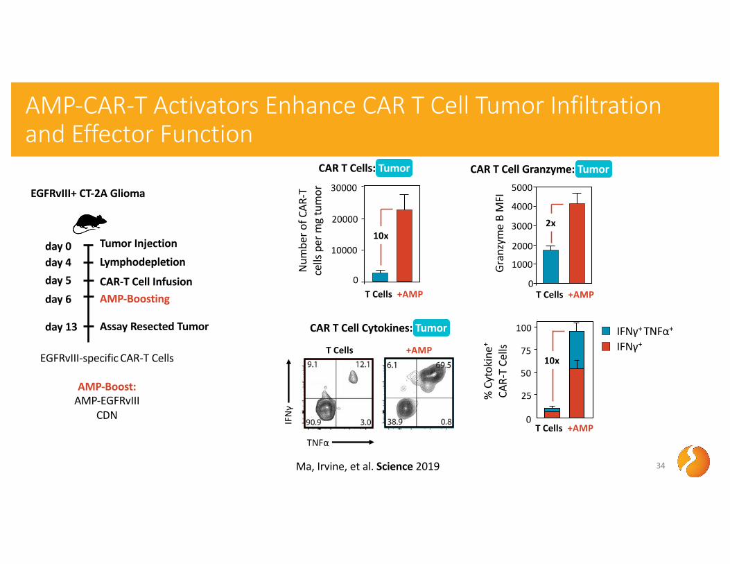
AMP-CAR-T Activators Enhance CAR T Cell Tumor Infiltration and Effector Function
34
day 5day 6
day 4 Lymphodepletion
AMP-BoostingCAR-T Cell Infusion
day 0 Tumor Injection
day 13 Assay Resected Tumor
EGFRvIII-specific CAR-T Cells
AMP-Boost:AMP-EGFRvIII
CDN IFN
γ
TNFα
% C
ytok
ine+
CAR-
T Ce
lls
0
25
50
75
100
IFNγ+IFNγ+ TNFα+
10x
CAR T Cell Cytokines: Tumor
T Cells +AMP
T Cells +AMP
Num
ber o
f CAR
-T
cells
per
mg
tum
or
0
10000
20000
30000
10x
CAR T Cells: Tumor
T Cells +AMP0
1000
2000
3000
4000
5000
Gran
zym
e B
MFI
2x
CAR T Cell Granzyme: Tumor
T Cells +AMP
Ma, Irvine, et al. Science 2019
EGFRvIII+ CT-2A Glioma
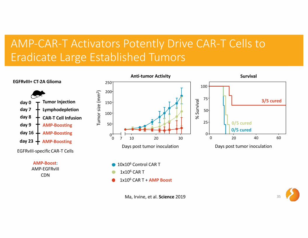
35
day 8day 9
day 7 Lymphodepletion
AMP-BoostingCAR-T Cell Infusion
day 0 Tumor Injection
day 16 AMP-Boosting
day 23 AMP-Boosting
Survival
EGFRvIII-specific CAR-T Cells
AMP-Boost:AMP-EGFRvIII
CDN
AMP-CAR-T Activators Potently Drive CAR-T Cells to Eradicate Large Established Tumors
Days post tumor inoculation
% S
urvi
val
0
50
100
20 40 600
25
75Tu
mor
size
(mm
2 )
0
50
200
100
150
0 7 10 20 30
250
Days post tumor inoculation
Anti-tumor Activity
1x10⁶ CAR T
1x10⁶ CAR T + AMP Boost
10x106 Control CAR T
3/5 cured
0/5 cured 0/5 cured
Ma, Irvine, et al. Science 2019
EGFRvIII+ CT-2A Glioma

AMP Boosted Responses Provide Durable Protection From Relapse and Antigen-loss
36
day 8day 9
day 7 Lymphodepletion
AMP-BoostingCAR-T Cell Infusion
day 0 Tumor Injection
day 16 AMP-Boostingday 23 AMP-Boosting
day 75 Tumor Injection
100% Protection
Tum
or si
ze (m
m2 )
0
50
200
100
150
0 4010 20 30
AMP-CAR-T CuredNaive
Challenged with EGFRvIII-expressing
tumor
Mediated by Memory EGFRvIII-CAR-T Response
AMP-CAR-T CuredNaive
Challenged with EGFRvIII-negative tumor
Mediated by Endogenous Memory T
cell Response
Days post tumor inoculation
Tum
or si
ze (m
m2 )
0
50
200
100
150
0 4010 20 30
EGFRvIII-specific CAR-T Cells
AMP-Boost:AMP-EGFRvIII
CDN
100% Protection
Ma, Irvine, et al. Science 2019
EGFRvIII+ CT-2A Glioma
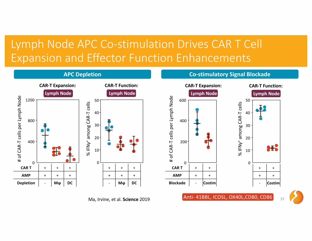
Lymph Node APC Co-stimulation Drives CAR T Cell Expansion and Effector Function Enhancements
37
# of
CAR
-T c
ells
per L
ymph
Nod
e
400
800
1200
CAR-T Expansion: Lymph Node
% IF
Nγ+
amon
g CA
R-T
cells
10
20
50
CAR-T Function: Lymph Node
30
40
APC Depletion Co-stimulatory Signal Blockade
Ma, Irvine, et al. Science 2019
CAR T + + +
AMP + + +
Depletion - Mϕ DC
0CAR T + +
AMP + +
Blockade - Costim
# of
CAR
-T c
ells
per L
ymph
Nod
e
200
400
600
CAR-T Expansion: Lymph Node
% IF
Nγ+
amon
g CA
R-T
cells
0
10
20
50
CAR-T Function: Lymph Node
30
40
Anti- 41BBL, ICOSL, OX40L,CD80, CD86
0+ +
+ +
- Costim
0+ + +
+ + +
- Mϕ DC

↑ Cytotoxic Activity
Promote T cell Fitness
↑ Clearance of Metastases
Overcome Tumor Exclusion
↑ TAA-targeting
Endogenous T Cells
Promote Tumor Recognition
↑ Lymph Node APC Activation
↑ T Cell Proliferation
in Tumor
↑ Tumor Penetration
↑ Effector Functionality
Promote T cell Proliferation
↑ Numbers in Blood and
Tumor ↑ Co-
stimulatory APC Support
?
?
?
Resist TIME Inhibition
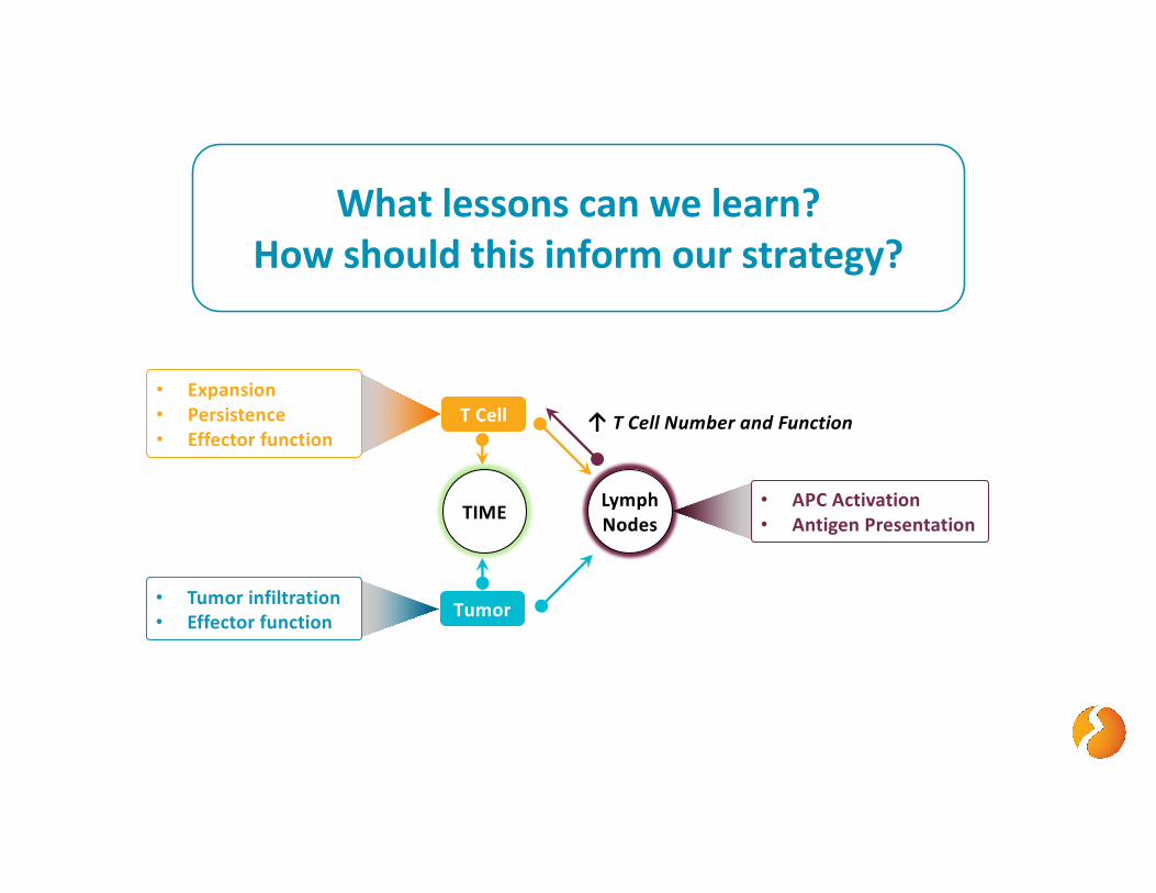
• Tumor infiltration• Effector function
T Cell
Tumor
TIME Lymph Nodes
What lessons can we learn?How should this inform our strategy?
↑ T Cell Number and Function
• Expansion• Persistence• Effector function
• APC Activation• Antigen Presentation
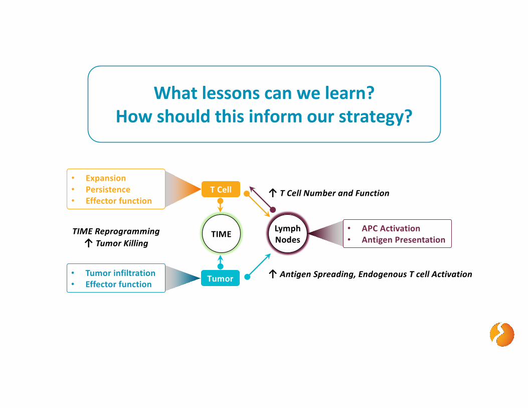
• Tumor infiltration• Effector function
T Cell
Tumor
TIME Lymph Nodes
What lessons can we learn?How should this inform our strategy?
↑ T Cell Number and Function
• Expansion• Persistence• Effector function
TIME Reprogramming↑ Tumor Killing
↑ Antigen Spreading, Endogenous T cell Activation
• APC Activation• Antigen Presentation
41
Dylan Drakes PhD, Abdul Abbas MS, Jackie Shields, Chris Haqq MD PhD
Darrell Irvine PhD, Leyuan Ma PhD
Ma, Irvine, et al. Science 2019

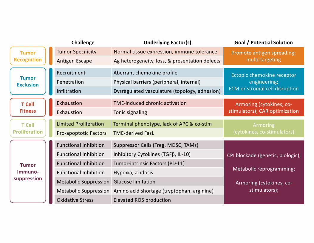
Recruitment Aberrant chemokine profile Ectopic chemokine receptor engineering;
ECM or stromal cell disruptionPenetration Physical barriers (peripheral, internal)
Infiltration Dysregulated vasculature (topology, adhesion)
Functional Inhibition Suppressor Cells (Treg, MDSC, TAMs)
CPI blockade (genetic, biologic);
Metabolic reprogramming;
Armoring (cytokines, co-stimulators);
Functional Inhibition Inhibitory Cytokines (TGFβ, IL-10)
Functional Inhibition Tumor-intrinsic Factors (PD-L1)
Functional Inhibition Hypoxia, acidosis
Metabolic Suppression Glucose limitation
Metabolic Suppression Amino acid shortage (tryptophan, arginine)
Oxidative Stress Elevated ROS production
Exhaustion TME-induced chronic activation Armoring (cytokines, co-stimulators); CAR optimizationExhaustion Tonic signaling
Tumor Specificity Normal tissue expression, immune tolerance Promote antigen spreading; multi-targetingAntigen Escape Ag heterogeneity, loss, & presentation defects
Limited Proliferation Terminal phenotype, lack of APC & co-stim Armoring (cytokines, co-stimulators)Pro-apoptotic Factors TME-derived FasL
Challenge Underlying Factor(s) Goal / Potential Solution
Tumor Recognition
Tumor Exclusion
T Cell Fitness
Tumor Immuno-
suppression
T Cell Proliferation
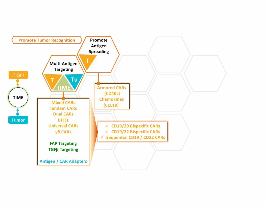
Promote Antigen
Spreading
T
T
Tu
Multi-Antigen Targeting
Promote Tumor Recognition
TIME
Mixed CARsTandem CARs
Dual CARsBITEs
Universal CARsγδ CARs
FAP TargetingTGFβ Targeting
Antigen / CAR Adapters
T Cell
Tumor
TIME
Armored CARs (CD40L)
Chemokines (CCL19)
ü CD19/20 Bispecific CARsü CD19/22 Bispecific CARs
ü Sequential CD19 / CD22 CARs

Local Delivery
Ectopic Chemokine &
Receptors
T
T
T TuTIME
ECM Disruption
Overcome Tumor Exclusion
TIME
Local Tumor InfusionSupportive Matrices
Hypoxia SensingProtease Sensing
CSF-1RCCR4
CCR2b
CCL19
FAP Targeting CARs
Heparinase
Hyaluronidase Combo
T Cell
Tumor
TIME

Cytokine / Co-stimulator
Armoring
T
T
CAR / TCR Optimization
Promote T cell Fitness
TIME
IL-12IL-15IL-18
CD40L41BB
IL-12IL-15IL-18
CD40L41BB
T Cell
Tumor
TIME
Affinity TuningCo-stimulatory Optimization
scFv SelectionHinge / TM Domain

Cytokine / Co-stimulator
Armoring
Infusion Product
Optimization
T
Promote T cell Proliferation
IL-12IL-1541BB
Kinase ConditioningCytokine Supplementation
Nutrient InclusionPhenotype Selection T
T Cell
Tumor
TIME

Cytokine / Co-stimulator
Armoring
Metabolic Reprogramming
CPI Combination
(genetic, biologic)
T
T
T
TuTIME
Resist TIME Inhibition
TIME
Glucose PathwayAA Supplementation
Catalase (ROS)
HIF Inhibition
CPI TransgeneDNR
Switch ReceptorsPD-1 KO
CPI Combo
TuTIME
IL-12IL-15IL-18
CD40L41BB
IL-12IL-15IL-18
CD40L41BB
T Cell
Tumor
TIME
ü HPV-E6 TCR +/- aPD-1 Transgeneü NY-ESO-1 TCR + TGFβ DNR
ü MSLN CAR + PD-1 KOü MUC-1 CAR + PD-1 KOü MSLN CAR + aPD-1ü CD19 CAR + aPD-L1

Local Delivery
Promote Antigen
Spreading
Cytokine / Co-stimulator
Armoring
Cytokine / Co-stimulator
Armoring
Cytokine / Co-stimulator
Armoring
Ectopic Chemokine Receptors
CPI Combination
(genetic, biologic)
Infusion Product
Optimization
T
T
T
T
T
T
T
T
T
T
T
T
Tu
Tu
Tu
Tu
TIME
TIME
TIMEECM
Disruption
CAR / TCR Optimization
Multi-Antigen Targeting
Promote T cell Fitness
Overcome Tumor Exclusion
Promote Tumor Recognition
Promote T cell Proliferation
Resist TIME Inhibition
TIME TIME
TIME
TIME
TuTIME
Metabolic Reprogramming

Promote T cell FitnessPromote Tumor Recognition
Promote T cell Proliferation
Resist TIME Inhibition
Overcome Tumor Exclusion

-2
-1
0
1
2
AMP-boosting Induces Coordinated Expression of Costimulation, Cytokine and Trafficking Genes without Negative Coregulation
9/9/21 51
T Cell Activation &Co-stimulation
• CD40• CD86 TLR
SignalingIFN-II
Signaling• STAT2• IL-12
ChemokineSignaling• CCL12• CXCL11• CCL2
Granzyme
Regulation of T cell Proliferation
• IL-15Ra
ADCC
MHCIPresentation• TAP1
MHCIIPresentation
T cell Anergy & Negative Activation
• FoxP3• CTLA-4• TIGIT
Z-score
D Drakes, PhD, A Abbas, MS, J Shields
Gene Expression: Lymph Node, Day 7T cells Boost
- AMP
5x10⁶ -
5x10⁶ AMP
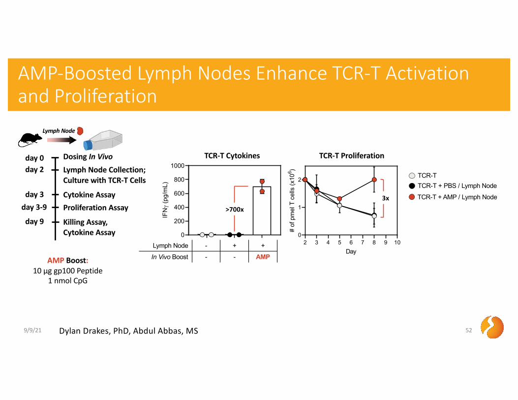
AMP-Boosted Lymph Nodes Enhance TCR-T Activation and Proliferation
9/9/21 52
0
200
400
600
800
1000IF
Ng
(pg/
mL)
TCR-T Cytokines
day 3-9
day 2 Lymph Node Collection;Culture with TCR-T Cells
Proliferation Assay
day 0 Dosing In Vivo
day 9 Killing Assay, Cytokine Assay
day 3 Cytokine Assay
Lymph Node - + +
In Vivo Boost - - AMP
2 3 4 5 6 7 8 9 100
1
2
Day
# of
pm
el T
cel
ls (x
106 )
TCR-T Proliferation
TCR-T TCR-T + PBS / Lymph Node
TCR-T + AMP / Lymph Node
Dylan Drakes, PhD, Abdul Abbas, MS
AMP Boost:10 µg gp100 Peptide
1 nmol CpG
>700x
Lymph Node
3x

AMP-Boosted Lymph Nodes Enhance TCR-T Killing and Cytokine Secretion In Culture with Tumor Cells
9/9/21 53
2.1 1.1 1.2 1.5 1.10 1.200
10
20
30
40
50
Effector:Target Ratio
Perc
ent L
ysis
TCR-T Tumor Killing
0
500
1000
1500
IFNg
(pg/
mL)
0
100
200
300
400
IL-2
(pg/
mL)TCR-T
TCR-T + PBS / Lymph Node
TCR-T + AMP / Lymph Node
TCR-T: IFNγ
TCR-T: IL-2
Lymph Node - + +
In Vivo Boost - - AMPDylan Drakes, PhD, Abdul Abbas, MS
day 3-9
day 2 Lymph Node Collection;Culture with TCR-T Cells
Proliferation Assay
day 0 Dosing In Vivo
day 9 Killing Assay, Cytokine Assay
day 3 Cytokine Assay
AMP-Boost:10 µg gp100 Peptide
1 nmol CpG
6x
33x
Lymph Node
>40x
9/9/21 Elicio Confidential 54
AMP-Boosting in the Lymph Nodes Enhances Activation and Functionality of TCR-T Cells
Murine Syngeneic Pmel TCR T Cell / B16F10 Melanoma
• Lymph Nodes from AMP-boosted mice potently stimulate TCR-T Cells
• Enhanced proliferation
• 700-fold enhanced cytokine secretion
• Enhanced functional and cytotoxic interaction with tumor targets
• Increased killing efficiency
• >6-30-fold increased cytokine secretion