Approach to the management of hyperbilirubinemia in...
50
Approach to the management of Hyperbilirubinemia in Term Newborn Infant Mohammad Bagher Hosseini MD Neonatologist Assosiated professor of Tabriz University of Medical science May 2011
-
Upload
nguyenkhue -
Category
Documents
-
view
218 -
download
0
Transcript of Approach to the management of hyperbilirubinemia in...

Approach to the management of
Hyperbilirubinemia in Term
Newborn Infant
Mohammad Bagher Hosseini MD
Neonatologist
Assosiated professor of Tabriz University of Medical science
May 2011

Case1
• You are called by the ER to see an infant
whose bili is 22.
• Must you admit?
• What information do you need to answer
this question?

Outline
• Review of physiology
• Kernicterus
• Risk factors
• Assessing the risk
• Therapies

Neonatal Hyperbilirubinemia
• Definition =
• Clinical ;
• Lab : (TSB) > 5 mg/dL
• Significance:
– Present in up to 60% of term newborns
– Severe complications possible• Deafness, CP (kirnicterus)
– Increase Kirnicterus 1990’s (related to early hospital discharge)

Recent concern
• JACHO alert due to several case
reports of kernicterus in healthy
newborns
• Term 35-38 weeks, dehydrated
breastfeeding, and with extremely
high bilirubin levels

Bilirubin Production
& Metabolism

Classification
• Benign
– Physiologic
– Breast Milk
– Breastfeeding
• Pathologic
– Many causes

Physiologic Jaundice
• Features– Elevated unconjugated bilirubin
– TSB generally peaks @ 5-6 mg/dL on day 3-4
and then declines to adult levels by day 10
• Asian infants peak at higher values (10 mg/dL)
– Exaggerated physiologic (up to 17 mg/dL)

Physiologic Jaundice
Asian infant
Breastfed infantNon-breastfed infant

Ethnic differences
• Exaggerated Hyperbilirubinemia (>12.8mg/dl)
– 4% African-Americans
– 6-10% Caucasian
– 25% Asian (>20mg% in 2%)

Effect of Type of
Feeding
• 2/3 of breastfeeding infants (BF) will have
chemical jaundice for 2-3 weeks
• TSB > 12mg% in 12% (BF) vs. 4%
Formula Fed infants (FF)
• TSB > 15mg% in 2% BF vs. 0.3% FF

Mechanism of Physiologic
Jaundice
Increased rbc’s
Shortened rbc lifespan
Immature hepatic uptake & conjugation
Increased enterohepaticCirculation

Breast feeding
Jaundice
• Elevated unconjugated bilirubin
• Benign or pathologic
– Elevated bilirubin in the 1st week of life tends to
worsen breast milk jaundice during later weeks
• Equivalent to starvation jaundice in adults
• Mandates improved/increased breastfeeding
– No water or dextrose supplementation
– Formula OK( Sometime , No bottle )

Breast Milk Jaundice
• Elevated unconjugated bilirubin
• Prolongation of physiologic jaundice– Slower decrease to adult
levels of bilirubin• 66% of breastfed babies
jaundiced into 3rd week of life
• May persist up to 3 months
– May have second peak @ day 10
• Average max TSB = 10-12 mg/dL
• TSB may reach 22-24 mg/dL
• ?Milk factor

BREASTMILK/BREASTFEEDING
JAUNDICE
• Breastfeeding jaundice occurs early
• It is due to the lack of breast milk
• It is often associated with poor passage of
meconium.
• Treatment should be aimed at supporting
breastfeeding while supplementing as
needed to avoid extreme weight loss,
dehydration, and worsening jaundice.

BREASTMILK/BREASTFEEDING
JAUNDICE
• Breast milk jaundice is a different, more benign entity, which tends to occur late in the first week or afterwards.
• It is actually due to something in the breast milk which tends to prolong jaundice.
• Usually weight gain is good, and the baby is otherwise well.
• Jaundice might persist as late as 3-4 weeks, but usually will peak by 2 weeks.
• Textbook treatment is to interrupt breastfeeding (I usually do not do this).

Pathologic Jaundice
• Features
– Jaundice in 1st 24 hrs
– Rapidly rising TSB (> 5
mg/dL per day)
– TSB > 17 mg/dL
• Categories
– Increased bilirubin load
– Decreased conjugation
– Impaired bilirubin excretion

Increased
Bilirubin Load
Hemolytic Disease
– Features: elevated reticulocytes, decreased Hgb
– Coomb’s + Rh incompatibility, ABO incompatibility, minor antigens
– Coomb’s - G6PD, spherocytosis, pyrovate kinase deficiency

Pathologic Jaundice
• Non-hemolytic Disease
– normal reticulocytes
– Extravascular sources – I.e.
cephalohematoma
– Polycythemia
– Exaggerated enterohepatic circulation –
I.e. CF, GI obstruction

G6PD Deficiency
• A cause of kernicterus in up to 35% of cases
• Always suspect if severe hyperbili or poor response to phototherapy
• Ethnic origin– 11-13% of African Americans
– Mediterranean, Middle East, Arabian peninsula, SE Asia, Africa
• Requires intervention at lower TSB levels
• Testing– Levels may be normal or elevated early
• Especially in presence of hemolysis
– Repeat level at 3 months

Decreased Bilirubin Conjugation
• Elevated unconjugated bilirubin
• Genetic Disorders
– Crigler-Najjar
• 2 types
• Severe hyperbilirubinemia
– Gilbert Syndrome
• Mild hyperbilirubinemia
• Hypothyroidism

Impaired Bilirubin Excretion
• Elevated unconjugated and conjugated bilirubin (> 2 mg/dL or > 20% of TSB)
• Biliary Obstruction– Structural defects – I.e. biliary atresia
– Genetic defects – Rotor’s & Dubin-Johnson syndromes
• Infection – sepsis, TORCH
• Metabolic Disorders – I.e. alpha1 antitrypsin deficiency
• Chromosomal Abnormalities – Turner’s syndrome
• Drugs – I.e. ASA, sulfa, erythromycin

Diagnosis & Evaluation
• Physical Exam
– Bilirubin > 5 mg/dL
– Milder jaundice - face & upper thorax
– Caudal progression generally signifies higher bilirubine levels
• Should not rely on this system
• Liberally check bilirubin values
• Laboratory
– Blood
– Transcutaneous
• Generally within 2mg/dL of serum test
• Most useful if serum bili < 15

Physical Exam
•Poor correlation inter-observer and with serum bilirubin
• Best cut appears to be jaundice to nipples for bili > 12.0 mg/dl
• 97% sensitive
• 19% specificArch Pediatr Adolesc Med. 2000; 154:391-4
• Zone 1 head - clavicle 5
• Zone 2 clavicle-umbilicus 6-8
• Zone 3 umbilicus-knee 9-12
• Zone 4 knees-ankles 13-15
• Zone 5 palms + soles 15
• Clinical Exam: Unreliable
• Clinical Exam: Unreliable

2004 AAP Guidelines
Management of Hyperbilirubinemia in the
Newborn Infant 35 or More Weeks of
Gestation
Subcommittee on HyperbilirubinemiaPediatrics 2004; 114;297-316

Prevention
• Breastfeeding
– Should be encouraged for most women
• Separate AAP guidelines
– 8-12 times/day for 1st several days
– Assistance and education
– Avoid supplements in non-dehydrated
infants
• Do not decrease level & severity of hyperbili

Prevention
• Ongoing assessments for risk of
developing severe hyperbilirubinemia
– Monitor at least every 8-12 hours
– Don’t rely on clinical exam
– Blood testing
• Prenatal (Mom): ABO & Rh type, antibody
• Infant cord blood
– Mom not tested, Rh (-): Coomb’s, ABO, Rh
– Mom O or Rh (+): optional to test cord blood

Laboratory investigation
• Indicated (if bilirubin concentrations reach phototherapy levels) – Serum total or unconjugated bilirubin concentration
Serum conjugated bilirubin concentration
Blood group with direct antibody test (Coombs’ test)
Hemoglobin and hematocrit determinations
• Optional (in specific clinical circumstances)
Complete blood count including manual differential white cell count – Blood smear for red cell morphology
– Reticulocyte count
– Glucose-6-phosphate dehydrogenase screen
• Serum electrolytes and albumin or protein concentrations
Copyright ©2004 American Academy of Pediatrics
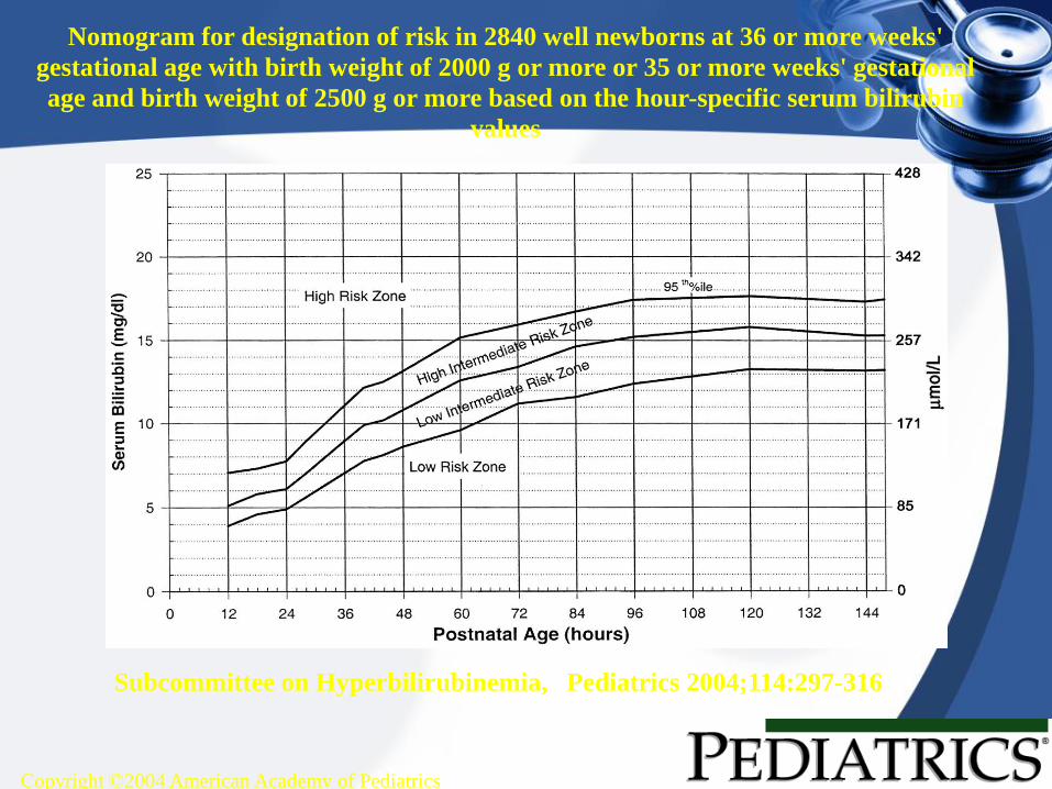
Subcommittee on Hyperbilirubinemia, Pediatrics 2004;114:297-316
Nomogram for designation of risk in 2840 well newborns at 36 or more weeks'
gestational age with birth weight of 2000 g or more or 35 or more weeks' gestational
age and birth weight of 2500 g or more based on the hour-specific serum bilirubin
values

ASSESSING THE RISK OF
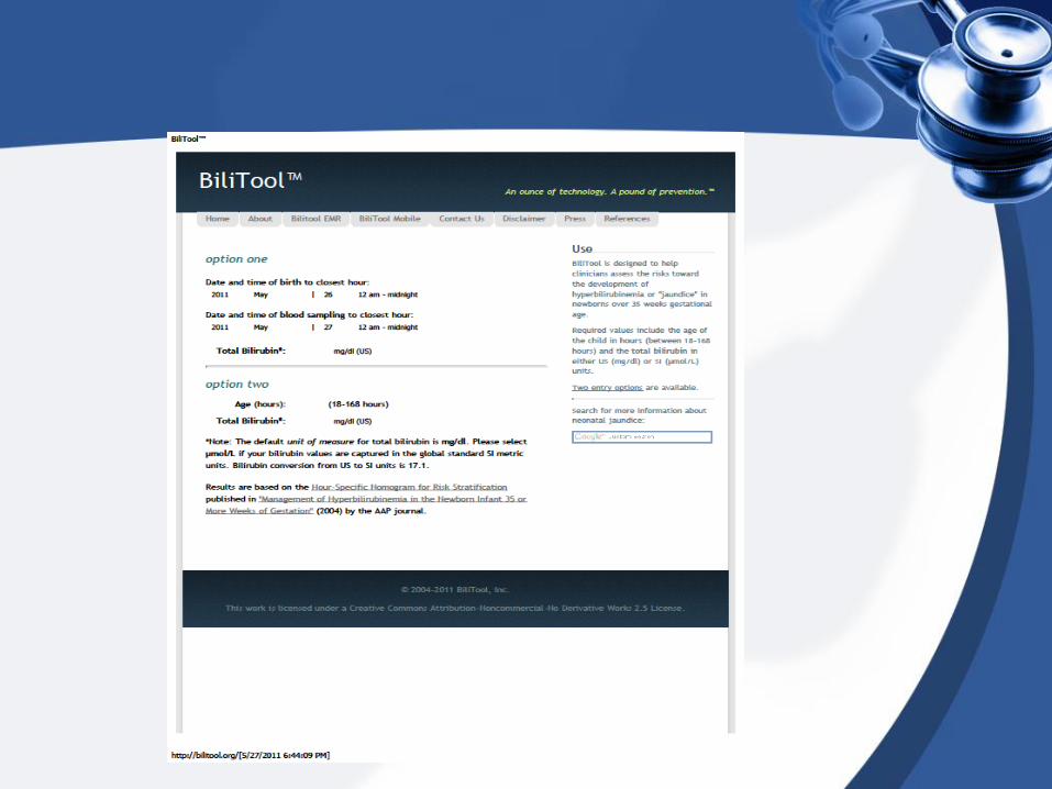
JAUNDICE BY THE NUMBERS
•www.bilitool.org
• Palm downloadable!

Risk Factors for Severe Hyperbilirubinemia
• Major risk factors– Predischarge bili in high-risk
zone
– Jaundice in 1st 24 hrs
– Blood group incomp with + direct antiglobulin test, other known hemolytic disease (eg, G6PD deficiency)
– Gestational age 35–36 wk
– Previous sibling received phototherapy
– Cephalohematoma or significant bruising
– Exclusive breastfeeding
– East Asian race
• Minor risk factors– Bili in high intermed-risk zone
– Gestational age 37–38 wk
– Jaundice before discharge
– Previous sibling with jaundice
– Macrosomia infant with diabetic mother
– Maternal age ≥ 25
– Male
• Decreased Risk– Bili in low-risk zone
– ≥ 41 wks gestation
– Exclusive bottle feed
– Black race
– D/c from hospital > 72hrs

Discharge
TSB Zone Newborns
(%)
% with TSB
>95th %
High risk 6 39.5
High intermed 12.5 12.9
Low intermed 19.6 2.26
Low 61.8 0
• Assess risk
– Predischarge bili
• Use nomogram to determine risk zone
– And/or Assessment of risk factors

Discharge
Infant
Discharge
Should be Seen
by
< 24 hours 72 hours
24-48 hours 96 hours
48-72 hours 120 hours
• Close follow-up necessary
– Individualize based on risk
– Weight, % change from BW, intake, voiding
habits, jaundice
Copyright ©2004 American Academy of Pediatrics
Subcommittee on Hyperbilirubinemia, Pediatrics 2004;114:297-316
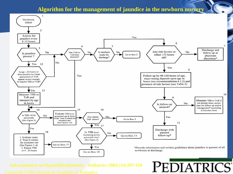
Algorithm for the management of jaundice in the newborn nursery

Phototherapy
• Mechanism: converts bilirubin to water
soluble form that is easily excreted
• Forms
– Fluorescent lighting
– Fiberoptic blankets
• Goal is to decrease TSB by 4-5 mg/dL or <
15 mg/dL total
• Breastfed infants are slower to recover

Phototherapy
• Severe rebound hyperbilirubinemia is
rare
– Average increase is 1 mg/dL
• Intensive
– Special blue tube with light in blue-green
spectrum
– Close to infant
– Expose maximum surface area

Copyright ©2004 American Academy of Pediatrics
Subcommittee on Hyperbilirubinemia, Pediatrics 2004;114:297-316
Guidelines for phototherapy in hospitalized infants of 35 or more weeks' gestation

Exchange Transfusion
• Mechanism: removes bilirubin and
antibodies from circulation and correct
anemia
• Most beneficial to infants with hemolysis
• Generally never used until after
intensive phototherapy attempted

Complications
• Toxicity to Basal Ganglia and brainstem nuclei
• 2 terms– Acute bilirubin encephalopathy
– Kernicterus
• Multiple phases

Risk of Kirnicterus
• TSB level > 25-30 mg/dl
• Acidosis
• Increased free bilirubin
• low albumin, drug displacement
• Blood-brain barrier disruption
• prematurity, sepsis, ischemia

Kernicterus cases
potentially correctable causes
• Early discharge (<48hrs) without f/u within 48 hrs
• Failure to check bilirubin level if onset in first 24 hours
• Failure to note risk factors
• Visual assessment underestimate of severity
• Delay in testing jaundiced newborns or treating elevated levels
• Lack of concern for presence of jaundice or parental concern
• Pediatrics 2001; 108:763-765

Common Clinical Risk Factors
for Severe Hyper-bilirubinemia
• Jaundice in the first 24 hours
• Visible jaundice at discharge
• Previous jaundiced sibling
• Near term gestation 35-38 weeks
• Exclusive breastfeeding
• East Asian (4), Mediterranean (1), African origin (12) (G6PD deficiency), 19/61 kernicterus cases = G6PD
• Bruising, cephalohematoma, birth trauma
• Hemolysis risk, O + maternal blood type, sepsis

Medications increasing
bilirubin toxicity
• Sulfisoxazole (displacement or G6PD
hemolysis)
• Ceftriaxone (displacement from
albumin)

Trans cutaneous bilirubin
• Older devices affected by skin pigmentation
• Newer multi-wavelength spectral reflectance
correlate 0.88 with the serum value,
• example SpectRx, ± 3 mg/dl
• ? Confirm values > 40% per age
• Carbon monoxide exhaled

Direct Coombs Testing
Strongly positive:
– Rh
– Kell
– Kidd
– Duffy
• Negative or “weakly positive:
– Anti-A

Hemolysis consider present
• Hct < 45%
• Abnormal blood smear with 3-4+
spherocytes
• Reticulocyte count is 4.5% in the first
72 hrs, or
• Reticulocyte count is >1-2% in the first
1-2 wks

Review of Case 2
• How old is the patient?
• What is the fractionation?
• Breast or bottle fed?
• Other risk factors?
– 10 days
– 22 total / 0.8 direct
– Breast fed
– None

QUESTIONS?

References
• American Academy of Pediatrics, Subcommittee on Hyperbilirubinemia. Management of hyperbilirubinemia in the newborn infant 35 or more weeks of gestation. Pediatrics. 2004;114:297-316
• Johnson LH, Bhutani VK, Brown AK. System-based approach to management of neonatal jaundice and prevention of kernicterus. J Pediatr. 2002;140:396-403
• American Academy of Pediatrics, Steering Committee on Quality Improvement and Management. Classification of recommendations for clinical practice guidelines. Pediatrics. 2004;114:874-877
• Gartner LM, Herschel M. Jaundice and breastfeeding. Pediatr Clin North Am. 2001;48:389-399
• Moyer VA, Ahn C, Sneed S. Accuracy of clinical judgment in neonatal jaundice. Arch Pediatr Adolesc Med. 2000;154:391-394
• Ip S, Glicken S, Kulig J, Obrien R, Sege R, Lau J. Management of Neonatal Hyperbilirubinemia. Rockville, MD: US Department of Health and Human Services, Agency for Healthcare Research and Quality; 2003. AHRQ Publication 03-E011
• Bhutani VK, Johnson LH, Sivieri EH. Predictive ability of a predischarge hour-specific serum bilirubin for subsequent hyperbilirubinemia in healthy term and near-term newborns. Pediatrics. 1999;103:6-14.
• American Academy of Pediatrics, Subcommittee on Neonatal Hyperbilirubinemia. Neonatal jaundice and kernicterus. Pediatrics. 2001;108:763-765