Applied Surface Science 424, 275-281 (2017)
16
1 Applied Surface Science 424, 275-281 (2017) Mesoporous silica obtained with methyltriethoxysilane as co-precursor in alkaline medium Ana-Maria Putz 1 , Kunzhou Wang 2 , Adél Len 3 , Jiri Plocek 4 , Petr Bezdicka 4 , Gennady P. Kopitsa 5,8 , Tamara V. Khamova 5 , Cătălin Ianăşi 1 , Liviu Săcărescu 6 , Zuzana Mitróová 7 , Cecilia Savii 1 , Minhao Yan *2 and László Almásy *2,3 1 Institute of Chemistry Timisoara of Romanian Academy, Laboratory of Inorganic Chemistry, Bv. Mihai Viteazul, No.24, RO-300223 Timisoara, Romania 2 State Key Laboratory Cultivation Base for Non-metal Composites and Functional Materials, Southwest University of Science and Technology, Mianyang 621010, China. 3 Wigner Research Centre for Physics, Institute for Solid State Physics and Optics, Hungarian Academy of Sciences, Budapest, Hungary 4 Institute of Inorganic Chemistry, Academy of Sciences of the Czech Republic, v.v.i, Husinec-Řež 1001, 25068 Řež, Czech Republic 5 Institute of Silicate Chemistry of RAS, nab. Makarova 2, 199034 St. Petersburg, Russia 6 Institute of Macromolecular Chemistry „Petru Poni” Iaşi, Romania 7 Institute of Experimental Physics, SAS, Watsonova 47, 04001 Kosice, Slovakia 8 Petersburg Nuclear Physics Institute, Gatchina, Russia * Corresponding author [email protected] and [email protected] Abstract Mesoporous silica particles have been synthesized by sol-gel method from tetraethoxysilane (tetraethylorthosilicate, TEOS) and methyltriethoxysilane (MTES), in ethanol and water mixture, at different ratios of the of the silica precursors. Ammonia was used as catalyst at room temperature and hexadecyltrimethylammonium bromide (cetyltrimethylammonium bromide, CTAB) as the structure directing agent. Nitrogen sorption, X-ray diffraction and small-angle neutron scattering gave information on the evolution of the gel structure and pore morphologies in the function of MTES/TEOS molar ratio. Thermogravimetric and differential thermal analysis showed that with addition of MTES the exothermic peak indicating the oxidation of the low molecular weight organic fragments shift to higher temperature. A room-temperature, one-pot synthesis of MCM-41 type materials is presented, in which the variation of the MTES concentration allows to change the hydrophobicity, preserving the specific properties materials, like the ordered pore structure, large specific surface area and high porosity, making them suitable for selective uptake of guest species in drug loading applications. Specifically, the obtained materials had cylindrical pores, specific surface areas up to 1101 m 2 /g and total pore volumes up to 0.473 cm 3 /g. The obtained mesoporous materials are susceptible for further functionalization to improve their selective uptake of guest species in drug delivery applications. Keywords: Sol–gel process; MTES/TEOS; Hydrophobic silica; MCM-41
Transcript of Applied Surface Science 424, 275-281 (2017)

1
Applied Surface Science 424, 275-281 (2017)
Mesoporous silica obtained with methyltriethoxysilane as co-precursor in alkaline medium
Ana-Maria Putz1, Kunzhou Wang
2, Adél Len
3, Jiri Plocek
4, Petr Bezdicka
4 , Gennady P.
Kopitsa5,8
, Tamara V. Khamova5, Cătălin Ianăşi
1, Liviu Săcărescu
6, Zuzana Mitróová
7, Cecilia
Savii1, Minhao Yan
*2 and László Almásy
*2,3
1Institute of Chemistry Timisoara of Romanian Academy, Laboratory of Inorganic Chemistry, Bv. Mihai
Viteazul, No.24, RO-300223 Timisoara, Romania 2State Key Laboratory Cultivation Base for Non-metal Composites and Functional Materials, Southwest
University of Science and Technology, Mianyang 621010, China. 3Wigner Research Centre for Physics, Institute for Solid State Physics and Optics, Hungarian Academy of Sciences,
Budapest, Hungary 4Institute of Inorganic Chemistry, Academy of Sciences of the Czech Republic, v.v.i, Husinec-Řež 1001, 25068 Řež,
Czech Republic 5Institute of Silicate Chemistry of RAS, nab. Makarova 2, 199034 St. Petersburg, Russia
6Institute of Macromolecular Chemistry „Petru Poni” Iaşi, Romania
7Institute of Experimental Physics, SAS, Watsonova 47, 04001 Kosice, Slovakia
8Petersburg Nuclear Physics Institute, Gatchina, Russia
*Corresponding author [email protected] and [email protected]
Abstract
Mesoporous silica particles have been synthesized by sol-gel method from tetraethoxysilane
(tetraethylorthosilicate, TEOS) and methyltriethoxysilane (MTES), in ethanol and water mixture,
at different ratios of the of the silica precursors. Ammonia was used as catalyst at room
temperature and hexadecyltrimethylammonium bromide (cetyltrimethylammonium bromide,
CTAB) as the structure directing agent. Nitrogen sorption, X-ray diffraction and small-angle
neutron scattering gave information on the evolution of the gel structure and pore morphologies
in the function of MTES/TEOS molar ratio. Thermogravimetric and differential thermal analysis
showed that with addition of MTES the exothermic peak indicating the oxidation of the low
molecular weight organic fragments shift to higher temperature. A room-temperature, one-pot
synthesis of MCM-41 type materials is presented, in which the variation of the MTES
concentration allows to change the hydrophobicity, preserving the specific properties materials,
like the ordered pore structure, large specific surface area and high porosity, making them
suitable for selective uptake of guest species in drug loading applications. Specifically, the
obtained materials had cylindrical pores, specific surface areas up to 1101 m2/g and total pore
volumes up to 0.473 cm3/g. The obtained mesoporous materials are susceptible for further
functionalization to improve their selective uptake of guest species in drug delivery applications.
Keywords: Sol–gel process; MTES/TEOS; Hydrophobic silica; MCM-41

2
Introduction
Monodisperse spherical silica particles can be synthesised by the Stöber method, starting from
tetraalkoxysilane in water and alcohol as solvent, and ammonia catalyst [1]. Mesoporous silica
nanoparticles with surfactant templated pores became well known since their synthesis by
Kresge and co-workers in Mobil Corporation laboratories in the early 90's [2]. MCM-41 and
related, surfactant templated materials are notable for their morphological properties such as
large surface area, high porosity, and controllable mesopores with narrow size distribution [3].
Spherical, mesoporous MCM-41 like particles were first synthesized and reported by Grun,
Lauer and Unger, modifying the Stöber method by adding the cationic surfactant [4]. Regarding
the order, the regularity and the thermal stability of the structure, the MCM-41 type materials
obtained in basic medium at room temperature, usually are qualitatively inferior to those
obtained by hydrothermal synthesis [5]. In the ordinary conditions of temperature and pressure,
lower condensation grade of the silica can be reached [6,7]. On the other hand, the low
temperature sol-gel method is cheaper and advantageous as scalable technology. The presence of
alcohol in the reaction mixture, as mutual solvent of water and alcoxide, facilitates
homogenisation, favouring formation of spherical MCM-41 [8,9]. In surfactant templated
spherical mesoporous silica, the channels are usually closed at one end, and the typical
hexagonal long range order is much weaker compared to the classical MCM-41 materials [10].
For applications in adsorption and catalysis, the variation of surface hydrophobicity is
required. Surface modifications of mesoporous materials are generally performed by two
common ways: post-synthetic method and direct co-condensation method [11]. The latter
involves a one-step co-condensation between tetraalkoxysilanes (Si(OR)4) with one or more
organoalkoxysilanes (RSi(OR)3) through the sol-gel process in presence of a structure directing
agent [12,13]. Inagaki and co-workers reported for the first time the synthesis of ordered
mesoporous silica with organic groups evenly incorporated in the walls (so-called periodic
mesoporous organosilicas, PMOs) using silsesquioxane precursor 1,2-bis(trimethoxysilyl) ethane
as the silica source and octadecyltrimethylammonium chloride as the structure directing agent,
under basic reaction conditions [14].
Organic modification of the silicate allows precise control over the surface properties and
pore sizes of the mesoporous sieves and at the same time stabilizing the materials towards
hydrolysis. Using organically modified precursors, like methylalkoxysilanes, the hydrophobicity
of the materials can be changed in an easily controllable way. Various examples for using
methyltriethoxysilane (MTES) as a hydrophobic reagent for xerogel preparation were presented
in the literature attempting to achieve superhydrophobic surfaces. The character of the sol–gel
derived hybrid film surface shifts from hydrophilic to superhydrophobic due to the incorporation
of CH3 groups in the silica surface from MTES precursor [15]. An enhancement of the surface
hydrophobicity is retarding the leaching [7] and improves also the hydrothermal stability [16]. S-
shape leaching curves have been obtained with an induction time increasing with the MTES

3
content, while the more hydrophilic samples prepared without MTES exhibited immediate rapid
release [16].
In a recent study, ordered mesoporous silica was synthesized at 60 ºC with NaOH
catalyst, by using MTES together with TEOS, as silica precursors, in different molar ratio,
without adding alcohol in the reaction mixture. It has been shown that he increased amount of
MTES destroyed the mesoporous channels and the materials became amorphous at 30% MTES
content [17].
In our present work, we prepared mesoporous particles, by room temperature sol-gel
synthesis, using tetraethyl orthosilicate (TEOS) and methyltriethoxysilane (MTES) at various
molar ratios, a cationic surfactant as templating agent, ammonia catalyst and adding alcohol into
the reaction mixture. Spherical mesoporous particles of MCM-41 type have been obtained in a
cost-effective way in one pot synthesis at room temperature. The hydrophobicity of the pore
walls could be efficiently tuned varying the proportion of the hydrophobic silica precursor.
Template removal was carried out using two methods: by calcinations and by extraction with
solvent, and the morphology of the resulted materials has been characterized by several
experimental techniques.
2. Experimental
2.1 Synthesis
All chemicals were commercially available: Tetraethyl orthosilicate (TEOS), (99%, for
analysis, Fluka); Methyltriethoxysilane (MTES 97%, Merck); Ethanol Absolute (Riedel de
Haen); NH4OHaq (25%, SC Silal Trading SRL); Hexadecyltrimethyl-ammonium bromide
(CTAB, Sigma).
The reactants molar ratio was: (TEOS+MTES): Ethanol: H2O: NH3= 1:58:144:11. The
molar ratio Si:CTAB was kept constant at 0.3 and through the series where TEOS was partially
replaced with MTES. Typically, 4.92 g CTAB were dissolved in a mixture of 116.64 mL
distilled water and 152.4 mL of ethanol. Subsequently 37.1 mL of ammonia 25% were added;
the mixtures were vigorously magnetic stirred in a closed vessel at room temperature for 30 min.
Then, the sol–gel precursors (10 mL TEOS or TEOS+MTES mixed in advance) were slowly
dripped (in 10 minutes) into the reaction mixture and the resulting mixture was vigorously stirred
for 2 h (rotation speeds 250 rpm). The formed gel was aged for 16 h, in static condition. In the
next day, the resulted suspension was several times washed with distilled water and then
centrifuged at 9000 rpm (2 times, 5 minutes each and two times, 10 minutes each) until the pH of
the supernatant approached neutral value. To the decanted precipitate, ca. 110 mL ethanol was
added and left for 48 hours; then, the samples were dried at 40 ºC for 1 hour, and then at 60 ºC
for 21 hours. These xerogel samples were labelled as Cx-60 (Table 1.).
Template removal was carried out using two methods: by calcination and by extraction
with ethanol.
For calcination, a part of each dried sample has been further heated up to 550 °C and kept
for 5 hours, and were labeled as Cx-550 (Table 1).

4
In the extraction procedure, 1g of Cx-60 samples were mixed with 200 ml of acidified
ethanol (1ml of concentrated HCl: 100 ml of ethanol) under magnetic stirring (350 rot/min) for
one hour, at room temperature following the slightly modified method of Du et al. [18]. Then,
the samples were dried at 60 ºC for 12 hours. These samples were labeled as Cx-EtOH (Table 1).
Table 1. Sample names and synthesis conditions
MTES / TEOS
molar ratio
Drying at 60 ºC Calcination at 550 ºC Template extraction
by ethanol
0 / 100 C0-60 C0-550 C0-EtOH
10 / 90 C1-60 C1-550 C1-EtOH
20 / 80 C2-60 C2-550 C2-EtOH
30 / 70 C3-60 C3-550 C3-EtOH
40 / 60 C4-60 C4-550 C4-EtOH
50 / 50 C5-60 C5-550 C5-EtOH
2.2. Characterization
FTIR spectra were taken on KBr pellets with a JASCO –FT/IR-4200 apparatus.
Measurements of specific surface area of the composite silica samples were performed by low
temperature nitrogen adsorption using QuantaChrome Nova 1200e analyzer. Before
measurements the samples were outgassed at 120 °C for 17÷18 hours in vacuum. Determination
of the surface area SBET (m2/g) was carried out by Brunauer–Emmett–Teller (BET) method.
When the BET plot produces either a too large, or negative C constant, the Langmuir surface
area may be considered [19]. The total pore volume is measured at a relative pressure of
P/P0=0.99. The pore diameter was calculated from adsorption by BJH method. Pore size
distribution was calculated from nitrogen adsorption isotherms by DFT, assuming cylindrical
pore shape and NLDFT model.
Transmission Electron Microscopy (TEM) analysis have been carried out with Hitachi High-
Tech HT7700 instrument, operated in high resolution mode at 100 kV accelerating voltage.
Samples have been prepared by drop casting from diluted dispersions of nanoparticles in ethanol
on 300 mesh holey carbon coated copper grids (Ted Pella) and vacuum dried.
Small-angle neutron scattering (SANS) measurements were performed on the Yellow Submarine
instrument at the BNC in Budapest, Hungary [20]. Neutron wavelength of 5 and 10Å, and
sample-detector distances 1.3 and 5.5 m were used; the measurements were carried out at room
temperature. Preliminary SANS experiments were conducted on Suanni SANS instrument
installed at CMRR, Mianyang, China [21]. X-ray powder diffraction (XRD) measurements were
performed on Panalytical X’Pert Pro MPD diffractometer equipped with Cu-anode (Cu Kα;

5
λ=1.54181Å) and X’Celerator detector. Measurements were done at room temperature in
transmission mode with Mylar foil in range from 0.65 to 10° in 2θ.
The thermogravimetric analysis was carried out between 25 °C and 800 °C using a 851-LF 1100-
Mettler Toledo apparatus in air flow and a heating rate of 5 °C min-1
.
3. Results and Discussion
3.1. Thermal analysis
According to the TGA and DTA curves, four characteristic weight loss domains occur at: 25-150
°C; 150-280 °C; 280-380 °C; 380-800 °C. The TGA and DTA curves are presented in
Supporting Information, and the relative weight losses are collected in Table 2.
The highest total weight loss (49.57%) occurs in the sample synthesised by using TEOS as the
only silica precursor. Generally, with the increase of MTES content, the total weight losses are
decreasing.
The first weight loss in the 25-150 °C temperature interval is accompanied by an endothermic
effect on the DTA curves, due to desorption of the physisorbed and chemisorbed water [22] and
of the residual solvents [23]. The weight loss in this region, decrease (from 6.8% at C0-60
sample to 4.3% at C5-60 sample) with the increase of MTES content, because the methyl groups
within the framework hinder the adsorption of water.
The second weight loss in the 150-280 °C temperature interval was assigned to partial
decomposition and reduction of organics to carbon [22,24], therefore to the CTAB
decomposition and from the evaporation of byproducts, generated by the polycondensation of
TEOS and MTES [17].
The third weight loss in the 280-380 °C temperature interval is accompanied by an exothermic
effect on DTA, indicating oxidation of the low molecular weight fragments of the surfactant and
methyl groups [24-27]. The positions of the exothermic maxima varied between 293.8 and 316.5
°C, shifting to higher temperature with the increase of MTES content.
For the last temperature interval (380-800 ºC), between 400 to 550 °C in case of methyl-
modified silica gel, the redistribution reactions between Si–O and Si–C bonds occurred, with the
loss of oligomeric siloxanes and, above 600 °C, the final densification and loss of residual atoms
took place.
Table 2. Weight losses in different temperature regions
Sample 25-150 ºC 150-280 ºC 280-380 ºC 380-800 ºC Total weight loss
C0-60 6.8% 28.9% 9.4% 4.4% 49.6%
C2-60 5.2% 28.8% 6.5% 3.4% 43.8%
C3-60 4.7% 29.1% 5.2% 3.8% 42.8%
C5-60 4.3% 25% 2.3% 4.3% 36%

6
3.2. FT-IR measurements
All samples (xerogels, thermally treated materials and EtOH treated materials) show the
characteristic vibration bands for the silica network, the hydrogen boned water molecules and the
surface silanol groups [28-31]. The vibration bands of surfactant molecules are observed in the
Cx-60 samples, and disappear for the ethanol-treated and calcined samples [32]. The Si-C
stretching band of MTES at 1270 cm-1
[17,33] disappears in the thermally treated samples,
indicating the decomposition of methyl groups of MTES, while in the ethanol treated materials
this specific peak is not changed. Overall, the IR data show the expected behavior of surfactant
templated mesoporous silica materials. IR spectra and more detailed description are given in the
Supporting Information.
3.3. Nitrogen adsorption/desorption measurements
In Figure 1 are presented the nitrogen adsorption/desorption isotherms of samples calcinated at
550 ºC and the xerogels after template extraction by ethanol. All isotherms are of IV type with
no hysteresis, exhibiting sharp capillary condensation step around P/Po = 0.25 characteristic for
MCM-41 materials with elongated pores [4,8,34]. For the calcined samples, the capillary
condensation step is shifted toward lower relative pressure with increasing MTES content.
Rabbani et al. reported a similar decrease of order for mesoporous silica synthesized with TEOS
and MTES already at lower MTES content [17]. Thus, the ethanol added into the reaction
mixtures promotes formation of ordered porosity for increased MTES content. For the ethanol-
treated materials, the sharp capillary condensation step is present in all samples, proving that the
ethanol added into the reaction mixture leads to pore uniformity even at high MTES
concentrations. Further, the extraction with solvent is not destroying the weaker silica structure
containing more methyl groups.

7
Figure 1. Nitrogen adsorption/desorption isotherms for the ethanol-treated and calcined samples.
The isotherm for C0-EtOH sample differs from the others isotherms of this series. The SSA and
the total pore volume for C0-EtOH are much lower compared with the others samples, indicating
that in that particular sample the surfactant extraction was not complete. Similar result was
obtained in a repeated adsorption measurement. Only after further extraction steps, the surfactant
could be washed out completely by ethanol. It is concluded that template extraction with ethanol
is more efficient for the samples containing also MTES, while the samples made only with
TEOS need longer or repeated extraction steps.

8
Figure 2. Pore size distributions calculated by DFT method of the samples calcinated at 550 °C
0 1 2 3 4 5
0
200
400
600
800
1000
1200
1400
Sp
ecific
Su
rfa
ce
Are
a (
m2/g
)
MTES content
SSA BET: Cx-550
SSA Langmuir: Cx-550
SSA BET: Cx-EtOH
SSA Langmuir: Cx-EtOH
0 1 2 3 4 5
1.1
1.2
1.3
1.4
1.5
1.6
1.7
1.8
1.9
2.0
Po
re d
iam
ete
r, n
m
MTES content
Cx-550
Cx-EtOH
Figure 3. (a) Specific surface area and (b) BJH pore diameters (ads) in the calcinated and the
EtOH treated samples.
The pore size distributions calculated by DFT are shown in Figure 2, and the development of
porosity and specific surfaces with the MTES content are shown in Figure 3. The specific surface

9
areas increase until MTES:TEOS molar ratio of 2:8 and 3:7, in Cx-550 and Cx-EtOH series,
respectively. For the calcinated samples, the Langmuir analysis results in consistently higher
surface areas compared to BET data, which might be related to different microporosity due the
different degree of densification of the silica framework in heat treatment and solvent extraction
procedures. Increasing MTES content, the pore diameters first increase, then, starting at
MTES:TEOS=2:8, the pore size is nearly constant or decreasing slightly in both series, Cx-550
and Cx-EtOH (Figure 3). Decrease of pore size with increasing MTES/TEOS ratio was reported
for core-shell nanoparticles with mesoporous shell, and attributed to occlusion of the pore
channels as OH groups are replaced with CH3 groups [16].
Nitrogen sorption data show that for small to moderate amounts of MTES, mesostructural
integrity and the large pore dimensions are retained, thus allowing the variation of the
hydrophobicity in a broad range. In previous studies at MTES:TEOS=2:8 molar ratio, the
structural order weakened and was lost at a MTES:TEOS =3:7 [17]. In the present work, we have
shown that using ethanol in the reaction mixture leads to better pore structure even at the highest
tested MTES concentration.
3.4. Electron microscopy
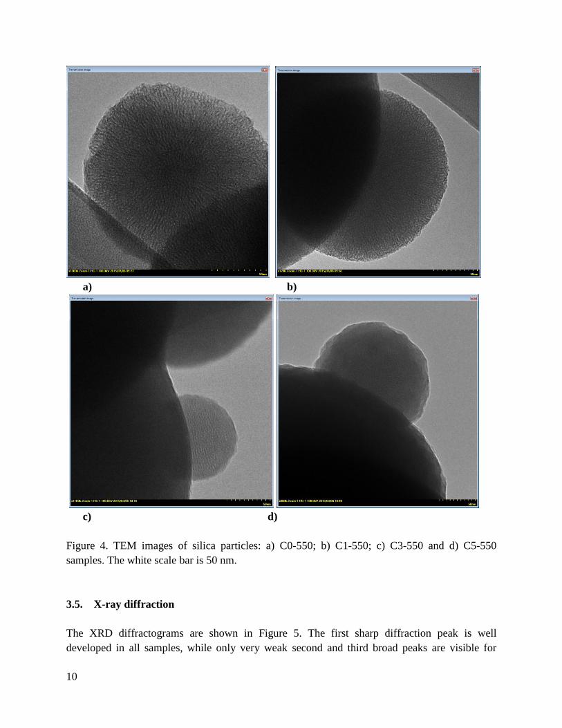
Selected TEM images of the calcined samples are shown in Figure 4. The low magnification
images show that all samples contain spherical particles of 200-800 nm diameters. At higher
magnification, the inner porous structure of the particles is resolved, and the structure of the
particle surface is visible. The comparison of several images shows that the most ordered
channels are developed in the C0-550 sample (Figure 4a). At higher MTES content the porous
region is visible only in the middle of the particles, while their outer part appears to be
amorphous. Also, the particle surface is less sharp for the C3-550 and C5-550 samples (Figure
4c,d). This difference in the outer layer of the particles prepared with methyl containing
precursors is probably due to the uneven distribution of the precursors during particle formation,
since the hydrophobic, methyl containing silica atoms tend to locate near the outer surface of the
particles. During calcination, the burning of the methyl groups leads to a rougher surface.
10
a) b)
c) d)
Figure 4. TEM images of silica particles: a) C0-550; b) C1-550; c) C3-550 and d) C5-550
samples. The white scale bar is 50 nm.
3.5. X-ray diffraction
The XRD diffractograms are shown in Figure 5. The first sharp diffraction peak is well
developed in all samples, while only very weak second and third broad peaks are visible for

11
some of them. These data indicate the locally hexagonal cylindrical pore structure, while the
correlation to larger distances is weak. This is in good agreement with other studies on
mesoporous silica synthesis using molar ratio TEOS: EtOH = 1:58 [35]. The peak positions in
the function of the MTES content are shown in Figure 6. The gradual decrease of the lattice
spacing with increasing MTES content is observed for the as-prepared, as well as for the
template-removed materials. A notable feature is the asymmetric broadening of the first peak in
most of the Cx-60 samples: a shoulder is apparent at 0.3 degrees to the right of the maximum.
This can be explained as a concurrent cubic pore arrangement, encountered on specific
mesoporous Stöber particles and induced by the sphericity of the particle that prevents the
formation of long-range parallel cylinders [35,36]. After calcination at 550 °C, the peaks are
broadened and this shoulder is not visible. The peak position changes from 2.4-2.6° of the
xerogels to 2.8-2.9° for the calcined samples, showing shrinkage of the silica by 12% in average.
In the samples treated with ethanol, the shrinkage is only Large angle XRD data contain a broad
peak at about 23 degrees, which is characteristic for amorphous silica glass (see in Supporting
Information).
1 2 3 4 5 6 7 8
2, degree
C0 - 550
C1 - 550
C2 - 550
C3 - 550
C4 - 550
C5 - 550
1 2 3 4 5 6 7 8
C0 - EtOH
C1 - EtOH
C2 - EtOH
C3 - EtOH
C4 - EtOH
C5 - EtOH
2degree1 2 3 4 5 6 7 8
Inte
nsi
ty
2, degree
C0 - 60
C1 - 60
C2 - 60
C3 - 60
C4 - 60
C5 - 60
Figure 5. XRD diffraction patterns of xerogels, solvent treated and calcined samples.

12
Figure 6. Position of the first XRD and SANS diffraction maximum as a function of MTES
content.
3.6. Small angle neutron scattering
SANS scattering curves of the Cx-60 and Cx-550 sample series are shown in Figure 7. The first
diffraction peak is broader compared to the XRD data due to the instrumental resolution. At
small angles, the logarithmic decay of the scattering curves reflects the surface roughness of the
spherical particles. An empirical model was fitted to the data, consisting of a power law decay at
the small q region, and a Gaussian peak. The fitting was done separately for the two q-ranges.
The power exponent varied between -3.5 and -4.2, showing no regularity with MTES content or
thermal treatment. Value of -4 would correspond to an ideally smooth interface, while the
observed deviations from this value point to a rough particle surface [37,38]. The variation of the
pore distance is similar to the XRD results, the trends being smoother due to the lower resolution
(Figure 6).

13
Figure 7. SANS scattering curves of the xerogels and the calcinated materials. The curves are
shifted vertically for better visibility. Lines are least squares fits to power law model at low q,
and Gaussian peak at high q regions.
4. Conclusions
A simple room temperature sol-gel method for one-pot synthesis of spherical MCM-41 type
materials of varying hydrophobicity was developed, employing a mixture of silica precursors
TEOS and MTES. Structural as well as thermal stability has been analysed. Electron microscopy
shows spherical particles of several hundreds of nanometers, with cylindrical pores arranged
mostly in radial direction. Nitrogen adsorption, XRD and SANS measurements revealed the
integrity of the pore structure for all samples, and a monotonous variation of the pore size with
the precursor composition. With the increase of MTES content, the numbers of CH3 groups on
the silica surfaces is increasing, increasing the hydrophobic character of the material. The high
porosity and specific surface, as well as the monotonous changes of structural and surface
parameters with varying MTES content allows tuning these parameters in a broad range for
potential applications, comparatively to similar materials prepared with TEOS only.
Using ethanol as co-solvent in this room-temperature synthesis proved to be essential for
formation of the mesoporous structure at high MTES concentrations, providing therefore an easy
way of variation of the hydrophobicity without changing synthesis temperature.

14
Acknowledgements
Authors thank the Romanian Academy and the Inter-Academic Exchange Programs: between
Romanian Academy and Hungarian Academy of Sciences and between Academy of Sciences of
the Czech Republic and Romanian Academy. L. A. thanks the Hungarian and Slovak Academies
for supporting a research visit to Institute of Experimental Physics, Kosice. Neutron scattering
experiments from this research project has been supported by the European Commission under
the 7th
Framework Programme through the key action: Strengthening the European Research
Area, Research Infrastructures. Grant Agreement N 283883-NMI3-II, and the Open Project of
the Key Laboratory of Neutron Physics and Institute of Nuclear Physics and Chemistry, China
Academy of Engineering Physics (No. 2014BB06).
References:
[1] W. Stöber, A. Fink, E. Bohn, Controlled growth of monodisperse silica spheres in the
micron size range, J. Colloid Interf. Sci. 26 (1968) 62-69.
[2] C.T. Kresge, M.E. Leonowicz, W.J. Roth, J.C. Vartuli, J.S. Beck, Ordered mesoporous
molecular sieves synthesized by a liquid-crystal template mechanism, Nature 359 (1992) 710-
712.
[3] B. Pauwels, G. van Tendeloo, C. Thoelen, W. van Rhijn, P.A. Jacobs, Structure
Determination of Spherical MCM-41 Particles, Adv. Mater. 13 (2001) 1317 -1320.
[4] M. Grün, I. Lauer, K.K. Unger, The synthesis of micrometer- and submicrometer-size
spheres of ordered mesoporous oxide MCM-41, Adv. Mater. 9 (1997) 254-257.
[5] Y. Yang, S. Lim, G. Du, Y. Chen, D. Ciuparu, G.L. Haller, Synthesis and characterization
of highly ordered Ni-MCM-41 mesoporous molecular sieves, J. Phys. Chem. B 109 (2005)
13237-13246.
[6] N. Igarashi, S. Kidani, R. Ahemaito, K. Hashimoto, T. Tatsumi, Direct organic
functionalization of Ti–MCM-41: Synthesis condition, organic content, and catalytic activity,
Micropor. Mesopor. Mater. 81 (2005) 97-105.
[7] K. Lin, P.P. Pescarmona, K. Houthoofd, D. Liang, G. van Tendeloo, P.A. Jacobs, Direct
room-temperature synthesis of methyl-functionalized Ti-MCM-41 nanoparticles and their
catalytic performance in epoxidation, J. Catal. 263 (2009) 75-82.
[8] M. Grün, K.K. Unger, A. Matsumoto, K. Tsutsumi, Novel pathways for the preparation of
mesoporous MCM-41 materials: Control of porosity and morphology, Micropor. Mesopor.
Mater. 27 (1999) 207-216.
[9] A. Szegedi, Z. Kónya, D. Méhn, E. Solymár, G. Pál-Borbély, Z.E. Horváth, L.P. Biró, I.
Kiricsi, Spherical mesoporous MCM-41 materials containing transition metals: synthesis and
characterization, Appl. Catal. A 272 (2004) 257-266.

15
[10] A. Szegedi, M. Popova, V. Mavrodinova, M. Urbán, I. Kiricsi, C. Minchev, Synthesis
and characterization of Ni-MCM-41 materials with spherical morphology and their catalytic
activity in toluene hydrogenation, Micropor. Mesopor. Mater. 99 (2007) 149-158.
[11] S. Shylesh, P.P. Samuel, S. Sisodiya, A.P. Singh, Periodic Mesoporous Silicas and
Organosilicas: An Overview Towards Catalysis, Catal. Surv. Asia 12 (2008) 266–282.
[12] S.L. Burkett, S.D. Sims, S. Mann, Synthesis of hybrid inorganic–organic mesoporous
silica by co-condensation of siloxane and organosiloxane precursors, Chem. Commun. 11
(1996) 1367-1368.
[13] D.J. Macquairre, Direct preparation of organically modified MCM-type materials.
Preparation and characterisation of aminopropyl–MCM and 2-cyanoethyl–MCM, Chem.
Commun. 16 (1996) 1961-1962.
[14] S. Inagaki, S. Guan, Y. Fukushima, T. Oshuna, O. Tersaki, Novel Mesoporous Materials
with a Uniform Distribution of Organic Groups and Inorganic Oxide in Their Frameworks, J.
Am. Chem. Soc. 121 41 (1999) 9611-9614.
[15] Y.F. Wen, K. Wang, P.H. Pi, J.X. Yang, Z.Q. Cai, L.J. Zhang, Y. Qian, Z.R. Yang, D.F.
Zheng, J. Cheng, Organic–inorganic hybrid superhydrophobic surfaces using
methyltriethoxysilane and tetraethoxysilane sol–gel derived materials in emulsion, Appl. Surf.
Sci. 258 3 (2011) 991-998.
[16] X. Jiang, C.J. Brinker, Aerosol-Assisted Self-Assembly of Single-Crystal
Core/Nanoporous Shell Particles as Model Controlled Release Capsules, J. Am. Chem. Soc.
128 (2006) 4512-4513.
[17] M.M. Rabbani, W. Oh, D.-G. Nam, Synthesis and Characterization of
Methyltriethoxysilyl-Mediated Mesoporous Silicalites, Trans. Electr. Electron. Mater. 12
(2011) 119-122.
[18] P.D. Du, D.Q. Khieu, T.T. Hoa, Removal of Organic Template from Mesoporous MCM-
41, Journal of Science, Hue University, 69 (2011) 35-41.
[19] M.C. Goncalves, Physisorption data for methyl-hybrid silica gels, J. Sol-Gel Sci.
Technol. 75 (2015) 508–518.
[20] L. Rosta, Cold neutron research facility at the Budapest Neutron Centre, Appl. Phys. A
74 Suppl. (2002) S52–S54.
[21] M. Peng, L. Sun, L. Chen, G. Sun, B. Chen, C. Xie, Q. Xia, G. Yan, Q. Tian, C. Huang,
B. Pang, Y. Zhang, Y. Wang, Y. Liu, W. Kang, J. Gong, A new small-angle neutron
scattering spectrometer at China Mianyang research reactor, Nucl. Instr. Methods Phys. Res.
Sect. A 810 (2016) 63-67.
[22] D. Zhao, Q. Huo, J. Feng, B.F. Chmelka, G.D. Stucky, Nonionic Triblock and Star
Diblock Copolymer and Oligomeric Surfactant Syntheses of Highly Ordered, Hydrothermally
Stable, Mesoporous Silica Structures, J. Am. Chem. Soc. 120 (1998) 6024-6036.
[23] Y. Zhang, J.M. Kim, D. Wu, Y. Sun, D. Zhao, S. Peng, Sol-gel synthesis of methyl-
modified mesoporous materials with dual porosity, J. Non-Cryst. Solids 351 (2005) 777-783.

16
[24] Q. Huo, R. Leon, P.M. Petroff, G.D. Stucky, Mesostructure design with gemini
surfactants: Supercage formation in a three-dimensional hexagonal array, Science 268 (1995)
1324-1327.
[25] D. Zhao, J. Feng, Q. Huo, N. Melosh, G.H. Fredickson, B.F. Chmelka, G.D. Stucky,
Triblock copolymer syntheses of mesoporous silica with periodic 50 to 300 angstrom pores,
Science 279 (1998) 548-552.
[26] F. Babonneau, L. Leite, S. Fontlupt, Structural characterization of organically-modified
porous silicates synthesized using CTA+ surfactant and acidic conditions, J. Mater. Chem. 9
(1999) 175-178.
[27] A.V. Rao, R.R. Kalesh, Comparative studies of the physical and hydrophobic properties
of TEOS based silica aerogels using different co-precursors, Sci. Technol. Adv. Mater. 4
(2003) 509–515.
[28] R. Al-Oweini, H. El-Rassy, Synthesis and characterization by FTIR spectroscopy of
silica aerogels prepared using several Si(OR)4 and R′′Si(OR′)3 precursors, J. Mol. Struct. 919
(2009) 140-145.
[29] J. Gallardo, A. Durán, Di Martino D., R.M. Almeida, Structure of inorganic and hybrid
SiO2 sol–gel coatings studied by variable incidence infrared spectroscopy, J. Non-Cryst.
Solids 298 (2002) 219-225.
[30] C.J. Brinker, G.W. Scherer, Sol–gel Science, The Physics and Chemistry of Sol–gel
Processing, Academic, New York 1990, pp. 581–585.
[31] R.F.S. Lenza, W.L. Vasconcelos, Preparation of silica by sol-gel method using
formamide, Mater. Res. 4 (3) (2001) 175-179.
[32] J.M. Berquier, L. Teyssedre, C. Jacquiod, Synthesis of transparent mesoporous and
mesostructured thin silica films, J. Sol-Gel Sci. Technol. 13 (1998) 739-742.
[33] J. Bu, H.-K. Rhee, Improvement in hydrophobicity of Ti-MCM-41 using a new silylation
agent MSTFA, Catal. Lett. 65 (2000) 141-145.
[34] M. Kruk, M. Jaroniec, A. Sayari, Application of Large Pore MCM-41 Molecular Sieves
to Improve Pore Size Analysis Using Nitrogen Adsorption Measurements, Langmuir 13
(1997) 6267-6273.
[35] O.I. Lebedev, G. van Tendeloo, O. Collart, P. Cool, E.F. Vansant, Structure and
microstructure of nanoscale mesoporous silica spheres, Solid State Sci. 6 (2004) 489-498.
[36] O.I. Lebedev, S. Turner, S. Liu, P. Cool, G. van Tendeloo, New nano-architectures of
mesoporous silica spheres analyzed by advanced electron microscopy, Nanoscale 4 (2012)
1722-1727.
[37] A.M. Putz, C. Savii, C. Ianăşi, D. Dudás, K.N. Székely, J. Plocek, P. Sfârloagă, L.
Săcărescu, L. Almásy, Pore ordering in mesoporous matrices induced by different directing
agents, J. Porous Mater. 22 (2014) 321-331.
[38] E. Fagadar-Cosma, Z. Dudás, M. Birdeanu, L. Almásy, Hybrid organic-silica materials
based on novel A3B mixed substituted porphyrin, Mater. Chem. Phys. 148 (2014) 143-152.