Applications of transmission electron microscopy to …formatex.info/microscopy4/128-136.pdf ·...
9
Applications of transmission electron microscopy to virus detection and identification F.F. Vale 1 , A.C. Correia 2 , B. Matos 2 , JF Moura Nunes 3 and A.P. Alves de Matos 2,4 1 Faculty of Engineering, Catholic University of Portugal. Estrada Octávio Pato, 2635-631 Rio de Mouro, Portugal. 2 Pathological Anatomy Department, Curry Cabral Hospital, Rua da Beneficência 8, 1069-166 Lisboa, Portugal. 3 Electron Microscopy, Portuguese Institute of Oncology, 1099-023 Lisbon, Portugal. 4 Center for Environmental and Marine Studies, University of Aveiro, Campus Universitário de Santiago, 3810-193 Aveiro. The size of viruses is with a few exceptions (the mimiviruses, poxviruses and some iridoviruses) below the resolution of the optical microscope. The 1000 times higher resolution of the transmission electron microscope (TEM), compared with the light microscope, allows direct observation of small virus particles. The improvement of both TEM equipment and technical procedures allows quick preparative methods and diagnosis of viruses at the family and genus level. Besides routine diagnosis this approach may prove essential to the identification of emerging pathogens in animals and humans. A review of the literature and a description of the main methods and applications of TEM to virus diagnosis and characterization are presented. Keywords Transmission electron microscopy; virus; diagnosis 1. Introduction Virus diagnosis by electron microscopy is based on the visualization and morphological identification of virus particles in samples of diseased tissues or organic fluids [1-3]. Before the advent of electron microscopy, viruses were only known by their manifestations, especially their capability to induce disease in susceptible hosts. Because of their small size they can not usually be imaged with a conventional light microscope, and their particulate nature is apparent only indirectly through filtration and ultracentrifugation experiments [4, 5]. This fact set the stage for the rapidly growing interest in viruses as soon as the electron microscope was invented [6]. The approach met with great success since viruses, with their small size and particulate nature, were almost ideal samples for early transmission electron microscope (TEM) observations. In fact, the development of the microscope by Ernst Ruska was heavily influenced by the observations of viruses carried out by his brother Helmut during the early stage of development of the methodology [5]. An historical review of the field was recently made by Roingeard [7]. The use of electrons for imaging allows the achievement of a resolution of about 0.2 nm that can be used to visualize macromolecules, such as the capsid proteins or the nucleic acids of viruses. However, the need for high vacuum, thin specimens, and the very high energy of the electron beam irradiating the specimen placed terrible constraints to the kind of objects that could be visualized with the instrument [6]. Decades of technical advances that continue today, were needed to observe the macromolecular world in a state of preservation close to the living one [8, 9]. The accumulated knowledge on virus ultrastructure formed the grounds for the first classification of viruses based on virus intrinsic properties rather than on host characteristics and diseases produced by them [10]. As mentioned before, direct morphological observation of most virus particles is not possible with conventional light microscopy combined with immunocytochemical methods, that can reveal the nature of the viral antigens and their intracellular distribution, and constitutes a very powerful diagnostic method in virology [11]. Molecular or immunological methods, such as PCR (Polymerase Chain Reaction) and ELISA (Enzyme Linked Immuno Sorbent Assay), are very effective in detecting known viruses and can be used to scrutinize a large amount of samples. Nontheless, for new or unknown pathogens that may occur in the context of bioterrorism attacks or as a result of the manifestation of new pathogens, electron microscopy remains the only method that can provide a quick assessment of all pathogens present in a sample, providing an “open view”, which is why electron microscopy is considered a “catch- all method” [2, 3, 12, 13]. Electron microscopy can also be used to investigate viruses isolated in cell cultures, which is useful in controlling the results of the virus isolation process, among other applications [3]. In this paper we aim to lay down a few useful guidelines for those wishing to use electron microscopy in the context of virus diagnosis, on the basis of personal experience with human and veterinary samples and information from relevant literature. 2. Overview of the transmission electron microscope The resolution of a compound microscope is limited to half of the wavelength of the radiation used for imaging. This limits the light microscope to a maximum resolution of 200 nm, when using visible light with a wavelength of 400- Microscopy: Science, Technology, Applications and Education A. Méndez-Vilas and J. Díaz (Eds.) 128 ©FORMATEX 2010 ______________________________________________
Transcript of Applications of transmission electron microscopy to …formatex.info/microscopy4/128-136.pdf ·...
Applications of transmission electron microscopy to virus detection and
identification
F.F. Vale1, A.C. Correia
2, B. Matos
2, JF Moura Nunes
3 and A.P. Alves de Matos
2,4
1 Faculty of Engineering, Catholic University of Portugal. Estrada Octávio Pato, 2635-631 Rio de Mouro, Portugal. 2 Pathological Anatomy Department, Curry Cabral Hospital, Rua da Beneficência 8, 1069-166 Lisboa, Portugal. 3 Electron Microscopy, Portuguese Institute of Oncology, 1099-023 Lisbon, Portugal. 4 Center for Environmental and Marine Studies, University of Aveiro, Campus Universitário de Santiago, 3810-193
Aveiro.
The size of viruses is with a few exceptions (the mimiviruses, poxviruses and some iridoviruses) below the resolution of
the optical microscope. The 1000 times higher resolution of the transmission electron microscope (TEM), compared with
the light microscope, allows direct observation of small virus particles. The improvement of both TEM equipment and
technical procedures allows quick preparative methods and diagnosis of viruses at the family and genus level. Besides
routine diagnosis this approach may prove essential to the identification of emerging pathogens in animals and humans. A
review of the literature and a description of the main methods and applications of TEM to virus diagnosis and
characterization are presented.
Keywords Transmission electron microscopy; virus; diagnosis
1. Introduction
Virus diagnosis by electron microscopy is based on the visualization and morphological identification of virus particles
in samples of diseased tissues or organic fluids [1-3]. Before the advent of electron microscopy, viruses were only
known by their manifestations, especially their capability to induce disease in susceptible hosts. Because of their small
size they can not usually be imaged with a conventional light microscope, and their particulate nature is apparent only
indirectly through filtration and ultracentrifugation experiments [4, 5]. This fact set the stage for the rapidly growing
interest in viruses as soon as the electron microscope was invented [6]. The approach met with great success since
viruses, with their small size and particulate nature, were almost ideal samples for early transmission electron
microscope (TEM) observations. In fact, the development of the microscope by Ernst Ruska was heavily influenced by
the observations of viruses carried out by his brother Helmut during the early stage of development of the methodology
[5]. An historical review of the field was recently made by Roingeard [7].
The use of electrons for imaging allows the achievement of a resolution of about 0.2 nm that can be used to visualize
macromolecules, such as the capsid proteins or the nucleic acids of viruses. However, the need for high vacuum, thin
specimens, and the very high energy of the electron beam irradiating the specimen placed terrible constraints to the kind
of objects that could be visualized with the instrument [6]. Decades of technical advances that continue today, were
needed to observe the macromolecular world in a state of preservation close to the living one [8, 9].
The accumulated knowledge on virus ultrastructure formed the grounds for the first classification of viruses based on
virus intrinsic properties rather than on host characteristics and diseases produced by them [10].
As mentioned before, direct morphological observation of most virus particles is not possible with conventional light
microscopy combined with immunocytochemical methods, that can reveal the nature of the viral antigens and their
intracellular distribution, and constitutes a very powerful diagnostic method in virology [11]. Molecular or
immunological methods, such as PCR (Polymerase Chain Reaction) and ELISA (Enzyme Linked Immuno Sorbent
Assay), are very effective in detecting known viruses and can be used to scrutinize a large amount of samples.
Nontheless, for new or unknown pathogens that may occur in the context of bioterrorism attacks or as a result of the
manifestation of new pathogens, electron microscopy remains the only method that can provide a quick assessment of
all pathogens present in a sample, providing an “open view”, which is why electron microscopy is considered a “catch-
all method” [2, 3, 12, 13]. Electron microscopy can also be used to investigate viruses isolated in cell cultures, which is
useful in controlling the results of the virus isolation process, among other applications [3].
In this paper we aim to lay down a few useful guidelines for those wishing to use electron microscopy in the context
of virus diagnosis, on the basis of personal experience with human and veterinary samples and information from
relevant literature.
2. Overview of the transmission electron microscope
The resolution of a compound microscope is limited to half of the wavelength of the radiation used for imaging. This
limits the light microscope to a maximum resolution of 200 nm, when using visible light with a wavelength of 400-
Microscopy: Science, Technology, Applications and Education A. Méndez-Vilas and J. Díaz (Eds.)
128 ©FORMATEX 2010
______________________________________________
800nm. The limited resolution of the light microscope only allows the observation of the general morphology of
bacterial cells and extra-large viruses. The internal structure is not clearly observed by light microscopes. Bacteria
dimensions range from 100 to 200 nm for the smallest bacteria (e.g. some members of Mycoplasma, which equals the
large-sized poxvirus), to 7 µm in diameter for the largest sized specimens (e.g. cyanobacterium Oscillatoria, with the
same size as a red blood cell), including medium sized bacteria with 1.1 µm by 2.0 to 6.0 long (e.g. Escherichia coli). In
summary, bacteria can be observed using a light microscope, but subcellular bacterial structures, bacterial appendages,
and most viruses are not usually visible since their largest dimension is smaller than 0.2 µm [14].
A huge improvement in resolution is achieved with TEM by replacing the large wavelength light beam with an
electron beam that has a wavelength of approximately 0.005 nm. When using TEM, electron beams generated either by
a tungsten or LaB6 filament, or by a field emission gun, substitute visible light. The monochromatic beam of electrons
is accelerated under vacuum through a voltage of 40 to 100 kV for biological work, and goes through magnetic fields
that work as lenses. These electromagnetic fields are generated by solenoids that can focus the electron beam. The
overall optics is very similar to the light microscope [6, 15].
Electron beams are notoriously unable to penetrate through matter, and therefore only the thinnest possible
specimens kept under high vacuum are suitable for imaging with this type of microscope. This posed enormous
constraints to the development of the field and a complex array of technical achievements in producing very thin
samples had to mature before relevant data could be extracted from electron microscopic images [16].
The images of biological samples produced by the electron microscope are inherently of very low contrast, since
organic matter is made of relatively light atoms of carbon, oxygen and hydrogen, that have low electron scattering
power. Staining methods requiring the introduction of heavy atoms into the specimen have to be employed in order to
reveal ultrastructural detail [17]. Positive staining, shadow casting, and negative staining procedures were developed for
visualization of thin sections and particulate samples. In particular, the negative staining method introduced by Brenner
and Horne provides a suitable and very fast way of revealing virus particles in TEM, and is thus extensively used for
diagnosis [18].
Another difficulty arises from the nature of the electron beam that is invisible to the human eye. Hence, auxiliary
devices that convert the electron image into a visible light image are needed.
For a well corrected optics, the resolution limit of the microscope is dependent on the wavelength of the radiation
beam. But it is difficult to correct spherical aberration in electron lenses. Energy spread in the electron beam, which can
be caused by the specimen also introduces chromatic aberration [15]. These defects limit the electron microscope
resolution to about 0.2 nm although the small wavelength of the electron beam could in theory allow much better
resolutions. Recently, correction of spherical aberration and chromatic aberrations has been achieved, improving
significantly electron optics [19].
The maximum useful magnification a microscope is able to achieve is set by the resolution, as the goal is to make
visible to the human eye the smallest object that the microscope can image (the size of the resolution limit). Since the
human eye can distinguish objects down to 0.2 mm, the useful magnification for a microscope with a resolution of 0.2
µm (light microscope), is about 0.2 mm/0.2 µm or 1,000x. There is really no restriction to the magnification that can be
achieved, except for the fact that higher magnifications are not needed. In practical terms magnifications of 30000 to
50.000 are suitable for viral diagnostic observations. The resolution of 0.2 nm of the TEM allows for a useful
magnification of 1,000,000x, a factor 1000x higher than for the light microscope [14].
3. Role of the electron microscope in virus identification
3.1 Sample collection
A successful virus diagnosis by electron microscopy depends heavily on the collection methods and on specimen
preparation [20, 21].
Collection is usually made by personnel not specifically trained in electron microscopy, and more general methods
used in other applications, such as molecular biology, are unsuitable for TEM or can even damage the specimen beyond
any hope of recovery [22]. Freezing the samples or fixing in formalin are common mistakes frequently found in the
practice of TEM.
Biological fluids can be easily and quickly studied by negative staining. Fresh fluid from lesions or dried fluid onto a
glass microscope slide are suitable specimens, while swabs offer significantly lower success rates [23].
Solid specimens can be embedded and sectioned with the methods mastered by any biological TEM lab. This takes
considerable more time than negative staining. For these methods, very small pieces of fresh tissue (less than 1 mm3)
must be immersed in glutaraldehyde fixative immediately after collection, as autolytic and anoxic changes that can
obscure the true pathological alterations of the tissue settle within minutes of the removal of the sample [17].
Knowledge of the ultrastructure of the virus particles gathered in these studies, together with the nature of its nucleic
acid formed the basis of an early classification of viruses that is still valid today [10, 24]. Morphological traits allow for
Microscopy: Science, Technology, Applications and Education A. Méndez-Vilas and J. Díaz (Eds.)
©FORMATEX 2010 129
______________________________________________

their identification up to the family or genus level [25]. Further information on the virus type must be obtained by
means of serologic or molecular methods.
3.2 Handling of infectious specimens
Specimens for TEM diagnosis are concentrated suspensions of virus particles, which can be highly infectious. This is
particularly important when dealing with human samples. All manipulations must therefore be done under adequate
biosafety level laboratories. Diagnosis of exotic viruses such as the hemorrhagic fever agents Marburg and Ebola or
viruses producing encephalitis like the West Nile virus or the Dengue virus, must be performed in specialized centers
with high biosafety level facilities [2].
A particularly important point is that viruses are not completely inactivated even after being subjected to the vacuum
and electron beam of the electron microscope. Therefore, the sample and specimen holder must be disposed off and
decontaminated, respectively. If inactivation of the sample is needed, the virus particles can be treated with
glutaraldehyde fixative before or after adsorption to the grid without significantly changing their morphology [12].
3.3 Concentration methods
The minimum virus concentration necessary for successful detection by TEM is about 106 particles per ml. This is a
rather concentrated suspension and viruses are often present in diseased tissues and organic fluids at considerably lower
concentrations [12]. Another problem is the presence of other structures like bacteria and cell debris that may make
virus identification difficult, like in faecal samples, for instance [3]. In these cases, viruses must be separated from
contaminating debris and concentrated to get a workable preparation for electron microscopy.
Clearing the preparation from debris usually involves centrifugation at 1000-2000g for 5 to 10 minutes. The
supernatant is then submitted to ultracentrifugation at 100000g for thirty minutes to one hour to generate a pellet of
concentrated virus particles, which is ressuspended in a small drop (10-15 µl) of bidistilled water [3]. Alternatively, the
centrifugation can be done directly onto the grid, using a bench ultracentrifuge with a specially designed rotor
(Beckman Airfuge® with a EM-90 rotor), a procedure that greatly improves the sensitivity of the method [26].
3.4 Immunomethods
Concentration of the virus preparation may also be achieved by immunological means. The virus suspension may be
treated with antiserum against a known virus that may come from the patient himself. If the appropriate virus antigen is
present, clumps of virus particles are formed and easily visible in the electron microscope image. In the presence of an
excess of antibody, particles may become covered with a layer of antibody, which is visible by electron microscopy,
instead of forming clumps. Alternatively, the antibody can be adsorbed to a membrane-covered electron microscope
grid. Virus particles attach specifically to the treated membrane, and contaminants can be washed away, allowing for an
increased sensitivity of the method [3].
The use of specific antibodies also allows a more precise identification of the virus particles. If this is required, the
viruses adsorbed to the membrane-covered grid or thin sections of virus infected tissues, can be treated with gold-
labelled antibody. Colloidal gold particles with a definite size (5-15 nm) are attached to the virus antigens and can be
ready visualized in the electron microscope [2].
3.5 Specimen preparation for TEM observation
For negative staining, a drop of about 10 µl of the virus suspension to be studied is applied to the hydrophobic surface
of a parafilm square in a Petri dish. A formvar-coated grid is floated onto this drop for one minute, with the formvar
side of the grid in contact with the liquid. The excess liquid is removed from the grid by touching its border with a cut
piece of filter paper. The grid is immediately floated in a drop of 1.5% phosphotungstic acid, 2% ammonium molybdate
or 1% aqueous uranyl acetate, depending on the specimen. For a better assessment of the samples, two or three grids
should be prepared, each stained with one of these stains. After staining for one minute, the excess stain is removed
with filter paper and the grid left to dry for a few minutes, before insertion into the microscope column [3].
If solid samples are to be studied, such as organ fragments from biopsies or autopsy specimens, it may be necessary
to make thin sections of the pathologically altered tissues. This calls for a complex sequence of fixation in appropriate
fixatives, epoxy embedding, thin sectioning and staining, which is usually mastered by well trained personnel in
electron microscopy laboratories. Detailed accounts of the methods can be found elsewhere [16, 17]. If these methods
become necessary, an electron microscopy facility should be contacted for obtaining the appropriate primary fixative
and instructions for sample collection and submission to further processing. Common mistakes, such as freezing the
sample or leaving it unfixed for even “short” periods of time can ruin the sample and are to be avoided at all cost.
Microscopy: Science, Technology, Applications and Education A. Méndez-Vilas and J. Díaz (Eds.)
130 ©FORMATEX 2010
______________________________________________
4. Application of EM to the study of several viruses
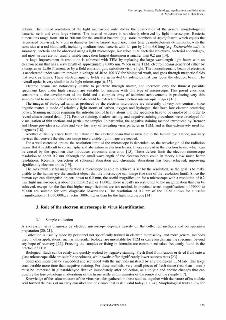
For an example of a TEM application to the study of viruses Fig.1 shows virus particles recovered by cell culture from
tissues Triturus marmoratus, from a high mortality episode that occurred in a lagoon at the Peneda-Gerês National Park,
Portugal. The same virus had previously been observed in ulcers from diseased animals [27]. Among the many possible
causes for such an event, TEM was able to point the right direction with little effort and with no need for exhaustive
testing of specific suspected pathogens. The viruses have the size and overall morphology of Ranaviruses, and this was
subsequently confirmed by PCR. This case illustrates the advantages of TEM in virus identification, making it probably
the most common method for detecting new viruses [3]. Isolation of viruses in cell cultures is also extensively used in
research and diagnostic virology labs. The infected cells and isolated agents can conveniently be assessed by TEM, in
particular when atypical cytopathic effects occur.
Fig. 1 Thin section of ranaviruses in cell cultures. Skin ulcers of dying Triturus marmoratus from a lagoon in Peneda Gerês
Natural Park, showed virus-like particles suggestive of ranaviruses. Cell culture isolation combined with TEM and PCR studies
subsequently confirmed the presence of the viruses.
4.1 General virus particle morphology
The virus nucleocapsid contains the genetic material and a protective protein coat. Nucleocapsids can be icosahedral or
helical, and can be naked or enveloped in a protective lipoprotein coat. When present, the lipoprotein coat usually
shows projections that are important for virus identification. The poxviruses shown below have a more complex
arrangement [10, 28].
4.2 DNA viruses
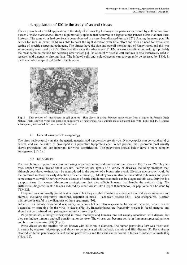
The morphology of poxviruses observed using negative staining and thin sections are show in Fig. 2a and 2b. They are
brick-shaped with a size of about 300 nm. Poxviruses are agents of a variety of diseases, including smallpox that,
although considered extinct, may be reintroduced in the context of a bioterrorist attack. Electron microscopy would be
the preferred method for early detection of such a threat [3]. Monkeypox can also be transmitted to humans and poses
some concern as well. Other Poxviruses diseases of cattle and domestic animals can be diagnosed this way. Orfvirus is a
parapox virus that causes Muluscum contagiosum that also affects humans that handle the animals (Fig. 2b).
Differential diagnosis in skin lesions induced by other viruses like Herpes (Chickenpox) or papilloma can be done by
TEM [2].
Herpesviruses are usually found in skin lesions, but they are able to induce a wide spectrum of diseases in human and
animals, including respiratory infections, hepatitis in birds – Pacheco’s disease [29] – and encephalitis. Electron
microscopy is useful in the diagnosis of these specimens [30].
Adenoviruses mainly cause mild respiratory infections but are also responsible for canine hepatitis, which can be
diagnosed by searching for the virus in faeces (Fig. 3). Bacteriophages are frequently present in faecal material and
should not be confused with pathogenic animal viruses (Fig 4).
Polyomaviruses, although widespread in mice, monkeys and humans, are not usually associated with disease, but
they can induce tumours and cell transformation in vitro. The viruses can become active in immunossupressed patients
and be excreted in urine [28] (Fig. 5).
Parvoviruses are the smallest viruses known with 20-25nm in diameter. The human parvovirus B19 was discovered
in serum by electron microscopy and shown to be associated with aplastic anemia and fifth disease [3]. Parvoviruses
also induce feline panleukopenia and canine parvovirosis and the virus can be found in faeces of infected animals (Fig
6) [31, 32].
Microscopy: Science, Technology, Applications and Education A. Méndez-Vilas and J. Díaz (Eds.)
©FORMATEX 2010 131
______________________________________________
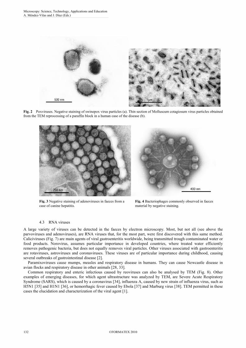
Fig. 2 Poxviruses. Negative staining of swinepox virus particles (a). Thin section of Molluscum cotagiosum virus particles obtained
from the TEM reprocessing of a paraffin block in a human case of the disease (b).
4.3 RNA viruses
A large variety of viruses can be detected in the faeces by electron microscopy. Most, but not all (see above the
parvoviruses and adenoviruses), are RNA viruses that, for the most part, were first discovered with this same method.
Caliciviruses (Fig. 7) are main agents of viral gastroenteritis worldwide, being transmitted trough contaminated water or
food products. Norovirus, assumes particular importance in developed countries, where treated water efficiently
removes pathogenic bacteria, but does not equally removes viral particles. Other viruses associated with gastroenteritis
are rotaviruses, astroviruses and coronaviruses. These viruses are of particular importance during childhood, causing
several outbreaks of gastrointestinal disease [2].
Paramixoviruses cause mumps, measles and respiratory disease in humans. They can cause Newcastle disease in
avian flocks and respiratory disease in other animals [28, 33].
Common respiratory and enteric infectious caused by reoviruses can also be analysed by TEM (Fig. 8). Other
examples of emerging diseases, for which agent ultrastructure was analyzed by TEM, are Severe Acute Respiratory
Syndrome (SARS), which is caused by a coronavirus [34], influenza A, caused by new strain of influenza virus, such as
H5N1 [35] and H1N1 [36], or hemorrhagic fever caused by Ebola [37] and Marburg virus [38]. TEM permitted in these
cases the elucidation and characterization of the viral agent [1].
Fig. 3 Negative staining of adenoviruses in faeces from a
case of canine hepatitis.
Fig. 4 Bacteriophages commonly observed in faeces
material by negative staining.
Microscopy: Science, Technology, Applications and Education A. Méndez-Vilas and J. Díaz (Eds.)
132 ©FORMATEX 2010
______________________________________________
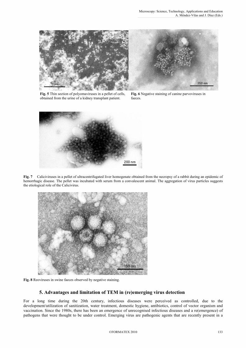
Fig. 7 Caliciviruses in a pellet of ultracentrifugated liver homogenate obtained from the necropsy of a rabbit during an epidemic of
hemorrhagic disease. The pellet was incubated with serum from a convalescent animal. The aggregation of virus particles suggests
the etiological role of the Calicivirus.
Fig. 8 Reoviruses in swine faeces observed by negative staining.
5. Advantages and limitation of TEM in (re)emerging virus detection
For a long time during the 20th century, infectious diseases were perceived as controlled, due to the
development/utilization of sanitization, water treatment, domestic hygiene, antibiotics, control of vector organism and
vaccination. Since the 1980s, there has been an emergence of unrecognised infectious diseases and a re(emergence) of
pathogens that were thought to be under control. Emerging virus are pathogenic agents that are recently present in a
Fig. 5 Thin section of polyomaviruses in a pellet of cells,
obtained from the urine of a kidney transplant patient.
Fig. 6 Negative staining of canine parvoviruses in
faeces.
Microscopy: Science, Technology, Applications and Education A. Méndez-Vilas and J. Díaz (Eds.)
©FORMATEX 2010 133
______________________________________________
community or, that existed before in a specific population and are experiencing an increase of incidence or geographic
spread. A re-emerging pathogenic agent was previously present in a specific population and experienced a decrease of
incidence and reappears in the population, causing an increase of the incidence rate. The emergence of infectious
diseases is due to anthropogenic, social, and environmental changes. Modifications in medical care (nosocomial
infectious) and medical technology (blood transfusion, organ transplantation, re-used syringes, contamination of
vaccines, etc.) are important as anthropogenic causes. Social causes include general social changes, such as worldwide
urbanization, intravenous drug abuse, changing sexual practices, accelerated human mobility and refugee populations,
intensive food production and commercial trading. Finally, climate changes refer to global warming and ecosystem
disturbances, such as deforestation, eutrophication of waterways, reduction in the population of predator of disease
vector organisms and changes in the habitat or migratory routes of vector organisms [39].
For instance, gastrointestinal viruses are the leading cause of death among children. Most of these fatalities occur in
Africa, where health services are deficient and misdiagnosis is common, such as in the case of misdiagnosis of viral
diseases as malaria cases [40]. However, the aetiology of gastroenteritis is currently poorly investigated even in
developed countries. The routine detection methods used evolved over time. Initially, diagnosis was done by electron
microscopy, but this was replaced by molecular detection methods, using reverse transcription-PCR and confirmation of
positive samples by sequencing. There are cases of gastroenteritis that are negative for all RT-PCR techniques, for
which other methods must be applied, such as new molecular methods, electron microscopy or cell culture isolation. An
investigation of the aetiology of outbreaks in the Netherlands leaves 12.2% of these unexplained [41], in these the use
of TEM is still useful for at least determining the viral origin of the outbreak.
In addition to the capability of detection and identification of virus, electron microscopy can also help differentiate
between similar viruses, such as the Ebola and Marburg viruses. Both are from the filovirus family, but Marburg virions
are shorter than Ebola virions, and the spikes are also different [3, 42].
Electron microscopy has several advantages in emerging virus diagnosis. It allows an “open view” of whatever is
present (“catch-all method”), while molecular methods require previous knowledge of the virus to be tested. Electron
microscopy has also the advantage of being able to identify dual infectious (caused by more than one virus), which
could be missed by molecular or antigen tests. Indeed, no prior virus selection is necessary. Electron microscopy
permits a rapid diagnostic, since negative staining can be made in a few minutes. Viruses can be identified to the
group/family level on a morphological basis. Hence, even in the age of molecular diagnostics, the use of electron
microscopy is still a mainstay in detecting new and unusual outbreaks of unknown viruses and those that were not
considered for analysis by the clinician [3, 11].
Viruses stored in various solutions for long periods of time may not be viable for culture detection or molecular
testing, but may be useful for electron microscopy, since this technique does not require live or intact viruses for
identification [3]. However, if virus particles are in low concentration, or too altered morphologically due to sample
degradation, or as a result of inappropriate preservation procedures, the use of electron microscopy may be not useful,
which is a clear limitation of the technique.
The emergence of viral infectious diseases affecting humans and animals will continue both in developed and
developing countries and is very difficult to predict. Surveillance systems and the use of a large range of diagnostic
methodologies can contribute to control outbreaks. The use of immunological methods, such as production of antibodies
against specific virus, and the use of molecular biology tools like PCR imply some previous knowledge of the virus.
The identification of new unknown virus depends on the initial characterization of viruses by TEM. The study of
samples using TEM may achieve quick virus identification, or provide enough information on the pathological process
to point to the right direction. Additionally, as electron microscopy is a rapid procedure, it can be of major importance
in the surveillance of viruses that can be used as biological weapons [3]. Indeed, electron microscopy can be used to
detect viral agents, such as Ebola virus and smallpox virus. However, before becoming involved in the monitoring of
virus bioterrorism samples, laboratories must consider several issues, including their expertise in virus identification,
capacity for handling biohazards, and the health and immune status of the laboratory staff [43]. The identification on the
basis of virus morphology requires well trained and experienced microscopists, which are hardly present in every virus
diagnosis laboratory. The use of tele-microscopy, i.e., the control of the microscope and diagnosis by a spatially remote
expert, can guaranty an additional quality assurance level in the morphologic diagnostic [39, 40].
6. Conclusions
Electron microscopy was the gold standard for virus diagnosis for decades (from the 30s to 80s of the 20th century), but
the interest in the technique decreased with the implementation of molecular biology techniques. Indeed, electron
microscopy was successfully used in both infected cell lines and “dirty” clinical samples, such as plasma, urine, faeces
and vesicle fluids [1, 7]. Because electron microscopy is not a high throughput technique, many molecular and
immunologic methods have been developed. Immunological methods have almost unlimited throughput and are very
specific, which can contribute to the failure of identification of viruses with different antigenic determinants. Similarly
molecular techniques, such as PCR rely on previous knowledge of genomic content so that primers can be designed.
Mutations, which are very common in viruses, may lead to false negative results, because the primers may not recognise
Microscopy: Science, Technology, Applications and Education A. Méndez-Vilas and J. Díaz (Eds.)
134 ©FORMATEX 2010
______________________________________________
the complementary mutated sequence [12]. Nonetheless, electron microscopy is still the gold standard for the
identification of unknown viruses, in situations where no probes or primers are available, which is often the case of
emergent viruses.
Electron microscopy was is and will be always necessary for new virus characterization. This especially applies to
veterinary virology, in which several agents are continuously being identified with this technique [1]. The future of
electron microscopy goes through rapid diagnosis of viruses, the exclusion of presence of pathogenic virus in
bioterrorism samples, and in quality control of produced vaccines. Considering that its use in diagnosis, although
declining, is of prime importance for both diagnosis and research, it is important to prepare a new generation of
microscopists with the appropriate technical skills [2]. The recourse to tele-microscopy should also be considered, so
that excellence centres in electron microscopy can concentrate their expertise and better interact with the diagnostic labs
[44, 45]. Besides being applied in diagnostic virology, electron microscopy can be used to understand the mechanisms
of virus attachment and replication within the host cell. This information can be useful in the design of novel antiviral
drugs and vaccines. The study of viral effects on cells and tissues enriches the knowledge of which cells and organs are
involved in disease. Ultrastructural studies contribute for both the design of novel drugs against viruses, but also help
elucidating the function of several viral elements [3, 7]. Surely electron microscopy will still be needed for the
identification of new unknown virus. The “open view” approach of electron microscopy permits rapid and “catch-all”
detection of viruses, if these are present in a high concentration, and makes it especially useful in emergency situations.
The nature of the samples to be analyzed can be tremendously diverse, from body fluids or biopsies performed after or
before cell culture, or environment samples [12].
Finally, an additional advantage of electron microscopy is its promoting value. It is reassuring to have the picture of
the screened virus and “a picture is worth a thousand words”. Among the disadvantages of electron microscopy usage
there are the need for training microscopists, the capital expenses in instrument acquisition and maintenance, need for
an appropriate room for its operation, the unsuitability of use for all viruses, the intensive labour, the imprecision in
distinguishing between many similar viruses, and the low throughput [1]. All methods have advantages and
disadvantages and their applications should be careful planned. The combined use of several methods should not be
discarded. For example, results obtained by TEM can be confirmed at the species level by directly testing the virus
adhered to grids by PCR [46], joining and taking advantage of two distinct techniques.
Acknowledgements The English review by Patrícia Fonseca is gratefully acknowledged.
References
[1] Biel SS, Gelderblom HR. Diagnostic electron microscopy is still a timely and rewarding method. J Clin Virol 1999;
13:105-119.
[2] Curry A, Appleton H, Dowsett B. Application of transmission electron microscopy to the clinical study of viral and
bacterial infections: present and future. Micron 2006;37:91-106.
[3] Goldsmith CS, Miller SE. Modern uses of electron microscopy for detection of viruses. Clin Microbiol Rev 2009;22:552-
563.
[4] Levine AJ, Enquist LW. History of Virology. In: Knipe DM, Howley PM, editors. Fields Virology. 5th ed. New York:
Lippincott Williams & Wilkins; 2007. p. 4-24.
[5] Kruger DH, Schneck P, Gelderblom HR. Helmut Ruska and the visualisation of viruses. Lancet 2000;355:1713-1717.
[6] Ruska E. Nobel lecture. The development of the electron microscope and of electron microscopy. Biosci Rep 1987;7:607-
629.
[7] Roingeard P. Viral detection by electron microscopy: past, present and future. Biol Cell 2008;100:491-501.
[8] Crowther RA. Viruses and the development of quantitative biological electron microscopy. Notes Rec R Soc Lond
2004;58:65-81.
[9] Pierson J, Sani M, Tomova C, Godsave S, Peters PJ. Toward visualization of nanomachines in their native cellular
environment. Histochem Cell Biol 2009;132:253-262.
[10] Fenner F, McAuslan BR, Mims CA, Sambrook J, White DO. The Biology of Animal Viruses. New York: Academic Press;
1974.
[11] Kennedy M. Methodology in diagnostic virology. Vet Clin North Am Exot Anim Pract 2005;8:7-26.
[12] Hazelton PR, Gelderblom HR. Electron microscopy for rapid diagnosis of infectious agents in emergent situations. Emerg
Infect Dis 2003;9:294-303.
[13] Biel SS, Madeley D. Diagnostic virology--the need for electron microscopy: a discussion paper. J Clin Virol 2001;22:1-9.
[14] Willey J, Sherwood L, Woolverton C. Prescott, Harley and Klein's Microbiology. 7th ed. London: McGraw-Hill; 2008.
[15] Agar AW, Alderson RH, Chescoe D, Glauert AM. Principles and Practice of Electron Microscope Operation. In: Glauert
AM, editor. Practical methods in electron microscopy. London: Elsevier Science & Technology; 1974.
[16] Glauert AM, Lewis PR. Biological Specimen Preparation for Transmission Electron Microscopy. New Jersey: Princeton
University Press; 1988.
[17] Hayat MA. Principles and Techniques of Electron Microscopy: Biological Applications. Cambridge: Cambridge
University Press; 2000.
[18] Brenner S, Horne RW. A negative staining method for high resolution electron microscopy of viruses. Biochim Biophys
Acta 1959;34:103-110.
Microscopy: Science, Technology, Applications and Education A. Méndez-Vilas and J. Díaz (Eds.)
©FORMATEX 2010 135
______________________________________________
[19] Rose HH. Future trends in aberration-corrected electron microscopy. Philos Transact A Math Phys Eng Sci 2009;367:3809-
3823.
[20] Doane FW, Anderson N. Electron Microscopy in Diagnostic Virology. A Practical Guide and Atlas. London: Cambridge
University Press; 1987.
[21] Lew JF, LeBaron CW, Glass RI, Torok T, Griffin PM, Wells JG, et al. Recommendations for collection of laboratory
specimens associated with outbreaks of gastroenteritis. MMWR Recomm Rep 1990;39:1-13.
[22] Gelderblom HR, Hazelton PR. Specimen collection for electron microscopy. Emerg Infect Dis 2000;6:433-434.
[23] Hammond GW, Hazelton PR, Chuang I, Klisko B. Improved detection of viruses by electron microscopy after direct
ultracentrifuge preparation of specimens. J Clin Microbiol 1981;14:210-221.
[24] Almeida JD, Waterson AP. Some implications of a morphologically oriented classification of viruses. Arch Gesamte
Virusforsch 1970;32:66-72.
[25] Doane FW. Virus morphology as an aid for rapid diagnosis. Yale J Biol Med 1980;53:19-25.
[26] Rice SJ, Phillips AD. Rapid preparation of faecal specimens for detection of viral particles by electron microscopy. Med
Lab Sci 1980;37:371-372.
[27] Soares C, Alves de Matos AP, Arntzen JW, Carretero M, Loureiro A. Amphibian mortality in a National Park in the North
of Portugal. Froglog 2003;56:1.
[28] Levy JA, Fraenkel-Conrat H, Owens RA. Virology. New Jersey: Prentice Hall, Englewood Cliffs; 1994.
[29] Tsai SS, Park JH, Hirai K, Itakura C. Herpesvirus infections in psittacine birds in Japan. Avian Pathol 1993;22:141-56.
[30] White CL, III, Taxy JB. Early morphologic diagnosis of herpes simplex virus encephalitis: advantages of electron
microscopy and immunoperoxidase staining. Hum Pathol 1983;14:135-139.
[31] Vieler E, Herbst W. [Electron microscopic demonstration of viruses in feces of dogs with diarrhea]. Tierarztl Prax
1995;23:66-69.
[32] Marshall JA, Kennett ML, Rodger SM, Studdert MJ, Thompson WL, Gust ID. Virus and virus-like particles in the faeces
of cats with and without diarrhoea. Aust Vet J 1987;64:100-105.
[33] McFerran JB, McNulty MS, Curran WL. Diagnosis of avian viral diseass by electron microscopy. Am J Vet Res
1978;39:505-508.
[34] Goldsmith CS, Tatti KM, Ksiazek TG, Rollin PE, Comer JA, Lee WW, et al. Ultrastructural characterization of SARS
coronavirus. Emerg Infect Dis 2004;10:320-326.
[35] Ng AK, Zhang H, Tan K, Li Z, Liu JH, Chan PK, et al. Structure of the influenza virus A H5N1 nucleoprotein:
implications for RNA binding, oligomerization, and vaccine design. FASEB J 2008;22:3638-3647.
[36] Giocondi MC, Ronzon F, Nicolai MC, Dosset P, Milhiet PE, Chevalier M, et al. Organization of influenza A virus
envelope at neutral and low pH. J Gen Virol 2010;91:329-338.
[37] Noda T, Ebihara H, Muramoto Y, Fujii K, Takada A, Sagara H, et al. Assembly and budding of Ebolavirus. PLoS Pathog
2006;2:e99.
[38] Mavrakis M, Kolesnikova L, Schoehn G, Becker S, Ruigrok RW. Morphology of Marburg virus NP-RNA. Virology
2002;296:300-307.
[39] Kuiken T, Fouchier R, Rimmelzwaan G, Osterhaus A. Emerging viral infections in a rapidly changing world. Curr Opin
Biotechnol 2003;14:641-646.
[40] Schoub BD. Surveillance and management of influenza on the African continent. Expert Rev Respir Med 2010;4:167-169.
[41] Svraka S, Duizer E, Vennema H, de BE, van d, V, Dorresteijn B, et al. Etiological role of viruses in outbreaks of acute
gastroenteritis in The Netherlands from 1994 through 2005. J Clin Microbiol 2007;45:1389-1394.
[42] Geisbert TW, Jahrling PB. Differentiation of filoviruses by electron microscopy. Virus Res 1995;39:129-50.
[43] Miller SE. Bioterrorism and electron microscopic differentiation of poxviruses from herpesviruses: dos and don'ts.
Ultrastruct Pathol 2003;27:133-140.
[44] Gelderblom HR. Electron microscopy in diagnosis of bioterrorism agents. Bundesgesundheitsblatt - Gesundheitsforschung
- Gesundheitsschutz 2003;46:984-988.
[45] Schroeder JA, Gelderblom HR, Hauroeder B, Schmetz C, Milios J, Hofstaedter F. Microwave-assisted tissue processing for
same-day EM-diagnosis of potential bioterrorism and clinical samples. Micron 2006;37:577-590.
[46] Johnsen CK, Bottiger B, Blom J. Confirmation of electron microscopy results by direct testing of viruses adhered to grids
using nucleic acid amplification techniques. J Virol Methods 2006;134:92-98.
Microscopy: Science, Technology, Applications and Education A. Méndez-Vilas and J. Díaz (Eds.)
136 ©FORMATEX 2010
______________________________________________