Applications of the Universal DNA Microarray in Molecular ...
34
2 Applications of the Universal DNA Microarray in Molecular Medicine Reyna Favis, Norman P. Gerry, Yu-Wei Cheng, and Francis Barany Summary Integration of molecular medicine into standard clinical practice will require the availability of diagnostics that are sensitive, rapid, and robust. The backbone technology underlying the diag- nostic will likely serve double duty during clinical trials in order to first validate the biomarkers that contribute to both drug response and disease stratification. PCR/LDR/Universal DNA microarray is a promising technology to help drive the transition from the current paradigms of clinical decision making to the new era of personalized medicine. By uncoupling the mutation detection step from array hybridization, this technology becomes fully programmable. It exploits full use of the sensitivity that the ligase detection reaction can provide, while maintaining a rapid read out on a universal microarray. Thus, PCR/LDR/Universal DNA microarray is 50-fold more sensitive and 10-fold more rapid than conventional hybridization-only arrays. The intent of this article is to provide investigators with a perspective on current uses of this approach, as well as to serve as a practical guide to implementation. Key Words: Microarray; ligase detection reaction; high throughput; multiplexing; cancer genomics; SNP; DNA methylation; mutation detection; thermostable ligase. 1. Introduction 1.1. The Future of Molecular Medicine Molecular medicine holds the promise of maximizing patient benefit while minimizing risk. In a vision of the future, standard care for a patient would involve the selection of the drug most likely to be efficacious, given the particulars of an individual’s indication, and an adjustment of the drug dose to accommodate the patient’s drug metabolism (absorption, distribution, metabolism, and excretion [ADME]) profile. Guiding the physician in making these decisions will be information contained on the drug label, as well as results obtained from point-of-care diagnostic kits and/or the diagnostic laboratory. 25 From: Methods in Molecular Medicine, Vol. 144, Microarrays in Clinical Diagnostics Edited by: T. Joos and P. Fortina © Humana Press Inc., Totowa, NJ
Transcript of Applications of the Universal DNA Microarray in Molecular ...

2
Applications of the Universal DNA Microarray in Molecular Medicine
Reyna Favis, Norman P. Gerry, Yu-Wei Cheng, and Francis Barany
SummaryIntegration of molecular medicine into standard clinical practice will require the availability
of diagnostics that are sensitive, rapid, and robust. The backbone technology underlying the diag-nostic will likely serve double duty during clinical trials in order to first validate the biomarkersthat contribute to both drug response and disease stratification. PCR/LDR/Universal DNAmicroarray is a promising technology to help drive the transition from the current paradigms ofclinical decision making to the new era of personalized medicine. By uncoupling the mutationdetection step from array hybridization, this technology becomes fully programmable. It exploitsfull use of the sensitivity that the ligase detection reaction can provide, while maintaining a rapidread out on a universal microarray. Thus, PCR/LDR/Universal DNA microarray is 50-fold moresensitive and 10-fold more rapid than conventional hybridization-only arrays. The intent of thisarticle is to provide investigators with a perspective on current uses of this approach, as well asto serve as a practical guide to implementation.
Key Words: Microarray; ligase detection reaction; high throughput; multiplexing; cancergenomics; SNP; DNA methylation; mutation detection; thermostable ligase.
1. Introduction1.1. The Future of Molecular Medicine
Molecular medicine holds the promise of maximizing patient benefitwhile minimizing risk. In a vision of the future, standard care for a patientwould involve the selection of the drug most likely to be efficacious, giventhe particulars of an individual’s indication, and an adjustment of the drug doseto accommodate the patient’s drug metabolism (absorption, distribution,metabolism, and excretion [ADME]) profile. Guiding the physician in makingthese decisions will be information contained on the drug label, as well as resultsobtained from point-of-care diagnostic kits and/or the diagnostic laboratory.
25
From: Methods in Molecular Medicine, Vol. 144, Microarrays in Clinical DiagnosticsEdited by: T. Joos and P. Fortina © Humana Press Inc., Totowa, NJ
02_chap_Joos.qxd 25/04/2005 02:50 pm Page 25

Although the present state of medical care bears little resemblance to this sce-nario, appropriate technologies to support this standard of care are activelybeing developed.
To bring molecular medicine a step closer to becoming a reality, the technolo-gy platforms of today must have the flexibility to make the transition from clini-cal trial to diagnostic laboratory. In the recent Food and Drug Administration draftguidance for pharmacogenomic data submission (http:/ /www.fda.gov/cder/guid-ance/5900dft.pdf), it was recommended that the test and the drug be codevelopedand complete information on the test be submitted to the agency. Thus, a cost-effective strategy is to apply the test used during the clinical trial, very likely infundamentally the same form, to execute the same function in a diagnostic capa-bility after the drug is launched. The standard requirements for a marketable testinclude high sensitivity and specificity, reproducibility, rapid throughput, and costeffectiveness; however, clinical trials require extra considerations. Of note, testsmust assess only those factors for which subjects have provided consent. A suit-able technology platform must provide for the aforementioned requirements, aswell as be able to accommodate molecular versatility, since there are numerousindications that would benefit from the use of biomarkers.
Molecular approaches for identifying single-base variants fall into threegeneral strategies:
1. Target amplification, e.g., (real-time polymerase chain reaction [PCR], usuallywith TaqMan® or molecular beacon probes), whole-genome amplification (WGA),and strand displacement amplification (SDA).
2. Probe amplification, e.g., ligase chain reaction (LCR), ligase detection reaction(LDR), and rolling circle amplification (RCA).
3. Signal amplification, e.g., 3D dendrimer labeling systems, enzymatic cascadereporters, and invader assay (see ref. 1 for review) (Fig. 1; see Color Plate 8 followingp. 18).
These amplification strategies are often combined with identification of theproduct sequence by hybridization to its complement on an array. Althoughmany of these approaches have great utility in certain contexts, various draw-backs exist that limit their use in other contexts. For example, direct hybridiza-tion schemes cannot always distinguish between closely related sequences andcannot find mutant sequence in an excess of normal sequence (Fig. 1, hybridiza-tion array on bottom left). Another fundamental stumbling block is that allamplification techniques have the risk of false-positive signal. Whereas PCRamplification suffers from carryover contamination and amplification of falseproducts, LCR and RCA may amplify probe in the absence of the correct target,and signal amplification schemes may create signal arising from nonspecificbinding. By combining two such techniques, i.e., PCR and LDR, we have been
26 Favis et al.
02_chap_Joos.qxd 25/04/2005 02:50 pm Page 26

Universal DNA Microarray 27
able to integrate the best features of each approach, achieving high sensitivitywhile avoiding false positives (2–14). Furthermore, we have been able to over-come the limitations of hybridization arrays by introducing the concept of diver-gent zip-code sequences with similar Tm values to guide products to their correctaddresses on universal arrays (Fig. 1, universal arrays on bottom right).
1.2. Development and Proliferation of Ligase-Based Detection Strategies
The ability to cycle ligation reactions efficiently was first made possible inthe early 1990s, following the cloning of the Thermus thermophilus (Tth) DNAligase-encoding gene by our laboratory (15,16). That same year, we demon-strated that LCR (the exponential amplification of DNA by ligating four adja-cent primers on both DNA strands) could be used for the detection of geneticdisease. LDR (linear amplification by ligating two adjacent primers on oneDNA strand) was subsequently developed as a versatile method for discrimi-nating single-base mutations or polymorphisms in a multiplexed fashion(17,18). LDR is ideal for multiplexing when combined with PCR, since sever-al primer sets can ligate along a gene without the interference encountered inpolymerase-based systems. This advance paved the way for LDR/PCR (2),which permitted amplification of the signal from the ligation detection event.The development of the Universal DNA microarray allowed different LDRproducts tagged with unique “zip-code” sequences to be guided to complemen-tary addresses on a universal DNA chip (3).
The techniques and approaches described above have been shown to be ver-satile, robust, and accessible, having been successfully validated and extendedin other laboratories (see Subheading 1.4. below). Indeed, another measure ofutility and accessibility has been the conversion of ligase-based strategies intocommercial products. Our industrial collaborators (Applied Biosystems [ABI],Foster City, CA) and others have subsequently adopted our universal zip-codedesign and/or ligation assays for distinguishing multiple signals in capillary,liquid, bead, or microchip arrays (11,19–35). For example, ABI has extendedthe multiplexing utility of LDR (see Subheading 1.4.3. below).
1.3. Principles of the Universal DNA Microarray
The Universal DNA microarray is a technology platform that provides an alter-nate strategy in microarray design. It differs from the conventional approaches tomicroarray technology in that mutation detection and hybridization to the arraysurface are completely separate events. Since the specificity for the UniversalDNA microarray is determined by LDR, it avoids the false negatives and falsepositives associated with direct DNA hybridization arrays. For high-throughputdetection of specific multiplexed LDR products, unique zip-code sequences are
02_chap_Joos.qxd 25/04/2005 02:50 pm Page 27

28 Favis et al.
Fig. 1. (Color Plate 8 following p. 18). Overview of target amplification, probe ampli-fication, signal amplification, and arrays used in molecular diagnostics (see ref. 1 for orig-inal references). Target amplification: PCR/TaqMan—The 5′–3′ exonuclease activity ofTaq polymerase releases a fluorescent group from its quencher during primer extension,allowing real-time monitoring of the PCR reaction. WGA—random oligonucleotideprimers are used in conjunction with a processive polymerase with strand displacement
02_chap_Joos.qxd 25/04/2005 02:50 pm Page 28

Universal DNA Microarray 29
Fig. 1. (Continued from previous page) activity, allowing isothermal, nonspecificamplification total genomic DNA with little amplification bias. SDA—two primershybridize to each strand of the target DNA and are extended so that the upstream primerdisplaces the downstream primer. Two more primers anneal to the displaced product andextend in the same way, followed by restriction endonuclease nicking and extension of thedouble-stranded product. Probe amplification: LCR—two primers anneal to each strandof the target DNA region and are ligated if there is perfect complementarity at the junc-tions. The product molecules can act as templates for the next round of LCR, resulting inexponential amplification. LDR—a fluorescently labeled allele-specific primer and adownstream primer anneal adjacently on one strand of the target DNA. If there is perfectcomplementarity at the junction, a thermostable ligase joins the primers, and the resultingproduct is detected on an array or by electrophoresis. RCA—both sides of a long primerhybridize to the target sequence and, when ligated, produce a circular product molecule,which is then amplified by a strand-displacing DNA polymerase. Signal amplification:3D dendrimer labeling systems—a dendrimer has a core that consists of a matrix of dou-ble-stranded DNA, and an outer surface with hundreds of single-stranded “arms,” whichare available for hybridization to a specific sequence or to oligonucleotides that carry sig-nal molecules. Enzymatic cascade reporters—an event, such as hybridization of the correctDNA molecule to a position on an array, stimulates an enzyme to produce a cofactor thatis specific for a second enzyme. This second enzyme can, in turn, separate a fluorescentgroup from its quencher. Invader assay—an allele-specific oligonucleotide and a secondoligonucleotide hybridize to the target so there is a single nucleotide overlap at the base inquestion, and cleavase exonuclease releases the “flap” at the 5′ end of the oligonucleotideonly if there is perfect complementarity at the junction. This “flap” is then able to bind toa specific fluorescence resonance energy transfer (FRET) moiety, allowing cleavase torelease a fluorescent signal. Array format: Hybridization array—biological oligonu-cleotides that are specific for a particular organism (e.g., cDNAs) are covalently attachedto an array surface at specific locations, allowing detection of their labeled complements(e.g., RNAs). This format has difficulty distinguishing all single-base mutations. Universalarray—unique oligonucleotides (zip-codes) are located at specific positions on an arraysurface, allowing fluorescently labeled LDR products, with a tails that are complementaryto the zip-codes, to hybridize. Appropriate primer design allows a single Universal array tobe used for many different assays.
attached to each LDR product, allowing for specific capture at complementaryaddresses on a DNA microarray (3). The Universal DNA microarray is thus pro-grammable and can accommodate any gene without redesigning the array. Thedetails of the methodology provide explanations as to why this technology issensitive, rapid, and robust.
The major components of the process are simple to perform and involve PCRfollowed by LDR and then hybridization of the product to the Universal DNAmicroarray (Fig. 2; see Color Plate 9 following p. 18). To amplify multiplegenomic regions while minimizing primer-specific differences in efficiency,
02_chap_Joos.qxd 25/04/2005 02:50 pm Page 29

30 Favis et al.
Fig. 2. (Color Plate 9 following p.18) Schematic diagram and results for the detectionof multiple mutations using PCR/PCR/LDR/Universal Array. Upper panel shows schemat-ic. p53 exons 4–8 are PCR-amplified in a multiplex format with gene-specific primersbearing 5′ universal sequences, and in a second PCR, amplified simultaneously with uni-versal PCR primers. Multiplexed LDR is performed on all PCR products with fluores-cently labeled allele-specific LDR primers and common primers containing complemen-tary zip-code sequences. LDR products are hybridized to universal array containing zip-codes. Address and color of the microarray spot scores are given for the mutation. Lowerpanelshows chip results. The p53 chip can detect 110 different mutations in exons
02_chap_Joos.qxd 25/04/2005 02:50 pm Page 30

Universal DNA Microarray 31
two rounds of PCR are used. PCR primers are constructed to have gene-specif-ic portions connected to universal sequences on the 5′ ends. The first round ofPCR relies on a limited number of cycles using the gene-specific portions of theprimers. The reaction is supplemented with a PCR cocktail containing primerscomplementary to the universal sequences and the majority of the amplificationcycles are completed. Following this multiplex PCR/PCR amplification of thegenomic regions of interest, each mutation is simultaneously detected using athermostable ligase that joins adjacent pairs of oligonucleotides complementaryto the sequences of interest. Attached to one of the paired oligonucleotides(referred to as the common oligo) are nongenomic 24-base sequences that arecomplementary to 24-base zip-code sequences present at known locations onthe microarray surface. The remaining oligonucleotide of the pair (referred toas the discriminating oligo) is fluorescently labeled. Ligation occurs only whenthe sequence at the junction between the paired oligos is exactly complemen-tary to the template sequence. Thus, when the variant of interest is present, lig-ation joins the oligonucleotide bearing the fluorescent label to the oligonu-cleotide bearing the zip-code complement. Hybridizing the LDR product to theUniversal DNA microarray reveals the presence of a variant. If the variant ofinterest is present, a fluorescent signal will be visible on the address bearing thezip-code sequence that captures the complementary zip-code on the LDR oligo.If the variant is not present, the LDR oligo with the complementary zip-codewill still hybridize to the appropriate address, but no fluorescent signal will bejoined to it.
The sensitivity of this system is augmented by the use of a 3D polyacry-lamide surface. This surface permits hybridization times of 30–60 min and signalintensities 100-fold better than conventional microarrays (e.g., poly-L-lysine oramino/silane-coated slides) (3).
The most significant advantage of our technique is the ability to separate andtherefore optimize mutation identification independently of array hybridization.
Fig. 2. (Continued from previous page) 5, 6, 7, and 8. A total of 216 LDR primerswere required for detection. The mutation status of each sample and the zip-codesexpected to capture signal are indicated at the bottom of each array; fiducials are alongthe top and right side of all arrays. Two reactions were performed for each sample con-taining LDR primers that were designed to hybridize to the upper strand or lower strandof p53 sequence. The array was imaged on a Lumonics ScanArray 5000 to visualize theCy3, Cy5, and FAM signals. The 16-bit grayscale images for each dye were capturedusing the MetaMorph Imaging System (Universal Imaging), rendered in color, overlaid,and merged. R72R is a polymorphism in exon 4, and R273 C→T is the mutant signal.PCR, polymerase chain reaction; LDR, ligase detection reaction.
02_chap_Joos.qxd 25/04/2005 02:50 pm Page 31

32 Favis et al.
The background signal from each step can be minimized, and consequently, theoverall sensitivity and accuracy of our method can be significantly enhancedover other strategies. Direct hybridization DNA microarrays suffer from differ-ential hybridization efficiencies owing either to sequence variation or to theamount of target present in the sample. Consequently, hybridizations are per-formed at low temperatures, often for several hours to overnight, and this resultsin increased background noise and false signals caused by mismatch hybridiza-tion and nonspecific binding, for example, on small insertions and deletions inrepeat sequences (36–39). In contrast, our approach of designing divergent zip-code sequences with similar Tms, allows for a more stringent and rapidhybridization at 65°C.
1.4. Applications of PCR/LDR and the Universal DNA Microarray
When our approaches are combined with PCR, they have been successfullyapplied to the simultaneous multiplex detection of numerous genetic diseases(see Subheading 1.4.1. below). In our own laboratory, the approach has beenvalidated on hundreds of clinical tumor samples during detection of 19 K-rasand 110 p53 gene mutations in non-microdissected tumors, as well as stool,demonstrating the ability to find mutations despite a large quantity of back-ground normal sequence (5,10,12–14). Our approach has the sensitivity todetect 1 in 100 for a p53 mutation in a wild-type sequence, which is impossibleto achieve using standard commercial hybridization chips (3,12).
The Universal DNA microarray allows for the detection of (1) dozens tohundreds of polymorphisms in a single-tube multiplex format, (2) small inser-tions and deletions in repeat sequences, (3) low-level mutations in a back-ground of normal DNA (3,5,6,17,18), and (4) methylation status of gene pro-moters. In addition, it requires less manipulation of the DNA. Directhybridization methods require (1) multiple rounds of PCR or PCR/T7 tran-scription and (2) processing of PCR-amplified products into fragments or ren-dering them single stranded. In contrast, our approach allows multiplexed PCRin a single reaction (18) but does not require an additional step to convert prod-uct into a single-stranded form.
PCR/LDR and the Universal DNA microarray have been successfullyemployed in studies that required the following capabilities.
1.4.1. Multiplexed Detection of Single-Nucleotide Polymorphisms and Point Mutations
PCR/LDR has been successfully applied to simultaneous multiplex detectionof 61 cystic fibrosis alleles (40,41), 6 hyperkalemic periodic paralysis alleles (42),and 20 21-hydroxylase deficiency alleles (17,43). In addition to point mutations,
02_chap_Joos.qxd 25/04/2005 02:50 pm Page 32

Universal DNA Microarray 33
we demonstrated that PCR/LDR could detect instability within the transform-ing growth factor-β type II receptor gene and the APCI1307K mononucleotiderepeat allele in DNA derived from both blood and paraffin-embedded tumorsamples (4,6). The cystic fibrosis test is commercialized by our corporatecollaborators, ABI and Celera Diagnostics, and is used throughout the world forprenatal testing of this inherited disease.
1.4.2. Multiplexed PCR for Amplifying Many Regions of Chromosomal DNA Simultaneously
We have developed a coupled multiplex PCR/PCR/LDR assay for use inarmed forces personnel. This technique was developed to mitigate the problemsof false amplicons, allele dropout, and uneven amplifications, which often marattempts to perform highly multiplexed PCR (18). A comparison of LDR pro-files of several individuals demonstrated the ability of PCR/LDR to distinguishboth homozygous and heterozygous genotypes at each locus (18). Others haveindependently validated the use of PCR/PCR in human identification to ampli-fy 26 loci simultaneously (44) or ligase-based detection to distinguish 32 alle-les, although the latter was in individual reactions (45). We have also developeda PCR/PCR/LDR assay to detect the founder Jewish BRCA1 and BRCA2insertion and deletion mutations associated with breast cancer (8).
1.4.3. Multiplexed LDR/PCR to Determine DNA Copy Number or Score SNPs
We initially developed multiplexed LDR followed by PCR to score chro-mosomal instability in tumors (2). Others have extended our approach todetection of deletions in the DMD gene, deletions in the hMLH1 and hMSH2genes, and chromosomal trisomy (46–48). Our corporate collaborators atABI have extended our LDR/PCR protocol for typing single-nucleotidepolymorphisms (SNPs) directly on genomic DNA. In their protocol, one ofeach LDR primer pair contains a unique zip-code sequence, and locus-spe-cific sequences are flanked by universal primer sequences. Consequently, allLDR products may be amplified in a single PCR step, and each product maybe identified by its unique zip-code sequence. The products may be renderedsingle stranded and hybridized on a universal array (Fig. 3; see Color Plate10 following p. 18), or alternatively used to capture premade fluorescentlylabeled “zip-chutes” (developed by ABI) (Fig. 4; see Color Plate 11 follow-ing p. 18), each with a unique size, for scoring by electrophoretic separation.The technique has been validated on 3000 SNPs using 96 genomic DNAsamples (11).
02_chap_Joos.qxd 25/04/2005 02:50 pm Page 33
34 Favis et al.
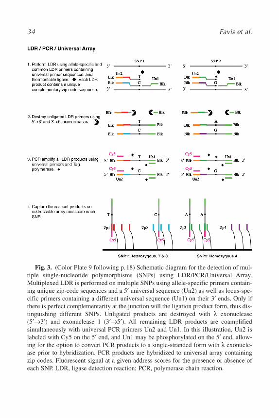
Fig. 3. (Color Plate 9 following p.18) Schematic diagram for the detection of mul-tiple single-nucleotide polymorphisms (SNPs) using LDR/PCR/Universal Array.Multiplexed LDR is performed on multiple SNPs using allele-specific primers contain-ing unique zip-code sequences and a 5′ universal sequence (Un2) as well as locus-spe-cific primers containing a different universal sequence (Un1) on their 3′ ends. Only ifthere is perfect complementarity at the junction will the ligation product form, thus dis-tinguishing different SNPs. Unligated products are destroyed with λ exonuclease(5′→3′) and exonuclease 1 (3′→5′). All remaining LDR products are coamplifiedsimultaneously with universal PCR primers Un2 and Un1. In this illustration, Un2 islabeled with Cy5 on the 5′ end, and Un1 may be phosphorylated on the 5′ end, allow-ing for the option to convert PCR products to a single-stranded form with λ exonucle-ase prior to hybridization. PCR products are hybridized to universal array containingzip-codes. Fluorescent signal at a given address scores for the presence or absence ofeach SNP. LDR, ligase detection reaction; PCR, polymerase chain reaction.
02_chap_Joos.qxd 25/04/2005 02:50 pm Page 34

35
Fig
. 4.
(Col
or P
late
11
foll
owin
g p.
18)
Ele
ctro
phor
etog
ram
of
30-p
lex
dete
ctio
n in
gen
omic
DN
A. T
he d
ata
dem
onst
rate
the
abi
lity
of L
DR
/PC
R to
cha
ract
eriz
e 60
alle
les
sim
ulta
neou
sly
in a
n SN
Pge
noty
ping
ass
ay. T
he b
lue
and
gree
n pe
aks
are
eith
er F
AM
or V
ic la
bele
dm
obili
ty m
odif
ied
zip-
chut
es. T
he x
-axi
s is
the
size
of
zip-
chut
e an
d th
e y-
axis
is f
luor
esce
nt in
tens
ity o
f ca
pilla
ry e
lect
roph
ores
is. T
he z
ip-
chut
es h
ave
the
sam
e co
lor
but
diff
eren
t m
otili
ties
for
both
of
alle
le-1
and
alle
le-2
. For
exa
mpl
e, 1
T(f
irst
blu
e pe
ak)
is h
omoz
ygou
s, T
;17
C (
gree
n) a
nd 1
7G (
gree
n) a
re h
eter
ozyg
ous
C/G
, etc
.
02_chap_Joos.qxd 25/04/2005 02:50 pm Page 35

36 Favis et al.
1.4.4. Detection of K-ras, BRCA1, BRCA2, and p53 Mutations Using Multiplex PCR/LDR and Zip-Code Capture
Since the zip-code sequences remain constant and their complements canbe appended to any set of LDR primers, our zip-code arrays are universal, andwe and our cancer collaborators at The Rockefeller University and the InstitutCurie have applied this array-based mutation detection to mutations in the K-ras,BRCA1, BRCA2, and p53 genes (3,8,9,12–14). Figure 2 shows the schematicand results for p53 mutation analysis, where by 110 mutations could be queriedsimultaneously. Mutations present at 1% of the wild-type DNA level, or inpooled samples could be distinguished (8,12).
1.5. Back to the Future
The practice of molecular medicine will require technology platforms thatcan span the progression from clinical trial to diagnostic laboratory and rapidlydeliver an answer. En route, the incipient diagnostic will be challenged with avariety of genes containing diverse assortments of genetic variation.
The Universal DNA microarray is a strong candidate to serve this need.Because the platform is programmable, it is robust enough to accommodatechanges in the genes, SNPs, or mutations of interest without reengineering thearray. In addition, our zip-code concept can be used for displaying mutations ona variety of platforms: universal array surfaces, gel or capillary electrophoresis,and universal encoded beads (see Fig. 5 [Color Plate 12 following p. 18] andSubheading 3.3.). This programmability and display versatility are addedboons in the context of clinical trials, in which the list of genes for which sub-jects provide consent for genotyping can vary from trial to trial, the SNP con-tent must reflect the targeted patient/volunteer population, and the number ofrecruited subjects can vary by orders of magnitude.
We and others have also shown that our assay can detect a greater range ofhuman genetic variation compared with other systems. In addition to SNPs andlow-level mutations (10), insertion/deletion mutations (8) and length polymor-phisms in mononucleotide (6) and dinucleotide repeats (7) can also be reliablydetected—two blind spots for direct hybridization arrays (49–51). Even thoughthis latter type of variation is invisible to most mutation detection technologies,such variations are known to have pharmacological relevance owing to theirprevalence in ADME genes. For example, CYP2D6 has numerous insertion/dele-tions that impact enzyme activity (52); UGT1A1 contains a promoter polymor-phism consisting of variable numbers of TA repeats that influences enzyme con-centration (53).
This technology is also amenable to the rapid provision of results in a clinicalsetting. Given that LDR can be performed in 5–10 min (54), a microfabricated
02_chap_Joos.qxd 25/04/2005 02:50 pm Page 36

Universal DNA Microarray 37
Fig. 5. (Color Plate 12 following p. 18) Ligase-based multiplexed single-nucleotidepolymorphism (SNP) assays combined with zip-code universal display. The originalLDR/PCR procedure combines ligation of multiple oligonucleotide probes on multipletarget sequences followed by PCR coamplification of ligation products using universalprimer sequences (2). The generic version for SNP detection is illustrated in the middleof the figure (D). Allele-specific oligonucleotides may hybridize on target DNA adjacentto a downstream locus-specific sequence containing a specific zip-code sequence andwill ligate if there is perfect complementarity at the junction. Use of blocked ends allowsfor exonuclease to degrade unligated probes but not the ligation products. UniversalprimersUn1, Un2, and Un3 are used to coamplify all the products. A variation of thisscheme uses only two universal primers but distinguishes alleles by appending differentzip-code sequences to the allele-specific oligonucleotides (as shown in Fig. 3). In
02_chap_Joos.qxd 25/04/2005 02:50 pm Page 37

38 Favis et al.
device that integrates sample prep with multiplexed genetic variation identifi-cation (PCR/LDR) and molecular profiling (LDR/PCR) would be anticipated toprovide results in less than 30 min. A reason for optimism lies in the significantprogress recently made in developing microfluidic and microchip-based plat-forms for point-of-care analysis. Although they are beyond the scope of thischapter, platforms that integrate sample preparation (55–57), purification andconcentration of the DNA/RNA component (58,59), component delivery(60–63), thermocycling (58,64,65), and capillary or channel-based separationand signal detection (54,66–80) will be considered for enriching our assaydevelopment.
2. Materials 1. Oligonucleotides (see Note 1).2. dNTPs (PE Biosystems, cat. no. 4303441).3. AmpliTaq Gold (PE Biosystems, cat. no. 4311820).4. Taq DNA ligase (NEB, cat. no. M0208L).5. Proteinase K (Qiagen, cat. no. 19131).6. Three 1-in microscope slides with etched circles (VWR, cat. no. 48349-057 or
Erie, cat. no. 2960; see Note 2).7. 24 × 50-mm Cover slips (VWR, cat. no. 48393-081; see Note 2).8. Corning crystallizing dishes, 170-mm diameter × 90-mm height (VWR, cat. no.
25411-140 or Corning, cat. no. 3140-170; see Note 3). 9. 20-Slide glass slide racks (VWR, cat. no. 25463-009 or Wheaton, cat. no. 900204).
10. Glass slide rack handle (VWR, cat. no. 25464-001 or Wheaton, cat. no. 900205).11. 50-Slide rack and staining dish (VWR, cat. no. 25461-024 or Wheaton, cat. no.
900400).12. Acrylamide (Boehringer Mannheim, cat. no. 1871757). 13. Bis-acrylamide (Boehringer Mannheim, cat. no. 1685830).14. Acrylic acid (Aldrich, cat. no. 14,723-0).
Fig. 5. (Continued from previous page) another variation of this scheme, allele-spe-cific oligonucleotide primers are extended with a polymerase prior to ligation to the com-mon oligonucleotide (A [23]). Alternatively, the nonligating ends of the upstream anddownstream probes may also be blocked by synthesizing a single long probe, which issubsequently linearized after exonuclease digestion in preparation for the PCR step (B[21]) In one version of this variation, an extra polymerase step is used to add a singlebase prior to ligation (B), although the approach works as well by using straight ligation(C [19]). In the examples illustrated here, two different fluorescent labels are used forthe UN2 and Un3 PCR primers, although schemes using a single label or more may beused. The PCR products may be displayed using gel or capillary electrophoresis (E[2,11]), universal array surfaces (F [3,8–10,12–14,19,21]), or universal encoded beads(G [22–24]).
02_chap_Joos.qxd 25/04/2005 02:50 pm Page 38

15. Ammonium persulfate (APS; VWR, cat. no. JT0762-11).16. 1-[3-(Dimethylamino)propyl]-3-ethylcarbodimide hydrochloride (Aldrich, cat. no.
16,146-2).17. N-hydroxysuccinimide (NHS; Aldrich, cat. no. 13,067-2).18. 3-(Trimethoxysilyl)propyl methacrylate (Aldrich, cat. no. 44,015-9).19. Concentrated NH4OH (VWR, cat. no. JT9721-4).20. 30% Hydrogen peroxide (Aldrich, cat. no. 21,676-3).21. Concentrated HCl (VWR, cat. no. JT9535-4).22. High-performance liquid chromatography (HPLC) grade methanol (VWR, cat. no.
JT9093-3).23. HPLC grade acetone (VWR, cat. no. JT9002-3).24. HPLC grade chloroform (Fisher, cat. no. C606-4).25. Triethylamine (Aldrich, cat. no. 47,128-3).26. 0.4 M K2HPO4 /KH2PO4, pH 5.5.27. 1 M 2-Morpholinoethanesulfonic acid (MES), pH 6.0 (light sensitive; filter-sterilize
and store at room temperature; stable for 3–4 mo; do not use if the solution appearsyellow).
28. 2X Hybridization buffer: 0.6 M MES, pH 6.0, 20 mM MgCl2, 0.2% sodium dodecylsulfate (SDS; light sensitive; store at room temperature; stable for 3–4 mo).
29. 1X Wash buffer: 0.3 M bicine, pH 8.0, filter-sterilized, 0.1% SDS (store at roomtemperature).
30. Drierite (VWR, cat. no. WLC3712T).31. Cover wells, 9-mm diameter × 1-mm height (Grace, Sunriver, OR). 32. Razor blade.33. Plastic slide box for hybridization (SPI Supplies, cat. no. 01253A-CF).34. Dessicator.35. Shaker. 36. Heat blocks.37. Hot plates.38. Thermocycler.39. Rotating hybridization oven.40. Cartesian Pixsys 5500 array spotting robot with a quill-type spotter in a controlled
atmosphere chamber or similar instrument.41. Perkin-Elmer ScanArray 5000 or similar instrument.
3. Methods3.1. Array Fabrication
Below is a description of the process used to produce universal arrays.Commercially available arrays with 3D surfaces are an alternative to in-houseproduction.
3.1.1. Clean Slides to Remove Oxidized Surfaces
1. Assemble slides in the glass slide racks. Put slides in back to back so there are twoslides per slot (see Note 4).
Universal DNA Microarray 39
02_chap_Joos.qxd 25/04/2005 02:50 pm Page 39

40 Favis et al.
2. In a hood using hot plates, boil 600 mL of ultrapure water in two glass crystalliz-ing dishes partially covered with a glass plate to prevent evaporation. Fill a thirddish with water, and heat to 60–70°C.
3. When the water boils, add 120 mL concentrated NH4OH and 120 mL 30% perox-ide to the first dish (5:1:1 water/NH4OH/peroxide; the solution will bubble vigor-ously). Place racked slides in solution, and boil for 10 min (see Note 5).
4. Rinse slides in 60–70°C dish of water for 2–3 min. Drain excess water from slideracks.
5. Add 120 mL concentrated HCl and 120 mL 30% peroxide to second dish of boilingwater (5:1:1 water/HCl/peroxide; the solution will bubble vigorously). Add slideracks, and boil for 10 min.
6. Rinse slides in water followed by HPLC-grade methanol and finally HPLC-gradeacetone. Air-dry (see Note 6).
3.1.2. Silanize Slides
1. Place cleaned slides in 50-slide rack and immerse in 400 mL CHCl3 solution con-taining 2% γ-(trimethoxysilyl)propyl methacrylate and 0.2% triethylamine. Agitategently at room temperature on a shaker table for 30 min (see Note 7).
2. Wash slides in 400 mL CHCl3 for 15 min on a shaker table. Repeat wash with freshCHCl3 (see Note 8).
3. Drain excess CHCl3, blot racked slides on paper towel, and let air-dry in a hood.Store slides in slide box until ready to prepare polymer surface. Do not touch sur-face of slides; handle slides by frosted end only (see Note 9).
3.1.3. Preparation of Polymer Surface1. Heat heating blocks to 70°C, and then invert blocks so the solid bottom surface is up.2. Make up monomer solution. The individual monomer solutions should be made
fresh (no more than 1or 2 d in advance). The solution is as follows: 200 µL 40%w/v acrylamide, 50 µL 40% w/v acrylic acid, 20 µL 1% w/v bis-acrylamide, 80 µL10% APS, and 650 µL water.
3. Vortex to mix, and allow mixture to sit at room temperature for 1 h before makingslides (see Note 10).
4. Spot 20 µL of monomer solution onto the center of a silanized slide from above.
a. Cover with a cover slip by lowering the cover slip parallel to the surface of theslide until it makes contact with the top of the monomer solution droplet.
b. Release the cover slip, and allow the monomer solution to spread. c. The solution should spread to the edges of the cover slip on its own. d. If numerous or large bubbles are caught under the cover slip, it can be slid off,
the surface gently wiped with a Kimwipe, and the procedure repeated using afresh cover slip (see Note 11).
5. Place slide on heated blocks for 4.5 min with cover slip side up (see Note 12).6. Following polymerization, place the slide into a slide rack immersed in water for
5 min.
02_chap_Joos.qxd 25/04/2005 02:50 pm Page 40

Universal DNA Microarray 41
7. Use a razor blade to wedge up one of the short sides of the cover slip, and thenslowly peel the cover slip from the polymer.
8. Place slides that have new polymer surfaces in racks. (We use test tube racks.) 9. Rinse the slides under running ultrapure water, use a forced air line to gently blow
off excess liquid, and then allow slides to air-dry. Store at room temperature inslide boxes until ready to activate surfaces for spotting (see Note 13).
3.1.4. Activation of Polymer Surfaces
1. Place polymer-coated slides in a 50-slide rack, and immerse in 400 mL 0.1 M potas-sium phosphate, pH 6.0, containing 0.1 M (EDC) 1-[3-(Dimethylamino)propyl]-3-ethylcarbodiimide hydrochloride and 20 mM NHS. Agitate gently at room temper-ature on a shaker table for 30 min.
2. Dunk racked slides in receptacle of ultrapure water approx 20 times to rinse.Replace water, and immerse approx 10 times.
3. Tap racked slides on paper towel to blot excess water, and blow off water usingforced air.
4. Heat slides at 65°C in an oven (cracked open slightly for good circulation) untilcompletely dry (30 min to 1 h).
5. Store slides desiccated at room temperature in slide boxes (see Note 14).
3.1.5. Array Spotting
1. Prepare spotting plates by mixing 5 µL 1000 µM zip-code oligonucleotide solu-tions with 5 µL 0.4 M K2HPO4/KH2PO4, pH 8.5, in 384 conical well spottingplates (see Note 15).
2. Place slides in spotter, and set relative humidity to 60–70%. Allow the slides toincubate for 15–20 min (see Note 16).
3. Spot slides in desired layout (see Note 17).4. Following spotting, removed uncoupled oligonucleotides from the polymer sur-
faces by soaking the slides in 300 mM bicine, pH 8.0/300 mM NaCl/0.1% SDS for30 min at 65°C (see Note 18).
5. Rinse the slides with ultrapure water, and dry as described above in Subheading 3.1.4.6. Store slides desiccated at room temperature in slide boxes (see Note 19).
3.1.6. Array Quality Control
Spotting failures are detected by staining two newly minted arrays from thebeginning and end of a spotting run with SYBR Green II via the method ofBattaglia et al. (81) (Fig. 6A; see Color Plate 13 following p. 18) This dye clear-ly shows whether addresses failed to spot during the array printing process; ifcritical addresses used by all assays are affected, defective arrays can be dis-carded. If addresses deeper into the array are affected, these arrays can be setaside for use in small assays that will be unaffected by the missing addresses(e.g., K-ras mutation detection only requires the first four addresses).
02_chap_Joos.qxd 25/04/2005 02:50 pm Page 41

42 Favis et al.
Fig. 6. (Color Plate 13 following p. 18) Validation of arrays following spotting. (A)Arrays are stained with a solution of SYBR Green II to determine whether all the zip-codes have been spotted successfully. The left panel shows an array, which has had aspotting failure, and the right panel shows the complete array. (B) Arrays are hybridizedwith mixtures of fluorescein-labeled zip-code complements to look for cross-talkresulting from either well-plate cross-contamination or poor washing of the pinsbetween spotting cycles.
Four random arrays are next chosen from array sets that pass this first roundof inspection and are subjected to hybridization with mixtures of fluorescein-labeled zip-code complements (see Subheading 3.2. for conditions). Mixturesare prepared for the even and odd rows, and the even and odd columns. Thistest confirms that there is no crosstalk between addresses caused by either well-plate crosscontamination or poor washing of the pins between spotting cycles.Figure 6B shows an array that has passed this level of inspection: specifichybridization to odd or even rows and odd or even columns are visible with noextraneous signals.
02_chap_Joos.qxd 25/04/2005 02:50 pm Page 42

Universal DNA Microarray 43
3.2. PCR/LDR/Array Hybridization3.2.1. PCR
1. Multiplex PCR is performed with 50–100 ng of genomic DNA in a 25 µL reactionusing AmpliTaq Gold and 2 pmol of each gene-specific primer bearing the univer-sal primer on the 5′ ends.
a. Overlay the reaction with mineral oil and princubate for 10 min at 95°C. b. Amplifiy for 15 cycles using conditions optimum for the genes of interest. c. Add a second 25 µl aliquot of the reaction mixture through the mineral oil con-
taining 25 pmol universal primer. d. Continue cycling for 25 cycles at a higher annealing temperature than that used
for the gene-specific primers.
2. Digest the reaction with the addition of 1 µL proteinase K (18 mg/mL) and incu-bation at 70°C for 10 min. Inactivate the proteinase K by incubating at 95°C for15 min.
3. Analyze a 1–2 µL aliquot by agarose gel electrophoresis to verify the presence ofamplification product of the expected size.
3.2.2. LDR
1. LDR reactions are carried out under oil. The following reactants are combined ina PCR tube for each sample: 1 µL 10X ligase buffer, 1 µL 100 mM dithiothreitol(DTT), 1 µL 10 mM NAD+, 500 fmol of each primer, and 2 µL of PCR productfrom Subheading 3.2.1., step 3 in a total volume of 10 µL.
2. Dilute the 40,000 U/mL (0.3 pmol/µL) Tth DNA ligase to 2.5 fmol/µL in 1X ligasebuffer. The volume of the diluted ligase should be sufficient to add 10 µL to eachreaction.
3. Heat-denature the 10 µL reaction at 94°C for 2 min, and add 10 µL (25 fmol) ofdiluted Tth DNA ligase.
4. Cycle the reaction for 20 rounds of 94°C for 30 s and 65°C for 4 min.
3.2.3. Array Hybridization
1. Dilute LDR reactions with an equal volume of 2X hybridization buffer to producea final buffer concentration of 300 mM MES, pH 6.0, 10 mM MgCl2, 0.1% SDS(see Note 20).
2. Denature the mixture at 94°C for 3 min and chill on ice.3. Preincubate arrays for 15 min at 25°C in 1X hybridization buffer. Remove the
arrays from the buffer and blow the surface dry of excess liquid.4. Attach cover wells to the arrays and fill with 35 µL of the diluted LDR products (see
Note 21). Use adhesive plastic to seal the cover well openings to prevent drying.5. Place the arrays in plastic slide holders and humidify using a moistened sponge.
Secure the arrays to the rotisseary of the rotating hybridization oven, and incubatefor 1 h at 65°C and 20 rpm (see Note 22).
6. After hybridization, wash the arrays in 300 mM bicine, pH 8.0/0.1% SDS for 10 minat 25°C. Rinse the arrays briefly in water, dry, and scan.
02_chap_Joos.qxd 25/04/2005 02:50 pm Page 43

44 Favis et al.
3.3. Variations on a Theme
In addition to the standard PCR/LDR technique outlined in Subheading 3.2.above, there are several variations of LDR as an assay tool for mutation identi-fication, SNP detection and DNA methylation analysis.
3.3.1. LDR/PCR
To genotype hundreds of thousands SNPs accurately in multiple samples in ahigh-throughput format, one variation is to perform LDR on the DNA samplesfollowed by PCR amplification of the ligation products. By performing LDRdirectly on the DNA samples using primers bearing universal sequences on the5′ end (see Fig. 3), ligation products can be subsequently simultaneously ampli-fied using universal primers. There are several advantages to performing LDRprior to PCR amplification: (1) it eliminates the time-consuming steps of designand optimization of multiplex gene-specific PCR primers; (2) it reduces the costof synthesizing hundreds of fluorescently labeled allele-specific LDR primers,since product labeling can be accomplished using fluorescently labeled univer-sal PCR primers; (3) it reduces the complexity of the assay system to ensure anaccurate and efficient DNA analysis by avoiding the common pitfalls associatedwith multiplex PCR amplification (e.g., formation of primer dimers and othernonspecific amplicons that may interfere with downstream applications); (4) itreduces the time required for LDR primer design and reaction optimization; and(5) it provides an initial linear amplification of the targeted genomic informationthat is nonbiased, it promotes allelic balance, and it may minimize the need forlater PCR cycles that may detract from this balance.
3.3.1.1. LDR/PCR/UNIVERSAL MICROARRAY
The use of multiplex LDR followed by PCR was initially developed to scorechromosomal instability in tumors (see Subheading 1.4. above). A schematicdiagram of multiplex LDR/PCR/Universal Array to determine DNA copy num-ber or score SNPs is shown in Fig. 3. In this approach, the universal primersequences are added to the 5′ end of discriminating LDR primers and to the 3′end of common LDR primers. After ligase detection reaction, the excess unli-gated LDR primers and DNA templates can be digested using 5′→3′ and 3′→5′exonucleases. The ligation products are protected from digestion, since block-ing groups are added at both their 5′ and 3′ ends. This exonuclease digestionstep reduces the potential of nonspecific hybridization and false-positive resultson the universal array readout. The ligation products are simultaneously ampli-fied with universal PCR primers. Only one of the universal PCR primers is fluo-rescently labeled to serve as the detection signal when these amplicons are cap-tured on a universal array.
02_chap_Joos.qxd 25/04/2005 02:50 pm Page 44

3.3.1.2. LDR/PCR/UNIVERSAL DISPLAY
ABI recently extended the LDR/PCR concept with the development of anultra-high-throughput genotyping method, SNPlex. This technology utilizesmultiplexed oligonucleotide ligation assay on genomic DNA. Each LDR primerpair was synthesized with universal primer sequences flanking the locus-specificsequences. A unique zip-code sequence is designed within the LDR primers touniquely identity each LDR product. The excess LDR primers and genomicDNA are eliminated through enzymatic digestion. Consequently, all LDRproducts may be amplified in a single PCR step with two universal PCRprimers, one of which is biotinylated. Biotinylated amplicons may be renderedsingle stranded and captured on streptavidin-coated plates. Each single-strandedPCR product may be identified by its unique zip-code sequence through inter-rogation with a set of universal ZipChute probes. These probes have fluores-cence labels, unique complementary zip-code sequences, and ABI mobilitymodifiers. ZipChute probes can be eluted and electrophoretically separated onan ABI 3730xl DNA analyzer. Figure 4 shows an example of a 60-plex reac-tion using this approach. This technique has been validated on 3,000 SNPsusing 96 genomic DNA samples. Compared with other genotyping platforms,the SNPlex system demonstrates 98.7 and 99.2% concordance with dideoxysequencing and TaqMan assays, respectively. This variation is an alternativeapproach to existing genotyping methodologies and has the advantage of arobust detection strategy and low DNA consumption.
3.3.2. Bisulfite/PCR-PCR/LDR/Universal Microarray
One application of LDR/PCR/Universal Array is to study DNA methylation.In particular, this variation focuses on the detection of aberrant promotermethylation occurring at the 5-position of cytosine within the CpG dinu-cleotide. Sodium bisulfite conversion of cytosines to uracils is one of the mostcommonly used methods to study DNA methylation. 5-Methylcytosines areresistant to conversion, and deamination only occurs on unmethylated cytosines.The modified DNA sequences can then either be amplified and sequenced, or onecan perform methylation-specific PCR (MSP) to determine cytosine methylationstatus. Our bisulfite/PCR-PCR/LDR/Universal Array approach provides a sen-sitive and accurate high-throughput format that can detect methylation status invirtually any gene sequence of interest.
A multiplex PCR-PCR/LDR assay is shown in Fig. 7; (see Color Plate 14 fol-lowing p. 18) to illustrate this approach. When possible, the gene-specific PCRprimers used for multiplexing are designed to avoid CpG sites present in the pro-moter sequences. As a further improvement to accommodate situations in whichbisulfite-modified bases cannot be avoided, pyrimidine and purine nucleotide
Universal DNA Microarray 45
02_chap_Joos.qxd 25/04/2005 02:50 pm Page 45

46 Favis et al.
Fig. 7. (Color Plate 14 following p. 18) Schematic diagram, illustrating the proce-dure for high-throughput detection of promoter methylation status with the combinationof bisulfite treatment, multiplex PCR, multiplex LDR, and universal array approaches.The different fluorescently labeled (Cy3 and Cy5) LDR products are captured on thesame addressable array. PCR, polymerase chain reaction; LDR, ligase detection reaction.
02_chap_Joos.qxd 25/04/2005 02:50 pm Page 46

analogs are incorporated within the PCR primers. These modified bases, designat-ed P and K, show considerable promise as degenerate bases. The pyrimidinederivative P base pairs with either A or G, whereas the purine derivative K basepairs with either C or T; thus the target DNA can be amplified regardless of itsmethylation status. Multiple promoter regions are amplified in a two-stage nestedPCR reaction simultaneously. The first-stage multiplex amplification uses pairs ofgene-specific PCR primers with universal sequences attached to their 5′ ends. Thesecond-stage amplification uses universal PCR primers to amplify the first-stagePCR products. The final PCR products are usually verified on an agarose gel priorto LDR analysis.
The details of LDR primer design have been described in Subheading 3. andin Note 1. Briefly, three LDR primers are designed for each CpG dinucleotidesite. Two discriminating primers are labeled at the 5′ end with either Cy3 orCy5 and at the 3′ end with a G or A, respectively. The single common primerfor the reaction consists of a 5′ phosphate and terminates at the 3′ end with azip-code complement sequence. Degeneracy is also accommodated in the LDRprimers by using pyrimidine and purine nucleotide analogs. After the LDRproducts are captured on a universal array, the methylated cytosine residues aredetected by the presence of Cy3 signals; the presence of unmethylatedcytosines is revealed by the presence of Cy5 signals.
Typically, 1–2 µg of genomic DNA in a volume of 40 µL is incubated with0.2 N NaOH at 37°C for 10 min. Then 30 µL freshly made 10 mM hydro-quinone and 520 µL of freshly made 3 M sodium bisulfite, pH 5.0 (Sigma, ACSgrade) is added. This mixture is next incubated for 16 h in a DNA thermocyclerusing alternating cycles of 50°C for 20 min followed by a denaturing step of85°C for 15 s. The bisulfite-treated DNAs can be desalted using MICROCONcentrifugal filter devices (Millipore, Bedford, MA) or, alternatively, cleanedwith a Wizard DNA clean-up kit (Promega, Madison, WI). The eluted DNAsare incubated with 1/10 volume of 3 N NaOH at room temperature for 5 minprior to ethanol precipitation. The DNA pellet is then resuspended in 20 µLdeionized H2O and stored at 4°C. Bisulfite-modified DNA is stable at 4°C forat least 1 mo.
The current assay is designed to detect the extent of DNA methylation with-in the promoters of the tumor suppressor genes p15INK4b, p16INK4a, p19ARF,p21CIP, p27KIP, p53, and BRCA1, as well as the imprinted gene small nuclearribonucleoprotein N (SNRPN). Using the same design parameters, the promoterregions of seven additional genes were chosen to investigate their promotermethylation status in human tumors. These include O6 methyl guanine DNAmethyl transferase (MGMT), adenomatous polyposis coli (APC), retinoic acidreceptor (RARb), tissue inhibitor metalloproteinase (TIMP-3), death-associatedprotein kinase (DAPK), E-cadherin (ECAD), glutathione S-transferase (GSTP1),
Universal DNA Microarray 47
02_chap_Joos.qxd 25/04/2005 02:50 pm Page 47

48 Favis et al.
and Ras association domain family 1 (RASSF1). The hemimethylated SNRPN isused as a positive internal control.
As seen in Fig. 8 (see Color Plate 15 following p.178), to demonstrate thatLDR primers are working properly, genomic DNAs of normal lymphocyteswith and without in vitro methylation are included in experiments as controls.DNA extracted from colorectal cancer cell lines SW1116 and DLD1 is alsoused to validate this strategy. All experiments were performed minimally induplicate to avoid ambiguity. For each promoter region, three CpG sites werechosen to analyze their methylation status. The presence of Cy5 signals indi-cates efficient amplification during multiplexing PCR steps. The promoterregions will be considered to be hypermethylated only when at least two CpGsites can be detected by Cy3 labeling. In most cases, the universal arrays pro-vide very high capture specificity. The methylated promoters identified in thismethod may be reconfirmed by either bisulfite sequencing or uniplexPCR/PCR/LDR under more stringent hybridization conditions on a fresharray in a separate experiment.
In contrast to MSP-based methods, the bisulfite/PCR/LDR approach cir-cumvents the issues of incomplete bisulfite conversion (C to U modification isnot 100% efficient) and the potential primer extension of unmethylated DNAby extension of a G:U mismatch. The requirement of scoring methylation atthree CpG sites per promoter using LDR should help the assay retain itsexquisite specificity.
4. Notes1. We recommend the software program Oligo for LDR primer design. This program
is also useful in designing PCR and multiplex PCR primers. This program calcu-lates Tm using the nearest neighbor method. Gene-specific PCR and LDR primersare generally designed with Tms around 70°C. To perform multiplex PCR/PCR,gene-specific oligonucleotide primers with universal primer sequence attached tothe 5′ ends are required. The sequence of the universal primer is 5′-ggagcacgctatc-ccgttagac-3′. LDR discriminating primers are labeled at the 5′ end with fluorophoresthat can be detected by the array-scanning instrument and that have sufficient spec-tral separation to avoid confounding owing to overlapping signals. The final baseon the discriminating primer is the query base. The LDR common primer is mod-ified with a 5′ phosphate and the 24-base zip-code complement is appended to the3′ end (see Table 1 for sequences). When synthesized on a 1-µmol scale or larger,zip-code oligonucleotides used for spotting arrays should be gel purified. Whensynthesized on a smaller scale, a reversed-phase, solid-phase extraction columnproduces satisfactory results. The zip-code oligonucleotides are synthesized on a 3′amino modifier C3 column (Glen Research) with a spacer C18 (Glen Research)inserted before the first base.
02_chap_Joos.qxd 25/04/2005 02:50 pm Page 48

Universal DNA Microarray 49
Fig. 8. (Color Plate 15 following p. 178) Universal array images of methylationprofiles of selected promoter regions (SNRPN, p15, p16, p19, p21, p27, p53, andBRCA1) in normal and colorectal tumor cell line genomic DNAs. False color greenrepresents the status of unmethylated promoter regions detected by Cy5-labeled LDRprimers. False color red represents the status of methylated promoter regions detectedby Cy3-labeled LDR primers. (A) LDR results of normal human lymphocyte genomicDNAs in the presence (right panel) and absence (left panel) of in vitro methylationusing SssI methylase. (B,C) The methylation profiles of two colorectal cancer cell linegenomic DNAs were analyzed. Among the eight genes that were analyzed in cell lineSW1116, Cy3 labeled LDR products only present on the p16 promoter region. Thisindicates that only the p16 promoter was hypermethylated. The presence of Cy3 signalon both p16 and p19 promoters in cell line DLD-1 indicates that both of these promot-ers are hypermethylated.
02_chap_Joos.qxd 25/04/2005 02:50 pm Page 49

50 Favis et al.
Table 1Sequences of Zip-Code-Related Oligonucleotides
Zip-code Zip-code complement
TTGAAATCCAGCGCAAAATCTGCG CGCAGATTTTGCGCTGGATTTCAATTGAAAAGCCTACACGACGGCGAA TTCGCCGTCGTGTAGGCTTTTCAATTGATCTGCCATACGGGCTTACGG CCGTAAGCCCGTATGGCAGATCAATTGACTTGTCCCCAGCACGGCCAT ATGGCCGTGCTGGGGACAAGTCAATTGACGTTGACCAGCCCGTTGCAA TTGCAACGGGCTGGTCAACGTCAATTGACGAAGCTTTCCCCCATGATG CATCATGGGGGAAAGCTTCGTCAATTGAGCAAGGACGACCGCAAACGG CCGTTTGCGGTCGTCCTTGCTCAATTGAGATGACGGACGGTGCGGCAA TTGCCGCACCGTCCGTCATCTCAATTGATCCCATCGAAAGGGACGATG CATCGTCCCTTTCGATGGGATCAATTGATGCGTCTGGGACGTGCCTTG CAAGGCACGTCCCAGACGCATCAATTGACACGTCGTCAGCTCCCGTGC GCACGGGAGCTGACGACGTGTCAATTGACAGCCTGTTGCGGTGCGTCT AGACGCACCGCAACAGGCTGTCAATTGAGTGCGGTACTTGCAGCGATG CATCGCTGCAAGTACCGCACTCAATTGAACGGTCTGCACGTCCCAGCC GGCTGGGACGTGCAGACCGTTCAATGATTCTGGTGCGTGCCAGCCAGC GCTGGCTGGCACGCACCAGAATCATGATTGTCGCTTTCTGACGGAGCC GGCTCCGTCAGAAAGCGACAATCATGATCGTTTGCGGGTATCCCTCGT ACGAGGGATACCCGCAAACGATCATGATCGAAAGGACAGCAGCCTCCC GGGAGGCTGCTGTCCTTTCGATCATGATGCAAGCAACGAACACGCTGT ACAGCGTGTTCGTTGCTTGCATCATGATTGCGAGTGGACCATCGCCAT ATGGCGATGGTCCACTCGCAATCATGATCACGCTTGCCATGGACGGAC GTCCGTCCATGGCAAGCGTGATCATGATGTGCCTCAACGGGTGCAGCC GGCTGCACCCGTTGAGGCACATCATGATGGACCGTTAGCCGATGTTGA TCAACATCGGCTAACGGTCCATCATGATACGGAGGAGGACTGCGTGCG CGCACGCAGTCCTCCTCCGTATCATTAGGATGAGCCAGCCTGCGAGCC GGCTCGCAGGCTGGCTCATCCTAAAATCTCGTCGTTTCCCCTCATGCG CGCATGAGGGGAAACGACGAGATTAATCGCAACTGTCGTTCACGGTGC GCACCGTGAACGACAGTTGCGATTAATCAGGACACGCAGCGACCTGCG CGCAGGTCGCTGCGTGTCCTGATTAATCGACCCTGTGTCTGCTTTGCG CGCAAAGCAGACACAGGGTCGATTAATCAGCCAAAGCGAAGTGCGATG CATCGCACTTCGCTTTGGCTGATTATACGACCTCGTGAGTTCCCGCAA TTGCGGGAACTCACGAGGTCGTATAAAGCTTGACCTATCGAGCCGTGC GCACGGCTCGATAGGTCAAGCTTTAAAGAGCCGCTTGAGTCGAAATCG CGATTTCGACTCAAGCGGCTCTTTTCTGCTTGCTCACCTACCATTGCG CGCAATGGTAGGTGAGCAAGCAGATCTGATCGCCTAGGTAACGGGGAC GTCCCCGTTACCTAGGCGATCAGATCTGCAGCGGTACTGTGGACCCAT ATGGGTCCACAGTACCGCTGCAGATCTGAGCCACCTAATCTCCCACGG CCGTGGGAGATTAGGTGGCTCAGATGTCTCGTTCCCACCTCCATTCCC GGGAATGGAGGTGGGAACGAGACATGTCATCGCAGCGAGTCAGCCACG CGTGGCTGACTCGCTGCGATGACA
(Continued)
02_chap_Joos.qxd 25/04/2005 02:50 pm Page 50

Universal DNA Microarray 51
Table 1(Continued)
Zip-code Zip-code complement
TGTCCCTAACCTGATGGTGCGCAA TTGCGCACCATCAGGTTAGGGACATGTCTGCGGTCTCCATATCGGTGC GCACCGATATGGAGACCGCAGACATGTCCACGCGTTACCTTGTCGATG CATCGACAAGGTAACGCGTGGACATGTCGTGCTCTGACCTTGCGCTCA TGAGCGCAAGGTCAGAGCACGACATGTCACGGAATCGTGCTGCGGCTT AAGCCGCAGCACGATTCCGTGACATCGTTCTGGCTTGGACGCTTCTCA TGAGAAGCGTCCAAGCCAGAACGATCGTTGCGTGTCGGACCTTGGATG CATCCAAGGTCCGACACGCAACGACTTGCGTTGATGCGAATCGTCGAA TTCGACGATTCGCATCAACGCAAGCTGTCACGCTCAACCTTCCCCGTT AACGGGGAAGGTTGAGCGTGACAGCTGTGTGCCGTTTCGTGTGCAGTG CACTGCACACGAAACGGCACACAGCCATGCAATCCCAGGATGTCGGTA TACCGACATCCTGGGATTGCATGGCCATCAGCTCTGGCAATGCGGAGT ACTCCGCATTGCCAGAGCTGATGGCGAATCTGGGTAAGGAAGCCATCG CGATGGCTTCCTTACCCAGATTCGCGAATGTCCTGTCCATCGAATGCG CGCATTCGATGGACAGGACATTCGCGAACGTTTACATGCGTCGTAGCC GGCTACGACGCATGTAAACGTTCGCGAAAGTGAGCCGCAACTTGGGAC GTCCCAAGTTGCGGCTCACTTTCGCGAAGGACAGTGAGTGTGCGCACG CGTGCGCACACTCACTGTCCTTCGGCAAAGCCATACCTTGGCTTGCTT AAGCAAGCCAAGGTATGGCTTTGCACCTCTTGCCTACGAACAGCCGAA TTCGGCTGTTCGTAGGCAAGAGGTACCTGCAAGTGCCCATGTGCCCTA TAGGGCACATGGGCACTTGCAGGTAGGATCTGGACCGGACTCCCCGAA TTCGGGGAGTCCGGTCCAGATCCTAGGAACGGCAGCTACACACGAGCC GGCTCGTGTGTAGCTGCCGTTCCTGATGGACCACCTCAGCGCTTGACC GGTCAAGCGCTGAGGTGGTCCATCTCCCTTAGGACCCAGCGTCTGTGC GCACAGACGCTGGGTCCTAAGGGATGCGCCATAAAGGACCTTAGCCAT ATGGCTAAGGTCCTTTATGGCGCATGCGAGGACCATGGTAGGTAAGCC GGCTTACCTACCATGGTCCTCGCA
2. Use only sealed boxes of slides. Opened boxes do not clean well, probably becauseof to either heavy oxidation or reaction with chemicals present in the lab.
3. Each dish holds two glass slide racks.4. We use slides with two etched circles when making arrays. A single array is spot-
ted within each circle.5. This process can be repeated for a second set of slides with the existing solutions
before the reagents boil out and too much liquid evaporates. Use fresh solutions forthe next rounds.
6. Silanize as soon as possible or leave in water overnight (rinse #1, step 7) if youcannot silanize immediately.
7. Do not touch the surface of slides after cleaning; handle slides by frosted endonly.
02_chap_Joos.qxd 25/04/2005 02:50 pm Page 51

52 Favis et al.
8. If you are preparing a large number of slides, the second wash of one batch can beused as the first wash for the next batch of slides.
9. The silanized slides are stable for several weeks prior to coating with the polymer.For long-term storage, place in a desiccator.
10. Allowing the solution to sit produces more uniform surfaces. 11. If slides were cleaned and silanized properly, the solution should form a rela-
tively small, well-defined droplet on the surface. Do not worry about small voidsor bubbles at the edges of the cover slip; you will not be using that area of thepolymer.
12. Using two to four blocks, you can heat 8–16 slides simultaneously if you staggerthem by 20 s. The two outer slides on the blocks will hang off the edges slightly,but this does not effect the polymerization.
13. The slides are stable for at least 6 mo prior to activation.14. The activated slides are stable for a minimum of 6 mo if stored properly desiccated.15. Plates can be sealed and stored refrigerated for spotting, on consecutive days.
Prior to spotting, spin the plates to collect all the liquid in the bottom of the wells.For long-term storage, dry the spotting plate, seal, and store at −20°C. The daybefore spotting, redissolve the wells in the appropriate amount of water (assume~0.5 µL loss in volume/spotting run), vortex the plate several times, and storerefrigerated. The day of spotting, vortex several times, and then spin the plate tocollect all the liquid in the bottom of the wells.
16. The slides are very hydroscopic when dry. If not allowed to partially rehydrate priorto spotting, the first few slides will suck all the spotting solution out of the pin.
17. We normally spot each zip-code in duplicate. Additionally, we spot a set of fiducialsalong the top and one side of the array (see bottom panel of Fig. 2) for alignmentpurposes. The fiducials are made on a DNA synthesizer and have the followingstructure: amino-group-T-T-fluorescent dye. The fiducials should be printed afterthe zip-code oligonucleotides because the dyes are sticky, and carryover contami-nation can be a problem. The pins should be sonicated thoroughly after spotting ofthe fiducials to prevent contaminating the next spotting run with fluorescent dye.
18. This can be done immediately following spotting or any time just prior tohybridization.
19. The spotted slides are stable for a minimum of 6 mo.20. The LDR can remain in the original reaction tube/plate, and the 2X buffer can be
added through the oil. Following denaturation, the tubes/plate should not need tobe spun down, and the hybridization mix can be drawn out from under the oil.
21. There are cover wells available in a variety of sizes for different size arrays. Theimportant thing is not to fill the chamber completely so there is an air bubble presentto facilitate mixing.
22. Avoid excessive liquid in the sponge, since this may create unnecessary moisture andresult in a capillary stream between the adjacent hybridization chambers under coverwell gaskets that have not been securely sealed against the array surface. This cross-contamination issue can easily be solved by just slightly dampening the sponge, thatis, make the sponge damp but without the ability to squeeze out any liquid.
02_chap_Joos.qxd 25/04/2005 02:50 pm Page 52

Universal DNA Microarray 53
AcknowledgmentsSupport for this work was provided by the National Cancer Institute (grouts
P01-CA65930 and RO1-CA81467).
References1. Kirk, B. W., Feinsod, M., Favis, R., Kliman, R. M., and Barany, F. (2002) Single
nucleotide polymorphism seeking long term association with complex disease.Nucleic Acids Res. 30, 3295–3311.
2. Barany, F. and Lubin, M. (1997) Detection of nucleic acid sequence differencesusing coupled ligase detection and polymerase chain reactions. International PatentApplication (WO9745559A1).
3. Gerry, N. P., Witowski, N. E., Day, J., Hammer, R. P., Barany, G., and Barany, F.(1999) Universal DNA microarray method for multiplex detection of low abun-dance point mutations. J. Mol. Biol. 292, 251–262.
4. Khanna, M., Park, P., Zirvi, M., et al. (1999) Multiplex PCR/LDR for detection ofK-ras mutations in primary colon tumors. Oncogene 18, 27–38.
5. Khanna, M., Cao, W., Zirvi, M., Paty, P., and Barany, F. (1999) Ligase detectionreaction for identification of low abundance mutations. Clin. Biochem. 32, 287–290.
6. Zirvi, M., Nakayama, T., Newman, G., McCaffrey, T., Paty, P., and Barany, F.(1999) Ligase-based detection of mononucleotide repeat sequences. Nucleic AcidsRes. 27, e40.
7. Zirvi, M., Bergstrom, D. E., Saurage, A. S., Hammer, R. P. and Barany, F. (1999)Improved fidelity of thermostable ligases for detection of microsatellite repeatsequences using nucleoside analogs. Nucleic Acids Res. 27, e41.
8. Favis, R., Day, J. P., Gerry, N. P., Phelan, C., Narod, S., and Barany, F. (2000)Universal DNA array detection of small insertions and deletions in BRCA1 andBRCA2. Nat. Biotechnol. 18, 561–564.
9. Favis, R. and Barany, F. (2000) Mutation detection in K-ras, BRCA1, BRCA2, andp53 using PCR/LDR and a universal DNA microarray. Ann. N. Y. Acad. Sci. 906,39–43.
10. Dong, S. M., Traverso, G., and Johnson, C. (2001) Detecting colorectal cancer instool with the use of multiple genetic targets. J. Natl. Cancer Inst. 93, 858–865.
11. Day, J. P. (2003) The 53rd American Society of Human Genetics Annual Meeting,Poster #1589.
12. Favis, R., Huang, J., Gerry, N., et al. (2003) Harmonized microarray/mutationscanning analysis of TP53 mutations in undissected colorectal tumors. Hum. Mutat.24, 63–75.
13. Fouquet, C., Antoine, M., Tisserand, P., et al. (2003) Rapid and sensitive p53alteration analysis in biopsies from lung cancer patients using a functional assayand a universal oligonucleotide array: a prospective study. Clin. Cancer Res. 10,3479–3489.
14. Overholtzer, M., Rao, P. H., Favis, R., et al. (2003) The presence of p53 mutationsin human osteosarcomas correlates with high levels of genomic instability. Proc.Natl. Acad. Sci. USA 100, 11547–11552.
02_chap_Joos.qxd 25/04/2005 02:50 pm Page 53

15. Barany, F. and Gelfand, D. H. (1991) Cloning, overexpression and nucleotidesequence of a thermostable DNA ligase-encoding gene. Gene 109, 1–11.
16. Barany, F. (1991) The ligase chain reaction in a PCR world. PCR Methods Appl.1, 5–16.
17. Day, D. J., Speiser, P. W., White, P. C., and Barany, F. (1995) Detection of steroid21-hydroxylase alleles using gene-specific PCR and a multiplexed ligation detec-tion reaction. Genomics 29, 152–162.
18. Belgrader, P., Devaney, J. M., Del Rio, S. A., Turner, K. A., Weaver, K. R., andMarino, M. A. (1996) Automated polymerase chain reaction product sample prepa-ration for capillary electrophoresis analysis. J. Chromatogr. B. Biomed. Appl. 683,109–114.
19. Baner, J., Isaksson, A., Waldenstrom, E., Jarvius, J., Landegren, U., and Nilsson,M. (2003) Parallel gene analysis with allele-specific padlock probes and tagmicroarrays. Nucleic Acids Res. 31, e103.
20. Consolandi, C., Busti, E., Pera, C., et al. (2003) Detection of HLA polymorphismsby ligase detection reaction and a universal array format: a pilot study for low res-olution genotyping. Hum. Immunol. 64, 168–178.
21. Hardenbol, P., Baner, J., Jain, M., et al. (2003) Multiplexed genotyping withsequence-tagged molecular inversion probes. Nat. Biotechnol. 21, 673–678.
22. Iannone, M. A., Taylor, J. D., Chen, J., et al. (2000) Multiplexed single nucleotidepolymorphism genotyping by oligonucleotide ligation and flow cytometry.Cytometry. 39, 131–140.
23. Oliphant, A., Barker, D. L., Stuelpnagel, J. R., and Chee, M. S. (2002) BeadArraytechnology: enabling an accurate, cost-effective approach to high-throughputgenotyping. Biotechniques Suppl., 56–80, 60–61.
24. Han, M., Gao, X., Su, J. Z., and Nie, S. (2001) Quantum-dot-tagged microbeadsfor multiplexed optical coding of biomolecules. Nat. Biotechnol. 19, 631–635.
25. Busti, E., Bordoni, R., Castiglioni, B., et al. (2002) Bacterial discrimination bymeans of a universal array approach mediated by LDR (ligase detection reaction).BMC Microbiol. 2, 27.
26. Chen, J., Iannone, M. A., Li, M. S. T., et al. (2000) A microsphere-based assay formultiplexed single nucleotide polymorphism analysis using single base chainextension. Genome Res. 10, 549–557.
27. Epstein, J. R., Ferguson, J. A., Lee, K. H., and Walt, D. R. (2003) Combinatorialdecoding: an approach for universal DNA array fabrication. J. Am. Chem. Soc. 125,13753–13759.
28. Hirschhorn, J. N., Sklar, P., Lindblad-Toh, K., et al. (2000) SBE-TAGS: an array-based method for efficient single-nucleotide polymorphism genotyping. Proc. Natl.Acad. Sci. USA 97, 12164–12169.
29. Jarvius, J., Nilsson, M., and Landegren, U. (2003) Oligonucleotide ligation assay.Methods Mol. Biol. 212, 215–228.
30. Ladner, D. P., Leamon, J. H., Hamann, S., et al. (2001) Multiplex detection ofhotspot mutations by rolling circle-enabled universal microarrays. Lab. Invest. 81,1079–1086.
54 Favis et al.
02_chap_Joos.qxd 25/04/2005 02:50 pm Page 54

31. Mikhailovich, V., Lapa, S., Gryadunov, D., et al. (2001) Identification of rifampin-resistant Mycobacterium tuberculosis strains by hybridization, PCR, and ligasedetection reaction on oligonucleotide microchips. J. Clin. Microbiol. 39,2531–2540.
32. Taylor, J. D., Briley, D., Nguyen, Q., et al. (2001) Flow cytometric platform forhigh-throughput single nucleotide polymorphism analysis. Biotechniques 30,661–666, 68–169.
33. Zhong, X. B., Lizardi, P. M., Huang, X. H., Bray-Ward, P. L., and Ward, D. C.(2001) Visualization of oligonucleotide probes and point mutations in interphasenuclei and DNA fibers using rolling circle DNA amplification. Proc. Natl. Acad.Sci. USA 98, 3940–3945.
34. Zhong, X. B., Reynolds, R., Kidd, J. R., et. al. (2003) Single-nucleotide polymor-phism genotyping on optical thin-film biosensor chips. Proc. Natl. Acad. Sci. USA100, 11559–11564.
35. Pastinen, T., Raitio, M., Lindroos, K., Tainola, P., Peltonen, L., and Syvanen, A. C.(2000) A system for specific, high-throughput genotyping by allele-specific primerextension on microarrays. Genome Res. 10, 1031–1042.
36. Cronin, M. T., Fucini, R. V., Kim, S. M., Masino, R. S., Wespi, R. M., and Miyada,C. G. (1996) Cystic fibrosis mutation detection by hybridization to light-generatedDNA probe arrays. Hum. Mutat. 7, 244–255.
37. Hacia, J. G., Brody, L. C., Chee, M. S., Fodor, S. P., and Collins, F. S. (1996)Detection of heterozygous mutations in BRCA1 using high density oligonucleotidearrays and two-colour fluorescence analysis [see comments]. Nat. Genet. 14,441–447.
38. Southern, E. M. (1996) DNA chips: analysing sequence by hybridization tooligonucleotides on a large scale. Trends Genet. 12, 110–115.
39. Wang, Y., Blandino, G., Oren, M., and Givol, D. (1998) Induced p53 expression inlung cancer cell line promotes cell senescence and differentially modifies the cyto-toxicity of anti-cancer drugs. Oncogene 17, 1923–1930.
40. Grossman, P. D., Bloch, W., Brinson, E., et al. (1994) High-density multiplexdetection of nucleic acid sequences: oligonucleotide ligation assay and sequence-coded separation. Nucleic Acids Res. 22, 4527–14534.
41. Eggerding, F. A., Iovannisci, D. M., Brinson, E., Grossman, P., and Winn-Deen, E. S.(1995) Fluorescence-based oligonucleotide ligation assay for analysis of cystic fibro-sis transmembrane conductance regulator gene mutations. Hum. Mutat. 5, 153–165.
42. Feero, W. T., Wang, J., Barany, F., et al. (1993) Hyperkalemic periodic paralysis:rapid molecular diagnosis and relationship of genotype to phenotype in 12 fami-lies. Neurology 43, 668–673.
43. Day, D. J., Speiser, P. W., Schulze, E., et al. (1996) Identification of non-amplify-ing CYP21 genes when using PCR-based diagnosis of 21-hydroxylase deficiencyin congenital adrenal hyperplasia (CAH) affected pedigrees. Hum. Mol. Genet. 5,2039–2048.
44. Lin, Z., Cui, X. and Li, H. (1996) Multiplex genotype determination at a largenumber of gene loci. Proc. Natl. Acad. Sci. USA 93, 2582–2587.
Universal DNA Microarray 55
02_chap_Joos.qxd 25/04/2005 02:50 pm Page 55

45. Nickerson, D. A., Whitehurst, C., Boysen, C., Charmley, P., Kaiser, R., and Hood,L. (1992) Identification of clusters of biallelic polymorphic sequence-tagged sites(pSTSs) that generate highly informative and automatable markers for genetic link-age mapping. Genomics 12, 377–387.
46. Schouten, J. P., McElgunn, C. J., Waaijer, R., Zwijnenburg, D., Diepvens, F., andPals, G. (2002) Relative quantification of 40 nucleic acid sequences by multiplexligation-dependent probe amplification. Nucleic Acids Res. 30, e57.
47. Gille, J. J., Hogervorst, F. B., Pals, G., et al. (2002) Genomic deletions of MSH2and MLH1 in colorectal cancer families detected by a novel mutation detectionapproach. Br. J. Cancer 87, 892–897.
48. White, S., Kalf, M., Liu, Q., et al. (2002) Comprehensive detection of genomicduplications and deletions in the DMD gene, by use of multiplex amplifiable probehybridization. Am. J. Hum. Genet. 71, 365–374.
49. Ahrendt, S., Halachmi, S., Chow, J., et al. (1999) Rapid p53 sequence analysis inprimary lung cancer using an oligonucleotide probe array. Proc. Natl. Acad. Sci.USA 96, 7382–7387.
50. Wikman, F. P., Lu, M. L., Thykjaer, T., et al. (2000) Evaluation of the performanceof a p53 sequencing microarray chip using 140 previously sequenced bladdertumor samples. Clin. Chem. 46, 1555–1561.
51. Wen, W. H., Bernstein, L., Lescallett, J., et al. (2000) Comparison of TP53 muta-tions identified by oligonucleotide microarray and conventional DNA sequenceanalysis. Cancer Res. 60, 2716–2722.
52. Ingelman-Sundberg, M., Daly, A. K., and Nebert, D. W. (2003). Feb. 18, 2003 Ed.Karolinska Institute, Vol. 2003.
53. Lampe, J. W., Bigler, J., Horner, N. K., and Potter, J. D. (1999) UDP-glucurono-syltransferase (UGT1A1*28 and UGT1A6*2) polymorphisms in Caucasians andAsians: relationships to serum bilirubin concentrations. Pharmacogenetics 9,341–349.
54. Wabuyele, M. B., Farquar, H., Stryjewski, W., et al. (2003) Approaching real-timemolecular diagnostics: single-pair fluorescence resonance energy transfer(spFRET) detection for the analysis of low abundant point mutations in K-rasoncogenes. J. Am. Chem. Soc. 125, 6937–6945.
55. Belgrader, P., Okuzumi, M., Pourahmadi, F., Borkholder, D. A., and Northrup, M.A. (2000) A microfluidic cartridge to prepare spores for PCR analysis. Biosens.Bioelectron 14, 849–852.
56. Soper, S. A., Ford, S. M., Xu, Y., et al. (1999) Nanoliter-scale sample preparationmethods directly coupled to polymethylmethacrylate-based microchips and gel-filled capillaries for the analysis of oligonucleotides. J. Chromatogr. A 853,107–120.
57. Taylor, M. T., Belgrader, P., Furman, B. J., Pourahmadi, F., Kovacs, G. T., andNorthrup, M. A. (2001) Lysing bacterial spores by sonication through a flexibleinterface in a microfluidic system. Anal. Chem. 73, 492–496.
58. Belgrader, P., Benett, W., Hadley, D., et al. (1998) Rapid pathogen detection usinga microchip PCR array instrument. Clin. Chem. 44, 2191–2194.
56 Favis et al.
02_chap_Joos.qxd 25/04/2005 02:51 pm Page 56

59. Broyles, B. S., Jacobson, S. C., and Ramsey, J. M. (2003) Sample filtration, con-centration, and separation integrated on microfluidic devices. Anal. Chem. 75,2761–2777.
60. Belgrader, P., Elkin, C. J., Brown, S. B., et al. (2003) A reusable flow-through poly-merase chain reaction instrument for the continuous monitoring of infectious bio-logical agents. Anal. Chem. 75, 3114–3118.
61. Groisman, A., Enzelberger, M., and Quake, S. R. (2003) Microfluidic memory andcontrol devices. Science 300, 955–958.
62. Handique, K., Burke, D. T., Mastrangelo, C. H., and Burns, M. A. (2000) Nanoliterliquid metering in microchannels using hydrophobic patterns. Anal. Chem. 72,4100–4109.
63. Liu, J., Hansen, C., and Quake, S. R. (2003) Solving the “world-to-chip” interfaceproblem with a microfluidic matrix. Anal. Chem. 75, 4718–4723.
64. Belgrader, P., Benett, W., Hadley, D., Richards, J., Stratton, P., Mariella, R. Jr., andMilanovich, F. (1999) PCR detection of bacteria in seven minutes. Science 284,449–450.
65. Lagally, E. T., Medintz, I., and Mathies, R. A. (2001) Single-molecule DNA ampli-fication and analysis in an integrated microfluidic device. Anal. Chem. 73, 565–570.
66. Khandurina, J., McKnight, T. E., Jacobson, S. C., Waters, L. C., Foote, R. S., andRamsey, J. M. (2000) Integrated system for rapid PCR-based DNA analysis inmicrofluidic devices. Anal. Chem. 72, 2995–3000.
67. Wang, Y., Vaidya, B., Farquar, H. D., et al. (2003) Microarrays assembled inmicrofluidic chips fabricated from poly(methyl methacrylate) for the detection oflow-abundant DNA mutations. Anal. Chem. 75, 1130–1140.
68. Burns, M. A., Johnson, B. N., Brahmasandra, S. N., et al. (1998) An integratednanoliter DNA analysis device. Science 282, 484–487.
69. Culbertson, C. T., Jacobson, S. C. and Ramsey, J. M. (2000) Microchip devices forhigh-efficiency separations. Anal. Chem. 72, 5814–5819.
70. Emrich, C. A., Tian, H., Medintz, I. L., and Mathies, R. A. (2002) Microfabricated384-lane capillary array electrophoresis bioanalyzer for ultrahigh-throughputgenetic analysis. Anal. Chem. 74, 5076–5083.
71. Haab, B. B. and Mathies, R. A. (1999) Single-molecule detection of DNA separa-tions in microfabricated capillary electrophoresis chips employing focused molec-ular streams. Anal. Chem. 71, 5137–5145.
72. Hansen, C. and Quake, S. R. (2003) Microfluidics in structural biology: smaller,faster em leader better. Curr. Opin. Struct. Biol. 13, 538–544.
73. Henry, A. C., Tutt, T. J., Galloway, M., et al. (2000) Surface modification ofpoly(methyl methacrylate) used in the fabrication of microanalytical devices. Anal.Chem. 72, 5331–5337.
74. Hearps, A., Zhang, Z., and Alexandersen, S. (2002) Evaluation of the portableCepheid SmartCycler real-time PCR machine for the rapid diagnosis of foot-and-mouth disease. Vet. Rec. 150, 625–628.
75. Hong, J. W. and Quake, S. R. (2003) Integrated nanoliter systems. Nat. Biotechnol.21, 1179–1183.
Universal DNA Microarray 57
02_chap_Joos.qxd 25/04/2005 02:51 pm Page 57

76. McWhorter, S. and Soper, S. A. (2000) Near-infrared laser-induced fluorescencedetection in capillary electrophoresis. Electrophoresis 21, 1267–1280.
77. McWhorter, S. and Soper, S. A. (2000) Conductivity detection of polymerase chainreaction products separated by micro-reversed-phase liquid chromatography. J.Chromatogr. A 883, 1–9.
78. Scherer, J. R., Kheterpal, I., Radhakrishnan, A., Ja, W. W., and Mathies, R. A.(1999) Ultra-high throughput rotary capillary array electrophoresis scanner for flu-orescent DNA sequencing and analysis. Electrophoresis 20, 1508–1517.
79. Thomas, G. A., Williams, D. L., and Soper, S. A. (2001) Capillary electrophoresis-based heteroduplex analysis with a universal heteroduplex generator for detectionof point mutations associated with rifampin resistance in tuberculosis. Clin. Chem.47, 1195–203.
80. Wabuyele, M. B., Ford, S. M., Stryjewski, W., Barrow, J., and Soper, S. A. (2001)Single molecule detection of double-stranded DNA in poly(methylmethacrylate)and polycarbonate microfluidic devices. Electrophoresis 22, 3939–3948.
81. Battaglia, C., Salani, G., Consolandi, C., Rossi Bernardi, L., and De Bellis, G.(2000) Analysis of DNA microarrays by non-destructive fluorescent staining usingSYBR® Green II. Biotechniques 29, 78–81.
58 Favis et al.
02_chap_Joos.qxd 25/04/2005 02:51 pm Page 58