APPLICATION FOR FEDERAL ASSISTANCE 3. DATE RECEIVED …Project Narrative. Parkinson disease is a...
36
State Application Identifier Applicant Identifier 1. * TYPE OF SUBMISSION 4. a. Federal Identifier 5. APPLICANT INFORMATION * Organizational DUNS: * Legal Name: Department: Division: * Street1: Street2: * City: * State: * ZIP / Postal Code: * Country: Person to be contacted on matters involving this application * First Name: Middle Name: * Last Name: Suffix: * Phone Number: Fax Number: Email: 6. * EMPLOYER IDENTIFICATION (EIN) or (TIN): 7. * TYPE OF APPLICANT: Other (Specify): Women Owned Socially and Economically Disadvantaged Small Business Organization Type If Revision, mark appropriate box(es). 9. * NAME OF FEDERAL AGENCY: A. Increase Award B. Decrease Award C. Increase Duration D. Decrease Duration E. Other (specify): 10. CATALOG OF FEDERAL DOMESTIC ASSISTANCE NUMBER: * Is this application being submitted to other agencies? TITLE: 11. * DESCRIPTIVE TITLE OF APPLICANT'S PROJECT: 2. DATE SUBMITTED 3. DATE RECEIVED BY STATE APPLICATION FOR FEDERAL ASSISTANCE SF 424 (R&R) County / Parish: Province: Prefix: What other Agencies? Pre-application Application Changed/Corrected Application 063690705 University of Alabama at Birmingham Grants and Contracts Admin 1530 3rd Avenue South AB 1170 Birmingham Jefferson 352940111 Ms. JD ShaDel Nix Williams 2059755977 [email protected] 1636005396A6 93.853 Extramural Research Programs in the Neurosciences and Neurological Disorders Role of complement in alpha-synuclein based models of Parkinson disease Yes No USA: UNITED STATES AL: Alabama H: Public/State Controlled Institution of Higher Education National Institutes of Health 2059347073 New Resubmission Renewal Continuation Revision 8. * TYPE OF APPLICATION: OMB Number: 4040-0001 Expiration Date: 06/30/2011 b. Agency Routing Identifier 12. PROPOSED PROJECT: * Start Date * Ending Date 07/01/2011 06/30/2014 * 13. CONGRESSIONAL DISTRICT OF APPLICANT AL-007 14. PROJECT DIRECTOR/PRINCIPAL INVESTIGATOR CONTACT INFORMATION * First Name: Middle Name: * Last Name: Suffix: Position/Title: * Organization Name: Department: Division: * Street1: Street2: * City: * ZIP / Postal Code: * Country: * Phone Number: Fax Number: * Email: * State: County / Parish: Province: Prefix: B.S. Allen E. Heather Ms. 352940021 Jefferson CIRC 525 Medicine Birmingham 1719 6th Avenue South Neurology MD/PhD Student University of Alabama at Birmingham 2059966580 [email protected] AL: Alabama USA: UNITED STATES 2059993797
Transcript of APPLICATION FOR FEDERAL ASSISTANCE 3. DATE RECEIVED …Project Narrative. Parkinson disease is a...

State Application Identifier
Applicant Identifier
1. * TYPE OF SUBMISSION 4. a. Federal Identifier
5. APPLICANT INFORMATION * Organizational DUNS:* Legal Name:
Department: Division:
* Street1:
Street2:
* City:
* State:
* ZIP / Postal Code:* Country:
Person to be contacted on matters involving this application
* First Name: Middle Name:
* Last Name: Suffix:
* Phone Number: Fax Number:
Email:
6. * EMPLOYER IDENTIFICATION (EIN) or (TIN):
7. * TYPE OF APPLICANT:
Other (Specify):
Women Owned Socially and Economically DisadvantagedSmall Business Organization Type
If Revision, mark appropriate box(es).
9. * NAME OF FEDERAL AGENCY:
A. Increase Award B. Decrease Award C. Increase Duration D. Decrease Duration
E. Other (specify):
10. CATALOG OF FEDERAL DOMESTIC ASSISTANCE NUMBER:
* Is this application being submitted to other agencies?
TITLE:
11. * DESCRIPTIVE TITLE OF APPLICANT'S PROJECT:
2. DATE SUBMITTED
3. DATE RECEIVED BY STATEAPPLICATION FOR FEDERAL ASSISTANCE
SF 424 (R&R)
County / Parish:
Province:
Prefix:
What other Agencies?
Pre-application Application Changed/Corrected Application
063690705
University of Alabama at Birmingham
Grants and Contracts Admin
1530 3rd Avenue South
AB 1170
Birmingham Jefferson
352940111
Ms.
JD
ShaDel Nix
Williams
2059755977
1636005396A6
93.853
Extramural Research Programs in the Neurosciences and Neurological Disorders
Role of complement in alpha-synuclein based models of Parkinson disease
Yes No
USA: UNITED STATES
AL: Alabama
H: Public/State Controlled Institution of Higher Education
National Institutes of Health
2059347073
New Resubmission
Renewal Continuation Revision
8. * TYPE OF APPLICATION:
OMB Number: 4040-0001 Expiration Date: 06/30/2011
b. Agency Routing Identifier
12. PROPOSED PROJECT:* Start Date * Ending Date07/01/2011 06/30/2014
* 13. CONGRESSIONAL DISTRICT OF APPLICANT
AL-007
14. PROJECT DIRECTOR/PRINCIPAL INVESTIGATOR CONTACT INFORMATION* First Name: Middle Name:
* Last Name: Suffix:
Position/Title:
* Organization Name:
Department: Division:
* Street1:
Street2:
* City:
* ZIP / Postal Code:* Country:
* Phone Number: Fax Number:
* Email:
* State:
County / Parish:
Province:
Prefix:
B.S.Allen
E.HeatherMs.
352940021
Jefferson
CIRC 525
Medicine
Birmingham
1719 6th Avenue South
Neurology
MD/PhD Student
University of Alabama at Birmingham
2059966580
AL: Alabama
USA: UNITED STATES
2059993797

Project Summary.
This proposal aims to determine the contribution of the complement system to pathology in an alpha-synuclein based, mouse model of Parkinson disease (PD). Our lab has pursued the idea that alpha-synuclein, an intracellular protein abnormally aggregated in PD brains, is a trigger for innate immune system activation associated with PD. We have shown that targeted overexpression of alpha-synuclein in the substantia nigra of mice driven by an adeno-associated virus vector (AAV-SYN) recapitulates the microgliosis and slow progressive cell death observed in human PD. Additionally, knocking out microglial Fc-gamma-receptors reduces alpha-synuclein induced neuronal degeneration, suggesting an innate immunity-based disease mechanism. The proposed research attempts to build upon this characterization, by investigating the hypothesis that activation of the complement cascade is required for mediating the dopaminergic neurotoxicity of alpha-synuclein in vivo.
First, we will determine whether alpha synuclein can lead to complement activation in our AAV-SYN mouse model of PD. We will measure expression of complement system proteins and mRNAs in mice injected with AAV-SYN or a control AAV vector (AAV-GFP) at 2 and 4 weeks post-injection.
Next, we will determine the effect of alpha-synuclein on microglial expression of complement components and receptors, and microglial effector function. We will measure changes in mRNA and protein expression of complement components expressed by microglia, changes in surface complement receptors by flow cytometry, changes in phagocytosis by commercially available assays, and changes in cytokine expression by ELISA.
Lastly, we will test the rationale that blocking the complement system will reduce inflammation and neurodegeneration associated with alpha-synuclein overexpression. We will inject both wild type mice and mice expressing a complement inhibitor (Crry) under an astrocyte-specific promoter with AAV-GFP and AAV-SYN in the substantia nigra. We will assess dopaminergic neuron loss at 6 months post-injection with unbiased stereology, microglial activation at 4 weeks post-injection through immunofluorescence staining and confocal microscopy, and cytokine expression at 2 and 4 weeks post-injection through previously characterized qPCR.
The proposed training plan is sponsored by Dr. David Standaert. The overall goal of the training plan is to provide the PI with a solid foundation for a successful career as a physician scientist. Included in the training plan are experiences that help the PI: 1) gain competence in a variety of techniques integrating neurobiology and immunology, 2) collaborate with other scientists, 3) develop hypothesis-driven research, 4) present data in a written and oral format, 5) effectively integrate research with clinic, and 6) responsibly conduct research.
Relevance to Public Health: Parkinson disease is the second most common neurodegenerative disorder, and the risk of disease increases with age. At least 3% of the United States population above age 65 is diagnosed with Parkinson disease. However, there are currently no neuroprotective treatments for PD. This proposal aims to provide evidence for a role for the complement system in PD pathology. This study would serve as rationale for the complement system as an innovative target for the development of PD therapies and biomarkers.
Project Description Page 6
Principal Investigator/Program Director (Last, first, middle): Allen, Heather, E.

Project Narrative. Parkinson disease is a progressive neurodegenerative movement disorder that affects 3% of the population over age 65 and results in an economic cost to the United States of more than $35.5 billion dollars annually. All approved treatments only address symptoms of the disease; no treatment is able to prevent further neurodegeneration. We aim to provide new targets for neuroprotective therapies by characterizing the mechanisms by which the complement system affects pathology in a mouse model of Parkinson disease.
Public Health Relevance Statement Page 7
Principal Investigator/Program Director (Last, first, middle): Allen, Heather, E.

Bibliography. 1. Alexander, J.J., Anderson, A.J., Barnum, S.R., Stevens, B., Tenner, A.J. "The complement cascade:
Yin-Yang in neuroinflammation-- neuro-protection and -degeneration." Journal of Neurochemistry 107, no. 5 (2008): 1169-1187.
2. Baba, M., Nakajo, S., Tu, P.H., Tomita, T., Nakaya, K., Lee, V.M., Trojanowski, J.Q., Iwatsubo, T. "Aggregation of alpha-synuclein in Lewy bodies of sporadic Parkinson's disease and dementia with Lewy bodies." American Journal of Pathology 152, no. 4 (1998): 879-884.
3. Barnum, S.R. "Complement Biosynthesis in the Central Nervous System." Critical Reviews in Oral Biology and Medicine 6, no. 2 (1995): 132-146.
4. Bonifati, D.M., Kishore, U. "Role of complement in neurodegeneration and neuroinflammation." Molecular Immunology 44 (2007): 999-1010.
5. Boos, L.A., Szalai, A.J., Barnum, S.R. "Murine complement C4 is not required for experimental autoimmune encephalomyelitis." Glia 49, no. 1 (2005): 158-160.
6. Brochard, V., Combadiere, B., Prigent, A., Laouar, Y., Perrin, A., Beray-Berthat, V., Bonduelle, O., Alvarez-Fischer, D., Callebert, J., Launay, J.M., Duyckaerts, C., Flavell, R.A., Hirsch, E.C., Hunot, S. "Infiltration of CD4+ lymphocytes into the brain contributes to neurodegeneration in a mouse model of Parkinson disease." Journal of Clinical Investigation 119, no. 1 (2009): 182-92.
7. Bullard, D.C., Hu, X., Adams, J.E., Schoeb, T.R., Barnum, S.R. "p150/95 (CD11c/CD18) expression is required for the development of experimental autoimmune encephalomyelitis." American Journal of Pathology 170, no. 6 (2007): 2001-2008.
8. Cao, S., Theodore, S., Standaert, D.G. "Fc-gamma receptors are required for NF-kappa-B signaling, microglial activation and dopaminergic neurodegeneration in an AAV-synuclein mouse model of Parkinson's disease." Molecular Neurodegeneration 5 (2010): 42.
9. Czlonkowska, A., Kohutnicka, M., Kurkowska-Jastrzebska, I., Czlonkowski, A. "Microglial reaction in MPTP (1-methyl-4-phenyl-1,2,3,6-tetrahydropyridine) induced Parkinson's disease mouse model." Neurodegeneration 5, no. 2 (1996): 137-143.
10. Davoust, N., Nataf, S., Reiman, R., Holers, M.V., Campbell, I.L., Barnum, S.R. "Central nervous system-targeted expression of the complement inhibitor sCrry prevents experimental allergic encephalomyelitis." Journal of Immunology 163, no. 12 (1999): 6551-6556.
11. Farrer, M., Wavrant-De Vrieze, F., Crook, R., Boles, L., Perez-Tur, J., Hardy, J., Johnson, W.G., Steele, J., Maraganore, D., Gwinn, K., Lynch, T. "Low frequency of alpha-synuclein mutations in familial Parkinson's disease." Annals of Neurology 43, no. 3 (1998): 394-397.
12. Finehout, E.J., Franck, Z., Lee, K.H. "Complement protein isoforms in CSF as possible biomarkers for neurodegeenrative disease." Disease Markers 21 (2005): 93-101.
13. Fuchs, J., Nilsson, C., Kachergus, J., Munz, M., Larsson, E.M., Schule, B., Langston, J.W., Middleton, F.A., Ross, O.A., Hulihan, M., Gasser, T., Farrer, M.J. "Phenotypic variation in a large Swedish pedigree due to SNCA duplication and triplication." Neurology 68, no. 12 (2007): 916-922.
14. Gagne, J.J., Power, M.C. "Anti-inflammatory drugs and risk of Parkinson disease: a meta-analysis." Neurology 74, no. 12 (2010): 995-1002.
15. Giasson, B.I., Duda, J.E., Quinn, S.M., Zhang, B., Trojanowski, J.Q., Lee, V.M. "Neuronal alpha-synucleinopathy with severe movement disorder in mice expressing A53T human alpha-synuclein." Neuron 34, no. 4 (2002): 521-33.
16. Goldknopf, I. L., Sheta, E.A., Bryson, J., Folsom, B., Wilson, C., Duty, J., Yen, A.A., Appel, S.H. "Complement C3c and related protein biomarkers in amyotrophic lateral sclerosis and Parkinson's disease." Biochemical and Biophysical Research Communications 342 (2006): 1034-1039.
17. Hamza, T.H., Zabetian, C.P., Tenesa, A., Laederach, A., Montimurro, J., Yearout, D., Kay, D.M., Doheny, K.F., Paschall, J., Pugh, E., Kusel, V.I., Collura, R., Roberts, J., Griffith, A., Samii, A., Scott, W.K., Nutt, J., Factor, S.A., Payami, H. "Common genetic variation in the HLA region is associated with late-onset sporadic Parkinson's disease." Nature genetics 42, no. 9 (2010): 781-785.
18. Hardy, J., Cai, H., Cookson, M.R., Gwinn-Hardy, K., Singleton, A. "Genetics of Parkinson's Disease and Parkinsonism." Annals of Neurology 60 (2006): 389-398.
19. He, Y., Appel, S., Le, W. "Minocycline inhibits microglial activation and protects nigral cells after 6-hydroxydopamine injection into mouse striatum." Brian Research 909 (2001): 187-193.
References Cited Page 8
Principal Investigator/Program Director (Last, first, middle): Allen, Heather, E.

20. Imamura, K., Hishikawa, N., Sawada, M., Nagatsu, T., Yoshida, M., Hashizume, Y. "Distribution of major histocompatibility complex class II-positive microglia and cytokine profile of Parkinson's disease brains." Acta Neuropathologica 106 (2003): 518-526.
21. Kim, S., Cho, S.H., Kim, K.Y., Shin, K.Y., Kim, H.S., Park, C.H., Chang, K.A., Lee, S.H., Cho, D., Suh Y.H. "Alpha-synuclein induces migration of BV-2 microglial cells by up-regulation of CD44 and MT1-MMP." Journal of Neurochemistry 109, no. 5 (2009): 1483-96.
22. Klegeris, A., McGeer, P.L. "Complement activation by islet amyloid polypeptide (IAPP) and alpha-synuclein 112." Biochemical and Biophysical Research Communications 357 (2007): 1096-1099.
23. Kobayashi, H., Ujike, H., Hasegawa, J., Yamamoto, M., Kanzaki, A., Sora, I. "Identification of a risk haplotype of the alpha-synuclein gene in Japanese with sporadic Parkinson's disease." Movement Disorders 21, no. 12 (2006): 2157-2164.
24. Lang, A.E., Lozano, A.M. "Parkinson's disease. First of two parts." New England Journal of Medicine 339, no. 15 (1998): 1044-53.
25. Maroteaux, L., Campanelli, J.T., Scheller, R.H. "Synuclein: A Neuron-specific Protein Localized to the Nucleus and Presynaptic Nerve Terminal." The Journal of Neuroscience 8, no. 8 (1988): 2804-2815.
26. McGeer, P.L., Itagaki, S., Boyes, B.E., McGeer, E.G. "Reactive microglia are positive for HLA-DR in the substantia nigra of Parkinson's and Alzheimer's disease brains." Neurology 38 (1988): 1285-1291.
27. McGeer, P.L., McGeer, E.G. "Inflammation and neurodegeneration in Parkinson's disease." Parkinsonism and Related Disorders 10 (2004): S3-S7.
28. Nataf, S., Carroll, S.L., Wetsel, R.A., Szalai, A.J., Barnum, S.R. "Attenuation of experimental autoimmune demyelination in complement-deficient mice." Journal of Immunology 165, no. 10 (2000): 5867-5873.
29. Obeso, J.A., Rodriguez-Oroz, M.C., Goetz, C.G., Marin, C., Kordower, J.H., Rodriguez, M., Hirsch, E.C., Farrer, M., Schapira, A.H.V., Halliday, G. "Missing pieces in the Parkinson's disease puzzle." Nature medicine 16, no. 6 (2010): 653-661.
30. Orr, C.F., Rowe, D.B., Mizuno, Y., Mori, H., Halliday, G.M. "A possible role for humoral immunity in the pathogenesis of Parkinson's disease." Brain 128 (2005): 2665-2674.
31. Polymeropoulos, M.H., Lavedan, C., Leroy, E., Ide, S.E., Dehejia, A., Dutra, A., Pike, B., Root, H., Rubenstein, J., Boyer, R., Stenroos, E.S., Chandrasekharappa, S., Athanassiadou, A., Papapetropoulos, T., Johnson, W.G., Lazzarini, A.M., et al. "Mutation in the Alpha-Synuclein Gene Identified in Families with Parkinson's Disease." Science 276 (1997): 2045-2047.
32. Ramos, T.N., Wohler, J.E., Barnum, S.R. "Deletion of both the C3a and C5a receptors fails to protect against experimental autoimmune encephalomyelitis." Neuroscience Letters 467, no. 3 (2009): 234-236.
33. Rancan, M., Morganti-Kossmann, M.C., Barnum, S.R., Saft, S., Schmidt, O.I., Ertel, W., Stahel, P.F. "Central nervous system-targeted complement inhibition mediates neuroprotection after closed head injury in transgenic mice." Journal of Cerebral Blood Flow and Metabolism 23, no. 9 (2003): 1070-1074.
34. Read, R.W., Szalai, A.J., Vogt, S.D., McGwin, G., Barnum, S.R. "Genetic deficiency of C3 as well as CNS-targeted expression of the complement inhibitor sCrry ameliorates experimental autoimmune uveoretinitis." Experimental Eye Research 82, no. 3 (2006): 389-394.
35. Reynolds, A.D., Glanzer, J.G., Kadiu, I., Ricardo-Dukelow, M., Chaudhuri, A., Ciborowski, P., Cerny, R., Gelman, B., Thomas, M.P., Mosley, R.L., Gendelman, H.E. "Nitrated alpha-synuclein-activated microglial profiling for Parkinson's disease." Journal of Neurochemistry 104, no. 6 (2008): 1504-25.
36. Ricklin, D., Hajishengallis, G., Yang, K., Lambris, J.D. "Complement: a key system for immune surveillance and homeostasis." Nature Immunology 11, no. 9 (2010): 785-797.
37. Roodvelt, C., Labrador-Garrido, A., Gonzalez-Rey, E., Fernandez-Montesinos, R., Caro, M., Lachaud, C.C., Waudby, C.A., Delgado, M., Dobson, C.M., Pozo, D. "Glial innate immunity generated by non-aggregated alpha-synuclein in mouse: differences between wild-type and Parkinson's disease-linked mutants." PLoS One 5, no. 10 (October 2010): e13481.
38. Ross, O.A., Braithwaite, A.T., Skipper, L.M., Kachergus, J., Hulihan, M.M., Middleton, F.A., Nishioka, K., Fuchs, J., Gasser, T., Maraganore, D.M., Adler, C.H., Larvor, L., Chartier-Harlin, M.C., Nilsson, C., Langston, J.W., Gwinn, K., Hattori, N., Farrer. "Genomic Investigation of alpha-synuclein multiplication and parkinsonism." Annals of Neurology 63, no. 6 (2008): 743-750.
References Cited Page 9
Principal Investigator/Program Director (Last, first, middle): Allen, Heather, E.

39. Sherer, T.B., Betarbet, R., Kim, J.H., Greenamyre, J.T. "Selective microglial activation in the rat rotenone model of Parkinson's disease." Neuroscience Letters 341, no. 2 (2003): 87-90.
40. Spillantini, M.G., Schmidt, M.L., Lee, V.M.-Y., Trojanowski, J., Jakes, R., Goedert, M. "Alpha-synuclein in Lewy bodies." Nature 388 (1997): 839-840.
41. St. Martin, J.L, Klucken, J., Outeiro, T.F., Nguyen, P., Keller-McGandy, C., Cantuti-Castelvetri, I., Grammatopoulos, T.N., Standaert, D.G., Hyman, B.T., McLean, P.J. "Dopaminergic neuron loss and up-regulation of chaperone protein mRNA induced by targeted over-expression of alpha-synuclein in mouse substantia nigra." Journal of Neurochemistry, 2007.
42. Su, X., Maguire-Zeiss, K.A., Giuliano, R., Prifti, L., Venkatesh, K., Federoff, H.J. "Synuclein activates microglia in a model of Parkinson's disease." Neurobiology of Aging, 2007.
43. Theodore, S., Cao, S., McLean, P.J., Standaert, D.G. "Targeted overexpression of human alpha-synuclein triggers microglial activation and an adaptive immune response in a mouse model of Parkinson disease." Journal of neuropathology and experimental neurology 67, no. 12 (2008): 1149-1158.
44. Tribouillard-Tanvier, D., Striebel, J.F., Peterson, K.E>, Chesebro, B. "Analysis of protein levels of 24 cytokines in scrapie agent-infected brain and glial cell cultures from mice differing in prion protein expression levels." Journal of Virology 83, no. 21 (2009): 11244-11253.
45. Wang, X., Yan, Z., Lu, G. Stuart, S., Chen, S. "Parkinson disease IgG and C5a-induced synergistic dopaminergic neurotoxicity: Role of microglia." Neurochemistry International 50 (2007): 39-50.
46. Wang, Z., Gerstein, M., Snyder, M. "RNA-Seq: a revolutionary tool for transcriptomics." Nature Reviews Genetics 10, no. 1 (2009): 57-63.
47. Whitton, P.S. "Inflammation as a causative factor in the aetiology of Parkinson's disease." British Journal of Pharmacology 150 (2007): 963-976.
48. Wu, B., Liu, Q., Duan, C., Li, Y., Yu, S., Chan, P., Ueda, K., Yang, H. "Phosphorylation of alpha-synuclein upregulates tyrosine hydroxylase activity in MN9D cells." Acta Histochemica, Aug 2009.
49. Yamada, T., McGeer, P.L. "Lewy bodies in Parkinson's disease are recognized by antibodies to complement proteins." Acta Neuropathologica 84 (1992): 100-104.
50. Zhang, W., Wang, T., Pei, Z., Miller, D.S., Wu, X., Block, M.L., Wilson, B., Zhang, W., Zhou, Y., Hong, J., Zhang, J. "Aggregated alpha-synuclein activates microglia: a proces leading to disease progression in Parkinson's disease." The FASEB Journal 19 (2005): 533-542.
References Cited Page 10
Principal Investigator/Program Director (Last, first, middle): Allen, Heather, E.

Resources. Laboratory: Dr. Standaert's laboratory occupies newly remodeled and refurbished 1,900 sq. ft on the fifth floor of the CIRC building at UAB. The laboratory has space dedicated for tissue preparation, tissue staining and RNA work, including a fume hood, a tissue culture room with 2 incubators and 2 biosafety hoods, a microscopy suite, and a cold room.
Clinical: The UAB Hospital is a 900 bed facility which provides patients with a complete range of primary and specialty care services. Opened in 2004, the 885,000-foot, 11-story hospital includes 37 operating suites, 2 procedure rooms, 3 medical surgical units, 4 intensive care units (trauma and burn intensive care, surgical intensive care, neuroscience intensive care, and cardiovascular intensive care), and a 38,000 square foot emergency department. The hospital was established in 1945 as the teaching hospital for the University of Alabama School of Medicine. Today it is a major regional tertiary care center and a modern medical complex serving approximately 35,000 patients annually.
The UAB Movement Disorders Clinic currently sees over 4000 patients per year for diagnostic and clinical care/treatment, the majority of which are for PD or related disorders. The Movement Disorders Division currently has 7 full-time faculty and 2 fellows. Nursing support is excellent, as well as access to 4 full time neuropsychologists within the department. Patients are seen at UAB’s The Kirklin Clinic (TKC) (a large, well-organized multi-specialty clinic composed of UAB School of Medicine full-time faculty). The TKC has 10,000 ft.2 of clinical examination, and support staff space available for the project.
UAB has a very active program in clinical trials for PD, with more than 15 studies active and additional studies planned for the coming year. The setting for most of these is the newly renovated Neurology Clinical Trials Unit on the 4th floor of the Sparks Neurosciences Building, which has 9 patient examination rooms and a spacious waiting room for families, a laboratory for blood and other specimen handling, meeting and conference rooms, a nurses/doctors station with computer access to our electronic medical record and research database, and associated storage rooms for supplies, records, and drugs. Of note, this Unit is located one floor above the newly renovated Movement Disorders Division faculty and staff offices on the 3rd floor of Sparks.
Animal: Experimental animals will be housed at UAB animal facilities staffed by a full time veterinarian. Trained veterinarian technicians (Animal Resources Program staff) provide daily care for all animals. All research involving animals at UAB is reviewed by the Institutional Animal Care and Use Committee to ensure compliance with federal, state and local government regulations and animal welfare organization guidelines.
Computer: The laboratories are equipped with many computers for data analysis. All laboratory space is equipped for network access to printers and the internet.
Office: Dr. Standaert has office space adjacent to the laboratory.
Other: The university contains machine, electrical, plumbing and biomedical engineering departments.
Facilities Page 11
Principal Investigator/Program Director (Last, first, middle): Allen, Heather, E.

Equipment. The laboratory has a confocal microscope (Leica TCS SP5) equipped with an argon laser and two helium-neon lasers, a Bio-Rad iQ5 iCycler with a 96 well optical reaction module for real time PCR, an Alpha Innotech FluorChem Imaging System with 1.92 megapixel camera, and a Biotek Synergy 2 microplate reader. A Zeiss Axio Observer is equipped for live-cell light and fluorescent imaging. Two Microbrightfield systems for unbiased stereology and morphometric analysis are available. Additional equipment in the laboratory include a Shandon Cryostat, -20 C and -70 C freezers, analytical balances, centrifuges, equipment for protein and DNA electrophoresis, ovens and incubators, 2 incubators, and 2 biosafety hoods.
Equipment Page 12
Principal Investigator/Program Director (Last, first, middle): Allen, Heather, E.

! Olympus BX60 fluorescence microscope Resources available to all Graduate and Professional Students:
The Center for Clinical and Translational Science (CCTS) is a research center at UAB designed to develop a transformative infrastructure to enhance the efficiency and quality of clinical and translational research. It is 1 of 55 institutional recipients of the National Center for Research Resources (NCRR)-sponsored Clinical and Translational Science Awards (CTSA). One of the programs it offers to graduate students and faculty is the Professional Skills Training Program (PSTP). This program is designed to provide practical assistance in the areas of scientific writing, scientific presentations, career development, and leadership. Additionally, the CCTS sponsors the Pittman Society for Clinical and Translational Science. The Pittman Society for Clinical and Translational Science serves as a campus wide forum for discussions between students, faculty and invited speakers on all aspects of clinical and translational science. The Graduate School also sponsors a Professional Development Program that offers formal courses in Academic Writing, Academic Spoken English, Research Writing and Style, Developing a Teaching Portfolio, Principles of Scientific Integrity, Teaching at the College Level and Beyond, Advanced Pronunciation Workshop, and Presentation and Discussion Skills. They also sponsor Professional Development Workshops in Word Processing for Theses and Dissertations, Career Development, Writing Fellowships, Writing Successfully, Presenting Effectively, and Grants and Fellowships 101.
Institutionalenvironment Page 18
Principal Investigator/Program Director (Last, first, middle): Allen, Heather, E.

Description of UAB’s Medical Scientist Training Program. The MSTP curriculum at UAB is composed of three phases: the preclinical phase (2 years), the research phase (usually 3.5-4.5 years, and the clinical phase (14-18 months). The Preclinical Phase (August 2008-July 2010)
At UAB, MSTP students take the first-year graduate curriculum in conjunction with the first two years of medical school. This curriculum replaces the majority of the Fundamentals I Medical School course, and therefore MD/PhD students are exempt from the majority of this course. All other medical school basic science courses taught in the first-year are taken by MD/PhD students.
In the 2007-2008 year, a new curriculum was introduced for the UASOM. The curriculum begins with a “Pre-clerkship” phase beginning with a “Patient, Doctor and Society” course, followed by "Fundamentals" biomedical courses, integrated organ or system-based modules (including cardiovascular, pulmonary, gastrointestinal, renal, musculoskeletal and skin, neurosciences, hematology/oncology, endocrine, reproductive), and a concluding integration module. In addition, the MD/PhD students also take "Introduction to Clinical Medicine".
The first semester of the graduate core curriculum includes modules focused on Biochemistry and Metabolism, Genetics and Molecular Biology, and Biological Organization. The spring semester of the graduate curriculum is composed of electives given in four week modules. Students can select theme modules that fit their own interests.
During the first two years there are three research rotations allowing an in depth experience in different laboratories prior to selecting a lab for the PhD dissertation research. The first rotation is a full-time commitment during a six week period in the summer prior to the beginning of the first year of medical school (June/July). Students have the option of continuing this rotation on a part-time basis during the fall of the MS1 year. The second rotation occurs during the summer break between the first and second year of medical school. This rotation is a full-time commitment for 10 weeks to gain in depth knowledge in a particular mentor's laboratory. The third and final rotation occurs after the second year classes are over, and also consists of a 10 week, full-time commitment.
Selection of the dissertation thesis research mentor must occur by early September of the 3rd year, which is about 15 weeks after the end of second year classes. Part I of the USMLE exam must be taken prior to the start of this final research rotation. After each of the two full-time research rotations, students will present their work at the fall Medical Student Research Day (held in October each year), along with other Medical Students who completed a research experience during the summer. The Research Phase (August 2010 - December 2014)
The research phase of the program is the heart of the MSTP training experience. After the first two years of classwork and laboratory rotations, students select a dissertation research laboratory to complete their PhD degree. The requirements for completion of the PhD depend on the individual graduate program selected by the student in consultation with their research mentor. All of the Graduate Biomedical Sciences Themes in the Joint Health Sciences Departments are affiliated with the MSTP. In addition, the Departments of Epidemiology, Biostatistics and Nutrition in the School of Public Health, the Department of Biomedical Engineering in the School of Engineering, the Department of Computer Science in the School of Natural Sciences and Mathematics, the Departments of Medical Sociology and Psychology in the School of Social and Behavioral Sciences, and the Department of Vision Sciences in the School of Optometry all offer PhD Programs that are affiliated with the MSTP.
In general, there are relatively few formal course requirements beyond those taken in during the first two years that are required for the PhD degree in the graduate themes associated with the School of Medicine Joint Health Sciences Departments. Because the bulk of the content for other PhD Programs are often not thoroughly covered during the standard curriculum, additional coursework is required for completion of the PhD degree in these disciplines. Each graduate program has multiple additional advanced course offerings which vary from year to year.
Throughout the program, each student is assigned an individual program mentor from the MSTP committee to provide guidance on an individual basis. This one-on-one focus by an experienced investigator, who has worked with the student since the beginning of the program, greatly facilitates the optimal selection of
Mstpdescription Page 19
Principal Investigator/Program Director (Last, first, middle): Allen, Heather, E.

a research mentor from among the large and diverse faculty in the program. After selection of the thesis laboratory, the MSTP mentor serves as an official member of the dissertation committee. The MSTP requires the early formation of a dissertation committee and regular meetings (each six months) of the dissertation committee. We have found these policies help students formulate a focused project early during their research work and complete their dissertation research and an appropriate number of publications in the medical literature in a timely fashion.
Since most of the elective requirements for graduation with the MD degree are satisfied by completion of the PhD research, the clinical phase curriculum requires only approximately 1½ years. Since medical internships and residencies usually began in July, most students elect to return to the clinical phase of the program in January, with either 3½ or 4½ years of full time research to complete their dissertation. Although entry at other times is possible, this schedule allows the most flexibility in completion of the combined degree requirements in synchrony with the beginning of further clinical training. Prior to formal reentry into the clinical clerkship rotations, MSTP students may elect to participate in a structured refresher course for clinical skills of obtaining medical histories and performing physical examination. The Clinical Phase (January 2015 – June 2016) The clinical phase of the program includes most of the clerkships normally taken by third-year medical students, two acting internships (4 weeks each), and one additional four-week elective that are normally part of the fourth year. MD/PhD students are required to take a family medicine rotation (this can be either the third year clerkship or a fourth year elective), but are not required to take the MSIII three-week selective or scholar’s week. The clinical phase comprises 14 months of required classes. An extra 2-3 months of electives are allowed to facilitate appropriate scheduling. Additional time is allowed off for residency interviews and time for study and preparation for the part 2 of the National Boards (USMLE). All MSTP students are assigned to the Birmingham campus for their clinical training. However, elective rotations at other institutions (potential sites for residency and fellowship training) are available. The MSTP covers tuition for up to three semesters of clinical training. The bulk of the clinical phase consists of the clinical clerkships normally taken during the third year of Medical School. These include 8-week blocks in Internal Medicine, Surgery, Pediatrics, and Obstetrics and Gynecology and 4-week blocks in Psychiatry, Neurology, and Family Practice. The Family Practice clerkship may either be taken during the third and fourth clinical years of the curriculum, or it may be taken immediately following the completion of the second year of medical school, prior to starting the final summer research rotation. The normal requirements for the fourth year of medical school are substantially abbreviated for MSTP students because the elective requirements can be satisfied by the Ph.D. research. Only 3 elective rotations (4-week blocks) are required, two of which must be acting internships. Depending on the month during the calendar year a student returns to the clinical clerkships, up to three additional four-week block electives may be taken with additional time off for travel to residency interviews and completion of part two of the USMLE boards. Additional Information The UAB MSTP sponsors a monthly evening seminar series (Monthly Translational Research Seminar, PAT 01-794) which invites speakers from university faculty currently involved in translational research, in addition to presentations from senior students in the program. The Annual MSTP Retreat is held in the summer to provide students with the opportunity for fellowship and collegiality. Student oral presentations, discussion sessions, panel discussions, and a keynote address are some features of this event.
Mstpdescription Page 20
Principal Investigator/Program Director (Last, first, middle): Allen, Heather, E.

Sponsor Information a. Research Support Available
Sponsorinformation Page 22
Principal Investigator/Program Director (Last, first, middle): Allen, Heather, E.

b. Sponsor’s Previous Fellows/Trainees:
Dr. Standaert relocated from Massachusetts General Hospital / Harvard Medical School in 2006. Because of the location and organization of the MGH research laboratories, there are very few predoctoral students involved in MGH research programs. Dr. Standaert has trained 16 previous postdoctoral trainees; of these five were MD/PhD’s, and three were supported by NIH K01 or K08 awards mentored by Dr. Standaert. Dr. Standaert has three current PhD thesis students for which he is the principal mentor (one is supported by an
Sponsorinformation Page 23
Principal Investigator/Program Director (Last, first, middle): Allen, Heather, E.

F30 award) and two additional co-mentored PhD students along with one current postdoctoral fellow.
Faculty Member Past and Current Trainees
Pre or
Post
Training Period
Prior Acade
mic Degree
(s)
Prior Academic
Degree Year(s)
Prior Academic
Degree Institutions(s)
Title of Research Project
Current Position (past
trainees) Source of Support (current trainees)
David Albers, PhD
Post 1997-1999
Ph.D. 1997 Rutgers Univ.
Research Scientist, Curis Inc.
Sarah Augood, PhD
Post 1997-1999
Ph.D.
1992 Council for National
Academic Awards,
London, UK.
Assistant Professor in Neurology, Harvard Medical School
Rosario Moratalla, PhD
Post 1997-1998
Ph.D. 1985 Univ. Complutense
Madrid
Faculty, Cajal Institute, Madrid
Karsten Kueppenbender, MD, PhD
Post 1997-1999
M.D. Albert-Ludwigs
Univ., Friberg,
Germany
Instructor in Psychiatry, Harvard Medical School
Anthone Dunah, PhD
Post 1997-2000
Ph.D. 1997 Georgetown Univ.
Regulation of NMDA receptor
trafficking by dopamine
Senior Scientist, Biogen Idec Inc
Jinhong Li, MD, PhD
Post 1999-2002
Ph.D. 1999 Georgetown Univ.
Surface Expression of
NMDA receptors
Pathologist, Associate of Geisinger Medical Laboratories
Ippolita Cantuti-Castelvetri, PhD
Post 2000-2003
Ph.D. 1998 Tuft’s University
Gene array studies in human
PD
Assistant Professor of Neurology, Harvard Medical School
Nutan Sharma, MD, PhD
Post 2001-2006
M.D., Ph.D.
1995 SUNY Stony Brook
The role of DYT1 mutation in
dystonia
Assistant Professor of Neurology, Harvard Medical
Sponsorinformation Page 24
Principal Investigator/Program Director (Last, first, middle): Allen, Heather, E.

School
Wendy Galpern, MD, PhD
Post 2002-2003
M.D., Ph.D.
1998 University of Massachuset
ts
Fly model of PD Program Director, Division of Extramural Research, NIH/NINDS
Penelope Hallett, PhD
Post 2002-2006
Ph.D. 2002 Univ. of Manchester
Trafficking of NMDA receptors
in PD
Instructor in Neurology, Harvard Medical School
Tom Grammatopoulos, PhD
Post 2004-2006
Ph.D. Gene array profiling in a mouse AAV model of PD
Research Scientist, Link Medicine
Talene Yacoubian, MD, PhD
Post 2005-2007
M.D., Ph.D.
2001 Duke University
Role of 14-3-3 proteins in neuro-
degeneration
Assistant Professor, UAB
Anne Marie Wills, MD
Post 2005-2006
M.D. Stanford University
Sirtuins in aging and PD
Instructor, Harvard Medical School
Shaji Theodore, PhD
Post 2006-2009
Ph.D. 2006 Univ. of Kentucky
Viral Vector Models of PD
Scientist, Virginia Commonwealth University
Qingmin Ruan, PhD
Post 2006-2010
Ph.D. 2006 Univ. of Alabama at Birmingham
VPS41 as a Target for
Parkinson’s Disease Therapy
Psychiatry Resident, University of Texas Southwestern Medical Center
Michelle Gray, PhD
Post 2008-2010
Ph.D. 2004 Ohio State University
Huntington’s Disease
Pathogenesis
Assistant Professor, UAB
Travis Lewis Pre 2007 - present
B.A. 2002 Washington University
Development of a novel model for
Parkinson’s disease
therapeutics
NIH
1F30NS065661
Sponsorinformation Page 25
Principal Investigator/Program Director (Last, first, middle): Allen, Heather, E.
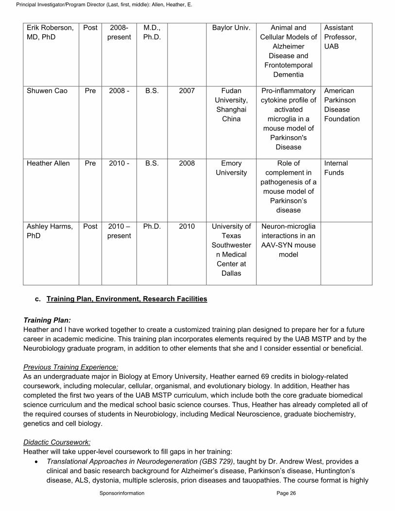
Erik Roberson, MD, PhD
Post 2008-present
M.D., Ph.D.
Baylor Univ. Animal and Cellular Models of
Alzheimer Disease and
Frontotemporal Dementia
Assistant Professor, UAB
Shuwen Cao Pre 2008 - B.S. 2007 Fudan University, Shanghai
China
Pro-inflammatory cytokine profile of
activated microglia in a
mouse model of Parkinson's
Disease
American Parkinson Disease Foundation
Heather Allen Pre 2010 - B.S. 2008 Emory University
Role of complement in
pathogenesis of a mouse model of
Parkinson’s disease
Internal Funds
Ashley Harms, PhD
Post 2010 – present
Ph.D. 2010 University of Texas
Southwestern Medical Center at
Dallas
Neuron-microglia interactions in an AAV-SYN mouse
model
c. Training Plan, Environment, Research Facilities
Training Plan: Heather and I have worked together to create a customized training plan designed to prepare her for a future career in academic medicine. This training plan incorporates elements required by the UAB MSTP and by the Neurobiology graduate program, in addition to other elements that she and I consider essential or beneficial. Previous Training Experience: As an undergraduate major in Biology at Emory University, Heather earned 69 credits in biology-related coursework, including molecular, cellular, organismal, and evolutionary biology. In addition, Heather has completed the first two years of the UAB MSTP curriculum, which include both the core graduate biomedical science curriculum and the medical school basic science courses. Thus, Heather has already completed all of the required courses of students in Neurobiology, including Medical Neuroscience, graduate biochemistry, genetics and cell biology. Didactic Coursework: Heather will take upper-level coursework to fill gaps in her training:
! Translational Approaches in Neurodegeneration (GBS 729), taught by Dr. Andrew West, provides a clinical and basic research background for Alzheimer’s disease, Parkinson’s disease, Huntington’s disease, ALS, dystonia, multiple sclerosis, prion diseases and tauopathies. The course format is highly
Sponsorinformation Page 26
Principal Investigator/Program Director (Last, first, middle): Allen, Heather, E.

interactive and participatory consisting of critical debates, discussions and presentations. Emphasis is placed on identifying common barriers to the development of successful therapeutics and possible strategies to address these barriers.
! The American Association of Immunologists Advanced Course in Immunology is a week-long intensive course designed for serious students of immunology. The course is held over the summer in Minnesota. Leading experts present recent research advances in understanding the biology of the immune system and its role in health and disease. Because the course is intended for advanced trainees and scientists, it is a perfect course for Heather to expand her understanding of the field.
! Intermediate Statistical Analysis I and II (BST 611 and 612) gives students a thorough understanding of basic analysis methods, elementary concepts, statistical models and applications of probability, commonly used sampling distributions, parametric and non-parametric one and two sample tests, confidence intervals, applications of analysis of two-way contingency table data, simple linear regression, and simple analysis of variance. Additionally, students will be introduced to the basic principle of tools of simple and multiple regression. The goal is to establish a firm foundation in the discipline upon which the applications of statistical and epidemiologic inference will be built. Students are taught to conduct the relevant analysis using current software such as the Statistical Analysis System (SAS).
Research Training We have crafted a research training plan, described under Research Strategy, that will provide Heather with opportunities to master new in vitro and in vivo techniques in neurobiology and immunology, including stereotactic injections, primary microglial culture, flow cytometry, imaging with confocal microscopy, and a range of molecular biology approaches. Continuing Education To stay current on the latest developments in related fields, Heather will attend:
! Neurobiology Seminar (NBL 703; Thursdays at 1:30). Seminars relate to all aspects of neurobiology, including synaptic physiology and plasticity, learning and memory, development, glial biology, neurobiology of disease, and systems neuroscience. Over 80% of the speakers are from outside institutions. Heather and the other neurobiology students are invited to lunch with the speaker weekly.
! Journal Club. Heather must participate in a formal journal club every semester and will choose between Neurodegenerative Disease (NBL 787) and Biology of Glial Cells (NBL 788). In addition, she will have the option of participating in an informal monthly journal club addressing clinical studies.
! MSTP Translational Research Seminar (PAT 794; monthly). Seminars feature case studies highlighting key elements in translating basic biomedical understanding into medical practice. Invited speakers will choose a particular scientific concept or candidate drug that is being developed or has been translated into an approved therapy with clinical impact. The speaker will attempt to highlight the milestones along the developmental pathway, including their own contributions, but focus on the overall development of the field over a significant period of time.
! NMSS Collaborative Research Meeting. Seminars are conducted by neuroimmunologists interested in neurodegenerative diseases. These informal monthly meetings facilitate collaboration and discussion amongst researchers and interested students by catering to a wide variety of expertise.
! National Meetings. Heather will attend at least one national meeting per year, to be selected from the annual Society for Neuroscience meeting, the International Society for Neuroimmunology or the International Movement Disorders Congress.
Oral Presentations Heather will participate in several forums, ranging in formality, allowing her to develop skills in public speaking and organizing an effective presentation in both poster and platform format.
Sponsorinformation Page 27
Principal Investigator/Program Director (Last, first, middle): Allen, Heather, E.

! Lab Meeting. Members in the lab share their progress every 1-2 weeks with one another and me in an informal setting. Heather will be expected to clearly state which experiments she did, why she did them, and present her results. She will give input on other lab members’ projects, and receive constructive criticism on her interpretations of results and troubleshooting of experiments.
! Center for Neurodegeneration and Experimental Therapeutics (CNET) Seminars. Members of the core CNET labs, which study a variety of cognitive and movement disorders, meet monthly for a seminar series. Heather will present a polished 50-min. seminar in this forum as she nears completion of her Ph.D.
! Parkinson’s Disease Group Meetings. PD group meetings occur about every 6 weeks and incorporate scientists from several laboratories at UAB as well as the University of Alabama. This provides the opportunity for a detailed discussion of data and potential collaborations. Heather will talk at one of these meetings about once every year.
! Neurobiology Retreat. The Department of Neurobiology convenes annually for two nights at a lakeside conference center with an outside keynote speaker, 10-15 internal speakers giving 20-min. talks, and poster sessions. Heather will present a poster annually and give a talk at least once.
! MSTP Retreat. All MSTP students attend an annual retreat, giving an oral presentation at least once during the graduate phase of their training. A keynote speaker discusses translational research discussion groups offer mentoring on the unique issues related to combined degree training.
! National Meetings. I expect Heather to present annually at the Society for Neuroscience meeting, the International Society for Neuroimmunology or the International Movement Disorders Congress.
Scientific Writing The ability to write clearly is one of the most important skills to acquire during this phase of training. Heather will receive formal and hands-on training in scientific writing.
! To graduate, Heather will be required to publish at least two original research articles in high-quality, peer-reviewed journals. I also anticipate providing Heather the opportunity to coauthor a review article on a topic related to her work, as I receive frequent invitations to contribute such manuscripts.
! I will periodically offer Heather the opportunity to assist me in peer-reviewing manuscripts, from which she will learn to accurately, fairly and confidentially judge others’ work and writing.
Grantsmanship and Professional Skills First, Heather participated in a mock grant writing and reviewing exercise last year as part of a Special Topics course called “Survival Skills for Physician Scientists.” In addition to discussing grant writing, this course also held group discussions around readings relating to professional skills, including how to manage a productive career in academic medicine and function as a leader of research and/or clinical staff. The textbook used was “Making the Right Moves. A Practical Guide to Scientific Management for Postdocs and New Faculty, Second Edition (Burroughs Wellcome Fund /Howard Hughes Medical Institute)”. Additionally, writing this proposal is a major aspect of Heather’s practical training in grantsmanship. I will also encourage her to participate in the Professional Skills Workshop at the annual Society for Neuroscience meeting. Biomedical Ethics Heather will take Ethics and Scientific Publications (PHY 792), which covers a range of topics related to the responsible conduct of biomedical research. Advisory Committee We have assembled a thesis committee for Heather composed of mentors who contribute different areas of expertise. The committee will meet every six months to review Heather’s progress. The members of the committee are: David Standaert, M.D., Ph.D., Scott Barnum, Ph.D., Andrew West, Ph.D., Etty Benveniste, Ph.D., and Victor Thannickal, M.D. (MSTP mentor).
Sponsorinformation Page 28
Principal Investigator/Program Director (Last, first, middle): Allen, Heather, E.

Clinical Exposure I encourage Heather to periodically attend Movement Disorders Clinic on Friday mornings and neurology ward rounds when I am attending for one month of the year. Additionally, she will attend Neurology Grand Rounds, which occurs every Tuesday morning. Research Environment and Facilities UAB is one of the Southeast's premier biomedical research institutions, ranking among the top 20 in funding from the National Institutes of Health and earning more than $470 million per year in contract and grant support. The University of Alabama at Birmingham seeks to foster academics, clinical research, training and clinical care in an interdisciplinary and collaborative atmosphere. To encourage such interaction, UAB has developed a large number of centers, many of which serve multiple disciplines across campus. The Neuroscience community at UAB has expanded dramatically in the last five years and several factors make UAB an ideal environment for the proposed research. Center for Neurodegeneration and Experimental Therapeutics (CNET). CNET was established in 2007 and serves as UAB’s focal point for translational studies in neurodegenerative diseases, including Parkinson’s, Alzheimer’s, ALS, and Huntington’s disease. I was recruited from Massachusetts General Hospital and Harvard Medical School to serve as the first director of CNET. Five senior investigators are core faculty members: Dr. Standaert, Erik Roberson, M.D., Ph.D., Andrew West, Ph.D., Talene Yacoubian, M.D., Ph.D., and Michelle Gray, Ph.D. Additional investigators will be recruited within the next three years. CNET resources include an imaging core facility, which contains instrumentation for conventional light microscopy, confocal microscopy, laser capture microdissection, and computer-assisted unbiased stereology. CNET also has shared facilities for cell culture, quantitative PCR, large instrumentation, such as centrifuges and scintillation counters, and other standard molecular laboratory equipment. Blueprint Grant and Core Facilities. UAB was one of four institutions to compete successfully for funding from the NIH Blueprint for Neuroscience in 2006. The Alabama Neuroscience Blueprint Core Center (P30NS057098) provides extensive infrastructure support for basic studies in neurobiology and animal models of neurological disease. These include a Molecular Engineering Core, a Cellular and Molecular Pathology Core, a rodent Neuroimaging Core, an in vivo Physiology and Phenotyping Core, and a Cellular and Synaptic Physiology Core. Comprehensive Neuroscience Center. CNET is part of a much larger and ongoing expansion of the neuroscience community at UAB. Since 2005, new leadership has been recruited to the Department of Neurology (Ray Watts, M.D., from Emory University who has recently been named Dean of the School of Medicine), Neurobiology (David Sweatt, Ph.D., from Baylor College of Medicine), and Psychiatry (James Meador-Woodruff, M.D., from the University of Michigan), and to the Center for Neurodegeneration and Experimental Therapeutics (David Standaert, M.D., Ph.D., from Harvard Medical School). In addition, the new chair of Pathology is a neuropathologist (Kevin Roth, M.D., Ph.D.) These leaders have fostered a unique degree of collaboration between their departments, manifest in many ways such as joint hires, and perhaps most notably, by the formation of a Comprehensive Neuroscience Center (CNC). The mission of the CNC is to develop interdisciplinary neuroscience research, clinical care, and education at UAB. There are 196 faculty, drawn from many different schools and programs. Collectively, the departments of Neurology, Psychiatry, Neurobiology, and the divisions of Neurosurgery and Neuropathology have seen a net increase of 41 new tenure-track faculty members since 2006, a 39% increase (to a current census of 145). There has been parallel growth in extramural funding: support from NIH institutes in the neurosciences has increased to $68M in 2009, from $42M in 2006. This rapid expansion makes UAB an exciting place to be a neuroscientist. The absence of inter-departmental boundaries to collaboration or
Sponsorinformation Page 29
Principal Investigator/Program Director (Last, first, middle): Allen, Heather, E.

resource sharing garnered UAB recognition as one of the Scientist magazine’s “Best Places to Work in Academia” in 2009.
d. Number of Fellows/Trainees to be supervised during the Fellowship
Dr. Standaert is a mentor for two current Graduate Students in addition to Heather engaged in thesis research, two additional co-mentored students (with Dr. Yacoubian and Gray), one Postdoctoral Fellow, and several additional predoctoral rotation students, medical students and undergraduates. He intends to accept at most one more thesis student during Heather’s training period.
e. Applicant’s Qualifications and potential for a research Career:
I think Heather Allen is an outstanding candidate for training as a physician-scientist and will have a bright
future in academic neurology. Heather has a very strong academic background, with undergraduate education at Emory University. Coursework there and summer research fellowships sparked her interest in biology and medicine, and she applied for training in MSTP programs. She was accepted to UAB in 2008 and has proven to be a very strong student, especially in the clinically-oriented components of the program. She first approached me about working in my lab in 2009, in the summer after her first year of medical school classwork. One of the most interesting and active areas in my lab is studies of the immunological basis of Parkinson disease. This is a relatively new area for the field of neurodegeneration but is increasingly recognized as holding the potential for novel approaches to therapies designed to prevent disease progression. As an initial project, Heather took on a study which was in its early stages, a collaborative project involving our lab and Dr. Howard Gendelman at the University of Nebraska. This is a study in which we exploring changes in peripheral immune cells in human PD, in particular T regulatory and suppressor cells. This work, which is funded by the Michael J. Fox Foundation for Parkinson Research, requires recruiting patients with PD and matched controls, gathering diagnostic and demographic information, isolating T cell subsets by flow cytometry and statistical analysis of the data. Heather took a lead role in our work on this at UAB, attending clinic, helping to identify suitable patients for recruitment, gathering data, organizing sample collection, and working with our colleagues in Nebraska to complete the analysis and analyze the data. This project has progressed surprisingly well; we have reached the stage where we have very intriguing preliminary data, and we are undertaking a second validation study to confirm our observation. I can honestly say that this project would never have moved forward as it did without Heather’s dedicated and committed work on the project. While I anticipate Heather will stay involved in the T cell study, this is a complex multi-site investigation not well suited to a thesis project. In the laboratory, Heather has been exploring the role of complement in alpha-synuclein related inflammation and neurotoxicity. This work is the result of a fortuitous interaction between my lab and Dr. Scott Barnum, a UAB faculty member who is a leading expert on the role of complement in the nervous system. Heather has tackled our initial questions with enthusiasm and hard work, and has produced the preliminary data described in the grant. These data do support the view that complement activation is likely to be involved in synuclein-related neurodegeneration, and may open new avenues to treatment. In her time in my laboratory, Heather has proven to be bright, hardworking and creative. She has an excellent ability to read and understand the relevant literature and to extract the important questions. Her work is careful and conscientious, and she is very good at anticipating the potential pitfalls of an experimental approach. She writes well and is comfortable speaking in both scientific as well as lay person settings. I have had the privilege of working with many MSTP students, and they are all outstanding in some way. This is not surprising, given the competitive nature of the programs. Still, I think Heather stands out from this distinguished crowd because of her passion for her work, and her genuine desire to work with patients and
Sponsorinformation Page 30
Principal Investigator/Program Director (Last, first, middle): Allen, Heather, E.

improve the treatments we have to offer. She is truly a humanist, and I see her as developing a career that is anchored in hands-on patient care and seeks to bring new solutions to common clinical problems. In short, I think Heather has the potential to be the kind of physician-scientist we need, closely connected to the world of clinical medicine but skilled in the use of modern scientific approaches. I think an F31 award at this stage of her training would have a profoundly beneficial effect on her long-term career trajectory. I am committed to her success, and can assure you that I, the UAB Department of Neurology, and the MSTP Program will provide her with every opportunity to succeed. I hope that the review committee shares my enthusiasm for Heather and the training program we have developed together.
Sponsorinformation Page 31
Principal Investigator/Program Director (Last, first, middle): Allen, Heather, E.

Specific Aims.
Parkinson disease (PD) is a neurodegenerative disorder characterized by a progressive loss of dopamine producing neurons in the substantia nigra pars compacta resulting in tremor, rigidity, bradykinesia and postural instability in 3-5% of people above age 65. Although dopamine replacement based therapies are quite effective at alleviating symptoms in PD, they fail to halt neuronal loss (Obeso, 2010). Alpha-synuclein aggregation is found in Lewy bodies in injured dopaminergic neurons in PD (Baba, 1998), and alpha-synuclein gene duplications and triplications cause PD in a dose-dependent manner (Ross, 2008). These observations demonstrate this protein’s importance in PD pathogenesis although the mechanisms by which it produces toxicity remain unclear. Recently, research has focused on the possibility that immune activation may be important for PD neurodegeneration: reactive microgliosis has been observed by PET imaging in vivo (Gerhard, 2006) and in PD brains post mortem (McGeer, 1988, Imamura, 2003). A polymorphism in the HLA region has been found to be associated with late-onset PD (Hamza, 2010!.
Our lab has pursued the idea that alpha-synuclein (a-syn) itself may be the trigger for immune activation in PD. We have shown that targeted overexpression of a-syn in the substantia nigra (SN) of mice driven by an adeno-associated virus vector recapitulates the reactive microgliosis observed in human PD (Theodore, 2008) and leads to a 30% reduction in the total number of dopaminergic neurons six months post-injection (St. Martin, 2007). Furthermore, knocking out microglial Fc-gamma-receptors reduces this a-syn induced neuronal degeneration, suggesting that interactions between innate and adaptive immunity are important (Cao, 2010).
The complement system is a critical part of the innate immune system, and is involved in not only the immune response to infection of the CNS, but also the immune response to many native CNS pathologies, including neurodegenerative diseases (Alexander, 2008). In PD, Lewy bodies are positive for C3d, C4d, C7 and C9 (Yamada, 1992), and C1q and C9 mRNA expression are increased in the SN of PD patients (McGeer, 2004). The overall hypothesis of this study is that activation of complement is required for mediating the dopaminergic neurotoxicity of alpha-synuclein in vivo.
Completion of the following experiments will determine whether the complement system contributes to neuronal loss in an alpha-synuclein mouse model of Parkinson disease. This study will determine whether the complement system is a potential target for future immune system-based therapeutics for PD. Hypothesis I: Alpha-synuclein overexpression in SNc in vivo leads to complement activation through the classical pathway. Aim 1: Using the AAV-Syn in vivo model of PD, determine whether a-syn expression leads to activation of classical pathway specific C1q and C4, common pathway C3 and C5, and terminal deposition of C9. We will use the previously characterized AAV-syn model of PD to study the effects of a-syn expression at 2 and 4 weeks after administration, when the inflammatory process is established but neuronal loss has not yet occurred. Complement activation will be evaluated by qPCR, western blot analysis and immunohistochemistry. Hypothesis II: Alpha-synuclein triggers classical complement activation by interaction with microglial cells leading to induction of C1q, C3 and upregulation of microglial complement receptors. Aim 2: Using primary mouse microglia in culture, determine the effect of a-syn on the expression of microglial C1q and C3, and complement receptors, CR1, CR3, CR4, calreticulin (a C1q receptor), C3aR, and C5aR, as well as their corresponding effector functions: phagocytosis and cytokine expression. Cultures of primary mouse microglia activated by a-syn in vitro will be assessed by immunocytochemistry and ELISA for expression of C1q and C3; flow cytometry will additionally be used to characterize complement receptor expression. Commercially available phagocytosis assays and ELISAs to assess cytokine expression will quantify changes in normal microglial function in response to a-syn. Hypothesis III: Inhibition of complement activation will reduce inflammation and neurodegeneration induced by alpha-syn overexpression in vivo. Aim 3: Using a transgenic mouse with astrocyte-specific expression of a soluble form of the murine complement control protein Crry, determine whether inhibition of complement prevents microglial activation, cytokine expression and neuron loss in the AAV-syn mouse model of PD. We will use the AAV-syn model of Aim 1 in the transgenic mice expressing Crry. As previously characterized, we will examine microglial activation through immunohistochemistry, cytokine expression through qPCR and neuron loss through stereology.
Specific Aims Page 47
Specific Aims
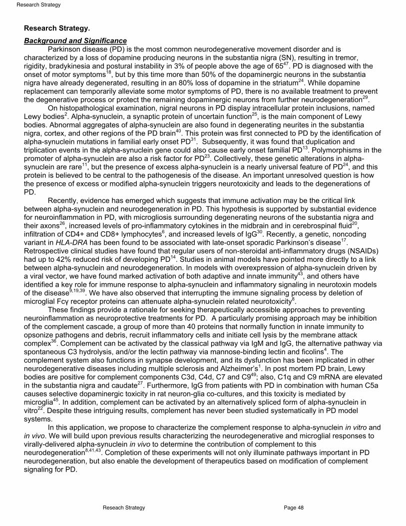
Research Strategy. Background and Significance
Parkinson disease (PD) is the most common neurodegenerative movement disorder an! is characterized by a loss of dopamine producing neurons in the substantia nigra (SN), resulting in tremor, rigidity, bradykinesia and postural instability in 3% of people above the age of 6547. PD is diagnosed with the onset of motor symptoms18, but by this time more than 50% of the dopaminergic neurons in the substantia nigra have already degenerated, resulting in an 80% loss of dopamine in the striatum24. While dopamine replacement can temporarily alleviate some motor symptoms of PD, there is no available treatment to prevent the degenerative process or protect the remaining dopaminergic neurons from further neurodegeneration29.
On histopathological examination, nigral neurons in PD display intracellular protein inclusions, named Lewy bodies2. Alpha-synuclein, a synaptic protein of uncertain function25, is the main component of Lewy bodies. Abnormal aggregates of alpha-synuclein are also found in degenerating neurites in the substantia nigra, cortex, and other regions of the PD brain40. This protein was first connected to PD by the identification of alpha-synuclein mutations in familial early onset PD31. Subsequently, it was found that duplication and triplication events in the alpha-synuclein gene could also cause early onset familial PD13. Polymorphisms in the promoter of alpha-synuclein are also a risk factor for PD23. Collectively, these genetic alterations in alpha-synuclein are rare11, but the presence of excess alpha-synuclein is a nearly universal feature of PD24, and this protein is believed to be central to the pathogenesis of the disease. An important unresolved question is how the presence of excess or modified alpha-synuclein triggers neurotoxicity and leads to the degenerations of PD.
Recently, evidence has emerged which suggests that immune activation may be the critical link between alpha-synuclein and neurodegeneration in PD. This hypothesis is supported by substantial evidence for neuroinflammation in PD, with microgliosis surrounding degenerating neurons of the substantia nigra and their axons26, increased levels of pro-inflammatory cytokines in the midbrain and in cerebrospinal fluid20, infiltration of CD4+ and CD8+ lymphocytes6, and increased levels of IgG30. Recently, a genetic, noncoding variant in HLA-DRA has been found to be associated with late-onset sporadic Parkinson’s disease17. Retrospective clinical studies have found that regular users of non-steroidal anti-inflammatory drugs (NSAIDs) had up to 42% reduced risk of developing PD14. Studies in animal models have pointed more directly to a link between alpha-synuclein and neurodegeneration. In models with overexpression of alpha-synuclein driven by a viral vector, we have found marked activation of both adaptive and innate immunity43, and others have identified a key role for immune response to alpha-synuclein and inflammatory signaling in neurotoxin models of the disease9,19,39. We have also observed that interrupting the immune signaling process by deletion of microglial Fc! receptor proteins can attenuate alpha-synuclein related neurotoxicity8. These findings provide a rationale for seeking therapeutically accessible approaches to preventing neuroinflammation as neuroprotective treatments for PD. A particularly promising approach may be inhibition of the complement cascade, a group of more than 40 proteins that normally function in innate immunity to opsonize pathogens and debris, recruit inflammatory cells and initiate cell lysis by the membrane attack complex36. Complement can be activated by the classical pathway via IgM and IgG, the alternative pathway via spontaneous C3 hydrolysis, and/or the lectin pathway via mannose-binding lectin and ficolins4. The complement system also functions in synapse development, and its dysfunction has been implicated in other neurodegenerative diseases including multiple sclerosis and Alzheimer’s1. In post mortem PD brain, Lewy bodies are positive for complement components C3d, C4d, C7 and C949; also, C1q and C9 mRNA are elevated in the substantia nigra and caudate27. Furthermore, IgG from patients with PD in combination with human C5a causes selective dopaminergic toxicity in rat neuron-glia co-cultures, and this toxicity is mediated by microglia45. In addition, complement can be activated by an alternatively spliced form of alpha-synuclein in vitro22. Despite these intriguing results, complement has never been studied systematically in PD model systems.
In this application, we propose to characterize the complement response to alpha-synuclein in vitro and in vivo. We will build upon previous results characterizing the neurodegenerative and microglial responses to virally-delivered alpha-synuclein in vivo to determine the contribution of complement to this neurodegeneration8,41,43. Completion of these experiments will not only illuminate pathways important in PD neurodegeneration, but also enable the development of therapeutics based on modification of complement signaling for PD.
Reseach Strategy Page 48
Research Strategy

Approach Aim 1: Using the AAV-SYN in vivo model of PD, determine whether a-syn expression leads to activation of classical pathway specific C1q and C4, common pathway C3 and C5, and terminal deposition of C9. Rationale: In PD post-mortem brain tissue, there are increased amounts of complement components, C1q and C9, mRNA in the substantia nigra and caudate27. Also, different complement component isoforms are expressed in CSF from PD patients as compared to healthy controls and other neurodegenerative diseases12,16.
Previous work in our laboratory on mice stereotactically injected with an AAV that overexpresses alpha-synuclein (AAV-SYN) shows a 30% loss of dopaminergic neurons in the substantia nigra 6 months post- injection41. The nigral pathology found in these animals contains a strong immune system component as evident by increased deposition of IgG, microglial activation, increased cytokine secretion, and increased recruitment of B and T cells43. The dopaminergic neurotoxicity in this model can be attenuated through knockout of the Fc!R, suggesting a causal link between the immune system and cell death8.
The complement cascade can be initiated through complexes of IgG binding to C1q, via the classical pathway36. Since our mice overexpress IgG43, and there are changes in complement mRNA and protein expression in PD patients27, we hypothesized that alpha-synuclein overexpression in the substantia nigra leads to complement activation through the classical pathway in our alpha-synuclein overexpressing mouse model of PD. We have conducted preliminary studies of complement in this model, and found that AAV-mediated expression of a-syn leads to a marked increase C3 mRNA expression 2 weeks post-injection (Figure 1). In addition, C3 protein is deposited in TH neurons at 6 months (Figure 2).
Experimental Design Mouse Model: C57BL/6 mice will be injected stereotactically under isoflurane anesthesia with 2 !L of a recombinant adeno-associated virus 2 containing the gene for human alpha-synuclein (AAV-SYN) or green fluorescent protein (AAV-GFP) of the same viral titer. The stereotaxic coordinates target the right substantia nigra, and are: anterior-posterior (-3.2 mm from bregma), medio-lateral (-1.2 from midline) and dorso-ventral (-4.6 from the dura). After injection, mice will be sacrificed at prescribed time points as described below. Additionally, some mice will be injected with LPS acutely as a positive control in the same stereotaxic area. These mice will be sacrificed within 24 hours of injection. In previous studies, this injection leads to a consistent, progressive selective dopaminergic neuron loss of up to 30% by 6 months41. Time Points: 2 week and 4 week time points were chosen for this experiment. Initial inflammatory reactions are seen at 2 weeks post-injection of AAV-GFP, when viral proteins begin to express at high levels. At the 2 week time point, in the PD mouse model, we see an increase in IgG as assessed by immunofluorescence staining and an increase in pro-inflammatory cytokine expression as assessed by qPCR. At 4 weeks post-
Figure 1. Expression of C3 mRNA. qPCR on cDNA from AAV-GFP and AAV-SYN mice 2 weeks post-injection reveals increased expression of C3 in AAV-SYN mice. N= 6(GFP), 5(SYN). Mann-Whitney U test; *p < 0.05.
Figure 2. C3 expression in TH+ neurons in AAV-SYN mice 6 months post-injection. AAV-SYN mice were injected into the right substantia nigra, and at 6 months post-injection, the tissue was stained for tyrosine hydroxylase (TH), using a mouse antibody and C3 using a chicken antibody. Images were taken at 63x power on the Leica TCS-SP5 laser scanning confocal.
Reseach Strategy Page 49
Research Strategy

injection of AAV-SYN mice, the inflammatory process is established, as there is microglial activation paired with pro-inflammatory cytokine expression, IgG deposition, and B and T cell infiltration in the substantia nigra. Assessing complement component mRNA by qPCR: Mice sacrificed at 2 weeks and 4 weeks post-injection of either AAV-GFP (n=8) or AAV-SYN (n=8) will be dissected for substantia nigra ipsilateral to the injection site. Brain tissue will be homogenized and processed for mRNA. cDNA will be reverse transcribed from substantia nigra mRNA, and assessed by SybrGreen qPCR. Primers against C3, C5 will assess the common portion of the complement cascade; primers against C9 will assess the terminal portion of the complement cascade; primers against C1q and C4 will assess the classical pathway of activation; primers against Factor B will assess the alternative pathway of activation. Primers against GAPDH will allow for normalization of mRNA expression between animals. Ratios of target complement component mRNA expression to GAPDH mRNA expression will be compared between mice injected with AAV-GFP and AAV-SYN. A student’s T-test will be performed if the data are parametric; otherwise, a Mann-Whitney U-test will be performed. A p<0.05 will be considered significant. Preliminary data shows an increase in transcription of C3 mRNA in AAV-SYN mice as compared to AAV-GFP mice (Figure 1). Assessing complement component protein by Western blot: Mice sacrificed at 4 weeks post-injection of either AAV-GFP (n=8) or AAV-SYN (n=8) will be dissected for substantia nigra ipsilateral to the injection site. Brain tissue will be homogenized and processed for protein. Protein samples will be run on an SDS-PAGE gel, transferred to nitrocellulose or PVDF membrane, and probed for C3, C5, C9, C1q, C4, Factor B and "-tubulin. Western blots will be quantified with an integrated area method. Ratios of target complement component expression to "-tubulin expression will be compared between mice injected with AAV-GFP (negative control), AAV-SYN (test condition) or acute LPS (positive control). A student’s T-test will be performed if the data are parametric; otherwise, a Mann-Whitney U-test will be performed. A p<0.05 will be considered significant. Assessing complement component protein by immunofluorescence staining and confocal microscopy: Mice sacrificed and perfused at 4 weeks post-injection of either AAV-GFP or AAV-SYN will be dissected for their brains and processed for immunohistochemistry. Free floating substantia nigra and striatum slices (40 !m) will be stained for tyrosine hydroxylase to label the substantia nigra, in addition to one of the following complement components: C3, C5, C9, C1q, C4, and Factor B. Confocal images will be captured using a Leica TCS-SP5 laser scanning confocal microscope. For quantitation of images, slides will be observed using a Nikon Eclipse E800 M fluorescent microscope. Coded slides will be scored using a scale from 0 (no staining) to 4 (most intense staining) by an observer blind to the treatment paradigm. Only staining in proximity to SN neurons will be scored. Staining along the needle tract will be ignored. Scores obtained from 8 mice per group will be statistically analyzed using the Mann-Whitney U test. A p<0.05 will be considered significant. Preliminary data show that robust C3 expression occurs at 6 months post-injection in AAV-SYN mice (Figure 2). Expected Results and Alternative Methods: If the hypothesis is correct, it is expected that expression of C1q, C4, C3, C5 and C9 mRNA and protein will be increased in AAV-SYN mice and expression of factor B mRNA will be unchanged between AAV-GFP and AAV-SYN mice. Alternatively, if complement is activated by the alternative pathway, expression of C1q and C4 will be unchanged between AAV-GFP and AAV-SYN mice, but expression of factor B, C3, C5 and C9 mRNA and protein will be increased. Additionally, none or both of the pathways could be activated, in which cases, none or all complement component mRNA or protein levels would change respectively. It is also conceivable that the pathway may terminate early due to inhibitors of the latter parts of the cascade being naturally expressed. In these cases, one would expect to see activation of either the classical (C1q and C4) or alternative pathways, with initiation of the terminal part of the cascade (C3) and possibly C5 expression, depending on where along the pathway the signal terminates. Because our lab has experience working with this mouse model, and has previously characterized the microglial response to alpha-synuclein in these mice, I do not anticipate technical problems with recreating the mice or completing these experiments. However, other transgenic PD model mice could be used to characterize the response of complement to alpha-synuclein overexpression in vivo if necessary, including A53T human alpha-synuclein transgenic mice15. To assess RNA transcription, I could alternatively use RNA-Seq to compare transcriptomes of AAV-GFP and AAV-SYN mice46. Aim 2: Using primary mouse microglia in culture, determine the effect of a-syn on the expression of microglial C1q and C3, and complement receptors, CR1, CR3, CR4, calreticulin (a C1q receptor), C3aR, and C5aR, as well as their corresponding effector functions: phagocytosis (CR1, CR3, CR4, calreticulin) and cytokine expression (C3aR, C5aR).
Reseach Strategy Page 50
Research Strategy

Rationale: Because primary murine microglia are activated by alpha-synuclein42,50, and microglia are responsible for some dopaminergic neurotoxicity seen in the AAV-SYN model of PD8, we desired to test the hypothesis that alpha-synuclein could trigger complement activation of the classical pathway by interaction with microglial cells. The first way that microglia could initiate and propagate complement is by secreting increased amounts of C1q and C3 complement components. The second way that microglia could influence susceptibility to complement activation is by upregulation and downregulation of their complement receptors on the cell surface3. In addition to using flow cytometry to assess changes in cell surface receptors, we will use functional assays of phagocytosis and cytokine expression in microglia to correlate with receptor function. Experimental Design Isolation and Culture of Primary Murine Microglia: Whole brains from wild type postnatal day 3-5 (P3-P5) C57BL/6 pups will be isolated, minced, and placed in ice-cold dissociation media containing sterile filtered DNase 1, dispase II and papain. Cells will be dissociated for 30 minutes with agitation every 10 minutes. Following mechanical and chemical dissociation, the population of mixed glial cells will be filtered through a 40µm pore filter and plated on T75 flasks in DMEM/F12 supplemented with fetal bovine serum, penicillin/streptomycin and L-glutamine. Mixed glial cultures will be maintained until confluence, approximately 14-18 days. Primary microglial cells were isolated by mechanical agitation on an orbital shaker (150 rpm) for 1 hour. Following isolation, cells will be plated at a density of 50,000 cells per well in a 24 well plate for immunocytochemical analysis or multiplexed immunoassay experiments, or plated at a density of 100,000 cells per well in a 96 well plate for phagocytosis experiments. We have conducted preliminary experiments using this method, which yields cultures of high purity with a vigorous response to stimuli such as LPS, with change in morphology as well as secretion of TNF# (Figure 3). Activation of Primary Murine Microglia by Alpha-Synuclein: After plating, cells will be allowed to adhere for 8 hours. Upon adherence, cells will be treated with a vehicle (negative control), soluble alpha-synuclein or aggregated alpha-synuclein at a range of concentrations between 100-1000nM for 24 hours, or lipopolysaccharide (LPS) (positive control). To assess activation of microglia by cell morphology, immunocytochemistry will be performed. Cells will be fixed, permeabilized, and incubated with antibodies: anti-NeuN and anti-GFAP as negative controls and anti-CD45 as a general microglial marker. Assessment of C1q and C3 Expression by Microglia: Immunocytochemistry. To assess microglial production of C1q and C3 qualitatively, immunocytochemistry to determine protein localization will be performed. After plating, cells will be treated with alpha-synuclein and appropriate controls as described earlier. Cells will be fixed, permeabilized, and incubated with antibodies: anti-CD45 microglial marker and anti-C1q or anti-C3. ELISA. To assess microglial production of C1q and C3 quantitatively, conditioned media and cell lysate will be collected from microglia treated with alpha-synuclein and appropriate controls as described earlier. C1q and C3 protein levels will be measured by ELISA per the manufacturer’s instructions. Spectrophotometric data obtained from 3 or more replicates of each condition will be analyzed by an appropriate statistical test. A p<0.05 will be considered significant. Alternatively, western blots could be performed to discover protein levels of secreted and intracellular microglial C1q and C3 in response to alpha-synuclein treatment. Assessment of Complement Receptors on Microglia: Immunocytochemistry. To visualize complement receptor expression on microglia, immunocytochemistry will be performed. After plating, microglia will be treated with alpha-synuclein and appropriate controls as described earlier. Cells will be fixed, permeabilized, and incubated with antibodies. Anti-CD35, anti-CD11b,
Figure 3. Primary Microglia Activation by LPS. Microglia treated with vehicle or LPS (10 ng/mL) were incubated for 24 hours at 37"C. Conditioned media was removed to assay for TNF via ELISA. LPS stimulated cells were shown to be activated by morphology. LPS stimulated cells demonstrated increased production of TNF. n=3 per treatment condition; Student’s unpaired t-test; ***p<0.001.
Reseach Strategy Page 51
Research Strategy

anti-CD11c, anti-calreticulin, anti-C3aR, anti-CD88 visualize each of the complement receptors, CR1, CR3, CR4, calreticulin, C3aR, and C5aR, respectively. Flow cytometry: To determine if alpha-synuclein treatment changes complement receptor expression on microglia, we will use flow cytometry. After isolation and plating of microglia, the cells will be treated with alpha-synuclein as well as appropriate controls. After treatment, the cells will be isolated and stained with appropriate fluorescently conjugated antibodies to the CR1, CR3, CR4, calreticulin, C3aR and C5aR receptors, washed, and fixed for flow cytometric analysis. Assessment of Microglial Phagocytosis: Microglial phagocytosis of fluorescent E.coli bioparticles will be measured using a Vybrant Phagocytosis Assay kit from Invitrogen. Briefly, cells will be treated with vehicle, soluble alpha-synuclein, aggregated alpha-synuclein or LPS as described earlier. Spectrophotometric data obtained from 3 or more replicates of each condition will be analyzed by an appropriate statistical test; a p<0.05 will be considered significant. Assessment of Cytokine Expression by Microglia: Conditioned media will be collected from microglia treated with vehicle, soluble alpha-synuclein, aggregated alpha-synuclein or LPS as described earlier. Analysis for IL-1, IL-6, TNF-" and ICAM-1 cytokines by ELISA will be performed. Spectrophotometric data obtained from 3 or more replicates of each condition will be analyzed by a student’s T-test if the data are parametric; otherwise, a Mann-Whitney U-test will be performed. A p<0.05 will be considered significant. Expected Results and Alternative Methods: If the hypothesis is true that activated microglia begin the classical complement cascade in response to alpha-synuclein, it would be expected that increased levels of secreted C1q and C3 would be found. However, if C1q and C3 expression are not increased, microglia could simply become more susceptible to complement through upregulation of complement receptors. If activated microglia begin the alternative complement cascade in response to alpha-synuclein, increased levels of secreted C3 may be found. The complement receptors CR1, CR3, CR4, and calreticulin are known to induce phagocytosis; if increased levels of CR1, CR3, CR4 or calreticulin in response to alpha-synuclein are seen, it would also be expected that increases in phagocytosis would also be seen. Likewise, the complement receptors, C3aR and C5aR, are known to induce cytokine expression via pro-inflammatory signaling. Thus, if C3aR or C5aR are upregulated, it would also be expected that corresponding increases in cytokine expression would be seen. If changes in phagocytosis or cytokine expression are seen without any changes in complement receptors, it is likely due to another mechanism of alpha-synuclein activation of microglia. If soluble and aggregated alpha-synuclein are not strong enough stimuli to activate microglia, modified alpha-synuclein could be used, including nitrated35, phosphorylated (Ser129)48, or mutated alpha-synuclein (A53T, A30P or E46K)37. Alternative techniques for assessing C1q, C3 and complement receptor expression in microglia include qPCR for mRNA and Western blot for protein expression. Alternative measurements of microglial effector function include cell migration21, or a larger cytokine panel assayed by a multiplex ELISA44. Aim 3: Using a transgenic mouse with astrocyte-specific expression of a soluble form of the murine complement control protein Crry, determine whether inhibition of complement prevents microglial activation, cytokine expression and neuron loss in the AAV-syn mouse model of PD. Rationale: The goal of this aim is to determine if inhibition of the complement cascade in vivo will attenuate dopaminergic neuronal cell death in the AAV-SYN model of PD. In this aim, we will use a novel mouse model that expresses soluble Crry (sCrry), a murine complement inhibitor, under the GFAP promoter in astrocytes. Crry is a rodent complement regulatory protein that acts to inhibit C3 and C5 convertases by the same mechanisms of action as the human homologues: DAF (decay accelerating factor) and MCP (membrane cofactor protein). This model has been characterized previously by our collaborator, Dr. Scott Barnum, and has been shown to delay onset and decrease severity of disease in several neurodegenerative paradigms, including experimental allergic encephalomyelitis (a mouse model of multiple sclerosis)10, experimental autoimmune uveoretinitis34, and closed head injury33. It is reasonable to hypothesize that inhibition of complement activation by this mechanism will also reduce inflammation and neurodegeneration induced by alpha-synuclein overexpression in vivo. The primary outcome measure for these studies will be based on counting the dopaminergic neurons in the substantia nigra using an unbiased stereological method. Secondarily, we will assess potential effects on inflammation by assessing known changes in response to targeted overexpression of alpha-synuclein in vivo, namely microglial activation by imaging, and cytokine expression by qPCR. The design for these studies is based on our recent investigation in which we used a mouse with inactivation of the gamma chain of the FcR receptor, and observed protection against AAV-SYN induced cell loss8.
Reseach Strategy Page 52
Research Strategy

Experimental Design Mouse Model: As in Aim 1, C57BL/6 mice (wild type) and transgenic mice from a C57BL/6 background expressing soluble Crry under the control of a GFAP promoter will be injected stereotactically under isoflurane anesthesia with 2 µL of a recombinant adeno-associated virus 2 containing the gene for human alpha-synuclein (AAV-SYN) or green fluorescent protein (AAV-GFP) of the same viral titer. The stereotaxic coordinates are the same as in Aim 1 and correspond to the right substantia nigra. After injection, mice will be sacrificed at prescribed time points as described below. Time Points: 2 week, 4 week and 24 week time points were chosen for this experiment. Initial inflammatory reactions are seen at 2 weeks post-injection of AAV-GFP, when viral proteins begin to express at high levels. At 4 weeks post-injection of AAV-SYN mice, the inflammatory process is established, with microglial activation, pro-inflammatory cytokine expression, IgG deposition, and B and T cell infiltration in the substantia nigra. Assessment of Microglial Activation: Wild type and Crry-expressing mice sacrificed and perfused at 4 weeks post-injection of either AAV-GFP or AAV-SYN will be dissected for their brains and processed for immunohistochemistry. Free floating substantia nigra and striatum slices (40 !m) will be stained for tyrosine hydroxylase to label the substantia nigra, in addition to a microglial marker, CD68. Confocal images will be captured using a Leica TCS-SP5 laser scanning confocal microscope. For quantitation of images, coded slides will be scored using a numerical scale from 0 (no staining) to 4 (most intense staining) by an observer blind to the treatment paradigm. Only staining in close proximity to SN neurons will be considered for scoring. Staining along the needle tract will be ignored. Scores obtained from 8 mice per group will be statistically analyzed using a 2-way ANOVA. A p<0.05 will be considered significant. Assessment of Cytokine mRNA Expression: Wild type and Crry-expressing mice sacrificed at 2 weeks and 4 weeks post-injection of either AAV-GFP (n=8 for each group) or AAV-SYN (n=8 for each group) will be dissected for substantia nigra ipsilateral to the injection site. Brain tissue will be homogenized and processed for mRNA. cDNA will be reverse transcribed from substantia nigra mRNA, and assessed by SybrGreen qPCR. Primers against ICAM-1, IL-1a, IL-6 and TNF will assess changes in cytokine expression. Primers against GAPDH will allow for normalization of mRNA expression between animals. Ratios of target complement component mRNA expression to GAPDH mRNA expression will be compared between both genotypes of mice injected with AAV-GFP and AAV-SYN. A 2-way ANOVA will be performed. A p<0.05 will be considered significant. Alternatively, ELISA could be used to assess cytokine protein expression as in Aim 2, and in fact, ELISA might be considered a better choice because it would assess protein levels; however, qPCR was chosen to facilitate comparison to previous data from our lab on cytokine expression in the AAV-SYN mouse model of PD assessed by qPCR. Assessment of Neuron Loss by Unbiased Stereology: Wild type and Crry-expressing mice sacrificed and perfused at 24 weeks post-injection of either AAV-GFP (n=8 for each genotype) or AAV-SYN (n=8 for each genotype) will be dissected for brains and processed for immunohistochemistry. Free floating substantia nigra slices (40 !m) will be stained for tyrosine hydroxylase (TH) to label the substantia nigra. A peroxidase-conjugated secondary antibody will be incubated with the sections and developed with diaminobenzidine to obtain a brown colored stain indicative of TH neurons. TH immunoreactive dopamine neurons will be quantified using unbiased stereology. Coded slides will be scanned, and SN from both the uninjected and injected sides of the brain will be contoured. TH-positive neurons will be counted by an optical fractionator method using Stereoinvestigator 7.0 software. A total of 4 sections covering the rostro-caudal extent of the SN will be counted and the number weighted section thickness will be used to correct for variations in tissue thickness at different sites. The number of TH+ cells estimated per substantia nigra ipsilateral to injection site will be compared between wt animals injected with AAV-GFP (n=8) and AAV-SYN (n=8), and sCrry-Tg animals injected with AAV-GFP (n=8) and AAV-SYN (n=8) by 2-way ANOVA. A p<0.05 will be considered significant. Expected Results and Alternative Methods: If complement inhibition in the CNS ameliorates inflammation and neuronal loss in the AAV-SYN model of PD, there will be reduced microglial activation, reduced expression of cytokines and increased survival of neurons. If there is increased microglial activation, increased expression of cytokines and decreased survival of neurons, it will be clear that the complement system is acting in a protective manner. If there is no change in inflammation or neuronal loss, there are 2 possibilities: 1) a more specific inhibitor is needed or 2) there is no effect of complement inhibition in the AAV-SYN model. In the event that more specific inhibitors are needed, our collaborator, Dr. Scott Barnum, has many knock-out mice targeting particular points within the complement system, including both components of the classical and alternative pathways of complement, as well as receptors for complement5,7,28,32. Alternative endpoints for this aim are a hypothesized decreased infiltration of lymphocytes and reduced degeneration of neurites."
Reseach Strategy Page 53
Research Strategy

12. Vertebrate Animals 1. Description of use of animals. All of the animals to be used in this study are mice. These mice will be ordered from Jackson Laboratory, or are already housed in Dr. David Standaert’s (sponsor) colony and Dr. Scott Barnum’s (collaborator) colony at the University of Alabama at Birmingham. For Aims 1 and 3, male mice will be unilaterally injected into the right substantia nigra with 2 rAAV2 vectors: one overexpressing human alpha-synuclein driven by the hybrid CMV immediate-early enhancer/chicken beta-actin promoter, and a control vector missing the human alpha-synuclein gene. Depending on the experiment, 2 weeks to 6 months post-injection, mice will be sacrificed, and the experiments described in this proposal will be performed. For Aim 2, it is necessary to isolate primary murine microglia. First, breeding cages containing adult male C57/BL6J mice and adult female C57/BL6J mice will be set up. Next, brains from both male and female mouse pups, age postnatal day 3 to postnatal day 5, resulting from this breeding will be used to create primary murine microglia cultures. 2. Justification of the use of animals. We have attempted to propose studies that will utilize a minimum number of animals and to obtain the maximum amount of information from each animal. Unfortunately, all of the studies cannot be carried out in tissue culture or in any other artificial system since the hypotheses apply directly to the functioning of the intact mammalian central nervous system. The mouse model was chosen because 1) previous publications support this model as an appropriate model of Parkinson disease, 2) our laboratory has experience in using this model, and 3) our laboratory has previously characterized the immune response in this model. Allowing for the requirements of breeding, we estimate that a total of about 2000 mice will be produced and used during the 4-year period of this project. This number is the minimum that will generate sufficient animals for breeding and creation of primary microglia, as well as give enough statistical power for the other in vivo experiments to be valid.
At UAB, the use of mice will be supervised by the UAB Animal Resources Program, and all protocols will be reviewed by the Institutional Animal Use and Care Committee (IACUC) prior to initiation of research studies. 3. Veterinary Care. All animal facilities at the University of Alabama at Birmingham are under the direction of full time veterinarians and are accredited by the Association for Assessment and Accreditation of Laboratory Animal Care International (AAALAC). UAB has a Letter of Assurance (A3255-01) on file with NIH’s Office of Laboratory Animal Welfare (OLAW) and is licensed as a research facility (64-R-0004) by the United States Department of Agriculture (USDA). 4. Procedures for limitation of discomfort, distress, pain and injury. Discomfort, stress, and pain will be minimized by the appropriate use of anesthetic agents. Because use of NSAIDs, which inhibit inflammation, may confound the results of our studies, we decided to treat mice for pain control with buprenex (0.05-0.1 mg/kg) and Tylenol dissolved in drinking water (300 mg/kg) subcutaneously as needed. Any animal observed to be experiencing unexpected stress or discomfort will be euthanized by CO2 inhalation. 5. Method of Euthanasia. Animals will be euthanized by CO2 inhalation or by an intraperitoneal injection of 200 mg/kg pentobarbital followed by decapitation. This method is consistent with the recommendations of the Panel on Euthanasia of the American Veterinary Medical Association.
Vertebrate Animals Page 54
Vertebrate Animals

Respective Contributions.
Dr. Standaert introduced me to the intersection of Parkinson disease (PD) and immunity during my second lab rotation, when I became involved in a clinical study examining T-cell function in PD patients. I thought that the hypothesis that the immune system was involved in the pathogenesis of PD was provocative. After many discussions over the next year with Dr. Standaert, I became convinced that I wanted to work on immunity and PD, particularly innate immunity. Much of our lab’s previous work explored the role of microglia in PD, but I wanted to expand this research into the complement system. I wrote the initial draft of the specific aims and the research strategy based upon my reading and previous discussions. Dr. Standaert reviewed the aims and strategy with me several times, and suggested people to bring into the discussion. I showed the drafts to Dr. Barnum, a complement expert, and Dr. Fineberg, a biostatistics expert, as well as other scientists, who suggested additional revisions, upon which I refined my aims and research strategy further.
Respective Contributions Page 55
Respective Contributions

Selection of Sponsor and Institution.
I was an undergraduate at Emory University, in Atlanta, GA, when I was applying to Medical Scientist Training Programs. I chose to apply to many great programs, but UAB stood out to me from the beginning because of their professionalism when dealing with applicants. It was clear that Robin Lorenz, the program director, and Paula Willey, the program manager, were not only organized, but also given the resources to do what was necessary for their students’ success. When I came to my interview, I was amazed by the quality of the students in the program. They were smart, pleasant, and pleased with their training and life in Birmingham. I felt comfortable with them and very much wanted to become one of them. At the time, I was not completely certain what type of research I was interested in, but UAB had the resources to convince me that whatever I wanted to do, there would be multiple people willing to help me. I went back to Emory, and several weeks later, I received an acceptance. When I mentioned this to my professors, they all told me that they respected the university for its medical training and fantastic research. I knew that I shouldn’t pass up this opportunity, so I started the MSTP program at UAB that summer.
When picking a research mentor, I knew that I wanted a reliable mentor, with whom I felt comfortable, and did fantastic science. During my first rotation, I worked on UCH-L1 deficient mice in Dr. Wilson’s lab and found myself reading quite a bit and becoming interested in movement disorders and Parkinson disease. I mentioned to one of the graduate students what I wanted in a mentor, and he wisely suggested I look at Dr. Standaert’s lab, given my research interests, experience and personality. I took the advice and decided to do a rotation. At the beginning of the rotation, we talked about what I was interested in, and he offered the opportunity to participate in a clinical study discovering whether subsets of T-cells were changed in patients with Parkinson disease as compared to controls. I was surprised and excited, because I had wanted an opportunity to learn how to complete human studies well. The science was truly novel, as it was an attempt to show in PD patients an immunological deficit.
After I started work in the lab, the first thing I noticed about Dr. Standaert’s mentorship is his skill as a teacher – I found myself respecting his ability to make complex topics simpler by carefully going over each issue involved, whether it was the latest thing covered in medical school or the latest confusion over troubleshooting experiments. Next, I noticed that he had a strong belief in the translational component of his lab. He invited not only his MSTP students, but also his graduate students, to come to clinic with him because he believed it was important for them to see and understand what Parkinson disease is and how it affected his patients. I then discovered the value in his support staff. They make life much easier for the students by showing us where to find information like which training courses need to be completed to start a new project, so that we can focus on the science and what needs to get done. In fact, I’m still discovering things that I consider brilliant about Dr. Standaert’s organization and management of the lab, because he clearly put a lot of thought into why we do things in a certain way. I believe that the care he puts into the management of the lab reflects the care he puts into training his students to reach their full potential.
Selection of Sponsor and Institution Page 56
Selection of Sponsor and Institution
Training in the Responsible Conduct of Research. By the time Heather is finished with her training, she will have taken 3 formal courses covering different aspects of responsible conduct of research.
1. Medical Ethics is part of the new Patient, Doctor, and Society course taught by several faculty members, including Dr. Gregory Pence, Ph.D., a Professor in the Department of Philosophy. Patient, Doctor, and Society (PDS) is a two week multidisciplinary required module for first year medical students. The PDS module is designed to introduce selected principles, behaviors and skills that are essential for a student’s professional development as a physician-in-training. These fundamentals are expected of all physicians in practice and serve to complement his/her competency in medical knowledge and are necessary for effective patient care. The fact that PDS was chosen to be the first module in the revised curriculum serves to emphasize the importance of the concepts introduced in this course. Physicians today are being asked by the public to pay greater attention to issues that cross the boundaries of biomedical science into those of professionalism in clinical medicine, such as communication skills, compassion, honesty, cultural competency, medical decision making, ethics, patient safety, leadership and health policy. In addition, the effective physician will need to be reflective and attentive to his or her own needs for life-long learning, personal health and well-being.
2. The UAB MSTP requires its students to complete a summer special topics course called Survival
Skills for Physician Scientists, taught by Robin Lorenz, M.D., Ph.D. The course focuses on grant preparation and review, developing scientific presentations, career development, and time and data management. In addition, the course covers topics including scientific ethics, professionalism, and interpersonal and communication skills.
3. MSTP trainees, as part of their graduate training, take Principles of Scientific Integrity. This course was developed by Harold Kincaid, PhD, and provides systematic instruction about the responsible conduct of science. The three-credit hour, semester long course provides a survey of ethical issues and principles in the practice of science. It is offered twice a year (Fall and Spring). Among the topics discussed are: the nature, extent and causes of fraud in science; UAB policies on fraud; ideals of good science; the responsibilities of authorship and peer review; bias and sloppy practices; responsible use of the press; potential problems raised by the commercialization of research; scientists as public policy advisors; and ethical issues involved in animal experimentation and in clinical trials. Famous cases from the history of science as well as fictional case studies are used to involve students in discussion of the above issues including extensive use of video clips to engage students. This course is also required for all graduate students trained in the biomedical sciences.
In addition, informal meetings with her sponsor, Dr. David Standaert, M.D., Ph.D., collaborator, Dr. Scott Barnum, Ph.D., and MSTP mentor Dr. Victor Thannickal, M.D., as well as other scientists will illuminate issues immediately and directly related to her research and career. Indeed, she has already been instructed in the proper treatment of human and animal research subjects, mentor/mentee responsibilities, collaborative research and the proper policies for data acquisition, management, and ownership. She will gain first-hand experience in other issues including authorship and publication of data obtained as she writes papers to present her data, and peer-review by helping her sponsor review journal articles in submission.
Responsible Conduct and Research Page 57
Responsible Conduct and Research

Goals for Fellowship Training and Career. During my undergraduate training, I learned to love science because it paired imagination with practicality. Realizing that I could propose mechanisms for everything, and test them, showed me how powerful science is at explaining the world. I chose to become a physician-scientist because I believed that people should benefit from scientists’ imaginations, and I wanted to translate good ideas into reality. So, I joined UAB’s MSTP program, because I knew that my professors and fellow students would train me in how to bridge the gap between a good idea on paper and a happy patient in clinic. Since starting medical school, I have learned that I love the geriatric patient population, gaining knowledge through physical exams, and managing chronic diseases, so Parkinson disease is a natural fit for my clinical interests. However, beyond appreciation for the clinical aspects of the disorder, I am enthusiastic about the science and its potential. Despite work by many scientists, there have been no successful treatments that modify the natural history of disease. Clearly, research on Parkinson disease needs more creative scientists and physicians, because so many ideas on how to confirm a Parkinson diagnosis or protect a patient’s dopaminergic neurons from further death have failed. I envision a career, as a physician-scientist, conducting the types of patient-oriented research that the field of neurodegeneration and Parkinson disease needs. Upon graduation, I will seek a residency at a nationally acclaimed research hospital with a strong reputation for supporting physician-scientist trainees. I will seek out mentors willing to encourage and support my ambitions to become a true academic physician. Ideally, I would like to have a career in an academic hospital with a culture of extensive translational and clinical research. The proposed project illuminates a role for the complement cascade in the pathology of a mouse model of Parkinson disease. Studying disease mechanisms in a mouse model of Parkinson disease provides good training and practice for me in thinking about the implications of my results clinically, beginning at the earliest stages of the project. As a result of this project, studying the complement system in humans becomes a viable option that may lead to new biomarkers and therapies. Thus, I can make a direct impact on the Parkinsonian patient’s problems. Additionally, I will gain experience in bringing ideas and people together from multiple fields of research (neurobiology and immunology), a skill that MD/PhD graduates are uniquely positioned to fulfill. Beyond giving me perspective into how to design patient-oriented research and begin productive collaborations, the proposed training plan will provide training and practice in several skills necessary for a successful career in research. First, I will take two semesters of statistics courses, to strengthen my skills in experimental design and data analysis. Next, I will take courses in advanced neurobiology and immunology, which teach the most innovative research relevant to my project. Also, I will learn to effectively communicate my results by not only giving formal and informal oral presentations to a wide variety of audiences, but also writing multiple journal articles and a review. I will learn to effectively judge my work and that of others through journal clubs, seminars, and by participating in peer review of manuscripts with Dr. Standaert. Lastly, I will learn how to integrate my work in clinic with my work in science by going to Movement Disorders Clinic regularly, observing and learning from Dr. Standaert’s example, and ultimately applying for a residency and fellowship that combines research with clinical care.
Goals for Fellowship Training and Career Page 58
Goals for Fellowship Training and Career

Activities Planned Under This Award. Year 1: July 2011 – June 2012
! Non-dissertation Research (74%): Begin Aim 1; learn microglia dissection for Aim 2; begin stereotactic injections for Aim 3
! Coursework (12%): Translational Approaches in Neurodegeneration (GBS 729), AAI Advanced Course in Immunology, Minneapolis, Minnesota, Intermediate Statistical Analysis (BST 611 and 612)
! Seminars, Journal Clubs, Other Meetings (5%): Neurobiology Seminar Series (NBL 703), MSTP Translational Research Seminar (PAT 794), Neurodegenerative Disease Journal Club (NBL 787), Translational Research Journal Club, NMSS Collaborative Research Meeting
! Scientific Conferences (3%): Attend and present at annual Society for Neuroscience meeting, the International Society for Neuroimmunology or the International Movement Disorders Congress.
! Oral Presentations (3%): Parkinson Disease Group Meeting, MSTP and Neurobiology Retreats
! Clinical Education (3%): Attend Movement Disorders Clinic, Ward Rounds, Grand Rounds Year 2: July 2012 – June 2013
! Dissertation Research (83%): Finish Aim 1, author manuscript by January 2013; begin stereology and other assessments of wt and Crry-expressing mice for Aim 3.
! Coursework (3%): Principles of Scientific integrity (PHY 792)
! Seminars, Journal Clubs, Other Meetings (5%): Neurobiology Seminar Series (NBL 703), MSTP Translational Research Seminar (PAT 794), Selected Neurobiology Journal Club, Translational Research Journal Club, NMSS Collaborative Research Meeting
! Scientific Conferences (3%): Attend and present at annual Society for Neuroscience meeting, the International Society for Neuroimmunology or the International Movement Disorders Congress.
! Oral Presentations (3%): Parkinson Disease Group Meeting, MSTP and Neurobiology Retreats
! Clinical Education (3%): Attend Movement Disorders Clinic, Ward Rounds, Grand Rounds Year 3: July 2013 – June 2014
! Dissertation Research (87%): Finish Aim 3, author manuscript by June 2014; optimize flow cytometry and phagocytosis assays of primary microglia for Aim 2.
! Seminars, Journal Clubs, Other Meetings (4%): Neurobiology Seminar Series (NBL 703), MSTP Translational Research Seminar (PAT 794), Selected Neurobiology Journal Club, Translational Research Journal Club, NMSS Collaborative Research Meeting
! Scientific Conferences (3%): Attend and present at annual Society for Neuroscience meeting, the International Society for Neuroimmunology or the International Movement Disorders Congress.
! Oral Presentations (3%): Parkinson Disease Group Meeting, MSTP and Neurobiology Retreats
! Clinical Education (3%): Attend Movement Disorders Clinic, Ward Rounds, Grand Rounds Year 4: July 2014 – June 2015
! Dissertation Research and Defense (87%): Finish Aim 2, author manuscript by June 2015; write and defend thesis by December 2014.
! Seminars, Journal Clubs, Other Meetings (4%): Neurobiology Seminar Series (NBL 703), MSTP Translational Research Seminar (PAT 794), Selected Neurobiology Journal Club, Translational Research Journal Club, NMSS Collaborative Research Meeting
! Scientific Conferences (3%): Attend and present at annual Society for Neuroscience meeting, the International Society for Neuroimmunology or the International Movement Disorders Congress.
! Oral Presentations (3%): Parkinson Disease Group Meeting, MSTP and Neurobiology Retreats
! Clinical Education (3%): Attend Movement Disorders Clinic, Ward Rounds, Grand Rounds
Activities Planned under this Award Page 59
Activities Planned under this Award

Doctoral Dissertation and Other Research Experience. Dr. Hanjoong Jo, Department of Cardiology, Emory University June 2004 - September 2005
My first research experience occurred upon beginning college at Emory University when I joined Dr.
Hanjoong Jo’s laboratory. His laboratory focuses on the blood vessel endothelium’s response to shear stress, particularly with regards to atherosclerosis. For a year and a half, I studied the effects of bone morphogenic protein 4 (BMP-4) in wild type mice and atherosclerotic ApoE knockout mice, with regard to angiogenesis, plaque development, and hypertension with two postdoctoral fellows, Dr. Susan Lessner, and Dr. Sumitra Miriyala. Because atherosclerosis occurs preferentially in areas of disturbed blood flow within blood vessels, and BMP-4 is upregulated in mechanosensitive endothelial cells undergoing disturbed flow, we hypothesized that BMP-4 accelerates atherosclerosis in ApoE null mice. By subdermally administering BMP-4 and Noggin (a BMP inhibitor), and then staining aortas for plaque, we discovered that BMP-4 administration increased the amount of atherosclerotic plaque within the vessel, and Noggin could prevent atherosclerotic change induced by BMP-4.
Because vascularization occurs in atherosclerotic plaques, we also hypothesized that BMP-4 is involved in stimulating angiogenesis due to its role in promoting ectopic bone growth. By implanting Matrigel chambers embedded with differing concentrations of BMP-4, we showed that BMP-4 increased capillary invasion in a dose-dependent manner, where unexpectedly, smaller doses increased the effect. The work on angiogenesis resulted in a third author abstract and poster at the Engineering Tissues Conference in March of 2005.
During my last summer in the lab, I applied for a position in the competitive Summer Undergraduate Research Experience at Emory University where I measured blood pressure in mice administered BMP-4. Previous in vitro studies in the laboratory also suggested that BMP-4 might reduce nitric oxide availability by increasing amounts of reactive oxygen species, like superoxide. We hypothesized that wild type mice administered BMP-4 would develop hypertension due to inhibition of vasodilation. Once again, BMP-4 raised the average blood pressure in mice, and Noggin, could prevented hypertension induced by BMP-4. I presented a poster at the SURE symposium on these results and won first prize for my poster presentation. During these projects, I gained a variety of skills including handling mice, performing surgeries and dissections, staining and sectioning tissues, and microscopy and image analysis. Dr. Stefan Lutz, Department of Chemistry, Emory University October 2005 - October 2007
I moved to Dr. Stefan Lutz’s protein engineering laboratory in the chemistry department at Emory University, because I believed that I needed some molecular biology experience, and the confidence to design and perform experiments on my own. Here, I studied the substrate specificity of human deoxycytidine kinase with a graduate student, Pinar Iyidogan, using cloning and microbiology techniques, and kinase assays. Human deoxycytidine kinase directly controls anti-cancer and anti-HIV drugs efficacy through catalysis of the rate-limiting phosphorylation step of nucleosides A, C, and G. Since hdCK already had broad substrate specificity and a known crystal structure, it was a great candidate for protein engineering. The first project I attempted was the creation of an amino acid library at the Ala100 site, important for changing the enzyme’s specificity between purines and pyrimidines. Unfortunately, a primer we ordered was constructed poorly by the company, and prevented my success.
The second project I attempted was subcloning five constructs differing at the Arg104 and Asp133 residues and isolating and measuring the kinetics of each of their corresponding enzymes. Eventually, all four enzymes turned out to denature at 37 degrees Celsius and had little to no activity on any nucleoside substrate, leaving my contribution to the research minimal. Ultimately, Pinar’s research using other amino acids at the 104 and 133 residues showed that the Arg104 and Asp133 residues selected against thymidine and uracil nucleosides via hydrogen bonding, limiting normal hdCK’s specificity to adenosine, cytosine and guanine nucleosides. During these projects, I gained an understanding of cloning, PCR, E.coli manipulation, and enzyme activity assays.
Doctoral Dissertation and Other Research Experience Page 60
Doctoral Dissertation and Other Research Experience

Dr. Otto Froehlich, Department of Physiology, Emory University January 2008 - June 2008
I moved to Dr. Otto Froehlich’s physiology laboratory at Emory during my senior year. My research project attempted to determine how cyclic GMP regulates urea flux through the UT-A1 transporter in the renal tubule through fluxing of radioactive urea by Madin-Darby canine kidney cells. I presented a poster in the biology department on my results at Undergraduate Research Day at Emory University in 2008. During this project, I gained experience in handling radioactive isotopes, scintillation counting, cell culture, and interpretation of data.
Dr. Scott Wilson, Department of Neurobiology, University of Alabama at Birmingham Summer 2008
During the medical portion of my MD/PhD training at the University of Alabama at Birmingham School of Medicine (UAB), I did two laboratory rotations. My first rotation was in the lab of Dr. Scott Wilson, whose interest in the ubiquitin-proteasome pathway led me to discover neuromuscular junction pathology in mice with a mutation in the Uch-L1 gene by immunofluorescence staining and confocal microscopy. This correlated with the motor deficits observed through gait analysis by Catwalk software. The motor deficits were partially rescued with a transgene overexpressing ubiquitin. I presented a poster on these results at Medical Student Research Day in 2008. Dr. David Standaert, Department of Neurology, University of Alabama at Birmingham Summer 2009 - Present My second and third rotations were in Dr. David Standaert’s Parkinson’s disease laboratory, where I ultimately decided to pursue my thesis. In this rotation, I studied different aspects of the immune response to Parkinson’s disease. One of the responsibilities was to do a quantitative PCR project regarding whether overexpression of alpha-synuclein by an adeno-associated virus changed levels of Nurr1, a dopaminergic neuron transcription factor known to be regulated by NFkB, 2 weeks and 4 weeks post-injection. There was no change. A second project was a stereology project to count the numbers of infiltrating T-cells into the substantia nigra 6 months post injection. Results were inconclusive due to the extremely small numbers of T-cells.
The last project was a clinical project that has continued for the past year in collaboration with the Howard Gendelman laboratory in Nebraska; normal controls and Parkinson’s patients early in disease progression were recruited to participate in the study. Blood was drawn, and we looked for differences in the subsets of peripheral T-cells by flow cytometry. We have finished analyzing the initial set of nonblinded patients, and are about to begin the set of blinded patients. I presented a poster on these results at Medical Student Research Day in 2009 and 2010.
Doctoral Dissertation and Other Research Experience Page 61
Doctoral Dissertation and Other Research Experience