ApoTox-Glo(TM) Triplex Assay Technical Manual TM322/media/Files/Resources/Protocols/Technical...
17
Revised 4/15 TM322 TECHNICAL MANUAL ApoTox-Glo™ Triplex Assay Instrucons for Use of Products G6320 and G6321
Transcript of ApoTox-Glo(TM) Triplex Assay Technical Manual TM322/media/Files/Resources/Protocols/Technical...

Revised 4/15 TM322
T E C H N I C A L M A N U A L
ApoTox-Glo™ Triplex AssayInstructions for Use of Products G6320 and G6321

Promega Corporation · 2800 Woods Hollow Road · Madison, WI 53711-5399 USA · Toll Free in USA 800-356-9526 · 608-274-4330 · Fax 608-277-2516 1www.promega.com TM322 · Revised 4/15
All technical literature is available at: www.promega.com/protocols/ Visit the web site to verify that you are using the most current version of this Technical Manual.
E-mail Promega Technical Services if you have questions on use of this system: [email protected]
ApoTox-Glo™ Triplex Assay
1. Description .........................................................................................................................................1
2. Product Components and Storage Conditions ........................................................................................4
3. Reagent Preparation and Storage of Prepared Reagents .........................................................................4
4. Protocol .............................................................................................................................................54.A. Materials to Be Supplied by the User ............................................................................................54.B. Before You Begin ........................................................................................................................54.C. Example Assay Protocol for 96-Well Plate Format .........................................................................64.D. Example Assay Protocol for Standard 384-Well Plate Format .........................................................74.E. Recommended Controls ..............................................................................................................8
5. General Considerations ..................................................................................................................... 11
6. References ........................................................................................................................................ 14
7. Additional Resources......................................................................................................................... 14
8. Related Products ............................................................................................................................... 15
9. Summary of Changes ......................................................................................................................... 16
1. Description
The ApoTox-Glo™ Triplex Assay(a,b) combines three Promega assay chemistries to assess viability, cytotoxicity and caspase activation events within a single assay well. The first part of the assay simultaneously measures two protease activities; one is a marker of cell viability, and the other is a marker of cytotoxicity. The live-cell protease activity is restricted to intact viable cells and is measured using a fluorogenic, cell-permeant, peptide substrate (glycyl-phenyl-alanyl-aminofluorocoumarin; GF-AFC). The substrate enters intact cells, where it is cleaved by the live-cell protease activity to generate a fluorescent signal proportional to the number of living cells. This live-cell protease becomes inactive upon loss of cell membrane integrity and leakage into the surrounding culture medium. A second, fluorogenic cell-impermeant peptide substrate (bis-alanylalanyl-phenylalanyl-rhodamine 110; bis-AAF-R110) is used to measure dead-cell protease activity, which is released from cells that have lost membrane integrity (Figure 1). Because bis-AAF-R110 is not cell-permeant, essentially no signal from this substrate is generated by intact, viable cells. The live- and dead-cell proteases produce different products, AFC and R110, which have different excitation and emission spectra, allowing them to be detected simultaneously (1).

2 Promega Corporation · 2800 Woods Hollow Road · Madison, WI 53711-5399 USA · Toll Free in USA 800-356-9526 · 608-274-4330 · Fax 608-277-2516TM322 · Revised 4/15 www.promega.com
1. Description (continued)
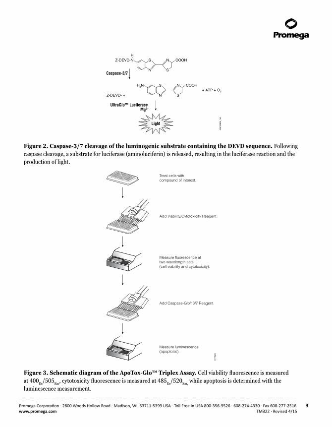
The second part of the assay uses the Caspase-Glo® Assay Technology by providing a luminogenic caspase-3/7 substrate, which contains the tetrapeptide sequence DEVD, in a reagent optimized for caspase activity, luciferase activity and cell lysis. Adding the Caspase-Glo® 3/7 Reagent in an “add-mix-measure” format results in cell lysis, followed by caspase cleavage of the substrate and generation of a “glow-type” luminescent signal produced by luciferase (Figure 2; 2). Luminescence is proportional to the amount of caspase activity present. The Caspase-Glo® 3/7 Reagent relies on the properties of a proprietary thermostable luciferase (Ultra-Glo™ Recombinant Luciferase), which is formulated to generate a stable “glow-type” luminescent signal and improve performance across a wide range of assay conditions.
Advantages of the ApoTox-Glo™ Assay:
Measure Viability, Cyototoxicity and Apoptosis in the Same Sample Well: Accurately determine the mechanism of cell death in less time with less sample.
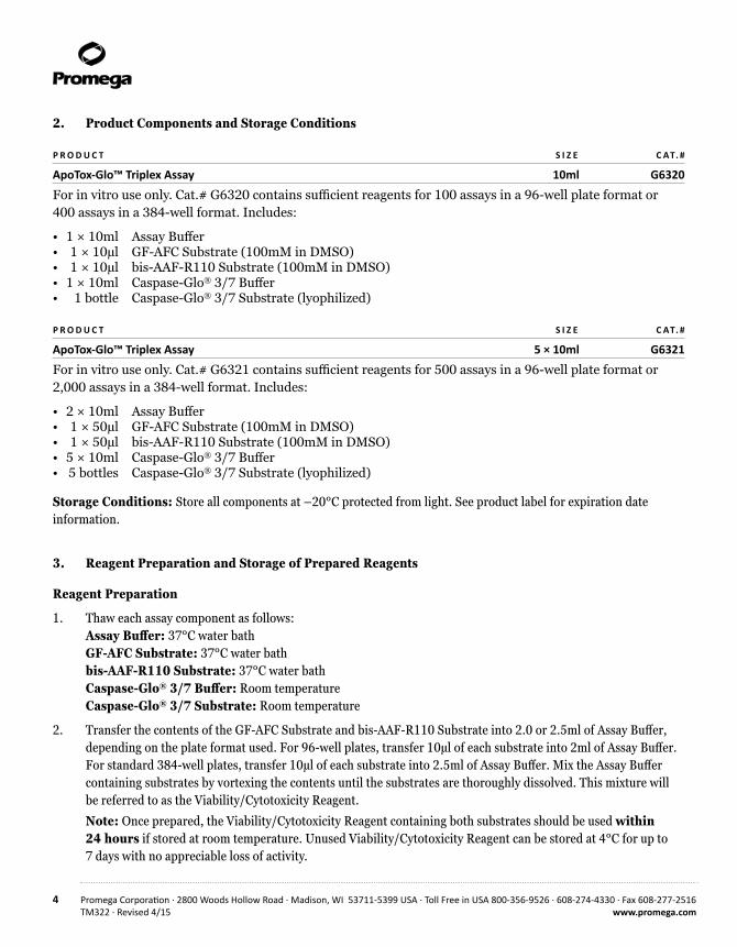
Easy to Implement: The assay uses a simple sequential “add-mix-read” format (Figure 3).
Normalize Data with a Built-In Internal Control: The ratio of the number of live cells to the number of dead cells is independent of cell number and normalizes data. This normalization makes results more comparable well-to-well, plate-to-plate and day-to-day.
Flexible and Easily Automated: The volumes of each assay component can be scaled to meet throughtput needs and is amenable to automation in 96- and 384-well plates.
5847
MA
cell-permeantGF-AFCsubstrate
GF-AFCsubstrate
cell-impermeantbis-AAF-R110
substrate
live-cellprotease
AFC
inactive live-cell
protease
active dead-cell protease
R110
Viable Cell Dead Cell
Figure 1. The biology of the viability/cytotoxicity assay. The GF-AFC Substrate can enter live cells where it is cleaved by the live-cell protease to release AFC. The bis-AAF-R110 Substrate cannot enter live cells but instead can be cleaved by the dead-cell protease to release R110.
Promega Corporation · 2800 Woods Hollow Road · Madison, WI 53711-5399 USA · Toll Free in USA 800-356-9526 · 608-274-4330 · Fax 608-277-2516 3www.promega.com TM322 · Revised 4/15
4061
MB
04_3
A
Caspase-3/7N
SH
S
NZ-DEVD-N COOH
-
N
S
S
NH2N COOH+ ATP + O2
UltraGlo™ LuciferaseMg2+
Light
Z-DEVD- +
Figure 2. Caspase-3/7 cleavage of the luminogenic substrate containing the DEVD sequence. Following caspase cleavage, a substrate for luciferase (aminoluciferin) is released, resulting in the luciferase reaction and the production of light.
8175
MA
Treat cells withcompound of interest.
Add Viability/Cytotoxicity Reagent.
Add Caspase-Glo® 3/7 Reagent.
Measure fluorescence at two wavelength sets(cell viability and cytotoxicity).
Measure luminescence (apoptosis).
Figure 3. Schematic diagram of the ApoTox-Glo™ Triplex Assay. Cell viability fluorescence is measured at 400Ex/505Em, cytotoxicity fluorescence is measured at 485Ex/520Em, while apoptosis is determined with the luminescence measurement.
4 Promega Corporation · 2800 Woods Hollow Road · Madison, WI 53711-5399 USA · Toll Free in USA 800-356-9526 · 608-274-4330 · Fax 608-277-2516TM322 · Revised 4/15 www.promega.com
2. Product Components and Storage Conditions
P R O D U C T S I Z E C AT. #
ApoTox-Glo™ Triplex Assay 10ml G6320
For in vitro use only. Cat.# G6320 contains sufficient reagents for 100 assays in a 96-well plate format or 400 assays in a 384-well format. Includes:
• 1 × 10ml Assay Buffer• 1 × 10µl GF-AFC Substrate (100mM in DMSO)• 1 × 10µl bis-AAF-R110 Substrate (100mM in DMSO)• 1 × 10ml Caspase-Glo® 3/7 Buffer• 1 bottle Caspase-Glo® 3/7 Substrate (lyophilized)
P R O D U C T S I Z E C AT. #
ApoTox-Glo™ Triplex Assay 5 × 10ml G6321
For in vitro use only. Cat.# G6321 contains sufficient reagents for 500 assays in a 96-well plate format or 2,000 assays in a 384-well format. Includes:
• 2 × 10ml Assay Buffer• 1 × 50µl GF-AFC Substrate (100mM in DMSO)• 1 × 50µl bis-AAF-R110 Substrate (100mM in DMSO)• 5 × 10ml Caspase-Glo® 3/7 Buffer• 5 bottles Caspase-Glo® 3/7 Substrate (lyophilized)
Storage Conditions: Store all components at –20°C protected from light. See product label for expiration date information.
3. Reagent Preparation and Storage of Prepared Reagents
Reagent Preparation
1. Thaw each assay component as follows: Assay Buffer: 37°C water bath GF-AFC Substrate: 37°C water bath bis-AAF-R110 Substrate: 37°C water bath Caspase-Glo® 3/7 Buffer: Room temperature Caspase-Glo® 3/7 Substrate: Room temperature
2. Transfer the contents of the GF-AFC Substrate and bis-AAF-R110 Substrate into 2.0 or 2.5ml of Assay Buffer, depending on the plate format used. For 96-well plates, transfer 10µl of each substrate into 2ml of Assay Buffer. For standard 384-well plates, transfer 10µl of each substrate into 2.5ml of Assay Buffer. Mix the Assay Buffer containing substrates by vortexing the contents until the substrates are thoroughly dissolved. This mixture will be referred to as the Viability/Cytotoxicity Reagent.
Note: Once prepared, the Viability/Cytotoxicity Reagent containing both substrates should be used within 24 hours if stored at room temperature. Unused Viability/Cytotoxicity Reagent can be stored at 4°C for up to 7 days with no appreciable loss of activity.

Promega Corporation · 2800 Woods Hollow Road · Madison, WI 53711-5399 USA · Toll Free in USA 800-356-9526 · 608-274-4330 · Fax 608-277-2516 5www.promega.com TM322 · Revised 4/15
3. Transfer the contents of the Caspase-Glo® 3/7 Buffer bottle into the amber bottle containing Caspase-Glo® 3/7 Substrate. Mix by swirling or inverting the contents until the substrate is thoroughly dissolved to form the Caspase-Glo® 3/7 Reagent (~20 seconds).
Note: Reconstituted Caspase-Glo® 3/7 Reagent can be stored according to the table below.
Storage TemperatureSignal Intensity Compared to Freshly Prepared Reagent
4°CUp to 3 days with no signal loss Stored for 1 week = ~90% signal Stored for 4 weeks = ~75% signal
–20°CStored up to 1 week = ~75% signal Stored up to 4 weeks = ~60% signal
4. Protocol
4.A. Materials to Be Supplied by the User• 96- or 384-well opaque-walled tissue culture plates with clear or solid bottoms• multichannel pipette or automated pipetting workstation• reagent reservoirs• orbital or linear plate shaker capable of 300–500rpm for 96-well plates or 1,300–1,500rpm for 384-well plates• microplate reader capable of measuring both luminescence and fluorescence at the following sets of wavelengths:
Excitation ~400nm and Emission ~505nm Excitation ~485nm and Emission ~520nm
• positive controls (see Section 4.E for recommendations)
4.B. Before You Begin
Before starting the assay, prepare the Assay Buffer with both substrates and Caspase-Glo® 3/7 Reagent as directed in Section 3. Because of the sensitivity of this assay, be careful not to touch pipette tips to the wells containing samples to avoid cross-contamination. Between dispensings, cover the plate with a lid or plate seal to minimize exposure to contaminants in the air. If you are reusing pipette tips, do not touch pipette tips to the wells containing samples to avoid cross-contamination.
Note: Temperature fluctuations can affect the luminescence readings. If the room temperature fluctuates, use a constant-temperature incubator. Total incubation time for the Caspase-Glo® 3/7 Assay depends upon the culture system, but typically peak luminescent signal will be reached in 1–2 hours. For optimal results, the maximum recommended incubation time is 3 hours. In general, the luminescent signal remaining at 3 hours is greater than 70% of peak luminescence.
6 Promega Corporation · 2800 Woods Hollow Road · Madison, WI 53711-5399 USA · Toll Free in USA 800-356-9526 · 608-274-4330 · Fax 608-277-2516TM322 · Revised 4/15 www.promega.com
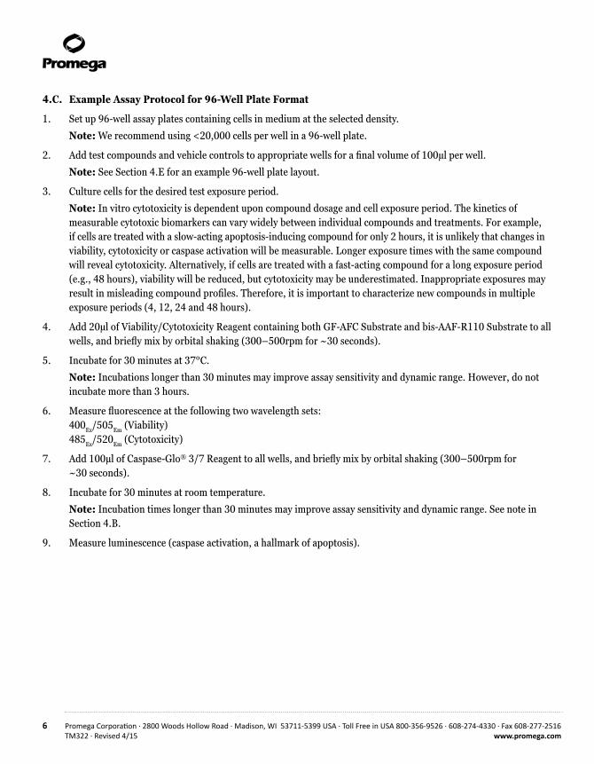
4.C. Example Assay Protocol for 96-Well Plate Format
1. Set up 96-well assay plates containing cells in medium at the selected density.
Note: We recommend using <20,000 cells per well in a 96-well plate.
2. Add test compounds and vehicle controls to appropriate wells for a final volume of 100µl per well.
Note: See Section 4.E for an example 96-well plate layout.
3. Culture cells for the desired test exposure period.
Note: In vitro cytotoxicity is dependent upon compound dosage and cell exposure period. The kinetics of measurable cytotoxic biomarkers can vary widely between individual compounds and treatments. For example, if cells are treated with a slow-acting apoptosis-inducing compound for only 2 hours, it is unlikely that changes in viability, cytotoxicity or caspase activation will be measurable. Longer exposure times with the same compound will reveal cytotoxicity. Alternatively, if cells are treated with a fast-acting compound for a long exposure period (e.g., 48 hours), viability will be reduced, but cytotoxicity may be underestimated. Inappropriate exposures may result in misleading compound profiles. Therefore, it is important to characterize new compounds in multiple exposure periods (4, 12, 24 and 48 hours).
4. Add 20µl of Viability/Cytotoxicity Reagent containing both GF-AFC Substrate and bis-AAF-R110 Substrate to all wells, and briefly mix by orbital shaking (300–500rpm for ~30 seconds).
5. Incubate for 30 minutes at 37°C.
Note: Incubations longer than 30 minutes may improve assay sensitivity and dynamic range. However, do not incubate more than 3 hours.
6. Measure fluorescence at the following two wavelength sets: 400Ex/505Em (Viability) 485Ex/520Em (Cytotoxicity)
7. Add 100µl of Caspase-Glo® 3/7 Reagent to all wells, and briefly mix by orbital shaking (300–500rpm for ~30 seconds).
8. Incubate for 30 minutes at room temperature.
Note: Incubation times longer than 30 minutes may improve assay sensitivity and dynamic range. See note in Section 4.B.
9. Measure luminescence (caspase activation, a hallmark of apoptosis).

Promega Corporation · 2800 Woods Hollow Road · Madison, WI 53711-5399 USA · Toll Free in USA 800-356-9526 · 608-274-4330 · Fax 608-277-2516 7www.promega.com TM322 · Revised 4/15
4.D. Example Assay Protocol for Standard 384-Well Plate Format
1. Set up 384-well assay plates containing cells in medium at the desired density.
Note: We recommend using <5,000 cells per well in a 384-well plate.
2. Add test compounds and vehicle controls to appropriate wells for a final volume of 20µl per well.
Note: See Section 4.E for an example 96-well plate layout.
3. Culture cells for the desired test exposure period.
Note: In vitro cytotoxicity is dependent upon compound dosage and cell exposure period. The kinetics of mea-surable cytotoxic biomarkers can vary widely between individual compounds and treatments. For example, if cells are treated with a slow-acting apoptosis-inducing compound for only 2 hours, it is unlikely that changes in viability, cytotoxicity or caspase activation will be measurable. Longer exposure times with the same compound will reveal cytotoxicity. Alternatively, if cells are treated with a fast-acting compound for a long exposure period (e.g., 48 hours), viability will be reduced, but cytotoxicity may be underestimated. Inappropriate exposures may lead to misleading profiles. Therefore, it is important to characterize new compounds in multiple exposure periods (4, 12, 24 and 48 hours).
4. Add 5µl of Viability/Cytotoxicity Reagent containing both GF-AFC Substrate and bis-AAF-R110 Substrate to all wells, and briefly mix by orbital shaking (1,300–1,500rpm for ~30 seconds).
5. Incubate for 30 minutes at 37°C.
Note: Incubations longer than 30 minutes may improve assay sensitivity and dynamic range. However, do not incubate more than 3 hours.
6. Measure fluorescence at the following two wavelength sets: 400Ex/505Em (Viability) 485Ex/520Em (Cytotoxicity)
7. Add 25µl of Caspase-Glo® 3/7 Reagent to all wells, and briefly mix by orbital shaking (1,300–1,500rpm for ~30 seconds).
8. Incubate for 30 minutes at room temperature.
Note: Incubation times longer than 30 minutes may improve assay sensitivity and dynamic range. See note in Section 4.B.
9. Measure luminescence (caspase activation, a hallmark of apoptosis).

8 Promega Corporation · 2800 Woods Hollow Road · Madison, WI 53711-5399 USA · Toll Free in USA 800-356-9526 · 608-274-4330 · Fax 608-277-2516TM322 · Revised 4/15 www.promega.com
4.E. Recommended Controls
No-Cell Control: Set up triplicate wells with medium but without cells to serve as the negative control for determining background fluorescence and luminescence.
Untreated Cells Control: Set up triplicate wells with untreated cells to serve as a vehicle control. Add the same percent solvent and medium vehicle used to deliver the test compounds to the vehicle control wells.
Optional Test Compound Control: Set up triplicate wells without cells containing the vehicle and test compound to test for possible interference with the assay chemistries.
Positive Controls
Cell Viability and Cytotoxicity: Set up triplicate wells containing cells treated with a compound known to be toxic to the cells used in your model system (e.g., final concentration of 30µg/ml digitonin for 15 minutes).
Necrosis: Set up triplicate wells containing cells treated with a compound known to be toxic to the cells used in your model system (e.g., 100µM ionomycin for 4–6 hours).
Apoptosis: Set up triplicate wells containing cells treated with a compound known to induce apoptosis in the cells used in your model system (e.g., 10µM staurosporine for 6 hours).
Note: It is important to use identical cell numbers and volumes for the assay and the control samples. You may need to empirically determine the optimal cell number, apoptosis induction treatment and incubation time for the cell culture system. We recommend using <20,000 cells per well in a 96-well plate and <5,000 cells per well in a 384-well plate.
Recommended Control Experiment (96-well format)
1. Choose the control compounds (ionomycin or staurosporine or both) appropriate for your experiment. Use 200µM ionomycin and 20µM staurosporine as the starting concentration. See Figure 4 for plate layout.
2. Add 50µl of RPMI 1640 + 10% FBS to columns 2–12 of a 96-well assay plate.
3. Add 50µl of control compound to replicate wells in columns 1 and 2. Mix the contents of column 2 by pipetting and transfer to column 3.
4. Repeat the mixing and transfer of compound until column 10. Discard the 50µl removed from column 10. This creates twofold serial dilutions from column 1 through column 10.
5. Prepare Jurkat cells at a concentration of 200,000 cells/ml, and dispense 50µl (a total of 10,000 cells) to all wells except column 12.
6. Add 50µl of medium and vehicle to column 12. Final volume in all wells will be 100µl.
7. Incubate the cells for 6 hours at 37ºC.
8. Add 20µl of Viability/Cytotoxicity Reagent containing 10µl of each substrate in 2ml of Assay Buffer to all wells, and briefly mix by orbital shaking (300–500rpm for ~30 seconds).
9. Incubate for at least 30 minutes at 37°C.

Promega Corporation · 2800 Woods Hollow Road · Madison, WI 53711-5399 USA · Toll Free in USA 800-356-9526 · 608-274-4330 · Fax 608-277-2516 9www.promega.com TM322 · Revised 4/15
10. Measure fluorescence at the following two wavelength sets: 400Ex/505Em (Viability) 485Ex/520Em (Cytotoxicity)
11. Add 100µl of Caspase-Glo® 3/7 Reagent to all wells, and briefly mix by orbital shaking (300–500rpm for ~30 seconds).
12. Incubate for 30 minutes at room temperature.
13. Measure luminescence (apoptosis).
8186
MA
A
B
C
D
E
F
G
H
1 2 3 4 5 6 7 8 9 10 11 12
10 5 2.5 1.25 0.62 0.31 0.16 0.08 0.04 0.02
100 50 25 12.5 6.25 3.12 1.56 0.78 0.39 0.20
Staurosporine Treatment (µM)Ionomycin Treatment (µM)Untreated Control (UTC)Background Control
UTC No cells
Figure 4. ApoTox-Glo™ Assay plate layout following Steps 2–6.

10 Promega Corporation · 2800 Woods Hollow Road · Madison, WI 53711-5399 USA · Toll Free in USA 800-356-9526 · 608-274-4330 · Fax 608-277-2516TM322 · Revised 4/15 www.promega.com
8141
MA
–7 –6 –5 –40
3,000
6,000
9,000
12,000
Cytotoxicity (bis-AAF-R110) EC50 = 6.87µM
Viability (GF-AFC) EC50 = 6.89µM
Apoptosis (Caspase-3/7) EC50 = N.D.
0
1,000
2,000
3,000
4,000
Log10 [ionomycin], M
Cyto
toxi
city
Flu
ores
cenc
e (R
FU)
Viab
ility
Flu
ores
cenc
e (R
FU)
and
Apo
ptos
is L
umin
esce
nce
(RLU
)
Figure 5. Expected results for ionomycin treatment of Jurkat cells. Ionomycin treatment for 6 hours should result in a dose-dependent decrease in viability, increase in cytotoxicity with no caspase-3/7 activation, which is consistent with primary necrosis.
8140
MA
–7 –6 –5 –40
5,000
10,000
15,000
20,000
25,000
30,000
Cytotoxicity (bis-AAF-R110) EC50 = 380nMViability (GF-AFC) EC50 = 463nM
Apoptosis (Caspase-3/7) EC50 = 491nM
0
200,000
400,000
600,000
800,000
Log10 [staurosporine], M
Viab
ility
or
Cyto
toxi
city
Fluo
resc
ence
(RF
U)
Apop
tosi
s Lu
min
esce
nce
(RLU
)
Figure 6. Expected results for staurosporine treatment of Jurkat cells. Staurosporine treatment for 6 hours should result in a dose-dependent decrease in viability, increase in cytotoxicity with an increase in caspase-3/7 activity consistent with apoptosis.

Promega Corporation · 2800 Woods Hollow Road · Madison, WI 53711-5399 USA · Toll Free in USA 800-356-9526 · 608-274-4330 · Fax 608-277-2516 11www.promega.com TM322 · Revised 4/15
5. General Considerations
This section contains a list of general factors to consider when designing your assay plate layout, interpreting your data accurately and troubleshooting the assay chemistry.
Length of Compound Exposure
The kinetics of cytotoxicity vary among compounds. The biomarkers of cytotoxicity and apoptosis may degrade in a time-dependent manner. Therefore, consider using this assay at different time points to establish optimal detection of cytotoxic affects or apoptosis. Primary necrosis (or catastrophic cell lysis) tends to occur very quickly after adding a toxic compound (i.e., 2 hours or less), whereas apoptosis proceeds in a more orderly manner over a longer period (i.e., 4–48 hours).
During most cytotoxicity events, viability and cytotoxicity measures will be inversely proportional. That is, if viability assay relative fluorescent units (RFU) are high, then cytotoxicity assay RFU values will be low, or vice versa. However, depending on the length of compound exposure, this inverse relationship does not always hold true. For example, after long-term exposure >24 hours, particularly after early primary necrosis occurs, the cytotoxicity biomarker will degrade after release into the extracellular environment and may lead to an underestimation of cytotoxicity. A reduction in viability without an increase in cytotoxicity might also be seen with compounds that alter normal cell division (cell-cycle arrest) without producing membrane integrity changes (Figure 7).
8170
MA
–9 –8 –7 –60
2,500
5,000
7,500
10,000
Viability/Cytotoxicity Reagent (Viability)Viability/Cytotoxicity Reagent (Cytotoxicity)Caspase-Glo® 3/7 Reagent (Apoptosis)
0
5,000
10,000
15,000
20,000
25,000
Log10 [camptothecin], M
Viab
iility
or C
ytot
oxic
ityFl
uore
scen
ce (
RFU)
Apop
tosi
s Lu
min
esce
nce
(RLU
)
Figure 7. An example of cytostasis seen using the ApoTox-Glo™ Triplex Assay. Camptothecin treatment of 10,000 K562 human erythroleukemia cells for 48 hours resulted in a dose-dependent decrease in apparent viability with no cytotoxicity, but an increase in caspase-3/7 activity, a profile consistent with cell cycle arrest and early phase apoptosis.

12 Promega Corporation · 2800 Woods Hollow Road · Madison, WI 53711-5399 USA · Toll Free in USA 800-356-9526 · 608-274-4330 · Fax 608-277-2516TM322 · Revised 4/15 www.promega.com
5. General Considerations (continued)
Selection of Compound Concentration(s)
Consider using serial dilutions of compounds instead of just one concentration in your assay. Many high-throughput screens are performed using a single compound concentration (e.g., 10µM final) to test larger numbers of compounds. However, using only one concentration can be problematic due to factors including biological variation in response and physiochemical concerns such as compound solubility. The approach of quantitative high-throughput screening (qHTS; 3) involves examining each compound in a screen in broad serial-dose dilutions. This approach can be more technically involved but can produce high-quality response curves that allow greater characterization of cytotoxic effects while mitigating false-positive or false-negative test results.
Interpreting the Mechanism of Cell Death
All three assay measures (viability, cytotoxicity, and caspase activation) are important for developing an accurate profile for your compound. In most circumstances, viability and cytotoxicity will be inversely correlated. However, it is well-appreciated that prototypical anticancer therapeutics may exhibit antiproliferative effects for sustained time periods prior to actual changes in membrane integrity. This period of cell cycle arrest will manifest as an apparent decline in viability with no concomitant increase in cytotoxic biomarker. Caspase activation may or may not be measurable during this period. Conversely, a measurable decline in apparent viability may be paired with a substantially reduced or unmeasurable cytotoxicity biomarker if cells died early (typically by primary necrosis) in the exposure period. If no caspase activation is indicated, primary necrosis or fast-acting apoptosis should be confirmed in a shorter exposure period (4).
Microplate Reader Settings
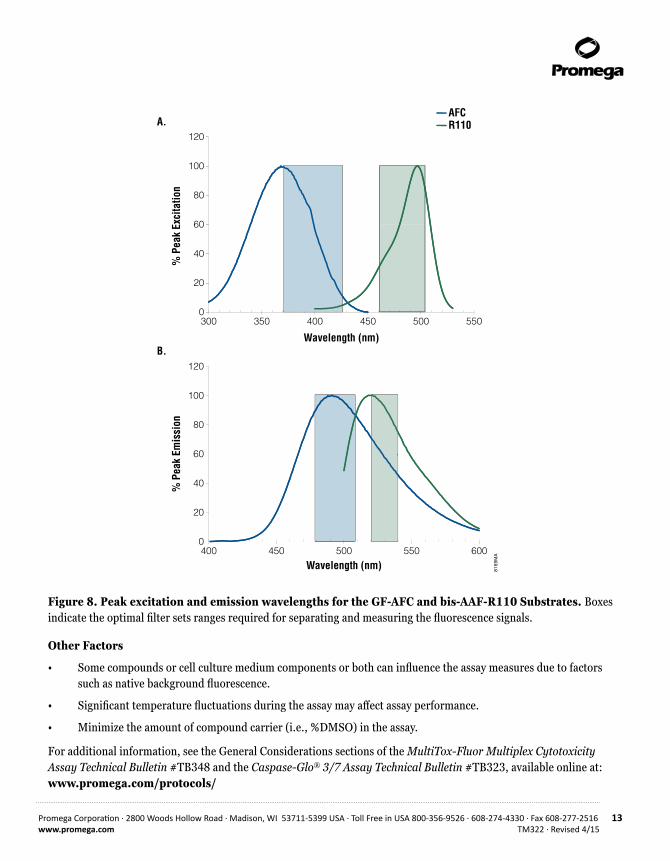
Fluorescent measurements: Carefully set the excitation and emission settings on your reader (as closely as possible) as follows: Viability: Excitation at 400nm / Emission at 505nm Cytotoxicity: Excitation at 485nm / Emission at 520nm
Results may suffer if the incorrect settings are selected. See Figure 8 for excitation and emission ranges.
Luminescence measurements: Confirm that the integration time is set within the following ranges: 96-well plates: 0.5–1 second 384-well plates: 0.25–0.5 second
Plotting Data
Consider plotting your data using a log-based transform for the compound concentration. Since the intensity of the fluorescent and luminescent measures (RFU vs. RLU) can differ significantly, consider plotting your data using two Y-axes. Refer to Section 4.E for examples.
Promega Corporation · 2800 Woods Hollow Road · Madison, WI 53711-5399 USA · Toll Free in USA 800-356-9526 · 608-274-4330 · Fax 608-277-2516 13www.promega.com TM322 · Revised 4/15
8169
MA
AFCR110
60
80
100
120
0
20
40
Wavelength (nm)600
% P
eak
Emis
sion
400 450 500 550
60
80
100
120
0
20
40
% P
eak
Exci
tatio
n
Wavelength (nm)
300 350 400 450 500 550
A.
B.
Figure 8. Peak excitation and emission wavelengths for the GF-AFC and bis-AAF-R110 Substrates. Boxes indicate the optimal filter sets ranges required for separating and measuring the fluorescence signals.
Other Factors
• Some compounds or cell culture medium components or both can influence the assay measures due to factors such as native background fluorescence.
• Significant temperature fluctuations during the assay may affect assay performance.
• Minimize the amount of compound carrier (i.e., %DMSO) in the assay.
For additional information, see the General Considerations sections of the MultiTox-Fluor Multiplex Cytotoxicity Assay Technical Bulletin #TB348 and the Caspase-Glo® 3/7 Assay Technical Bulletin #TB323, available online at: www.promega.com/protocols/

14 Promega Corporation · 2800 Woods Hollow Road · Madison, WI 53711-5399 USA · Toll Free in USA 800-356-9526 · 608-274-4330 · Fax 608-277-2516TM322 · Revised 4/15 www.promega.com
6. References
1. Niles, A.L. et al. (2007) A homogeneous assay to measure live and dead cells in the same sample by detecting different protease markers. Anal. Biochem. 366, 197–206.
2. O’Brien, M.A. et al. (2005) Homogeneous, bioluminescent protease assays: Caspase-3 as a model. J. Biomol. Screen. 10, 137-48.
3. Inglese, J. et al. (2006) Quantitative high-throughput screening: A titration-based approach that efficiently identifies biological activities in large chemical libraries. Proc. Natl. Acad. Sci. USA 103, 11473–8.
4. Niles, A.L., Moravec, R.A. and Riss, T.L. (2008) Update on in vitro cytotoxicity assays for drug development. Expert Opin. Drug Discovery 3, 655–69.
7. Additional Resources
Niles, A.L., Moravec, R.A. and Riss, T.L. (2009) In vitro viability and cytotoxicity testing and same-well multi- parametric combinations for high throughput screening. Curr. Chem. Genomics 3, 31–41.
Shultz, S. et al. (2008) Utilization of an automated triplex assay: New tool assesses cell viability, cytotoxicity, and apoptosis. GEN 28, 36–7.
Zakowicz, H. et al. (2008) Measuring cell health and viability sequentially by same-well multiplexing using the GloMax®-Multi Detection System. Promega Notes 99, 25–8.
Worzella, T., Busch, M. and Niles, A.L. (2008) High-throughput automation of multiplexed cell-based methods for viability and cytotoxicity. Cell Notes 20, 26–9.
Niles, A.L. et al. (2007) Using protease biomarkers to measure viability and cytotoxicity. Cell Notes 19, 16–20.
Niles, A.L. et al. (2007) Measure relative numbers of live and dead cells and normalize assay data to cell number. Cell Notes 18, 15–20.
Niles, A.L. et al. (2006) Monitor the ratio of live and dead cells within a population: MultiTox-Fluor Multiplex Cytotoxicity Assay. Promega Notes 94, 22–6.
Niles, A.L. et al. (2006) Multiplexed viability, cytotoxicity and apoptosis assays for cell-based screening. Cell Notes 16, 12–5.
Niles, A.L. et al. (2006) MultiTox-Fluor Multiplex Cytotoxicity Assay technology. Cell Notes 15, 11–5.
Riss, T.L. and Moravec, R.A. (2004) Use of multiple assay endpoints to investigate the effects of incubation time, dose of toxin, and plating density in cell-based cytotoxicity assays. Assay Drug Dev. Technol. 2, 51–62.

Promega Corporation · 2800 Woods Hollow Road · Madison, WI 53711-5399 USA · Toll Free in USA 800-356-9526 · 608-274-4330 · Fax 608-277-2516 15www.promega.com TM322 · Revised 4/15
8. Related Products
Multiplexed Viability and Cytotoxicity Assays
Product Size Cat.#MultiTox-Glo Multiplex Cytotoxicity Assay 10ml G9270
MultiTox-Fluor Multiplex Cytotoxicity Assay 10ml G9200
Available in Additional Sizes.
Viability Assays
Product Size Cat.#CellTiter-Glo® Luminescent Cell Viability Assay 10ml G7570
CellTiter-Fluor™ Cell Viability Assay 10ml G6080
Available in Additional Sizes.
Cytotoxicity Assays
Product Size Cat.#CytoTox-Glo™ Cytotoxicity Assay 10ml G9290
CytoTox-Fluor™ Cytotoxicity Assay 10ml G9260
Available in Additional Sizes.
Apoptosis Assays
Product Size Cat.#Caspase-Glo® 2 Assay 10ml G0940
Caspase-Glo® 3/7 Assay 10ml G8091
Caspase-Glo® 6 Assay 10ml G0970
Caspase-Glo® 8 Assay 10ml G8201
Caspase-Glo® 9 Assay 10ml G8211
Apo-ONE® Homogeneous Caspase-3/7 Assay 10ml G7790
Available in Additional Sizes.
Oxidative Stress Assays
Product Size Cat.#GSH-Glo™ Glutathione Assay 10ml V6911
Available in Additional Sizes.

16 Promega Corporation · 2800 Woods Hollow Road · Madison, WI 53711-5399 USA · Toll Free in USA 800-356-9526 · 608-274-4330 · Fax 608-277-2516TM322 · Revised 4/15 www.promega.com
8. Related Products (continued)
Detection Instrumentation
Product Size Cat.#GloMax®-Multi+ Detection System with Instinct® Software: Base Instrument with Shaking 1 each E8032
GloMax®-Multi+ Detection System with Instinct® Software: Base Instrument with Heating and Shaking 1 each E9032
9. Summary of Changes
The following changes were made to the 4/15 revision of this document:
1. The patent/license statements were updated.
2. The document design was updated.
(a)U.S. Pat. Nos. 7,416,854, 7,553,632 and other patents pending.(b)U.S. Pat. Nos. 6,602,677, 7,241,584 and 8,030,017, European Pat. No. 1131441, Japanese Pat. Nos. 4537573 and 4520084 and other patents pending.
© 2009, 2011, 2012, 2015 Promega Corporation. All Rights Reserved.
Apo-ONE, Caspase-Glo, Cell-Titer-Glo, GloMax and Instinct are registered trademarks of Promega Corporation. ApoTox-Glo, CellTiter-Fluor, CytoTox-Fluor, CytoTox-Glo, GSH-Glo and Ultra-Glo are trademarks of Promega Corporation.
Products may be covered by pending or issued patents or may have certain limitations. Please visit our Web site for more information.
All prices and specifications are subject to change without prior notice.
Product claims are subject to change. Please contact Promega Technical Services or access the Promega online catalog for the most up-to-date information on Promega products.