
Apoptosis induction in human breast cancer (MCF-7) … fetal bovine serum and 0.01 mg/ml bovine...
15
ORIGINAL PAPER Apoptosis induction in human breast cancer (MCF-7) cells by a novel venom L-amino acid oxidase (Rusvinoxidase) is independent of its enzymatic activity and is accompanied by caspase-7 activation and reactive oxygen species production Ashis K. Mukherjee 1,2 • Anthony J. Saviola 1,3 • Patrick D. Burns 1 • Stephen P. Mackessy 1 Published online: 29 August 2015 Ó Springer Science+Business Media New York 2015 Abstract We report the elucidation of a mechanism of apoptosis induction in breast cancer (MCF-7) cells by an L-amino acid oxidase (LAAO), Rusvinoxidase, purified from the venom of Daboia russelii russelii. Peptide mass fingerprinting analysis of Rusvinoxidase, an acidic mono- meric glycoprotein with a mass of *57 kDa, confirmed its identity as snake venom LAAO. The enzymatic activity of Rusvinoxidase was completely abolished after two cycles of freezing and thawing; however, its cytotoxicity toward MCF-7 cells remained unaffected. Dose- and time-depen- dent induction of apoptosis by Rusvinoxidase on MCF-7 cells was evident from changes in cell morphology, cell membrane integrity, shrinkage of cells and apoptotic body formation accompanied by DNA fragmentation. Rusvi- noxidase induced apoptosis in MCF-7 cells by both the extrinsic (death-receptor) and intrinsic (mitochondrial) signaling pathways. The former pathway of apoptosis operated through activation of caspase-8 that subsequently activated caspase-7 but not caspase-3. Rusvinoxidase- induced intrinsic pathway of apoptosis was accompanied by a time-dependent depolarization of the mitochondrial mem- brane through the generation of reactive oxygen species, followed by a decrease in cellular glutathione content and catalase activity, and down-regulation of expression of anti- apoptotic proteins Bcl-XL and heat-shock proteins (HSP-90 and HSP-70). Rusvinoxidase treatment resulted in increase of the pro-apoptotic protein Bax, subsequently leading to the release of cytochrome c from mitochondria to the cytosol and activating caspase-9, which in turn stimulated effector cas- pase-7. Rusvinoxidase at a dose of 4 mg/kg was non-toxic in mice, indicating that it may be useful as a model for the development of peptide-based anticancer drugs. Keywords Catalase Glutathione depletion Heat-shock protein Mitochondrial transmembrane potential Russell’s viper Introduction A recent epidemiological survey conducted by the Interna- tional Agency for Research on Cancer [1] highlighted an alarming increase in occurrence of breast cancer, which is the most frequently diagnosed cancer among women in 140 of 184 countries. Therefore, in order to decrease the global burden of cancer, research must continue to examine novel ways to inhibit cancer progression as well as to develop more effective chemotherapeutic agents against the malig- nancy. Further, exploring natural resources for potential therapeutic compounds [2] could lead to the development of novel, less toxic anticancer treatments with significantly less severe side effects than currently available treatments. Cellular homeostasis is maintained via a tightly regu- lated apoptotic (programmed cell death) mechanism Electronic supplementary material The online version of this article (doi:10.1007/s10495-015-1157-6) contains supplementary material, which is available to authorized users. & Ashis K. Mukherjee [email protected] 1 School of Biological Sciences, University of Northern Colorado, Greeley, CO 80639-0017, USA 2 Microbial Biotechnology and Protein Research Laboratory, Department of Molecular Biology and Biotechnology, Tezpur University, Tezpur, Assam 784028, India 3 Present Address: Department of Pharmacology, Weill Medical College of Cornell University, New York, NY 10065, USA 123 Apoptosis (2015) 20:1358–1372 DOI 10.1007/s10495-015-1157-6
Transcript of Apoptosis induction in human breast cancer (MCF-7) … fetal bovine serum and 0.01 mg/ml bovine...

ORIGINAL PAPER
Apoptosis induction in human breast cancer (MCF-7) cellsby a novel venom L-amino acid oxidase (Rusvinoxidase) isindependent of its enzymatic activity and is accompaniedby caspase-7 activation and reactive oxygen species production
Ashis K. Mukherjee1,2 • Anthony J. Saviola1,3 • Patrick D. Burns1 •
Stephen P. Mackessy1
Published online: 29 August 2015
� Springer Science+Business Media New York 2015
Abstract We report the elucidation of a mechanism of
apoptosis induction in breast cancer (MCF-7) cells by an
L-amino acid oxidase (LAAO), Rusvinoxidase, purified
from the venom of Daboia russelii russelii. Peptide mass
fingerprinting analysis of Rusvinoxidase, an acidic mono-
meric glycoprotein with a mass of *57 kDa, confirmed its
identity as snake venom LAAO. The enzymatic activity of
Rusvinoxidase was completely abolished after two cycles
of freezing and thawing; however, its cytotoxicity toward
MCF-7 cells remained unaffected. Dose- and time-depen-
dent induction of apoptosis by Rusvinoxidase on MCF-7
cells was evident from changes in cell morphology, cell
membrane integrity, shrinkage of cells and apoptotic body
formation accompanied by DNA fragmentation. Rusvi-
noxidase induced apoptosis in MCF-7 cells by both the
extrinsic (death-receptor) and intrinsic (mitochondrial)
signaling pathways. The former pathway of apoptosis
operated through activation of caspase-8 that subsequently
activated caspase-7 but not caspase-3. Rusvinoxidase-
induced intrinsic pathway of apoptosis was accompanied by a
time-dependent depolarization of the mitochondrial mem-
brane through the generation of reactive oxygen species,
followed by a decrease in cellular glutathione content and
catalase activity, and down-regulation of expression of anti-
apoptotic proteins Bcl-XL and heat-shock proteins (HSP-90
and HSP-70). Rusvinoxidase treatment resulted in increase of
the pro-apoptotic protein Bax, subsequently leading to the
release of cytochrome c from mitochondria to the cytosol and
activating caspase-9, which in turn stimulated effector cas-
pase-7. Rusvinoxidase at a dose of 4 mg/kg was non-toxic in
mice, indicating that it may be useful as a model for the
development of peptide-based anticancer drugs.
Keywords Catalase � Glutathione depletion � Heat-shockprotein � Mitochondrial transmembrane potential �Russell’s viper
Introduction
A recent epidemiological survey conducted by the Interna-
tional Agency for Research on Cancer [1] highlighted an
alarming increase in occurrence of breast cancer, which is
the most frequently diagnosed cancer among women in 140
of 184 countries. Therefore, in order to decrease the global
burden of cancer, research must continue to examine novel
ways to inhibit cancer progression as well as to develop
more effective chemotherapeutic agents against the malig-
nancy. Further, exploring natural resources for potential
therapeutic compounds [2] could lead to the development of
novel, less toxic anticancer treatments with significantly less
severe side effects than currently available treatments.
Cellular homeostasis is maintained via a tightly regu-
lated apoptotic (programmed cell death) mechanism
Electronic supplementary material The online version of thisarticle (doi:10.1007/s10495-015-1157-6) contains supplementarymaterial, which is available to authorized users.
& Ashis K. Mukherjee
1 School of Biological Sciences, University of Northern
Colorado, Greeley, CO 80639-0017, USA
2 Microbial Biotechnology and Protein Research Laboratory,
Department of Molecular Biology and Biotechnology,
Tezpur University, Tezpur, Assam 784028, India
3 Present Address: Department of Pharmacology, Weill
Medical College of Cornell University, New York,
NY 10065, USA
123
Apoptosis (2015) 20:1358–1372
DOI 10.1007/s10495-015-1157-6
[3].This is achieved via two major pathways—the extrinsic
pathway that occurs through death receptors present in the
outer membrane of the cell, and the intrinsic pathway,
which is a mitochondria-dependent pathway [3]. Any
critical defect in the apoptotic signaling pathways may
result in uncontrolled proliferation and growth of cells
which may ultimately lead to cancer, and the use of
chemotherapeutic agents to induce apoptosis in cancer is
one of the effective ways to overcome this deadly disease.
However, despite the development of new therapies,
acquired multidrug drug resistance in cancer cells has
become one of the major impediments against successful
treatment. Therefore, new anticancer drugs capable of
targeting cancer through multiple mechanisms can provide
a significant therapeutic advantage.
Snake venoms show promise in the treatment of several
diseases, including cancer [4, 5]. Among the different
components, L-amino acid oxidase (LAAO, E.C.1.4.3.2) is
a well-studied, important component of snake venom
which inhibits growth of mammalian cancer cells by
induction of apoptosis and inhibition of angiogenesis,
suggesting its potential as a lead compound for anticancer
drug development [5, 6]. However, there is significant
controversy regarding the anticancer mechanism(s) of
snake venom LAAO; it has been shown that the cytotoxic
activity of LAAOs is only partly dependent on H2O2 pro-
duction, indicating the presence of LAAO-specific recep-
tors or targets on the cell surface which are involved in the
induction of apoptosis [5]. In contrast, it has been reported
that the LAAO purified from Bothrops leucurus (Bl-
LAAO) venom induces apoptosis in cancer cells through
the generation of high amounts of H2O2 [7].
The venom of Russell’s viper (Daboia russelii russelii)
is rich in LAAO (which gives venom its characteristic
yellow color [8]); however, at present no attempt has been
made to characterize the anticancer potential and mecha-
nism of apoptosis induction in breast cancer cells by
LAAO purified from venom of D. r. russelii. In the present
study, we explore the cytotoxic mechanism of a novel
LAAO, named Rusvinoxidase, purified from venom of
Russell’s viper from Pakistan.
Materials and method
Venom of Daboia r. russelii was a gift from Kentucky
Reptile Zoo, USA; venom was extracted from snakes
originating in Pakistan. Protein concentration standard
reagents were purchased from BioRad Inc., USA. Chro-
mogenic and fluorogenic caspase substrates were pur-
chased from Sigma-Aldrich, USA. Pre-cast
NuPAGENovex� Bis–Tris gels, buffers and Mark 12
unstained molecular mass standards were obtained from
Life Technologies (Invitrogen Inc.), USA. Primary anti-
bodies against pro- and anti-apoptotic proteins were a gift
from Bioss Antibodies, MA, USA. All other chemicals
used were of analytical grade and procured from Sigma-
Aldrich, USA.
Purification of a cytotoxic protein (Rusvinoxidase)
from Russell’s viper venom (RVV)
One hundred seventy-four mg (dry protein weight) of lyo-
philized D. r. russelii venom was dissolved in 1.5 ml of
25 mM HEPES buffer containing 100 mM NaCl and 5 mM
CaCl2, pH 6.8 and the solution was centrifuged at
10,0009g for 10 min (Microfuge 18 Centrifuge, Beckman
Coulter, USA). The resulting clear yellow supernatant was
fractionated through a Bio Gel P-100 gel filtration column
(2.8 9 80 cm) previously equilibrated with the same buffer
[9]. The gel filtration fractions showing cytotoxicity (peak 1)
were pooled, desalted and lyophilized. The lyophilized pro-
teins were dissolved in 1.0 ml of buffer A (20 mMTris–HCl,
pH 8.0) and loaded on a Mono Q 5/50 GL anion exchange
columnpreviously equilibratedwith the bufferA. The column
was washed with 3 column volumes of equilibration buffer to
elute unbound proteins. The bound proteinswere elutedwith a
linear gradient from 0 to 350 mMNaCl in 20 mM Tris–HCl,
pH 8.0 (buffer B) at a flow rate of 0.75 ml/min for 80 min and
the elution of proteins was monitored at 280 nm [10]. The
protein peaks were desalted and each protein peak was then
screened for cytotoxic activity against MCF-7 cells.
Determination of purity and molecular weight
of Rusvinoxidase
The homogeneity andmolecularmass of the protein showing
cytotoxic activity was determined by SDS-PAGE analysis of
5 lg purified protein under both reduced and non-reduced
conditions followed by staining with Coomassie Brilliant
Blue R-250 [9]. Mobility of purified protein was compared
with Mark 12 (Invitrogen) molecular weight markers
(2.5–200 kDa) and a linear dependency of log MW versus
migration distance of protein bands was observed.
Identification of Rusvinoxidase by peptide mass
finger printing analysis
In-gel tryptic digestion of the protein was performed as
described by Mukherjee and Mackessy [9]. For LC/MS/MS
analysis, an aliquot of trypsin-digested peptide fragments
was withdrawn from the digest and the peptides were
purified and concentrated using an on-line enrichment
column (Agilent Zorbax C18, 5 lm, 5 9 0.3 mm). LC/
MS/MS was performed on a LTQ linear ion trap mass
spectrometer (Thermo Scientific) using a reversed-phase
Apoptosis (2015) 20:1358–1372 1359
123
nanospray column (Agilent 1100 nanoHPLC, Zorbax C18,
5 lm, 75 lm ID 9 150 mm column).The peptides were
eluted from the column with a 42 min linear gradient from
25 to 55 % buffer B (90 % acetonitrile, ACN, and 0.1 %
formic acid) at a flow rate of 0.3 ll/min. Spectra were
collected over a m/z range of 200–2000 Da using a
dynamic exclusion limit of 2 MS/MS spectra of a given
peptide mass for 30 s (exclusion duration of 90 s). Com-
pound lists of the resulting spectra were generated using
Bioworks 3.0 software (Thermo Scientific) with an inten-
sity threshold of 5000 and 1 scan/group. MS/MS spectra
were searched against the NCBInr protein database using
the Mascot database search engine (version 2.3). Scaffold 3
proteomic software (Proteome Software Inc., Portland,
OR) was used to validate MS/MS-based peptide and pro-
tein identifications.
The tryptic sequences of those peptides (obtained by
LC/MS/MS) showing more than 95 % probability were
subjected to a BLAST search in NCBInr data base, Swis-
sprot protein sequences (swissprot), and Protein Databank
(pdb) proteins against a snake venom protein database
(snakes, taxid:8570) using the blastp algorithm (http://
blast.ncbi.nlm.nih.gov/Blast.cgi).
Assay of enzyme activity of Rusvinoxidase
The LAAO activity was assayed using L-kynurenine as a
substrate [11]. The unit of LAAO activity is defined as one
nano mole of kynurenic acid produced/min under the assay
condition. Metalloprotease activity was assayed using
azocasein as described by Aird and de Silva [12]. Ester-
olytic activity was assayed using N-a-p-Tosyl-L-argininemethyl ester hydrochloride (TAME) and N-a-benzoyl-L-arginine ethyl ester hydrochloride (BAEE) as substrates
[13, 14].
Cell proliferation assay
In vitro cytotoxicity assays were conducted on the MCF-7
human breast adenocarcinoma cell line and human
embryonic kidney cell (HEK 293) (ATCC; Manassas, VA,
USA) cultured and maintained in Eagle’s minimum
essential medium (EMEM) and Dulbecco’s modified eagle
medium, respectively supplemented with 10 % heat-inac-
tivated fetal bovine serum and 0.01 mg/ml bovine insulin
at 37 �C in a humidified CO2 incubator (5 % CO2, 95 %
air). For cytotoxicity assays, 100 ll aliquots of 1 9 105
cells/ml were plated into 96-well plates and treated with
various concentrations (0–80.0 lg/ml) of Rusvinoxidase or
cytosine-b-D-arabinofuranoside-HCl (AraC, an anticancer
drug; 0–80 lg/ml, positive control), or treated with growth
medium (negative control), and cells were incubated at
37 �C for 24 h. Complete medium without cells was used
for blank absorbance readings. After 24 h, cytotoxicity of
Rusvinoxidase or AraC was measured using the colori-
metric MTT [3-(4,5-dimethylthiazol-2-yl)-2,5-diphenylte-
trazolium bromide] assay following manufacturer
instructions (ATCC). A standard curve was generated for
each assay performed, and the IC50 values were calculated
from the regression analysis of growth curves of MCF-7
cells in the presence of Rusvinoxidase. All assays were
performed in triplicate per treatment and repeated at least
three times.
Cellular and nuclear morphological changes
in MCF-7 cells induced by Rusvinoxidase
To study morphological changes induced by Rusvinoxi-
dase, 1 ml of 5 9 105 MCF-7 cells were seeded in a 24 well
plate and allowed to adhere overnight at 37 �C. The fol-
lowing day, the medium was replaced with complete fresh
medium and after treatment with different doses of Rusvi-
noxidase (� 9 IC50, 1 9 IC50, and 2 9 IC50 doses;
IC50 = 83 nM) for differing time periods (0–24 h), both the
non-adherent and adherent cells were collected (by
trypsinization). For every time point, a control was run in
parallel where the cells were treated with only medium. The
cells were washed with PBS, re-suspended in culture
medium and then stained with 5 ll each of ethidium bro-
mide and acridine orange (AO) (10 mg/ml in PBS). After
15 min of incubation at 37 �C in a humidified CO2 incu-
bator, the cells were washed twice in PBS and then observed
under a fluorescence microscope at 960 magnification.
Rusvinoxidase-induced nuclear damage in MCF-7 cells
was observed by Hoechst 33258 staining [15, 16]. Briefly,
after exposure to Rusvinoxidase (� 9 IC50–2 9 IC50;
IC50 = 83 nM), both non-adherent and adherent cells were
collected, washed in PBS and then fixed in 1 %
formaldehyde (in PBS) for 30 min at room temperature. A
control was run in parallel in which cells were treated with
only medium. After washing the cells with 1 9 PBS, they
were re-suspended in 100 ll growth medium and incubated
with 5 ll of Hoechst 33258 (10 mg/ml) for 30 min at
37 �C. The cells were then washed with PBS, placed on to
a glass slide and observed under a fluorescence microscope
at 960 magnification. The percentage of apoptotic cells
were counted at four randomly selected microscopic fields.
DNA fragmentation assay of Rusvinoxidase-treated
cells
For qualitative assay of DNA fragmentation, 1 ml of MCF-
7 cells were plated in 24 well plates at a density of 1 9 106
cells/well and allowed to adhere overnight at 37 �C. Thenext day, the medium was replaced with fresh medium
containing an IC50 dose (83 nM) of Rusvinoxidase and
1360 Apoptosis (2015) 20:1358–1372
123

returned to 37 �C for an additional 24 h. The cells treated
with only growth medium served as a control. The adherent
cells were harvested by trypsinization and combined with
non-adherent cells, washed in PBS and DNA was prepared
from the pelleted cells following the procedure described
by Herrmann et al. [17]. For the quantitative DNA frag-
mentation assay, the cells were cultured and treated with
Rusvinoxidase as above. Following lysis of cells, the lysate
was centrifuged at 11,4009g for 10 min to separate the
fragmented DNA (supernatant) from the intact chromatin
(pellet). Both fractions were treated with 1.0 ml of 0.5 M
trichloroacetic acid (TCA) overnight at 4 �C. The next day,both mixtures were centrifuged and the pelleted DNA was
treated with 160 ll of 5 % TCA at 90 �C for 15 min [18].
The DNA content of both fractions was estimated at
260 nm using a NanoDrop 2000 spectrophotometer. The
percent fragmentation was determined by calculating the
ratio of DNA in the supernatant to the total DNA recovered
in the supernatant and pellet, multiplied by 100.
Flow cytometric analysis of apoptosis induction
in MCF-7 cells by Rusvinoxidase
The induction of apoptosis in MCF-7 cells by Rusvinoxi-
dase was detected using an APO-BrdUTM TUNEL Assay
Kit (Invitrogen, USA). Briefly, 1 ml of 1 9 106 cells/well
were plated in 24-well plate and incubated for 18 h at
37 �C, 5 % CO2. Following incubation, the medium was
replaced with complete fresh medium containing � 9,
1 9, or 2 9 IC50 doses (IC50 = 83 nM) of Rusvinoxidase,
or growth medium (control) and incubated for additional
24 h at 37 �C. The adherent cells were harvested by
trypsinization and combined with non-adherent (detached)
cells. The cells were washed with PBS (pH 7.4) and then
5.0 ml of 1 % paraformaldehyde was added and cells were
incubated on ice for 15 min. The cells were washed twice
with PBS, pH 7.4 and then re-suspended in 0.5 ml PBS and
stored in 70 % (v/v) ice-cold ethanol at -20 �C for 18 h
before proceeding with the TUNEL assay, following the
instructions of the manufacturer. The samples were ana-
lyzed using flow cytometry (FACscan, Becton–Dickinson,
Bedford, MA, USA) within 3 h after staining with pro-
pidium iodide.
Estimation of total glutathione and catalase activity
in Rusvinoxidase-treated MCF-7 cells
One ml of MCF-7 cells were seeded in a 24-well plate at a
density of 1 9 106 cells/well and then allowed to attach to
the plate overnight at 37 �C. Culture medium was then
replaced with new medium containing an IC50 dose
(83 nM) of Rusvinoxidase and incubated for additional
0–24 h. For every time point, a control was run in parallel
where the cells were treated with only medium. After a
specific time interval, both the free non-adherent and
adherent cells were collected, and the cell lysate was used
to determine total glutathione content and catalase activity
[19]. Catalase activity or glutathione content per mg pro-
tein of control (untreated) MCF-7 cell lysate at each time
point was considered as 100 % activity and treated values
were compared to this control.
Assay for caspase activity in MCF-7 cells
MCF-7 cells (1 9 106 cells/ml) were treated with an IC50
dose (83 nM) of Rusvinoxidase or growth medium (con-
trol) for various time periods (0–24 h); at the end of
treatment, cells were lysed with RIPA lysis buffer (Sigma-
Aldrich, USA). The cell lysate was centrifuged at
10,0009g for 10 min at 4 �C and 25 ll of the supernatant
was assayed for caspase-3 or caspase-9 using Ac-Asp-Met-
Gln-Asp-pNA or Ac-Leu-Glu-His-Asp-pNAas a substrate
[9]. The unit of caspase activity (amidolytic activity) was
defined as lmoles of 4-nitroaniline released per minute by
the enzyme under the assay condition [9] and enzyme
activity was expressed as units of enzyme activity per mg
of protein. Caspase-8 and caspase-7 activities were assayed
by using the fluorogenic substrate Ac-Val-Glu-Thr-Asp-
AMCandAc-Asp-Glu-Val-Asp-AMC (20 lM, respec-
tively). After 30 min incubation at 37 �C, release of
methylcoumaryl-7-amine (AMC) was monitored in a
spectrofluorimeter (Perkin Elmer) at an excitation wave-
length of 380 nm and emission wavelength of 460 nm [20].
Caspase activity was expressed as units of enzyme activity
per mg protein. For every time period, a control was run in
which the cells were treated with growth medium only, and
the experimental values were compared to that baseline
value.
Determination of total ROS generation in MCF-7
cells
Intracellular reactive oxygen species (ROS) levels gener-
ated upon LAAO exposure to 1 9 106 cells were measured
using the non-fluorescent compound 20,70-dichlorofluores-cein-diacetate (DCFH-DA; Sigma-Aldrich, USA). MCF-7
cells were incubated with different doses of Rusvinoxidase
or growth medium (control) for 4 h and thereafter both the
adherent and free non-adherent cells were collected and
washed twice with PBS, pH 7.4. The cells were incubated
with 10 lM DCFH-DA at 37 �C for 30 min in the dark
followed by washing twice with chilled PBS. The fluo-
rescence intensity of 20,70-dichlorodihydrofluorescein(DCF) produced by intracellular reactive oxygen species
was analyzed on a flow cytometer with excitation and
emission at 480 and 530 nm, respectively.
Apoptosis (2015) 20:1358–1372 1361
123

Assay for mitochondrial transmembrane potential
of MCF-7 cells
The toxin-induced change in mitochondrial transmembrane
potential (MMP) in MCF-7 cells was determined using
5,50,6,60-tetrachloro-1,10,3,30-tetraethylbenzimidazolylcar-
bocyanine iodide (JC-1) (MitoProbeTM JC-1 Assay kit,
Sigma). After exposure of 1 9 106 cells to 1 9 IC50
(83 nM) or� 9 IC50 of Rusvinoxidase, or growth medium
(control) for 0-24 h, the cells were treated with JC-1 or
carbonyl cyanide m-chlorophenylhydrazone (CCCP; posi-
tive control) following the instructions of the manufacturer,
and the fluorescence intensity was determined by flow
cytometry (BD AccuriTMC6 Cytometer) with an excitation
at 488 nm and emission at 533 ± 30 nm (FL-1 green
channel) and 585 ± 40 nm (FL-2 red channel). Cells were
initially gated from debris using side scatter and forward
scatter. Data were presented as percentage of cells with
altered MMP. The Rusvinoxidase-induced change in MMP
potential was also detected by observing the Rusvinoxi-
dase-treated and control cells (JC-1 stained) with a con-
focal laser microscope (Olympus1X81).
Analysis of expression of pro- and anti-apoptotic
proteins in MCF-7 cells after Rusvinoxidase
treatment through Western blot analysis
For analyzing the expression of pro- and anti-apoptotic
proteins, 1 9 106 MCF-7 cells were incubated with
Rusvinoxidase (IC50 dose, 83 nM) or growth medium
(control) for 0–24 h, and both the detached and adhered
cells were collected and washed with PBS, pH 7.4. Cells
were lysed in 200 ll of RIPA lysis buffer (containing
protease inhibitor cocktail) in ice and then centrifuged at
10,0009g for 20 min at 4 �C. The protein content of the
supernatant was estimated by Bradford method (BioRad,
Inc.) and 50 lg of protein from each sample was run in
12.5 % NuPAGENovex� Bis–Tris mini gels (Invitrogen).
The proteins were electrophoretically transferred to a
nitrocellulose membrane and non-specific sites were
blocked by incubating the membrane for 1 h at room
temperature with 5 % (W/V) skimmed milk in 20 mM Tris
buffer saline (TBS), pH 7.4, containing 0.1 % Tween-20.
After washing the membrane with TBS (pH 7.4), it was
incubated with primary antibodies (1:750 dilution) against
Bcl-XL, Bax, HSP-70, HSP-90, cytochrome c or b-actin(internal standard) for 1 h at room temperature and then
overnight at 4 �C. The following day, the membrane was
washed with TBS (pH 7.4) and then incubated with
horseradish peroxidase-conjugated mouse anti-rabbit sec-
ondary antibodies (1:3000) for 1 h at room temperature,
and the Western blot was developed with SuperSignalTM
Western Blot Enhancer (Fisher Scientific). The signal was
recorded with a CCD camera and analyzed through
Quantity One 1-D analysis software (VersaDocTM Imaging
System, BioRad, USA). The experiment was repeated three
times to assure reproducibility.
Determination of in vivo toxicity of Rusvinoxidase
on mouse model
All experimental protocols for animal use were approved by
the Institutional Animal Care and Use Committee, UNC
(protocol 9401). For determining lethal toxicity, Rusvinoxi-
dase was dissolved in 0.2 ml of PBS, pH 7.4 and injected i.p
(1.0–4.0 lg/g body weight) into a group of three non-Swiss
albino (NSA) mice weighing between 18 and 20 g. Control
animals received only 0.2 ml of PBS, pH 7.4. The animals
were observed at regular intervals up to 48 h post-injection
for death or any physical or behavioral changes [9, 10].
Statistical analysis
To determine any significant differences, data were ana-
lyzed by a Student’s t test using the software SigmaPlot
11.0 for Windows (version 7.0), with p B 0.05 considered
as statistically significant.
Results
Purification and identification of a cytotoxic protein
(Rusvinoxidase) from Russell’s viper venom
Fractionation of crude RVV through size exclusion resulted
in separation of twelve protein/peptide peaks, named GF1-
GF12 (supplementary Fig. S1). The pooled fractions of
peak GF1 (showing highest cytotoxic activity) were frac-
tionated into eight peaks following anion exchange FPLC
(supplementary Fig. S2). The FPLC fraction (shown with
an arrow in Fig. S2) demonstrating significant cytotoxic
activity against MCF-7 cells (Table 1) was found to be
homogenous by 12 % SDS-PAGE (Fig. 1), displaying a
single band with an apparent molecular weight of 57.5 kDa
(reduced) and 55.4 kDa (non-reduced). This protein,
named Rusvinoxidase (Russell’s Viper L-amino acid oxi-
dase), represents 0.3 % of the total protein of crude RVV.
A summary of the purification of Rusvinoxidase from
crude RVV is shown in Table 1.
The LC–MS/MS analysis of the Rusvinoxidase trypsin
digest peptides unambiguously demonstrated its identity
(100 % probability, rank 1, 11 % sequence coverage) as an
LAAO with high homology to an LAAO (accession no.
Q4F867) purified from D. r. siamensis venom. BLASTP
analysis of 5 unique tryptic peptide sequences of Rusvinoxi-
dase against the NCBI snake venom protein database
1362 Apoptosis (2015) 20:1358–1372
123

demonstrated that this protein has significant similarity with
LAAOs isolated from other snake venoms, especially with
those isolated from other viperid venoms (Table 2).
Biochemical characterization of Rusvinoxidase
Rusvinoxidase was yellow in color due to presence of
flavin adenine dinucleotide, and it exhibited LAAO specific
activity of 23.8 U/mg protein. The LAAO activity dimin-
ished progressively after storage at 4 �C yet cytotoxic
activity of Rusvinoxidase was not affected (supplementary
Fig S3). The LAAO enzyme activity of Rusvinoxidase
following two freeze and thaw cycles was completely
abolished and after six cycles of freeze and thaw, the
enzyme activity could not be regained after incubation for
24 h at 37 �C; again, its cytotoxic property remained
unaffected. Rusvinoxidase did not show protease (azoca-
seinolytic, fibrinolytic, fibrinogenolytic), phospholipase
A2, TAME- or BAEE-esterase activities.
Rusvinoxidase significantly inhibits proliferation
of MCF-7 breast cancer cells
Rusvinoxidase demonstrated significantly higher
(p\ 0.05) dose-dependent cytotoxic activity toward MCF-
7 cells compared with the commercial anticancer drug
cytosine-b-D-arabinofuranoside (AraC), an antitumor agent
which selectively inhibits DNA synthesis (Fig. 2). From
regression analysis, the IC50 value of Rusvinoxidase
towards MCF-7 cells, after 24 h incubation, was 5.5 lg/ml
(83 nM). After 24 h of treatment with Rusvinoxidase at a
dose of 10 lg/ml (*2 9 IC50), MCF-7 cell viability was
zero (Fig. 2). Following a 24 h treatment with Rusvinoxi-
dase at its IC50 dose, most of the MCF-7 cells were
detached from the culture flasks and were found non-ad-
herent in the culture media.
In a sharp contrast, Rusvinoxidase at a concentration of
5 and 10 lg/ml inhibited 6 ± 1.2 and 12 ± 2.1 %
(mean ± SD, n = 3), respectively of HEK cells after 24 h
treatment suggesting it has marginal cytotoxicity against
normal human cells.
Rusvinoxidase induces morphological changes,
chromatin condensation and DNA fragmentation
in MCF-7 cells
Microscopic analysis also revealed a dose-dependent and
time-dependent decrease (p\ 0.01) in the population of
Rusvinoxidase-treated MCF-7 cells as compared to con-
trols (Fig. 3a). Induction of apoptosis in MCF-7 cells by
Rusvinoxidase was evident from the light microscopic
observations of changes in cell morphology, loss of cell
Table 1 Summary of purification of Rusvinoxidase, a cytotoxic L-amino acid oxidase from RVV
Purification steps Total protein
(mg)
Protein
yield (%)
Cytotoxicity
(% cell death)*
Specific activity (% cell
death/mg protein)
Purification
(fold)
Crude RVV 174 100 13.8 2760.0 111.0
GF-I 17.9 10.4 25.9 5180.0 1.9
FPLC-3 (Rusvinoxidase) 0.6 0.3 48.5 9700.0 3.5
The data represent a typical experiment
* Cytotoxicity (5 lg/ml) against MCF-7 cells after 24 h incubation at 37 �C, 5 % CO2. Control cells (treated with growth medium) under
identical experimental conditions showed 99 % cell viability
Fig. 1 Determination of the purity and molecular mass of Rusvi-
noxidase by 12 % SDS-PAGE. Lanes 2–4 reducing conditions; lanes
6–8 non-reduced conditions. Lanes 1 and 5 protein mass standards
(Dalton Mark 12); lanes 2 and 6, crude RVV (20 lg); lanes 3 and 7,
gel filtration fraction (GF1) (15 lg); lanes 4 and 8, Rusvinoxidase
(5.0 lg)
Apoptosis (2015) 20:1358–1372 1363
123

membrane integrity, and shrinkage of cells; apoptotic body
formation was detected in a dose-and time-dependent
manner (Fig. 3a). Twenty-four hour incubation of MCF-7
cells with 2 9 IC50 of Rusvinoxidase resulted in pro-
nounced apoptosis, and apoptotic cells had undergone
secondary necrosis (Fig. 3a).The changes in nuclear mor-
phology of treated breast cancer cells were evident from
chromatin condensation and formation of apoptotic cells
(Fig. 3a). Further, using Hoechst 33258 staining, dose- and
time-dependent apoptosis induction in MCF-7 cells by
Rusvinoxidase was observed (Fig. 3b). In addition to being
dependent on Rusvinoxidase concentration, percent apop-
tosis of MCF-7 cells induced by Rusvinoxidase increased
with exposure time (Fig. 3c).
Incubation of MCF-7 cells with Rusvinoxidase resulted
in an increase in DNA fragmentation of cancer cells as
compared to controls (Fig. 4a). This result confirmed that
apoptosis was accompanied by DNA fragmentation in
MCF-7 cells after exposure to Rusvinoxidase. Using the
APO-BrdUTuNEL assay, a routine method to quantify the
extent of apoptosis induction by anticancer agents, Rusvi-
noxidase was found to induce apoptosis (DNA fragmen-
tation) dose-dependently in treated cancer cells as
compared with control cells (Fig. 4b).
Rusvinoxidase induces apoptosis in MCF-7 cells
via activation of caspases-8, 9 and 7
Increase or decrease in caspase-9, caspase-8, caspase-7 and
caspase-3 expression as compared to control (untreated)
cells was determined by release of chromophore/fluo-
rophore from their respective chromogenic/fluorogenic
substrates by cell lysates of Rusvinoxidase-treated (at 1 9
IC50 dose) MCF-7 cells. Caspase-9 activity of MCF-7 cells
marginally increased after 60 min of treatment with
Rusvinoxidase, and peak activity for caspase-8 and cas-
pase-9 was attained 1 and 3 h after treatment, respectively
(Fig. 5). Thereafter, a gradual decrease in the level of these
enzymes was observed; however, the caspase-9 activity in
Rusvinoxidase-treated MCF-7 cells was significantly
Table 2 Homology of Rusvinoxidase tryptic peptide sequences generated by LC–MS/MS
MS/MS derived
peptide sequence
Accession Homologous protein/reference sequence Maximum
identity (%)
Source organism
HDDIFAYEK P81375 L-amino-acid oxidase 100 Macrovipera lebetina
P0CC17 L-amino-acid oxidase (Batrox LAAO) 100 Bothrops atrox
DQ104365.1 L-Amino acid oxidase, partial 100 Daboia russelii siamensis
Q4F867 L-Amino-acid oxidase 100 Daboia russelii siamensis
EU663622.1 L-Amino acid oxidase precursor 100 Daboia russelii
Q6TGQ8 L-Amino-acid oxidase (BmooLAAO-I) 100 Bothrops moojeni
VTVTYQTTQK AAZO8620.1 L-Amino acid oxidase 100 Daboia russelii siamensis
Q4F867.2 L-Amino-acid oxidase (DRS-LAAO) 100 Daboia russelii siamensis
ACF70483.1 Secreted L-amino acid oxidase precursor 100 Daboia russelii
FWEDDGIQGGK/
KFWEDDGIQGGK
AAZO8620.1 L-Amino acid oxidase 100 Daboia russelii siamensis
Q4F867.2 L-Amino-acid oxidase (DRS-LAAO) 100 Daboia russelii siamensis
ACF70483.1 Secreted L-amino acid oxidase precursor 100 Daboia russelii
B5U6Y8 L-Amino-acid oxidase 100 Echis ocellatus
SAGQLYQESLGK AAZO8620.1 L-Amino acid oxidase 100 Daboia russelii siamensis
Q4F867.2 L-Amino-acid oxidase (DRS-LAAO) 100 Daboia russelii siamensis
ACF70483.1 Secreted L-amino acid oxidase precursor 100 Daboia russelii
Fig. 2 A comparison of the dose-dependent cytotoxicity of Rusvi-
noxidase and cytosine-b-D-arabinofuranoside hydrochloride (AraC)
towards MCF-7 cells (1 9 105 cells/ml) after 24 h incubation at
37 �C, 5 % CO2. Data represent mean ± SD of three determinations.
The cytotoxic effect of Rusvinoxidase was significantly higher
(*p\ 0.01) compared to AraC under identical experimental
conditions
1364 Apoptosis (2015) 20:1358–1372
123

Apoptosis (2015) 20:1358–1372 1365
123

higher than the baseline value (untreated MCF-7 cells)
after 24 h of treatment (Fig. 5). Treatment of MCF-7 cells
with Rusvinoxidase resulted in a time-dependent decrease
in the level of caspase-3 compared to the same enzyme
activity displayed by control (untreated) MCF-7cells
(Fig. 5). Conversely, extracts of MCF-7 cells treated with
bFig. 3 a Dose- and time-dependent morphological changes induced
by Rusvinoxidase in MCF-7 cells. Light micrographs were obtained
after ethidium bromide-acridine orange staining (960 magnification).
b Dose- and time-dependent nuclear changes induced by Rusvinox-
idase in MCF-7 cells. The cells were observed under a light
microscope after Hoeschst 33258 staining (960 magnification). The
white arrows indicate cells with membrane blebbing and shrunken
nuclei, the black (solid) arrows show chromatin condensation, and
black dashed arrows indicate secondary cellular necrosis. c Quanti-
tation of dose and time-dependent apoptosis induction in MCF-7 cells
by Rusvinoxidase. The percentage of apoptotic cells (control as well
as Rusvinoxidase-treated) after Hoeschst 33258 staining was counted
from four random microscopic fields at 960 magnification for each
treatment. Data represent mean ± SD of three determinations.
Significant differences with respect to controls are indicated by
different letters *p\ 0.05; **p\ 0.01
Fig. 3 continued
Fig. 4 a DNA fragmentation analysis in Rusvinoxidase-treated
MCF-7 cells. The MCF-7 cells (1 9 106 cells/well) were incubated
with an IC50 dose of Rusvinoxidase (83 nM) or growth medium
(control) for 6 or 24 h at 37 �C in a humidified CO2 incubator. After
exposure, DNA fragmentation was analyzed by agarose gel elec-
trophoresis. Lane 1 1 kb DNA ladder, lane 2 DNA from control
MCF-7 cells (6 h); lane 3 DNA from Rusvinoxidase-treated MCF-7
cells (6 h); lane 4 DNA from control MCF-7 cells (24 h); lane 5 DNA
from Rusvinoxidase-treated MCF-7 cells (24 h). b Analysis of
apoptosis (DNA fragmentation) by APO-BrdUTuNEL assay. MCF-
7 cells (1 9 106 cells/well) were plated in a 24-well plate and
incubated with different doses (�IC50, IC50, and 2 9 IC50) of
Rusvinoxidase for 24 h at 37 �C. The samples were analyzed by flow
cytometry within 3 h after staining with PI. Values are mean ± SD of
three experiments.*p\ 0.01 compared to controls
Fig. 5 Analysis of the time-dependent activation of caspases in
MCF-7 cells after Rusvinoxidase treatment. MCF-7 (1 9 106) cells
were treated with an IC50 dose (83 nM) of Rusvinoxidase for various
time periods (0–24 h) at 37 �C, 5 % CO2. The activity of control
(cells treated with growth medium) at each time point was considered
as baseline and other values were compared to that. Values are
mean ± SD of three determinations. Significant differences with
respect to controls are indicated by different symbols *p\ 0.05;
**p\ 0.01
1366 Apoptosis (2015) 20:1358–1372
123

Rusvinoxidase exhibited a time-dependent increase in
caspase-7 activity after 3 h of treatment and the peak value
was observed 6 h post treatment (Fig. 5). Thereafter, a
progressive decrease in caspase-7 activity was observed,
although its activity remained significantly higher
(p\ 0.05) in Rusvinoxidase-treated MCF-7 cells com-
pared to control (untreated MCF-7) cells 24 h after treat-
ment (Fig. 5).
Effect of Rusvinoxidase on the level of cellular
glutathione and catalase activity in MCF-7 cells
Rusvinoxidase at its IC50 valuedepleted the cellular glutathione
(GSH) of treated-MCF-7 cells in a time-dependent manner as
compared to baseline values (considered as 100 %) shown by
control MCF-7 cells (Fig. 6). Total glutathione levels in
Rusvinoxidase-treated MCF-7 cells were lowest at 3 h post-
treatment; however, glutathione levels progressively increased
with increasing treatment time beyond 3 h (Fig. 6). At 18 h
glutathione levelswere at*85 %of baseline and they returned
to baseline values 24 h after Rusvinoxidase-treatment (Fig. 6).
Treatment of MCF-7 cells with Rusvinoxidase resulted in a
similar time-dependent decrease in catalase activity of cell-free
extract compared to the same activity of control cells (consid-
ered as 100 % activity) (Fig. 6). Catalase activity in Rusvi-
noxidase-treated MCF-7 cells was lowest at 3–6 h post-
treatment and thereafter a gradual increase in activity was
observed (Fig. 6).Nevertheless, catalase activitywas still lower
(p\0.05) than that of control cells 24 h after treatment.
Rusvinoxidase increases the mitochondrial
transmembrane potential and ROS generation
in MCF-7 cell
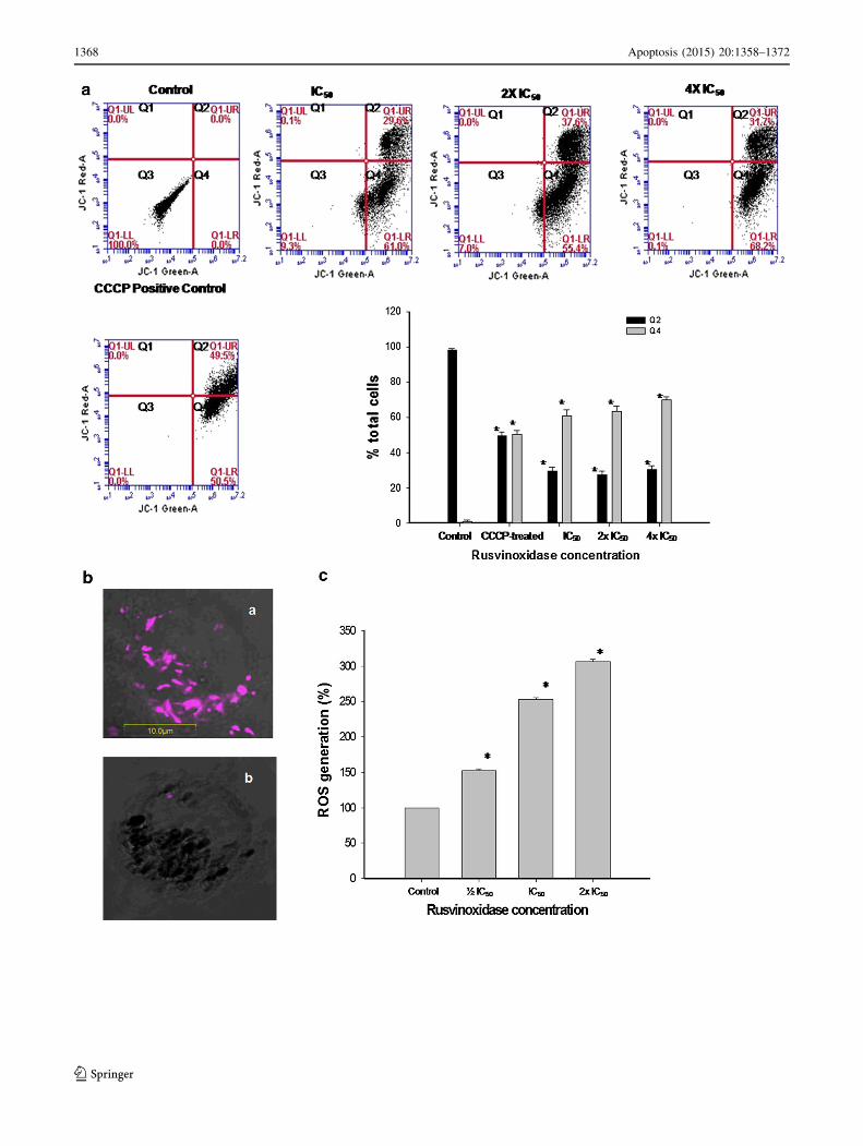
Flow cytometric analysis of mitochondrial membrane
depolarization using the MitoProbeTMJC-1 assay demon-
strated that Rusvinoxidase dose- and time-dependently
enhanced the depolarization of mitochondrial membranes
of treated MCF-7 cells as compared to control cells
(Fig. 7a). Confocal laser microscope images of MCF-7
cells stained with JC-1 showed the loss of red J-aggregate
fluorescence following exposure to Rusvinoxidase, indi-
cating loss of mitochondrial transmembrane potential of
MCF-7 cells after Rusvinoxidase treatment (Fig. 7b).
Rusvinoxidase dose-dependently increased the total
ROS production in MCF-7 cells after a 4 h incubation with
Rusvinoxidase; at a dose of 2 9 IC50, reactive oxygen
species’ concentrations had increased threefold as com-
pared with controls (Fig. 7c).
Rusvinoxidase increases the expression of
pro-apoptotic proteins and down-regulates
the expression of anti-apoptotic proteins
in MCF-7 cell
Investigation of the time-dependent expression of pro-
and anti-apoptotic proteins in MCF-7 cells following
exposure to Rusvinoxidase (at 1 9 IC50 dose)
Fig. 6 Determination of time-dependent decrease in GSH and
catalase activity of MCF-7 cells after exposure to Rusvinoxidase.
MCF-7 (1 9 106) cells were treated with an IC50 dose (83 nM) of
Rusvinoxidase for various time periods (0–24 h) at 37 �C, 5 % CO2.
The cellular GSH and catalase activity of the untreated MCF-7 cell
extract at each time point was considered as 100 % activity and other
values were compared to that. Values are mean ± SD of three
determinations. Significant differences with respect to controls are
indicated by different symbols *p\ 0.05; **p\ 0.01
cFig. 7 a Flow cytometric analysis of Rusvinoxidase-induced changes
in mitochondrial transmembrane potential in MCF-7 cells. After
exposure of 1 9 106 MCF-7cells to different doses of Rusvinoxidase
for 4 h, cells were treated with JC-1 and CCCP (positive control) and
the fluorescence intensity was determined with excitation at 488 nm
and emission at 530 nm (monomeric form of JC-1, indicating
unhealthy mitochondria or dissipation of mitochondrial membrane
potential) and 585 nm (aggregated form of JC-1, indicating healthy
mitochondria or intact mitochondrial membrane potential). In each
graph, the lower left quadrant (Q3) represents endogenous fluores-
cence of MCF-7 mitochondria, used to set the gating to determine
changes in mitochondrial potential in response to treatments. The
lower right quadrant (Q4) shows the green-fluorescing population of
MCF-7 cells that exhibit reduced mitochondrial potential, an indicator
of possible apoptotic cells. The upper right quadrant (Q2) illustrates
MCF-7 cells in which mitochondria are red-fluorescing as an
indication of high mitochondrial membrane potential, suggestive of
healthy, non-apoptotic cells. The bar diagram represents mean ± SD
from three independent experiments. *p\ 0.01 compared to controls.
b Confocal laser microscopic appearance of JC-1 stained (a) control
MCF-7 cells, and (b) Rusvinoxidase-treated (IC50 dose, 4 h) MCF-7
cells. c Flow cytometric analysis of Rusvinoxidase-induced ROS
generation in MCF-7 cells after 4 h of Rusvinoxidase (�IC50, IC50, 2
9 IC50 dose) treatment. The ROS level in control MCF-7 cells was
considered as baseline (100 %) and other values were compared to
that. Data represent mean ± SD of three determinations. *p\ 0.01
compared to control
Apoptosis (2015) 20:1358–1372 1367
123
1368 Apoptosis (2015) 20:1358–1372
123
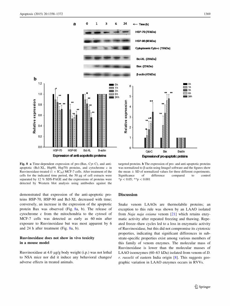
demonstrated that expression of the anti-apoptotic pro-
teins HSP-70, HSP-90 and Bcl-XL decreased with time;
conversely, an increase in the expression of the apoptotic
protein Bax was observed (Fig. 8a, b). The release of
cytochrome c from the mitochondria to the cytosol of
MCF-7 cells was detected as early as 60 min after
exposure to Rusvinoxidase but was most apparent by 6
and 24 h after treatment (Fig. 8a, b).
Rusvinoxidase does not show in vivo toxicity
in a mouse model
Rusvinoxidase at 4.0 lg/g body weight (i.p.) was not lethal
to NSA mice nor did it induce any behavioral changes/
adverse effects in treated animals.
Discussion
Snake venom LAAOs are thermolabile proteins; an
exception to this rule was shown by an LAAO isolated
from Naja naja oxiana venom [21] which retains enzy-
matic activity after repeated freezing and thawing. Repe-
ated freeze–thaw cycles led to a loss in enzymatic activity
of Rusvinoxidase, but this did not compromise its cytotoxic
properties, indicating that significant differences in sub-
strate-specific properties exist among various members of
this family of venom enzymes. The molecular mass of
Rusvinoxidase is lower than the molecular masses of
LAAO isoenzymes (60–63 kDa) isolated from venom of D.
r. russelii of eastern India origin [8]. This suggests geo-
graphic variation in LAAO enzymes occurs in RVVs.
Fig. 8 a Time-dependent expression of pro-(Bax, Cyt C), and anti-
apoptotic (Bcl-XL, Hsp90, Hsp70) proteins, and cytochrome c in
Rusvinoxidase-treated (1 9 IC50) MCF-7 cells. After treatment of the
cells for the indicated time period, the 50 lg of cell extracts were
separated by 12 % SDS-PAGE and the expressions of proteins were
detected by Western blot analysis using antibodies against the
targeted proteins. b The expression of pro- and anti-apoptotic proteins
was normalized to b-actin using ImageJ software and the figures show
the mean ± SD of normalized values for three different experiments.
Significance of difference compared to control
*p\ 0.05; **p\ 0.001
Apoptosis (2015) 20:1358–1372 1369
123
The cytotoxic activity of snake venom LAAOs has been
shown to be linked to production of H2O2, a reactive
oxygen species, which accumulates on the cell surface and
triggers oxidative stress in cancer cells, leading to apop-
tosis [22, 23]. Nevertheless, recent studies suggest that
apart from production of H2O2-stimulated cell apoptosis,
SV-LAAOs may induce apoptosis via intrinsic (mito-
chondrial) or extrinsic (death-receptor) pathways [24, 25].
Because Rusvinoxidase lost its enzymatic activity but
retained cytotoxicity after freezing and thawing, apoptosis
induction by this protein may not be correlated directly
with H2O2 production on the MCF-7 cell surface. Similar
to H2O2 production by doxorubicin, an anticancer drug
used to treat solid tumors, Rusvinoxidase may also induce
perturbation of the cellular redox system that leads to an
increase in intracellular H2O2 production [26].
Because programmed cell death may follow either the
apoptotic or necrotic pathway [3], the mechanism of cell
death induced by an anticancer drug prior to its pre-clinical
trial should be critically assessed. Rusvinoxidase-induced
death of MCF-7 cells demonstrated some hallmarks of
apoptosis; however, after prolonged ([24 h) incubation
with Rusvinoxidase, MCF-7 cells that have shown apop-
totic signs may also show some of the morphological
phenotypes associated with necrosis [27]. Moreover, as an
indicator of its high potency against MCF-7 cells, the IC50
value of Rusvinoxidase was significantly lower than the
IC50 values of anticancer drugs commonly used to treat
breast cancer, such as AraC (present study), cisplatin [28],
doxorubicin [29] and tamoxifen [30].
The induction of apoptosis through both the death
receptor (extrinsic) and mitochondrial (intrinsic) pathways
provides opportunity to develop potent therapeutic mole-
cules against cancer, particularly against multidrug resis-
tant cancers. Both pathways activate a cascade of
proteolytic enzymes, caspases that participates in the
cleavage of aspartic acid-containing motifs and ultimately
results in induction of apoptosis [31]. The extrinsic path-
way of apoptosis is triggered by the binding of apoptosis-
inducing ligands with their cognate death receptors [32]
that results in formation of the death-inducing signaling
complex (DISC). Consequently, through a series of events,
procaspase-8 is activated to caspase-8 which is then
released from the DISC to further activate the effecter
caspase-3 or caspase-7 [33]. Notably, activation of inactive
procaspase-8 to active caspase-8 occurs through the death
receptor, while caspase-9 activation is associated with
mitochondrial pathway [31].
The results of present study provide evidence that
Rusvinoxidase can induce apoptosis in MCF-7 cells by both
the extrinsic and intrinsic pathways; nevertheless, a signif-
icantly higher level (p\ 0.01) of expression of caspase-9
compared to caspase-8 suggests that the intrinsic pathway is
more significant in Rusvinoxidase-induced apoptosis in
MCF-7 cells. Rusvinoxidase may promote apoptosis
through interaction with putative ‘‘death receptors’’ in the
plasma membrane of MCF-7 cells that results in initial
cleavage of procaspase-8 and activation of subsequent
downstream events, leading to apoptosis by activating pro-
caspase 3/7 to caspase-3 or caspase-7 [31, 33]. However, our
results agree with the findings of McGee et al. [20] who
demonstrated that PBOX-6-induced apoptosis in MCF-7
cells was accompanied by DNA fragmentation due to acti-
vation of caspase-7 rather than activation of caspase-3;
Rusvinoxidase-treated MCF-7 cells showed a similar
increase in caspase-7 and a decrease in caspase-3 activities.
It has been suggested that the key role of caspase-3 is reg-
ulation of apoptotic DNA fragmentation via proteolysis of
caspase-activated DNase [31], but the decrease of caspase-
3 activity in MCF-7 cells in response to Rusvinoxidase
suggests that caspase-3 may not be essential for execution of
apoptosis in MCF-7 cells [20, 34].
The intrinsic pathway of apoptosis has been demon-
strated to be associated with key mitochondrial events such
as depolarization of the mitochondrial membrane through
generation of reactive oxygen species (ROS). This is
accompanied by depletion of cellular glutathione that
subsequently leads to release of cytochrome c from the
mitochondria to the cytosol, activating caspase-9, which is
an integral mechanism for initiating and regulating the
caspase cascade [31]. The initiator caspase-9 in turn acti-
vates effecter caspase-3 or caspase-7 [35]. Under normal
cellular conditions, production of excess ROS is down-
regulated by an antioxidant enzyme such as catalase, as
well as by non-protein antioxidants such as GSH, which
provide electrons for glutathione peroxidase to reduce
H2O2 to H2O [35, 36]. Therefore, reduction in catalase
activity as well as depletion of GSH (a hallmark of apop-
tosis) in MCF-7 cells following exposure to Rusvinoxidase
results in the accumulation of excess ROS within the
cytoplasm, which in turn induces apoptosis [37, 38]. Glu-
tathione depletion was initially correlated with its oxidation
by ROS generated during oxidative stress; however, more
recently it has been demonstrated that activation of death
receptors also leads to glutathione depletion, involving its
extrusion across the plasma membrane [37]. Therefore,
Rusvinoxidase-mediated dose- and time-dependent initial
depletion of total cellular glutathione may result via both
intrinsic and extrinsic pathways of apoptosis.
The time-dependent increase in the level of catalase
enzyme activity and cellular glutathione contents in MCF-7
cell at approximately 4-6 h after Rusvinoxidase-treatment
may be correlated with the increased level of ROS pro-
duction. Anticancer drug resistance in human cancer cell
lines is found to be associated with marked increase of
glutathione synthesis as well as enhanced stability of
1370 Apoptosis (2015) 20:1358–1372
123
mRNA of catalase, the major antioxidant defense systems
in cells, to down-regulate the excess ROS production [39,
40]. Therefore, it is suggested that MCF-7 cells may have
attempted to counteract Rusvinoxidase-induced apoptosis
by increasing (insufficiently) cellular glutathione content
and catalase activity; however, the precise mechanism of
action still needs to be elucidated. Treatment with cur-
cumin, a pro-apoptotic antitumor agent, has also resulted in
elevation of glutathione levels and/or prevention of glu-
tathione depletion, as well as inhibition of caspase-3 acti-
vation in human lymphoid Jurkat cells [41].
Apoptosis is regulated by the Bcl-2 protein family,
including the anti-apoptotic proteins such as Bcl-XL and pro-
apoptotic proteins such as Bax. A balance between the
expression levels of both Bcl-XL and Bax is a significant
determinant for cell survival or death [42]. It has been well
documented that Bcl-XL inhibits apoptotic processes by act-
ing upstream of caspase-3 activation and preventing the
release of cytochrome c from the mitochondria, while Bax
acts in the mitochondria to cause the release of cytochrome c,
leading to the activation of caspase-9 and the subsequent
activation of caspase-3 (or caspase-7 in the case of MCF-7
cells) [43, 44]. Therefore, it is expected that diminished
expression of Bcl-XL and increased regulation of expression
of Bax, leading to alteration of mitochondrial transmembrane
potential along with the discharge of cytochrome c to the
cytosol, are key events associated with the intrinsic mecha-
nism of apoptosis induction in MCF-7 cells by Rusvinoxidase.
Heat shock proteins (Hsp) are molecular chaperones and
have been demonstrated to be inhibitors of apoptosis [45].
Hsp90 is regarded as the inhibitor of oligomerization of the
Apaf-1 complex to halt apoptosis, while the function of
Hsp70 is to inhibit the signaling pathway from surface
receptors to retard apoptosis [45], implying that Hsp could
serve as pharmaceutical targets to modulate apoptosis in
cancer cells. In the present study, the observed time-de-
pendent down-regulation of expression of Hsp70 and
Hsp90 by Rusvinoxidase are likely involved in induction of
apoptosis in MCF-7 cells. Furthermore, Rusvinoxidase
(4 mg/kg) does not show toxicity or adverse pharmaco-
logical effects in mice as well as against normal (non-
cancerous) HEK cells, indicating the suitability of Rusvi-
noxidase as a model compound for the development of
peptide-based drugs for the treatment of breast cancer.
Conclusion
Our study suggests that Rusvinoxidase induces apoptosis in
MCF-7 cells via both intrinsic and extrinsic pathways;
however, the mitochondrial intrinsic pathway predomi-
nates. Apoptosis in MCF-7 cells is accompanied by DNA
fragmentation due to activation of caspase-7 by
Rusvinoxidase rather than activation of caspase-3. More-
over, apoptosis induction by Rusvinoxidase may not be
correlated directly with H2O2 production on the MCF-7
cell surface. Assessment of anticancer potential, including
pharmacokinetics study of Rusvinoxidase in experimental
animals, is the goal of our next study.
Acknowledgments Authors thank Dr. R. Mukhopadhyay, TU for
HEK cell cytotoxicity assay. AKM is the recipient of DBT-Crest
award from the Department of Biotechnology, Ministry of Science
and Technology, Govt. of India, which supported his participation in
this study. Support was also provided by a BioScience Discovery
award from COEDIT (to SPM).
References
1. World Health Organization (2012) GLOBOCAN 2012: estimated
cancer incidence, mortality and prevalence worldwide in 2012.
http://globocan.iarc.fr/Default.aspx
2. Takacs Z, Nathan S (2014) Animal venoms in medicine. In:
Wexler P (ed) Encyclopedia of toxicology, 3rd edn. Elsevier,
London, pp 252–259
3. Ellmore S (2007) Apoptosis: a review of programmed cell death.
Toxicol Pathol 35:495–516
4. Mukherjee AK, Saikia D, Thakur R (2011) Medical and diag-
nostic application of snake venom proteomes. J Proteins Pro-
teomics 21:31–40
5. Costa TR, Burin SM, Menaldo DL, de Castro FA, Sampaio SV
(2014) Snake venom L-amino acid oxidases: an overview on their
antitumor effects. J Venom Anim Toxins Incl Trop Dis 20:23
6. Guo C, Liu S, Yao Y, Zhang Q, Sun MZ (2012) Past decade study
of snake venom L-amino acid oxidase. Toxicon 60:302–311
7. Naumann GB, Silva LF, Silva l, Faria G, Richardson M, Evan-
gelista K, Kohlhoff M, Gontijo CM, Navdaev A, de Rezende FF,
Eble JA, Sanchez EF (2011) Cytotoxicity and inhibition of pla-
telet aggregation caused by an L-amino acid oxidase from
Bothrops leucurus venom. Biochim Biophys Acta 1810:683–694
8. Mandal S, Bhattacharyya D (2008) Two L-amino acid oxidase
isoenzymes from Russell’s viper (Daboia russelli russelli) venom
with different mechanisms of inhibition by substrate analogs.
FEBS J 275:2078–2095
9. Mukherjee AK, Mackessy SP (2013) Biochemical and pharma-
cological properties of a new thrombin-like serine protease
(Russelobin) from the venom of Russell’s viper (Daboia russelii
russelii) and assessment of its therapeutic potential. Biochim
Biophys Acta-General Subj 1830:3476–3488
10. Mukherjee AK, Mackessy SP, Dutta S (2014) Characterization of
a Kunitz-type protease inhibitor peptide (Rusvikunin) purified
from Daboia russelii russelii venom. Int J Biol Macromol
67:154–162
11. Weissbach H, Robertson AV, Witkop B, Udenfriend S (1961)
Rapid spectrophotometric assays for snake venom L-amino acid
oxidase based on the oxidation of L-kynurenine or 3,4-dehydro-L-
proline. Anal Biochem 1:286–290
12. Aird SD, da Silva NJ Jr (1991) Comparative enzymatic compo-
sition of Brazilian coral snake (Micrurus) venoms. Comp Bio-
chem Physiol 99B:287–294
13. Costa FLS, Rodrigues RS, Izidoro LFM, Menaldo DL, Ham-
aguchi A, Homsi-Brandeburgo MI, Fuly AL, Soares SG, Selistre-
de-Araujo HS, Barraviera B, Soares AM, Rodrigues VM (2009)
Biochemical and functional properties of a thrombin-like enzyme
isolated from Bothrops pauloensis snake venom. Toxicon
54:725–735
Apoptosis (2015) 20:1358–1372 1371
123

14. Rutkowski RB (1996) Human plasma and serum trypsin-like
esterase activity. Clin Chem 12:350–356
15. Mukherjee AK, Mackessy SP (2014) Pharmacological properties
and pathophysiological significance of a Kunitz-type protease
inhibitor (Rusvikunin-II) and its protein complex (Rusvikunin
complex) purified from Daboia russelii russelii venom. Toxicon
89:55–66
16. Ioannou YA, Chen FW (1997) Quantitation of DNA fragmenta-
tion in apoptosis. Nucl. Acids Res. 24:992–993
17. Herrmann M, Lorenz HM, Voll R, Griinke M, Woith W, Kalden
JR (1994) A rapid and simple method for the isolation of apop-
totic DNA fragments. Nucl Acids Res. 22:5506–5550
18. Nigam M, Ranjan V, Srivastava S, Sharma R, Balapure AK
(2008) Centchroman induces G0/G1 arrest and caspase-dependent
apoptosis involving mitochondrial membrane depolarization in
MCF-7 and MDA MB-231 human breast cancer cells. Life Sci
82:577–590
19. Zargan J, Umar S, Sajad M, Naime M, Ali S, Khan HA (2011)
Scorpion venom (Odonto buthusdoriae) induces apoptosis by
depolarization of mitochondria and reduces S-phase population in
human breast cancer cells (MCF-7). Toxicol In Vitro
25:1748–1756
20. McGee MM, Hyland E, Campiani G, Ramunno A, Nacci V,
Zisterer DM (2002) Caspase-3 is not essential for DNA frag-
mentation in MCF-7 cells during apoptosis induced by the pyr-
rolo-1,5-benzoxazepine, PBOX-6. FEBS Lett 515:66–70
21. Samel M, Tonismagi K, Ronnholm G, Vija H, Siigur J, Kalkki-
nen N, Siigur E (2008) L-Amino acid oxidase from Naja naja
oxiana venom. Comp Biochem Physiol B 149:572–580
22. Torii S, Naito M, Tsuruo T (1997) Apoxin I, a novel apoptosis-
inducing factor with L-amino acid oxidase activity purified from
Western diamondback rattlesnake venom. J Biol Chem
272:9539–9542
23. Ande SR, Kommoju PR, Draxl S, Murkovic M, Macheroux P,
Ghisla S, Ferrando-May E (2006) Mechanisms of cell death
induction by L-amino acid oxidase, a major component of
ophidian venom. Apoptosis 11:1439–1451
24. Alves RM, Antonucci GA, Paiva HH, Cintra AC, Franco JJ,
Mendonca-Franqueiro EP, Dorta DJ, Giglio JR, Rosa JC, Fuly
AL, Dias BM, Soares AM, Sampaio SV (2008) Evidence of
caspase-mediated apoptosis induced by L-amino acid oxidase
isolated from Bothrops atrox snake venom. Comp Biochem
Physiol A 151:542–550
25. Burin SM, Ayres LR, Neves RP, Ambrosio L, de Morais FR, Dias-
Baruffi M, Sampaio SV, Pereira-Crott LS, de Castro FA (2013) L-
Amino acid oxidase isolated from Bothrops pirajai induces
apoptosis in BCR-ABL-positive cells and potentiates ima-
tinibmesylate effect. Basic Clin Pharmacol Toxicol 113:103–112
26. Wagner BA, Evig CB, Reszka KJ, Buettner GR, Burns CP (2005)
Doxorubicin increases intracellular hydrogen peroxide in PC3
prostate cancer cells. Arch Biochem Biophys 440:181–190
27. Riss TL, Moravec RL (2004) Use of multiple assay endpoints to
investigate the effects of incubation time, dose of toxin, and
plating density in cell-based cytotoxicity assays. Assay Drug Dev
Technol 2:51–62
28. Tassone P, Tagliaferri P, Prricelli A, Blotta S, Quaresima B,
Martelli ML, Goel A, Barbieri V, Costanzo F, Boland CR,
Venuta S (2003) BRCA expression modulates chemosensitivity
of BRCA1-defective HCC1937 human breast cancer cells. Br J
Cancer 88:1285–1291
29. Fornari FA, Randolph JK, Yalowich JC, Ritke MK, Gewirtz DA
(1994) Interference by doxorubicin with DNA unwinding in
MCF-7 breast tumor cells. Mol Pharmacol 45:649–656
30. Seeger H, Huober J, Wallwiener D, Mueck AO (2004) Inhibition
of human breast cancer cell proliferation with estradiol metabo-
lites is as effective as with tamoxifen. Horm Metab Res
36:277–280
31. Boatright KM, Salvesen GS (2003) Mechanisms of caspase
activation. Curr Opin Cell Biol 15:725–731
32. Ashkenazi A, Dixit VM (1998) Death receptors: signaling and
modulation. Science 281:1305–1308
33. Dıaz GD, Li Q, Roderick RH (2003) Caspase-8 and apoptosis-
inducing factor mediate a cytochrome c-independent pathway of
apoptosis in human colon cancer cells induced by the dietary
phytochemical chlorophyllin. Cancer Res 63:1254–1261
34. Liang Y, Yan C, Schor NF (2001) Apoptosis in the absence of
caspase 3. Oncogene 20:6570–6578
35. Circu ML, Aw TY (2010) Reactive oxygen species, cellular
redox systems, and apoptosis. Free Radic Biol Med 48:749–762
36. Pan M-H, Huang Y-T, Ho C-T, Chang C-I, Hsu P-C, Pan BS
(2006) Induction of apoptosis by Meretrix lusoria through reac-
tive oxygen species production, glutathione depletion, and cas-
pase activation in human leukemia cells. Life Sci 79:1140–1152
37. Franco R, Cidlowski JA (2009) Apoptosis and glutathione:
beyond an antioxidant. Cell Death Differ 16:1303–1314
38. Valdameria G, Trombetta-Limab M, Worfel PR, Pires ARA,
Martinez GR, Noleto GR, Cadena SMSC, Sogayar MC, Win-
nischofer SMB, Rocha MEM (2011) Involvement of catalase in
the apoptotic mechanism induced by apigenin in HepG2 human
hepatoma cells. Chem Biol Interact 193:180–189
39. Backos DS, FranklinCC Reigan P (2012) The role of glutathione
in brain tumor drug resistance. Biochem Pharmacol
83:1005–1012
40. Akman SA, Forrest G, Chu FF, Doroshow JH (1989) Resistance
to hydrogen peroxide associated with altered catalase mRNA
stability in MCF7 breast cancer cells. Biochim Biophys Acta
1009:70–74
41. Piwocka K, Jaruga E, Skierski J, Gradzka I, Sikora E (2001)
Effect of glutathione depletion on caspase-3 independent apop-
tosis pathway induced by curcumin in Jurkat cells. Free Radic
Biol Med 31:670–678
42. Barbosa IA, Machad NG, Machad AJ, Scott PM, Oliveira PJ
(2012) Mitochondrial remodeling in cancer metabolism and
survival: potential for new therapies. Biochim Biophys Acta
1826:238–254
43. Salakou S, Kardamakis D, Tsamandas AC, Zolota V, Apostolakis
E, Tzelepi V, Papathanasopoulos P, Bonikos DS, Papa-
petropoulos T, Petsas T, Dougenis D (2007) Increased Bax/Bcl-2
ratio up-regulates caspase-3 and increases apoptosis in the thy-
mus of patients with myasthenia gravis. In Vivo 1:123–132
44. Gupta SD, Gomes A, Debnath A, Saha A, Gomes A (2010)
Apoptosis induction in human leukemic cells by a novel protein
Bengalin, isolated from Indian black scorpion venom: through
mitochondrial pathway and inhibition of heat shock proteins.
Chem Biol Interact 183:293–303
45. Sreedhar AS, Csermely P (2004) Heat shock proteins in the
regulation of apoptosis: new strategies in tumor therapy. A
comprehensive review. Pharmacol Ther 101:227–257
1372 Apoptosis (2015) 20:1358–1372
123


![Toll-LikeReceptor2EnhancesZO-1–AssociatedIntestinal ... · and lot #1467609 [HTB-38], respectively) and kept in a humidified incubator at 37°C with 5% CO 2. Caco-2 were maintained](https://static.fdocuments.net/doc/165x107/5e2fb529416d8302253ff3ff/toll-likereceptor2enhanceszo-1aassociatedintestinal-and-lot-1467609-htb-38.jpg)