Apoptosis-induced effects of extract from Artemisia annua ... · Apoptosis-induced effects of...
12
RESEARCH ARTICLE Open Access Apoptosis-induced effects of extract from Artemisia annua Linné by modulating PTEN/p53/PDK1/Akt/ signal pathways through PTEN/p53-independent manner in HCT116 colon cancer cells Eun Ji Kim 1 , Guen Tae Kim 1 , Bo Min Kim 1 , Eun Gyeong Lim 1 , Sang-Yong Kim 2 and Young Min Kim 1* Abstract Background: The extracts from Artemisia annua Linné (AAE) has been known to possess various functions including anti-bacterial, anti-virus and anti-oxidant effects. However, the mechanism of those effects of AAE is not well known. Pursuantly, we determined the apoptotic effects of extract of AAE in HCT116 cell. In this study, we suggested that AAE may exert cancer cell apoptosis through PTEN/PDK1/Akt/p53signal pathway and mitochondria-mediated apoptotic proteins. Methods: We measured 3-(4,5-dimethylthiazol-2-yl)-2,5-diphenyltetrazolium bromide (MTT) assay, lactate dehydrogenase (LDH) assay, Hoechst 33342 staining, Annexin V-PI staining, Mitopotential assay, immunofluorescence (IF) and Western blotting. Accordingly, our study showed that AAE treatment to HCT116 cells resulted in inhibition of PDK1, Akt, MDM2, Bcl-2, and pro-caspase 3 as well as activation of PTEN, p53-upregulated modulator of apoptosis (PUMA), Bax and Bak expression. Also we measured in vivo assay that xenograft model, H&E assay, TUNEL assay and IHC. Results: AAE induced apoptosis via PTEN/p53/PDK1/Akt signal pathways through PTEN/p53-independent manner. AAE inhibit cell viability and increase LDH release in HCT116 colon cancer cell. Also, AAE increase apoptotic bodies, caspase -3,7 activation and reduces mitochondria membrane potential. AAE regulates cytochrome c translocation to the cytoplasm and Bax translocation to the mitochondrial membrane in an Immunofluorescence staining and increase PTEN and p53 expression in an in vivo tumor xenograft model. To elucidate the role of the PTEN/p53/PDK1/Akt signal pathways in cancer control, we conditionally inactivated PTEN/p53/PDK1/Akt signal pathways. We used inhibitors of PTEN, p53, PDK1, Akt. In consequence, these results indicate that AAE induced apoptosis by means of a mitochondrial event through the regulation of proteins such as Bax, Bak and cytochrome c in PDK1/Akt signaling pathways via PTEM/p53-independent manner. Conclusions: We confirmed the apoptotic effect of extracts of AAE by Modulating PTEN/p53/PDK1/Akt/Signal Pathways through PTEN/p53-independent pathwaysin HCT116 colon cancer cell. Keywords: Phosphatase and tensin homolog (PTEN), p53-independent manner, Artemisia annua Linné, Apoptosis, HCT116 colon cancer cell * Correspondence: [email protected] 1 Department of Biological Science and Biotechnology, College of Life Science and Nano Technology, Hannam University, 1646 Yuseong-daero, Yuseong-gu, Daejeon 34054, Korea Full list of author information is available at the end of the article © The Author(s). 2017 Open Access This article is distributed under the terms of the Creative Commons Attribution 4.0 International License (http://creativecommons.org/licenses/by/4.0/), which permits unrestricted use, distribution, and reproduction in any medium, provided you give appropriate credit to the original author(s) and the source, provide a link to the Creative Commons license, and indicate if changes were made. The Creative Commons Public Domain Dedication waiver (http://creativecommons.org/publicdomain/zero/1.0/) applies to the data made available in this article, unless otherwise stated. Kim et al. BMC Complementary and Alternative Medicine (2017) 17:236 DOI 10.1186/s12906-017-1702-7
Transcript of Apoptosis-induced effects of extract from Artemisia annua ... · Apoptosis-induced effects of...

RESEARCH ARTICLE Open Access
Apoptosis-induced effects of extract fromArtemisia annua Linné by modulatingPTEN/p53/PDK1/Akt/ signal pathwaysthrough PTEN/p53-independent manner inHCT116 colon cancer cellsEun Ji Kim1, Guen Tae Kim1, Bo Min Kim1, Eun Gyeong Lim1, Sang-Yong Kim2 and Young Min Kim1*
Abstract
Background: The extracts from Artemisia annua Linné (AAE) has been known to possess various functions includinganti-bacterial, anti-virus and anti-oxidant effects. However, the mechanism of those effects of AAE is not wellknown. Pursuantly, we determined the apoptotic effects of extract of AAE in HCT116 cell. In this study, we suggestedthat AAE may exert cancer cell apoptosis through PTEN/PDK1/Akt/p53signal pathway and mitochondria-mediatedapoptotic proteins.
Methods: We measured 3-(4,5-dimethylthiazol-2-yl)-2,5-diphenyltetrazolium bromide (MTT) assay, lactate dehydrogenase(LDH) assay, Hoechst 33342 staining, Annexin V-PI staining, Mitopotential assay, immunofluorescence (IF) and Westernblotting. Accordingly, our study showed that AAE treatment to HCT116 cells resulted in inhibition of PDK1, Akt, MDM2,Bcl-2, and pro-caspase 3 as well as activation of PTEN, p53-upregulated modulator of apoptosis (PUMA), Bax and Bakexpression. Also we measured in vivo assay that xenograft model, H&E assay, TUNEL assay and IHC.
Results: AAE induced apoptosis via PTEN/p53/PDK1/Akt signal pathways through PTEN/p53-independent manner.AAE inhibit cell viability and increase LDH release in HCT116 colon cancer cell. Also, AAE increase apoptoticbodies, caspase −3,7 activation and reduces mitochondria membrane potential. AAE regulates cytochrome c translocationto the cytoplasm and Bax translocation to the mitochondrial membrane in an Immunofluorescence staining and increasePTEN and p53 expression in an in vivo tumor xenograft model. To elucidate the role of the PTEN/p53/PDK1/Akt signalpathways in cancer control, we conditionally inactivated PTEN/p53/PDK1/Akt signal pathways. We used inhibitors of PTEN,p53, PDK1, Akt. In consequence, these results indicate that AAE induced apoptosis by means of a mitochondrialevent through the regulation of proteins such as Bax, Bak and cytochrome c in PDK1/Akt signaling pathways viaPTEM/p53-independent manner.
Conclusions: We confirmed the apoptotic effect of extracts of AAE by Modulating PTEN/p53/PDK1/Akt/SignalPathways through PTEN/p53-independent pathwaysin HCT116 colon cancer cell.
Keywords: Phosphatase and tensin homolog (PTEN), p53-independent manner, Artemisia annua Linné, Apoptosis,HCT116 colon cancer cell
* Correspondence: [email protected] of Biological Science and Biotechnology, College of LifeScience and Nano Technology, Hannam University, 1646 Yuseong-daero,Yuseong-gu, Daejeon 34054, KoreaFull list of author information is available at the end of the article
© The Author(s). 2017 Open Access This article is distributed under the terms of the Creative Commons Attribution 4.0International License (http://creativecommons.org/licenses/by/4.0/), which permits unrestricted use, distribution, andreproduction in any medium, provided you give appropriate credit to the original author(s) and the source, provide a link tothe Creative Commons license, and indicate if changes were made. The Creative Commons Public Domain Dedication waiver(http://creativecommons.org/publicdomain/zero/1.0/) applies to the data made available in this article, unless otherwise stated.
Kim et al. BMC Complementary and Alternative Medicine (2017) 17:236 DOI 10.1186/s12906-017-1702-7
BackgroundArtemisia annua Linné is an annual plant that is chrys-anthemum family. This plant is primarily found in thetropical zones of Asia along streets and in fields. Sinceancient times, Artemisia annua Linné has been used asan antipyretic, hemostatic, as a treatment for skindiseases, and an insecticide. In addition, its antibacterial,antiviral and antioxidant properties allow it has beenused as a traditional herbalmedicine [1]. This plant alsocontains various bioactive compounds [2–5].The antioxidant activity of phenolic compounds in
Artemisia annua Linné has been reported [6]. Artemisi-nin, the main element of sweet wormwood, is being usedfor medical uses, such as anti-malarial activity [7–9]. Inprevious studies, they determined that when usingArtemisia annua Linné on extracts from breast cancer,cervical carcinoma cells, stomach cancer, and for cellgrowth inhibitory effect that there is a cancer cell [10, 11].Furthermore, selective necrosis of breast cancer cells wasproven to be anti-cancer active. This brought attention tothe world to consider taking action with herbal remedies[12]. Some studies have reported the effect of Artemisiaannua Linné added to food and feed. However, themechanism of the effects of Artemisia annua Linné is notwell known [13].PTEN (Phosphatase and TENsin homolog deleted on
chromosome ten), a tumor-suppressorgene with dual lipidand protein phosphatase activity, antagonizes the PI3K/AKT signaling pathway and suppresses cell survival, aswell as cell proliferation. Also, PTEN can inhibit theactivation of Akt. This effect has been attributed to PTENreducing the availability of Phosphatidylinositol (3,4,5)-trisphosphate (PIP3; 2–3) [14–16]. The serine/threoninekinase Akt is phosphorylated and activated by PDK1(phosphoinositide-dependent protein kinase-1) [17].PDK1 activation phosphorylates Akt at thr308. Once
phosphorylated in T308, phosphorylation additionallyoccurs at S473by PDK2 [18]. Akt activation induces dif-ferent cell survival mechanisms [19]. Akt plays a centralrole in many cellular processes that establish survivalfactor and exert anti-apoptotic activation. Also, Akt acti-vation induces cell cycle progression [20]. In anothercase, Akt prevented apoptosis via phosphorylation andtranslocation of MDM2 (Murine double minute 2) intothe nucleus [21, 22]. MDM2 interacts with p53 and in-hibits it. Under normal circumstances, p53 is maintainedat very low levels by ubiquitination and degradation[23]. The p53 gene, a tumor suppressor, plays a key rolein the induction of apoptosis and cell cycle arrest in re-sponse to a variety of stress genes, including theblocker of cellular DNA damage repair [24]. p53 is anuclear DNA-binding phosphor-protein. It is a tran-scriptional activator that can exert transcriptional re-pression of specific targeted genes [25]. Also, p53
interacts directly with cell proliferation-mediated pro-teins. The direct interaction of p53 activates apoptoticproteins into mitochondrial outer membrane permea-bilization (MOMP) [26].Mitochondria are well known for playing a key role in
activating apoptosis. The mitochondrial apoptosis path-way is mediated via Bcl-2 family proteins [27, 28]. Bcl-2family proteins are divided into anti-apoptotic proteinssuch as Bcl-XL, Bcl-w, Mcl-1 and pro-apoptotic proteinssuch as Bax, Bak and Bok. In normal cells, Bax exists asa monomer in the cytosol and translocates to mitochon-dria, experiencing conformational changes to form oligo-mers during apoptosis. On the other hand, Bak resideson mitochondria. During apoptosis, Bak changes to formoligomers identical to Bax. The activation of Bax andBak regulates cytochrome c release to cytosol from themitochondria via alteration of MOMP [29, 30]. Releasedcytochrome c induces apoptosis by activating last effec-tors caspase (caspase-3/−7) [31].In this study, we investigated the effects of Artemisia
annua Linné extract (AAE) on apoptosis in HCT116coloncancer cells. We suggested that AAE induced apop-tosis through PTEN/PDK1/Akt/p53signal pathwaysand mitochondria-mediated apoptotic proteins.
MethodsReagents and chemicalsAAE was purchased from Daejeon Oriental HerbalMarket (Deojun, Korea).3-(4,5-Dimethylthiazol-2-yl)-2,5-Diphenyltetrazolium Bromide (MTT) was purchasedfrom Sigma-Aldrich (St. Louis, MO, USA). The Piercelactate dehydrogenase (LDH) Cytotoxicity Assay kit waspurchased from Thermo Fisher Scientific (Waltham,MA, USA). The Muse Annexin V and Dead Cell Assay Kit,The Muse Caspase-3/7 Kit and the Muse MitoPotentialKit were purchased from Millipore (Darmstadt, Germany).MitoTracker was purchased from Molecular Probes(Eugene, OR, USA). Specific antibodies that recog-nized phosphorylated (p)Akt (Ser473) (4060S), (p)Akt(Tre308) (3038S), (p)PTEN (9549P), (p)PDK1 (3430P),PUMA (4976P) Bax (5023P), Bak (6947), pro-caspase-3 (9665), Bcl-2 (2876) and β-actin (4967) total formed(t)Akt (4060P), (t)PDK1 (3062P) were obtained fromCell Signaling Technology (Beverly, MA, USA). Thetotal formed (t)PTEN (SC-7974) was purchased fromSanta Cruz Biotechnology (Dallas, TX, USA), (t)MDM2(NBPI-02158SS) was purchased from Novus Biologicals(Littleton, CO, USA) and (p)MDM2 was purchased fromAbcam (Cambridge, MA, USA). LY294002 (PI3K/Aktinhibitor), Pifithrin-α (p53 inhibitor) were purchased fromCalbiochem (San Diego, CA, USA), Nutlin-3 (MDM2 in-hibitor) and BX-795 (PDK1 inhibitor) were purchasedfrom Sigma-Aldrich (St Louis, MO, USA), BpV (PTEN in-hibitor) was purchased from Santa Cruz Biotechnology
Kim et al. BMC Complementary and Alternative Medicine (2017) 17:236 Page 2 of 12
(Dallas, TX, USA). Horseradish peroxidase (HRP)-con-jugated Goat Anti-Mouse (PA1–30126) and GoatAnti-Rabbit (166–2408) secondary antibodies werepurchased from Thermo Fisher Scientific, Inc., and Bio-Rad Laboratories, Inc., (Tokyo, Japan), respectively.
Preparation of Artemisia annua Linné extract100 g of the powdered AAE was extracted with 800 mLof 95% EthOH for 72 h. The extract was filtered throughqualitative filter paper no. 1 (Toyo Roshi Kaisha, Ltd.;Tokyo, Japan) and concentrated with a rotary evaporatorto remove the ethanol. AAE was dissolved in dimethylsulfoxide (DMSO) prior to treatment and stored at −20 °C.The final concentration of AAE in the culture medium wascontrolled at 30-60 μg/ml.
Cell cultureHCT116 cells were obtained from the American TypeCulture Collection (ATCC; Rockville, MD, USA). Thecells were grown in RPMI-1640 medium (HycloneLaboratories Inc.)containing 10% fetal bovine serum(FBS) and 1% antibiotics at 37 °C in a 5% CO2 incubator.The cells were sub-cultured by detachment with Trypsin-EDTA (Hyclone Laboratories Inc.)and re-seeded at 1 × 10⁶
cells/mL per 100 mm plate every 48 h.
3-(4,5-Dimethylthiazol-2-yl)-2,5-diphenyltetrazoliumbromide (MTT) assayHCT116 cells and fibroblast cells (1 × 10⁵) wereseeded onto 12-well plates and treated with AAE atconcentrations of 30, 40, and 60 μg/mL for 24 h. Cer-tain samples were pre-treated with a respective inhibi-tor (20 μMLY294002, 20 μM Nutlin, 20 μM pifithrin-α,1 μM BpV and 5μMBX-)for 30 min prior to treatmentwith AAE. The selective medium removed and then in-cubated with20μl of MTT solution (5 mg/ml MTT inPBS) for 1 h. Converted purple formazan dye fromMTT was solubilized in DMSO and optical densitieswere measured at 595 nm.
Lactate dehydrogenase (LDH)assayCells were seeded at 2.5 × 10⁵ cells/mL per well in a 96-well plate and incubated for 24 h. The cells were thentreated with AAE (30, 40, and 60 μg/mL) and incubatedat 37 °C in a 5% CO2 atmosphere. After 24 h, the highcontrol cells (maximum LDH release) were treated withcell lysis solution from the LDH Cytotoxicity Assay Kitfor 30 min. The absorbance of the solution in each wellwas determined using a microplate reader (Bio-RadLaboratories, Inc.) at 490 and 655 nm.
Cells morphologyCells were seeded at 1 × 10⁵ cells/mL in 6wellplatesand-treatedwithAAEfora 24 h time period at 30, 40, and60 μg/ml concentrations.
Hoechst 33342 stainingCells were seeded at 1 × 10⁴ cells/mL in a 12-well platewith cover glasses and incubated for 24 h. Following in-cubation, the cells were treated with the AAE (30, 40,and 60 μg/mL) for 24 h at 37 °C in a 5% CO2 atmos-phere. Cells were stained with Hoechst33342 for 30 min.Slides were washed with PBS and mounting fluid waspoured over them. The slides were covered with a coverslip and sealed with nail polish. Fluorescence was mea-sured by using a fluorescence microscope (Carl Zeiss,Germany).
Cell apoptosis assayHCT116 cell apoptosis was assayed using the Muse™Annexin V and Dead Cell Kit (Merck Millipore, Guyan-court, France) according to the user’s guide. A total of1x105cells were collected by centrifugation (3000 rpm,5 min) and washed with PBS. Cells were resuspended ina RPMI-medium with 1% bovine serum albumin and10% FBS, mixed with the Muse™ Annexin V and DeadCell reagent, and then incubated for 20 min at roomtemperature in the dark. Assay results were measuredusing the Muse™ Cell Analyzer.
Caspase-3/7 activity analysisCells were seeded at 1 × 10⁵ cells/mL per plate in a 6-well plate and incubated for 24 h. Following incubation,the cells were treated with AAE (30, 40, and 60 μg/mL)for 24 h at 37 °C in a 5% CO2 atmosphere. HCT116 cellcaspase activity was assayed using the Muse Caspase 3/7Assay Kit according to the user’s guide. A total of 1 x10⁵cells were collected by centrifugation (3000 rpm,5 min) and washed with PBS. Cells were resuspended ina 1X Assay Buffer BA, mixed with the Muse™ Caspase-3/7 reagent, and then incubated for 20 min at 37 °C in a5% CO2 atmosphere in the dark. After incubation,150 μL of Muse™ Caspase 7-AAD working solution wasadded to each tube. The solution was mixed thoroughlyby pipetting up and down, also know as vortexing, at amedium speed for 3 to 5 s. It was then incubated atroom temperature for 5 min in the dark. Assay resultswere measured using the Muse™ Cell Analyzer.
Western blottingCells were seeded at 1 × 10⁵ cells/mL per plate in a 6-well plate and incubated for 24 h. The cells were thentreated with AAE (30, 40, and 60 μg/mL) and incubatedat 37 °C in a 5% CO2 atmosphere. Certain samples werepre-treated with the respective inhibitor (20 μMLY294002,
Kim et al. BMC Complementary and Alternative Medicine (2017) 17:236 Page 3 of 12
20 μM Nutlin, 20 μM pifithrin-α, 1 μM Bp V and 5μMBX-795) for 30 min prior to treatment with AAE. Cellswere rinsed twice with ice cold PBS and scraped with alysis buffer (50 mM Tris-HCl pH 8.0, 150 mMNaCl, 1%NP40, 0.5% sodium deoxycholate, 1 mM PMSF) and sub-jected to western blot analysis. The primary antibody wasallowed to react overnight at 4 °C and the second antibodyreacted for 90 min at room temperature with gentle agita-tion. Following washing the samples four times with 1XTris Buffered Saline with Tween 20 (TBST) for 10 min atroom temperature, proteins were detected using SuperSignal West Pico Chemiluminescent Substrate (PI34080;Thermo Fisher Scientific, Inc., Waltham, MA, USA) andvisualized on CP-BU new X-ray film (Agfa HealthCare,Inc., Mortsel, Belgium).
Mitochondrial membrane potential assayCells were seeded at 1 × 10⁵ cells/mL per plate in a 6-well plate and incubated for 24 h. Following incubation,the cells were treated with the AAE (30, 40, and 60 μg/mL)for 24 h at 37 °C in a 5% CO2 atmosphere. HCT116 cellcaspase activity was assayed using the Muse MitoPotentialKit according to the user’s guide. A total of 1 x 10⁵cellswere collected by centrifugation (3000 rpm, 5 min) andwashed with PBS. The supernatant was then removed andthe cell pellets were stained with the Muse MitoPotential
Kit (Merck Millipore, Guyancourt, France) for 25 min at37 °C.The data was analyzed using the Muse™ CellAnalyzer Assay.
Fraction of mitochondria and cytosol proteinsWe used a Mitochondria/Cytosol Fraction Kit (Abcam,Cambridge, MA, USA). Cells were seeded at 1 × 10⁶/mlon a 100 mm plate and incubated for24 h. After incuba-tion, cells were treated with AAE for 24 h at 37 °C in a5% CO2 atmosphere. Cells were harvested by trypsiniza-tion, collected by centrifugation, washed with PBS, andhomogenized in an ice cold cytosol extraction buffermix containing DTT and protease inhibitor using a soni-cator. The homogenates were centrifuged at 3000 rpmfor 10 min at 4 °C and the supernatants were collected.The supernatants were centrifuged at 13000 rpm for30 min at 4 °C and collected. The supernatant cytosolproteins and pellets were resuspended with ice coldmitochondria extraction buffer containing DTT and aprotease inhibitor for mitochondria proteins.
Immunofluorescence (IF)stainingCells were seeded at 1 × 10⁴ cells/mL in a 12-well platewith cover glasses and incubated for 24 h. The cells weretreated with the AAE (30, 40, and 60 μg/mL) for 24 h at37 °C in a 5% CO2 atmosphere. The cells were stained
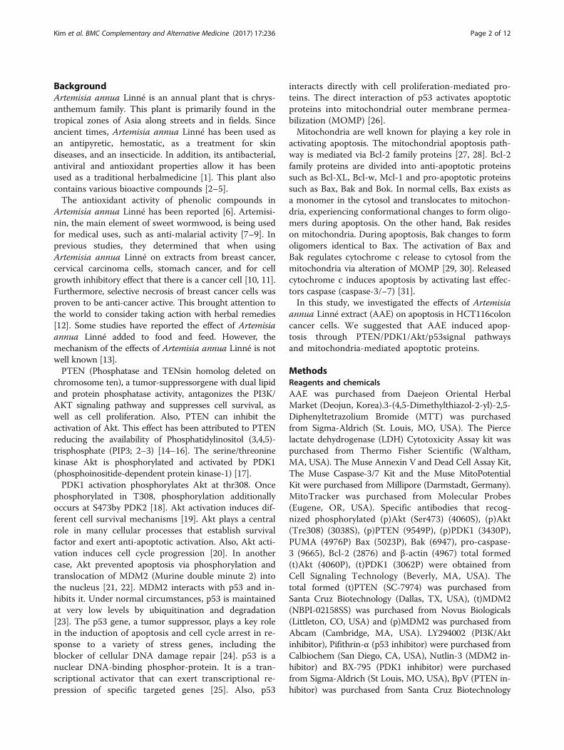
Fig. 1 AAE reduces cell proliferation in HCT116 colon cancer cells. a, c Cell viability was measured by MTT assay. a is HCT116 cell line. c is Fibroblastcell line. The statistical analysis of the data was carried out by use of an T-test. */#p < 0.05, **/##'p < 0.01 and ***/###p < 0.001 (each experiment, n = 3).b LDH assay was performed for assessing cell deaths. Cytotoxicity was induced by AAE. The statistical analysis of the data was carried out by use of anT-test. *p < 0.05, **p < 0.01 and ***p < 0.001 (each experiment, n = 3). d AAE affects the morphology of HCT116 cells, and promotes cell death in adose-dependent manner. HCT116 were treated with AAE (0, 30, 40, and 60 ìg/ml) for 24 h
Kim et al. BMC Complementary and Alternative Medicine (2017) 17:236 Page 4 of 12

with MitoTracker. for 30 min at 37 °C in a 5% CO2 at-mosphere. Cells were fixed with 3.7% formaldehyde for20 min and permeabilized with 0.2% Triton X-100 for20 min. Cells were washed with PBS twice and reactedwith cytochrome c, Bax and Bak antibodies overnight at4 °C. Cells were washed with PBS twice and reacted with asecondary antibody for 1 h 30 min. Fluorescence was de-tected by confocal microscopy (Olympus; Tokyo, Japan).
Xenograft modelFive-week-old male Balb/c nudemice were obtainedfrom SLC (SLC; Tokyo, Japan) Five mice made up thecontrol group while five other mice made up the experi-mental and delivery groups for each concentration. Fortumor induction, HCT116 human colon cancer cells(2.5 × 10⁵ cells/0.1 ml) were subcutaneously injectedinto the left flank of the mice (each group had 10 ani-mals). One week after the injection of cells, the micewere co-treated with AAE20, 40 mg/kg/day and 0.2cm3
PBS/DMSO for 21 days. Tumor size was measured by
taking two perpendicular diameter measurements, usinga caliper, every 2 days. The tumor volume was calculatedusing the following formula: V = 1/2 (length x width).The body weight of each animal was measured at a settime, once per week. After the 3-week treatment, thetumor was removed and frozen in liquid nitrogen forwestern blot analysis or fixed with formalin for immuno-histochemistry, TUNEL and H&E staining. All of the ani-mal experiments were approved by the Ethics Committeefor Animal Experimentation of Hannam University(Daejeon, Korea, HNU 2016–9).
TUNEL assayLevels of apoptosis in distal colon tissue were deter-mined using the TdT-mediated dUTP nickend labeling(TUNEL)method. Tumor specimens from mice werefixed in 10% formaldehyde, embedded in paraffin andsectioned into 5 μm thick slices. Tissue sections wereprocessed according to manufacturer’s instructions for
Fig. 2 AAE induces apoptosis and regulates pro-apoptotic proteins in HCT116 colon cancer cells. a Cell apoptosis observed using Hoechst 33342staining. HCT116 were treated with AAE (0, 30, 40, and 60 ìg/ml) for 24 h. Fluorescence was detected using a fluorescence microscope. Arrowsindicate apoptotic bodies, which were DNA fragments produced when apoptosis occurred. b Apoptotic effects of different concentration AAEwere evaluated by Muse™ Annexin V and Dead Cell Assay Kit. HCT116 were treated with AAE (0, 30, 40, and 60 ìg/ml) for 24 h. Data analyzedby flow cytometry. c HCT116 were treated with 0, 30, 40, and 60 ìg/ml of AAE 24 h, caspase 3/7 activity was analyzed using a Muse™ Caspase-3/7 kit,as described in Materials and Methods. d Cells were treated with the indicated concentrations of AAE for 24 h. The expression of PTEN, PDK1, Akt(Thr308 and Ser473), MDM2, p53, PUMA,Bcl-2, pro-caspase3 and the activation of Bax, Bak and cleaved PARP were analyzed by western blot analysis
Kim et al. BMC Complementary and Alternative Medicine (2017) 17:236 Page 5 of 12

the Apop Tag Peroxidase In Situ Apoptosis DetectionKit (Vector Laboratories; Burlingame, CA, USA).
ImmunohistochemistryTumor specimens from mice were fixed in 10% formal-dehyde, embedded in paraffin and sectioned into 5 μmthick slices. Consecutive thin cryosections (5 μm) ofOCT compound (Sakura Finetek; Torrance, CA,USA)embedded tumor tissues were fixed in acetone at 4 °Cfor 10 min. After washing in PBS, the sections weretreated with3% H2O2 for 10 min to block endogenousperoxidase activity. The sections were then blocked withnormal rabbit serum. Last, the sections were blockedand washed in PBS and incubated with specific anti-bodies overnight at 4 °C. Negative controls were incu-bated with the primary normal serum IgG for thespecies from which the primary antibody was obtained.
Statistical analysisCell viability was statistically analyzed using unpairedSPSS Student’s ANOVA-tests and t-tests (SPSSChicago,IL, USA). p < 0.05 was considered statistically significant.
ResultsAAE reduces cell proliferation in HCT116 colon cancer cellsWe investigated the cytotoxic effects of AAE through theuse of a MTT Assay and LDH Release Assay on HCT116colon cancer cells. Also, we determined the cytotoxic ef-fects of AAE on normal human fibroblast cells. We treatedcells with AAE (20–100 μg/ml) for 12 or 24 h, and thenassayed using a MTTAssay and LDH Assay, creating a cellmorphology image used to investigate cellular viability.Figure 1a showed a decrease in cell viability. As shown inFig. 1b, LDH release significantly increased following treat-ment with 20, 30, 40, 60, 80, and 100 μg/ml of AAE.
Fig. 3 AAE reduces the mitochondrial membrane potential. a Mitochondria membrane potential were evaluated by Muse™ Mitopotential kit. Cellswere treated with different concentration of AAE (HCT116 were treated with 0, 30, 40, and 60 ìg/ml of AAE) for 24 h. b Fraction of mitochondria/cytosol protein levels were analyzed by Western blotting
Kim et al. BMC Complementary and Alternative Medicine (2017) 17:236 Page 6 of 12
However, AAE had no effect on cellular viability in normalhuman fibroblast cells (Fig. 1c). Compared with the controlgroup, the group treated with AAE had induced typicalapoptotic cell morphology in HCT116 cells (Fig. 1d).
AAE induces apoptosis and regulates pro-apoptoticproteins in HCT116 colon cancer cellsTo determine whether AAE induction decreases cell via-bility involved cell death, we exerted a staining procedureusing the Hoechst 33342 dye and the Muse™ Annexin Vand Dead Cell Kit. Figure 2a shows that treatment withAAE (30–60 μg/ml) for 24 h, leads to the apoptotic bodiesincreasing in a dose-dependent manner. As shown in Fig.2b, the ratio of Annexin V-positive cells was low in thecontrol group, while the percent of Annexin V-positivecells increased in the AAE treatment groups.In order to determine the influence of AAE on caspase-3
activation, a caspase-3 activity assay was performed using aMuse Caspase 3/7 Activity Assay. HCT116 colon cancercells were treated with AAE at 30, 40, and 60 μg/mL for24 h. In Fig. 2c, the levels of caspase-3 increased in a dose-dependent manner in each cell line. Changes of PTEN, p-PDK1, p-Akt, p-MDM2, p53, Bcl-2 and apoptosis-relatedproteins such as Bak, Bax, PUMA and pro-caspase-3 aftertreatment with AAE were determined by western blot ana-lysis. The results showed that an increased concentration
of AAE increased the reduction of cell survival proteinssuch as p-PDK1, p-Akt, p-MDM2, Bcl-2and pro-caspase-3.Moreover, the levels of PTEN, p53 and the mitochondria-mediated apoptotic proteins Bak, Bax and PUMA in-creased in a dose-dependent manner (Fig. 2d).
AAE reduces the mitochondrial membrane potentialTo investigate the mechanism of AAE-induced apop-tosis, we employed a staining procedure using with theMuse MitoPotential Kit. After treatment with AAE(30-60 μg/mL) for 24 h, the mitochondrial membranepotential reduced in a dose-dependent manner (Fig. 3a).In order to examine whether AAE-induced apoptotic celldeath correlates with mitochondrial membrane potential,the levels of these proteins were observed in cytosol aswell as mitochondria fractions. In Fig. 3b, the levels ofpro-apoptotic mitochondrial proteins increased and anti-apoptotic mitochondrial proteins decreased in the mito-chondrial fraction.
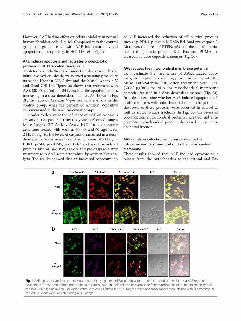
AAE regulates cytochrome c translocation to thecytoplasm and Bax translocation to the mitochondrialmembraneThese results showed that AAE induced cytochrome crelease from the mitochondria to the cytosol and Bax
Fig. 4 AAE regulates cytochrome c translocation to the cytoplasm and Bax translocation to the mitochondrial membrane. a AAE regulatedcytochrome C translocation from mitochondria to cytosol. Also, (b) AAE induced BAX activation from mitochondria outer membrane to cytosoland BAX/BAK oligomerization. Cell were treated with AAE (40ìg/ml) for 24 h. Target protein and mitochondria were stained with fluorescence lagand cell locations were indicated using a DIC image
Kim et al. BMC Complementary and Alternative Medicine (2017) 17:236 Page 7 of 12

translocation from the cytosol to the mitochondria. Wetreated AAE of 40 μg/mL for 24 h and stained cells inorder to visualize the mitochondria, cytochrome c, Baxand Bak (Fig. 4).
AAE-induced apoptosis by p53-independent mannerWe examined the association between AAE-inducedapoptosis and p53. First, we progressed a MTT assay,Annexin V and Dead cell staining, MitoPotential analysisand western blot after AAE treatment of 20 μMLY294002, 20 μM Nutlin, and 20 μM Pifithrin-α, alongwith20 μM Pifithrin-α prior to AAE, in HCT116 cells.Figure 5a showed that the group treated with Nutlin,Pifithrin-α only indicated similar cell viability to the con-trol group, while treatment with AAE, LY294002 andthe AAE co-treated Pifithrin-α group increased cell via-bility. As shown in Fig. 5b, the ratio of Annexin V-positive cells was low in the control and treatment withNutlin, Pifithrin-α only groups, however, the percent of
Annexin V-positive cells increased in the treatment ofLY294002, AAE and AAE co-treatment Pifithrin-αgroup. To examine the mechanism by association ofAAE with p53, we used a Muse MitoPotential Kit. Whiletreatment with AAE, 20 μM LY294002, 20 μM Nutlin,20 μM Pifithrin-α and 20 μM Pifithrin-α prior to AAEincubated for 24 h, the mitochondrial membrane potentialreduced in the LY294002, AAE and AAE co-treatmentPifithrin-α group however, treatment with the Nutlin andPifithrin-α only groups indicated similar mitochondrialmembrane potential to the control group (Fig. 5c).Changes of PTEN, p-PDK1, p-Akt, p-MDM2 and p53
after treatment with AAE, 20 μM LY294002, 20 μMNutlin, 20 μM Pifithrin-α and 20 μM Pifithrin-α co-treatment with AAE were determined by western blotanalysis. The results showed that the levels of cell sur-vival proteins such as p-PDK1, p-Akt, p-MDM2 de-creased but levels of PTEN and p53 increased in thetreatment of LY294002, AAE and AAE co-treatment
Fig. 5 AAE-induced apoptosis by p53-independent manner. Cells were treated with 20 μM LY294002, 20 μM Nutlin-3, 20 μM Pifithrin-á and 40 μg/mlAAE for 24 h. a Cells viability was measured by MTT assay (20 μM LY294002, 20 μM Nutlin-3, 20 μM Pifithrin-á 40 μg/ml AAE). The statistical analysis ofthe data was carried out by use of an an T-test. */#p < 0.05, **/##p < 0.01 and ***/###p < 0.001 (each experiment, n = 3). b Apoptotic effects ofdifferent concentration AAE were evaluated by Muse™ Annexin V and Dead Cell Assay Kit. HCT116 were treated with 20 μM LY294002, 20 μM Nutlin-3,20 μM Pifithrin-á 40 μg/ml AAE for 24 h. c Mitochondria membrane potential were evaluated by Muse™ Mitopotential kit. HCT116 were treated with20 μM LY294002, 20 μM Nutlin-3, 20 μM Pifithrin-á 40 μg/ml AAE for 24 h. d Cells were treated with 20 μM LY294002, 20 μM Nutlin-3, 20 μM Pifithrin-áand 40 μg/ml AAE for 6 h. The expression of PTEN, PDK1, Akt (Thr308 and Ser473), MDM2, p53 were analyzed by western blot analysis
Kim et al. BMC Complementary and Alternative Medicine (2017) 17:236 Page 8 of 12
Pifithrin-α group. However, treatment with the Nutlin,Pifithrin-α only group showed similar results of cell sur-vival proteins levels to that of PTEN and p53, the con-trol group (Fig. 5d).
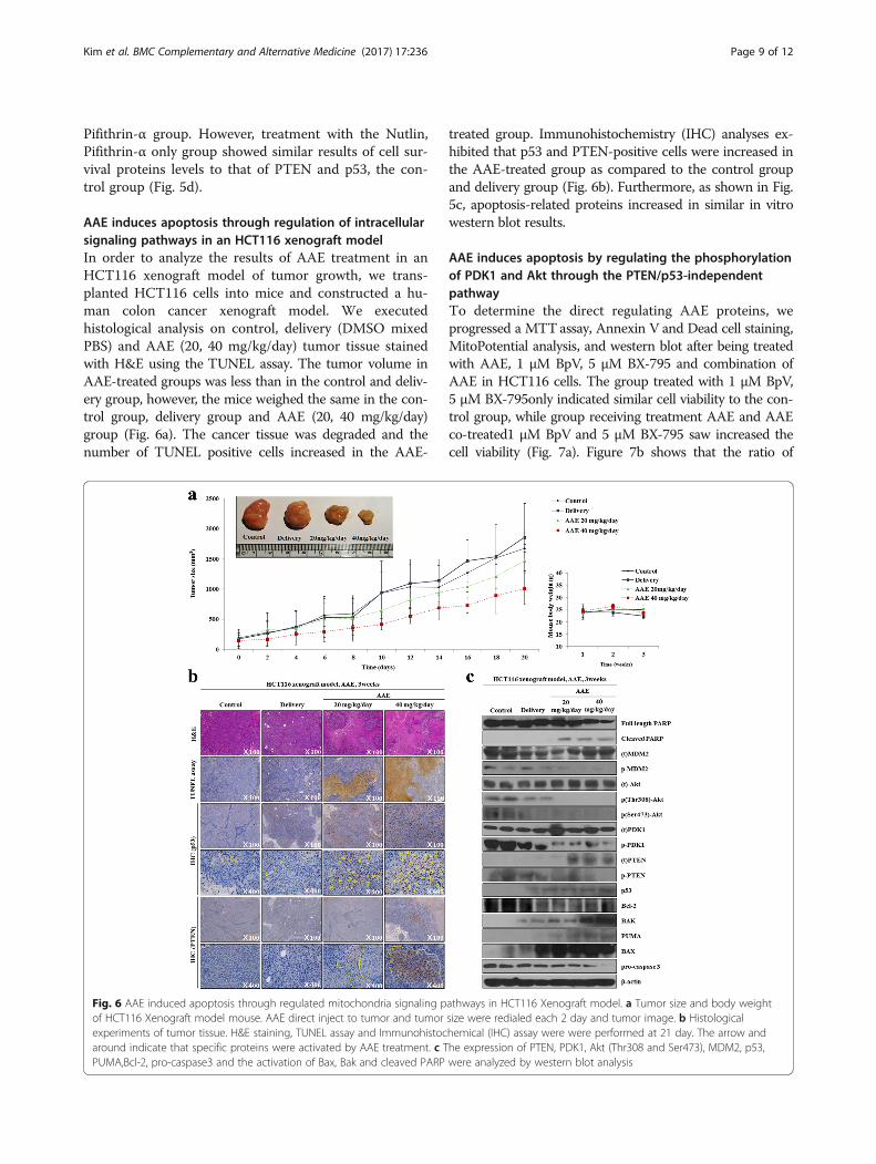
AAE induces apoptosis through regulation of intracellularsignaling pathways in an HCT116 xenograft modelIn order to analyze the results of AAE treatment in anHCT116 xenograft model of tumor growth, we trans-planted HCT116 cells into mice and constructed a hu-man colon cancer xenograft model. We executedhistological analysis on control, delivery (DMSO mixedPBS) and AAE (20, 40 mg/kg/day) tumor tissue stainedwith H&E using the TUNEL assay. The tumor volume inAAE-treated groups was less than in the control and deliv-ery group, however, the mice weighed the same in the con-trol group, delivery group and AAE (20, 40 mg/kg/day)group (Fig. 6a). The cancer tissue was degraded and thenumber of TUNEL positive cells increased in the AAE-
treated group. Immunohistochemistry (IHC) analyses ex-hibited that p53 and PTEN-positive cells were increased inthe AAE-treated group as compared to the control groupand delivery group (Fig. 6b). Furthermore, as shown in Fig.5c, apoptosis-related proteins increased in similar in vitrowestern blot results.
AAE induces apoptosis by regulating the phosphorylationof PDK1 and Akt through the PTEN/p53-independentpathwayTo determine the direct regulating AAE proteins, weprogressed a MTTassay, Annexin V and Dead cell staining,MitoPotential analysis, and western blot after being treatedwith AAE, 1 μM BpV, 5 μM BX-795 and combination ofAAE in HCT116 cells. The group treated with 1 μM BpV,5 μM BX-795only indicated similar cell viability to the con-trol group, while group receiving treatment AAE and AAEco-treated1 μM BpV and 5 μM BX-795 saw increased thecell viability (Fig. 7a). Figure 7b shows that the ratio of
Fig. 6 AAE induced apoptosis through regulated mitochondria signaling pathways in HCT116 Xenograft model. a Tumor size and body weightof HCT116 Xenograft model mouse. AAE direct inject to tumor and tumor size were redialed each 2 day and tumor image. b Histologicalexperiments of tumor tissue. H&E staining, TUNEL assay and Immunohistochemical (IHC) assay were were performed at 21 day. The arrow andaround indicate that specific proteins were activated by AAE treatment. c The expression of PTEN, PDK1, Akt (Thr308 and Ser473), MDM2, p53,PUMA,Bcl-2, pro-caspase3 and the activation of Bax, Bak and cleaved PARP were analyzed by western blot analysis
Kim et al. BMC Complementary and Alternative Medicine (2017) 17:236 Page 9 of 12
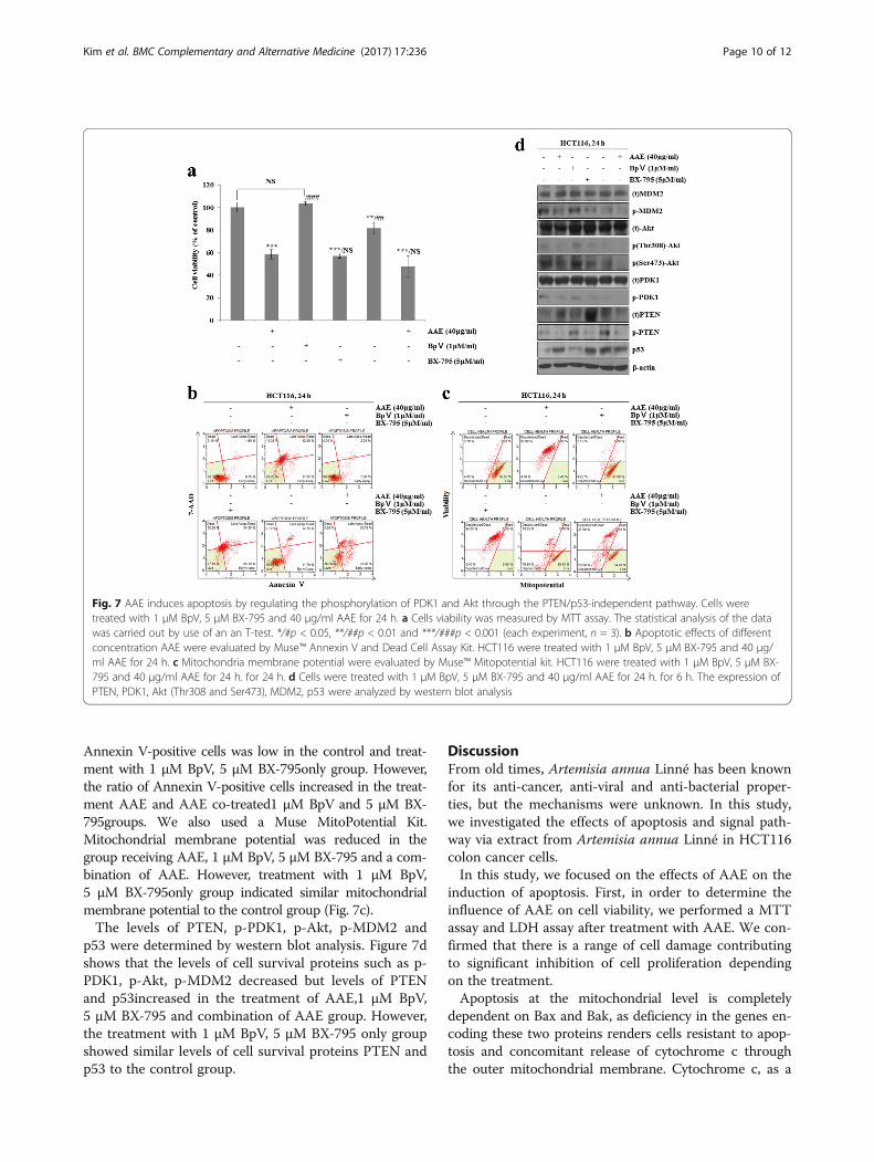
Annexin V-positive cells was low in the control and treat-ment with 1 μM BpV, 5 μM BX-795only group. However,the ratio of Annexin V-positive cells increased in the treat-ment AAE and AAE co-treated1 μM BpV and 5 μM BX-795groups. We also used a Muse MitoPotential Kit.Mitochondrial membrane potential was reduced in thegroup receiving AAE, 1 μM BpV, 5 μM BX-795 and a com-bination of AAE. However, treatment with 1 μM BpV,5 μM BX-795only group indicated similar mitochondrialmembrane potential to the control group (Fig. 7c).The levels of PTEN, p-PDK1, p-Akt, p-MDM2 and
p53 were determined by western blot analysis. Figure 7dshows that the levels of cell survival proteins such as p-PDK1, p-Akt, p-MDM2 decreased but levels of PTENand p53increased in the treatment of AAE,1 μM BpV,5 μM BX-795 and combination of AAE group. However,the treatment with 1 μM BpV, 5 μM BX-795 only groupshowed similar levels of cell survival proteins PTEN andp53 to the control group.
DiscussionFrom old times, Artemisia annua Linné has been knownfor its anti-cancer, anti-viral and anti-bacterial proper-ties, but the mechanisms were unknown. In this study,we investigated the effects of apoptosis and signal path-way via extract from Artemisia annua Linné in HCT116colon cancer cells.In this study, we focused on the effects of AAE on the
induction of apoptosis. First, in order to determine theinfluence of AAE on cell viability, we performed a MTTassay and LDH assay after treatment with AAE. We con-firmed that there is a range of cell damage contributingto significant inhibition of cell proliferation dependingon the treatment.Apoptosis at the mitochondrial level is completely
dependent on Bax and Bak, as deficiency in the genes en-coding these two proteins renders cells resistant to apop-tosis and concomitant release of cytochrome c throughthe outer mitochondrial membrane. Cytochrome c, as a
Fig. 7 AAE induces apoptosis by regulating the phosphorylation of PDK1 and Akt through the PTEN/p53-independent pathway. Cells weretreated with 1 μM BpV, 5 μM BX-795 and 40 μg/ml AAE for 24 h. a Cells viability was measured by MTT assay. The statistical analysis of the datawas carried out by use of an an T-test. */#p < 0.05, **/##p < 0.01 and ***/###p < 0.001 (each experiment, n = 3). b Apoptotic effects of differentconcentration AAE were evaluated by Muse™ Annexin V and Dead Cell Assay Kit. HCT116 were treated with 1 μM BpV, 5 μM BX-795 and 40 μg/ml AAE for 24 h. c Mitochondria membrane potential were evaluated by Muse™ Mitopotential kit. HCT116 were treated with 1 μM BpV, 5 μM BX-795 and 40 μg/ml AAE for 24 h. for 24 h. d Cells were treated with 1 μM BpV, 5 μM BX-795 and 40 μg/ml AAE for 24 h. for 6 h. The expression ofPTEN, PDK1, Akt (Thr308 and Ser473), MDM2, p53 were analyzed by western blot analysis
Kim et al. BMC Complementary and Alternative Medicine (2017) 17:236 Page 10 of 12

pro-apoptotic protein, plays an important role in trigger-ing programmed cell death [32]. The release of cyto-chrome c from mitochondria directly triggers caspase-3activation through formation of the cytochrome c contain-ing apoptosome complex [33]. To confirm the apoptosismechanism, we performed a Hoechst staining, anAnnexin V/Dead cell staining, caspase activity, west-ern blot, MitoPotential staining, fraction western blotand IF staining. Taking all of the results together, AAEinduced apoptotic cell death. This cell death is theAAE controlled cytochrome c released from mito-chondria to cytoplasm by formation of Bax/Bak oligo-meric complexes and led to translocation onto themitochondria outer membrane. Released cytochrome cby AAE treatment was an induced caspase-3 activityand this activity caused the apoptotic cell death.Former studies demonstrated special compound-induced
apoptosis via a p53-independent manner in HCT116 cells[34]. Thus, in order to confirm the association of AAE-induced apoptosis and p53, we treated Pifithrin-α (p53inhibitor) in HCT116 cells. Our results showed an AAE-induced apoptosis via a p53-independent manner, was incells treated with only AAE.Our in vivo results showed AAE-induced apoptosis in
a mouse xenograft model. The ratio of tumor growthwas reduced in the AAE-injected group compared to thecontrol and delivery groups. Also, the AAE-injectedgroup revealed a growth of pro-apoptosis proteins andp53 expressed in a p53-independent manner.When AAE-induced apoptosis occurred by p53-
independent pathway, we used BpV (PTEN inhibitor),and BX-795 (PDK1 inhibitor) to determine the directlyregulating proteins and signal pathway by AAE. In theMTT assay, through Annexin V staining and MitoPoten-tial staining, we confirmed that AAE-induced apoptoticcell death is mitochondria-mediated apoptotsis by PTENindependent, PDK1 dependent pathways. Through thewestern blot on equal terms, we detected that AAE-induced apoptosis induced activation of apoptosis re-lated proteins by regulating the PDK1 directly.
ConclusionsIn conclusion, our data reveals that AAE exerts apop-totic influences in the in vitro, in vivo situations viamodulation of PDK1/Akt signaling pathways and themitochondrial apoptosis pathway through the regulationof proteins such as Bax, Bak and cytochrome c in aPTEM/p53-independent manner.
AbbreviationsAAE: Extracts from Artemisia annua Linné; DMSO: Dissolved in dimethyl sulfoxide;FBS: Fetal bovine serum; IF: Immunofluorescence; IHC: Immunohistochemistry;LDH: Lactate dehydrogenase; MOMP: Mitochondrial outer membranepermeabilization; MTT: 3-(4,5-Dimethylthiazol-2-yl)-2,5-diphenyltetrazoliumbromide; PDK1: Phosphoinositide-dependent protein kinase-1; PTEN: Phosphataseand tensin homolog; TUNEL: TdT-mediated dUTP nickend labeling
FundingThis paper has been supported by 2017 Hannam University Research Fund.
Availability of data and materialsThe data and materials used in this study are available upon request fromthe authors.
Authors’ contributionsEJ, BM and EK carried out the cell culture, Annexin V/Death cell assay,Mitopotential assay. EJ, GT, BM and EK carried out the experiment that isxenograft model and conceived the study. EJ wrote the paper. All authorsread, designed and approved the final manuscript.
Competing interestsThe authors declare that there are no competing interests.
Consent for publicationNot applicable.
Ethics approval and consent to participateAll of the animal experiments were approved by the Ethics Committee forAnimal Experimentation of Hannam University (Daejeon, Korea, HNU 2016–9).
Author details1Department of Biological Science and Biotechnology, College of LifeScience and Nano Technology, Hannam University, 1646 Yuseong-daero,Yuseong-gu, Daejeon 34054, Korea. 2Department of Food Science & BioTechnology, Shinansan University, Daehakro Danwon-gu, Ansan-city,Gyeonggi-do, Korea.
Received: 23 January 2017 Accepted: 23 March 2017
References1. Romero MR, Serrano MA, Vallejo M, Efferth T, Alvarez M, Marin JJ. Antiviral
effect of artemisinin from Artemisia annua against a model member of theFlaviviridae family, the bovine viral diarrhoea virus (BVDV). Planta Med. 2006;72:1169–74.
2. Manulu CE, Adelakun EA, Abok JI. Metabolites Detected in the Crude N-Hexane Extract of Artemisia annua Linn (Asteraceae) Cultivated in Langtang,Plateau State, Nigeria. Chem Sci Int J. 2017;18(2):1–6.
3. Räth K, Taxis K, Walz G, Gleiter CH, Li SM, Heide L. Pharmacokineticstudy of artemisinin after oral intake of a traditional preparation ofArtemisia annua L. (annual wormwood). AmJTrop Med Hyg. 2004;70:128–32.
4. Towler MJ, Weathers PJ. Variations in key artemisinic and other metabolitesthroughout plant development in Artemisia annua L. for potentialtherapeutic use. Ind Crop Prod. 2015;67:185–91.
5. Weathers PJ, Arsenault PR, Covello PS, McMickle A, Teoh KH, Reed DW.Artemisinin production in Artemisia annua: studies in planta and results of anovel delivery method for treating malaria and other neglected diseases.Phytochem Rev. 2011;10:173–83.
6. Brisibe EA, Umoren UE, Brisibe F, Magalhaes PM, Ferreira JFS, Luthria D, WuX, Prior RL. Nutritional characterization and antioxidant capacity of differenttissues of Artemisia annua. L Food Chem. 2009;115:1240–6.
7. Ryu JH, Lee SJ, Kim MJ, Shin JH, Kang SK, Cho KM, Sung NJ. Antioxidant andanticancer activities of Artemisia annua L. and determination of functionalcompounds. J Korean Soc Food Sci Nutr. 2011;40:509–16.
8. Ryu JH, Kim RJ, Lee SJ, Kim IS, Lee HJ, Sung NJ. Nutritional properties andbiological activities of Artemisia annua L. J Korean Soc Food Sci Nutr. 2011;40:163–70.
9. Klayman DL. Qinghaosu (artemisinin): an antimalarial drug from China.Science. 1985;228:1049–55.
10. Avery MA, Chong WKM, Jennings-White C. Stereoselective total synthesis of(+)-artemisinin, the antimalarial constituent of Artemisia annua L. J AmChem Soc. 1992;114:974–9.
11. Schmid G, Hofheinz W. Total synthesis of qinghaosu. J Am Chem Soc. 1983;105:624–5.
12. Singh NP, Lai H. Selective toxicity of dihydroartemisinin and holotransferrintoward human breast cancer cells. Life Sci. 2001;70:49–56.
Kim et al. BMC Complementary and Alternative Medicine (2017) 17:236 Page 11 of 12
13. Kang JR, Lee SJ, Hwang CR, Shin J, Kang MJ, Sung NJ. Optimization ofextraction conditions for mixing beverage development of black garlicand gaeddongssuk by response surface methodology. J Agric Life Sci.2012;46:139–49.
14. Grego-Bessa J, Bloomekatz J, Castel P, Omelchenko T, Baselga J, AndersonKV. The tumor suppressor PTEN and the PDK1 kinase regulate formation ofthe columnar neural epithelium. elife. 2015; doi:10.7554/eLife.12034.
15. Tang J, Ning R, Zeng B, Li Y. Molecular evolution of PTEN Pseudogenes inmammals. PLoS One. 2016; doi:10.1371/journal.pone.0167851.
16. PTEN 3, Yin Y, Shen WH. PTEN: a new guardian of the genome. Oncogene.2008;27:5443–53.
17. PDK1 4, Itoh Y, Higuchi M, Koji O, Kishi Y, Okazaki T, Sakai H, Miyata T,Nakajima K, Gotoh Y. PDK1–Akt pathway regulates radial neuronal migrationand microtubules in the developing mouse neocortex. PNAS. 2016;doi:10.1073/pnas.1516321113.
18. Blanco-Aparicio C, Renner O, Leal JF, Carnero A. PTEN, more than the AKTpathway. Carcinogenesis. 2007;7:1379–86.
19. Vara JÁ, Casado E, de Castro J, Cejas P, Belda-Iniesta C, González-Barón M.PI3K/Akt signalling pathway and caner. Cancer Treat Rev. 2004;30:193–204.
20. Luo J, Manning BD, Cantley LC. Targeting the PI3K-Akt pathway in humancancer: rationale and promise. Cancer Cell. 2003;4:257–62.
21. Song G, Ouyang G, Bao S. The activation of Akt/PKB signaling pathway andcell survival. J Cell Mol Med. 2005;1:59–71.
22. Altomare DA, Testa JR. Perturbations of the AKT signaling pathway inhuman cancer. Oncogene. 2005;24:7455–64.
23. Ogawara Y, Kishishita S, Obata T, Isazawa Y, Suzuki T, Tanaka K, MasuyamaN, Gotoh Y. Akt enhances Mdm2-mediated Ubiquitination and degradationof p53. J Biol Chem. 2002;24:21843–50.
24. Chang H, Li C, Huo K, Wang O, Lu L, Zhang Q, Wang Y, Wang W. Luteolinprevents H2O2-induced apoptosis in H9C2 cells through modulating Akt-P53/Mdm2 signaling pathway. Biomed Res Int. 2016; http://dx.doi.org/10.1155/2016/5125836
25. Bellamy COC. p53 and apoptosis. Bnhih Medical Bulletin. 1996;3:522–38.26. Vaseva AV, Moll UM. The mitochondrial p53 pathway. J Cai et al/Biochimica
et Biophysica Acta. 2009;1787:414–20.27. Cai TY, Yang J, Jones DP. Mitochondrial control of apoptosis: the role of
cytochrome c. J Cai et al/Biochimica et Biophysica Acta. 1998;1366:139–49.28. Xiong SB, Mu TY, Wang GO, Jiang XJ. Mitochondria-mediated apoptosis in
mammals. Protein Cell. 2014;5(10):737–49.29. Wang CX, Youle RJ. The role of mitochondria in apoptosis. Annu Rev Genet.
2009;43:95–118.30. Elmore SS. Apoptosis: a review of programmed cell death. Toxicol Pathol.
2007;35(4):495–516.31. BT KILE. The role of the intrinsic apoptosis pathway in platelet life and
death. J Thromb Haemost. 2009;7:214–7.32. Zhao HM: Extrinsic and intrinsic apoptosis signal pathway review. Hongmei,
licensee InTech 2012, http://dx.doi.org/10.5772/5012933. Fulda S, Debatin KM. Extrinsic versus intrinsic apoptosis pathways in
anticancer chemotherapy. Oncogene. 2006;25:4798–811.34. Srivastava P, Yadav N, Lella R, Schneider A, Jones A, Marlowe T, Lovett G,
Loughlin KO, Minderman H, Gogada R, Chandra D. Neem oil limonoidsinduces p53-independent apoptosis and autophagy. Carcinogenesis. 2012;11:2199–207.
• We accept pre-submission inquiries
• Our selector tool helps you to find the most relevant journal
• We provide round the clock customer support
• Convenient online submission
• Thorough peer review
• Inclusion in PubMed and all major indexing services
• Maximum visibility for your research
Submit your manuscript atwww.biomedcentral.com/submit
Submit your next manuscript to BioMed Central and we will help you at every step:
Kim et al. BMC Complementary and Alternative Medicine (2017) 17:236 Page 12 of 12