
Apoptosis in Coxsackievirus B3-Caused Diseases: Interaction between the Capsid Protein VP2
7
JOURNAL OF VIROLOGY, 0022-538X/00/$04.0010 May 2000, p. 4284–4290 Vol. 74, No. 9 Copyright © 2000, American Society for Microbiology. All Rights Reserved. Apoptosis in Coxsackievirus B3-Caused Diseases: Interaction between the Capsid Protein VP2 and the Proapoptotic Protein Siva ANDREAS HENKE, 1 * HEIKE LAUNHARDT, 2 KATRIN KLEMENT, 1 AXEL STELZNER, 1 ROLAND ZELL, 1 AND THOMAS MUNDER 2 Institute of Virology, Medical Center, Friedrich Schiller University Jena, 1 and Department of Cell and Molecular Biology, Hans-Kno ¨ll-Institut fu ¨r Naturstoff-Forschung e.V., D-07745 Jena, 2 Germany Received 8 November 1999/Accepted 28 January 2000 Coxsackievirus B3 (CVB3) is a common factor in human myocarditis. Apoptotic events are present in CVB3- induced disease, but it is unclear how CVB3 is involved in apoptosis and which viral proteins may induce the apoptotic pathway. In this report we demonstrate that the human and murine proapoptotic protein Siva spe- cifically interact with the CVB3 capsid protein VP2. Furthermore, the transcription of Siva is strongly induced in tissue of CVB3-infected mice and is present in the same area which is positively stained for apoptosis, CD27, and CD70. It has been proposed that Siva is involved in the CD27/CD70-transduced apoptosis. Therefore, we suggest a molecular mechanism through which apoptotic events contributes to CVB3-caused pathogenesis. Coxsackievirus B3, a member of the picornavirus family, is an important human pathogen. Most CVB3-caused disease are mild, but some acute infections are severe and lethal. Clini- cally, coxsackievirus infections are known to be associated with different forms of subacute, acute, and chronic myocarditis (45, 50). CVB3 may cause cardiac arrhythmias and acute heart failure; chronic forms of this disease may supervene, leading to dilated cardiomyopathy, requiring heart transplantation, or to death. The pathogenesis of coxsackievirus infection has been studied extensively in different murine models, demonstrating that the outcome of the disease is determined by complex interactions among several variables, such as virus genotype (11, 25), mouse strains used (11, 24), and the sex (23, 24, 26), age (32), and immune status (18, 27, 37, 49) of the host. In addition, the molecular biology of CVB3 is well documented, especially in view of the availability of sequence data (9, 34, 35, 39, 46) and infectious cDNA molecules (30, 35) as well as the characterization of the genome organization (36) and RNA structures (52). Despite the accumulation of molecular data, so far there are no virus-specific preventive or therapeutic proce- dures available to protect humans against coxsackievirus-in- duced heart diseases. In addition, the mechanisms how CVB3 causes acute or chronic myocarditis are not well characterized (6). One detail of CVB3-induced pathogenesis is apoptosis. For example, apoptotic processes are present in myocardial tissue of patients with dilated cardiomyopathy (42), and depending on the mouse strain and the virus variant used, apoptotic cells are detectable in inflammatory lesions as well as myocardial tissue outside inflamed areas (12, 16, 18, 22, 25). However, it is not established which cell type undergoes apoptosis. Further- more, CVB3 infection of HeLa cells induces caspase-3 activa- tion, but this may not be responsible for the characteristic cytopathic effect produced by coxsackieviruses (8). It is not clear how CVB3 is involved in apoptotic processes and which viral proteins may interact with host cell proteins. To study these possible protein-protein interactions in more detail, we used the yeast two-hybrid system. A HeLa cDNA expression library was screened for proteins that interact with structural and functional proteins of CVB3. We identified the interaction between the CVB3 capsid protein VP2 and the proapoptotic protein Siva, which is involved in the CD27/CD70-transduced apoptotic pathway (44). CD27, a member of the tumor necro- sis factor receptor superfamily, is known to be expressed in T and B cells. Two basic functions of CD27 signaling are known so far. First, the binding of CD27 to CD70, a protein which is also expressed in T and B cells, can provide costimulatory signals in lymphocyte proliferation and immunoglobulin pro- duction (19, 44). For example, it was demonstrated that the cytoplasmic tail of CD27 directly associates with Traf-2 and signals to the Jun N-terminal kinase activation in primary mu- rine lymph node T cells (17). On the other hand, CD27 was shown to be involved in apoptotic processes (44). The cyto- plasmic tail of CD27 lacks the death domain, but Siva, which has a death domain-like region, can bind to this part of CD27 under in vitro conditions. Overexpression of Siva in different cell lines induces apoptosis in the absence of CD70. Therefore, only the association of CD27 with the intracellular Siva can result in the induction of apoptotic events. Furthermore, under in vivo conditions Siva was also found to be present in a number of nonlymphatic tissues (44). Using a rat model of acute ischemic injury, it was demonstrated (43) that the rat equivalent of Siva is produced within the kidney cells after injury and could be the mediator of apoptosis via an inter- action of CD27 which is expressed in the kidney as well. Using the murine model of CVB3 infection, we demonstrate that this virus infection induced the transcription of the murine equivalent of Siva (muSiva) in pancreas and heart tissue. The interaction between VP2 and muSiva was confirmed using a yeast two-hybrid approach. Interestingly, transcription of mu- Siva as well as CD27-, CD70-, active caspase-3-, and TUNEL (terminal deoxynucleotidyltransferase [TdT]-mediated dUTP- biotin nick end labeling) assay-positive cells were present in the same area of tissue in CVB3-infected mice. These findings indicate a newly discovered mechanism by which apoptosis may contribute to coxsackievirus-dependent pathogenesis. MATERIALS AND METHODS Mice. Inbred BALB/c (H-2 dd ) mice were obtained from the Friedrich Schiller University breeding colony. Adult males 7 to 9 weeks of age were used in this * Corresponding author. Mailing address: Institute of Virology, Medical Center, Friedrich Schiller University, Winzerlaer Str. 10, D-07745 Jena, Germany. Phone: (49) 3641 657215. Fax: (49) 3641 657202. E-mail: [email protected]. 4284 Downloaded from https://journals.asm.org/journal/jvi on 15 November 2021 by 77.53.233.27.
Transcript of Apoptosis in Coxsackievirus B3-Caused Diseases: Interaction between the Capsid Protein VP2

JOURNAL OF VIROLOGY,0022-538X/00/$04.0010
May 2000, p. 4284–4290 Vol. 74, No. 9
Copyright © 2000, American Society for Microbiology. All Rights Reserved.
Apoptosis in Coxsackievirus B3-Caused Diseases: Interaction betweenthe Capsid Protein VP2 and the Proapoptotic Protein Siva
ANDREAS HENKE,1* HEIKE LAUNHARDT,2 KATRIN KLEMENT,1 AXEL STELZNER,1
ROLAND ZELL,1 AND THOMAS MUNDER2
Institute of Virology, Medical Center, Friedrich Schiller University Jena,1 and Department of Cell andMolecular Biology, Hans-Knoll-Institut fur Naturstoff-Forschung e.V., D-07745 Jena,2 Germany
Received 8 November 1999/Accepted 28 January 2000
Coxsackievirus B3 (CVB3) is a common factor in human myocarditis. Apoptotic events are present in CVB3-induced disease, but it is unclear how CVB3 is involved in apoptosis and which viral proteins may induce theapoptotic pathway. In this report we demonstrate that the human and murine proapoptotic protein Siva spe-cifically interact with the CVB3 capsid protein VP2. Furthermore, the transcription of Siva is strongly inducedin tissue of CVB3-infected mice and is present in the same area which is positively stained for apoptosis, CD27,and CD70. It has been proposed that Siva is involved in the CD27/CD70-transduced apoptosis. Therefore, wesuggest a molecular mechanism through which apoptotic events contributes to CVB3-caused pathogenesis.
Coxsackievirus B3, a member of the picornavirus family, isan important human pathogen. Most CVB3-caused disease aremild, but some acute infections are severe and lethal. Clini-cally, coxsackievirus infections are known to be associated withdifferent forms of subacute, acute, and chronic myocarditis (45,50). CVB3 may cause cardiac arrhythmias and acute heartfailure; chronic forms of this disease may supervene, leading todilated cardiomyopathy, requiring heart transplantation, or todeath. The pathogenesis of coxsackievirus infection has beenstudied extensively in different murine models, demonstratingthat the outcome of the disease is determined by complexinteractions among several variables, such as virus genotype(11, 25), mouse strains used (11, 24), and the sex (23, 24, 26),age (32), and immune status (18, 27, 37, 49) of the host. Inaddition, the molecular biology of CVB3 is well documented,especially in view of the availability of sequence data (9, 34, 35,39, 46) and infectious cDNA molecules (30, 35) as well as thecharacterization of the genome organization (36) and RNAstructures (52). Despite the accumulation of molecular data, sofar there are no virus-specific preventive or therapeutic proce-dures available to protect humans against coxsackievirus-in-duced heart diseases. In addition, the mechanisms how CVB3causes acute or chronic myocarditis are not well characterized(6).
One detail of CVB3-induced pathogenesis is apoptosis. Forexample, apoptotic processes are present in myocardial tissueof patients with dilated cardiomyopathy (42), and dependingon the mouse strain and the virus variant used, apoptotic cellsare detectable in inflammatory lesions as well as myocardialtissue outside inflamed areas (12, 16, 18, 22, 25). However, it isnot established which cell type undergoes apoptosis. Further-more, CVB3 infection of HeLa cells induces caspase-3 activa-tion, but this may not be responsible for the characteristiccytopathic effect produced by coxsackieviruses (8). It is notclear how CVB3 is involved in apoptotic processes and whichviral proteins may interact with host cell proteins. To studythese possible protein-protein interactions in more detail, weused the yeast two-hybrid system. A HeLa cDNA expression
library was screened for proteins that interact with structuraland functional proteins of CVB3. We identified the interactionbetween the CVB3 capsid protein VP2 and the proapoptoticprotein Siva, which is involved in the CD27/CD70-transducedapoptotic pathway (44). CD27, a member of the tumor necro-sis factor receptor superfamily, is known to be expressed in Tand B cells. Two basic functions of CD27 signaling are knownso far. First, the binding of CD27 to CD70, a protein which isalso expressed in T and B cells, can provide costimulatorysignals in lymphocyte proliferation and immunoglobulin pro-duction (19, 44). For example, it was demonstrated that thecytoplasmic tail of CD27 directly associates with Traf-2 andsignals to the Jun N-terminal kinase activation in primary mu-rine lymph node T cells (17). On the other hand, CD27 wasshown to be involved in apoptotic processes (44). The cyto-plasmic tail of CD27 lacks the death domain, but Siva, whichhas a death domain-like region, can bind to this part of CD27under in vitro conditions. Overexpression of Siva in differentcell lines induces apoptosis in the absence of CD70. Therefore,only the association of CD27 with the intracellular Siva canresult in the induction of apoptotic events. Furthermore, underin vivo conditions Siva was also found to be present in anumber of nonlymphatic tissues (44). Using a rat model ofacute ischemic injury, it was demonstrated (43) that the ratequivalent of Siva is produced within the kidney cells afterinjury and could be the mediator of apoptosis via an inter-action of CD27 which is expressed in the kidney as well.
Using the murine model of CVB3 infection, we demonstratethat this virus infection induced the transcription of the murineequivalent of Siva (muSiva) in pancreas and heart tissue. Theinteraction between VP2 and muSiva was confirmed using ayeast two-hybrid approach. Interestingly, transcription of mu-Siva as well as CD27-, CD70-, active caspase-3-, and TUNEL(terminal deoxynucleotidyltransferase [TdT]-mediated dUTP-biotin nick end labeling) assay-positive cells were present inthe same area of tissue in CVB3-infected mice. These findingsindicate a newly discovered mechanism by which apoptosismay contribute to coxsackievirus-dependent pathogenesis.
MATERIALS AND METHODS
Mice. Inbred BALB/c (H-2dd) mice were obtained from the Friedrich SchillerUniversity breeding colony. Adult males 7 to 9 weeks of age were used in this
* Corresponding author. Mailing address: Institute of Virology,Medical Center, Friedrich Schiller University, Winzerlaer Str. 10,D-07745 Jena, Germany. Phone: (49) 3641 657215. Fax: (49) 3641657202. E-mail: [email protected].
4284
Dow
nloa
ded
from
http
s://j
ourn
als.
asm
.org
/jour
nal/j
vi o
n 15
Nov
embe
r 20
21 b
y 77
.53.
233.
27.

study. Experimental groups consisted of a minimum of four mice, and experi-ments were repeated at least twice and usually three or four times.
Viruses and cell lines. The CVB3 (Nancy) variant used is a cDNA-generatedvirus obtained after transfection of HeLa cells with plasmid pCVB3M2, origi-nally obtained by R. Zell (38). The virus was propagated in HeLa cells and thenpurified and quantified as described previously (18).
Constructions of plasmids. The baits for the two-hybrid screening were con-structed as follows. Sequences specific for the capsid proteins VP1 (851 bp), VP2(788 bp), and the protease 2A (440 bp) were amplified by PCR from the CVB3cDNA (35). The upstream primer contains an NdeI site. The downstream prim-ers contain a BamHI site 39 to the stop codon. VP1, VP2, and 2A samples wereamplified at 94°C (5 min) for 1 cycle followed by 30 cycles at 94°C (1 min), 58°C(1 min), and 72°C (2 min), and a final cycle at 72°C for 5 min in a total volumeof 100 ml. The PCR products were digested with NdeI and BamHI, gel purified,and inserted between the appropriate sites of pAS2-1 (Clontech LaboratoriesInc., Palo Alto, Calif.) to produce pAS2-1/VP1, pAS2-1/VP2, and pAS2-1/2A. Ina similar way, the coding sequences of PV1-VP2 and TMEV-VP2 were PCRamplified using the cDNA of the relevant virus as a template and cloned intopAS2-1. The fusion of the entire human Siva (huSiva) protein to the Gal4DNA-binding and activation domains (Gal4BD and Gal4AD) was achieved byPCR amplification of the identified prey plasmid lacking the first seven residuesof huSiva, using an upstream extended primer containing the missing codons anda cleavage site for EcoRI. In the downstream primer, the stop codon of huSivawas followed by an XhoI site. Digested PCR fragments were ligated to theEcoRI/XhoI sites of pGADGH (Clontech) or to the EcoRI/SalI sites of pAS2-1,yielding Gal4AD-huSiva and Gal4BD-huSiva, respectively. The same primerswere applied for the generation of a glutathione S-transferase (GST)–huSivafusion using the vector pGEX-4T-1 (Pharmacia, Freiburg, Germany). The in-frame fusions of all PCR-amplified fragments were confirmed by sequencing.
Yeast strains, transformation, and two-hybrid analyses. The Saccharomycescerevisiae strain used for the two-hybrid studies was Y190 (MATa ura3-52 his3-200 lys2-801 ade2-101 trp1-901 leu2-3,-112 gal4D gal80D cyhr2 LYS2::GAL1UAS-HIS3TATA-HIS3 URA3::GAL1UAS-GAL1TATA-lacZ; Clontech). Transformationof yeast cells was carried out by the method of Klebe et al. (33). Yeast transfor-mants were selected and cultivated on SD synthetic medium (2% glucose and0.67% yeast nitrogen base without amino acids) supplemented with the appro-priate nutrients. S. cerevisiae Y190 expressing each of the analyzed viral baitsfused to the Gal4BD was transformed with a Gal4AD-tagged HeLa cell cDNAlibrary (Clontech), and the cotransformants were initially selected for growth onmedium lacking histidine. To enhance the stringency of a two-hybrid interaction,the medium was particularly supplemented with 40 mM 3-amino-1,2,4-triazole.Growing yeast colonies were subsequently analyzed for b-galactosidase expres-sion using a colony lift filter assay with X-Gal (5-bromo-4-chloro-3-indolyl-b-D-galactopyranoside) as a substrate as specified by Breeden and Nasmyth (7). Thelibrary plasmids of yeast cells expressing both reporter genes were rescued bytransformation of total yeast DNA into Escherichia coli HB101. Transformantswere selected on M9 minimal medium lacking leucine. To ensure the identifi-cation of the correct cDNA preys, isolated plasmids were retransformed intoyeast strain Y190 containing the appropriate bait proteins, and the cotransfor-mants were again tested for b-galactosidase activity.
GST pull-down experiments. GST-huSiva was maintained and expressed in E.coli BL21 as instructed by the manufacturer (Pharmacia). The fusion protein waspurified from crude bacterial cell extracts with glutathione-Sepharose. Proteinsof noninfected and CVB3-infected HeLa cells were isolated by detaching thecells with 10 mM EDTA. Cells were centrifuged at 250 3 g, resuspended in NTEbuffer (10 mM Tris-HCl [pH 7.4], 100 mM NaCl, 0.5% NP-40), and immediatelyvortexed for 30 s. Cell debris were removed by centrifugation at 12,000 3 g at 4°Cfor 20 min. Protein concentration of the clear supernatant was determined by theBradford protein assay (Bio-Rad Laboratories, Hercules, Calif.). The GST-pulldown assays were performed by the protocol of MacDonald et al. (40), with slightmodification. Briefly, 30 mg of the HeLa cell crude extract was incubated with 2mg of GST-huSiva or 2 mg of GST alone in a total volume of 200 ml of bindingbuffer (20 mM Tris-HCl [pH 7.6], 100 mM NaCl, 0.1% NP-40, 0.1 nM phenyl-methylsulfonylfluoride, 1 mM dithiothreitol, pepstatin [50 mg/ml], aprotinin [2mg/ml], leupeptin [2 mg/ml]). Subsequently, 50 ml of a 50% slurry of glutathione-Sepharose, equilibrated with the binding buffer, was added. The mixture wasincubated for 1 h at 4°C under slight shaking. Associated proteins were pelletedby centrifugation and washed five times in 10 volumes of binding buffer. Finallythe pellet was resuspended in 10 ml of sodium dodecyl sulfate sample buffer andanalyzed for VP2 content by Western blotting. For this, the samples were loadedon a 10 to 20% Tris-glycine gradient gel. After electrophoresis, proteins wereelectroblotted on a nitrocellulose membrane. Detection of proteins was per-formed with the ProtoBlot II AP system (Promega Corp., Madison, Wis.). As aprimary antibody, a polyclonal anti-CVB3/VP4-2 rabbit antibody (dilution 1:500)was applied, and color reaction was obtained by using the Promega ProtoBlot IIAP detection system.
Preparation and staining of routine histology. Aseptically removed pancreasand heart tissue was fixed with for at least 24 h with 4% formaline and mountedin paraffin, and 6-mm sections were cut and stained with hematoxylin-eosin.
Reverse transcription-PCR (RT-PCR). Total RNA was isolated from pancreasand heart tissues of infected and noninfected BALB/c mice according to the acidguanidinium thiocyanate phenol chloroform method described in detail by
Chomczynski and Sacchi (10). Following ultraspeed homogenization, RNA wasextracted using 4 M guanidinium thiocyanate–25 mM sodium citrate–0.5% sar-cosyl–100 mM mercapthoethanol (pH 7.0). DNA and protein contaminationswere removed by phenol-chloroform treatment. After ethanol precipitation,RNA pellets were dissolved in diethyl pyrocarbonate-treated water and incu-bated with DNase I (Boehringer, Mannheim, Germany) for 15 min at roomtemperature (RT) to digest remaining DNA. The DNase I was inactivated byadding 10 mM EDTA and heating to 65°C for 10 min. Reverse transcription wasperformed as follows, using 5 mg of total RNA. Random hexamer primers wereallowed to anneal for 10 min at 70°C. Samples were heated to 42°C for 50 minusing 200 U of Superscript II reverse transcriptase (Life Technologies Inc.,Rockeville, Md.) in buffer containing 20 mM Tris-HCl (pH 8.4), 50 mM KCl, 1mM dithiothreitol, and 2.5 mM MgCl2. The reaction was terminated by heatingto 90°C for 5 min. The remaining RNA was digested with E. coli RNase H (LifeTechnologies). Primers were designed to correspond to the 59 and 39 ends ofmurine Siva, VP2, and b-actin cDNA sequences.
Immunohistochemistry. Immunohistochemical studies were carried out withcryomicrotome sections. Aseptically removed pancreas and heart tissue wasquickly frozen in frozen specimen embedding medium (Cryomatrix; Life Tech-nologies). For lymphocyte characterization, 10-mm sections were obtained, airdried for 2 h, fixed for 3 min with acetone at RT, washed with Hanks solution,and treated with 0.04% H2O2 to block cellular peroxidase activity. Thereafter,sections were incubated separately with an avidin solution (30 min, RT), biotinsolution (30 min, RT), and a 2% nonfat dry milk solution (30 min, RT) to blocknonspecific binding. Between each procedure, sections were washed three timeswith Hanks solution (3 min, RT). All incubation and washing procedures todetect active caspase-3 were performed in the presence of 0.05% saponin. Pri-mary antibodies were applied for 14 h at 4°C. These consisted of rabbit anti-active caspase-3 antibodies (clone 67341A; PharMingen, San Diego, Calif.),Armenian hamster anti-CD27 antibodies (clone LG.3A10; PharMingen), and ratanti-CD70 antibodies (clone FR70; PharMingen). After a 12-min wash withHanks solution, secondary antibodies (biotin-conjugated anti-rabbit [PharMin-gen]; biotin-conjugated mouse anti-hamster [PharMingen]; biotin-conjugatedgoat anti-rat [Jackson ImmunoResearch Laboratories, West Grove, Pa.]) wereapplied for 30 min at RT. A color reaction was obtained after washing of theslides for 12 min (RT) and sequential treatment with streptavidin-horseradishperoxidase conjugate and ACE Red peroxidase substrate kit (Camon Labor-Service GmbH, Wiesbaden, Germany). The sections were counterstained withMayer’s hemalaun solution.
In situ hybridization. Digoxigenin-labeled DNA probes for in situ detection ofviral RNA and RNA of muSiva were synthesized from plasmid pCMV/VP1 orpAS2-1/muSiva by PCR using digoxigenin-labeled nucleotides. Formalin-fixed,paraffin-embedded sections (6 mm) obtained from pancreas and heart tissuewere rehydrated by sequential incubation with xylene, 100, 95, 70, and 50%ethanol, and phosphate-buffered saline (PBS) for 5 min each and treated withprotease VIII solution (50 mg/ml; Sigma Biosciences, St. Louis, Mo.) for 1 h at37°C. After dehydration by sequential incubation with 70, 95, and 100% ethanol,sections were air dried, denatured at 90°C for 8 min, and hybridized at 37°C for18 h. Thereafter, slides were washed twice at 37°C for 10 min. Backgroundbinding was blocked with blocking solution (Kreatech Inc., Amsterdam, TheNetherlands), and the sections were incubated with antidigoxigenin antibodysolution (Boehringer) for 1 h at RT. A color reaction was obtained after washingof the slides three times with PBS at RT and treatment with nitroblue tetrazo-lium–5-bromo-4-chloro-3-indolylphosphate solution (Boehringer). The sectionswere counterstained with eosin or hematoxylin solution.
Apoptosis assay. Apoptotic cells in pancreas and heart tissue of CVB3-in-fected mice were detected by using a TACS Blue Label (TBL) in situ apoptosisdetection kit (Genzyme Corp., Cambridge, Mass.) as described in the Genzymemanual. Briefly, formalin-fixed paraffin sections (6 mm) were obtained frompancreas and heart tissue and rehydrated by sequential incubation with xylene,100, 95, and 70% ethanol, distilled H2O, and PBS for 5 min each. After proteaseK digestion for 15 min at RT and quenching of endogenous peroxidase using 2%H2O2 solution for 5 min at RT, tissue sections were incubated with the enzymeTdT, reaction buffer with Co21 cations, and digoxigenin-labeled nucleotides. Asa negative control, the TdT enzyme was omitted from the protocol. Singlepositive cells were detected after color reaction with TBL-streptavidin-horserad-ish peroxidase detection solution. The slides were counterstained with eosin.
RESULTS
Coxsackievirus protein VP2 interacts with the proapoptoticprotein huSiva. To identify human proteins that interact withCVB3 proteins, we used a yeast two-hybrid system based onthe yeast Gal4 transactivator (15). As viral baits we selected theCVB3 proteins VP1, VP2, and protease 2A. The full-lengthcDNAs of these proteins fused to the yeast Gal4BD wereapplied to screen a Gal4AD-tagged HeLa cell cDNA library.Our initial selection criteria for positive interactors were theexpression of the reporter genes HIS3 and lacZ. Out of ap-
VOL. 74, 2000 APOPTOSIS IN CVB3-MEDIATED DISEASES 4285
Dow
nloa
ded
from
http
s://j
ourn
als.
asm
.org
/jour
nal/j
vi o
n 15
Nov
embe
r 20
21 b
y 77
.53.
233.
27.
proximately 2.2 3 105 analyzed yeast cotransformants, weidentified two candidate clones expressing proteins which maybind to VP2. No reporter-positive clones were detected usingthe baits VP1 and 2A. The cotransformation of Gal4BD-2A-containing plasmids with the Gal4AD library plasmids resultedin a low transformation frequency. Additionally, the obtainedtransformants grew very slowly on selective media, which mayindicate a toxic effect of 2A expression on yeast cells as de-scribed earlier for the 2A protease of poliovirus type 1 (3).
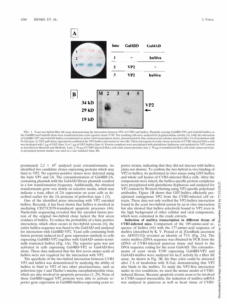
One of the identified preys interacting with VP2 encodedhuSiva. Recently, it has been shown that huSiva is involved inmediating CD27/CD70-transduced apoptotic processes (44).Nucleotide sequencing revealed that the encoded fusion pro-tein of the original two-hybrid clone lacked the first sevenresidues of huSiva. To reduce the probability of a false positiveinteraction, which often occurs in two-hybrid screens (4), theentire huSiva sequence was fused to the Gal4AD and analyzedfor interaction with Gal4BD-VP2. Yeast cells containing bothfusion proteins induced lacZ gene expression similarly to cellsexpressing Gal4BD-VP2 together with Gal4AD–amino-termi-nally truncated huSiva (Fig. 1A). The reporter gene was notactivated in cells expressing Gal4BD-VP2 or Gal4AD-Sivaalone. These data indicated that the first seven amino acids ofhuSiva were not required for the interaction with VP2.
The specificity of the two-hybrid interaction between CVB3VP2 and huSiva was demonstrated by analyzing the ability ofhuSiva to bind to VP2 of two other picornaviruses, such aspoliovirus type 1 and Theiler’s murine encephalomyelitis virus,which are also involved in apoptotic processes (1, 29). None ofthese Gal4BD-tagged VP2 proteins were able to activate re-porter gene expression in Gal4BD-huSiva-expressing yeast re-
porter strains, indicating that they did not interact with huSiva(data not shown). To confirm the two-hybrid in vivo binding ofVP2 to huSiva, we performed in vitro assays using GST-huSivaand whole cell lysates of CVB3-infected HeLa cells. After thecomponents were mixed, the huSiva-specific protein complexeswere precipitated with glutathione-Sepharose and analyzed forVP2 content by Western blotting using VP2-specific polyclonalantibodies. Figure 1B shows that GST-huSiva efficiently pre-cipitated endogenous VP2 from the CVB3-infected cell ex-tracts. These data not only verified the VP2-huSiva interactionfound in the yeast two-hybrid system by an in vitro interactionbut also showed that huSiva selectively bound to VP2 even inthe high background of other cellular and viral components,which were contained in the crude extracts.
Induction of muSiva transcription in different tissue ofCVB3-infected mice. Comparison of the 189-amino-acid se-quence of huSiva (44) with the 177-amino-acid sequence ofmuSiva (described by K. V. Prasad et al. [GenBank accessionno. AF033115]) revealed an identity of 71% (Fig. 2A). Theentire muSiva cDNA sequence was obtained by PCR from thecDNA of CVB3-infected pancreas tissue and fused to theDNA sequence coding for the yeast Gal4AD. The cotransfor-mants of yeast strain Y190 expressing Gal4BD-VP2 andGal4AD-muSiva were analyzed for lacZ activity by a filter liftassay. As shown in Fig. 2B, the blue color could be detectedafter 2 h of incubation with X-Gal, demonstrating that VP2also binds to the muSiva. To study the VP2-Siva interactionunder in vivo conditions, we used the mouse model of CVB3-induced disease. Because apoptotic events seem to be involvedin CVB3-caused myocarditis, the induction of muSiva mRNAwas analyzed in pancreas as well as heart tissue of CVB3-
FIG. 1. Yeast two-hybrid filter lift assay demonstrating the interaction between VP2 of CVB3 and huSiva. Plasmids carrying Gal4BD-VP2 and Gal4AD-huSiva orthe Gal4BD and Gal4AD alone were transformed into yeast reporter strain Y190. The resulting cells were analyzed for b-galactosidase activity (A). Only the interactionof Gal4BD-VP2 and Gal4AD-huSiva reconstituted an active Gal4 transcription factor, demonstrated by blue-stained yeast colonies detected after 2 h of incubation withX-Gal (lane 3). GST pull-down experiments confirmed the VP2-huSiva interaction in vitro (B). Thirty-microgram of crude extract proteins of CVB3-infected HeLa cellswas incubated with 2 mg of GST (lane 3) or 2 mg of GST-huSiva (lane 4). Protein complexes were precipitated with glutathione-Sepharose and analyzed for VP2 contentas described in Materials and Methods. Lane 2, 30 mg of CVB3-infected HeLa cell crude extract proteins; lane 5, 30 mg of noninfected HeLa cell crude extract proteins.A prestained protein marker was used as a size standard (lane M).
4286 HENKE ET AL. J. VIROL.
Dow
nloa
ded
from
http
s://j
ourn
als.
asm
.org
/jour
nal/j
vi o
n 15
Nov
embe
r 20
21 b
y 77
.53.
233.
27.

infected mice by RT-PCR 1 day and 7 days p.i., respectively.As demonstrated in Fig. 2C, CVB3 caused the induction ofhigh levels of muSiva mRNA in different tissue of individualmice compared to noninfected mice. This indicates that theCVB3-caused induction of muSiva expression might be in-volved in apoptosis during coxsackievirus-depending patho-genesis. Upon intraperitoneal (i.p.) inoculation of 106 PFU,CVB3 replicated primarily in tissue of the exocrine pancreas.High levels of viral progenies were detectable at the first day ofviral replication (Fig. 3B), causing massive tissue destruction 1to 3 days postinfection (p.i.) as demonstrated in Fig. 3A.Thereafter, only the tissue of the endocrine pancreas (islets ofLangerhans) was still present 5 to 7 days p.i. Via blood circula-tion CVB3 entered the heart tissue, causing myocytolysis andinfiltration of mononuclear cells into the infected area (Fig. 3A).
Presence of apoptotic, CD27-positive, and CD70-positivecells in different tissues of CVB3-infected mice. During CVB3infection many host cell functions are altered, including theinduction or suppression of several genes encoding the infor-mation of structural and nonstructural cellular proteins (51).These experiments indicate that CVB3 infections are dynamicmolecular processes in which timely interactions between viraland host proteins determine the outcome for both the virusand the host cells. To analyze whether the CVB3-caused in-duction of muSiva transcription (Fig. 2B) and the presence ofhigh amounts of infectious virus particles (Fig. 3B) were ac-companied by apoptosis, the TUNEL assay and immunohisto-chemistry to detect active caspase-3 protein were applied, us-ing pancreas and heart tissue 1 and 7 days p.i. As shown in Fig.
4C and D, TUNEL assay- and active caspase-3-positive cellswere easily detectable in pancreas and heart tissue in whichviral RNA (Fig. 4B) as well as muSiva RNA (Fig. 4A) werepresent. Furthermore, CVB3 infections activated also the ex-pression of CD27 and CD70 in cells which were localized in theinfected area (Fig. 4E and F), indicating that the CD27/CD70apoptotic pathway seemed to be induced in CVB3-causedpathogenesis. Tissue sections of noninfected animals were neg-ative relating to in situ hybridization of muSiva and CVB3 aswell as TUNEL assay and immunohistochemistry of activecaspase-3, CD27, and CD70 (data not shown).
DISCUSSION
In this report, we demonstrate that the structural proteinVP2 of CVB3 interacts specifically with the proapoptotic pro-tein Siva. This observation was obtained using the yeast two-hybrid system which was successfully applied for the identifica-tion of other virus-host cell protein interactions. For example,this technique was used to demonstrate that the IE2 protein ofcytomegalovirus interacts with several different human ribo-nucleoproteins (48) and that the core protein of hepatitis Cvirus is able to bind to the intracellular domain of the lympho-toxin b receptor, influencing hepatitis C virus-caused pathol-ogy (20).
CVB3 infections are usually accompanied by dramaticchanges of the cellular metabolism and the release of newlysynthesized virus particles. For a better understanding of thisdisease, several mouse model systems demonstrating the com-
FIG. 2. CVB3-caused induction of muSiva. Sequence comparison between the human (first row) and the murine (second row) proapoptotic protein Siva revealsa 71% identity (A). The interaction between VP2 of CVB3 and muSiva was confirmed using the yeast two-hybrid system (B), demonstrated by the blue-colored yeastcolonies coexpressing Gal4BD-VP2 and Gal4AD-muSiva (lane 1). Male BALB/c mice were infected with CVB3 i.p. RNA was isolated from pancreas and heart tissue1 or 7 days p.i. Transcription of muSiva, CVB3-VP2, and b-actin in tissue of individual noninfected or CVB3-infected mice was analyzed by RT-PCR, demonstratingthe induction of high levels of muSiva mRNA only in tissue of CVB3-infected mice (C).
VOL. 74, 2000 APOPTOSIS IN CVB3-MEDIATED DISEASES 4287
Dow
nloa
ded
from
http
s://j
ourn
als.
asm
.org
/jour
nal/j
vi o
n 15
Nov
embe
r 20
21 b
y 77
.53.
233.
27.

plexity of CVB3-caused pathogenesis have been established.Upon CVB3 binding to the coxsackievirus and adenovirus re-ceptor, the viral RNA enters the cytoplasm (5). There it istranslated into a single polyprotein which is proteolyticallyprocessed by virus-specific proteases into structural and non-structural proteins. The virus-encoded RNA-dependent RNApolymerase transcribes negative-strand RNA, which is the tem-plate for multiple rounds of virus genome synthesis. Duringthis viral replication several host cellular processes are altered,inducing host cellular protein synthesis shutoff; e.g., the virus-specific protease 2A cleaves the eucaryotic initiation factor 4gamma-1 and -2, stimulating the translation of uncappedmRNA like the CVB3 genome (14, 41). In addition, recently ithas been shown that 2A of CVB3 can also inactivate both thepoly(A)-binding protein (31) as well as the cytoskeletal proteindystrophin (2). Furthermore, 2B of CVB3 can modify plasmamembrane and endo-plasmic reticulum permeability (13), thusinducing an increased level of cytosolic-free calcium (28, 47).Using in vitro conditions, CVB3 infection results in tyrosinephosphorylation of two cellular proteins, increasing viral prog-eny production (21). With the help of the differential mRNAdisplay technique, it was demonstrated that in heart tissue
of CVB3-infected mice several genes were up- as well asdownregulated in comparison to cells of noninfected ani-mals. Among these genes, mRNA levels of the mouse Nip21were decreased. The human equivalent of Nip21, Nip2, mayinteracts with the Bcl-2 protein to promote cell survival. Down-regulation of Nip21 by CVB3 infection may therefore increasemyocyte cell death (51).
In our murine model of CVB3 infection, we were able todemonstrate that the transcription of muSiva was increased inpancreas and heart tissue in the presence of infectious virusparticles. A molecular model that illustrates the role of Siva inCD27/CD70-caused apoptosis is shown in Fig. 5A. With orwithout binding of CD70 to CD27 on the surface of the cellularmembrane, Siva interacts with the cytoplasmic tail of CD27,providing death domain-like structures. The further events ofthis apoptotic pathway are unknown so far. Due to the fact thatCD27 belongs to the tumor necrosis factor receptor super-family, it is quite possible that a protein which is similar toFADD—a protein which is necessary for the Fas/Fas ligand-caused apoptosis—binds to Siva and induces the activation of
FIG. 3. Coxsackievirus-induced pathology in pancreas and heart tissue ofBALB/c mice. BALB/c mice were infected with CVB3 i.p. (A) Pancreas tissueand heart tissue was isolated from infected animals 1 day and 7 days p.i.,respectively, and from noninfected animals. After hematoxylin-eosin staining,virus-caused tissue damage in the pancreas was obvious, demonstrated by mas-sive destruction of the exocrine pancreas up to 7 days p.i. (original magnification,3500). Only the islets of Langerhans remained unaffected (arrows). In the hearttissue, CVB3 infection caused massive inflammation accompanied by infiltrationof mononuclear cells 7 days p.i. (original magnification, 3500). (B) At theindicated time points, eight mice were sacrificed and virus titers were measuredby plaque formation assays. Average titers and standard deviations are shown aslog10 values of PFU/0.1 g of tissue.
FIG. 4. Detection of muSiva, apoptotic cells, and CD27- and CD70-positivecells in CVB3-infected tissue. BALB/c mice were infected with CVB3 i.p. Pan-creas tissue and heart tissue were isolated at 1 day and 7 days after infection,respectively, and used to perform in situ hybridization studies (A and B),TUNEL assays (C), and immunohistochemistry stainings (D to F). In the area ofCVB3-infected tissue (B), transcriptional activity of muSiva (A, arrows) as wellas TUNEL assay (C)- and active caspase-3 (D)-positive cells were detectable.CVB3-caused inflammation also induced the accumulation of CD27- and CD70-positive cells in both tissue (E and F). Original magnification, 31575.
4288 HENKE ET AL. J. VIROL.
Dow
nloa
ded
from
http
s://j
ourn
als.
asm
.org
/jour
nal/j
vi o
n 15
Nov
embe
r 20
21 b
y 77
.53.
233.
27.

a putative caspase. Therefore, this activation might be respon-sible for the activation of effector caspases (e.g., caspase-3) andfinally for the induction of programmed cell death. In CVB3-infected cells, the expression of Siva is induced (Fig. 5B) andboth CD27- and CD70-positive cells are present in the samearea of the infected tissue. Siva binds to the cytoplasmatic tailof CD27; thereafter, VP2 of CVB3 may take the role of aputative FADD-like protein, inducing apoptosis by caspaseactivation as shown in Fig. 4D by detecting the active form ofcaspase-3. One other possibility is that the direct binding be-tween Siva and VP2 in the cytoplasm may attract caspases andactivate the death pathway in the absence of CD70 ligation andCD27 complex formation. In addition, in yeast Siva formshomodimeric complexes (data not shown), but whether thisobservation has a physiological function in apoptotic pathwaysis not clear.
Given that coxsackievirus infections can cause pancreati-tis as well as acute or dilated cardiomyopathy and apoptoticevents are present in virus-infected tissue, our results indicatea molecular mechanism by which the regulation of cell deathproteins may be an important early event of CVB3 infectionbefore and after inflammation.
ACKNOWLEDGMENTS
We thank H.-P. Saluz for helpful discussions during the preparationof this article.
This work was partly supported by grant HE 2910/2-1 MU 1395/1-1from the Deutsche Forschungsgemeinschaft.
REFERENCES
1. Agol, V. I., G. A. Belov, K. Bienz, D. Egger, M. S. Kolesnikova, N. T.Raikhlin, L. I. Romanova, E. A. Smirnova, and E. A. Tolskaya. 1998. Twotypes of death of poliovirus-infected cells: caspase involvement in the apo-ptosis but not cytopathic effect. Virology 252:343–353.
2. Badorff, C., G. H. Lee, B. J. Lamphear, M. E. Martone, K. P. Campbell, R. E.Rhoads, and K. U. Knowlton. 1999. Enteroviral protease 2A cleaves dystro-phin: evidence of cytoskeletal disruption in an acquired cardiomyopathy.Nat. Med. 5:320–326.
3. Barco, A., and L. Carrasco. 1995. Poliovirus 2Apro expression inhibitsgrowth of yeast cells. FEBS Lett. 371:4–8.
4. Bartel, P., C. T. Chien, R. Sternglanz, and S. Fields. 1993. Elimination offalse positives that arise in using the two-hybrid system. BioTechniques 14:920–924.
5. Bergelson, J. M., J. A. Cunningham, G. Droguett, E. A. Kurt-Jones, A.Krithivas, J. S. Hong, M. S. Horwitz, R. L. Crowell, and R. W. Finberg. 1997.Isolation of a common receptor for coxsackie B viruses and adenoviruses 2and 5. Science 275:1320–1323.
6. Bowles, N. E., and J. A. Towbin. 1998. Molecular aspects of myocarditis.Curr. Opin. Cardiol. 13:179–184.
7. Breeden, L., and K. Nasmyth. 1985. Regulation of the yeast HO gene. ColdSpring Harbor Symp. Quant. Biol. 50:643–650.
8. Carthy, C. M., D. J. Granville, K. A. Watson, D. R. Anderson, J. E. Wilson,D. Yang, D. W. Hunt, and B. M. McManus. 1998. Caspase activation andspecific cleavage of substrates after coxsackievirus B3-induced cytopathiceffect in HeLa cells. J. Virol. 72:7669–7675.
9. Chapman, N. M., Z. Tu, S. Tracy, and C. J. Gauntt. 1994. An infectiouscDNA copy of the genome of a non-cardiovirulent coxsackievirus B3 strain:its complete sequence analysis and comparison to the genomes of cardio-virulent coxsackieviruses. Arch. Virol. 135:115–130.
10. Chomczynski, P., and N. Sacchi. 1987. Single-step method of RNA isolationby acid guanidinium thiocyanate-phenol-chloroform extraction. Anal. Bio-chem. 162:156–159.
11. Chow, L. H., K. W. Beisel, and B. M. McManus. 1992. Enteroviral infectionof mice with severe combined immunodeficiency. Evidence for direct viralpathogenesis of myocardial injury. Lab. Investig. 66:24–31.
12. Colston, J. T., B. Chandrasekar, and G. L. Freeman. 1998. Expression ofapoptosis-related proteins in experimental coxsackievirus myocarditis. Car-diovasc. Res. 38:158–168.
13. Doedens, J. R., and K. Kirkegaard. 1995. Inhibition of cellular proteinsecretion by poliovirus proteins 2B and 3A. EMBO J. 14:894–907.
14. Etchison, D., S. C. Milburn, I. Edery, N. Sonenberg, and J. W. Hershey.1982. Inhibition of HeLa cell protein synthesis following poliovirus infectioncorrelates with the proteolysis of a 220,000-dalton polypeptide associatedwith eucaryotic initiation factor 3 and a cap binding protein complex. J. Biol.Chem. 257:14806–14810.
15. Fields, S., and O. Song. 1989. A novel genetic system to detect protein-protein interactions. Nature 340:245–246.
16. Gebhard, J. R., C. M. Perry, S. Harkins, T. Lane, I. Mena, V. C. Asensio, I. L.Campbell, and J. L. Whitton. 1998. Coxsackievirus B3-induced myocarditis:perforin exacerbates disease, but plays no detectable role in virus clearance.Am. J. Pathol. 153:417–428.
17. Gravestein, L. A., D. Amsen, M. Boes, C. R. Calvo, A. M. Kruisbeek, and J.Borst. 1998. The TNF receptor family member CD27 signals to Jun N-
FIG. 5. Model for the putative induction of apoptosis in CD27/Siva-mediated pathways (A) and possible role of VP2 in CD27/Siva-mediated apoptotic events afterCVB3 infection (B).
VOL. 74, 2000 APOPTOSIS IN CVB3-MEDIATED DISEASES 4289
Dow
nloa
ded
from
http
s://j
ourn
als.
asm
.org
/jour
nal/j
vi o
n 15
Nov
embe
r 20
21 b
y 77
.53.
233.
27.
terminal kinase via Traf-2. Eur. J. Immunol. 28:2208–2216.18. Henke, A., S. Huber, A. Stelzner, and J. L. Whitton. 1995. The role of CD81
T lymphocytes in coxsackievirus B3-induced myocarditis. J. Virol. 69:6720–6728.
19. Hintzen, R. Q., S. M. Lens, G. Koopman, S. T. Pals, H. Spits, and R. A. vanLier. 1994. CD70 represents the human ligand for CD27. Int. Immunol. 6:477–480.
20. Hsieh, T. Y., M. Matsumoto, H. C. Chou, R. Schneider, S. B. Hwang, A. S.Lee, and M. M. Lai. 1998. Hepatitis C virus core protein interacts with het-erogeneous nuclear ribonucleoprotein K. J. Biol. Chem. 273:17651–17659.
21. Huber, M., H. C. Selinka, and R. Kandolf. 1997. Tyrosine phosphorylationevents during coxsackievirus B3 replication. J. Virol. 71:595–600.
22. Huber, S. A. 1997. Coxsackievirus-induced myocarditis is dependent on dis-tinct immunopathogenic responses in different strains of mice. Lab. Investig.76:691–701.
23. Huber, S. A., J. Kupperman, and M. K. Newell. 1999. Hormonal regulationof CD41 T-cell responses in coxsackievirus B3-induced myocarditis in mice.J. Virol. 73:4689–95.
24. Huber, S. A., and P. A. Lodge. 1984. Coxsackievirus B-3 myocarditis in Balb/cmice. Evidence for autoimmunity to myocyte antigens. Am. J. Pathol. 116:21–29.
25. Huber, S. A., A. Mortensen, and G. Moulton. 1996. Modulation of cytokineexpression by CD41 T cells during coxsackievirus B3 infections of BALB/cmice initiated by cells expressing the gd1 T-cell receptor. J. Virol. 70:3039–3044.
26. Huber, S. A., and B. Pfaeffle. 1994. Differential Th1 and Th2 cell responsesin male and female BALB/c mice infected with coxsackievirus group B type3. J. Virol. 68:5126–5132.
27. Huber, S. A., J. E. Stone, D. H. Wagner, Jr., J. Kupperman, L. Pfeiffer, C.David, R. L. O’Brien, G. S. Davis, and M. K. Newell. 1999. gd1 T cellsregulate major histocompatibility complex class II(IA and IE)-dependentsusceptibility to coxsackievirus B3-induced autoimmune myocarditis. J. Vi-rol. 73:5630–5636.
28. Irurzun, A., L. Perez, and L. Carrasco. 1993. Enhancement of phospholipaseactivity during poliovirus infection. J. Gen. Virol. 74:1063–1071.
29. Jelachich, M. L., and H. L. Lipton. 1999. Restricted Theiler’s murine en-cephalomyelitis virus infection in murine macrophages induces apoptosis.J. Gen. Virol. 80:1701–1705.
30. Kandolf, R., and P. H. Hofschneider. 1985. Molecular cloning of the genomeof a cardiotropic coxsackie B3 virus: full-length reverse-transcribed recom-binant cDNA generates infectious virus in mammalian cells. Proc. Natl.Acad. Sci. USA 82:4818–4822.
31. Kerekatte, V., B. D. Keiper, C. Badorff, A. Cai, K. U. Knowlton, and R. E.Rhoads. 1999. Cleavage of poly(A)-binding protein by coxsackievirus 2Aprotease in vitro and in vivo: another mechanism for host protein synthesisshutoff? J. Virol. 73:709–717.
32. Khatib, R., J. L. Chason, B. K. Silberberg, and A. M. Lerner. 1980. Age-dependent pathogenicity of group B coxsackieviruses in Swiss-Webster mice:infectivity for myocardium and pancreas. J. Infect. Dis. 141:394–403.
33. Klebe, R. J., J. V. Harriss, Z. D. Sharp, and M. G. Douglas. 1983. A generalmethod for polyethylene-glycol-induced genetic transformation of bacteriaand yeast. Gene 25:333–341.
34. Klump, W. M., I. Bergmann, B. C. Muller, D. Ameis, and R. Kandolf. 1990.Complete nucleotide sequence of infectious coxsackievirus B3 cDNA: twoinitial 59 uridine residues are regained during plus-strand RNA synthesis.J. Virol. 64:1573–83.
35. Knowlton, K. U., E. S. Jeon, N. Berkley, R. Wessely, and S. Huber. 1996. A
mutation in the puff region of VP2 attenuates the myocarditic phenotype ofan infectious cDNA of the Woodruff variant of coxsackievirus B3. J. Virol.70:7811–7818.
36. Krausslich, H. G., M. J. Nicklin, C. K. Lee, and E. Wimmer. 1988. Polypro-tein processing in picornavirus replication. Biochemie 70:119–130.
37. Leipner, C., M. Borchers, I. Merkle, and A. Stelzner. 1999. CoxsackievirusB3-induced myocarditis in MHC class II-deficient mice. J. Hum. Virol. 2:102–114.
38. Lindberg, A. M., R. L. Crowell, R. Zell, R. Kandolf, and U. Pettersson. 1992.Mapping of the RD phenotype of the Nancy strain of coxsackievirus B3.Virus Res. 24:187–196.
39. Lindberg, A. M., P. O. Stalhandske, and U. Pettersson. 1987. Genome ofcoxsackievirus B3. Virology 156:50–63.
40. MacDonald, P. N., D. R. Sherman, D. R. Dowd, S. C. Jefcoat, Jr., and R. K.DeLisle. 1995. The vitamin D receptor interacts with general transcriptionfactor IIB. J. Biol. Chem. 270:4748–4752.
41. Ohlmann, T., M. Rau, V. M. Pain, and S. J. Morley. 1996. The C-terminaldomain of eucaryotic protein synthesis initiation factor (eIF) 4G is sufficientto support cap-independent translation in the absence of eIF4E. EMBO J.15:1371–1382.
42. Olivetti, G., R. Abbi, F. Quaini, J. Kajstura, W. Cheng, J. A. Nitahara, E.Quaini, C. Di Loreto, C. A. Beltrami, S. Krajewski, J. C. Reed, and P.Anversa. 1997. Apoptosis in the failing human heart. N. Engl. J. Med. 336:1131–1141.
43. Padanilam, B. J., A. J. Lewington, and M. R. Hammerman. 1998. Expressionof CD27 and ischemia/reperfusion-induced expression of its ligand Siva inrat kidneys. Kidney Int. 54:1967–1975.
44. Prasad, K. V., Z. Ao, Y. Yoon, M. X. Wu, M. Rizk, S. Jacquot, and S. F.Schlossman. 1997. CD27, a member of the tumor necrosis factor receptorfamily, induces apoptosis and binds to Siva, a proapoptotic protein. Proc.Natl. Acad. Sci. USA 94:6346–6351.
45. Reyes, M. P., and A. M. Lerner. 1985. Coxsackievirus myocarditis—withspecial reference to acute and chronic effects. Prog. Cardiovasc. Dis. 27:373–394.
46. Tracy, S., N. M. Chapman, and Z. Tu. 1992. Coxsackievirus B3 from aninfectious cDNA copy of the genome is cardiovirulent in mice. Arch. Virol.122:399–409.
47. van Kuppeveld, F. J., J. G. Hoenderop, R. L. Smeets, P. H. Willems, H. B.Dijkman, J. M. Galama, and W. J. Melchers. 1997. Coxsackievirus protein2B modifies endoplasmic reticulum membrane and plasma membrane per-meability and facilitates virus release. EMBO J. 16:3519–3532.
48. Wang, Y. F., S. C. Chen, F. Y. Wu, and C. W. Wu. 1997. The interactionbetween human cytomegalovirus immediate-early gene 2 (IE2) protein andheterogeneous ribonucleoprotein A1. Biochem. Biophys. Res. Commun.232:590–594.
49. Woodruff, J. F. 1970. The influence of quantitated post-weaning undernu-trition on coxsackievirus B3 infection of adult mice. II. Alteration of hostdefense mechanisms. J. Infect. Dis. 121:164–181.
50. Woodruff, J. F. 1980. Viral myocarditis. A review. Am. J. Pathol. 101:425–484.
51. Yang, D., J. Yu, Z. Luo, C. M. Carthy, J. E. Wilson, Z. Liu, and B. M.McManus. 1999. Viral myocarditis: identification of five differentially ex-pressed genes in coxsackie-virus B3-infected mouse heart. Circ. Res. 84:704–712.
52. Zell, R., and A. Stelzner. 1997. Application of genome sequence informationto the classification of bovine enteroviruses: the importance of 59- and39-nontranslated regions. Virus Res. 51:213–229.
4290 HENKE ET AL. J. VIROL.
Dow
nloa
ded
from
http
s://j
ourn
als.
asm
.org
/jour
nal/j
vi o
n 15
Nov
embe
r 20
21 b
y 77
.53.
233.
27.


