Aplastic Anemia Stuart Goldberg MD - aamds.org Anemia Goldberg Newark.pdf · Anemia What you need...
11
9/19/2017 1 Aplastic Anemia What you need to know about Stuart Goldberg MD Aplastic Anemia is a bone marrow failure disease The bone marrow is the factory that makes blood The 4 major components of blood • Red Blood Cells: Carry oxygen (energy) • When low called “Anemia” • Weakness, pale, short of breath, leg swelling • Usually measured as hemoglobin • Normal male 14-16 gm/dl • Normal female 12-14 gm/dl The 4 major components of blood • Red Blood Cells: Carry oxygen (energy) • White Blood Cells: Immune system (fight infections) • Neutrophils: primary bacteria fighters • T- lymphocytes: recognize infections • B- lymphocytes: make antibodies to prevent repeat infections • Monocytes: deep penetrating infection fighting and recognition • Normal WBC 4.5-10 X 10 9 /L • Normal Neutrophil count 1.5-8 X 10 9 /L
-
Upload
duongduong -
Category
Documents
-
view
215 -
download
0
Transcript of Aplastic Anemia Stuart Goldberg MD - aamds.org Anemia Goldberg Newark.pdf · Anemia What you need...
9/19/2017
1
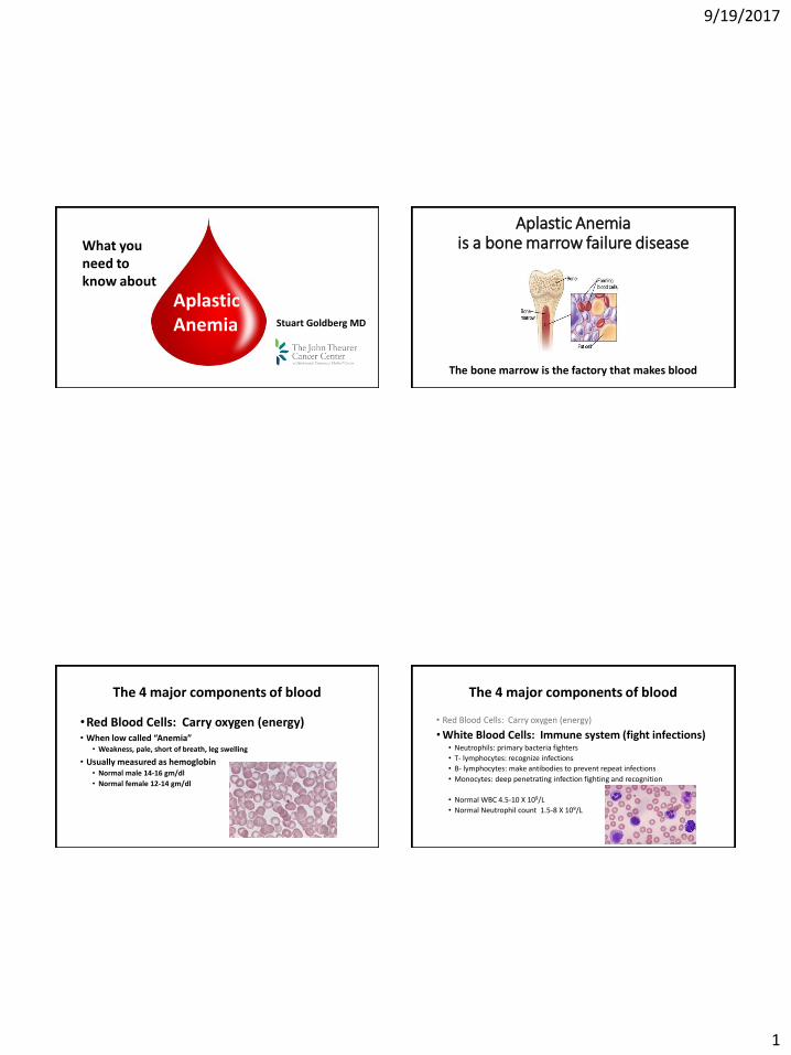
Aplastic Anemia
What you need to know about
Stuart Goldberg MD
Aplastic Anemia is a bone marrow failure disease
The bone marrow is the factory that makes blood
The 4 major components of blood
•Red Blood Cells: Carry oxygen (energy)• When low called “Anemia”
• Weakness, pale, short of breath, leg swelling
• Usually measured as hemoglobin • Normal male 14-16 gm/dl
• Normal female 12-14 gm/dl
The 4 major components of blood
• Red Blood Cells: Carry oxygen (energy)
•White Blood Cells: Immune system (fight infections)• Neutrophils: primary bacteria fighters
• T- lymphocytes: recognize infections
• B- lymphocytes: make antibodies to prevent repeat infections
• Monocytes: deep penetrating infection fighting and recognition
• Normal WBC 4.5-10 X 109/L
• Normal Neutrophil count 1.5-8 X 10⁹/L
9/19/2017
2
The 4 major components of blood
• Red Blood Cells: Carry oxygen (energy)
• White Blood Cells: Immune system (fight infections)
•Platelets: clotting (stop bleeding)• Normal platelet count 150,000 – 400,000
•Plasma: clotting (stop bleeding)
Too few blood cells leads to
•Weakness
• Shortness of breath
•Pale
• Fevers
• Infections
•Bruising
•Bleeding
In Aplastic Anemia The bone marrow fails to make enough blood cells
• Severe Aplastic Anemia (SAA) • Neutrophils less than 0.5 X 10⁹/L
• Platelets less than 20 X 109/L
• Reticulocytes (early red cells) less than 20 X 109/L
• Very Severe Aplastic Anemia (VSAA)• Neutrophils less than 0.2 X 10⁹/L
• Platelets less than 20 X 109/L
• Reticulocytes less than 20 X 109/L
At least 2
At least 2
Making the Diagnosis: The Bone Marrow Biopsy
9/19/2017
3
Making the Diagnosis: The Bone Marrow Biopsy
The “factories” have disappeared. Thus less blood is made.
Cytogenetic Tests from the Bone Marrow• Bone marrow cells may be examined for genetic changes
• Some chromosomal changes are more common, and may indicate more of a pre-leukemia (MDS) state with a poorer prognosis
• Genetic alterations, found by Next Generation Sequencing, are common but the implications are unknown
• Fragility studies are important to exclude Fanconi’s anemia (congenital AA) which change treatment
• Telomere length studies are newer, and results are conflicting but potentially patients with shorter telomeres may do worse
Diagnostic procedures in patients with low blood counts
Andrea Bacigalupo Blood 2017;129:1428-1436
©2017 by American Society of Hematology
Other tests often performed
• Hemoglobin electrophoresis and blood group testing• Elevated fetal hemoglobin and red cell I antigen in stress
• Coombs test• Exclude hemolysis (destruction)
• Liver tests and Rheumatology testing• Exclude other causes of low counts
• Viral tests• May cause low counts (especially hepatitis and HIV)
• PNH testing (paroxysmal nocturnal hemoglobinuria)
• Diepoxybutane incubation (Fanconi’s anemia)
• HLA testing for possible future transplantation
9/19/2017
4
Causes of Aplastic Anemia(The Biology Slides)
• “Damage” to the bone marrow especially the stem cell
• The stromal cells (supporting cells) are typically normal
• Immune mediated destruction of the blood cell precursors is the major cause of acquired AA
• Over representation of patients with HLA DR2
• Suppression of hematopoiesis by expanded population CD8+HLA-DR+ cytotoxic T lymphocytes. Produce inhibitory cytokines (gamma interferon and tumor necrosis factor). FAS-mediated apoptosis
Causes of Aplastic AnemiaBiology II
• Increased nitric oxide damage to cells
• Kinectin-derived peptides that suppress growth
• Increased expression of T-bet
• Diminished perforin and SAP proteins
• Decreased TREGs
• Variations in telomere length
Causes of Aplastic Anemia80% are Acquired
• Idiopathic (unknown)
• Infections – hepatitis, Epstein Barr virus, HIV, parvovirus, mycobacteria
• Exposure to ionizing radiation• Exposure to toxic chemicals (benzenes, pesticides)
• Transfusional graft-vs-host disease
• Orthotropic liver transplantation
• Pregnancy
• Eosinophillic fasciitis• Anorexia
• Severe nutritional deficiencies (B12, folate)
• Paroxysmal nocturnal hemoglobinuria
• Myelodysplastic syndrome
• Drugs (chloramphenicol, phenylbutazone, gold)
Causes of Aplastic Anemia20% are inherited / congenital
• Fanconi’s anemia
• Dyskeratosis congenital
• Familial AA
• Cartilage-hair hypoplasia
• Pearson syndrome
• Thrombocytopenia-absent radius syndrome
• Schwachman-Diamond
• Dubowitz syndrome
9/19/2017
5
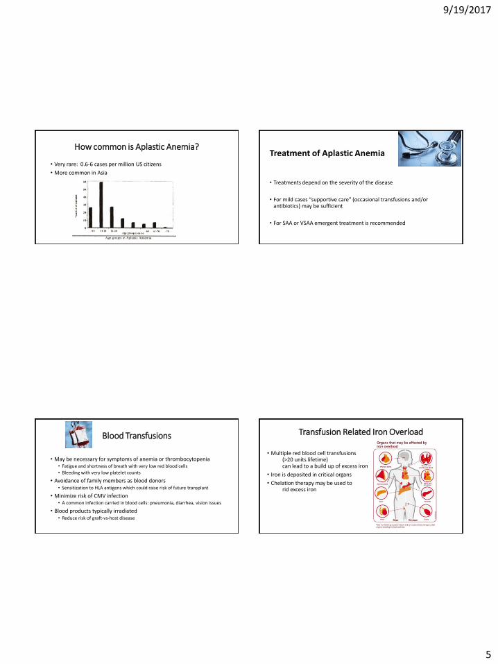
How common is Aplastic Anemia?
• Very rare: 0.6-6 cases per million US citizens
• More common in Asia
Treatment of Aplastic Anemia
• Treatments depend on the severity of the disease
• For mild cases “supportive care” (occasional transfusions and/or antibiotics) may be sufficient
• For SAA or VSAA emergent treatment is recommended
Blood Transfusions
• May be necessary for symptoms of anemia or thrombocytopenia• Fatigue and shortness of breath with very low red blood cells
• Bleeding with very low platelet counts
• Avoidance of family members as blood donors • Sensitization to HLA antigens which could raise risk of future transplant
• Minimize risk of CMV infection• A common infection carried in blood cells: pneumonia, diarrhea, vision issues
• Blood products typically irradiated• Reduce risk of graft-vs-host disease
Transfusion Related Iron Overload
• Multiple red blood cell transfusions (>20 units lifetime) can lead to a build up of excess iron
• Iron is deposited in critical organs
• Chelation therapy may be used torid excess iron
9/19/2017
6
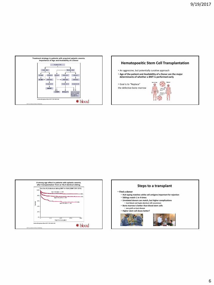
Treatment strategy in patients with acquired aplastic anemia.
Importance of Age and Availability of a Donor
Andrea Bacigalupo Blood 2017;129:1428-1436
©2017 by American Society of Hematology
Hematopoeitic Stem Cell Transplantation
• An aggressive, but potentially curative approach
• Age of the patient and Availability of a Donor are the major determinants of whether a BMT is performed early
• Goal is to “Replace”
the defective bone marrow
A strong age effect in patients with aplastic anemia,
after transplantation from an HLA identical sibling.
Andrea Bacigalupo Blood 2017;129:1428-1436
©2017 by American Society of Hematology
Steps to a transplant
• Find a donor• HLA typing matches white cell antigens important for rejection
• Siblings match 1 in 4 times
• Unrelated donors can match, but higher complications• Cord blood and haplo-identical still uncommon
• Bone marrow is better than blood stem cells• Less graft-vs-host disease
• Higher stem cell doses better?
9/19/2017
7
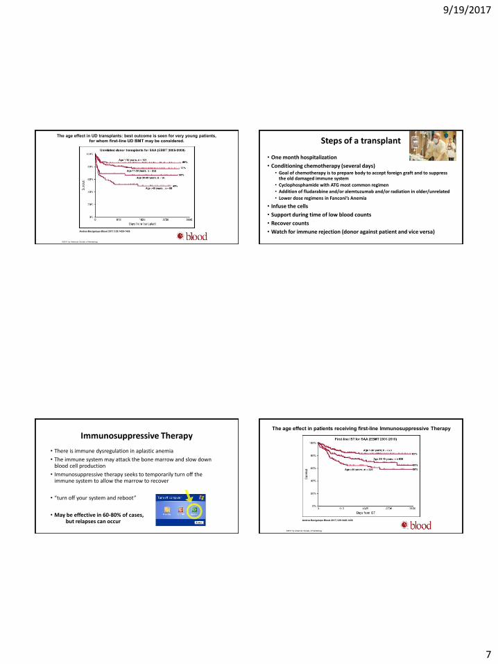
The age effect in UD transplants: best outcome is seen for very young patients,
for whom first-line UD BMT may be considered.
Andrea Bacigalupo Blood 2017;129:1428-1436
©2017 by American Society of Hematology
Steps of a transplant
• One month hospitalization
• Conditioning chemotherapy (several days)• Goal of chemotherapy is to prepare body to accept foreign graft and to suppress
the old damaged immune system
• Cyclophosphamide with ATG most common regimen
• Addition of fludarabine and/or alemtuzumab and/or radiation in older/unrelated
• Lower dose regimens in Fanconi’s Anemia
• Infuse the cells
• Support during time of low blood counts
• Recover counts
• Watch for immune rejection (donor against patient and vice versa)
Immunosuppressive Therapy
• There is immune dysregulation in aplastic anemia
• The immune system may attack the bone marrow and slow down blood cell production
• Immunosuppressive therapy seeks to temporarily turn off the immune system to allow the marrow to recover
• “turn off your system and reboot”
• May be effective in 60-80% of cases, but relapses can occur
The age effect in patients receiving first-line Immunosuppressive Therapy
Andrea Bacigalupo Blood 2017;129:1428-1436
©2017 by American Society of Hematology
9/19/2017
8
Immunosuppressive therapy
• Most common regimen consists of two medications
ATG with Cyclosporine
• Potentially a MAJOR change is coming (hold on – wait a couple of slides)
Anti-thymocyte globulin (ATG)
• ATG: Yes, it really is horse serum
• The T-cells in the horse’s blood attack the diseased human immune system – the human immune system temporarily shuts down and cannot attack the bone marrow – then the human immune system regrows
• Given over 4-5 days in the hospital via a large intravenous line
• May cause shaking chills and low blood pressure during administration
• May cause muscle aches and rashes (serum sickness)
• Takes 10-12 weeks to see improvement in counts
• Occasionally a second treatment (course) is given at 3-6 months
Rabbit ATG
• In some parts of the world horse ATG is not available
• Rabbit ATG was found to be less effective than horse ATG but can work, used in allergic patients and those failing prior treatment
• Higher early complication rate with rabbit ATG
Cyclosporine
• Usually added to ATG to prolong suppression of human T-cells in the original diseased immune system
• Pill is taken for a year or more, with slow taper• Some patients (a third) require years of cyclosporine
• May affect kidney function, so monitored closely
• May cause reversible tremors and hair growth

9/19/2017
9
G-CSF (filgrastim)
• A growth factor (“fertilizer”) for white blood cells
• Typically ineffective by itself in AA
• May be added to ATG+Cyclosporine (but benefit questionable)
Eltrombopag
• A pill developed to increase platelet production
• Has broader growth stimulation on the bone marrow
• Used (and now approved) to treat relapsed/ refractory AA
A Possible New Standard of Care
Eltrombopag added to ATG+Cyclosporine
Townsley D et al: N Engl J Med 2017; 376:1540
The complete response rates were 26-58% and
the overall response rates at 6 months
were 80%, 87%, and 94%, respectively.
The complete and overall response rates in the combined cohorts were higher than in the NIH historical cohort, in
which the rate of complete response was 10% and the overall response rate was 66%.
At a median follow-up of 2 years, the survival rate was 97%; one patient died during the study from a
nonhematologic cause.
Marked increases in bone marrow cellularity, CD34+ cell number, and frequency of early hematopoietic precursors
Similar relapse rates to historical cohort
Androgens
• Men have higher blood counts than women, driven in part by androgens such as testosterone
• Androgens may increase blood counts in AA (?especially in women)
• Androgens may lengthen telomeres – maybe important in AA
Calado RT et al. Blood. 2009;114:2236-
9/19/2017
10
High dose cyclophosphamide
• A chemotherapy drug (intravenous) that markedly suppresses the immune system and a component of most BMTs
• Requires hospitalization and vigorous fluids to prevent bladder damage
• Moderate nausea acutely
• May be effective in relapsed patients, but still mostly experimental
Brodsky RA et al. Blood. 2010;115:2136-
What not to do
If AA is severe – get definitive treatment
• Growth factors alone (erythopoeitin/filgrastim) are ineffective
• Corticosteroids alone are ineffective and raise infectious risks
• Androgens alone as initial therapy is too little and usually ineffective
• Cyclosporine alone as initial therapy is too little
Scheinberg P, Young NS. Blood. 2012;120:1185
Long-term follow-up after immunosuppression.
Phillip Scheinberg, and Neal S. Young Blood
2012;120:1185-1196
©2012 by American Society of Hematology
Late Complications of Aplastic Anemia
• Relapses of AA
• Clonal evolution to Acute Leukemia (up to 15%)
• Secondary cancers
• Infertility from treatments
• BUT THE MAJORITY OF PATIENTS TODAY CAN EXPECT
A LONG HEALTHY LIFE WITH CURRENT THERAPIES
9/19/2017
11
Pure Red Cell Aplasia
• A related disease
• Failure to make red cells (energy) but white cells and platelets normal
• Bone marrow often without red cell precursors
• Reticulocytes (early red cells) low
• Causes include congenital (Diamond Blackfan – deletion ribosomal protein RPS19) and acquired (idiopathic, autoimmune, thymoma, SLE, etc)
• Treatment includes immune suppression therapies including steroids, rituximab, cyclosporine, ATG, plasmapheresis
Thank you. Questions?