Antiphospholipid Antibodies and Antiphospholipid...
114
"Antiphospholipid Antibodies and Antiphospholipid Syndrome." Silvia S. Pierangeli, Ph.D. Professor, Director of the Antiphospholipid Standardization Laboratory University of Texas Medical Branch, Galveston, TX. CEO and Technical Director Louisville APL Diagnostics, Inc. Seabrook, TX.
Transcript of Antiphospholipid Antibodies and Antiphospholipid...
"Antiphospholipid Antibodies and Antiphospholipid Syndrome."
Silvia S. Pierangeli, Ph.D.
Professor, Director of the Antiphospholipid Standardization Laboratory
University of Texas Medical Branch, Galveston, TX.
CEO and Technical Director
Louisville APL Diagnostics, Inc. Seabrook, TX.

Disclosures
CEO and Technical Director of Louisville APL Diagnostics, Inc.
Objectives
Definition and Clinical Manifestations
Pathogenic Mechanisms
Classification Criteria
Treatments
Laboratory Diagnosis
Clinical Cases
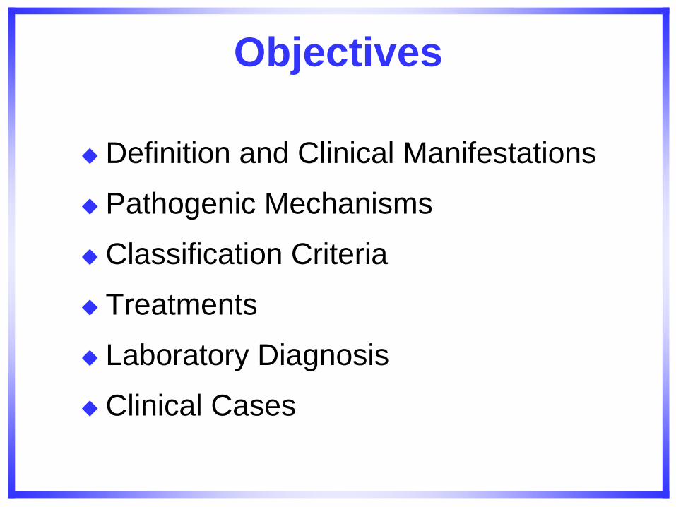
What is the Antiphospholipid Antibody Syndrome ?
An acquired autoimmune thrombophilia, characterized by:
a) vascular thrombosis.
b) recurrent pregnancy losses.
c) thrombocytopenia.
d) laboratory evidence for:
-antibodies against phospholipids or
phospholipid-binding protein cofactors in their blood.
APS morbidity
APS is the most common cause of acquired thrombophilia. Prevalence of aPL antibodies in general population: 2-4%
15-20% of all DVT with or without PE.
1/3 of new strokes in patients < 50 years age.
10-15% women with recurrent pregnancy losses.
aPL are now accepted as the most frequent acquired risk factor for a treatable cause of recurrent pregnancy loss and for pregnancy complications (early and severe pre-eclampsia)
APS: significant proportion of thromboembolic disease and pregnancy loss in SLE.
APL Abs present in 30-40% SLE. One third of those patients have clinical manifestations of APS.
aCL positivity may precede a more severe form of SLE.
MULTISYSTEMIC DISEASE WITH MULTIPLE PATHOGENIC MECHANISMS INVOLVED
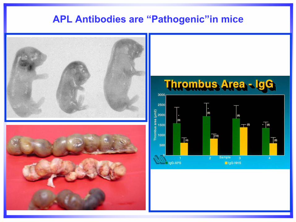
APL Antibodies are “Pathogenic”in mice
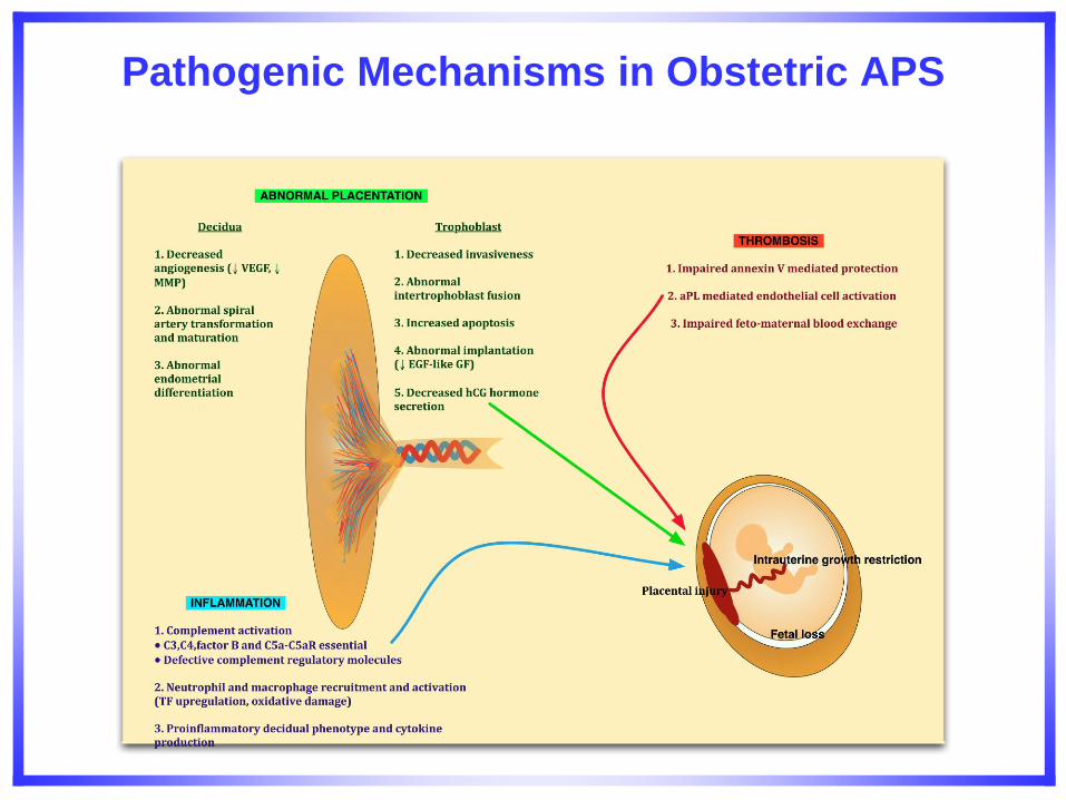
Pathogenic Mechanisms in Obstetric APS
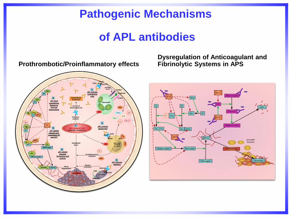
Pathogenic Mechanisms
of APL antibodies
Prothrombotic/Proinflammatory effects
Dysregulation of Anticoagulant and Fibrinolytic Systems in APS
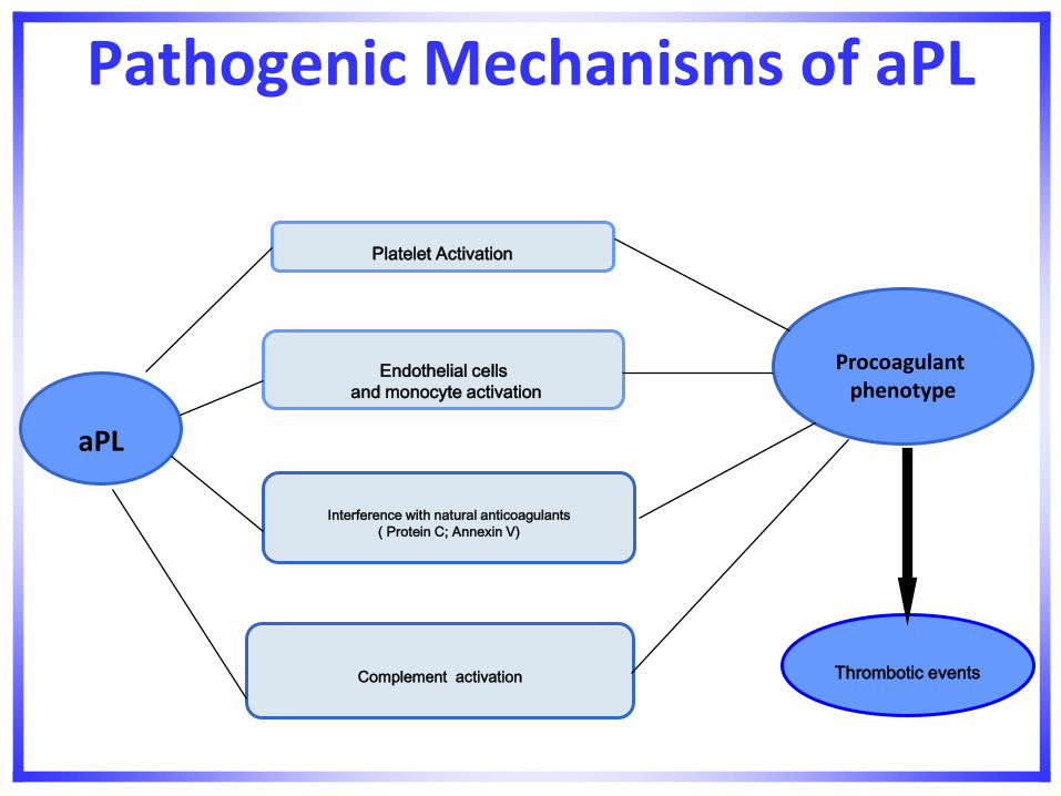
Pathogenic Mechanisms of aPL
Platelet Activation
aPL
Procoagulant phenotype
Endothelial cells
and monocyte activation
Thrombotic events Complement activation
Interference with natural anticoagulants
( Protein C; Annexin V)
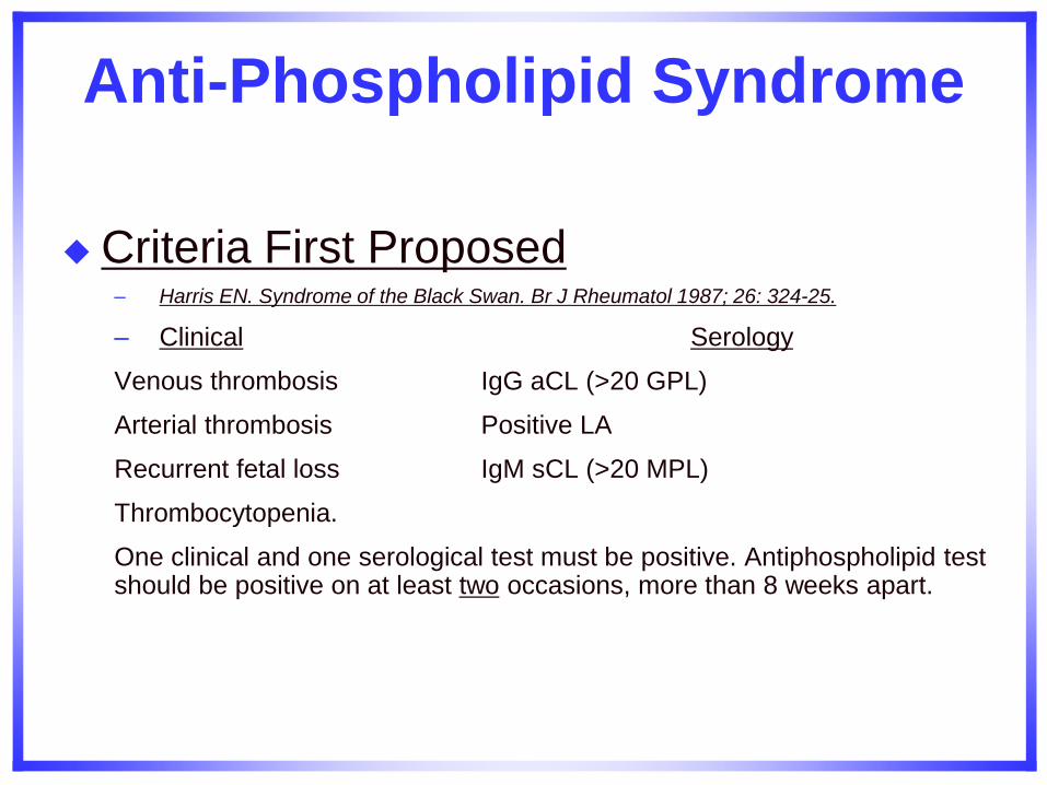
Anti-Phospholipid Syndrome
Criteria First Proposed – Harris EN. Syndrome of the Black Swan. Br J Rheumatol 1987; 26: 324-25.
– Clinical Serology
Venous thrombosis IgG aCL (>20 GPL)
Arterial thrombosis Positive LA
Recurrent fetal loss IgM sCL (>20 MPL)
Thrombocytopenia.
One clinical and one serological test must be positive. Antiphospholipid test should be positive on at least two occasions, more than 8 weeks apart.
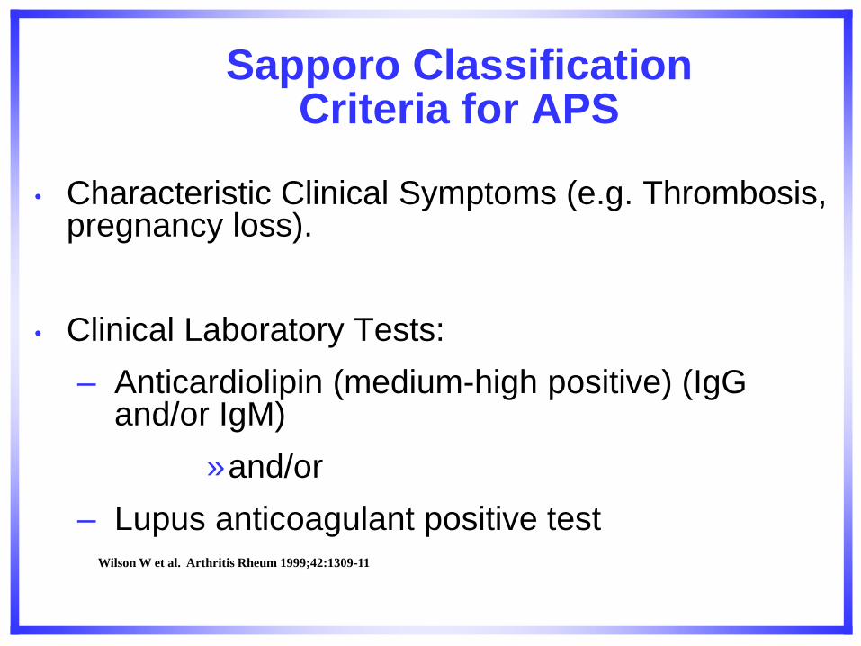
Sapporo Classification Criteria for APS
• Characteristic Clinical Symptoms (e.g. Thrombosis, pregnancy loss).
• Clinical Laboratory Tests:
– Anticardiolipin (medium-high positive) (IgG and/or IgM)
»and/or
– Lupus anticoagulant positive test
Wilson W et al. Arthritis Rheum 1999;42:1309-11
.
Giannakopoulos B et al. Blood 2009;113:985-994
©2009 by American Society of Hematology
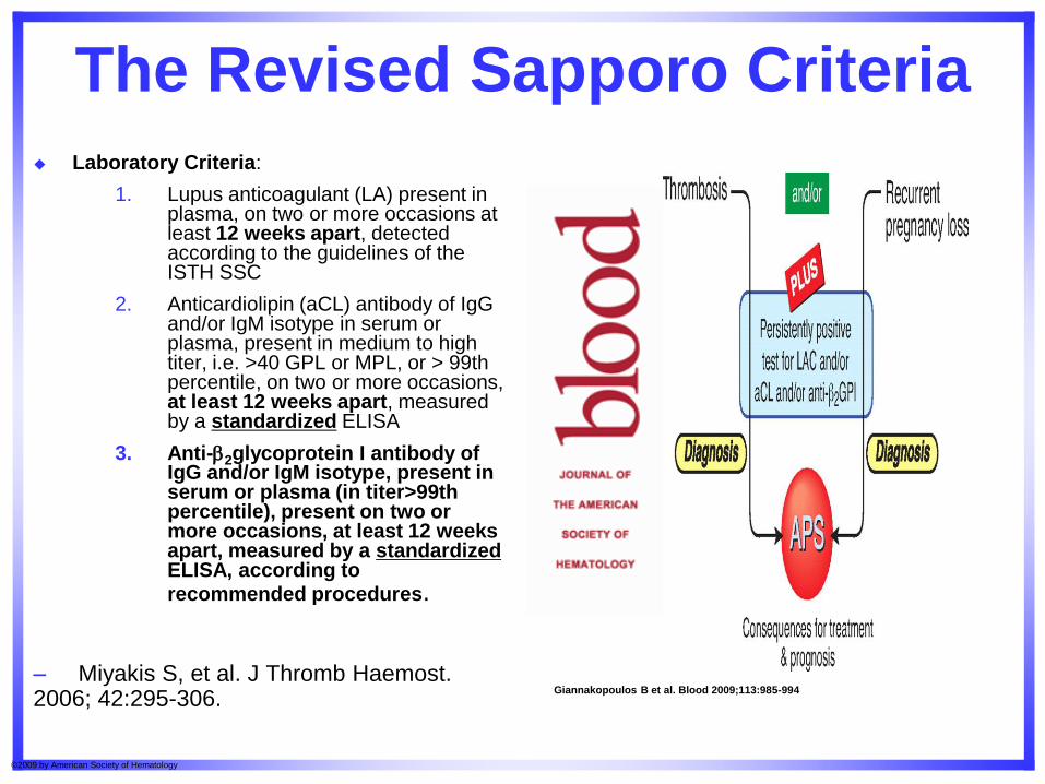
The Revised Sapporo Criteria
Laboratory Criteria:
1. Lupus anticoagulant (LA) present in plasma, on two or more occasions at least 12 weeks apart, detected according to the guidelines of the ISTH SSC
2. Anticardiolipin (aCL) antibody of IgG and/or IgM isotype in serum or plasma, present in medium to high titer, i.e. >40 GPL or MPL, or > 99th percentile, on two or more occasions, at least 12 weeks apart, measured by a standardized ELISA
3. Anti-2glycoprotein I antibody of IgG and/or IgM isotype, present in serum or plasma (in titer>99th percentile), present on two or more occasions, at least 12 weeks apart, measured by a standardized ELISA, according to recommended procedures.
– Miyakis S, et al. J Thromb Haemost. 2006; 42:295-306.
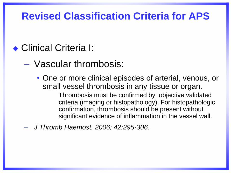
Revised Classification Criteria for APS
Clinical Criteria I:
– Vascular thrombosis:
• One or more clinical episodes of arterial, venous, or small vessel thrombosis in any tissue or organ. Thrombosis must be confirmed by objective validated criteria (imaging or histopathology). For histopathologic confirmation, thrombosis should be present without significant evidence of inflammation in the vessel wall.
– J Thromb Haemost. 2006; 42:295-306.
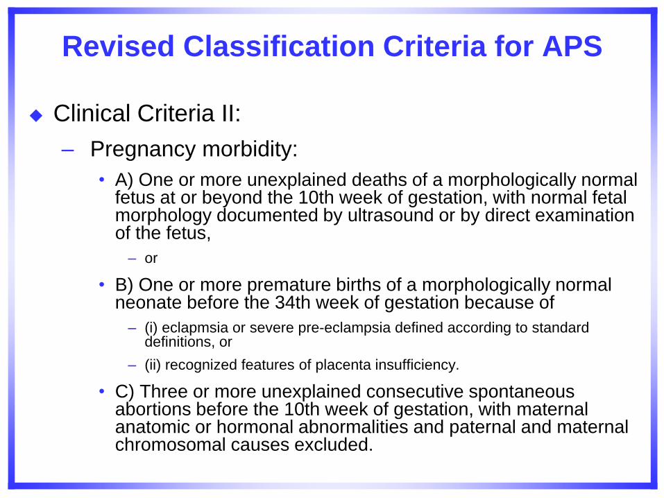
Revised Classification Criteria for APS
Clinical Criteria II:
– Pregnancy morbidity:
• A) One or more unexplained deaths of a morphologically normal fetus at or beyond the 10th week of gestation, with normal fetal morphology documented by ultrasound or by direct examination of the fetus,
– or
• B) One or more premature births of a morphologically normal neonate before the 34th week of gestation because of
– (i) eclapmsia or severe pre-eclampsia defined according to standard definitions, or
– (ii) recognized features of placenta insufficiency.
• C) Three or more unexplained consecutive spontaneous abortions before the 10th week of gestation, with maternal anatomic or hormonal abnormalities and paternal and maternal chromosomal causes excluded.
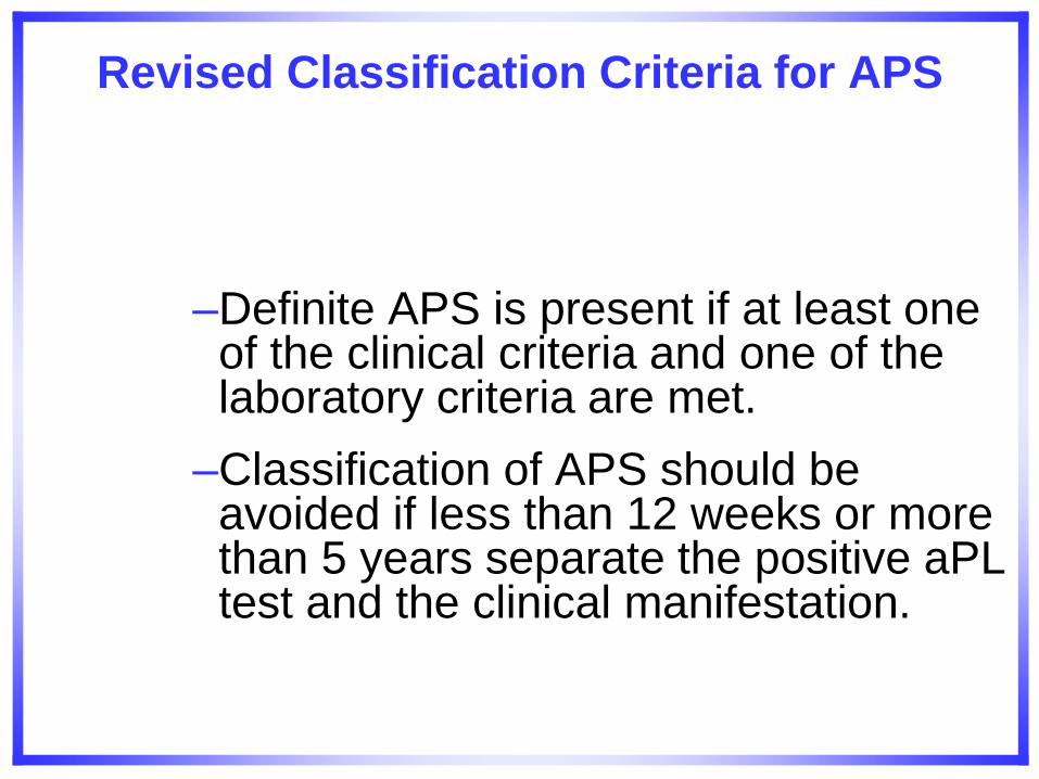
Revised Classification Criteria for APS
–Definite APS is present if at least one of the clinical criteria and one of the laboratory criteria are met.
–Classification of APS should be avoided if less than 12 weeks or more than 5 years separate the positive aPL test and the clinical manifestation.
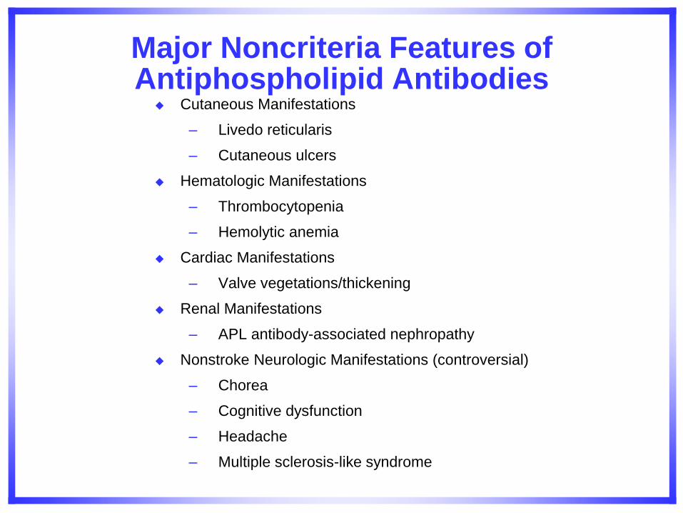
Major Noncriteria Features of Antiphospholipid Antibodies
Cutaneous Manifestations
– Livedo reticularis
– Cutaneous ulcers
Hematologic Manifestations
– Thrombocytopenia
– Hemolytic anemia
Cardiac Manifestations
– Valve vegetations/thickening
Renal Manifestations
– APL antibody-associated nephropathy
Nonstroke Neurologic Manifestations (controversial)
– Chorea
– Cognitive dysfunction
– Headache
– Multiple sclerosis-like syndrome
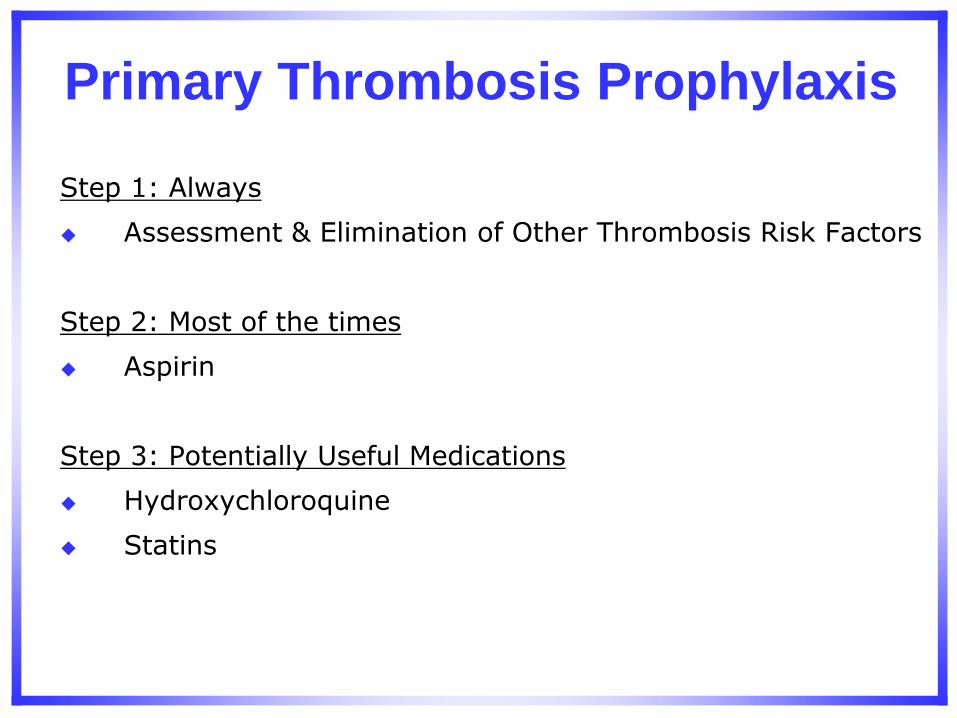
Step 1: Always
Assessment & Elimination of Other Thrombosis Risk Factors
Step 2: Most of the times
Aspirin
Step 3: Potentially Useful Medications
Hydroxychloroquine
Statins
Primary Thrombosis Prophylaxis
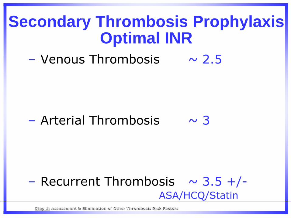
– Venous Thrombosis ~ 2.5
– Arterial Thrombosis ~ 3
– Recurrent Thrombosis ~ 3.5 +/- ASA/HCQ/Statin
Secondary Thrombosis Prophylaxis Optimal INR
Step 1: Assessment & Elimination of Other Thrombosis Risk Factors
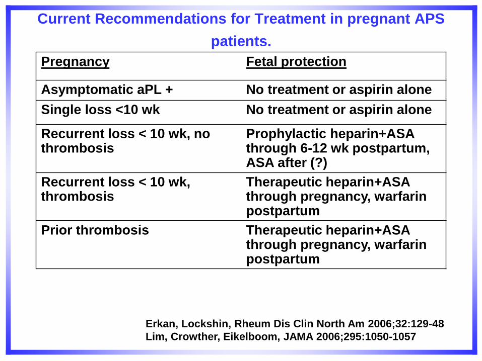
Erkan, Lockshin, Rheum Dis Clin North Am 2006;32:129-48
Lim, Crowther, Eikelboom, JAMA 2006;295:1050-1057
Current Recommendations for Treatment in pregnant APS
patients. Pregnancy Fetal protection
Asymptomatic aPL + No treatment or aspirin alone
Single loss <10 wk No treatment or aspirin alone
Recurrent loss < 10 wk, no thrombosis
Prophylactic heparin+ASA through 6-12 wk postpartum, ASA after (?)
Recurrent loss < 10 wk, thrombosis
Therapeutic heparin+ASA through pregnancy, warfarin postpartum
Prior thrombosis Therapeutic heparin+ASA through pregnancy, warfarin postpartum
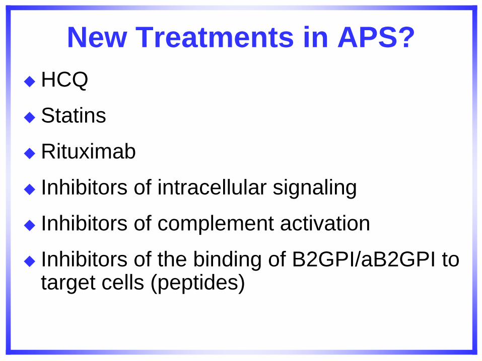
New Treatments in APS?
HCQ
Statins
Rituximab
Inhibitors of intracellular signaling
Inhibitors of complement activation
Inhibitors of the binding of B2GPI/aB2GPI to target cells (peptides)
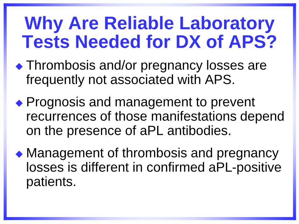
Why Are Reliable Laboratory Tests Needed for DX of APS?
Thrombosis and/or pregnancy losses are frequently not associated with APS.
Prognosis and management to prevent recurrences of those manifestations depend on the presence of aPL antibodies.
Management of thrombosis and pregnancy losses is different in confirmed aPL-positive patients.
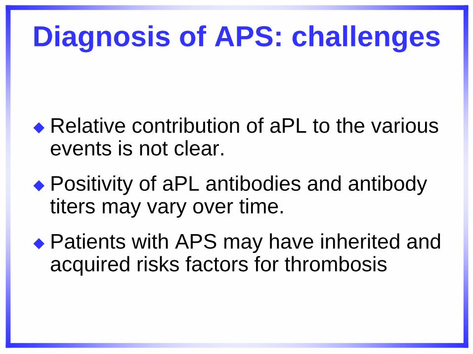
Diagnosis of APS: challenges
Relative contribution of aPL to the various events is not clear.
Positivity of aPL antibodies and antibody titers may vary over time.
Patients with APS may have inherited and acquired risks factors for thrombosis
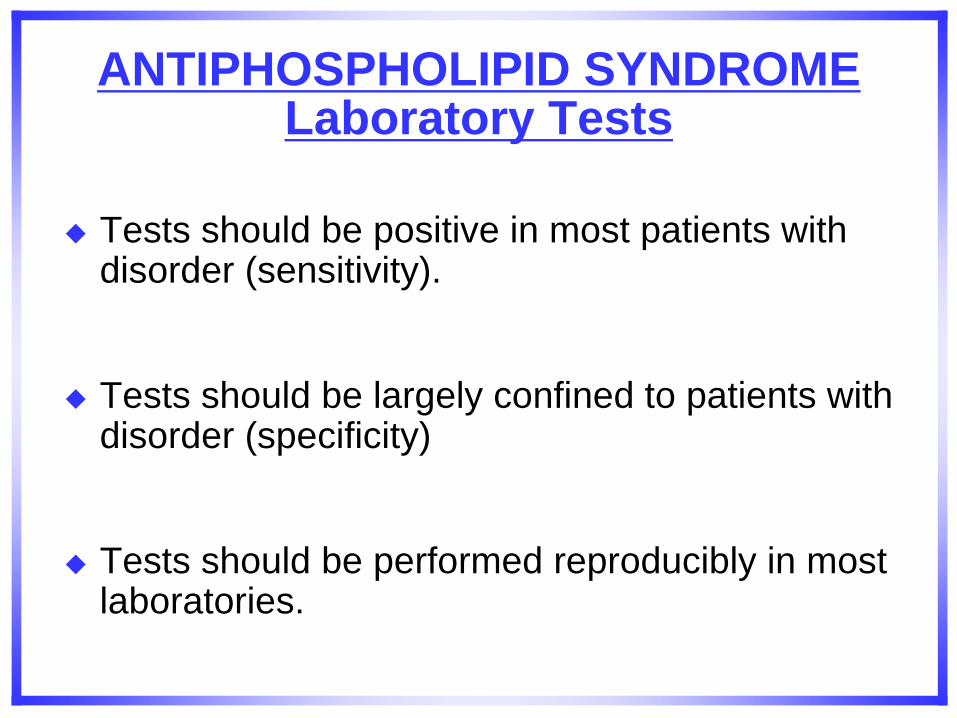
ANTIPHOSPHOLIPID SYNDROME Laboratory Tests
Tests should be positive in most patients with disorder (sensitivity).
Tests should be largely confined to patients with disorder (specificity)
Tests should be performed reproducibly in most laboratories.
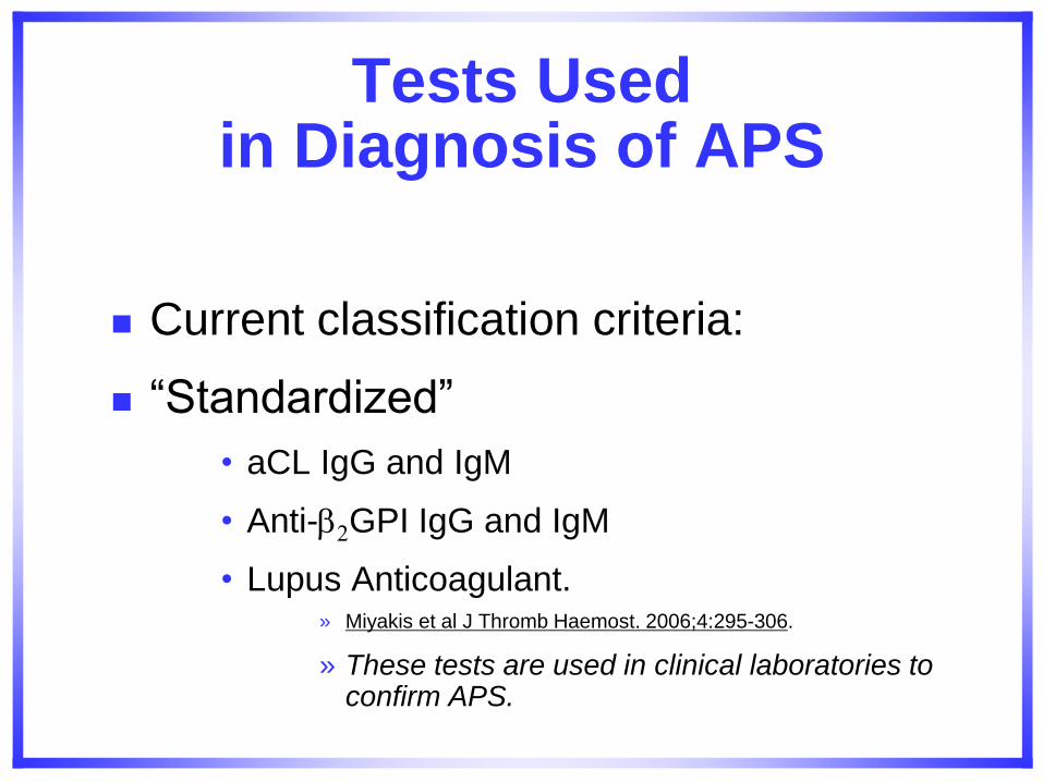
Tests Used in Diagnosis of APS
Current classification criteria:
“Standardized”
• aCL IgG and IgM
• Anti-2GPI IgG and IgM
• Lupus Anticoagulant. » Miyakis et al J Thromb Haemost. 2006;4:295-306.
» These tests are used in clinical laboratories to confirm APS.
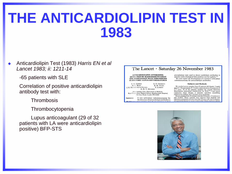
THE ANTICARDIOLIPIN TEST IN 1983
Anticardiolipin Test (1983) Harris EN et al Lancet 1983; ii: 1211-14
-65 patients with SLE
Correlation of positive anticardiolipin antibody test with:
Thrombosis
Thrombocytopenia
Lupus anticoagulant (29 of 32 patients with LA were anticardiolipin positive) BFP-STS
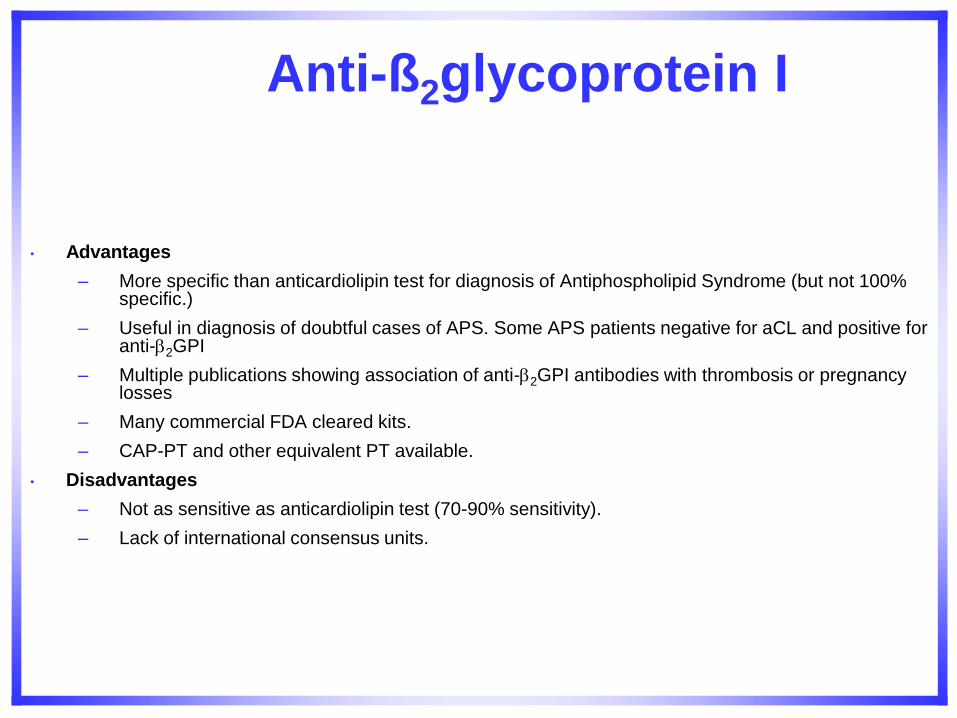
• Advantages
– More specific than anticardiolipin test for diagnosis of Antiphospholipid Syndrome (but not 100% specific.)
– Useful in diagnosis of doubtful cases of APS. Some APS patients negative for aCL and positive for anti-2GPI
– Multiple publications showing association of anti-2GPI antibodies with thrombosis or pregnancy losses
– Many commercial FDA cleared kits.
– CAP-PT and other equivalent PT available.
• Disadvantages
– Not as sensitive as anticardiolipin test (70-90% sensitivity).
– Lack of international consensus units.
Anti-ß2glycoprotein I
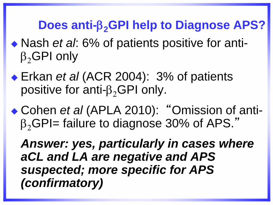
Does anti-2GPI help to Diagnose APS?
Nash et al: 6% of patients positive for anti-2GPI only
Erkan et al (ACR 2004): 3% of patients positive for anti-2GPI only.
Cohen et al (APLA 2010): “Omission of anti-2GPI= failure to diagnose 30% of APS.”
Answer: yes, particularly in cases where aCL and LA are negative and APS suspected; more specific for APS (confirmatory)
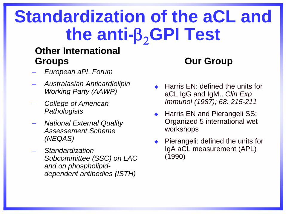
Standardization of the aCL and the anti-2GPI Test
Other International Groups
– European aPL Forum
– Australasian Anticardiolipin Working Party (AAWP)
– College of American Pathologists
– National External Quality Assessement Scheme (NEQAS)
– Standardization Subcommittee (SSC) on LAC and on phospholipid-dependent antibodies (ISTH)
Our Group
Harris EN: defined the units for aCL IgG and IgM.. Clin Exp Immunol (1987); 68: 215-211
Harris EN and Pierangeli SS: Organized 5 international wet workshops
Pierangeli: defined the units for IgA aCL measurement (APL) (1990)
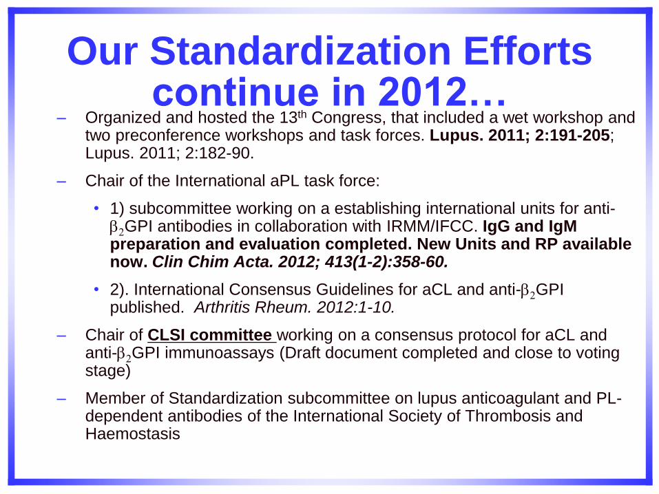
Our Standardization Efforts continue in 2012…
– Organized and hosted the 13th Congress, that included a wet workshop and two preconference workshops and task forces. Lupus. 2011; 2:191-205; Lupus. 2011; 2:182-90.
– Chair of the International aPL task force:
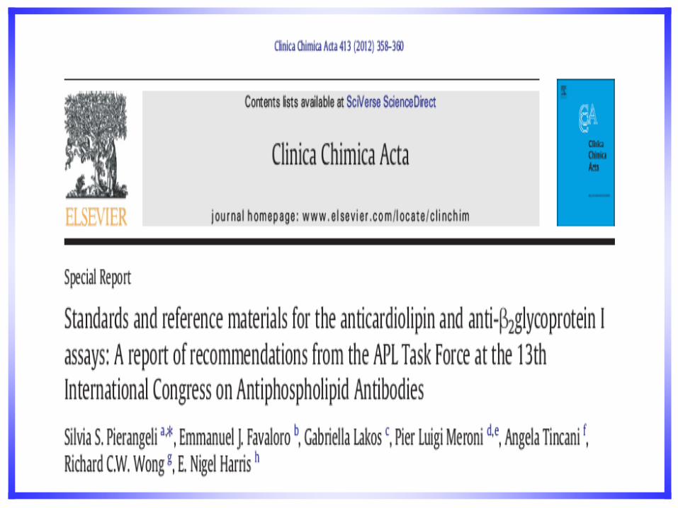
• 1) subcommittee working on a establishing international units for anti-2GPI antibodies in collaboration with IRMM/IFCC. IgG and IgM preparation and evaluation completed. New Units and RP available now. Clin Chim Acta. 2012; 413(1-2):358-60.
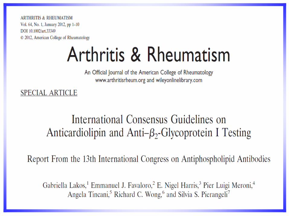
• 2). International Consensus Guidelines for aCL and anti-2GPI published. Arthritis Rheum. 2012:1-10.
– Chair of CLSI committee working on a consensus protocol for aCL and anti-2GPI immunoassays (Draft document completed and close to voting stage)
– Member of Standardization subcommittee on lupus anticoagulant and PL-dependent antibodies of the International Society of Thrombosis and Haemostasis
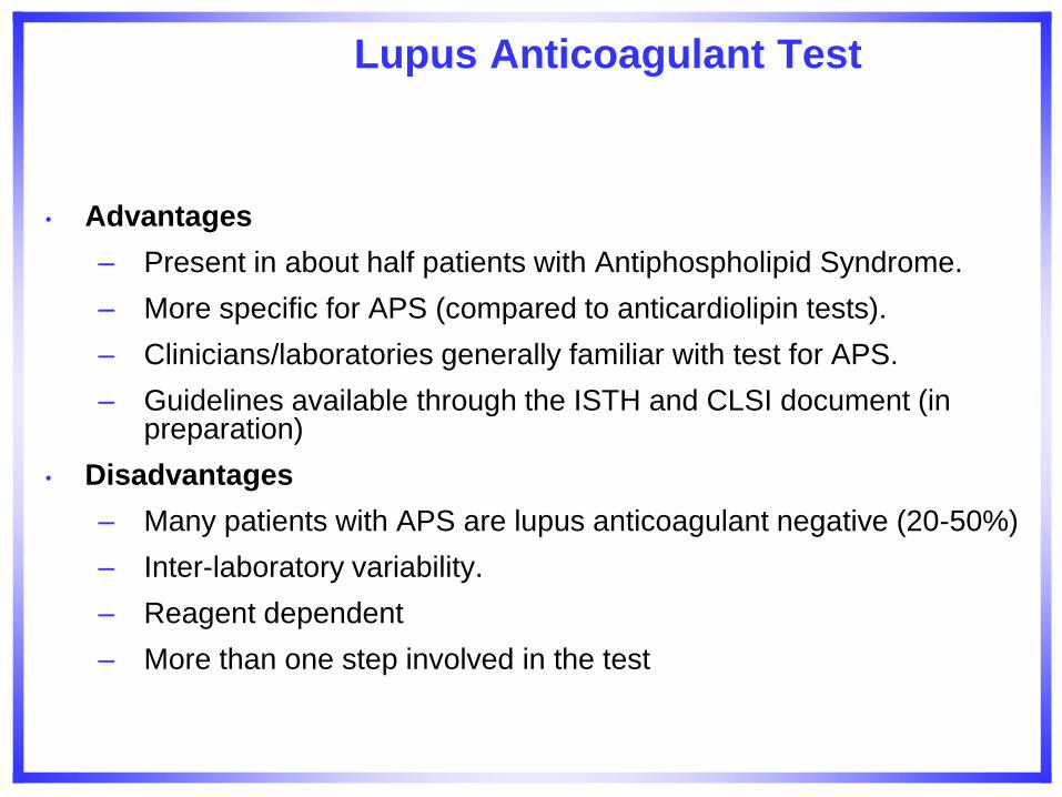
• Advantages
– Present in about half patients with Antiphospholipid Syndrome.
– More specific for APS (compared to anticardiolipin tests).
– Clinicians/laboratories generally familiar with test for APS.
– Guidelines available through the ISTH and CLSI document (in preparation)
• Disadvantages
– Many patients with APS are lupus anticoagulant negative (20-50%)
– Inter-laboratory variability.
– Reagent dependent
– More than one step involved in the test
Lupus Anticoagulant Test
Lupus Anticoagulant Guidelines
Brandt JT, et al. Thromb Haemost 1995; 74:1185-1190
Pengo V, et al. J Thromb Haemost 2009; 7: 1737-40.
CLSI document in progress
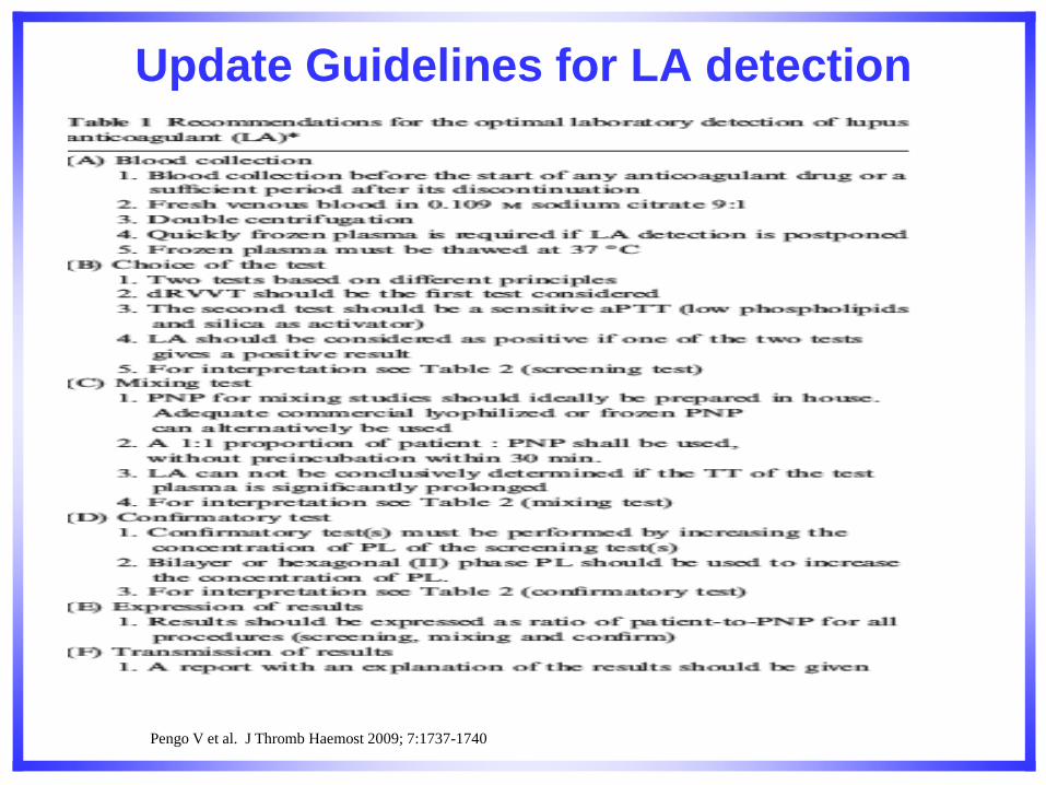
Update Guidelines for LA detection
Pengo V et al. J Thromb Haemost 2009; 7:1737-1740
Pengo V et al. J Thromb Haemost 2009; 7:1737-1740
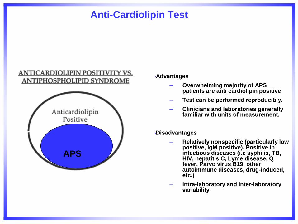
Anti-Cardiolipin Test
•Advantages
– Overwhelming majority of APS patients are anti cardiolipin positive
– Test can be performed reproducibly.
– Clinicians and laboratories generally familiar with units of measurement.
•Disadvantages
– Relatively nonspecific (particularly low positive, IgM positive). Positive in infectious diseases (i.e syphilis, TB, HIV, hepatitis C, Lyme disease, Q fever, Parvo virus B19, other autoimmune diseases, drug-induced, etc.)
– Intra-laboratory and Inter-laboratory variability.
APS
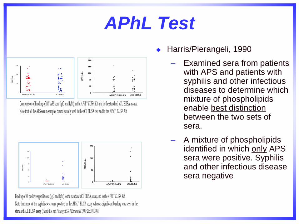
APhL Test
Harris/Pierangeli, 1990
– Examined sera from patients with APS and patients with syphilis and other infectious diseases to determine which mixture of phospholipids enable best distinction between the two sets of sera.
– A mixture of phospholipids identified in which only APS sera were positive. Syphilis and other infectious disease sera negative
-5
26
57
88
119
APhL™ ELISA Kit aCL ELISA
150
GP
L U
nits
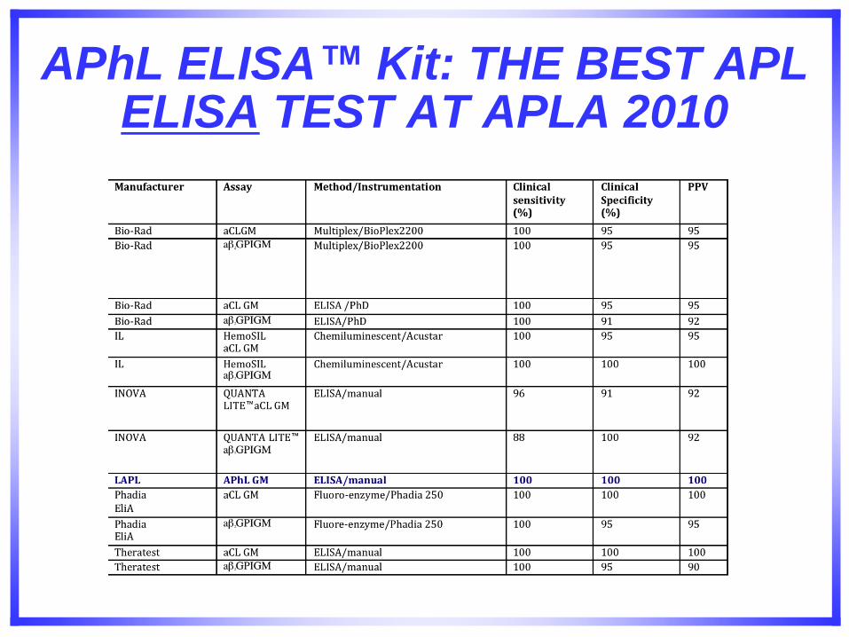
APhL ELISA™ Kit: THE BEST APL ELISA TEST AT APLA 2010
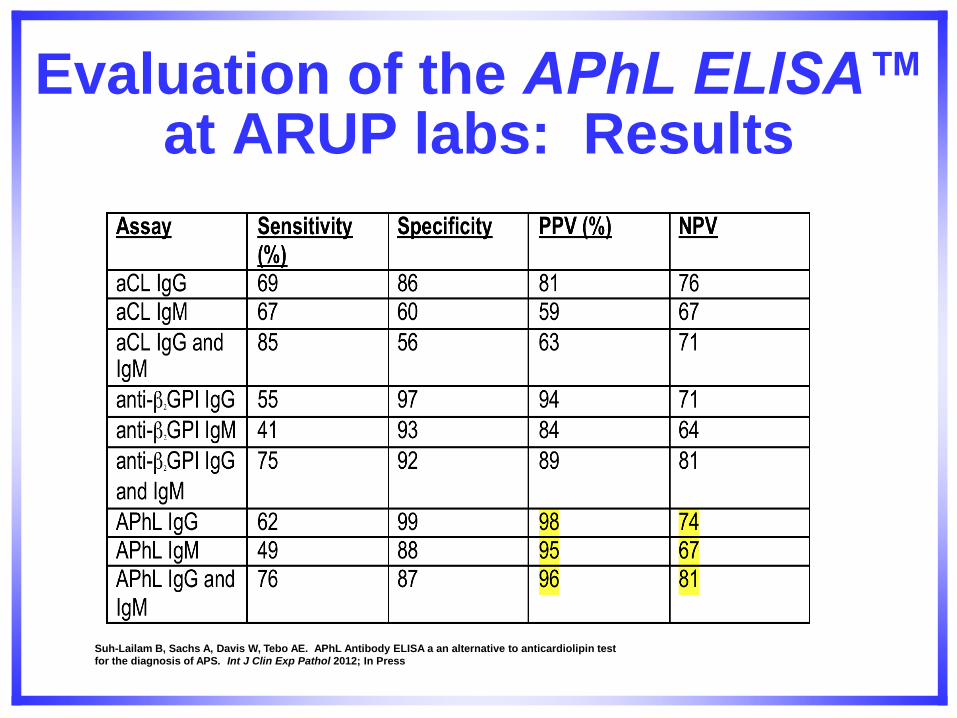
Evaluation of the APhL ELISA™ at ARUP labs: Results
Suh-Lailam B, Sachs A, Davis W, Tebo AE. APhL Antibody ELISA a an alternative to anticardiolipin test for the diagnosis of APS. Int J Clin Exp Pathol 2012; In Press
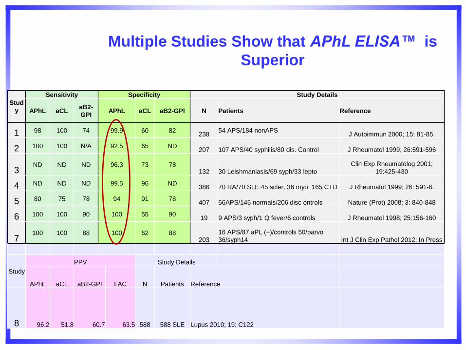
Stud
y
Sensitivity Specificity Study Details
APhL aCL aB2-
GPI APhL aCL aB2-GPI N Patients Reference
1 98 100 74 99.9 60 82 238
54 APS/184 nonAPS J Autoimmun 2000; 15: 81-85.
2 100 100 N/A 92.5 65 ND 207 107 APS/40 syphilis/80 dis. Control J Rheumatol 1999; 26:591-596
3 ND ND ND 96.3 73 78
132 30 Leishmaniasis/69 syph/33 lepto Clin Exp Rheumatolog 2001;
19:425-430
4 ND ND ND 99.5 96 ND 386 70 RA/70 SLE.45 scler, 36 myo, 165 CTD J Rheumatol 1999; 26: 591-6.
5 80 75 78 94 91 78 407 56APS/145 normals/206 disc ontrols Nature (Prot) 2008; 3: 840-848
6 100 100 90 100 55 90 19 9 APS/3 syph/1 Q fever/6 controls J Rheumatol 1998; 25:156-160
7 100 100 88 100 62 88
203 16 APS/87 aPL (+)/controls 50/parvo
36/syph14 Int J Clin Exp Pathol 2012; In Press
Study
PPV Study Details
APhL aCL aB2-GPI LAC N Patients Reference
8 96.2 51.8 60.7 63.5 588 588 SLE Lupus 2010; 19: C122
Multiple Studies Show that APhL ELISA™ is
Superior
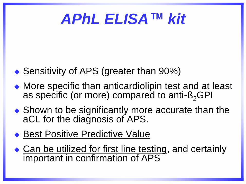
APhL ELISA™ kit
Sensitivity of APS (greater than 90%)
More specific than anticardiolipin test and at least as specific (or more) compared to anti-ß2GPI
Shown to be significantly more accurate than the aCL for the diagnosis of APS.
Best Positive Predictive Value
Can be utilized for first line testing, and certainly important in confirmation of APS
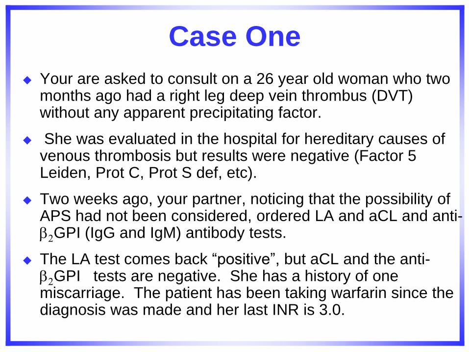
Case One
Your are asked to consult on a 26 year old woman who two months ago had a right leg deep vein thrombus (DVT) without any apparent precipitating factor.
She was evaluated in the hospital for hereditary causes of venous thrombosis but results were negative (Factor 5 Leiden, Prot C, Prot S def, etc).
Two weeks ago, your partner, noticing that the possibility of APS had not been considered, ordered LA and aCL and anti-2GPI (IgG and IgM) antibody tests.
The LA test comes back “positive”, but aCL and the anti-2GPI tests are negative. She has a history of one miscarriage. The patient has been taking warfarin since the diagnosis was made and her last INR is 3.0.
Question
Does this patient have APS?
What is the course of action?
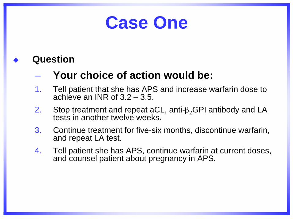
Case One
Question
– Your choice of action would be:
1. Tell patient that she has APS and increase warfarin dose to achieve an INR of 3.2 – 3.5.
2. Stop treatment and repeat aCL, anti-2GPI antibody and LA tests in another twelve weeks.
3. Continue treatment for five-six months, discontinue warfarin, and repeat LA test.
4. Tell patient she has APS, continue warfarin at current doses, and counsel patient about pregnancy in APS.
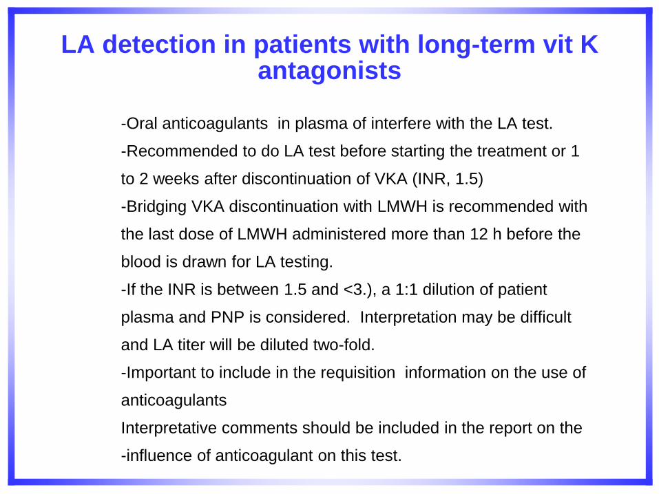
LA detection in patients with long-term vit K antagonists
-Oral anticoagulants in plasma of interfere with the LA test.
-Recommended to do LA test before starting the treatment or 1
to 2 weeks after discontinuation of VKA (INR, 1.5)
-Bridging VKA discontinuation with LMWH is recommended with
the last dose of LMWH administered more than 12 h before the
blood is drawn for LA testing.
-If the INR is between 1.5 and <3.), a 1:1 dilution of patient
plasma and PNP is considered. Interpretation may be difficult
and LA titer will be diluted two-fold.
-Important to include in the requisition information on the use of
anticoagulants
Interpretative comments should be included in the report on the
-influence of anticoagulant on this test.
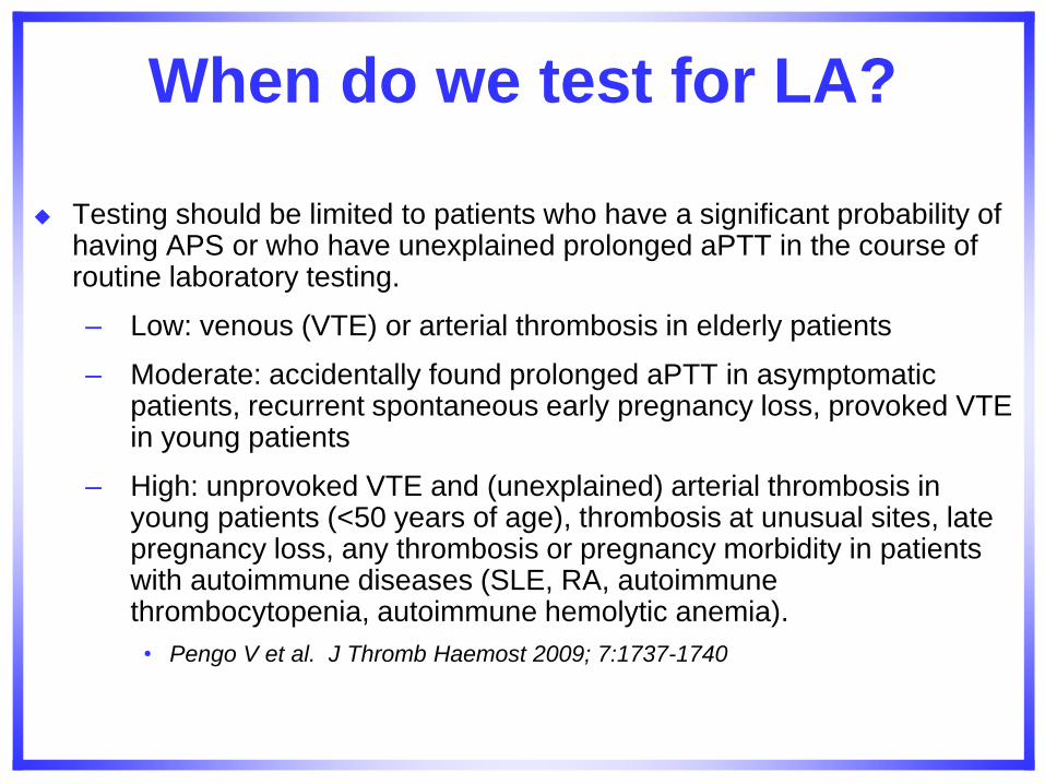
When do we test for LA?
Testing should be limited to patients who have a significant probability of having APS or who have unexplained prolonged aPTT in the course of routine laboratory testing.
– Low: venous (VTE) or arterial thrombosis in elderly patients
– Moderate: accidentally found prolonged aPTT in asymptomatic patients, recurrent spontaneous early pregnancy loss, provoked VTE in young patients
– High: unprovoked VTE and (unexplained) arterial thrombosis in young patients (<50 years of age), thrombosis at unusual sites, late pregnancy loss, any thrombosis or pregnancy morbidity in patients with autoimmune diseases (SLE, RA, autoimmune thrombocytopenia, autoimmune hemolytic anemia).
• Pengo V et al. J Thromb Haemost 2009; 7:1737-1740
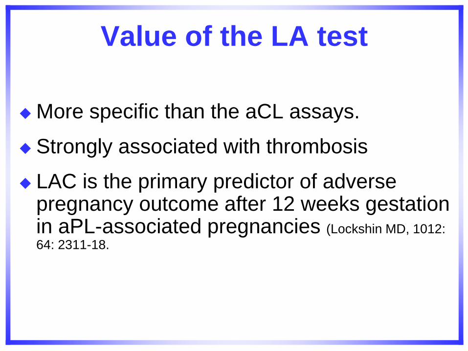
Value of the LA test
More specific than the aCL assays.
Strongly associated with thrombosis
LAC is the primary predictor of adverse pregnancy outcome after 12 weeks gestation in aPL-associated pregnancies (Lockshin MD, 1012:
64: 2311-18.
Case Two
You are managing a 35 year old woman with a diagnosis of APS.
She has a history of right CVA and associated mild left hemiparesis that occurred four years ago.
She has two children, ages 10 and 7. Both children were born at term and she has no history of pregnancy loss.
Her history is significant for mild hypertension that is controlled with a beta-blocker, and she had smoked cigarettes – one packet per day for 10 years – stopping at the time of her stroke.
Case Two (continued)
A review of her laboratory records showed that at diagnosis, IgG anticardiolipin was 22 GPL units and IgM anticardiolipin was 70 MPL units. Annual testing since diagnosis showed her IgG anticardiolipin results to be 15, 12, and 8 GPL units; and IgM anticardiolipin results were 70, 60, 49, and 40 MPL units.
Lupus anticoagulant was always negative. She is on Warfarin with INRs ranging from 2.6 to 3.2, and one low dose aspirin daily.
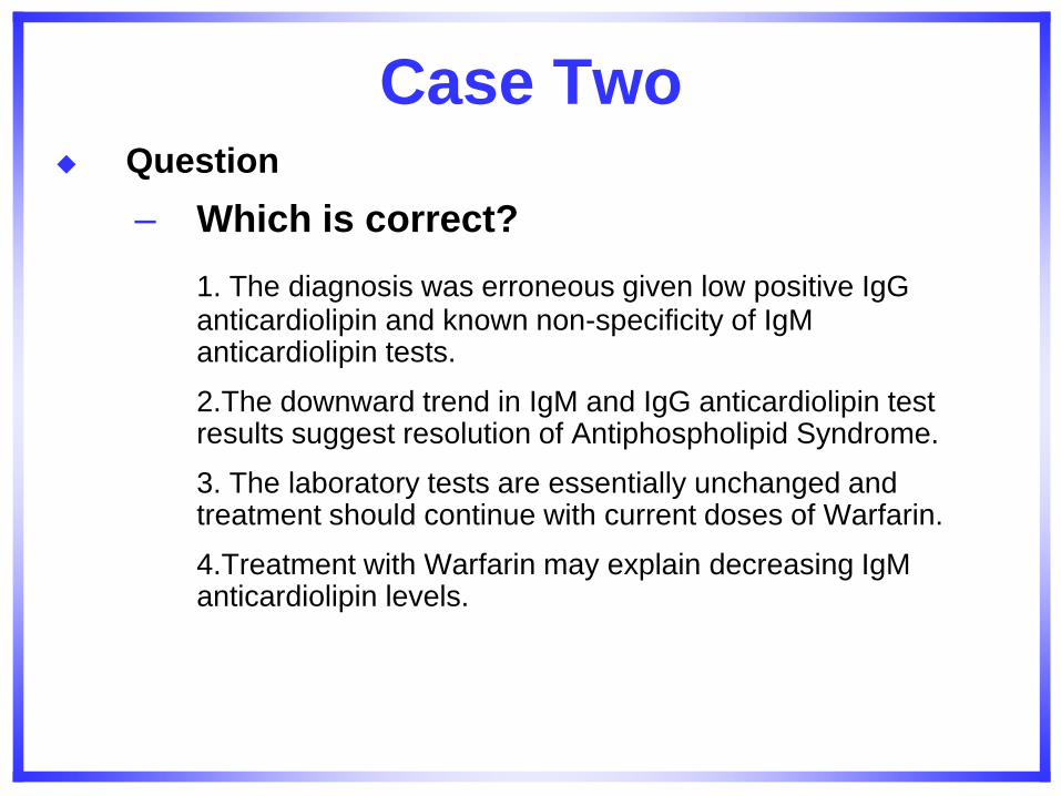
Case Two Question
– Which is correct?
1. The diagnosis was erroneous given low positive IgG anticardiolipin and known non-specificity of IgM anticardiolipin tests.
2.The downward trend in IgM and IgG anticardiolipin test results suggest resolution of Antiphospholipid Syndrome.
3. The laboratory tests are essentially unchanged and treatment should continue with current doses of Warfarin.
4.Treatment with Warfarin may explain decreasing IgM anticardiolipin levels.
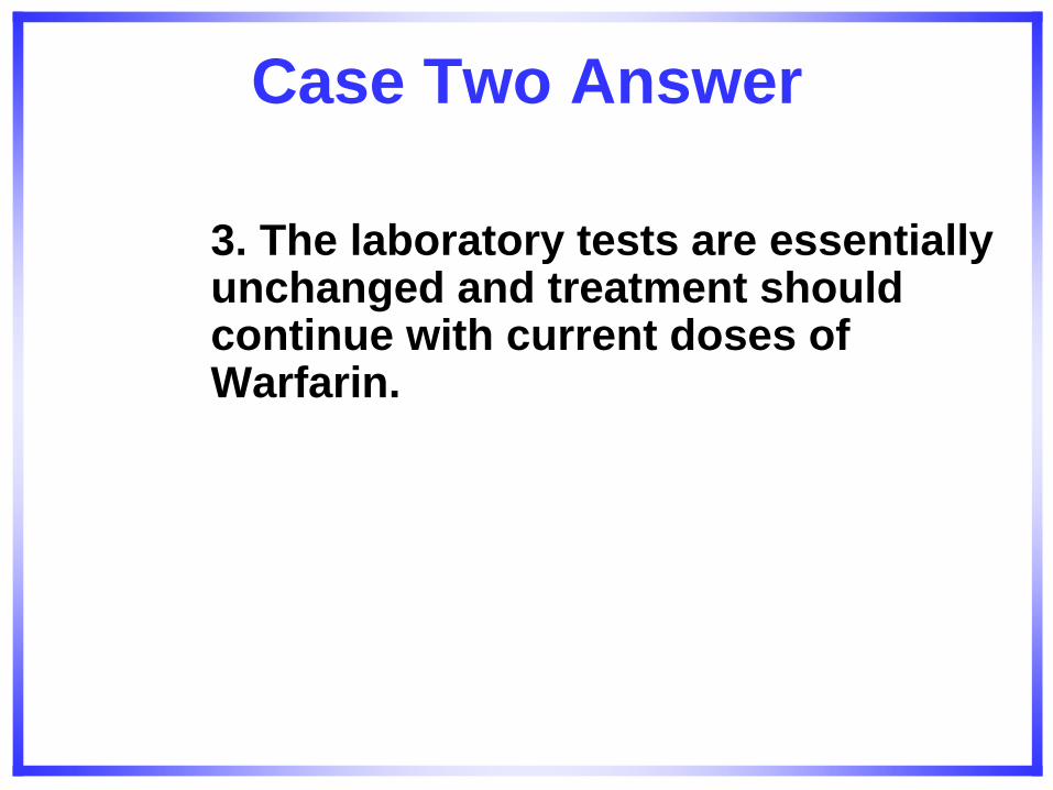
Case Two Answer
3. The laboratory tests are essentially unchanged and treatment should continue with current doses of Warfarin.
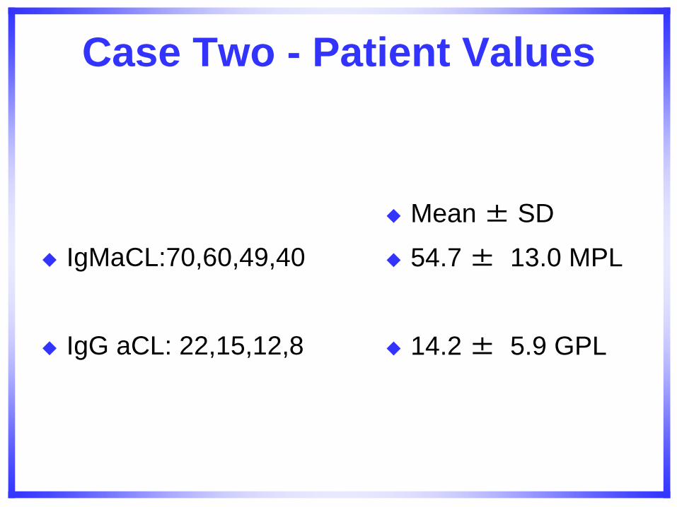
Case Two - Patient Values
IgMaCL:70,60,49,40
IgG aCL: 22,15,12,8
Mean ± SD
54.7 ± 13.0 MPL
14.2 ± 5.9 GPL
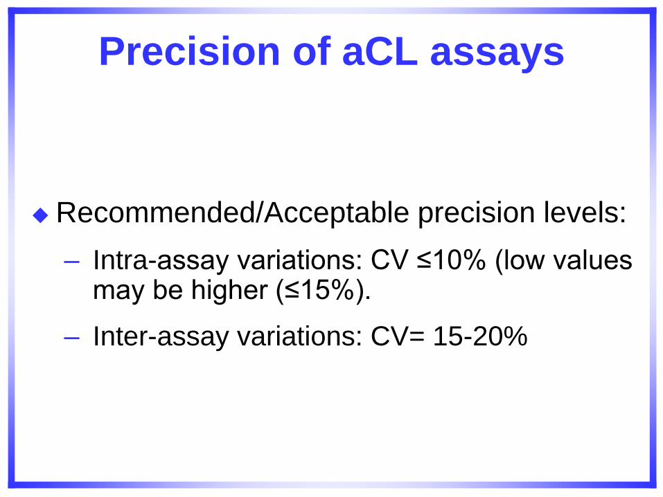
Precision of aCL assays
Recommended/Acceptable precision levels:
– Intra-assay variations: CV ≤10% (low values may be higher (≤15%).
– Inter-assay variations: CV= 15-20%
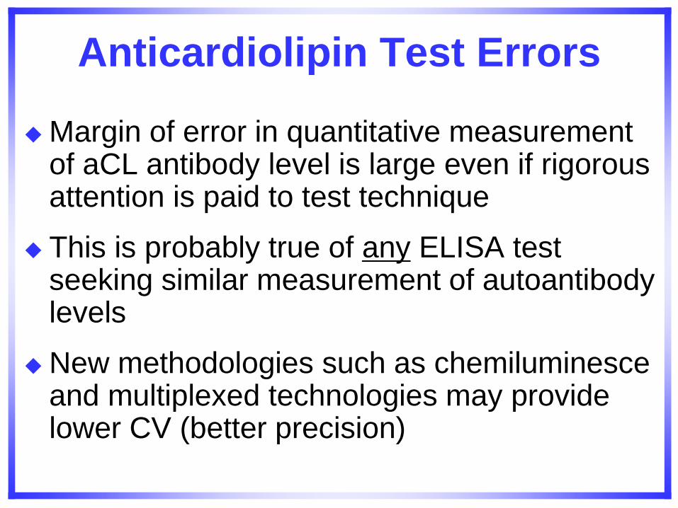
Anticardiolipin Test Errors
Margin of error in quantitative measurement of aCL antibody level is large even if rigorous attention is paid to test technique
This is probably true of any ELISA test seeking similar measurement of autoantibody levels
New methodologies such as chemiluminesce and multiplexed technologies may provide lower CV (better precision)
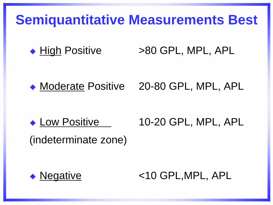
Semiquantitative Measurements Best
High Positive >80 GPL, MPL, APL
Moderate Positive 20-80 GPL, MPL, APL
Low Positive 10-20 GPL, MPL, APL
(indeterminate zone)
Negative <10 GPL,MPL, APL
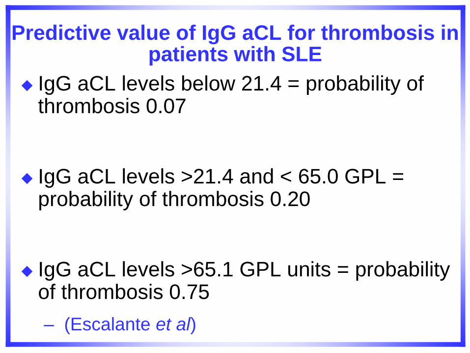
Predictive value of IgG aCL for thrombosis in patients with SLE
IgG aCL levels below 21.4 = probability of thrombosis 0.07
IgG aCL levels >21.4 and < 65.0 GPL = probability of thrombosis 0.20
IgG aCL levels >65.1 GPL units = probability of thrombosis 0.75
– (Escalante et al)
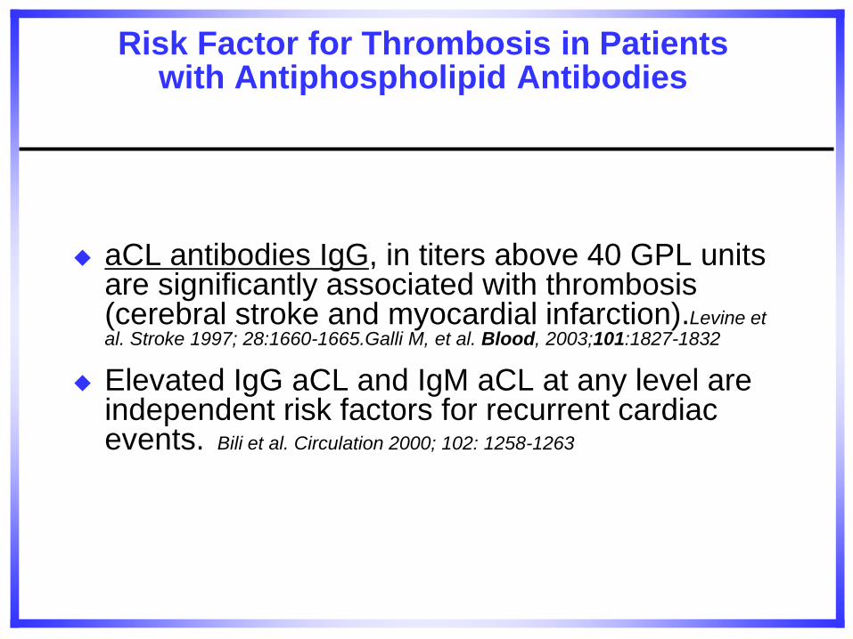
Risk Factor for Thrombosis in Patients with Antiphospholipid Antibodies
aCL antibodies IgG, in titers above 40 GPL units are significantly associated with thrombosis (cerebral stroke and myocardial infarction).Levine et al. Stroke 1997; 28:1660-1665.Galli M, et al. Blood, 2003;101:1827-1832
Elevated IgG aCL and IgM aCL at any level are independent risk factors for recurrent cardiac events. Bili et al. Circulation 2000; 102: 1258-1263
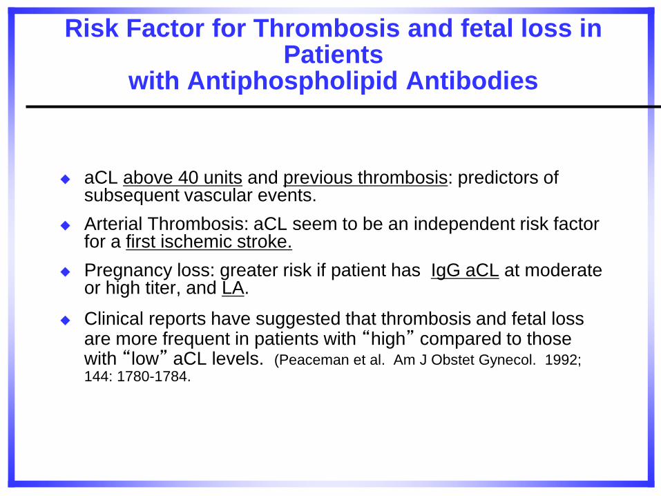
Risk Factor for Thrombosis and fetal loss in Patients
with Antiphospholipid Antibodies
aCL above 40 units and previous thrombosis: predictors of subsequent vascular events.
Arterial Thrombosis: aCL seem to be an independent risk factor for a first ischemic stroke.
Pregnancy loss: greater risk if patient has IgG aCL at moderate or high titer, and LA.
Clinical reports have suggested that thrombosis and fetal loss are more frequent in patients with “high” compared to those with “low” aCL levels. (Peaceman et al. Am J Obstet Gynecol. 1992; 144: 1780-1784.
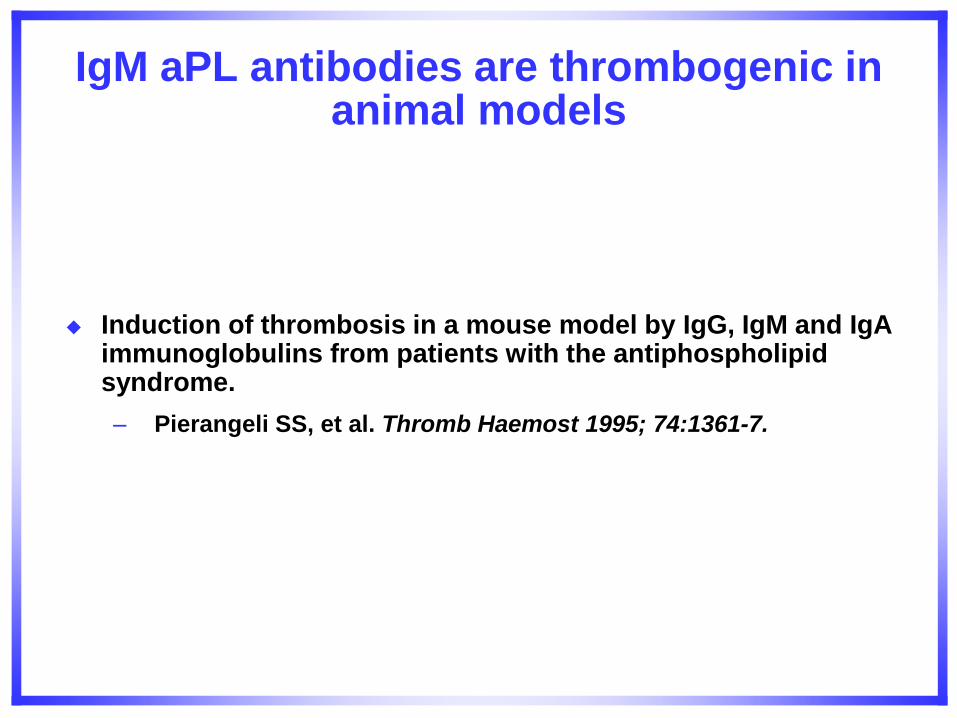
IgM aPL antibodies are thrombogenic in animal models
Induction of thrombosis in a mouse model by IgG, IgM and IgA immunoglobulins from patients with the antiphospholipid syndrome.
– Pierangeli SS, et al. Thromb Haemost 1995; 74:1361-7.
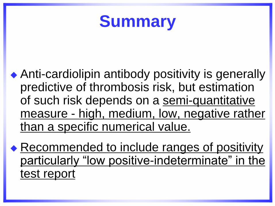
Summary
Anti-cardiolipin antibody positivity is generally predictive of thrombosis risk, but estimation of such risk depends on a semi-quantitative measure - high, medium, low, negative rather than a specific numerical value.
Recommended to include ranges of positivity particularly “low positive-indeterminate” in the test report
Case Three
You have just joined a discussion between two colleagues who are pondering the diagnosis of a 30 year old woman who is otherwise well, but has a history of three miscarriages at 6-weeks, 5-weeks, and -weeks gestation, respectively.
She has been extensively evaluated by an obstetrician in another state. The only positive tests were a low positive IgM anti-phosphatidyl inositol test and when repeated a low positive IgM anti-phosphatidyl ethanolamine test. ACL and LA tests have been repeatedly negative.
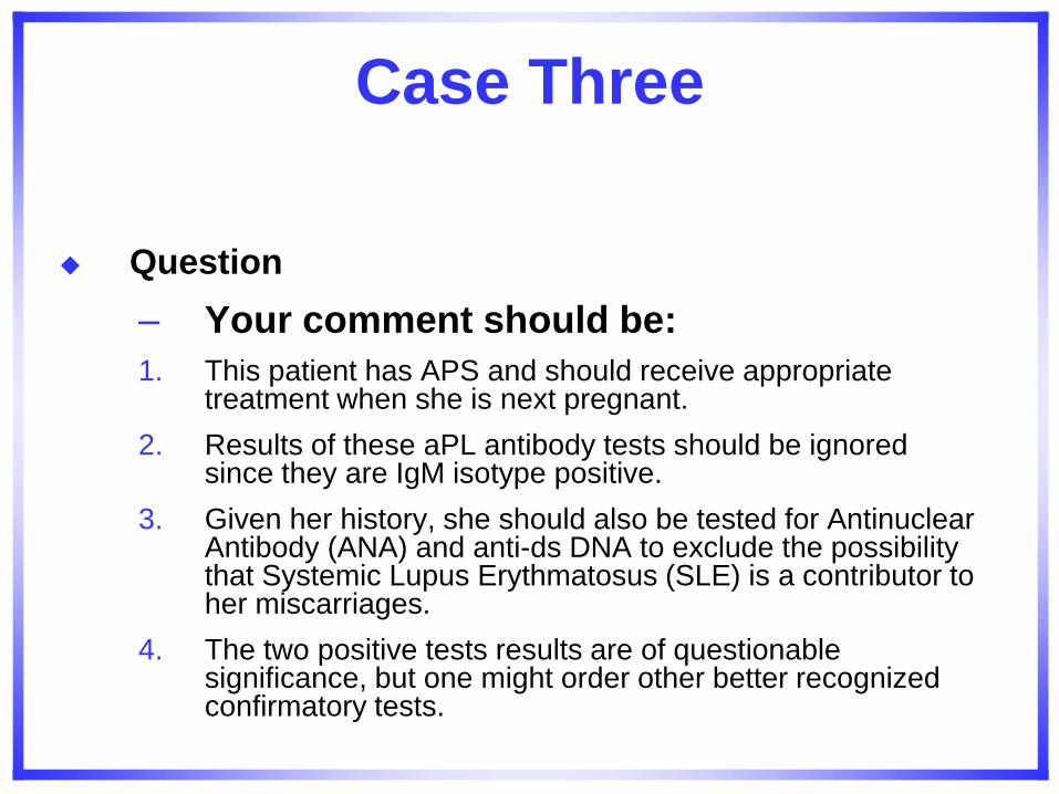
Case Three
Question
– Your comment should be:
1. This patient has APS and should receive appropriate treatment when she is next pregnant.
2. Results of these aPL antibody tests should be ignored since they are IgM isotype positive.
3. Given her history, she should also be tested for Antinuclear Antibody (ANA) and anti-ds DNA to exclude the possibility that Systemic Lupus Erythmatosus (SLE) is a contributor to her miscarriages.
4. The two positive tests results are of questionable significance, but one might order other better recognized confirmatory tests.
Case Three Answer 1
4. The two positive tests results are of questionable significance, but one might order other better recognized confirmatory tests.
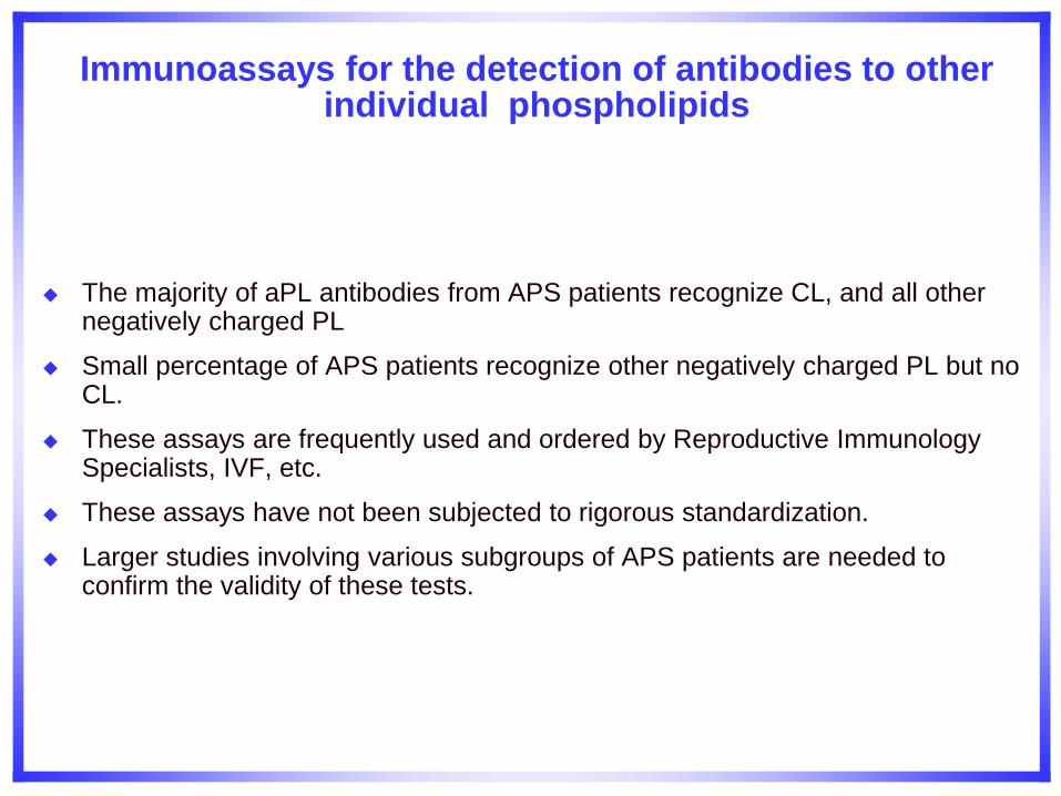
Immunoassays for the detection of antibodies to other individual phospholipids
The majority of aPL antibodies from APS patients recognize CL, and all other negatively charged PL
Small percentage of APS patients recognize other negatively charged PL but no CL.
These assays are frequently used and ordered by Reproductive Immunology Specialists, IVF, etc.
These assays have not been subjected to rigorous standardization.
Larger studies involving various subgroups of APS patients are needed to confirm the validity of these tests.
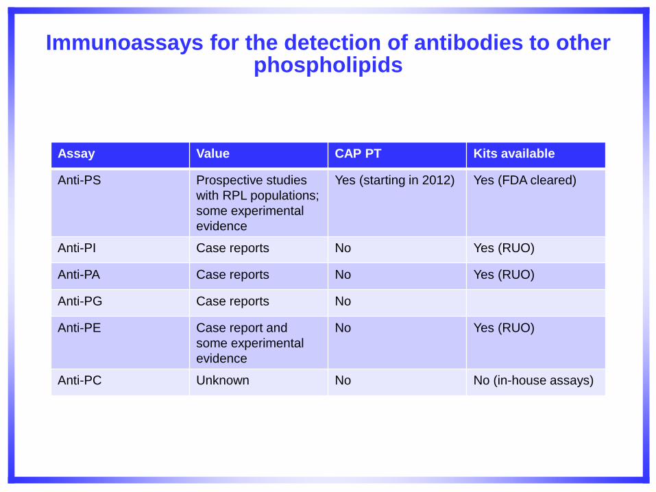
Immunoassays for the detection of antibodies to other phospholipids
Assay Value CAP PT Kits available
Anti-PS Prospective studies
with RPL populations;
some experimental
evidence
Yes (starting in 2012) Yes (FDA cleared)
Anti-PI Case reports No Yes (RUO)
Anti-PA Case reports No Yes (RUO)
Anti-PG Case reports No
Anti-PE Case report and
some experimental
evidence
No Yes (RUO)
Anti-PC Unknown No No (in-house assays)
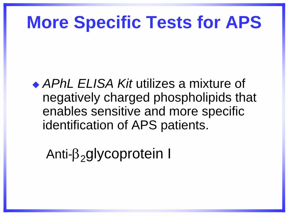
More Specific Tests for APS
APhL ELISA Kit utilizes a mixture of negatively charged phospholipids that enables sensitive and more specific identification of APS patients.
Anti-2glycoprotein I
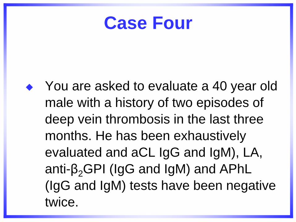
Case Four
You are asked to evaluate a 40 year old
male with a history of two episodes of
deep vein thrombosis in the last three
months. He has been exhaustively
evaluated and aCL IgG and IgM), LA,
anti-β2GPI (IgG and IgM) and APhL
(IgG and IgM) tests have been negative
twice.
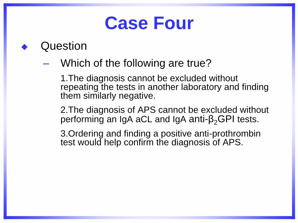
Case Four Question
– Which of the following are true?
1.The diagnosis cannot be excluded without repeating the tests in another laboratory and finding them similarly negative.
2.The diagnosis of APS cannot be excluded without performing an IgA aCL and IgA anti-β2GPI tests.
3.Ordering and finding a positive anti-prothrombin test would help confirm the diagnosis of APS.
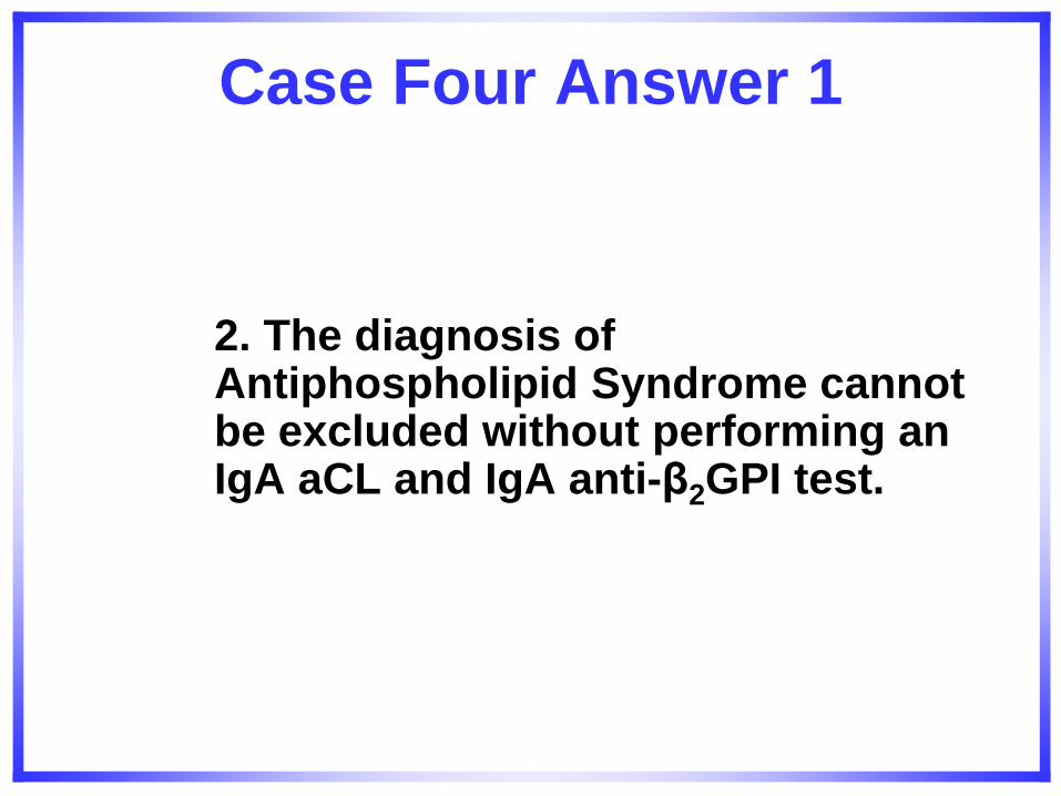
Case Four Answer 1
2. The diagnosis of Antiphospholipid Syndrome cannot be excluded without performing an IgA aCL and IgA anti-β2GPI test.
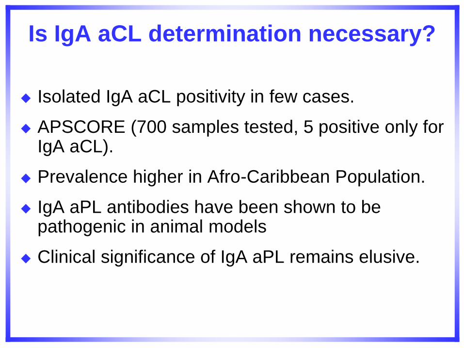
Is IgA aCL determination necessary?
Isolated IgA aCL positivity in few cases.
APSCORE (700 samples tested, 5 positive only for IgA aCL).
Prevalence higher in Afro-Caribbean Population.
IgA aPL antibodies have been shown to be pathogenic in animal models
Clinical significance of IgA aPL remains elusive.
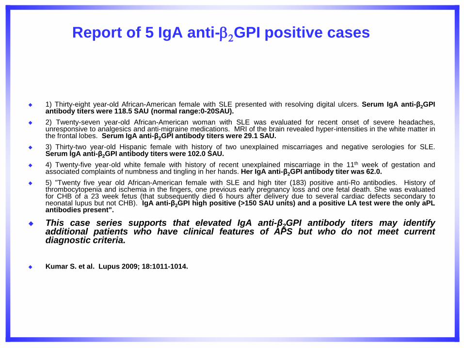
Report of 5 IgA anti-2GPI positive cases
1) Thirty-eight year-old African-American female with SLE presented with resolving digital ulcers. Serum IgA anti-β2GPI antibody titers were 118.5 SAU (normal range:0-20SAU).
2) Twenty-seven year-old African-American woman with SLE was evaluated for recent onset of severe headaches, unresponsive to analgesics and anti-migraine medications. MRI of the brain revealed hyper-intensities in the white matter in the frontal lobes. Serum IgA anti-β2GPI antibody titers were 29.1 SAU.
3) Thirty-two year-old Hispanic female with history of two unexplained miscarriages and negative serologies for SLE. Serum IgA anti-β2GPI antibody titers were 102.0 SAU.
4) Twenty-five year-old white female with history of recent unexplained miscarriage in the 11th week of gestation and associated complaints of numbness and tingling in her hands. Her IgA anti-β2GPI antibody titer was 62.0.
5) "Twenty five year old African-American female with SLE and high titer (183) positive anti-Ro antibodies. History of thrombocytopenia and ischemia in the fingers, one previous early pregnancy loss and one fetal death. She was evaluated for CHB of a 23 week fetus (that subsequently died 6 hours after delivery due to several cardiac defects secondary to neonatal lupus but not CHB). IgA anti-β2GPI high positive (>150 SAU units) and a positive LA test were the only aPL antibodies present".
This case series supports that elevated IgA anti-β2GPI antibody titers may identify additional patients who have clinical features of APS but who do not meet current diagnostic criteria.
Kumar S. et al. Lupus 2009; 18:1011-1014.
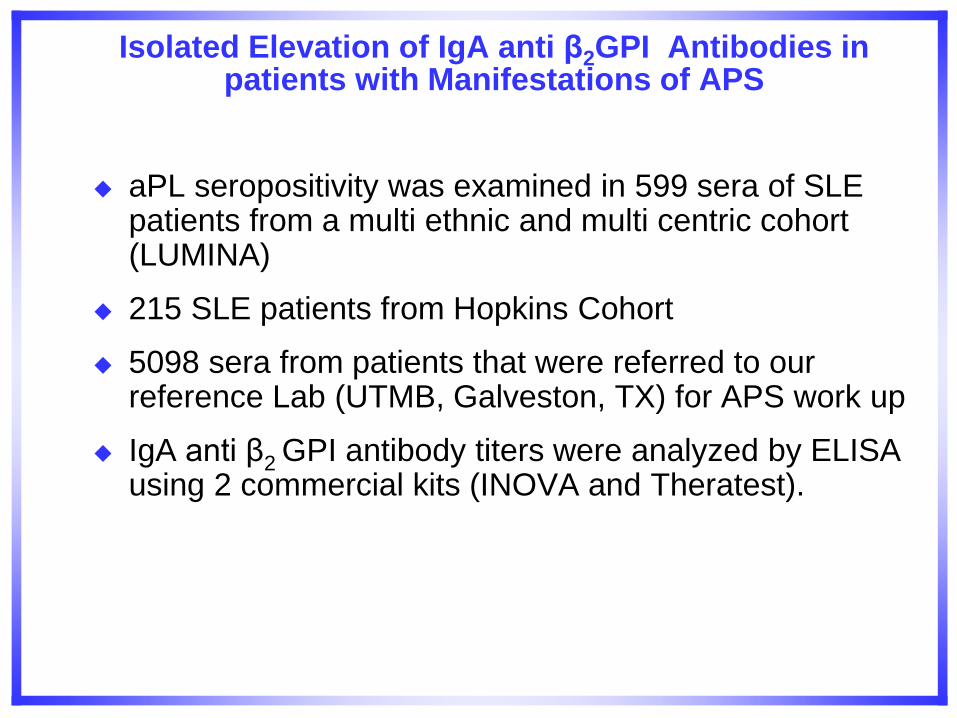
Isolated Elevation of IgA anti β2GPI Antibodies in patients with Manifestations of APS
aPL seropositivity was examined in 599 sera of SLE patients from a multi ethnic and multi centric cohort (LUMINA)
215 SLE patients from Hopkins Cohort
5098 sera from patients that were referred to our reference Lab (UTMB, Galveston, TX) for APS work up
IgA anti β2 GPI antibody titers were analyzed by ELISA using 2 commercial kits (INOVA and Theratest).
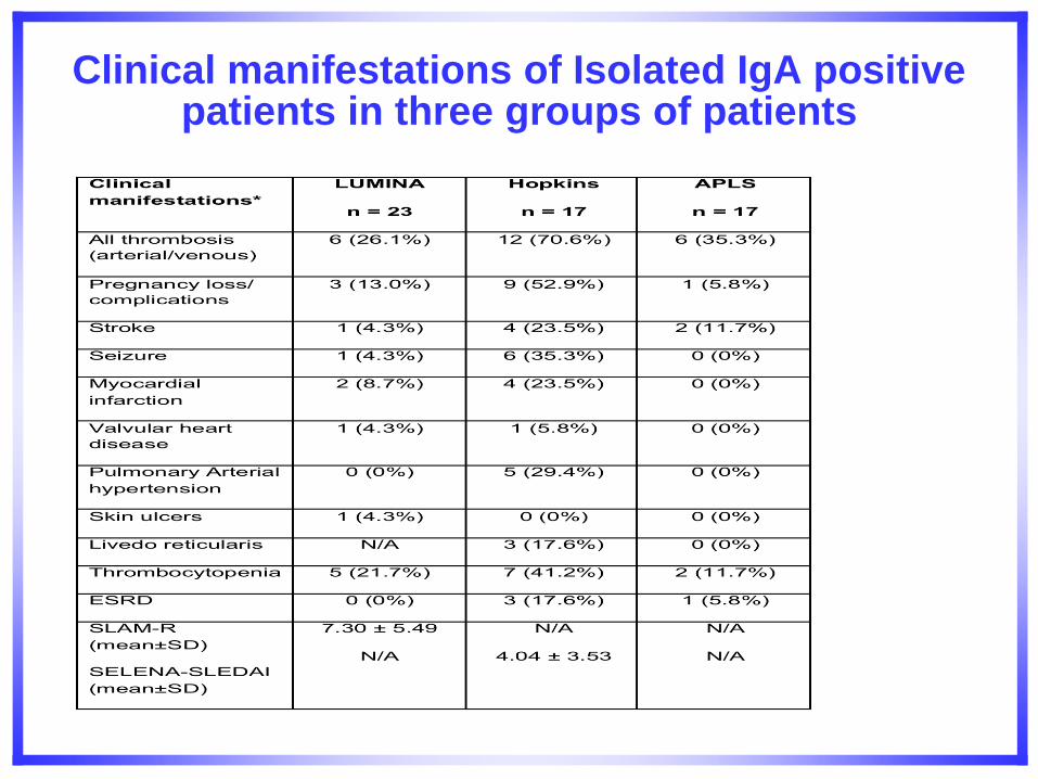
Clinical manifestations of Isolated IgA positive patients in three groups of patients
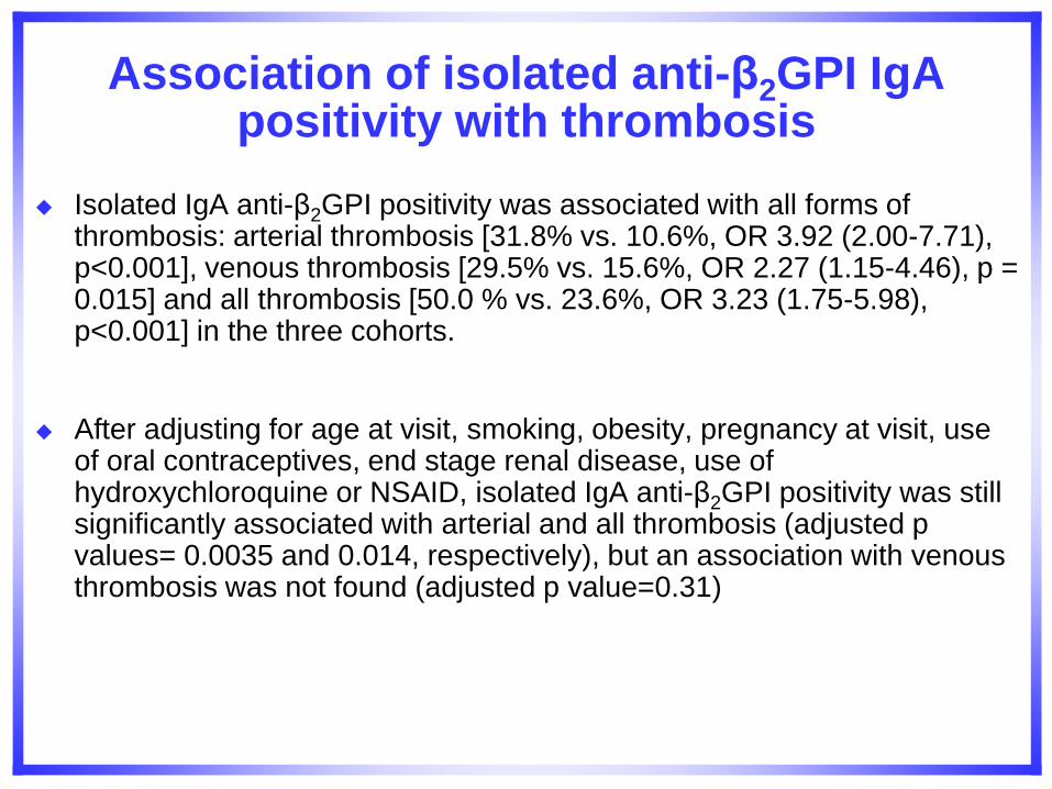
Association of isolated anti-β2GPI IgA positivity with thrombosis
Isolated IgA anti-β2GPI positivity was associated with all forms of thrombosis: arterial thrombosis [31.8% vs. 10.6%, OR 3.92 (2.00-7.71), p<0.001], venous thrombosis [29.5% vs. 15.6%, OR 2.27 (1.15-4.46), p = 0.015] and all thrombosis [50.0 % vs. 23.6%, OR 3.23 (1.75-5.98), p<0.001] in the three cohorts.
After adjusting for age at visit, smoking, obesity, pregnancy at visit, use of oral contraceptives, end stage renal disease, use of hydroxychloroquine or NSAID, isolated IgA anti-β2GPI positivity was still significantly associated with arterial and all thrombosis (adjusted p values= 0.0035 and 0.014, respectively), but an association with venous thrombosis was not found (adjusted p value=0.31)
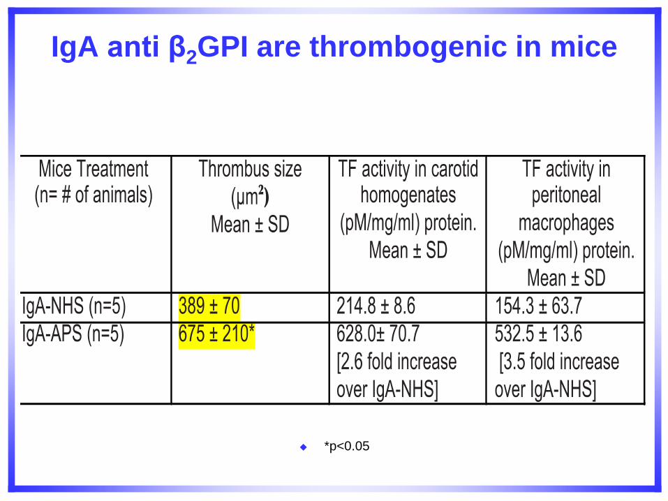
IgA anti β2GPI are thrombogenic in mice
*p<0.05
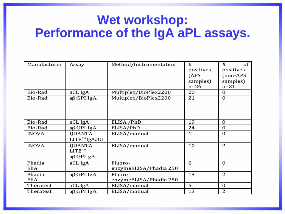
Wet workshop: Performance of the IgA aPL assays.
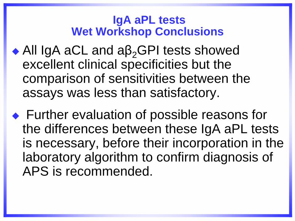
IgA aPL tests Wet Workshop Conclusions
All IgA aCL and aβ2GPI tests showed excellent clinical specificities but the comparison of sensitivities between the assays was less than satisfactory.
Further evaluation of possible reasons for the differences between these IgA aPL tests is necessary, before their incorporation in the laboratory algorithm to confirm diagnosis of APS is recommended.
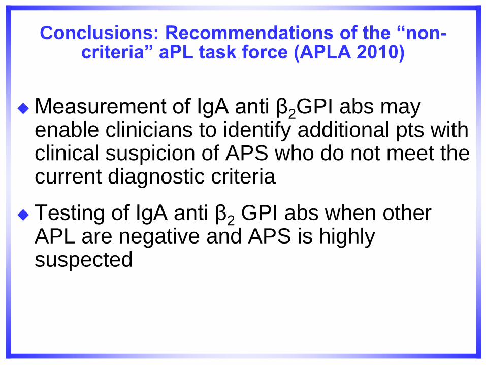
Conclusions: Recommendations of the “non-criteria” aPL task force (APLA 2010)
Measurement of IgA anti β2GPI abs may enable clinicians to identify additional pts with clinical suspicion of APS who do not meet the current diagnostic criteria
Testing of IgA anti β2 GPI abs when other APL are negative and APS is highly suspected

New Assays for Diagnosis of APS
Anti-prothrombin antibodies (aPT)
Anti-Phosphatidylserine/PT antibodies (aPS/PT
Annexin A5 resistance (A5R) assay
Domain I ELISA.
Anti-annexin A5
All these assays need to be further validated and standardized.
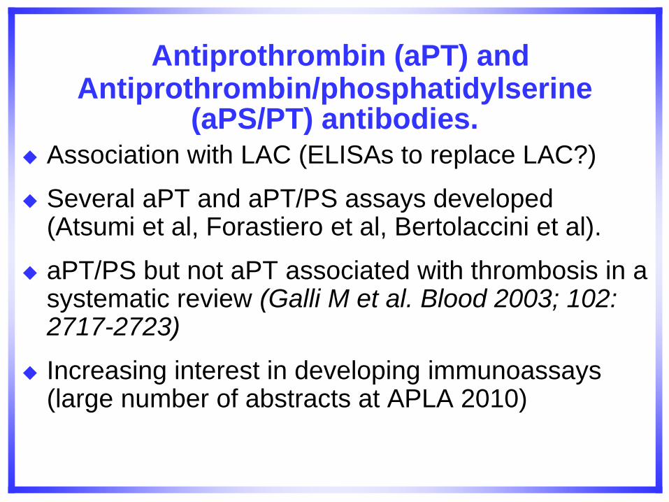
Antiprothrombin (aPT) and Antiprothrombin/phosphatidylserine
(aPS/PT) antibodies. Association with LAC (ELISAs to replace LAC?)
Several aPT and aPT/PS assays developed (Atsumi et al, Forastiero et al, Bertolaccini et al).
aPT/PS but not aPT associated with thrombosis in a systematic review (Galli M et al. Blood 2003; 102: 2717-2723)
Increasing interest in developing immunoassays (large number of abstracts at APLA 2010)
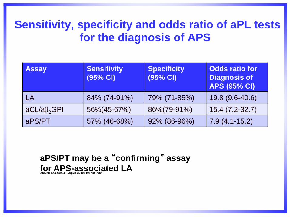
Sensitivity, specificity and odds ratio of aPL tests for the diagnosis of APS
Assay Sensitivity
(95% CI)
Specificity
(95% CI)
Odds ratio for
Diagnosis of
APS (95% CI)
LA 84% (74-91%) 79% (71-85%) 19.8 (9.6-40.6)
aCL/a2GPI 56%(45-67%) 86%(79-91%) 15.4 (7.2-32.7)
aPS/PT 57% (46-68%) 92% (86-96%) 7.9 (4.1-15.2)
aPS/PT may be a “confirming” assay
for APS-associated LA Atsumi and Koike. Lupus 2010: 19: 436-439.
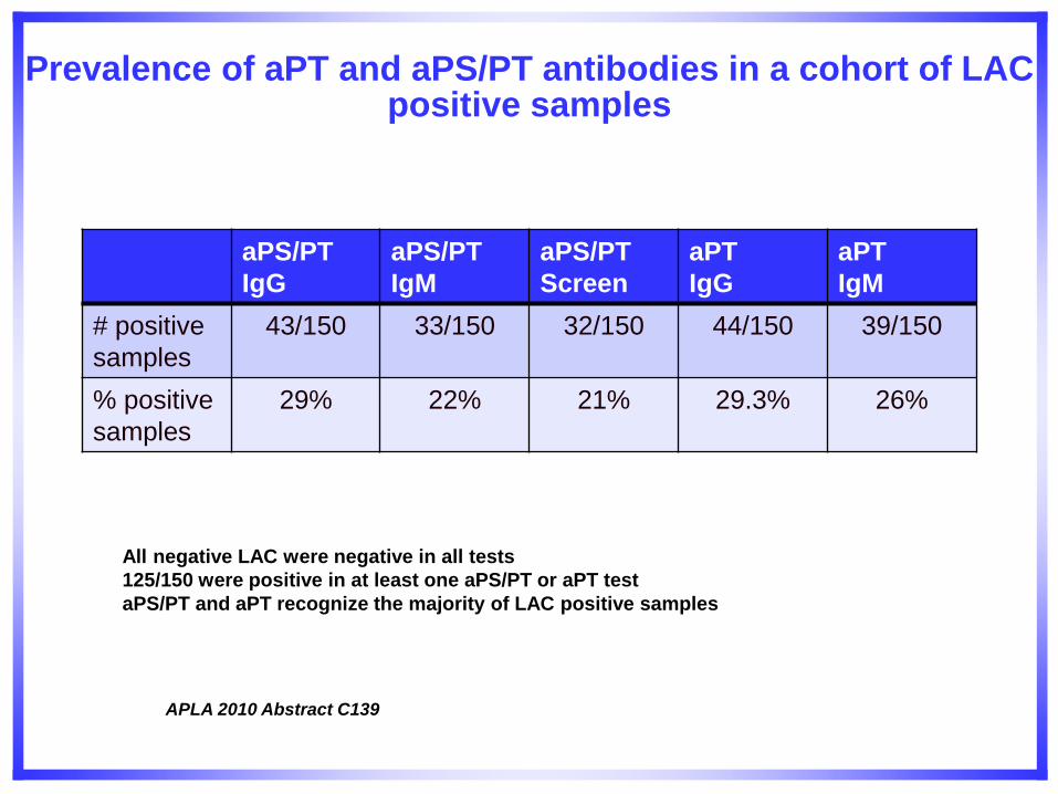
Prevalence of aPT and aPS/PT antibodies in a cohort of LAC positive samples
aPS/PT
IgG
aPS/PT
IgM
aPS/PT
Screen
aPT
IgG
aPT
IgM
# positive
samples
43/150 33/150 32/150 44/150 39/150
% positive
samples
29% 22% 21% 29.3% 26%
All negative LAC were negative in all tests
125/150 were positive in at least one aPS/PT or aPT test
aPS/PT and aPT recognize the majority of LAC positive samples
APLA 2010 Abstract C139
Conclusions aPL task force
– aPS-PT should be intensively standardized
– aPS-PT should be further investigated as a possible criterion for
APS
– Test seems to be specific for APS, not very sensitive
– Value of aPS-PT as a confirmatory test for LAC deserves further
investigation
– aPT-might be tested a a risk marker for APS manifestations
(Bertolaccini, Atsumi, Forastiero, Binder)
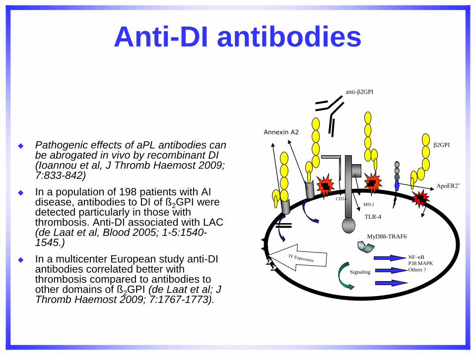
Anti-DI antibodies
Pathogenic effects of aPL antibodies can
be abrogated in vivo by recombinant DI (Ioannou et al, J Thromb Haemost 2009; 7:833-842)
In a population of 198 patients with AI disease, antibodies to DI of ß2GPI were detected particularly in those with thrombosis. Anti-DI associated with LAC (de Laat et al, Blood 2005; 1-5:1540-1545.)
In a multicenter European study anti-DI antibodies correlated better with thrombosis compared to antibodies to other domains of ß2GPI (de Laat et al; J Thromb Haemost 2009; 7:1767-1773).
Signaling
NF-B
P38 MAPK
Others ?
MD-2
TLR-4
β2GPI
ApoER2’
anti-β2GPI
Annexin A2
CD14
MyD88-TRAF6
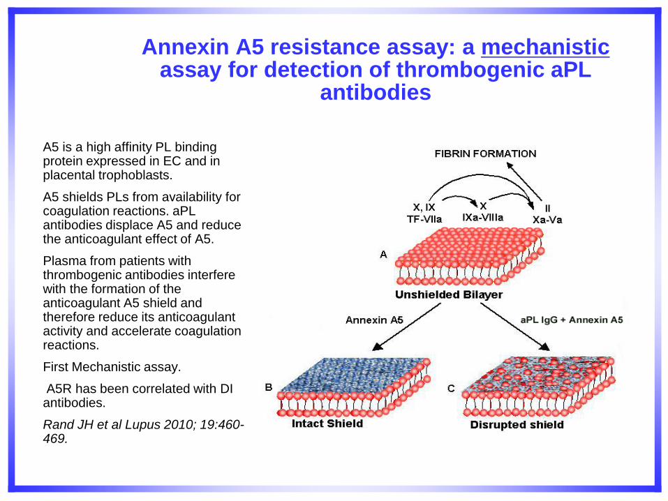
Annexin A5 resistance assay: a mechanistic assay for detection of thrombogenic aPL
antibodies
A5 is a high affinity PL binding protein expressed in EC and in placental trophoblasts.
A5 shields PLs from availability for coagulation reactions. aPL antibodies displace A5 and reduce the anticoagulant effect of A5.
Plasma from patients with thrombogenic antibodies interfere with the formation of the anticoagulant A5 shield and therefore reduce its anticoagulant activity and accelerate coagulation reactions.
First Mechanistic assay.
A5R has been correlated with DI antibodies.
Rand JH et al Lupus 2010; 19:460-469.
Case V
The supervisor of the immunology section of a university hospital received a call from an attending physician who requested advice about a 34-year old white woman he had seen with a history of 2 deep vein thrombosis (DVT).
The patient had been taking oral contraceptives and is overweight and did not have a history of pregnancy loss. Since her first DVT episode 4 years ago, the patient had received therapy with oral anticoagulants.
Despite this therapy, the patient had recently been diagnosed with having a transient ischemic attack (TIA). A CBC count performed on the patient revealed nothing abnormal, and the patient’s platelet count was within normal parameters. A LA test was not performed. Other findings are listed in the table (next). The physician suspected that the patient had APS but wondered what laboratory tests could be performed to make a more definitive diagnosis of APS.
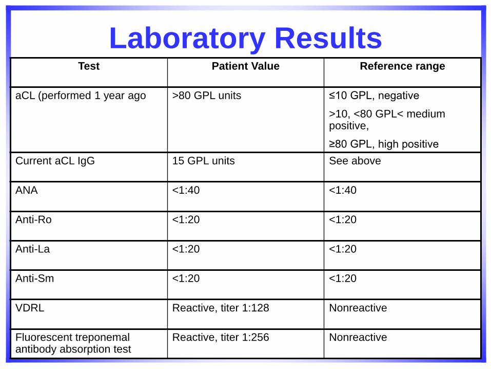
Laboratory Results Test Patient Value Reference range
aCL (performed 1 year ago >80 GPL units ≤10 GPL, negative
>10, <80 GPL< medium positive,
≥80 GPL, high positive
Current aCL IgG 15 GPL units See above
ANA <1:40 <1:40
Anti-Ro <1:20 <1:20
Anti-La <1:20 <1:20
Anti-Sm <1:20 <1:20
VDRL Reactive, titer 1:128 Nonreactive
Fluorescent treponemal antibody absorption test
Reactive, titer 1:256 Nonreactive
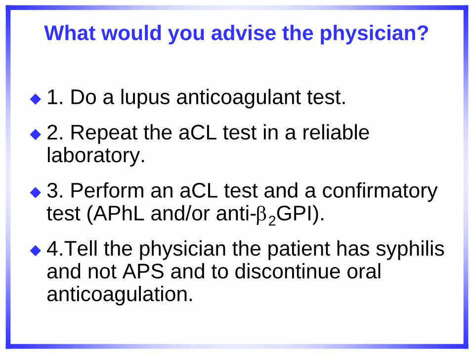
What would you advise the physician?
1. Do a lupus anticoagulant test.
2. Repeat the aCL test in a reliable laboratory.
3. Perform an aCL test and a confirmatory test (APhL and/or anti-2GPI).
4.Tell the physician the patient has syphilis and not APS and to discontinue oral anticoagulation.
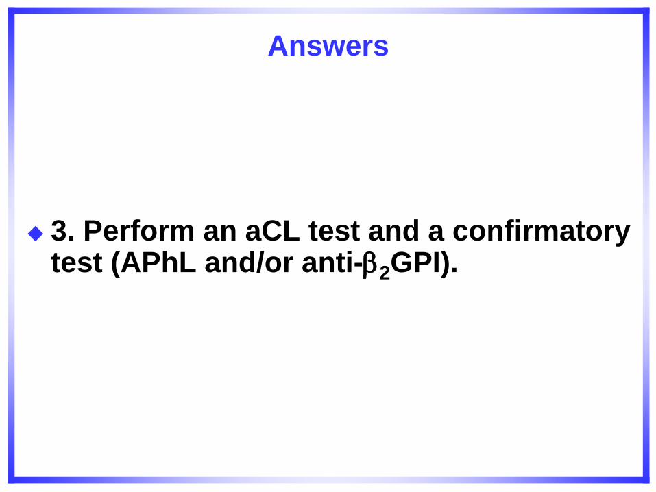
Answers
3. Perform an aCL test and a confirmatory test (APhL and/or anti-2GPI).
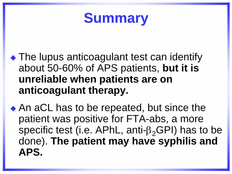
Summary
The lupus anticoagulant test can identify about 50-60% of APS patients, but it is unreliable when patients are on anticoagulant therapy.
An aCL has to be repeated, but since the patient was positive for FTA-abs, a more specific test (i.e. APhL, anti-2GPI) has to be done). The patient may have syphilis and APS.
Conclusions
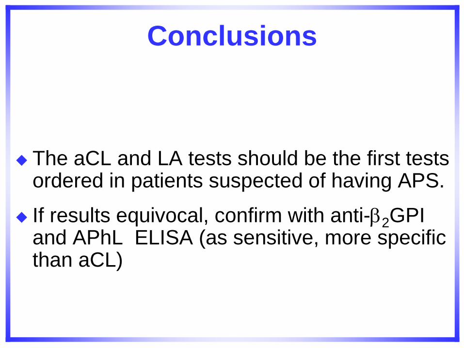
The aCL and LA tests should be the first tests ordered in patients suspected of having APS.
If results equivocal, confirm with anti-2GPI and APhL ELISA (as sensitive, more specific than aCL)
Case VI
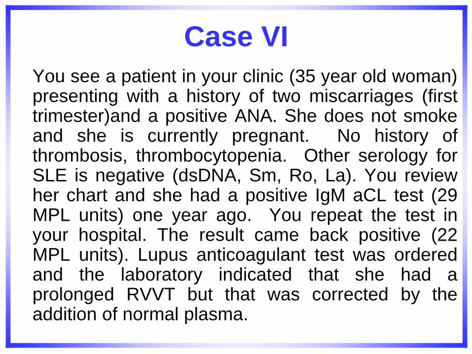
You see a patient in your clinic (35 year old woman) presenting with a history of two miscarriages (first trimester)and a positive ANA. She does not smoke and she is currently pregnant. No history of thrombosis, thrombocytopenia. Other serology for SLE is negative (dsDNA, Sm, Ro, La). You review her chart and she had a positive IgM aCL test (29 MPL units) one year ago. You repeat the test in your hospital. The result came back positive (22 MPL units). Lupus anticoagulant test was ordered and the laboratory indicated that she had a prolonged RVVT but that was corrected by the addition of normal plasma.
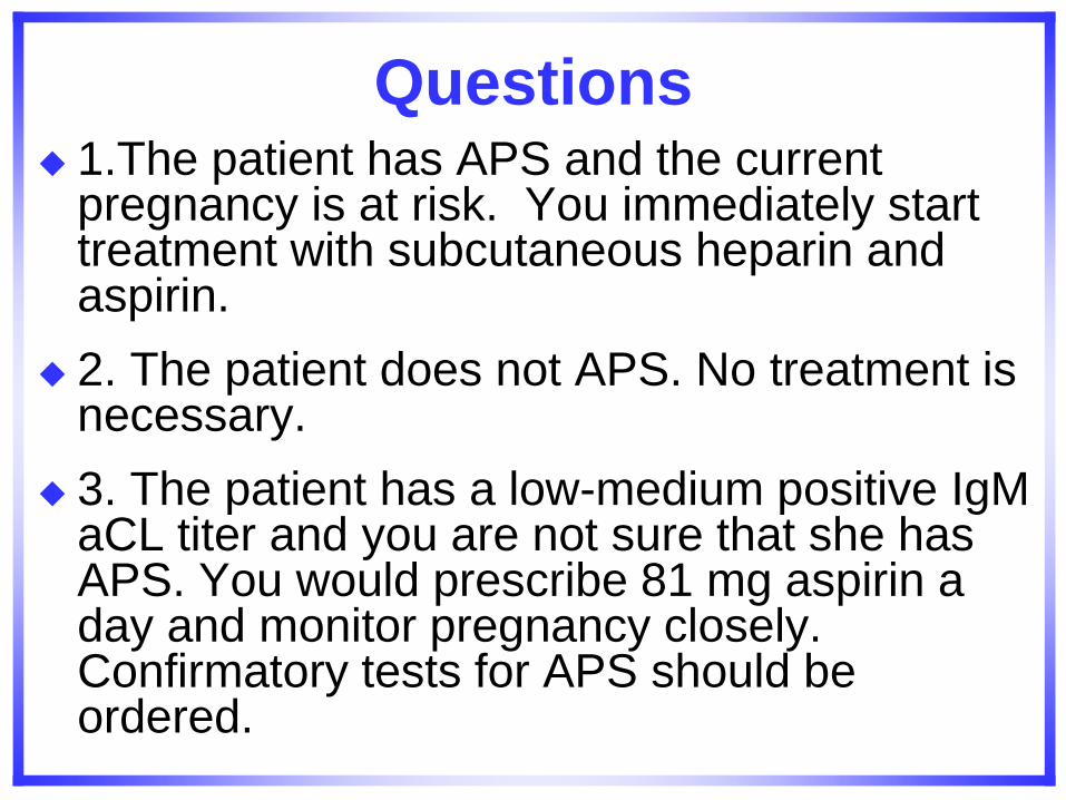
Questions 1.The patient has APS and the current
pregnancy is at risk. You immediately start treatment with subcutaneous heparin and aspirin.
2. The patient does not APS. No treatment is necessary.
3. The patient has a low-medium positive IgM aCL titer and you are not sure that she has APS. You would prescribe 81 mg aspirin a day and monitor pregnancy closely. Confirmatory tests for APS should be ordered.
Answer
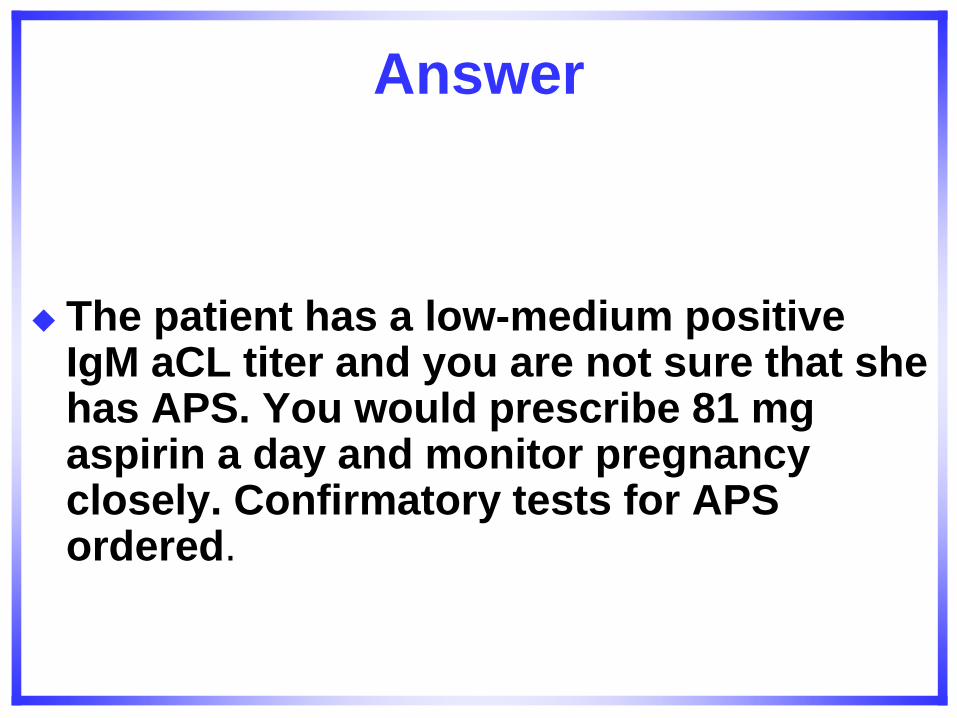
The patient has a low-medium positive IgM aCL titer and you are not sure that she has APS. You would prescribe 81 mg aspirin a day and monitor pregnancy closely. Confirmatory tests for APS ordered.
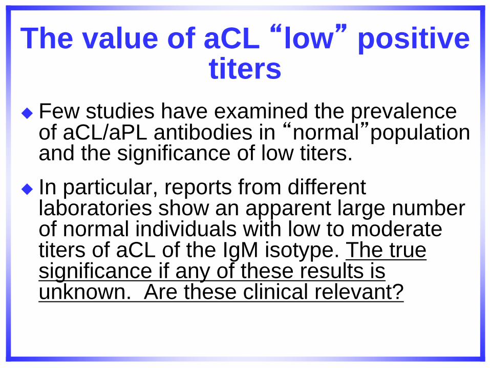
The value of aCL “low” positive titers
Few studies have examined the prevalence of aCL/aPL antibodies in “normal”population and the significance of low titers.
In particular, reports from different laboratories show an apparent large number of normal individuals with low to moderate titers of aCL of the IgM isotype. The true significance if any of these results is unknown. Are these clinical relevant?
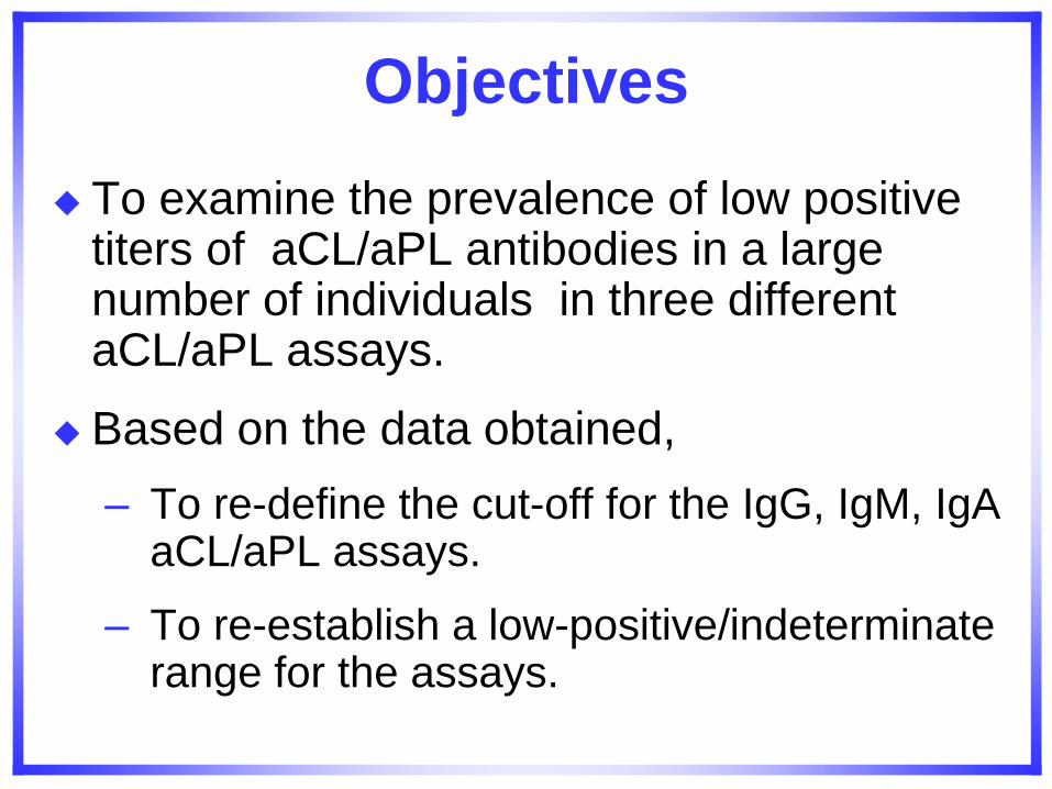
Objectives
To examine the prevalence of low positive titers of aCL/aPL antibodies in a large number of individuals in three different aCL/aPL assays.
Based on the data obtained,
– To re-define the cut-off for the IgG, IgM, IgA aCL/aPL assays.
– To re-establish a low-positive/indeterminate range for the assays.
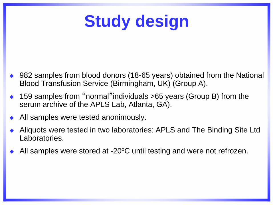
Study design
982 samples from blood donors (18-65 years) obtained from the National Blood Transfusion Service (Birmingham, UK) (Group A).
159 samples from “normal”individuals >65 years (Group B) from the serum archive of the APLS Lab, Atlanta, GA).
All samples were tested anonimously.
Aliquots were tested in two laboratories: APLS and The Binding Site Ltd Laboratories.
All samples were stored at -20ºC until testing and were not refrozen.

Determination of Cut-offs and Indeterminate Zones In Healthy Controls Using Three Different Assays.
N= 1141
BINDAZYME™
MPL U/mL
(A)
ACL standard ELISA
MPL U/mL
(B)
APhL® ELISA
kit
MPL U/mL
(C)
95th percentile
(lower limit of
indeterminate zone
11.7 5.5 10.6
99th percentile
(upper limit of
indeterminate zone)
27.0 14.8 38.4
Current stated
normal cut-off) <10 <10 <15
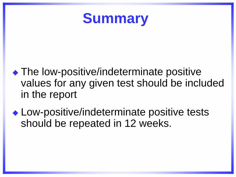
Summary
The low-positive/indeterminate positive values for any given test should be included in the report
Low-positive/indeterminate positive tests should be repeated in 12 weeks.
Case VII
A 25-year-old nulliparous woman presents with an objectively confirmed left leg proximal, deep vein thrombosis that occurred without precipitant.
Question 1: What laboratory testing should be ordered and when testing should be performed?
Answer to question 1:
Consider acquired or inherited “thrombophilia” due to her young age.
Laboratory testing is indicated because patients with APS and first episode of venous thromboembolism, have a high risk for recurrent event after anticoagulants are discontinued, because persistent positive test would mandate extended duration of anticoagulation.
Use Revised Sapporo criteria for laboratory
confirmation of diagnosis of APS.
Question 2
At the time of her presentation with DVT, laboratory testing is positive for LAC, aCL levels (IgG and IgM) are within normal limits,
Based on these results, what is the optimal type and intensity of anticoagulant therapy?
Answer to question 2:
This patient should be started on therapeutic doses of a rapidly acting parenteral anticoagulant (unfractionated heparin, LMWH or pentasaccharide) and an oral vitamin K antagonist such as warfarin. After 4-5 days of overlap and stable INR between 2.0 and 3.0, continue with oral anticoagulant.
Question 3:
How long oral anticoagulation should be continued?
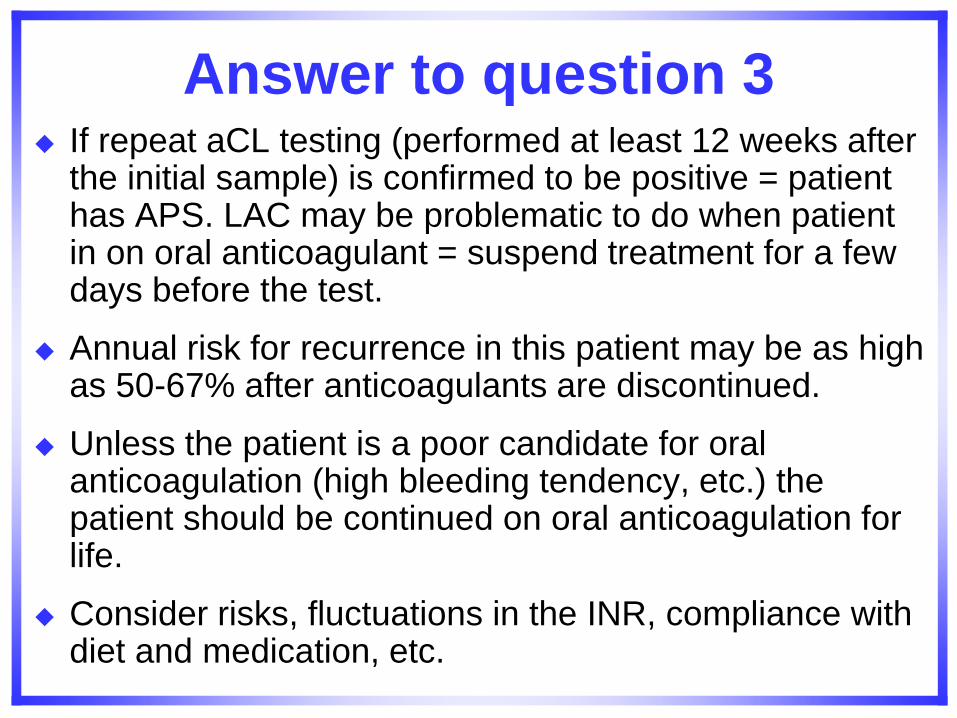
Answer to question 3 If repeat aCL testing (performed at least 12 weeks after
the initial sample) is confirmed to be positive = patient has APS. LAC may be problematic to do when patient in on oral anticoagulant = suspend treatment for a few days before the test.
Annual risk for recurrence in this patient may be as high as 50-67% after anticoagulants are discontinued.
Unless the patient is a poor candidate for oral anticoagulation (high bleeding tendency, etc.) the patient should be continued on oral anticoagulation for life.
Consider risks, fluctuations in the INR, compliance with diet and medication, etc.
Question 4
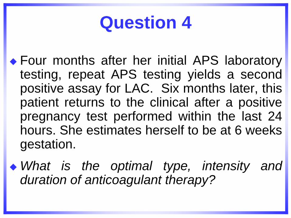
Four months after her initial APS laboratory testing, repeat APS testing yields a second positive assay for LAC. Six months later, this patient returns to the clinical after a positive pregnancy test performed within the last 24 hours. She estimates herself to be at 6 weeks gestation.
What is the optimal type, intensity and duration of anticoagulant therapy?
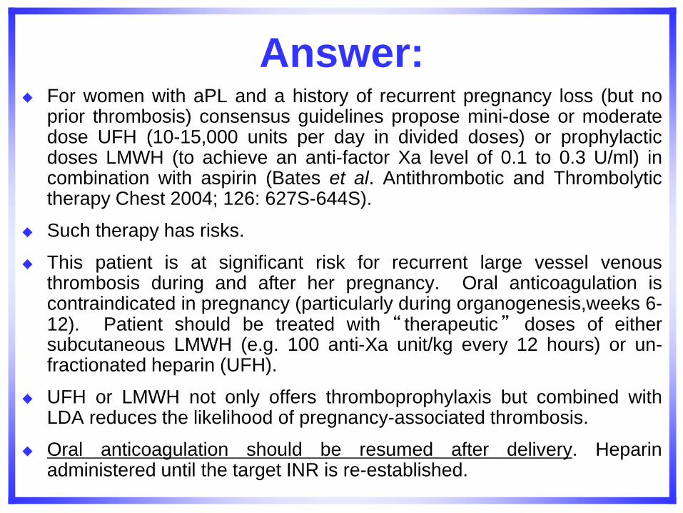
Answer: For women with aPL and a history of recurrent pregnancy loss (but no
prior thrombosis) consensus guidelines propose mini-dose or moderate dose UFH (10-15,000 units per day in divided doses) or prophylactic doses LMWH (to achieve an anti-factor Xa level of 0.1 to 0.3 U/ml) in combination with aspirin (Bates et al. Antithrombotic and Thrombolytic therapy Chest 2004; 126: 627S-644S).
Such therapy has risks.
This patient is at significant risk for recurrent large vessel venous thrombosis during and after her pregnancy. Oral anticoagulation is contraindicated in pregnancy (particularly during organogenesis,weeks 6-12). Patient should be treated with “ therapeutic” doses of either subcutaneous LMWH (e.g. 100 anti-Xa unit/kg every 12 hours) or un-fractionated heparin (UFH).
UFH or LMWH not only offers thromboprophylaxis but combined with LDA reduces the likelihood of pregnancy-associated thrombosis.
Oral anticoagulation should be resumed after delivery. Heparin administered until the target INR is re-established.
Recurrent Thrombosis in patients with APS
Three retrospective studies reported that recurrent thrombosis occurred in 52%, 69%, and 52 % of patients during the follow-up period (5, 6, and 6.4 years respectively) regardless of anti-thrombotic strategy.
Prospective studies have shown that for patients with an initial venous thromboembolic event who completed 6 months of oral anticoagulant therapy, the risk for recurrent thrombosis was 29% in patients with aCL antibodies compared to 14% in those without these antibodies (P = 0.0013).
The risk for recurrent thrombosis appears to be highest in the first few months after discontinuing anticoagulant therapy.
The role of aPL in recurrent stroke: is less clear than for venous disease.
Fifty % of Patients Who Have Experienced a Clinical Thrombosis
Will Re-thrombose.
Several retrospective studies have suggested that the only way to prevent re-thrombosis in these patients is high dose, life long anticoagulation.
It is now known that thrombosis can be triggered by other pathological and physiological events.
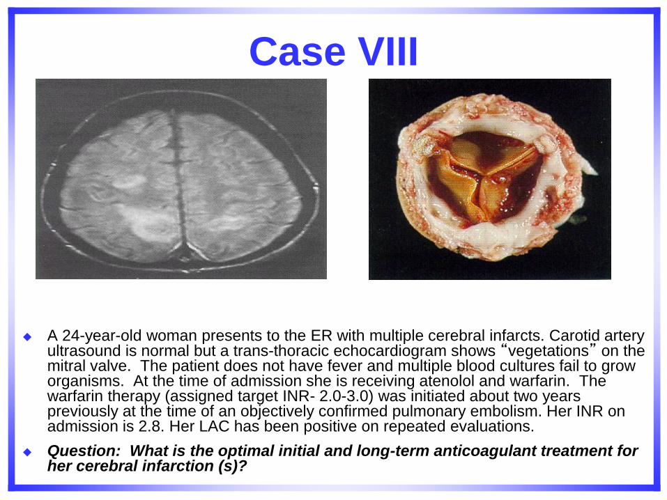
Case VIII
A 24-year-old woman presents to the ER with multiple cerebral infarcts. Carotid artery ultrasound is normal but a trans-thoracic echocardiogram shows “vegetations” on the mitral valve. The patient does not have fever and multiple blood cultures fail to grow organisms. At the time of admission she is receiving atenolol and warfarin. The warfarin therapy (assigned target INR- 2.0-3.0) was initiated about two years previously at the time of an objectively confirmed pulmonary embolism. Her INR on admission is 2.8. Her LAC has been positive on repeated evaluations.
Question: What is the optimal initial and long-term anticoagulant treatment for her cerebral infarction (s)?
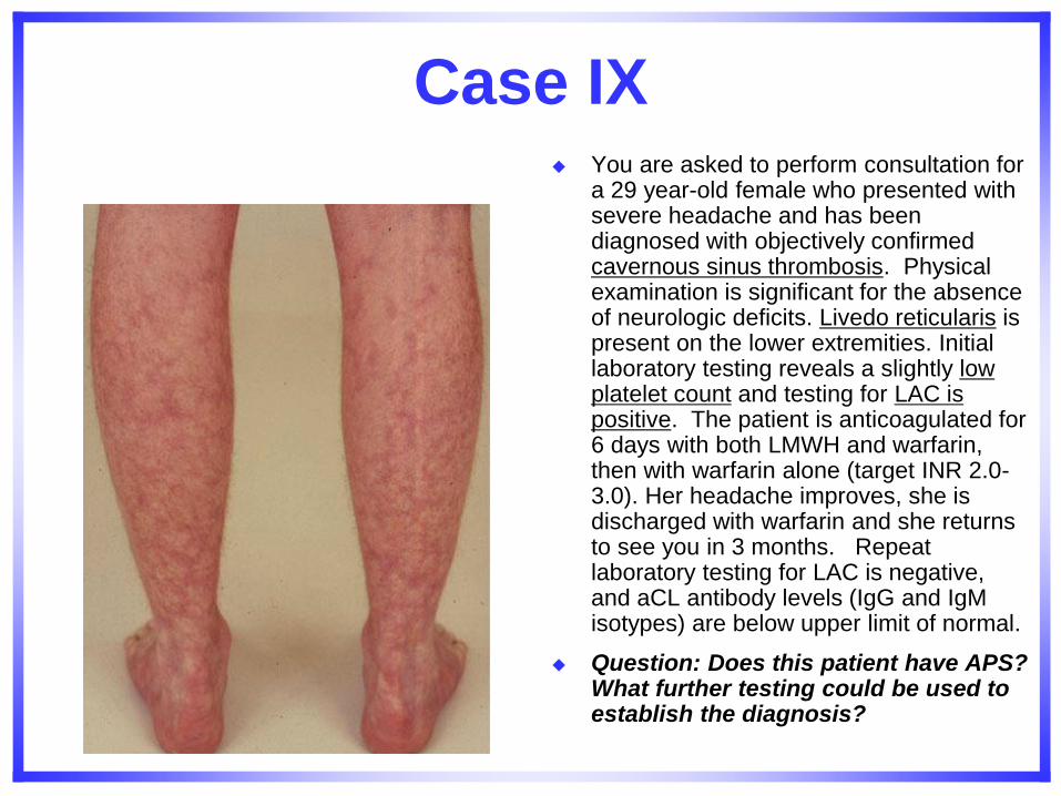
Case IX You are asked to perform consultation for
a 29 year-old female who presented with severe headache and has been diagnosed with objectively confirmed cavernous sinus thrombosis. Physical examination is significant for the absence of neurologic deficits. Livedo reticularis is present on the lower extremities. Initial laboratory testing reveals a slightly low platelet count and testing for LAC is positive. The patient is anticoagulated for 6 days with both LMWH and warfarin, then with warfarin alone (target INR 2.0-3.0). Her headache improves, she is discharged with warfarin and she returns to see you in 3 months. Repeat laboratory testing for LAC is negative, and aCL antibody levels (IgG and IgM isotypes) are below upper limit of normal.
Question: Does this patient have APS? What further testing could be used to establish the diagnosis?
Answers to Case IX
Patient suspicious of having APS (clinically) and one LAC positive.
Test for IgA aCL
Test for anti-2GPI IgG, IgM, IgA.
Test for anti-prothrombin antibodies
Do not discontinue anticoagulation on this patient.

The Lab Team

Thanks for your attention Any Questions?