Antimicrobial-Resistant Bacterial Populations and Antimicrobial ...
Upload
duongxuyenCategory
view
217download
1
ORIGINAL ARTICLE
Antimicrobial activities of a promising glycolipid biosurfactantfrom a novel marine Staphylococcus saprophyticus SBPS 15
P. Mani1 • G. Dineshkumar2 • T. Jayaseelan2 • K. Deepalakshmi3 • C. Ganesh Kumar4 •
S. Senthil Balan3
Received: 28 December 2015 / Accepted: 1 August 2016 / Published online: 9 August 2016
� The Author(s) 2016. This article is published with open access at Springerlink.com
Abstract Biosurfactants have gained a renewed interest
in the recent years for their commercial application in
diverse research areas. Recent evidences suggest that the
antimicrobial activities exhibited by biosurfactants make
them promising molecules for the application in the field
of therapeutics. Marine microbes are well known for their
unique metabolic and functional properties; however, few
reports are available till date regarding their biosurfactant
production and antimicrobial potential. In an ongoing
survey for bioactive microbial metabolites from microbes
isolated from diverse ecological niches, a marine Sta-
phylococcus saprophyticus SBPS 15 isolated from the
petroleum hydrocarbon contaminated coastal site, Pudu-
cherry, India, was identified as a promising biosurfactant
producer based on multiple screening methods. This
bacterium exhibited growth-dependent biosurfactant pro-
duction and the recorded yield was 1.345 ± 0.056 g/L (on
dry weight basis). The biosurfactant was purified and
chemically characterized as a glycolipid with a molecular
mass of 606.7 Da, based on TLC, biochemical estimation
methods, FT-IR spectrum and MALDI-TOF–MS analysis.
Further, the estimated molecular mass was different from
the earlier reports on biosurfactants. This new glycolipid
biosurfactant exhibited a board range of pH and temper-
ature stability. Furthermore, it revealed a promising
antimicrobial activity against many tested human patho-
genic bacterial and fungal clinical isolates. Based on these
observations, the isolated biosurfactant from the marine S.
saprophyticus revealed board physicochemical stabilities
and possess excellent antimicrobial activities which
proves its significance for possible use in various thera-
peutic and biomedical applications. To the best of our
knowledge, this is the first report of a biosurfactant from
the bacterium, S. saprophyticus.
Keywords Staphylococcus saprophyticus � Glycolipid �Biosurfactant � Puducherry coast � Antimicrobial potential
Introduction
Biosurfactants possess both hydrophilic and hydrophobic
moieties that tend to interact with the phase boundary
between two distinct phases in a heterogeneous system to
solubilize them (Ron and Rosenberg 2002). Biosurfactants
has several advantages over their synthetic counterparts
such as higher biodegradability; lower toxicity; good bio-
compatibility; stable at different physico-chemical condi-
tions; synthesis under user-friendly conditions, e.g., low
temperatures and pressures (Zhang et al. 2004; Ruggeri
et al. 2009). They exhibit diverse functional properties such
as emulsification, wetting, foaming, cleansing, phase sep-
aration, surface activity and reduction in viscosity of crude
oil, which makes them amenable for the application in
diverse niche areas such as agriculture, pharmaceuticals,
cosmetics, food industries, oil recovery and environmental
& S. Senthil Balan
1 Department of Biotechnology, Annai College of Arts and
Science, Kumbakonam 612503, Tamilnadu, India
2 Department of Zoology, A.V.V.M. Sri Pushpam College,
Poondi, Thanjavur 613503, Tamilnadu, India
3 Department of Medicinal Plant Biotechnology, Sharmila
Institute of Medicinal Products Research Academy,
Thanjavur 613007, Tamilnadu, India
4 Medicinal Chemistry and Pharmacology Division, CSIR-
Indian Institute of Chemical Technology, Tarnaka,
Hyderabad 500007, India
123
3 Biotech (2016) 6:163
DOI 10.1007/s13205-016-0478-7
remediation (Mulligan 2005; Campos et al. 2013; Sachdev
and Cameotra 2013; Gudina et al. 2016).
In addition to these, recent studies evidenced that bio-
surfactants exhibited anti-bacterial, anti-fungal and anti-
viral activities which makes them potential sources for
biomedical applications (Singh and Cameotra 2004). Many
of the known biosurfactants having bioactive potential
have been reported from terrestrial habitats, the well known
biosurfactants are surfactin, fengycin, bacillomycins and
iturin produced by Bacillus subtilis (Ahimou et al. 2000),
sophorolipids (Cavalero and Cooper 2003), rhamnolipids
from Pseudomonas aeruginosa (Benincasa et al. 2004) and
mannosylerythritol lipids from Candida antarctica
(Arutchelvi et al. 2008). Despite the fact that marine
environment forms[70 % of the Earth’s biosphere com-
prising of diverse group of microorganisms with unique
metabolic, structural and functional properties (Fenical
1993; Proksch et al. 2003); they were less studied till date.
The first antimicrobial marine biosurfactant was repor-
ted from Bacillus circulans isolated from the Andaman and
Nicobar Islands, India, which exhibited antimicrobial
potential against several multidrug resistant human patho-
gens with non-hemolytic property (Das et al. 2008).
Thereafter, some researchers explored the antimicrobial
activities of biosurfactants from marine microbes (Kho-
pade et al. 2012; Dusane et al. 2011; Kiran et al. 2010).
However, when compared to biosurfactants from terrestrial
isolates, few marine biosurfactants have been explored for
its antimicrobial potential, hence warrants this
investigation.
Staphylococcus is a well known genus for its human and
animal infections, but some species have been recognized
as common commensals. Recent studies have reported that
some secondary metabolites produced by Staphylococcus
species isolated from natural environments exhibited
biotechnological and biomedical significance (Popowicz
et al. 2006). Staphylococcus saprophyticus isolated from
the seawater in Jiangsu, China showed appreciable pro-
duction of lipase having excellent organic solvent tolerance
(Fang et al. 2006). Moreover, previous studies showed the
possibilities of biosurfactant production in the genus Sta-
phylococcus species (Eddouaouda et al. 2012). To the best
of our knowledge, no studies have been reported on Sta-
phylococcus saprophyticus for biosurfactant production.
Hence, the present study was undertaken with regard to the
isolation of potential biosurfactant strains using multiple
screening methods, followed by the production and
purification of the biosurfactant from a promising strain of
Staphylococcus saprophyticus, and its biochemical char-
acterization, pH and temperature stability studies and
evaluation of the antimicrobial potential of the biosurfac-
tant against different human pathogenic clinical isolates.
Materials and methods
Sample collection
Sediment samples were collected using Petersen grab
sampler from four different locations of the petroleum
hydrocarbon contaminated coastal sites of Puducherry,
India. Possible aseptic techniques were applied while
sampling to avoid contamination and the collected samples
were transferred to pre-sterilized bottle containers which
was kept in an icebox maintained at 4 �C till further pro-
cessing. The collected samples after reaching the labora-
tory were processed immediately.
Screening of potential biosurfactant producing
bacteria
All the collected four samples were processed individually,
in which 1 g of central portions of the samples was serially
diluted using sterilized natural seawater (34 ppt) and
spread plated on Bushnell Haas agar plates prepared in sea
water and supplemented with 1 % crude oil. After 5 days
of incubation at 37 �C, individual colonies with distinct
morphologies were isolated and further sub-cultured on
Zobell marine agar plates to obtain pure cultures, which
were maintained in lyophilized form for further studies. All
the axenic cultures were individually cultured in Zobell
marine broth 2216 for 48 h and the cell free supernatant
was used for screening the most promising biosurfactant
producers using multiple screening methods, viz., surface
activity (Tadros 2005), emulsification activity (Cooper and
Goldenberg 1987), lipase activity (Kiran et al. 2010) and
oil displacement test (Youssef et al. 2004).
Molecular identification
Molecular identification of the most potential strain was
performed based on 16S rRNA gene sequence analysis
using the bacterial universal primer set of Eubac 27F (50-AGAG TTTG ATCM TGGC TCAG-30) and 1492R (50-GGTT ACCT TGTT ACGA CTT-30). The PCR product
was purified using the Qiagen PCR purification kit and then
sequenced on an ABI Prism 377 automatic sequencer
(Applied Biosystems, CA, USA). The 16S rRNA gene
sequence from this promising strain was compared with the
available bacterial sequences using NCBI BLAST (http://
blast.ncbi.nlm.nih.gov/Blast.cgi) for their pair-wise identi-
ties. Neighbor-joining phylogenetic tree was plotted using
the UPGMA statistical method based on the Maximum-
Composite-Likelihood model using MEGA 6.0 software
(www.megasoftware.net).
163 Page 2 of 9 3 Biotech (2016) 6:163
123
Growth kinetics profile as a function of time
on biosurfactant production
The identified potential strain was standardized for its peak
time of biosurfactant production with reference to its cell
growth. The production process was carried out in a 3 L
laboratory fermentor (Scigenics, India) with 2.1 L working
volume using sea water prepared glucose mineral salt
medium as the production medium under the culture con-
ditions of pH 8.0, temperature of 37 �C, 34 ppt salinity,
agitation at 150 rpm and aeration at 1.0 vvm. The inocu-
lum was prepared using the exponential phase culture of
this promising strain in the same production medium,
where the optical density (OD 620 nm) of the inoculum
culture was adjusted to 0.1 based on McFarland turbidity
0.5 standards which was equivalent to the bacterial con-
centration of 1 9 108 cfu/mL. The biosurfactant produc-
tion was monitored based on the 24 h emulsification index
(E24) (Cooper and Goldenberg 1987) and the bacterial
growth was monitored based on the dry weight of cell
biomass. The fermentation process was monitored for 96 h
and the samples were withdrawn on periodic intervals of
6 h starting from the lag phase to stationary phase under
batch culture conditions. The values are represented as
mean ± standard deviation of triplicate experiments.
Purification of biosurfactant
At the standardized incubation time, the cell free super-
natant was subjected to acid precipitation using 6 N HCl
until pH 2 was attained (Nitschke and Pastore 2006). After
overnight incubation at 4 �C, the precipitated crude bio-
surfactant was collected by centrifugation at 5000 rpm for
15 minutes. The obtained crude biosurfactant was neu-
tralized using phosphate buffer (pH 7) and the resultant
biosurfactant was extracted with an equal volume of
chloroform. The organic phase was separated, concentrated
and rotary vacuum evaporated (Lablinks PBU-6, India)
which was further purified on normal phase silica gel
(60–120 mesh, HiMedia Laboratories Pvt. Ltd., Mumbai,
India) column chromatography using stepwise elution with
methanol and chloroform ranging from 1:20 to 1:1 (v/v).
Twenty fractions were collected and every fraction was
screened for the emulsification index (E24) and the purity
of biosurfactant was checked by thin layer chromatogra-
phy. The fraction(s) showing maximum activity were
pooled, rotary evaporated and lyophilized for further
studies.
Biochemical characterization
The purified biosurfactant was analyzed on silica gel 60
TLC plate (F254, Merck) which was separated using
CH3Cl:CH3OH:H2O (65/15/2, v/v/v) as developing system.
Visualizing reagents used were ninhydrin reagent (0.2 g
ninhydrin in 100 mL ethanol) to detect peptides, anthrone
reagent (1 g anthrone in 5 mL sulfuric acid mixed with
95 mL ethanol) to examine sugars and lipid portion was
evidenced using rhodamine B reagent (0.25 g in 100 mL
ethanol). Following this, the total content of protein, car-
bohydrate and lipid was estimated using Lowry’s method
(Lowry et al. 1951), phenol sulphuric acid method (Dubois
et al. 1956) and total free fatty acids (Folch et al. 1957),
respectively. Further, the FT-IR spectrum was used to
elucidate the functional groups present in the purified
unknown biosurfactant. One milligram of freeze-dried
biosurfactant was grounded with 100 mg of KBr. Infrared
absorption spectrum were recorded on a Thermo Nicolet,
AVATAR 330 FTIR system with a spectral resolution of
cm-1 with an average of 10 scans in the wave number
range of 400–4000 cm-1, and KBr pellet was used as the
background reference.
Molecular mass determination
MALDI-TOF MS was used to examine the molecular mass
of the purified biosurfactant. MS was carried out using a
Voyager DE-Pro MALDI-TOF spectrometer (Applied
Biosystems, Inc, CA, USA) in reflector mode with an
accelerating voltage of 20 kV. Equal volume (2 lL) of thepurified fraction was mixed with an equal volume of matrix
solution, i.e. 0.1 % a-cyano-4-hydroxycinnamic acid in
acetonitrile–water–TFA (50:50:0.01, v/v/v). After mixing,
the sample was spotted on the target plate, dried, placed
inside the sample cabinet and the molecules were separated
based on their molecular weight.
Stability studies
Stability of the biosurfactant was evaluated using 24 h
emulsification index (E24) (Sheppard and Mulligan 1987).
The emulsification activity was estimated using crude oil as
the solvent and the estimations were carried out after
exposure for an hour under the specified test conditions.
The purified biosurfactant at 4 mg/mL concentration in
distilled water was tested for the effect of different tem-
peratures ranging between 30 and 120 �C (Cooper and
Goldenberg 1987) and the influence of pH was estimated
by adjusting the initial pH of the medium from 2 to 10
(Nitschke and Pastore 2006).
Antimicrobial activity
The antimicrobial activity of the purified biosurfactant was
studied on Muller-Hinton agar (MHA) plates against a
panel of different human pathogens using antimicrobial
3 Biotech (2016) 6:163 Page 3 of 9 163
123
disk susceptibility tests as per the Clinical and Laboratory
Standards Institute (2015) recommendations. The human
bacterial pathogens used were Escherichia coli, Salmonella
typhi, S. paratyphi, Klebsiella pneumoniae, K. oxytoca,
Vibrio parahemolyticus, V. cholerae, Proteus mirabilis,
Streptococcus pneumoniae, Bacillus subtilis, B. cereus and
Staphylococcus aureus and human fungal pathogens used
were Aspergillus niger, A. flavus, Candida albicans,
Cryptococcus neoformans and C. gattii, which were kindly
provided by Rajah Muthiah Medical College Hospital,
Annamalai University, Tamilnadu, India. These strains
were cultured on nutrient broth at 37 �C and the OD of
these broth cultures were adjusted to 0.1 equivalent to an
inoculum concentration of 108 cfu mL-1 (according to
McFarland turbidity standard). MHA plates were swab
cultured with 100 lL of individual pathogenic strains and
the wells were impregnated with 50 lL of purified bio-
surfactant dissolved in phosphate buffer (pH 7) at different
concentrations (1–128 lg/mL) to obtain the minimum
inhibitory concentration (MIC) whereas phosphate buffer
solution was used as control. After incubation for 24 h at
37 �C, the plates were examined for zone diameter of
inhibition using an antibiotic zone scale.
Results and discussion
Isolation and screening of most promising
biosurfactant producing bacterium
After the specified incubation period, the Bushnell Haas
agar plates were examined for distinct morphological
colonies which were isolated and pure cultured on Zobell
marine agar plates. A total of 51 axenic strains were
isolated and named as SBPS 1–51. These isolates were
screened to identify the most promising biosurfactant
producer(s) using multiple screening tests. Only 11–30 %
of the isolates showed potent activity with respect to
surface tension reduction (13 %), emulsification activity
(11 %), lipase activity (20 %) and oil displacement test
(30 %). Among these isolates, only one strain SBPS 15
showed promising activity with respect to all the
screening tests, viz., surface tension reduction (32 mN/m),
emulsification activity (77.5 %), lipase activity (66 U/mL)
and oil displacement (3.3 cm), while the rest of the strains
showed highly variable results against the different
screening methods. In accordance to the present investi-
gation, Bodour and Maier (2000) earlier reported the
significance of petroleum contaminated sites as the best
sources for the isolation of promising biosurfactant pro-
ducers. Many researchers have also previously reported
the significance of multiple screening tests for isolation of
potential biosurfactant producers and some methods were
chosen as standard techniques for the effective isolation
of biosurfactant producers (Khopade et al. 2012; Kiran
et al. 2010). Based on the multiple screening methods
adopted in the present study, strain SBPS 15 was identi-
fied as a promising biosurfactant producer. The cell
morphology based on Gram staining and microscopic
observation revealed the strain SBPS 15 to be Gram-
positive cocci. The molecular identification was per-
formed by amplifying the 16S rRNA region and the
sequence homology was examined based on BLASTn
analysis. The total length of the amplified sequence was
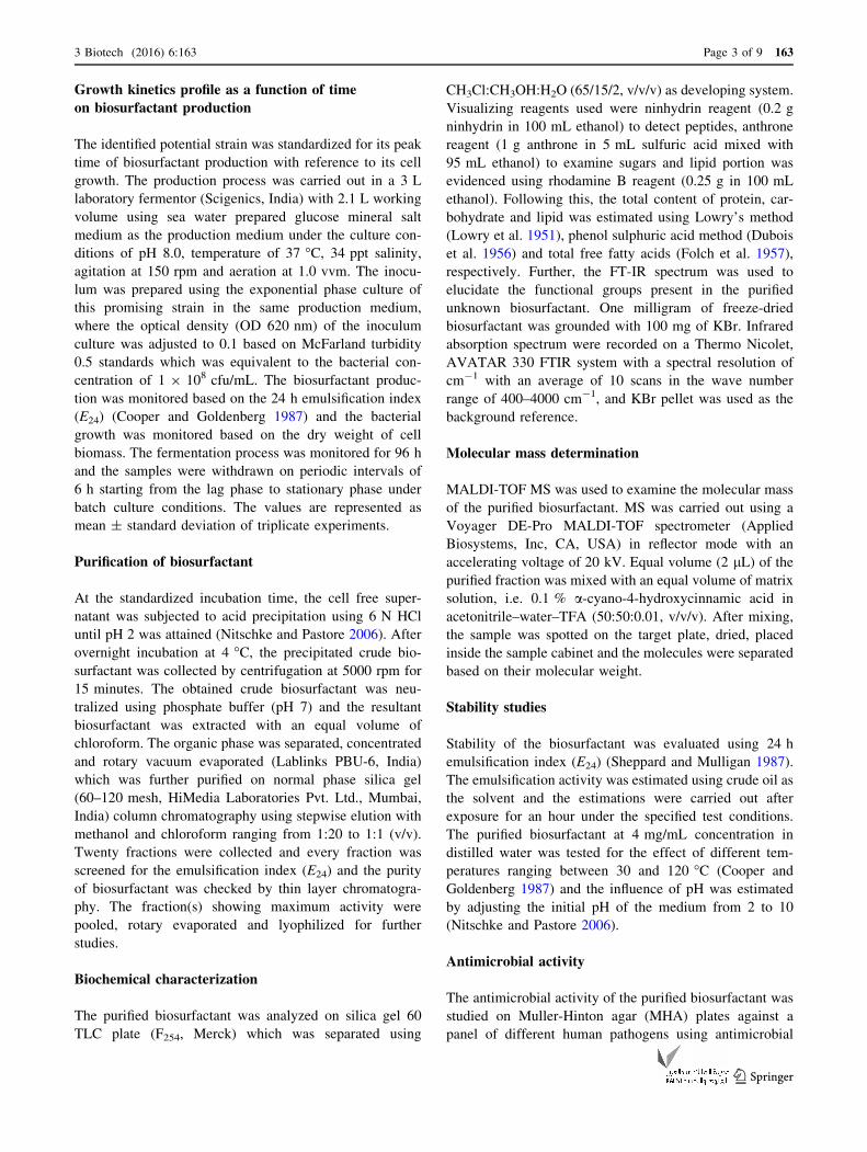
1441 bp. The BLASTn homology comparison of the 16S
rRNA gene sequence of the strain SBPS 15 against the
nucleotide sequence collection of the NCBI GenBank
sequence database showed 100 % sequence similarity
with Staphylococcus saprophyticus A6 (accession number
KX262676.1). Based on these comparisons, the strain
SBPS 15 was identified as Staphylococcus saprophyticus
and the 16S rRNA sequence was deposited in NCBI
GenBank with the accession number KX352162. The
genus Staphylococcus belongs to the family Staphylo-
coccaceae and the phylum Firmicutes. The phylogenetic
tree of Staphylococcus saprophyticus SBPS 15 plotted
with respect to their homology with NCBI strains is
shown in Fig. 1. To the best of our knowledge, Staphy-
lococcus saprophyticus was unexplored for the production
and characterization of biosurfactant, hence further studies
were undertaken to this regard.
Growth kinetics profile of biosurfactant production
The kinetic profile of biosurfactant production by strain
SBPS 15 as a function of time evidenced that the biosur-
factant secretion was observed from the early logarithmic
phase of bacterial growth and the peak emulsification index
(E24) was measured during the initiation of the stationary
growth phase of the bacterium (66th hour). Further, the
maximum emulsification activity (E24) of 77.8 ± 2.3 %
was maintained throughout the stationary phase of the
bacterium with cell biomass concentration of
8.13 ± 0.35 g/L (Fig. 2). The results revealed that the
biosurfactant production with reference to its biomass
concentration indicated that they are predominantly pro-
duced during the exponential phase and they function as
primary metabolites for the normal growth and nutrient
uptake, while in the other case they function as secondary
metabolite having an ecological role rather than growth,
similar to that of antibiotics and pigments (Mulligan et al.
2014). A previous study also reported a similar growth-
dependent pattern of biosurfactant production in marine
Streptomyces species B3 (Khopade et al. 2012).
163 Page 4 of 9 3 Biotech (2016) 6:163
123
Purification and characterization
of the biosurfactant
From the cell free supernatant, the crude biosurfactant was
recovered by acid precipitation, extracted and further puri-
fied using silica gel column chromatography. The maximum
emulsification activity quantified in methanol:chloroform
(1:11) eluted fraction was 1.345 ± 0.056 g/L on dry weight
basis. The silica gel TLC plate analysis of the purified bio-
surfactant revealed the presence of two spots corresponding
to lipid and carbohydrate with Rf values of 0.73 and 0.45.
The carbohydrate and lipid contents estimated were 738 mg/
g (nearly 74 %of carbohydrates) and 262 mg/g (nearly 26 %
of lipid) in the biosurfactant based on biochemical estimation
methods and protein was not detected. The purified biosur-
factant was further analyzed for its functional groups based
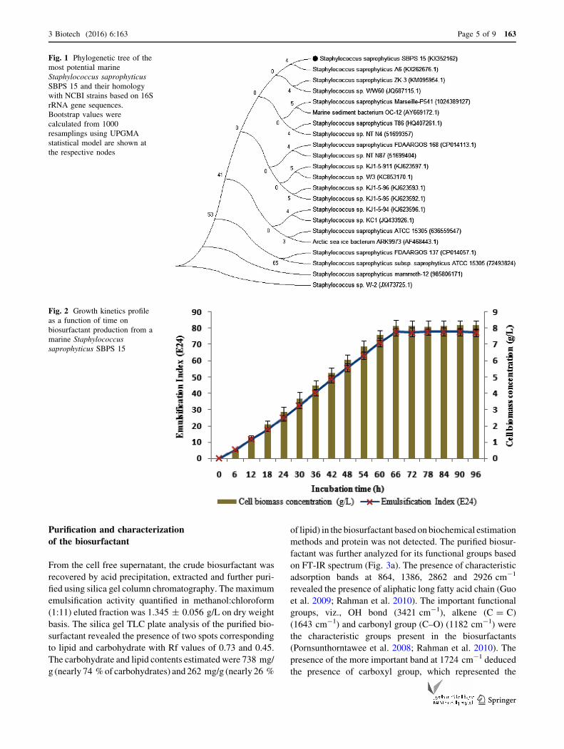
on FT-IR spectrum (Fig. 3a). The presence of characteristic
adsorption bands at 864, 1386, 2862 and 2926 cm-1
revealed the presence of aliphatic long fatty acid chain (Guo
et al. 2009; Rahman et al. 2010). The important functional
groups, viz., OH bond (3421 cm-1), alkene (C = C)
(1643 cm-1) and carbonyl group (C–O) (1182 cm-1) were
the characteristic groups present in the biosurfactants
(Pornsunthorntawee et al. 2008; Rahman et al. 2010). The
presence of the more important band at 1724 cm-1 deduced
the presence of carboxyl group, which represented the
Fig. 1 Phylogenetic tree of the
most potential marine
Staphylococcus saprophyticus
SBPS 15 and their homology
with NCBI strains based on 16S
rRNA gene sequences.
Bootstrap values were
calculated from 1000
resamplings using UPGMA
statistical model are shown at
the respective nodes
Fig. 2 Growth kinetics profile
as a function of time on
biosurfactant production from a
marine Staphylococcus
saprophyticus SBPS 15
3 Biotech (2016) 6:163 Page 5 of 9 163
123
linkage group between the sugar and fatty acid (Rodrigues
et al. 2006) and vibration at 1105 cm-1 was reported as
corresponded to the C–O–C stretching, which predicts the
presence of sugar moiety (Pornsunthorntawee et al. 2008).
These results suggest that the isolated biosurfactant belongs
to the family of glycolipids.
In support to the current prediction, prior studies have
reported thatmicrobial strains from the same species isolated
from different ecological niches produce different types of
biosurfactant, for example, Serratia marcescens isolated
from terrestrial habitats produced Serrawettin, an exolipid
biosurfactant (Li et al. 2005); however, Dusane et al. (2011)
reported a glycolipid biosurfactant produced by S. marces-
cens isolated from marine habitats. Likewise, Pseudomonas
aeruginosa is mostly reported to produce rhamnolipids, a
glycolipid biosurfactant (Pornsunthorntawee et al. 2008);
however, a lipopeptide biosurfactant was produced by P.
aeruginosa isolated from sea water of Tuticorin Harbor,
Fig. 3 FT-IR spectrum (a) and MALDI-TOF–MS analysis (b) of purified glycolipid biosurfactant from a marine Staphylococcus saprophyticus
SBPS 15
163 Page 6 of 9 3 Biotech (2016) 6:163
123
India (Thavasi et al. 2011). Further, in the present investi-
gation, the isolated biosurfactant from the marine bacterium
S. saprophyticus revealed an interesting observation in its
biochemical diversity, producing a glycolipid biosurfactant
which was unique as compared to an earlier report of Sta-
phylococcus sp. strain 1E isolated from terrestrial hydro-
carbon contaminated soil of Algeria which produced a
lipopeptide biosurfactant (Eddouaouda et al. 2012). In
addition,many researches have earlier evidenced thatmarine
microbes represent a distinctive group owing to their
immense genetic (Sogin et al. 2006) and biochemical
diversity (Rusch et al. 2007), and a rich resource for a wide
range of bioactive compounds (Debbab et al. 2010).
The molecular weight of the purified glycolipid bio-
surfactant from strain SBPS 15 was examined using
MALDI-TOF–MS analysis. The spectral analysis revealed
the presence of a cluster of three molecules at m/z 607.7,
629.7 and 645.7 which were attributed to the molecules of
protonated ion and to the adducts of sodium and potassium
ions (Fig. 3b). Based on the above observations, the
molecular mass of this glycolipid biosurfactant is 606.7 Da
and its molecular weight was found to be different from the
earlier reported glycolipid biosurfactants and a new addi-
tion to the existing list of glycolipid biosurfactants. This
result showed good arguments with Abdel-Mawgoud et al.
(2010) who suggested that the molecular mass of most
reported glycolipid biosurfactants are present within the
molecular mass range of 302–803 Da.
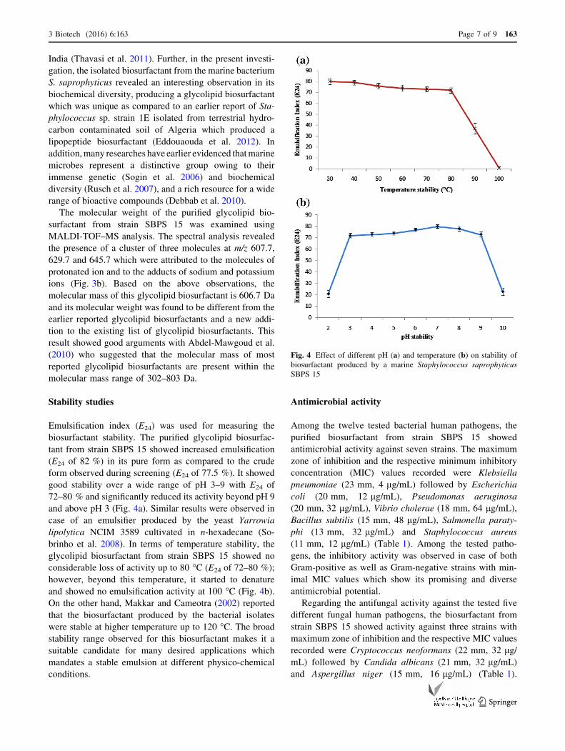
Stability studies
Emulsification index (E24) was used for measuring the
biosurfactant stability. The purified glycolipid biosurfac-
tant from strain SBPS 15 showed increased emulsification
(E24 of 82 %) in its pure form as compared to the crude
form observed during screening (E24 of 77.5 %). It showed
good stability over a wide range of pH 3–9 with E24 of
72–80 % and significantly reduced its activity beyond pH 9
and above pH 3 (Fig. 4a). Similar results were observed in
case of an emulsifier produced by the yeast Yarrowia
lipolytica NCIM 3589 cultivated in n-hexadecane (So-
brinho et al. 2008). In terms of temperature stability, the
glycolipid biosurfactant from strain SBPS 15 showed no
considerable loss of activity up to 80 �C (E24 of 72–80 %);
however, beyond this temperature, it started to denature
and showed no emulsification activity at 100 �C (Fig. 4b).
On the other hand, Makkar and Cameotra (2002) reported
that the biosurfactant produced by the bacterial isolates
were stable at higher temperature up to 120 �C. The broadstability range observed for this biosurfactant makes it a
suitable candidate for many desired applications which
mandates a stable emulsion at different physico-chemical
conditions.
Antimicrobial activity
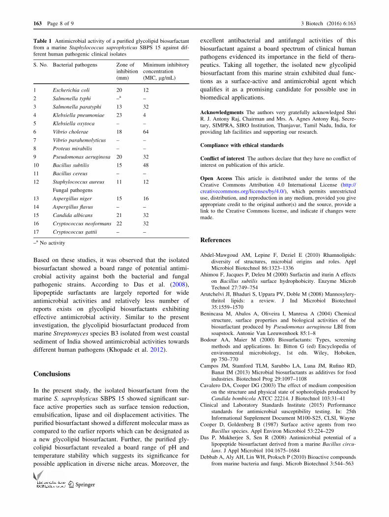
Among the twelve tested bacterial human pathogens, the
purified biosurfactant from strain SBPS 15 showed
antimicrobial activity against seven strains. The maximum
zone of inhibition and the respective minimum inhibitory
concentration (MIC) values recorded were Klebsiella
pneumoniae (23 mm, 4 lg/mL) followed by Escherichia
coli (20 mm, 12 lg/mL), Pseudomonas aeruginosa
(20 mm, 32 lg/mL), Vibrio cholerae (18 mm, 64 lg/mL),
Bacillus subtilis (15 mm, 48 lg/mL), Salmonella paraty-
phi (13 mm, 32 lg/mL) and Staphylococcus aureus
(11 mm, 12 lg/mL) (Table 1). Among the tested patho-
gens, the inhibitory activity was observed in case of both
Gram-positive as well as Gram-negative strains with min-
imal MIC values which show its promising and diverse
antimicrobial potential.
Regarding the antifungal activity against the tested five
different fungal human pathogens, the biosurfactant from
strain SBPS 15 showed activity against three strains with
maximum zone of inhibition and the respective MIC values
recorded were Cryptococcus neoformans (22 mm, 32 lg/mL) followed by Candida albicans (21 mm, 32 lg/mL)
and Aspergillus niger (15 mm, 16 lg/mL) (Table 1).
Fig. 4 Effect of different pH (a) and temperature (b) on stability of
biosurfactant produced by a marine Staphylococcus saprophyticus
SBPS 15
3 Biotech (2016) 6:163 Page 7 of 9 163
123
Based on these studies, it was observed that the isolated
biosurfactant showed a board range of potential antimi-
crobial activity against both the bacterial and fungal
pathogenic strains. According to Das et al. (2008),
lipopeptide surfactants are largely reported for wide
antimicrobial activities and relatively less number of
reports exists on glycolipid biosurfactants exhibiting
effective antimicrobial activity. Similar to the present
investigation, the glycolipid biosurfactant produced from
marine Streptomyces species B3 isolated from west coastal
sediment of India showed antimicrobial activities towards
different human pathogens (Khopade et al. 2012).
Conclusions
In the present study, the isolated biosurfactant from the
marine S. saprophyticus SBPS 15 showed significant sur-
face active properties such as surface tension reduction,
emulsification, lipase and oil displacement activities. The
purified biosurfactant showed a different molecular mass as
compared to the earlier reports which can be designated as
a new glycolipid biosurfactant. Further, the purified gly-
colipid biosurfactant revealed a board range of pH and
temperature stability which suggests its significance for
possible application in diverse niche areas. Moreover, the
excellent antibacterial and antifungal activities of this
biosurfactant against a board spectrum of clinical human
pathogens evidenced its importance in the field of thera-
peutics. Taking all together, the isolated new glycolipid
biosurfactant from this marine strain exhibited dual func-
tions as a surface-active and antimicrobial agent which
qualifies it as a promising candidate for possible use in
biomedical applications.
Acknowledgments The authors very gratefully acknowledged Shri
R. J. Antony Raj, Chairman and Mrs. A. Agnes Antony Raj, Secre-
tary, SIMPRA, SIRO Institution, Thanjavur, Tamil Nadu, India, for
providing lab facilities and supporting our research.
Compliance with ethical standards
Conflict of interest The authors declare that they have no conflict of
interest on publication of this article.
Open Access This article is distributed under the terms of the
Creative Commons Attribution 4.0 International License (http://
creativecommons.org/licenses/by/4.0/), which permits unrestricted
use, distribution, and reproduction in any medium, provided you give
appropriate credit to the original author(s) and the source, provide a
link to the Creative Commons license, and indicate if changes were
made.
References
Abdel-Mawgoud AM, Lepine F, Deziel E (2010) Rhamnolipids:
diversity of structures, microbial origins and roles. Appl
Microbiol Biotechnol 86:1323–1336
Ahimou F, Jacques P, Deleu M (2000) Surfactin and iturin A effects
on Bacillus subtilis surface hydrophobicity. Enzyme Microb
Technol 27:749–754
Arutchelvi JI, Bhaduri S, Uppara PV, Doble M (2008) Mannosylery-
thritol lipids: a review. J Ind Microbiol Biotechnol
35:1559–1570
Benincasa M, Abalos A, Oliveira I, Manresa A (2004) Chemical
structure, surface properties and biological activities of the
biosurfactant produced by Pseudomonas aeruginosa LBI from
soapstock. Antonie Van Leeuwenhoek 85:1–8
Bodour AA, Maier M (2000) Biosurfactants: Types, screening
methods and applications. In: Bitton G (ed) Encyclopedia of
environmental microbiology, 1st edn. Wiley, Hoboken,
pp 750–770
Campos JM, Stamford TLM, Sarubbo LA, Luna JM, Rufino RD,
Banat IM (2013) Microbial biosurfactants as additives for food
industries. Biotechnol Prog 29:1097–1108
Cavalero DA, Cooper DG (2003) The effect of medium composition
on the structure and physical state of sophorolipids produced by
Candida bombicola ATCC 22214. J Biotechnol 103:31–41
Clinical and Laboratory Standards Institute (2015) Performance
standards for antimicrobial susceptibility testing. In: 25th
Informational Supplement Document M100-S25, CLSI, Wayne
Cooper D, Goldenberg B (1987) Surface active agents from two
Bacillus species. Appl Environ Microbiol 53:224–229
Das P, Mukherjee S, Sen R (2008) Antimicrobial potential of a
lipopeptide biosurfactant derived from a marine Bacillus circu-
lans. J Appl Microbiol 104:1675–1684
Debbab A, Aly AH, Lin WH, Proksch P (2010) Bioactive compounds
from marine bacteria and fungi. Microb Biotechnol 3:544–563
Table 1 Antimicrobial activity of a purified glycolipid biosurfactant
from a marine Staphylococcus saprophyticus SBPS 15 against dif-
ferent human pathogenic clinical isolates
S. No. Bacterial pathogens Zone of
inhibition
(mm)
Minimum inhibitory
concentration
(MIC, lg/mL)
1 Escherichia coli 20 12
2 Salmonella typhi –a –
3 Salmonella paratyphi 13 32
4 Klebsiella pneumoniae 23 4
5 Klebsiella oxytoca – –
6 Vibrio cholerae 18 64
7 Vibrio parahemolyticus – –
8 Proteus mirabilis – –
9 Pseudomonas aeruginosa 20 32
10 Bacillus subtilis 15 48
11 Bacillus cereus – –
12 Staphylococcus aureus 11 12
Fungal pathogens
13 Aspergillus niger 15 16
14 Aspergillus flavus – –
15 Candida albicans 21 32
16 Cryptococcus neoformans 22 32
17 Cryptococcus gattii – –
–a No activity
163 Page 8 of 9 3 Biotech (2016) 6:163
123
Dubois M, Gilles KA, Hamilton JK, Rebers PA, Smith F (1956)
Colorimetric method for determination of sugars and related
substances. Anal Chem 28:350–356
Dusane DH, Pawar VS, Nancharaiah YV, Venugopalan VP, Kumar
AR, Zinjarde SS (2011) Anti-biofilm potential of a glycolipid
surfactant produced by a tropical marine strain of Serratia
marcescens. Biofouling 27:645–654
Eddouaouda K, Mnif S, Badis A, Younes SB, Cherif S, Ferhat S,
Mhiri N, Chamkha M, Sayadi S (2012) Characterization of a
novel biosurfactant produced by Staphylococcus sp. strain 1E
with potential application on hydrocarbon bioremediation.
J Basic Microbiol 52:408–418
Fang Y, Lu Z, Lv F, Bie X, Liu S, Ding Z, Xu W (2006) A newly
isolated organic solvent tolerant Staphylococcus saprophyticus
M36 produced organic solvent-stable lipase. Curr Microbiol
53:510–515
Fenical W (1993) Chemical studies of marine bacteria: developing a
new resource. Chem Rev 93:1673–1683
Folch J, Lees M, Sloane-Stanley GH (1957) A simple method for the
isolation and purification of total lipids from animal tissues.
J Biol Chem 226:497–509
Gudina EJ, Teixeira JA, Rodrigues LR (2016) Biosurfactants
produced by marine microorganisms with therapeutic applica-
tions. Mar Drugs 14:E38. doi:10.3390/md14020038
Guo Y, Hu Y, Gu RR, Lin H (2009) Characterization and
micellization of rhamnolipidic fractions and crude extracts
produced by Pseudomonas aeruginosa mutant MIG-N146.
J Colloid Interface Sci 331:356–363
Khopade A, Ren B, Liu X, Mahadik K, Zhang L, Kokare C (2012)
Production and characterization of biosurfactant from marine
Streptomyces species B3. J Colloids Interface Sci 367:311–318
Kiran GS, Thomas TA, Selvin J (2010) Production of a new
glycolipid biosurfactant from marine Nocardiopsis lucentensis
MSA04 in solid-state cultivation. Coll Surf B Biointerf 78:8–16
Li H, Tanikawa T, Sato Y, Nakagawa Y, Matsuyama T (2005)
Serratia marcescens gene required for surfactant serrawettin W1
production encodes putative aminolipid synthetase belonging to
nonribosomal peptide synthetase family. Microbiol Immunol
49:303–310
Lowry OH, Rosenbrough NJ, Farr AL, Randall RJ (1951) Protein
measurement with the folin phenol reagent. J Biol Chem
193:265–275
Makkar RS, Cameotra SS (2002) An update on the use of
unconventional substrates for biosurfactants production and their
new applications. Appl Microbiol Biotechnol 58:428–434
Mulligan CN (2005) Environmental applications for biosurfactants.
Environ Pollut 133:183–198
Mulligan CN, Mudhoo A, Sharma SK (2014) Biosurfactants: research
trends and applications. CRC Press, Boca Raton
Nitschke M, Pastore GM (2006) Production and properties of a
surfactant obtained from Bacillus subtilis grown on cassava
wastewater. Bioresour Technol 97:336–341
Popowicz GM, Dubin G, Stec-Niemczyk J, Czarny A (2006)
Functional and structural characterization of Sp1 protease from
Staphylococcus aureus. J Mol Biol 358:270–279
Pornsunthorntawee O, Wongpanit P, Chavadej S, Abe M, Rujiravanit
R (2008) Structural and physicochemical characterization of
crude biosurfactant produced by Pseudomonas aeruginosa SP4
isolated from petroleum-contaminated soil. Bioresour Technol
99:1589–1595
Proksch P, Edrada-Ebel R, Ebel R (2003) Drugs from the sea—
opportunities and obstacles. Mar Drugs 1:5–17
Rahman PKSM, Pasirayi G, Auger V, Ali Z (2010) Production of
rhamnolipid biosurfactant by Psuedomonas aeruginosa DS
10-129 in a microfluidic bioreactor. Biotechnol Appl Biochem
55:45–52
Rodrigues LR, Teixeira JA, Mei HCV, Oliveira R (2006) Physico-
chemical and functional characterization of a biosurfactant
produced by Lactococcus lactis. Colloids Surf B 49:78–85
Ron EZ, Rosenberg E (2002) Biosurfactants and oil bioremediation.
Curr Opin Microbiol 13:249–252
Ruggeri C, Franzetti A,Bestetti G, Caredda P, Colla PL, PintusM, Sergi
S, Tamburini E (2009) Isolation and characterization of surface
active compound producing bacteria from hydrocarbon-contam-
inated environments. Int Biodeterior Biodegrad 63:936–942
Rusch DB, Halpern AL, Sutton G, Heidelberg KB, Williamson S,
Yooseph S, Wu D, Elsen JA, Hoffman JM, Remington K,
Beeson K, Tran B, Smith H, Baden-Tillson H, Stewart C, Thorpe
J, Freeman J, Andrews-Pfannkoch C, Venter JE, Li K, Kravitz S,
Heidelberg JF, Utterback T, Rogers Y-H, Falcon LI, Souza V,
Bonilla-Rosso G, Eguiarte LE, Karl DM, Sathyendranath S, Platt
T, Bermingham E, Gallardo V, Tamayo-Castillo G, Ferrari MR,
Strausberg RL, Nealson K, Friedman R, Frazier M, Venter JC
(2007) The sorcer 11 global ocean sampling expedition: north-
west Atlantic through Eastern Tropical Pacific. PLoS Biol 5:e77.
doi:10.1371/journal.pbio.0050077
Sachdev DP, Cameotra SS (2013) Biosurfactants in agriculture. Appl
Microbiol Biotechnol 97:1005–1016
Sheppard JD, Mulligan CN (1987) The production of surfactin by
Bacillus subtilis grown on peat hydrolysate. Appl Microbiol
Biotechnol 27:110–116
Singh P, Cameotra SS (2004) Potential applications of microbial
surfactants in biomedical sciences. Trends Biotechnol 22:
142–146
Sobrinho HBS, Rufino RD, Luna JM, Salgueiro AA, Takaki GMC,
Leite LFC, Sarubbo LA (2008) Utilization of two agro industrial
by-products for the production of a surfactant by Candida
sphaerica UCP0995. Process Biochem 43:912–917
Sogin ML, Morrison HG, Huber JA, Welch DM, Huse SM, Neal PR,
Arrieta JM, Herndl GJ (2006) Microbial diversity in the deep sea
and the underexplored ‘‘rare biosphere’’. Proc Natl Acad Sci
USA 103:12115–12120
Tadros T (2005) Adsorption of surfactants at the air/liquid and liquid/
liquid interfaces. In: Applied surfactants: principles and appli-
cations. Wiley, Weinheim, pp 81–82
Thavasi R, Nambaru VRMS, Jayalakshmi S, Balasubramanian T,
Banat IM (2011) Biosurfactant production by Pseudomonas
aeruginosa from renewable resources. Indian J Microbiol
51:30–36
Youssef NH, Dunacn KE, Nagle DP, Savage KN, Knapp RM,
McInerney MJ (2004) Comparison of methods to detect
biosurfactant production by diverse microorganisms. J Microbiol
Methods 56:339–347
Zhang L, Somasundaran P, Singh SK, Felse AP, Gross RA (2004)
Synthesis and interfacial properties of sophorolipid derivatives.
Colloids Surf A Physicochem Eng Asp 240:75–82
3 Biotech (2016) 6:163 Page 9 of 9 163
123


![Deciphering the Glycolipid Code of Alzheimer’s and ... · decipher this code is to study protein/glycolipid interactions with minimal synthetic SBD peptides [13]. In the present](https://static.fdocuments.net/doc/165x107/5ecaf2582cb72d3ca35ba0a2/deciphering-the-glycolipid-code-of-alzheimeras-and-decipher-this-code-is-to.jpg)