Antigen Selection of Anti-DSG1 Autoantibodies During and ...
12
Antigen Selection of Anti-DSG1 Autoantibodies During and Before the Onset of Endemic Pemphigus Foliaceus Ye Qian 1 , Stephen H. Clarke 2 , Valeria Aoki 3 , Gunter Hans-Filhio 4 , Evandro A. Rivitti 3 and Luis A. Diaz 1 for the Cooperative Group on Fogo Selvagem Research Fogo selvagem (FS), the endemic form of pemphigus foliaceus (PF), is characterized by pathogenic anti- desmoglein 1 (DSG1) autoantibodies. To study the etiology of FS, hybridomas that secrete either IgM or IgG (predominantly IgG1 subclass) autoantibodies were generated from the B cells of eight FS patients and one individual 4 years before FS onset, and the H and L chain V genes of anti-DSG1 autoantibodies were analyzed. Multiple lines of evidence suggest that these anti-DSG1 autoantibodies are antigen selected. First, clonally related sets of anti-DSG1 hybridomas characterize the response in individual FS patients. Second, H and L chain V gene use seems to be biased, particularly among IgG hybridomas, and third, most hybridomas are mutants and exhibit a bias in favor of CDR (complementary determining region) amino acid replacement (R) mutations. Strikingly, pre-FS hybridomas also exhibit evidence of antigen selection, including an overlap in V H gene use and shared multiple R mutations with anti-DSG1 FS hybridomas, suggesting selection by the same or a similar antigen. We conclude that the anti-DSG1 response in FS is antigen driven and that selection for mutant anti- DSG1 B cells begins well before the onset of disease. Journal of Investigative Dermatology (2009) 129, 2823–2834; doi:10.1038/jid.2009.184; published online 2 July 2009 INTRODUCTION Pemphigus encompasses a group of autoimmune blistering diseases exhibiting pathogenic autoantibodies against desmogleins (DSG), a family of desmosomal cell adhesion glycoproteins (Lever, 1953; Beutner and Jordon, 1964; Ding et al., 1997; Udey and Stanley, 1999). The hallmark of these diseases is the presence of intraepidermal vesicles (Lever, 1953) and anti-epidermal autoantibodies (Beutner and Jordon, 1964; Ding et al., 1997; Udey and Stanley, 1999). Pemphigus foliaceus (PF) and pemphigus vulgaris (PV) are the two major phenotypes of pemphigus (Lever, 1953). Immunologically, the sera of PF patients show anti-DSG1 antibodies, whereas the sera of PV patients contain antibodies against DSG3 (mPV), or both DSG1 and DSG3 (mcPV) (Ding et al., 1997; Udey and Stanley, 1999). PV and PF in North America are sporadic (Lever, 1953; Udey and Stanley, 1999), but endemic PF is described in certain states of Brazil, where it is known as fogo selvagem (FS) (Diaz et al., 1989b). FS shows similar clinical, histological, and immunological features to those observed in non-endemic PF (Stanley et al., 1986; Diaz et al., 1989a). The published epidemiological studies of FS strongly suggest that this disease is precipitated by environmental agent(s) harbored in certain regions of Brazil. One of these sites, under investigation by our group for the last 15 years, is the Amerindian Reservation of Limao Verde (Hans-Filho et al., 1996). We have reported the serological transition from preclinical to clinical stage of FS in several cases from Limao Verde (Warren et al., 2000, 2003; Li et al., 2003; Qaqish et al., 2009). Fogo selvagem is mediated by pathogenic autoantibodies against DSG1 (Roscoe et al., 1985; Stanley et al., 1986). These pathogenic autoantibodies are IgG4 restricted (Rock et al., 1989) and their appearance in the serum heralds the onset of clinical disease (Warren et al., 2003). In fact, a recent study by our group has identified IgG4 anti-DSG1 auto- antibodies as the serological markers of disease in FS (Qaqish et al., 2009). Non-pathogenic IgG anti-DSG1 autoantibodies (Warren et al., 2000; Li et al., 2003) and IgM anti-DSG1 autoantibodies (Diaz et al., 2008) have been detected in healthy individuals living in endemic areas of FS. The under- lying mechanism of autoantibody formation in FS, however, & 2009 The Society for Investigative Dermatology www.jidonline.org 2823 ORIGINAL ARTICLE Received 4 February 2009; revised 23 April 2009; accepted 15 May 2009; published online 2 July 2009 1 Department of Dermatology, University of North Carolina at Chapel Hill, Chapel Hill, North Carolina, USA; 2 Department of Microbiology and Immunology, University of North Carolina at Chapel Hill, Chapel Hill, North Carolina, USA; 3 Departamento de Dermatologia, Universidade de Sao Paulo, Sao Paulo, Brazil and 4 Departamento de Dermatologia, Universidade Federal de Mato Grosso do Sul, Campo Grande, Brazil Correspondence: Dr Luis A. Diaz, Department of Dermatology, University of North Carolina at Chapel Hill, 3100 Thurston Building, CB #7287 Chapel Hill, North Carolina 27599, USA. E-mail: [email protected] Abbreviations: CDR, complementary determining region; DSG1, desmoglein 1; FS, fogo selvagem; FWR, framework region; PBMC, peripheral blood mononuclear cell; PF, pemphigus foliaceus; PV, pemphigus vulgaris; R, replacement mutations; S, silent mutations
Transcript of Antigen Selection of Anti-DSG1 Autoantibodies During and ...
Antigen Selection of Anti-DSG1 AutoantibodiesDuring and Before the Onset of Endemic PemphigusFoliaceusYe Qian1, Stephen H. Clarke2, Valeria Aoki3, Gunter Hans-Filhio4, Evandro A. Rivitti3 and Luis A. Diaz1
for the Cooperative Group on Fogo Selvagem Research
Fogo selvagem (FS), the endemic form of pemphigus foliaceus (PF), is characterized by pathogenic anti-desmoglein 1 (DSG1) autoantibodies. To study the etiology of FS, hybridomas that secrete either IgM or IgG(predominantly IgG1 subclass) autoantibodies were generated from the B cells of eight FS patients and oneindividual 4 years before FS onset, and the H and L chain V genes of anti-DSG1 autoantibodies were analyzed.Multiple lines of evidence suggest that these anti-DSG1 autoantibodies are antigen selected. First, clonallyrelated sets of anti-DSG1 hybridomas characterize the response in individual FS patients. Second, H and L chainV gene use seems to be biased, particularly among IgG hybridomas, and third, most hybridomas are mutantsand exhibit a bias in favor of CDR (complementary determining region) amino acid replacement (R) mutations.Strikingly, pre-FS hybridomas also exhibit evidence of antigen selection, including an overlap in VH gene useand shared multiple R mutations with anti-DSG1 FS hybridomas, suggesting selection by the same or a similarantigen. We conclude that the anti-DSG1 response in FS is antigen driven and that selection for mutant anti-DSG1 B cells begins well before the onset of disease.
Journal of Investigative Dermatology (2009) 129, 2823–2834; doi:10.1038/jid.2009.184; published online 2 July 2009
INTRODUCTIONPemphigus encompasses a group of autoimmune blisteringdiseases exhibiting pathogenic autoantibodies againstdesmogleins (DSG), a family of desmosomal cell adhesionglycoproteins (Lever, 1953; Beutner and Jordon, 1964;Ding et al., 1997; Udey and Stanley, 1999). The hallmarkof these diseases is the presence of intraepidermalvesicles (Lever, 1953) and anti-epidermal autoantibodies(Beutner and Jordon, 1964; Ding et al., 1997; Udey andStanley, 1999). Pemphigus foliaceus (PF) and pemphigusvulgaris (PV) are the two major phenotypes of pemphigus(Lever, 1953). Immunologically, the sera of PF patients showanti-DSG1 antibodies, whereas the sera of PV patientscontain antibodies against DSG3 (mPV), or both DSG1 and
DSG3 (mcPV) (Ding et al., 1997; Udey and Stanley, 1999).PV and PF in North America are sporadic (Lever, 1953; Udeyand Stanley, 1999), but endemic PF is described in certainstates of Brazil, where it is known as fogo selvagem (FS) (Diazet al., 1989b). FS shows similar clinical, histological, andimmunological features to those observed in non-endemic PF(Stanley et al., 1986; Diaz et al., 1989a). The publishedepidemiological studies of FS strongly suggest that thisdisease is precipitated by environmental agent(s) harboredin certain regions of Brazil. One of these sites, underinvestigation by our group for the last 15 years, is theAmerindian Reservation of Limao Verde (Hans-Filho et al.,1996). We have reported the serological transition frompreclinical to clinical stage of FS in several cases from LimaoVerde (Warren et al., 2000, 2003; Li et al., 2003; Qaqishet al., 2009).
Fogo selvagem is mediated by pathogenic autoantibodiesagainst DSG1 (Roscoe et al., 1985; Stanley et al., 1986).These pathogenic autoantibodies are IgG4 restricted (Rocket al., 1989) and their appearance in the serum heralds theonset of clinical disease (Warren et al., 2003). In fact, a recentstudy by our group has identified IgG4 anti-DSG1 auto-antibodies as the serological markers of disease in FS (Qaqishet al., 2009). Non-pathogenic IgG anti-DSG1 autoantibodies(Warren et al., 2000; Li et al., 2003) and IgM anti-DSG1autoantibodies (Diaz et al., 2008) have been detected inhealthy individuals living in endemic areas of FS. The under-lying mechanism of autoantibody formation in FS, however,
& 2009 The Society for Investigative Dermatology www.jidonline.org 2823
ORIGINAL ARTICLE
Received 4 February 2009; revised 23 April 2009; accepted 15 May 2009;published online 2 July 2009
1Department of Dermatology, University of North Carolina at Chapel Hill,Chapel Hill, North Carolina, USA; 2Department of Microbiology andImmunology, University of North Carolina at Chapel Hill, Chapel Hill, NorthCarolina, USA; 3Departamento de Dermatologia, Universidade de Sao Paulo,Sao Paulo, Brazil and 4Departamento de Dermatologia, Universidade Federalde Mato Grosso do Sul, Campo Grande, Brazil
Correspondence: Dr Luis A. Diaz, Department of Dermatology, University ofNorth Carolina at Chapel Hill, 3100 Thurston Building, CB #7287 ChapelHill, North Carolina 27599, USA. E-mail: [email protected]
Abbreviations: CDR, complementary determining region; DSG1, desmoglein1; FS, fogo selvagem; FWR, framework region; PBMC, peripheral bloodmononuclear cell; PF, pemphigus foliaceus; PV, pemphigus vulgaris;R, replacement mutations; S, silent mutations
specifically that of anti-DSG1 autoantibodies, is still poorlyunderstood. Whether these autoantibodies are developedthrough a polyclonal activation or an antigen-drivenmechanism is still a mystery, but this information is neededto identify the cause of FS. To provide a definite answer tothis question requires the genetic analysis of anti-DSG1autoantibody gene repertoire.
Analyses of the V gene sequences encoding the auto-antibodies in PV by our group (Qian et al., 2007) and byPayne et al. (2005) have yielded important clues to thedevelopment of anti-DSG1 and anti-DSG3 antibodies. Thepotentially pathogenic IgG anti-DSG response in PV has beenshown to be antigen selected (Qian et al., 2007). The resultsof these studies are similar to the findings reported in otherautoimmune diseases, such as systemic lupus erythematosusand rheumatoid arthritis. Recently, two single-chain variablefragments of pathogenic autoantibodies from a PF patientwere isolated and the H chain V regions of the autoantibodiesfrom this patient were shown to be encoded by a restrictednumber of genes (Ishii et al., 2008).
Although there has been no genetic study on the auto-antibodies in individuals before the onset of autoimmunediseases, the high prevalence of FS in Limao Verde providesus with a unique opportunity to address this question, as wehave preserved peripheral blood mononuclear cells (PBMCs)of selected FS and healthy individuals from this humansettlement. In this study, we have examined anti-DSG B cellsfrom multiple FS patients and from an individual who washealthy at the time of the blood draw, but developed FS4 years later. We conclude that (a) anti-DSG response in FS iscomposed of mutant B cells that have been subject toextensive antigen selection, and (b) the pre-clinical anti-DSGresponse is composed of mutant B cells that have undergonethe same or similar selection pressures. These findingsindicate the presence of inciting antigen(s) in FS endemicareas, and these inciting antigen(s) have an important role inthe etiology of FS.
RESULTSAnti-DSG1 in FS patients is oligoclonal
We fused Epstein-Barr virus-transformed PBMCs from eightFS patients with myeloma cells and screened for the resultinghybridomas for anti-DSG1 production by ELISA. We gene-rated 78 anti-DSG1 hybridomas, 40 IgM producers, and 38IgG producers (Table 1). Most IgG anti-DSG1 hybridomaswere IgG1. Despite the fact that the IgG4 autoantibodies arethe main pathogenic antibodies in the sera of FS patients(Rock et al., 1989), only two IgG4 hybridomas wereidentified from two FS patients. There are possible explana-tions for the disparity between the high levels of serum IgG4from FS patients and the low frequency of IgG4 hybridomasgenerated in this study. First, serum IgG4 antibodies wereproduced by plasma cells. Plasma cells reside mainly in thetissues and bone marrow, and are rarely present in peripheralblood (Benner et al., 1981; Slifka and Ahmed, 1996). Thus,they were not abundantly present in the FS patients’ bloodsamples collected. Second, Epstein-Barr virus transformationpredominantly immortalizes B cells (Middleton et al., 1991),
and the IgG4-secreting plasma cells might not be immor-talized and thus might be absent from the subsequenthybridoma generation. Third, this may simply reflect therelative amount of IgG1- and IgG4-expressing B cells in theperipheral blood of FS patients. The DSG1 specificity of theantibodies produced by these hybridomas was confirmed byimmunoprecipitation (Figure 1a) and indirect immunofluor-escence (Figure 1b). Most of these hybridomas (35 of 38) alsorecognized the ectodomain of DSG3 by ELISA (data notshown).
The mRNA of the expressed VH and VL genes of these 77anti-DSG1 hybridomas was PCR amplified and sequenced todetermine VH and VL gene use and to identify somaticmutations. Table 1 summarizes this analysis. Clonally relatedsets of hybridomas from an individual fusion are a hallmark ofsecondary responses to foreign antigen and are also acharacteristic of autoimmune responses, including those inhuman PV, as we have shown previously (Qian et al., 2007).This oligoclonality is because of the antigen-driven clonalexpansion of a limited number of B-cell clones. Clonallyrelated hybridomas express the identical V region genes andhave identical VH CDR3 sequences. By these criteria, weidentified 12 sets of clonally related hybridomas from sevenof the eight FS patients. In patient FS8, all seven of the anti-DSG1 hybridomas identified belonged to a single clonal set,whereas those from patients FS12 and FGS belonged to onlytwo clonal sets each, indicating the dominance of a smallnumber of clones in some patients. Most clonally relatedhybridomas were identical in sequence, suggesting that alimited number of clones in each patient had undergoneextensive clonal growth in the absence of somatic mutation.However, sequence identity raises the possibility that thepresence of clonal sets is an artifact of in vitro growth. Todiscriminate between these possibilities, we compared theextent of somatic mutation among the clonal sets beforedivergence with that of singlet hybridomas. On the basis ofsequence comparison with the most similar germline VH
genes, most (9 of 11) clonally related sets had at least 10differences from germline, indicating the occurrence ofextensive somatic mutation before clonal divergence. Thiswas significantly different from that of singlet hybridomas, inwhich only half had 10 or more mutations (16 of 35) (w2,P¼0.036). This argues against an in vitro artifact to explainthe presence of clonal sets in FS patients, as any in vitroexpansion will be independent of the extent of somaticmutation. This is further suggested by intraclonal sequencedifference in the clonal set from patient FS12 (FS12-1F10 andFS12-3A7). Thus, the presence of clonal sets of hybridomas inthese FS patients likely reflects in vivo clonal expansion,presumably because of the selective advantage in antigenbinding conferred by the somatic mutations they acquiredbefore clonal divergence.
We identified 18 VH genes used by 48 clonally indepen-dent anti-DSG1 hybridomas (Table 1 and Figure 2a upperpanels) and found that IgM and IgG anti-DSG1 hybridomasdiffered significantly in their expressed VH repertoires. TheVH3 gene family use increased from 43.5% among IgMhybridomas to 68.2% (w2, P¼0.095) among IgG hybridomas,
2824 Journal of Investigative Dermatology (2009), Volume 129
Y Qian et al.Autoantibody Antigen Selection in Endemic PF

Table 1. Anti-desmoglein 1 (Dsg1) hybridomas from fogo selvagem (FS) patients
FWRs CDRs
PBMC Clones1 Isotype VH Similarity R2 S3 P4(FWRs) R S P(CDRs) JH CDR3
FS6 FS6-1D10 IgM IGHV3-48*02 98.3 1 0 0.06073 4 0 0.00262 IGHJ4*02 DSGYNTFDY
FS6-2C12 IgG1 IGHV4-59*01 90.0 9 8 0.00311 9 3 0.04149 IGHJ5*02 QNCSPNSCFNYFAP
FS6-2F5(6) IgM IGHV3-23*01 94.2 8 7 0.19667 2 0 0.70918 IGHJ4*02 LIDGGY
FS6-2G3 IgG1 IGHV3-23*01 92.2 5 5 0.00031 12 1 0.00010 IGHJ6*02 EMSLQPVAGMDV
FS6-3A4(2) IgG1 IGHV1-69*01 94.9 3 7 0.00359 3 1 0.33725 IGHJ6*03 GRFYHMDV
FS6-3D7 IgM IGHV1-18*01 95.9 4 2 0.05007 5 1 0.03033 IGHJ4*02 DLGGTYFDY
FS6-4A3 IgM IGHV4-4*07 100.0 IGHJ6*03 DRGVGYCSSTSCYSGRYMDV
FS6-4D4 IgM IGHV1-3*01 96.6 7 2 0.76103 0 1 0.93314 IGHJ4*02 HYYDSSGYYDNFDY
FS6-5B7 IgM IGHV2-5*05 98.3 3 2 0.53435 0.81007 IGHJ4*02 STGAARTDNTYYFDY
FS7 FS7-1C11 IgG1 IGHV5-51*01 97.0 3 2 0.03761 5 0 0.01121 IGHJ4*02 HRIGYCSGSNCYDFDY
FS7-1H10 IgG2 IGHV1-18*01 91.3 6 9 0.00038 5 5 0.38273 IGHJ4*02 HFIPAPPDY
FS7-2A9 IgG1 IGHV3-23*02 95.2 2 3 0.00065 8 1 0.00054 IGHJ4*02 RDIYGDVGVGLVDY
FS7-2F8(4) IgG1 IGHV3-7*01 91.5 8 6 0.01155 9 1 0.01347 IGHJ4*02 TESATIFGVAYYYFDY
FS7-2G6 IgM IGHV3-23*01 95.3 5 3 0.05574 3 3 0.33725 IGHJ3*02 DPGAPCSTTNCYVSDAFDM
FS7-4C1 IgG1 IGHV3-23*01 96.6 3 1 0.04677 5 1 0.01110 IGHJ6*03 DSDSHYYMDV
FS7-4H7 IgM IGHV5-51*01 95.2 5 2 0.04332 5 2 0.05366 IGHJ4*02 GGEAYNLDY
FS8 FS8-1A9(7) IgM IGHV4-59*01 96.6 3 3 0.05258 2 2 0.40536 IGHJ6*03 GVYYGSGGYYGGGSYYYMDV
FS12 FS12-1D2(2) IgG1 IGHV3-7*01 96.3 2 2 0.00558 3 4 0.23564 IGHJ4*02 DSLTAYCGGDCPTVTFGY
FS12-1F10(2) IgG1 IGHV3-74*01 99.0 0 1 0.03950 2 0 0.04735 IGHJ3*02 AYYDFWSGHDDAFDI
FS12-3A7 IgG1 IGHV3-74*01 99.3 0 1 0.09205 1 0 0.18335 IGHJ3*02 AYYDFWSGHDDAFDI
FS33 FS33-1A9 IgM IGHV1-46*01 100.0 IGHJ3*02 DRPDSSGYYLGAFDI
FS33-1B8(4) IgM IGHV3-7*01 98.6 0 2 0.01657 2 0 0.09153 IGHJ6*04 DRQGIYYYYGLDV
FS33-1D4(2) IgM ND
FS33-2B12 IgG1 IGHV3-48*03 91.8 9 9 0.02832 5 1 0.35950 IGHJ5*02 GLPYSGSDRGLDP
FS33-2C6 IgG1 IGHV3-13*01 97.9 3 0 0.36241 3 0 0.03787 IGHJ4*02 RRVIRVRGVIPFFDY
FS33-2E6(4) IgG1 IGHV3-11*03 90.1 8 8 0.00121 9 3 0.04319 IGHJ4*02 GIDYYDSSGHYGSWGEDR
FS33-2F4 IgG1 IGHV4-39*06 96.6 3 1 0.03664 7 0 0.00080 IGHJ4*02 DWGTGWQPLNYFDY
FS33-3C6 IgM IGHV1-3*01 98.3 2 1 0.22538 2 0 0.13601 IGHJ4*02 STRITMITSGY
FS33-3C7 IgG1 ND
FS33-3E5 IgM IGHV1-46*01 96.6 4 1 0.13579 3 2 0.18203 IGHJ4*02 DPGRGAAGIGYYFDY
FS33-3H3 IgM IGHV3-30*18 97.0 2 1 0.02074 4 2 0.03590 IGHJ6*02 ERTVATLYHYYYYGMDV
FS33-4D4(2) IgM IGHV1-3*01 89.1 12 8 0.01015 8 4 0.16514 IGHJ5*02 NQQQLEQQNWYVP
FS33-4G10 IgM IGHV1-69*01 99.7 0.21008 0 1 0.58756 IGHJ4*02 SGYDFYSADY
FS33-5E11 IgM IGHV1-46*01 100.0 IGHJ3*02 DRPDSSGYYLGAFDI
FS33-5F5 IgG1 IGHV5-51*01 99.0 0 1 0.03400 2 0 0.04351 IGHJ4*02 RTMATITGPIGY
FS33-5F7 IgM IGHV3-73*02 98.7 2 1 0.39825 0 1 0.77521 IGHJ5*02 GRDGTVVSLGST
GCDS GCDS-1C2(2) IgG1 IGHV3-23*01 95.6 4 4 0.03017 4 1 0.12097 IGHJ4*02 RRFDWLLYFDY
GCDS-2A8 IgG1 IGHV3-11*01 95.9 2 2 0.00276 7 1 0.00127 IGHJ6*03 DGRGYNYNYNRYFYYMDV
Table 1 continued on the following page
www.jidonline.org 2825
Y Qian et al.Autoantibody Antigen Selection in Endemic PF

and VH1 gene family use decreased from 34.8 to 9.1%(w2, P¼0.038). No VH gene dominated the IgM repertoire,but IGHV3-23 may be favored in the IgG repertoire, as it wasused by 5 of 22 clonally independent IgG hybridomas,although this does not reach the level of significance incomparison with IgM hybridomas (2 of 23 clonally inde-pendent hybridomas: w2, P¼ 0.1942) (Figure 2a upper panels)or with healthy controls (12 of 71 B cells; w2, P¼0.5367)(Brezinschek et al., 1995). JH use by IgM and IgG anti-DSG1included JH3, 4, 5, and 6, and was not significantly differentbetween IgM and IgG hybridomas, and not different fromhealthy control B cells (Brezinschek et al., 1995). In addition,we observed no restriction in VH CDR3 length, as it rangedfrom 6 to 21 amino acids (Table 1).
We identified 11 k and 6 l VL genes used by 29 sequencedclonally independent anti-DSG1 hybridomas (12 IgM and 17IgG) (Table 2 and Figure 2b upper panels). As with VH geneuse, VL gene use seems to be more restricted among IgGhybridomas than among IgM hybridomas. With one exception,each IgM hybridoma expressed a unique VL gene, whereasfour VL genes were used by two or more clonally independentIgG hybridomas. IGKV1D-39 was expressed by four of 17independent hybridomas (23.5%), suggesting that this VL geneprovides a selective advantage in binding DSG. This biaseddistribution of VL is similar to that observed in responses toforeign antigens and, therefore, suggests that the anti-DSG1 Bcells in FS patients undergo antigen selection.
Antigen selection of mutant anti-DSG1 B cells
The anti-DSG1 hybridomas from FS patients were extensivelymutated (Table 1). The number of VHDJH mutations in IgMhybridomas (166 mutations in 23 sequences; 7.2 mutations/gene) was half that in IgG hybridomas (324 mutations in 22sequences; 14.7 mutations/gene) (w2, P¼ 0.021). Moreover,all five unmutated sequences were from IgM hybridomas(Table 1). We used a multinomial distribution model (Lossoset al., 2000) to determine bias in the frequency of amino acidreplacement (R) and silent (S) mutations in framework regions(FWRs) and complementary determining regions (CDRs).We found that 5 of 23 IgM hybridomas showed a biaseddistribution of mutations in either FWR- or CDR-encodingregions and 1 (FS33-3H3) showed a bias distribution in bothFWR- and CDR-encoding regions. In contrast, 15 of 22independent IgG hybridomas showed a biased distribution inboth FWR- and CDR-encoding regions, and most of theremaining hybridomas showed a biased distribution in eitherFWR- or CDR-encoding regions (Table 1). This differencebetween IgM and IgG hybridomas is statistically significant(w2, Po0.001). We conclude that anti-DSG B cells areantigen selected, and that mutant IgG B cells in particularhave a selective advantage in contributing to this response inFS patients.
The expressed VL genes from these hybridomas were alsoextensively mutated, although as is typical, VL mutationswere less frequent than VH mutations (195 mutations in
Table 1. Continued
FWRs CDRs
PBMC Clones1 Isotype VH Similarity R2 S3 P4(FWRs) R S P(CDRs) JH CDR3
GCDS-2D11 IgG4 IGHV2-5*09 97.3 2 2 0.04076 4 0 0.02137 IGHJ4*02 RRLGLRYCSTSSCFGDFDY
GCDS-3G9 IgG1 IGHV3-23*01 96.9 2 3 0.00534 6 0 0.00340 IGHJ4*02 DHCRSDSCYRADL
GCDS-5D2 IgG1 ND
GCDS-6A6 IgG1 ND
GCDS-6G10 IgM IGHV3-30*18 99.3 0 1 0.09014 1 0 0.17838 IGHJ5*02 GLLPEDIVVVPAAQTENWFDP
JLDO JLDO-1B2 IgM IGHV3-48*02 100.0 IGHJ4*02 DSDDYGDYGCFDY
JLDO-2A3 IgM IGHV3-30*03 97.0 3 1 0.08352 3 2 0.13312 IGHJ4*02 EEADFWSGYYFDY
JLDO-3F8 IgG1 IGHV3-30-3*01 98.0 2 1 0.10667 3 0 0.02760 IGHJ6*03 DQQPTIYYYYYMDV
JLDO-3G4 IgM IGHV3-9*01 99.0 1 1 0.23217 1 0 0.26683 IGHJ6*02 DKTLYYYYGMDV
JLDO-4B5 IgG1 IGHV3-73*02 92.6 3 6 0.00002 11 2 0.00040 IGHJ3*02 LIQVWSSQTFDI
FGS FGS-2E5 IgM IGHV4-59*01 100.0 IGHJ6*02 VEVKRDTGTTHPYYYGMDV
FGS-4E4(3) IgG4 IGHV3-48*01 94.2 5 4 0.01181 6 2 0.04083 IGHJ4*02 GRTTFGGEGQLFDY
CDR, complementary determining region; FWR, framework region; ND, not determined; PMBC, peripheral blood mononuclear cell.1Numbers in parentheses indicate the identical clones isolated from the same patient. Clones with evidence of antigen selection in both FWRs and CDRs areshown in bold. Clonally related hybridomas are shown with italics.2Number of replacement mutations.3Number of silent mutations.4P-value determined according to Lossos et al. (2000). Those values less than 0.05, which is the indication of antigen selection, are shown in bold. FWRsinclude FWR1, FWR2, and FWR3, whereas CDRs include CDR1 and CDR2. The P-values of clones with 100% similarity to germline genes are notdetermined.
2826 Journal of Investigative Dermatology (2009), Volume 129
Y Qian et al.Autoantibody Antigen Selection in Endemic PF

29 genes (6.7 mutations/gene) vs 490 mutations in 45 genes(10.9 mutations/gene) (w2, P¼ 0.055) (Table 2). In commonwith the VH mutations in these hybridomas, the frequency ofVL mutation in IgM hybridomas (5.2 mutations/gene) waslower than that in IgG (7.7 mutations/gene) (w2, P¼0.4434).However, few IgG VL genes exhibited a bias in the distri-bution of CDR mutations (2 of 17 VL genes) (Table 2), sugges-ting that VL mutations are less likely to improve antigenbinding than VH mutations.
Shared mutations among anti-DSG1 autoantibodies
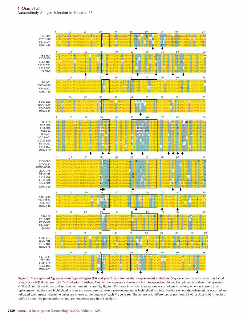
Bias in somatic mutation was also evident by the largenumber of shared mutations among these hybridomas(Figure 3). For example, four of seven IGHV3-23-expressinghybridomas acquired a mutation at position 31 in the CDR1-encoding region of this VH (Figure 3) with S31N occurringtwice. In addition, CDR1 mutation S35N occurred twice andS35T occurred twice, and in CDR2, both A50S and A50Goccurred twice. There were also shared mutations located inFWRs, such as N77K, Y80F, and M83I. Parallel mutationswere not limited to the more frequently used IGHV3-23 andIGHV3-30 genes, as they occurred in other VH genes
(Figure 3). Interestingly, S31N occurred in three othermembers of the VH1 family, and in a member each of theVH3 and VH5 families, further suggesting its importance toantigen binding.
Antigen selection for anti-DSG1 autoantibodies from anindividual at the pre-clinical stage of disease
We have shown that healthy individuals from Limao Verdepossess IgM and IgG anti-DSG1 autoantibodies (Warrenet al., 2000; Diaz et al., 2008), strongly suggesting theexistence of an environmental factor in endemic areas thatsensitizes and triggers anti-DSG1 autoantibody formation inthese individuals. To understand the development of anti-DSG1 antibodies in FS, we investigated the anti-DSG1response before the onset of disease. We have collectedserum samples and PBMCs from selected individuals with orwithout FS living in endemic regions for the past 20 years,and one non-FS individual developed FS 4 years later (patientFS45). The PBMCs of this individual, kept in liquid nitrogen,were Epstein-Barr virus transformed and fused with MSP-2Smyeloma cells.
We identified 28 anti-DSG1 hybridomas (17 IgM, 11IgG1) by ELISA (Table 3) and confirmed their specificity byimmunoprecipitation (data not shown). Two clonal sets ofhybridomas were identified, the largest of which consisted ofsix IgM hybridomas. A total of 14 VH genes encode anti-DSG1 antibodies, but only IGHV3-23 and IGHV3-30 wereused by both IgM and IgG hybridomas (Figure 2a lowerpanels). IGHV3-30, used by three independent FS IgMhybridomas, was the most frequently used VH gene by pre-FS IgG hybridomas (3 of 10 clonally independent pre-FS IgGhybridomas).
The VL gene use by pre-FS hybridomas (Table 4) wasalso diverse, similar to that of FS hybridomas. We identifiedonly two VL genes used by both pre-FS and FS hybridomas(Figure 2a lower panels and Figure 2b lower panels). TheIgG hybridomas were very restricted in VL use as five ofthe seven clonally independent hybridomas sequencedused IGKV2D-28 and IGKV4-1 (Figure 2a lower panel).Interestingly, we did not observe Vl gene use among pre-FShybridomas (0 of 17 vs 10 of 29 among FS hybridomas;w2, P¼ 0.006) (Figure 2b lower panel). Overall, although thepre-FS and FS anti-DSG1 responses overlap in VH gene use,they exhibit notable differences in VL gene use.
As with the FS hybridomas, the overall VH and VL mutationfrequencies differ (266 mutations in 20 VH genes; 59mutations in 17 VL genes; w2, Po0.001). All VH genes ofpre-FS hybridomas were somatically mutated (Table 3), andIgM and IgG anti-DSG1 hybridomas exhibited similar muta-tion rates (123 mutations in 10 genes for IgM; 143 mutationsin 10 genes for IgG; w2, P¼0.7451). These rates were alsosimilar to those of FS IgG hybridomas (w2, P¼0.6125 andP¼0.9404, respectively). Multiple pre-FS hybridomas exhi-bited evidence of antigen selection based on biases in VH Rand S mutations. However, only 3 of 12 clonally independentIgM hybridomas showed a significant bias, and then only inFWR-encoding regions, whereas 6 of 10 clonally indepen-dent IgG hybridomas exhibited a significant bias in mutation
Positiv
e
Negat
ive
FS6-2C
12
FS33-2
B12
FS33-1
B8
FS12-1
F10
FS7-2F
8
FGS-4E4
JLDo-
4B5
GCDS-6A6
GCDS-2D11
Monkeyesophegus
Pat
ient
ser
umF
GS
-4E
4Ir
rele
vant
MA
b
Figure 1. Anti-desmoglein 1 (DSG1) hybridoma antibodies are specific for
DSG1. (a) Representative anti-DSG1 MAbs (all are IgG1, except FGS-4E4 and
GCDS-2D11 (IgG4)) from different fogo selvagem (FS) patients were used to
immunoprecipitate DSG1. Hybridoma antibodies bound to protein G agarose
beads were used to immunoprecipitate His-tagged DSG1 ectodomain. The
immunoprecipitated samples were used in western blots using an anti-His
horseradish peroxidase conjugate to detect DSG1 protein. (b) Similar to serum
from an FS patient, anti-DSG1 MAbs (FGS-4E4) show typical indirect
immunofluorescence staining patterns using monkey esophagus as the
substrate. Bar in (b)¼ 50mm.
www.jidonline.org 2827
Y Qian et al.Autoantibody Antigen Selection in Endemic PF

in the regions encoding FWRs or CDRs, or both. Interestingly,a comparison of IGHV3-23 and IGHV3-30 sequencesfrom pre-FS and FS hybridomas reveals multiple sharedamino acid R mutations (Figure 3). The IGHV3-23 mutationsA23T, S31N, S35N, A50S, A50V, S57R, and N74S, as wellas IGHV3-30 mutation, S31N, occurred in both groups.In addition, the IGHV3-30 mutations, S30R and A88P, eachoccurred twice among pre-FS hybridomas. Thus, similar toFS hybridomas, the pre-FS anti-DSG1 hybridomas exhibitevidence of selection for mutant B cells, particularly amonganti-DSG1 IgG B cells. Moreover, the shared VH mutationssuggest that the same or a similar antigen is responsible forthe selective pressure in the pre-clinical and clinical stagesof the disease.
DISCUSSIONIn this study, we report the genetics of anti-DSG1 auto-antibodies from eight FS patients and one individual 4 yearsbefore the clinical onset of FS. Our results show that the anti-DSG1 response in FS patients living in endemic regions of thedisease in Brazil is antigen selected and that selection beginswell in advance of the onset of clinical disease.
The hypothesis of antigen selection of anti-DSG1 B cells inFS patients is based on several lines of evidence. First,multiple groups of clonally related hybridomas were identi-fied among hybridomas from each patient indicating thatcertain clones have a selective advantage in growth. Clonallyrelated hybridoma sets are a characteristic of secondaryresponses to foreign antigens (Clarke et al., 1985; Blier and
Bothwell, 1987; Scott et al., 1989) and have been observedamong hybridomas derived from autoimmune patients (Qianet al., 2007). Second, a limited VH and VL gene repertoiremay be used to encode these anti-DSG1 antibodies in pre-FSand FS patients. Most VH genes belong to VH families 1, 3,and 4, similar to those in normal and systemic lupuserythematosus individuals (Brezinschek et al., 1997; Dorneret al., 1999; de Wildt et al., 2000). However, IGHV3-23 iscommon among IgG anti-DSG1 hybridomas, increasing from9% among IgM hybridomas to 24% among IgG hybridomas,suggesting selective pressure in favor of its use in thisresponse. Selective VL gene use also occurs, as we find thatIGKV1D-39 increases from 8.3% among IgM hybridomas to23.5% among IgG hybridomas. Thus, V gene use by DSG1-specific B cells may become increasingly restricted duringthe course of the response. Third, FS anti-DSG1 hybridomasexhibit a bias favoring the accumulation of amino acid Rmutations in CDRs and S mutations in FWRs in either VH orVL, or both. This pattern is consistent with the selection for Rmutations that provide an advantage in antigen binding(in CDRs) and against R mutations that could harm antigenbinding or the structural integrity of the antibody molecule(FWRs). Consistent with this are the numerous parallel VH
mutations among these hybridomas, many of which occurredin CDRs. Taken together, the anti-DSG1 response of FSpatients resembles antigen-selected responses to foreignantigen, arguing that the anti-DSG response in FS is antigenselected. This parallels our findings with the anti-DSGresponse in PV (Qian et al., 2007).
3
2
1
5
4
3
2
1
2
1
3
2
1
0
Num
ber
of c
lone
s
Num
ber
of c
lone
s
FS
FS
Pre
-FS Pre
-FS
IGH
V1-
18*
IGH
V1-
3*IG
HV
1-46
IGH
V1-
69*
IGH
V2-
5IG
HV
3-11
*IG
HV
3-13
*IG
HV
3-23
*IG
HV
3-30
*IG
HV
3-48
IGH
V3-
7*IG
HV
3-73
*IG
HV
3-74
IGH
V3-
9*IG
HV
4-30
IGH
V4-
31IG
HV
4-39
*IG
HV
4-4*
IGH
V4-
59IG
HV
4-51
*
IgGIgG
IgM 2
1
4
3
2
1
2
1
3
2
1
0
IgM
IgM
IgM
IgGIgG
IGK
V1-
16IG
KV
1-27
IGK
V1-
5*IG
KV
1-6
IGK
V1D
-17
IGK
V1D
-33
IGK
V1D
-39*
IGK
V2-
29IG
KV
2D-2
8IG
KV
3-11
*IG
KV
3-15
*IG
KV
3-20
*IG
KV
3D-1
1IG
KV
4-1*
IGLV
1-40
IGLV
1-47
IGLV
2-11
IGLV
2-14
IGLV
3-9
IGLV
6-57
Vκ Vλ
Figure 2. V gene use by fogo selvagem (FS) and pre-FS clonally independent anti-desmoglein 1 (DSG1) hybridomas. (a) VH gene use. (b) VL gene use. IgM
and IgG are summarized separately for FS (upper panels) and pre-FS (lower panels). The number of hybridomas identified by sequence similarity to use
each gene is indicated. Genes used by both FS and pre-FS hybridomas are marked by an asterisk.
2828 Journal of Investigative Dermatology (2009), Volume 129
Y Qian et al.Autoantibody Antigen Selection in Endemic PF

Table 2. L chain V genes of anti-desmoglein 1 (Dsg1) hybridomas from fogo selvagem (FS) patients
FWRs CDRs
PBMC Clones1 Isotype VL Similarity R2 S3 P 4(FWRs) R S P(CDRs)
FS6 FS6-2C12 IgG1 IGKV1D-39*01 92.4 8 6 0.02417 4 3 0.34897
FS6-2F5(6) IgM IGKV3D-11*01 93.8 5 6 0.01309 3 2 0.35003
FS6-3D7 IgM IGLV3-9*01 97.9 1 2 0.02771 2 1 0.15775
FS6-4A3 IgM IGLV2-14*01 99.3 0 0 0.09251 2 0 0.01514
FS7 FS7-1H10 IgG2 IGKV1D-39*01 96.4 3 2 0.03390 2 3 0.35646
FS7-2F8(4) IgG1 IGKV1D-39*01 96.4 3 3 0.03390 3 1 0.13846
FS8 FS8-1A9 IgM IGKV1D-39*01 96.8 1 2 0.00465 3 2 0.07741
FS12 FS12-1D2(2) IgG1 IGKV3-15*01 97.6 2 1 0.11233 2 1 0.14244
FS12-1F10(2) IgG1 IGLV6-57*01 97.6 3 3 0.34730 0 0 0.84156
FS12-3A7 IgG1 IGLV6-57*01 97.6 3 3 0.34730 0 0 0.84156
FS33 FS33-1B8(4) IgM IGKV3-11*01 98.6 0 1 0.03299 2 0 0.03589
FS33-1D4(2) IgM IGLV1-40*01 99.0 0 1 0.09860 1 0 0.17658
FS33-2E6(4) IgG1 IGKV3-20*01 94.8 3 3 0.00559 4 3 0.10083
FS33-2F4 IgG1 IGKV4-1*01 99.0 2 0 0.61661 1 0 0.29558
FS33-3C7 IgG1 IGLV1-47*01 96.9 2 2 0.04690 2 2 0.28217
FS33-4D4(2) IgM IGKV1-16*01 95.4 4 2 0.06498 5 0 0.0113
FS33-5E11 IgM IGLV1-47*01 100.0
JLDO JLDO-1B2 IgM IGKV1D-17*01 98.9 1 0 0.20091 2 0 0.03448
JLDO-2A3 IgM IGLV2-14*03 97.9 1 1 0.44278 0 0 0.67306
JLDO-3F8 IgG1 IGLV2-11*01 98.6 1 2 0.12676 1 0 0.33844
JLDO-3G4 IgM IGKV3-20*01 97.6 0 2 0.01410 2 0 0.07237
JLDO-4B5 IgG1 IGKV4-1*01 97.7 0 1 0.00818 2 2 0.1599
GCDS GCDS-1C2(2) IgG1 IGKV1-6*01 96.1 4 3 0.06725 3 1 0.15831
GCDS-2A8 IgG1 IGLV2-14*01 97.6 1 3 0.12494 0 0 0.76729
GCDS-2D11 IgG4 IGLV6-57*01 96.5 1 3 0.01293 2 1 0.22925
GCDS-3G9 IgG1 IGKV3-15*01 98.6 0 0 0.03259 3 0 0.00184
GCDS-5D2 IgG1 IGKV3-20*01 98.3 2 2 0.21463 1 0 0.39272
GCDS-6A6 IgG1 IGKV1D-39*01 95.8 1 5 0.00091 4 0 0.03901
GCDS-6G10 IgM IGKV1-5*03 98.2 1 0 0.04899 2 2 0.11176
FGS FGS-4E4(3) IgG4 IGKV1D-33*01 97.2 1 2 0.01021 2 2 0.21164
CDR, complementary determining region; FWR, framework region; PMBC, peripheral blood mononuclear cell.1Numbers in parentheses indicate the identical clones isolated from the same patient. Clones with evidence of antigen selection in both FWRs and CDRs ofH chains as shown in Table 1 are in bold.2Number of replacement mutations.3Number of silent mutations.4P-value determined according to Lossos et al. (2000). Those values less than 0.05, which are the indication of antigen selection, are shown in bold. FWRsinclude FWR1, FWR2, and FWR3, whereas CDRs include CDR1 and CDR2.
www.jidonline.org 2829
Y Qian et al.Autoantibody Antigen Selection in Endemic PF
FS6-3D7FS7-1H10FS45-4C7IGHV1-18
FS6-4D4
FS6-3A4FS33-4G10
FS45-3C7IGHV1-69
FS33-2E6GCDS-2A8FS45-1C8IGHV3-11
FS7-2G6FS6-2G3
FS6-2F5
FS7-2A9FS7-4C1
GCDS-1C2GCDS-3G9
FS45-3E7FS45-5D3IGHV3-23
FS33-3H3JLDO-2A3
GCDS-6G10JLDO-3F8FS45-1B5FS45-3C9FS45-4A2FS45-5G6IGHV3-30
FS6-1D10FS33-2B12
FSG-4E4IGHV3-48
FS7-2F8FS12-1D2FS33-1B8FS45-2D3
IGHV3-7
FS33-5F7JLDO-4B5FS45-4D5IGHV3-73
FS7-1C11FS7-4H7
FS33-5F5FS45-1G7IGHV5-51
1 10 20 30 40 50 60 70 80 98
1009080706050403020101
1 10 20 30 40 50 60 70 80
80706050403020101
1 10 20 30 40 50 60 70 80 98
98
98
80706050403020101
1 10 20 30 40 50 60 70 80
98
98
FS33-3C6FS33-4D4
FS45-4F11FS45-4G4
IGHV1-3
981 10 20 30 40 50 60 70 80
981 10 20 30 40 50 60 70 80
981 10 20 30 40 50 60 70 80
Figure 3. The expressed VH genes from fogo selvagem (FS) and pre-FS hybridomas share replacement mutations. Sequence comparisons were conducted
using Vector NTI (Invitrogen Life Technologies, Carlsbad, CA). All the sequences shown are from independent clones. Complementary determining regions
(CDRs) (1 and 2) are boxed and replacement mutations are highlighted. Positions in which no mutations occurred are in yellow, whereas conservative
replacement mutations are highlighted in blue and non-conservative replacement mutations highlighted in white. Positions where shared mutations occurred are
indicated with arrows. Germline genes are shown at the bottom of each VH gene set. The amino acid differences at positions 33 (G or A) and 98 (K or R) of
IGHV3-30 may be polymorphisms and are not considered in this analysis.
2830 Journal of Investigative Dermatology (2009), Volume 129
Y Qian et al.Autoantibody Antigen Selection in Endemic PF

The endemic nature of FS, which allowed the freezing ofPBMCs from individuals with a relatively high probability ofdeveloping FS, makes it possible to examine the autoreactiveB-cell repertoire in humans before development of anautoimmune disease. Our analysis of one pre-FS individualindicates a remarkable similarity in pre-clinical and clinicalanti-DSG1 responses. Clonal sets of hybridomas were evidentamong pre-FS hybridoma panels indicating uneven clonalexpansion. In addition, VH and VL use by pre-FS and FShybridomas overlap, particularly with the use of IGHV3-23and IGHV3-30 (Figure 2a). There is also an overlap in Vk useby pre-FS and FS hybridomas (6 of 14 Vk genes, Figure 2b).Although IGKV1D-39 is expressed, it is not dominant, as it isin the FS response (Figure 2b). The most notable differencebetween pre-FS and FS hybridomas is the expression of Vl.One-third (10 of 29) of the FS hybridomas, but none of the 17pre-FS hybridomas expressed Vl genes (Figure 2b). Unfortu-nately, a PBMC sample from this patient after FS diagnosis isnot available. However, the analysis of anti-DSG1 antibodies
in sera from this patient indicates very low levels of l anti-DSG1 before FS onset compared with after FS diagnosis (datanot shown). In contrast, there was no change in the levels of kanti-DSG1 before and after active FS. This change in l anti-DSG1 antibodies was observed in 4 of 11 individuals forwhom sera before and after FS diagnosis were available(data not shown). This raises the possibility that l anti-DSGantibodies are a clinical marker for FS that may predictthe development of disease. We are currently testing thispossibility.
The CDR replacement mutations have the most strikingsimilarity between the pre-FS and FS hybridoma panels. Atotal of 18 amino-acid replacement mutations in CDR1 andCDR2 occurred in IGHV3-23 and IGHV3-30 of pre-FS and FShybridomas. In some cases, these mutations occurred multi-ple times within a panel. We interpret this to mean that manyof the same mutations provide a selective advantage inthe clonal expansion of anti-DSG1 B cells, providing strongevidence that the same or a similar antigen is responsible for
Table 3. Anti-desmoglein 1 (Dsg1) hybridomas from an individual at preclinical stage of fogo selvagem (FS)
FWRs CDRs
PBMC Clones1 Isotype VH Similarity R2 S3 P4(FWRs) R S P(CDRs) JH CDR3
Pre-FS FS45-1A3 IgM ND
FS45-1B5 IgM IGHV3-30*18 99.3 1 0 0.42458 1 0 0.17838 IGHJ4*02 DRQLVGHFWTHYFDY
FS45-1B9 IgM IGHV4-4*07 96.6 5 1 0.33430 3 1 0.16242 IGHJ4*02 GWSFFDY
FS45-1C10 IgG1 IGHV4-30-2*01 92.6 8 7 0.03897 6 1 0.15458 IGHJ6*02 GSLSAALKGCAMEV
FS45-1C4 IgM ND
FS45-1C8 IgM IGHV3-11*01 99 1 0 0.03869 1 1 0.27067 IGHJ4*02 GLVQQFSYLPYYFDY
FS45-1G7 IgG1 IGHV5-51*01 96.3 8 0 0.00003 1 2 0.74024 IGHJ4*02 ARVMLHLSGERTFYFDF
FS45-2C6 IgM IGHV3-9*01 98.6 2 1 0.38779 0 1 0.77375 IGHJ6*02 GRYSNSWYGYYSMDV
FS45-2D3 IgM IGHV3-7*01 99.7 0 1 0.21335 0 0 0.59333 IGHJ4*02 LGDY
FS45-3C7 IgG1 IGHV1-69*01 96.6 3 3 0.04427 4 0 0.05014 IGHJ3*01 GPNRRGSYRPNDAFDI
FS45-3C9 IgG1 IGHV3-30-3*01 96.6 4 3 0.10605 2 1 0.32406 IGHJ6*02 TSNWDGLDV
FS45-3E7(6) IgM IGHV3-23*01 95.6 7 2 0.39281 3 1 0.29244 IGHJ4*02 KKAQLSAPFDY
FS45-3H9 IgG1 IGHV4-39*06 96.6 3 1 0.03664 6 1 0.00522 IGHJ4*02 DSAERSELGYFDY
FS45-4A2(2) IgG1 IGHV3-30*18 96.6 4 3 0.14281 3 0 0.17202 IGHJ4*02 RAKWGSPQPYYFDY
FS45-4C7 IgM IGHV1-18*01 96.6 6 1 0.54624 3 0 0.17573 IGHJ6*02 DRDGYNHYYHYGMDV
FS45-4C8 IgG1 IGHV4-31*03 96.7 5 6 0.25173 0 0 0.94877 IGHJ4*02 DLRYNYDYEGSS
FS45-4D5 IgM IGHV3-73*02 81.1 21 19 0.00198 11 5 0.37278 IGHJ4*02 ATRF
FS45-4F11 IgM IGHV1-3*01 99.7 0 1 0.20903 0 0 0.59112 IGHJ6*02 GGLLMTTVTTYYYYYGMDV
FS45-4G1 IgG1 IGHV4-30-2*01 90.4 6 7 0.00086 11 1 0.00191 IGHJ4*02 AGSSYYYDSRGPIDY
FS45-4G4 IgM IGHV1-3*01 92.2 7 6 0.00426 7 3 0.07727 IGHJ3*02 ARRRKSIVVVPAARDDGFDI
FS45-5D3 IgG1 IGHV3-23*04 93.9 4 6 0.00153 7 1 0.01695 IGHJ6*02 DSASPVLYYYYGMDV
FS45-5G6 IgG1 IGHV3-30-3*01 94.5 4 5 0.00518 5 1 0.04133 IGHJ4*02 SVSVRLSHFDH
CDR, complementary determining region; FWR, framework region; ND, not determined; PMBC, peripheral blood mononuclear cell.1Numbers in parentheses indicate the identical clones isolated. Clones with evidence of antigen selection in both FWRs and CDRs are shown in bold.2Number of replacement mutations.3Number of silent mutations.4P-value determined according to Lossos et al. (2000). Those values less than 0.05, which are the indication of antigen selection, are shown in bold. FWRsinclude FWR1, FWR2, and FWR3, whereas CDRs include CDR1 and CDR2.
www.jidonline.org 2831
Y Qian et al.Autoantibody Antigen Selection in Endemic PF

clonal selection before and after active FS. The differencesbetween these responses, such as Vl gene use, may be due totargeting of different epitopes before and after active FS.Nevertheless, these data indicate that the selection of anti-DSG1 B cells begins well before the onset of clinical disease.What distinguishes between those clones that producepathogenic autoantibodies and those that are benign or evenprotective has yet to be elucidated, but the current datasuggest that antigen selection of mutant IgG1 anti-DSG1B cells is not sufficient for pathogenicity, as the pre-FSindividual analyzed here did not develop clinical disease foranother 4 years.
The two IgG4 hybridomas overlap in VH and VL gene usewith IgG1 hybridomas (IGHV3-48, IGHV2-5, and IGLV6-57)and, similar to IgG1 hybridomas, exhibit a bias in thedistribution of mutations (Table 1). Moreover, the IgG4hybridoma shares the CDR mutations, Y32F and S53T, withIgG1 hybridomas (Figure 3). Further analysis of more IgG4autoantibodies is required to conclusively determine whetherpotential pathogenic IgG4 B cells are subject to the same orsimilar selective pressures in vivo as IgG1 B cells. Never-theless, this study provides a view of the anti-DSG1 repertoirein FS patients indicating that the response to this self-antigenis antigen selected and begins well in advance of clinical
disease. The source of the driving antigen could beenvironmental or self. We previously showed that patientswith parasitic diseases, wherein insect bites are involved,such as onchocerciasis, leishmaniasis, and Chagas, oftenpossess serum anti-DSG1 autoantibodies (Diaz et al., 2004).Epidemiological studies of FS suggest that insect bites are arisk factor in FS (Aoki et al., 2004). It has been proposed thatarthropod salivary antigen(s) induces the production of cross-reactive anti-DSG1 antibodies. Alternatively, the inflamma-tory reaction to insect bites may expose DSG1, allowing ananti-DSG1 response.
In conclusion, we have shown that the development ofautoantibodies in FS is antigen driven, similar to otherautoimmune diseases, such as systemic lupus erythematosusand PV, and that antigen selection of anti-DSG1 B cells canbegin years before the onset of active FS.
MATERIALS AND METHODSPatient samples
Heparinized peripheral blood samples were collected from eight FS
patients and from an individual 4 years before the onset of clinical
FS. All FS patients were living in Limao Verde, Brazil, except three
who were hospitalized in the Penfigo Hospital in Campo Grande,
Brazil. The clinical and serological features of these patients have
Table 4. L chain V genes of anti-desmoglein 1 (Dsg1) hybridomas from a pre-fogo selvagem (FS) individual
FWRs CDRs
PBMC Clones1 Isotype VL Similarity R2 S3 P4(FWRs) R S P(CDRs)
Pre-FS FS45-1A3 IgM IGKV3-15*01 100.0
FS45-1C10 IgG1 IGKV1-5*03 95.4 3 3 0.01783 1 4 0.70848
FS45-1C4 IgM IGKV1-27*01 98.6 0 2 0.08044 0 0 0.64386
FS45-1C8 IgM IGKV3-15*01 99.6 0 0 0.20121 1 0 0.07719
FS45-1G7 IgG1 IGKV1D-39*01 97.2 2 3 0.05803 2 0 0.20386
FS45-2C6 IgM IGKV2D-28*01 100.0
FS45-2D3 IgM IGKV2-29*02 100.0
FS45-3C7 IgG1 IGKV4-1*01 98.3 1 3 0.06859 0 1 0.83586
FS45-3C9 IgG1 IGKV2D-28*01 99.7 0 0 0.21961 1 0 0.09507
FS45-3E7(3) IgM IGKV3-20*01 98.9 0 0 0.03441 3 0 0.00223
FS45-3H9 IgG1 IGKV4-1*01 99.7 0 0 0.21967 1 0 0.09985
FS45-4C7 IgM IGKV3-11*01 99.7 0 1 0.20203 0 0 0.57947
FS45-4D5 IgM IGKV2D-28*01 100.0
FS45-4F11 IgM IGKV1D-39*01 100.0
FS45-4G4 IgM IGKV1D-39*01 95.8 2 2 0.01459 3 2 0.10611
FS45-5D3 IgG1 IGKV2D-28*01 99.0 1 1 0.24701 0 1 0.73442
FS45-5G6 IgG1 IGKV4-1*01 94.4 5 4 0.04315 5 1 0.11207
CDR, complementary determining region; FWR, framework region; PMBC, peripheral blood mononuclear cell.1Numbers in parentheses indicate the identical clones isolated. Clones with evidence of antigen selection in both FWRs and CDRs of H chains as shown inTable 3 are in bold.2Number of replacement mutations.3Number of silent mutations4P-value determined according to Lossos et al. (2000). Those values less than 0.05, which are the indication of antigen selection, are shown in bold. FWRsinclude FWR1, FWR2, and FWR3, whereas CDRs include CDR1 and CDR2.
2832 Journal of Investigative Dermatology (2009), Volume 129
Y Qian et al.Autoantibody Antigen Selection in Endemic PF
been reported previously, that is, FS6 (patient no. 6), FS7 (patient no.
7), FS8 (patient no. 8), and FS 12 (patient no. 12) (Hans-Filho et al.,
1996). FS33 is a patient from Limao Verde who developed FS at age
44 and was not included in the previous report. The patient donating
blood 4 years before the onset of disease was FS45 who developed
FS at age 22 and is the son of FS12 and brother of FS46. Patient JLDO
was a 23-year-old female with a 5-month history of generalized FS.
Patient GCDS was a 13-year-old male with a 1-year history of
generalized FS. Finally, FGS was a 33-year-old female with a
5-month history of generalized FS. All patient samples and clinical
information were collected with written informed consent and
adherence to the Helsinki Guidelines. All experiments were
approved by the Institutional Review Boards of the University of
North Carolina and the University of Sao Paulo, Brazil.
Cell preparation and hybridoma generation
Hybridomas were generated by fusion of Epstein-Barr virus-
transformed PBMCs with mouse myeloma cells (P3X63Ag8.653)
(Kumpel, 2000) or MFP-2S myeloma cells (Kalantarov et al., 2002),
as described previously (Qian et al., 2007). MFP-2S myeloma cells
were kindly provided by Drs Kalantarov and Trakht from Columbia
University, New York, USA. Hybridomas secreting anti-DSG1
antibodies were screened using a DSG1-specific ELISA, as described
previously (Warren et al., 2000, 2003; Li et al., 2003; Diaz et al.,
2004). The specificity of these autoantibodies was confirmed by
immunoprecipitation (Warren et al., 2000; Li et al., 2003) and
indirect immunofluorescence using monkey esophagus (IF) as
described (Anhalt et al., 1982; Ding et al., 1997).
Sequence analysis of VH and VL region from cloned hybridomacells
Messenger RNA isolation, PCR amplification, and sequence analysis
were conducted as described previously (Qian et al., 2007). The
distribution of somatic mutations was analyzed according to the
multinomial distribution model established by Lossos et al. (2000)
and the P-values were calculated using the JAVA applet at http://
www-stat.stanford.edu/immunoglobin. A P-value of less than 0.05 is
taken as evidence for antigen selection at FWR and CDR (Lossos
et al., 2000).
CONFLICT OF INTERESTThe authors state no conflict of interests.
ACKNOWLEDGMENTSThis work was supported in part by the US Public Health Service GrantsR01-AR30281, RO1-AR32599, and T32 AR07369 awarded to L.A.D.,R01-AI43587 to S.H.C, and a Dermatology Foundation Research Grant andan American Skin Association Alice P. Melly Research Grant to Y.Q.
REFERENCES
Anhalt GJ, Labib RS, Voorhees JJ, Beals TF, Diaz LA (1982) Induction ofpemphigus in neonatal mice by passive transfer of IgG from patients withthe disease. N Engl J Med 306:1189–96
Aoki V, Millikan RC, Rivitti EA, Hans-Filho G, Eaton DP, Warren SJ et al.(2004) Environmental risk factors in endemic pemphigus foliaceus(fogo selvagem). J Investig Dermatol Symp Proc 9:34–40
Benner R, Hijmans W, Haaijman JJ (1981) The bone marrow: the majorsource of serum immunoglobulins, but still a neglected site of antibodyformation. Clin Exp Immunol 46:1–8
Beutner EH, Jordon RE (1964) Demonstration of skin antibodies in sera ofpemphigus vulgaris patients by indirect immunofluorescent staining.Proc Soc Exp Biol Med 117:505–10
Blier PR, Bothwell A (1987) A limited number of B cell lineages generatesthe heterogeneity of a secondary immune response. J Immunol 139:3996–4006
Brezinschek HP, Brezinschek RI, Lipsky PE (1995) Analysis of the heavy chainrepertoire of human peripheral B cells using single-cell polymerase chainreaction. J Immunol 155:190–202
Brezinschek HP, Foster SJ, Brezinschek RI, Dorner T, Domiati-Saad R,Lipsky PE (1997) Analysis of the human VH gene repertoire. Differe-ntial effects of selection and somatic hypermutation on humanperipheral CD5(+)/IgM+ and CD5(�)/IgM+ B cells. J Clin Invest 99:2488–501
Clarke SH, Huppi K, Ruezinsky D, Staudt L, Gerhard W, Weigert M (1985)Inter- and intraclonal diversity in the antibody response to influenzahemagglutinin. J Exp Med 161:687–704
de Wildt RM, Tomlinson IM, van Venrooij WJ, Winter G, Hoet RM (2000)Comparable heavy and light chain pairings in normal and systemic lupuserythematosus IgG(+) B cells. Eur J Immunol 30:254–61
Diaz L, Sampaio S, Rivitti E, Martins C, Cunha P, Lombardi C et al. (1989a)Endemic pemphigus foliaceus (fogo selvagem). I. Clinical features andimmunopathology. J Am Acad Dermatol 20:657–69
Diaz LA, Arteaga LA, Hilario-Vargas J, Valenzuela JG, Li N, Warren S et al.(2004) Anti-desmoglein-1 antibodies in onchocerciasis, leishmaniasisand Chagas disease suggest a possible etiological link to Fogo selvagem.J Invest Dermatol 123:1045–51
Diaz LA, Prisayanh PS, Dasher DA, Li N, Evangelista F, Aoki V et al. (2008)The IgM anti-desmoglein 1 response distinguishes Brazilian pemphigusfoliaceus (fogo selvagem) from other forms of pemphigus. J InvestDermatol 128:667–75
Diaz LA, Sampaio SA, Rivitti EA, Martins CR, Cunha PR, Lombardi C et al.(1989b) Endemic pemphigus foliaceus (Fogo Selvagem): II. Current andhistoric epidemiologic studies. J Invest Dermatol 92:4–12
Ding X, Aoki V, Mascaro JM Jr, Lopez-Swiderski A, Diaz LA, Fairley JA (1997)Mucosal and mucocutaneous (generalized) pemphigus vulgaris showdistinct autoantibody profiles. J Invest Dermatol 109:592–6
Dorner T, Farner NL, Lipsky PE (1999) Ig lambda and heavy chain gene usagein early untreated systemic lupus erythematosus suggests intensive B cellstimulation. J Immunol 163:1027–36
Hans-Filho G, dos Santos V, Katayama JH, Aoki V, Rivitti EA, Sampaio SAet al. (1996) An active focus of high prevalence of fogo selvagem on anAmerindian reservation in Brazil. Cooperative Group on Fogo SelvagemResearch. J Invest Dermatol 107:68–75
Ishii K, Lin C, Siegel DL, Stanley JR (2008) Isolation of pathogenic monoclonalanti-desmoglein 1 human antibodies by phage display of pemphigusfoliaceus autoantibodies. J Invest Dermatol 128:939–48
Kalantarov GF, Rudchenko SA, Lobel L, Trakht I (2002) Development ofa fusion partner cell line for efficient production of human monoclonalantibodies from peripheral blood lymphocytes. Hum Antibodies 11:85–96
Kumpel BM (2000) Human monoclonal antibodies to blood groupantigens. In: Monoclonal Antibodies: A Practical Approach (Phil S.Shepherd and Christopher Dean e, eds). London: Oxford UniversityPress, 114–23
Lever W (1953) Pemphigus. Medicine (Baltimore) 32:1–123
Li N, Aoki V, Hans-Filho G, Rivitti EA, Diaz LA (2003) The role ofintramolecular epitope spreading in the pathogenesis of endemicpemphigus foliaceus (fogo selvagem). J Exp Med 197:1501–10
Lossos IS, Tibshirani R, Narasimhan B, Levy R (2000) The inference of antigenselection on Ig genes. J Immunol 165:5122–6
Middleton T, Gahn TA, Martin JM, Sugden B (1991) Immortalizing genes ofEpstein-Barr virus. Adv Virus Res 40:19–55
Payne AS, Ishii K, Kacir S, Lin C, Li H, Hanakawa Y et al. (2005) Geneticand functional characterization of human pemphigus vulgaris mono-clonal autoantibodies isolated by phage display. J Clin Invest 115:888–99
www.jidonline.org 2833
Y Qian et al.Autoantibody Antigen Selection in Endemic PF

Qaqish BF, Prisayanh P, Qian Y, Andraca E, Li N, Aoki V et al. (2009)Development of an IgG4-based predictor of endemic pemphigusfoliaceus (fogo selvagem). J Invest Dermatol 129:110–8
Qian Y, Diaz LA, Ye J, Clarke SH (2007) Dissecting the anti-desmoglein autoreac-tive B cell repertoire in pemphigus vulgaris patients. J Immunol 178:5982–90
Rock B, Martins CR, Theofilopoulos AN, Balderas RS, Anhalt GJ, Labib RSet al. (1989) The pathogenic effect of IgG4 autoantibodies in endemicpemphigus foliaceus (fogo selvagem). N Engl J Med 320:1463–9
Roscoe J, Diaz L, Sampaio S, Castro R, Labib R, Takahashi Y et al. (1985)Brazilian pemphigus foliaceus autoantibodies are pathogenic to BALB/cmice by passive transfer. J Invest Dermatol 85:538–41
Scott MG, Tarrand JJ, Crimmins DL, McCourt DW, Siegel NR, Smith CE et al.(1989) Clonal characterization of the human IgG antibody repertoireto Haemophilus influenzae type b polysaccharide. II. IgG antibodiescontain VH genes from a single VH family and VL genes from at leastfour VL families. J Immunol 143:293–8
Slifka MK, Ahmed R (1996) Long-term humoral immunity against viruses:revisiting the issue of plasma cell longevity. Trends Microbiol 4:394–400
Stanley J, Klaus-Kovtun V, Sampaio S (1986) Antigenic specificity of fogoselvagem autoantibodies is similar to North American pemphigusfoliaceus and distinct from pemphigus vulgaris autoantibodies. J InvestDermatol 87:197–201
Udey MC, Stanley JR (1999) Pemphigus–diseases of antidesmosomalautoimmunity. JAMA 282:572–6
Warren S, Arteaga L, Rivitti E, Aoki V, Hans-Filho G, Qaqish B et al. (2003)The Role of subclass switching in the pathogenesis of endemicpemphigus foliaceus. J Invest Dermatol 120:104–8
Warren SJ, Lin MS, Giudice GJ, Hoffmann RG, Hans-Filho G, Aoki V et al.(2000) The prevalence of antibodies against desmoglein 1 in endemicpemphigus foliaceus in Brazil. Cooperative Group on Fogo SelvagemResearch. N Engl J Med 343:23–30
2834 Journal of Investigative Dermatology (2009), Volume 129
Y Qian et al.Autoantibody Antigen Selection in Endemic PF