Antibiotics from the nature - culturing of genetically...
32
Faculty of Natural Resources and Agricultural Sciences Antibiotics from the nature - culturing of genetically talented bacterial isolates Elinor Bertholtz Department of Microbiology Independent project • 15 hec • First cycle, G2E Biology with specialisation in Biotechnology - Bachelor's Programme • Examensarbete/Sveriges lantbruksuniversitet, Institutionen för mikrobiologi, 2014:8 • ISSN 1101-8151 Uppsala 2014
Transcript of Antibiotics from the nature - culturing of genetically...

Faculty of Natural Resources and
Agricultural Sciences
Antibiotics from the nature - culturing
of genetically talented bacterial
isolates
Elinor Bertholtz
Department of Microbiology
Independent project • 15 hec • First cycle, G2E
Biology with specialisation in Biotechnology - Bachelor's Programme •
Examensarbete/Sveriges lantbruksuniversitet,
Institutionen för mikrobiologi, 2014:8 • ISSN 1101-8151
Uppsala 2014


Antibiotics from the nature - culturing of genetically talented
bacterial isolates
Elinor Bertholtz
Supervisor: Joakim Bjerketorp, Swedish University of Agricultural Sciences,
Department of Microbiology
Assistant Supervisor: Jolanta Levenfors, Swedish University of Agricultural Sciences,
Department of Microbiology
Examiner: Bengt Guss, Swedish University of Agricultural Sciences,
Department of Microbiology
Credits: 15 hec
Level: First cycle, G2E
Course title: Independent project in Biology - bachelor project
Course code: EX0689
Programme/education: Biology with specialisation in Biotechnology - Bachelor's Programme
Place of publication: Uppsala
Year of publication: 2014
Title of series: Examensarbete/Sveriges lantbruksuniversitet, Institutionen för mikrobiologi
No: 2014:8
ISSN: 1101-8151
Online publication: http://stud.epsilon.slu.se
Keywords: antimicrobial, antibiotics, actinomycetes, secondary metabolites, NRPS, PKS-I,
PKS-II, DTS
Sveriges lantbruksuniversitet
Swedish University of Agricultural Sciences
Faculty of Natural Resources and Agricultural Sciences
Uppsala BioCenter
Department of Microbiology


ABSTRACT
Bacterial infections are a threat to the modern healthcare because of the increase of the antibiotic resistance
that many bacteria have developed during the history of antibiotics that began with the discovery of
penicillin 1928. In order to respond to the accumulating resistance, novel antibiotics have to be discovered
constantly. Therefore, there is an increasing need to exploit the ability of microorganisms to produce
secondary metabolites with an antimicrobial effect. The current report addresses methods to activate silent
genes coupled to the production of secondary metabolites. These procedures involve usage of microbial
culture media with various composition, different culturing conditions such as high or low oxygen level as
wells as addition of previously reported supplements that may trigger the expression of silent metabolic
pathways for novel antibiotics. The different approaches used affected the metabolite production both by
activating or suppressing the observed antimicrobial effects and possibly also resulted in the production of
previously not detected substances. Some of the obtained substances will be further investigated in order to
determine their structure and putative antimicrobial effects in more detail.


TABLE OF CONTENTS
INTRODUCTION ............................................................................................................................................. 7
Examples of antibiotic- producing organisms ............................................................................................... 7
Genetic approach in the discovery of novel antimicrobial agents ................................................................. 8
Activating silent genes ................................................................................................................................... 8
Culturing of talented bacterial isolates .......................................................................................................... 9
Scope of this investigation ............................................................................................................................. 9
MATERIALS AND METHODS ..................................................................................................................... 10
Growth media............................................................................................................................................... 10
Bacterial isolates .......................................................................................................................................... 12
Screening for the presence of genes involved in secondary metabolism ..................................................... 12
Culturing conditions and media composition .............................................................................................. 13
Zone inhibition assay ................................................................................................................................... 14
Microtiter plate bioassay against non-pathogenic microorganisms ............................................................. 14
High- Performance Liquid Chromatography (HPLC) ................................................................................. 14
Microtiter plate assay against pathogenic microorganisms ......................................................................... 15
LC-MS analysis of active fractions .............................................................................................................. 15
RESULTS ........................................................................................................................................................ 15
PCR screening of genes for secondary metabolites ..................................................................................... 15
Zone inhibition assay ................................................................................................................................... 16
Influence of culturing conditions on production of secondary metabolites ................................................. 18
Experiment 1- Oxygen exposure ............................................................................................................. 18
Experiment 2- Different growth media composition ............................................................................... 19
Experiment 3- Culturing in the presence of various supplements ........................................................... 20
Microtiter plate assay against pathogenic organisms................................................................................... 22
DISCUSSION .................................................................................................................................................. 24
CONCLUSION AND FUTURE PERSPECTIVES ........................................................................................ 25
ACKNOWLEDGMENT.................................................................................................................................. 26
REFERENCES ................................................................................................................................................ 27


7
INTRODUCTION
Infectious diseases caused by bacteria and fungi resistant to antibiotics are an increasingly severe threat to
the modern standards of healthcare. The morbidity and mortality continue to rise worldwide as well as the
costs coupled to the treatment of these infections. Finding treatments for these diseases has been and still are
of great importance. In the early mid 20´th century the development of antimicrobial medicines experienced
a breakthrough after Alexander Fleming found out that the mold Penicillium notatum produced a substance
currently known as penicillin that inhibited the growth of Staphylococcus aureus on plates (Fleming 1929).
Along with the emerging use of antibiotics the bacteria became resistant to the treatments. As early as 1944,
S. aureus was reported to make use of the enzyme β- lactamase to eliminate the effect of penicillin. To deal
with this problem, scientists created semisynthetic variants of penicillin, for example methicillin, that S.
aureus eventually also became resistant to (Neu 1992). Nowadays disease causing bacteria resistant to
various antibiotics is a serious problem in many hospitals, not to mention infections caused by Methicillin
Resistant S. aureus (MRSA). These bacteria are resistant to virtually all types of antibiotics (Enright et al.
2002). Hence there is a critical need to find new antibiotics, and one of the places to investigate is once
again among the secondary metabolites produced by microorganisms. One approach that is used in the
search of new antibiotics is testing the influence of different culturing conditions such as culture media with
different composition, aeration and so forth to investigate the impact on the production of secondary
metabolites. Research conducted by systematically varying the culture conditions has shown that one
talented microbial species can produce surprisingly many different compounds. This methodology is called
One Strain- Many Compounds (OSMAC) (Bode et al. 2002).
Another approach for the discovery of new antibiotics is to detect genes that are connected to the synthesis
of bioactive compounds such as secondary metabolites, and then try to activate them in order to obtain more
interesting substances (Ayuso-Sacido and Genilloud, 2005, Xie et al. 2014). There are still enormous
amounts of bacterial species left to be explored and even more substances to discover from these species that
may be used for various applications in both industry and healthcare.
Examples of antibiotic- producing organisms
Intense investigation in the last almost 80 years has resulted in the screening of many microorganisms.
Particularly genera of filamentous bacteria and fungi have been attracting much interest in the search for
active compounds. Also Pseudomonas, Bacillus and the cyanobacteria are included in the list of potential
producers of valuable substances (Donadio et al., 2002). Frequently mentioned in antibiotic discovery
related publications are species belonging to the phylum Actinobacteria. They are Gram- positive bacteria
known to produce not only antibiotics but also metabolites active against cancer and suppressors of the
immune system (Baltz 2008, Zotchev 2012). Of all antibiotics that were discovered in the years between
1940 and 1960, 70-80% was isolated from Streptomyces species (Bérdy 2005, Bush 2010). Streptomycin,
produced by Streptomyces griseus, is one example of an antibiotic that is produced by Actinobacteria spp.

8
Other examples of antibiotics produced by actinobacteria are Tetracycline, Erythromycin and Daptomycin
(Mahajan and Balachandran 2012). Both actinobacteria of marine origin (reviewed by Manivasagana et al.
2013, Zotchev 2012,) and terrestrial origin (reviewed by Kumar et al. 2013) have been revealed to produce
antimicrobial substances against both Gram-positive and Gram-negative bacteria that are multi-drug
resistant. Recent studies have been focused on rare actinobacteria also called non-streptomycetes in the
search for novel secondary metabolites as reviewed by Tiwari and Gupta (2012).
Genetic approach in the discovery of novel antimicrobial agents
The enzyme families type I polyketide synthase (PKS-I), type II polyketide synthase (PKS-II), non-
ribosomal peptide synthetase (NRPS) and diterpene synthases (DTS) has been linked to the synthesis of
secondary metabolites from different microorganisms (Ayuso-Sacido and Genilloud 2005, Xie et al.
2014).These large enzymes are considered to be multifunctional, meaning that one single protein has the
ability to generate various compounds. PKS-I manufacture reduced polyketides, PKS-II produce aromatic
polyketides and NRPS create non-ribosomal peptides. The fourth mentioned enzyme DTS generate
diterpenoides (Xie et al. 2014). A recent report proposes a method to achieve metabolites from the
respective enzymatic class by using primers targeted to the corresponding gene as a criterion which isolate
to cultivate (Xie et al. 2014). After screening 100 strains of actinobacteria, the authors choose 16 strains that
had the potential to produce all the four classes of natural products. One of them, a strain of Streptomyces
griseus, was showcased to produce several metabolites from all these different classes of natural products.
The advent of sequencing has facilitated the search of genes responsible of secondary metabolite production.
Bentley et al. (2002) reported the whole genome sequencing of Streptomyces coelicolor and found out that
its genome possess a large number of potentially interesting genes for antibiotic production. Baltz (2008)
compiled the total genome sizes and compared that to the size of PKS-I/-II and NRPS- genes of several
bacterial species. In addition to actinobacteria, species from the genus Pseudomonas showed big potential
for antibiotic production due to its relatively large proportions of PKS- and NRPS-genes in their genomes.
Activating silent genes
Sequencing of Streptomyces species reveals that they do contain many genes coding for the enzymatic
pathways for the synthesis of bioactive compound, but only a few of these end products were actually
detected when the organism was cultured at standard laboratory conditions (Bentley et al. 2002). The silent
genes can hold great potential to encode for enzymes that make novel antibiotics and several methods to
activate these pathways have been reported. For example, by introducing mutations in gene rpoB which
encode for the β- subunit in RNA polymerase, the production of secondary metabolites by some microbes
increased (Ochi and Hosaka 2013). Other approach for activating genes involved in secondary metabolite
expression is adding selected supplements that affect the relevant gene expression in the cultures. Rare earth
elements such as Scandium have been shown to trigger the production of both certain enzymes and also the
production of secondary metabolites in microorganisms (Inaoka and Ochi 2011). Triclosan is a formerly

9
used antimicrobial ingredient in toothpaste (which is still used in parts of the world) that recently has also
been examined for its effect as a trigger of antibiotic production (Craney et al. 2012).
As a defense mechanism, bacteria can use signaling substances as pheromones in a way called quorum
sensing, that allows them to communicate with each other. Sufficient concentration of these pheromones can
either activate or suppress genes encoding for enzymes involved in e.g. secondary metabolite production (de
Kievit and Iglewski 2000). One example of signaling molecules that particularly Gram-negative bacteria use
is N-Acylhomoserine lactones (AHLs), which chemical structure have been described (Yajima 2014). Gram-
positive bacteria in contrariwise, use γ-butyrolactone (GBL) as signal substance (Takano et al. 2000).
Culturing of talented bacterial isolates
When the technologies that are known as high-throughput screening (HTS) were introduced in the beginning
of 1990´s it was believed to revolutionize the search for new drugs (Persidis 1998). Older methods like
fermentation and testing against target pathogens had to step aside for the modern techniques. Unfortunately
HTS did not reach the expected success (Livermore 2011). Very few new antibiotics have been discovered
since the antibiotic golden age in the 1940´s, and nowadays the majority of the current antibiotics are
derivatives of the ones that were originally discovered as secondary metabolites of actinobacteria. Currently,
it is believed that lots of the antibiotics that can be produced by actinobacteria are still left to be discovered.
Research at companies and universities must only put more efforts and re-introduced screening of enough
numbers actinobacteria with improved methods (Baltz 2008).
Scope of this investigation
In the current report, a few Gram-positive isolates of actinobacteria that hopefully might be used as putative
antimicrobial agents were chosen for their potential ability to produce secondary metabolites. The choice of
microbes for antibiotic research, nowadays, can be based on their genetic potential to produce metabolites.
Genes that are linked to the production of metabolites can be screened in order to investigate if they are
present within the organism. Sometimes these genes can be weakly expressed or silent when culturing in
standard conditions. It is therefore desirable to try to switch on these genes using various culturing
conditions (Baltz 2008).
As a comparison, a representative of the Gram- negative bacterial species (Pseudomonas) belonging to the
phylum Proteobacteria was included in the set of experiments. Previous research has shown that species
from the genus Pseudomonas produce antibiotics against plant pathogens such as fungi (Raaijmakers et al.
1997).
The bacterial isolates were tested to see if the genes encoding for PKS-I, PKS-II, NRPS and DTS were
present in their genome, followed by methods aimed to activate or enhance their potential for production of
antimicrobial secondary metabolites. The basic hypothesis was that when talented microorganisms are
cultured in different conditions and with various supplements a change in antimicrobial activity may be
detected similar to what have been observed by previous research efforts.
10
MATERIALS AND METHODS
Growth media
The following culture media reported by Bisht et al. (2013) with some modifications were used in this study:
Arginine glycerol salt broth (AGS), Glucose soybean meal (GS) and Fermentation Medium 4 (FM4) but
without glycerol. A commercial Vegetable Peptone Broth (VPB) was used as a standard medium to allow
various comparisons. Afterwards, an additional medium denoted Glucose soy peptone (GSP), was composed
on the basis of some other media. The media composition is presented in Table 1. Glycerol (25 ml, 50%
w/v) was added to AGS medium after autoclaving.
For the inhibition zone bioassay two different media were used: Vegetable Peptone Agar (VPA) and COR
(Table 1). COR medium was composed as a mixture of three different media. At first, Mineral Medium
(MM, Table 1) previously described by Stainer et al. (1966) with modifications by Pohanka et al. (2005) was
made in 800 ml of deionized H2O. This medium was, afterwards, complemented by adding 100 ml each of
the two other media: Tryptone broth (O) and Casamino Acids- Dextrose Broth (R, Table 1). When needed
15 g agar was added to solidify the growth media. After autoclaving, 2 % (20 g/L) of glycerol was added to
the mixture.

11
Table 1: Media composition
Medium Composition (gram/liter H2O)
AGS 1.0 L-arginine (SIGMA-ALDRICH), 1.0 K2HPO4(MERCK), 1.0 NaCl
(SIGMA-ALDRICH), 0.5 MgSO4·7H2O (SIGMA-ALDRICH), 0.001
CuSO4·5H2O (SIGMA-ALDRICH), 0.01 Fe2(SO4)3·6H2O (SIGMA-
ALDRICH) and 0.001 ZnSO4·H2O (MERCK)
GS 10.0 glucose (VWR), 10.0 soybean meal (Grocery brand Risenta), 10.0 NaCl
(SIGMA-ALDRICH ) and 1.0 CaCO3 (SIGMA-ALDRICH)
VPB 15.0 Vegetable Peptone Broth (OXOID)
FM 4 10.0 glucose (VWR), 10.0 soluble starch (SIGMA-ALDRICH), 2.5 corn steep
liquor (SIGMA-ALDRICH), 5.0 peptone (BD), 2.0 yeast extract (BD), 1.0
NaCl (SIGMA-ALDRICH) and 3.0 CaCO3 (SIGMA-ALDRICH)
GSP 20.0 glucose (VWR), 10.0 soy peptone (BD), 10.0 NaCl (SIGMA-
ALDRICH) and 1.0 CaCO3 (SIGMA-ALDRICH)
VPA 10.0 Vegetable Peptone Broth (OXOID), 15.0 agar (SIGMA-ALDRICH)
MM 40 ml Na2HP04·KH2P04 (SIGMA-ALDRICH), 25 ml mineral stock A, 25 ml
mineral stock B, 1.0 (NH4)2SO4 (SIGMA-ALDRICH), 20 glycerol (Stainer et
al., 1966, Pohanka et al., 2005)
O 10.0 Tryptone (BD), 1 L-Tryptophan (SIGMA-ALDRICH), 5.0 NaCl
(SIGMA-ALDRICH)
R 10.0 Casamino Acids (BD), 2.0 Dextrose (VWR), 1.25 K2HPO4 (SIGMA-
ALDRICH), 1.0 yeast extract (BD)

12
Bacterial isolates
The different bacterial isolates were selected for their potential to produce secondary metabolites. The
isolate identities and origin is shown in Table 2.
Table 2: The identity of bacterial isolates and their origin
Isolate
denotation
Isolate identity Origin
AV130 Streptomyces sp. Coffee plant
GB 34:1 Nocardia sp. Geodia barretti (sponge)
GB 43 Pseudomonas sp. Geodia barretti (sponge)
T110 Streptomyces sp. Seaweed
T339 Brevibacterium sp. Seaweed
T306 Salinibacterium sp. Seaweed
AV226:2 Serratia sp. Grass
GB39 Mycobacterium sp. Geodia barretti (sponge)
GB59 Brevibacterium sp. Geodia barretti (sponge)
GB7 Bacillus sp. Geodia barretti (sponge)
MF503 Serratia sp. Plant material from field
CCUG11104T Streptomyces griseus Reference strain*
CCUG11110TT Streptomyces coelicolor Reference strain*
Screening for the presence of genes involved in secondary metabolism
DNA was isolated from the bacterial isolates AV130, GB34:1, GB43, T110, T339, CCUG11104T and
CCUG11110TT using FASTDNA SPIN kit for soil. The PCR primers targeting the four genes PKS-I, PKS-
II, NRPS and DTS are presented in Table 3 (Xie et al. 2014, Ayuso-Sacido and Genilloud, 2005). For the
PCR reaction DreamTaq master mix (Thermofisher Scentific) was used together with forward and reverse
primer with a final concentration of 0.4 µM each. The PCR program was as follows: initial melting at 95 ºC
for 5 min, followed by 30 cycles with 94 ºC for 30 sec, 52 ºC for 30 sec and 72 ºC for 1 min, and finally an
elongation step at 72 ºC for 7 min.
* Obtained from Culture Collection, University of Gothenburg, Sweden (CCUG)
13
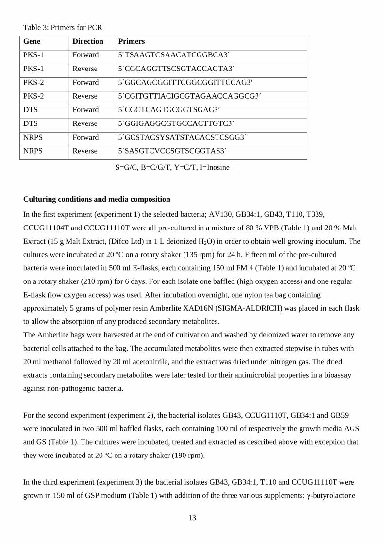
Table 3: Primers for PCR
Gene Direction Primers
PKS-1 Forward 5´TSAAGTCSAACATCGGBCA3´
PKS-1 Reverse 5´CGCAGGTTSCSGTACCAGTA3´
PKS-2 Forward 5´GGCAGCGGITTCGGCGGITTCCAG3′
PKS-2 Reverse 5´CGITGTTIACIGCGTAGAACCAGGCG3′
DTS Forward 5´CGCTCAGTGCGGTSGAG3′
DTS Reverse 5´GGIGAGGCGTGCCACTTGTC3′
NRPS Forward 5´GCSTACSYSATSTACACSTCSGG3´
NRPS Reverse 5´SASGTCVCCSGTSCGGTAS3´
Culturing conditions and media composition
In the first experiment (experiment 1) the selected bacteria; AV130, GB34:1, GB43, T110, T339,
CCUG11104T and CCUG11110T were all pre-cultured in a mixture of 80 % VPB (Table 1) and 20 % Malt
Extract (15 g Malt Extract, (Difco Ltd) in 1 L deionized H2O) in order to obtain well growing inoculum. The
cultures were incubated at 20 ºC on a rotary shaker (135 rpm) for 24 h. Fifteen ml of the pre-cultured
bacteria were inoculated in 500 ml E-flasks, each containing 150 ml FM 4 (Table 1) and incubated at 20 ºC
on a rotary shaker (210 rpm) for 6 days. For each isolate one baffled (high oxygen access) and one regular
E-flask (low oxygen access) was used. After incubation overnight, one nylon tea bag containing
approximately 5 grams of polymer resin Amberlite XAD16N (SIGMA-ALDRICH) was placed in each flask
to allow the absorption of any produced secondary metabolites.
The Amberlite bags were harvested at the end of cultivation and washed by deionized water to remove any
bacterial cells attached to the bag. The accumulated metabolites were then extracted stepwise in tubes with
20 ml methanol followed by 20 ml acetonitrile, and the extract was dried under nitrogen gas. The dried
extracts containing secondary metabolites were later tested for their antimicrobial properties in a bioassay
against non-pathogenic bacteria.
For the second experiment (experiment 2), the bacterial isolates GB43, CCUG1110T, GB34:1 and GB59
were inoculated in two 500 ml baffled flasks, each containing 100 ml of respectively the growth media AGS
and GS (Table 1). The cultures were incubated, treated and extracted as described above with exception that
they were incubated at 20 ºC on a rotary shaker (190 rpm).
In the third experiment (experiment 3) the bacterial isolates GB43, GB34:1, T110 and CCUG11110T were
grown in 150 ml of GSP medium (Table 1) with addition of the three various supplements: γ-butyrolactone
S=G/C, B=C/G/T, Y=C/T, I=Inosine

14
GBL and ScCl3 (SIGMA-ALDRICH), and Triclosan (MERCK). The isolates GB34:1, T110 and
CCUG11110T were inoculated in baffled flasks and isolate GB43 was inoculated in regular E-flasks. GBL
(0.5 µM), Triclosan (1 µM) and ScCl3 (0.2 mM) were added each to one set of cultures. Controls were
cultures without any additives. Cultures were in total incubated for a period of 9 days (20 ºC, 190 rpm).
Three days after inoculation supplements were added and 24 h after the Amberlite bags. Extraction
procedure was as described above.
Zone inhibition assay
The two test organisms Staphylococcus warneri and Pseudomonas resinovorans were diluted in VPB to a
concentration of 5.0·105 cfu/ml and 2.6·10
5 cfu/ml respectively. The bacterial solutions were each spread
onto two agar plates (Ø 14 cm), one containing the media VPA and the other one COR. Suspensions (10 µl)
of the isolates AV130, GB34:1, GB43, T110, T339, CCUG11104T, CCUG11110T, T306, AV226:2, GB39,
GB7 and MF503 were then spotted in a square pattern on the total four different plate/test organism
combination and then incubated at 25 ºC for 3 days.
Microtiter plate bioassay against non-pathogenic microorganisms
The ability of extracts obtained in all culturing experiments to inhibit the growth of two tested organisms S.
warneri and P. resinovorans was estimated in microtiter plate assay. Extracts from the experiment 1
(varying oxygen levels) were dissolved in 5 ml methanol plus 5 ml acetonitrile, whereas extracts from
experiments 2 (two culture media) and 3 (specific supplements) were dissolved in 2.5 ml methanol. Aliquots
of extracts (100, 80, 60, 40, 20 and 10 µl) were distributed to the wells (96-wells microtiter plates) and dried
overnight. Suspensions (100 µl) of S. warneri 3.0x10⁴ cfu/ml (experiment 1) and 3.3x10
5 cfu/ml
(experiment 2 and 3) and of P. resinovorans 4.3x10⁴
cfu/ml (experiment 1) and 2,9x10⁵ cfu/ml (experiment
2 and 3) were added to the wells. Plates were cover with adhesive plastic covers (NUNC) and incubated at
37 °C overnight. Inhibition of bacterial growth was estimated by eye using following scale: 0 – bacterial
growth not inhibited at all; 1 – little inhibition of bacterial growth; 2 – intermediate inhibition of bacterial
growth; 3 – full inhibition and no bacterial growth at all.
High- Performance Liquid Chromatography (HPLC)
In order to detect the possible metabolites with antimicrobial activity, the extracts collected from the
supplement test (experiment 3) were separated by HPLC with gradient 10-95 % acetonitrile during 10 min
with a flow of 10 ml min-1
(injection volume 1 ml, column HypersilGold, C-18, 5µm, 100 x 21.2 mm, and
guard column 10 x 10 mm). Aliquots of the fractions (100µl) were transferred to seven identical replica 96-
well microtiter plates, and chromatograms showing the fractionated compounds absorbance at 210 nm
wavelength were recorded. Solvents were, afterwards, evaporated in the fume cupboard.

15
Microtiter plate assay against pathogenic microorganisms
Suspensions (100µl, approximately 104 cfu/ml) of six different bacterial pathogens and one fungal pathogen
were distributed into wells. The plates were incubated at 37 ºC overnight. Inhibition of bacterial growth was
estimated as described above.
LC-MS analysis of active fractions
The fractions that showed the interesting activity profile against the pathogens were analyzed by using
Liquid Chromatography- Mass Spectrometry (LC-MS). The column used was Accucore RP-MS (50 x 3
mm, particle size 2.6 µm). The eluent consisted two parts: A (H2O, 0.2 % FA) and B (MeCN, 0.2 % FA).
One microliter of the active fractions were injected and analyzed with the gradient of 10-95 % eluent during
4 min, followed by 95 % eluent for 3 min, and ended with 10 % eluent for 3 min. The flow was set at 0.8
ml/min. The substances were detected in the mass spectrometer Bruker maXis Impact, Q- TOF (electrospray
ionization with positive ions), and compared against known microbial metabolites from the database
Antibase.
RESULTS
PCR screening of genes for secondary metabolites
Seven of the bacterial isolates in this report (AV130, GB34:1, GB43, T110, T339, CCUG11104T and
CCUG11110T) were tested if they harbor genes that encode for secondary metabolites associated enzymes
PKS-1, PKS-2, NRPS and DTS. A gel of the resulting PCR-products is shown in Figure 1. These results are
converted into a gene grading scale that corresponds to the number of PCR products obtained from each
bacterial isolates (Figure 2). For the bacterial isolate GB43 the results strongly indicate that is possesses all 4
genes, and the bacterial isolate GB34:1 seems to have all genes with doubts concerning PKS-II. The gene
encoding for DTS is present both in isolate CCUG11104T and T110, and probably also in the reference
strain CCUG11110TT but not in isolate AV130. In the strain CCUG11104T the presence of PKS-II could be
detected but not in the isolate T339. The remaining gene NRPS could be weakly detected in the isolates
T339, T110 and AV130.

16
Figure 2: Genetic potential of the tested isolates estimated after converting the bands for PCR- products on
the gel image in Figure 1. The following scale was used for conversion: 0- no PCR-product, 1- only one
PCR-product, 2- two PCR-products, 3- three or more PCR-products.
Zone inhibition assay
The bacterial isolates AV130, GB34:1, GB43, T110, T339, CCUG11104T and CCUG11110T were cultured
in baffled flasks at a relatively high speed on a rotary shaker for increased oxygen exposure. The results of
the zone inhibition assay that was performed on plates with different substrates are shown in Figure 3 and 4.
The bacterial isolate GB43 and two other isolates included in the test AV226:6 and MF503 showed
inhibition zones against S. warneri on both COR and VPA plates. The bacterial isolate CCUG11110T also
0 1 2 3 4 5 6 7 8 9 10 11 12 13
AV130
GB 34-1
GB 43
T110
T339
CCUG11104T
CCUG11110T
Number of PCR products
Bac
teri
al is
ola
tes
Gene score
PKS-I
PKS-II
DTS
NRPS
Figure 1: Gel image of PCR products. L (ladder)
A (PKS-II), B (DTS), C (PKS-I) and D (NRPS).
1(AV130), 2 (GB34:1), 3 (GB43), 4 (T110), 5
(T339) 6 (CCUG11104T), 7 (CCUGG11110T), 8
(Negative control).

17
created an inhibition zone against S. warneri but only on the plate with COR. The isolate AV226:2 only
showed a narrow inhibition zone against P. resinovorans (result not shown).
Figure 3: Inhibition zone bioassay.
Suspensions of the bacterial
isolates (starting from the top left)
AV226:2, T339, AV130, GB39,
11104 T, GB34:1, GB7,
CCUG11110T, GB43, MF503,
T306 and T110 were spotted (10
µl) on a COR plate with S.
warneri as test organism. Plates
were inspected after 72 h at 25 ºC.
Figure 4: Inhibition zone
bioassay. Suspensions of the
bacterial isolates (starting from
the top left) AV226:2, T339,
AV130, GB39, 11104 T, GB34:1,
GB7, CCUG11110T, GB43,
MF503, T306 and T110 were
spotted (10 µl) on a VPA plate
with S. warneri as test organism.
Plates were inspected after 72 h at
25 ºC.
18
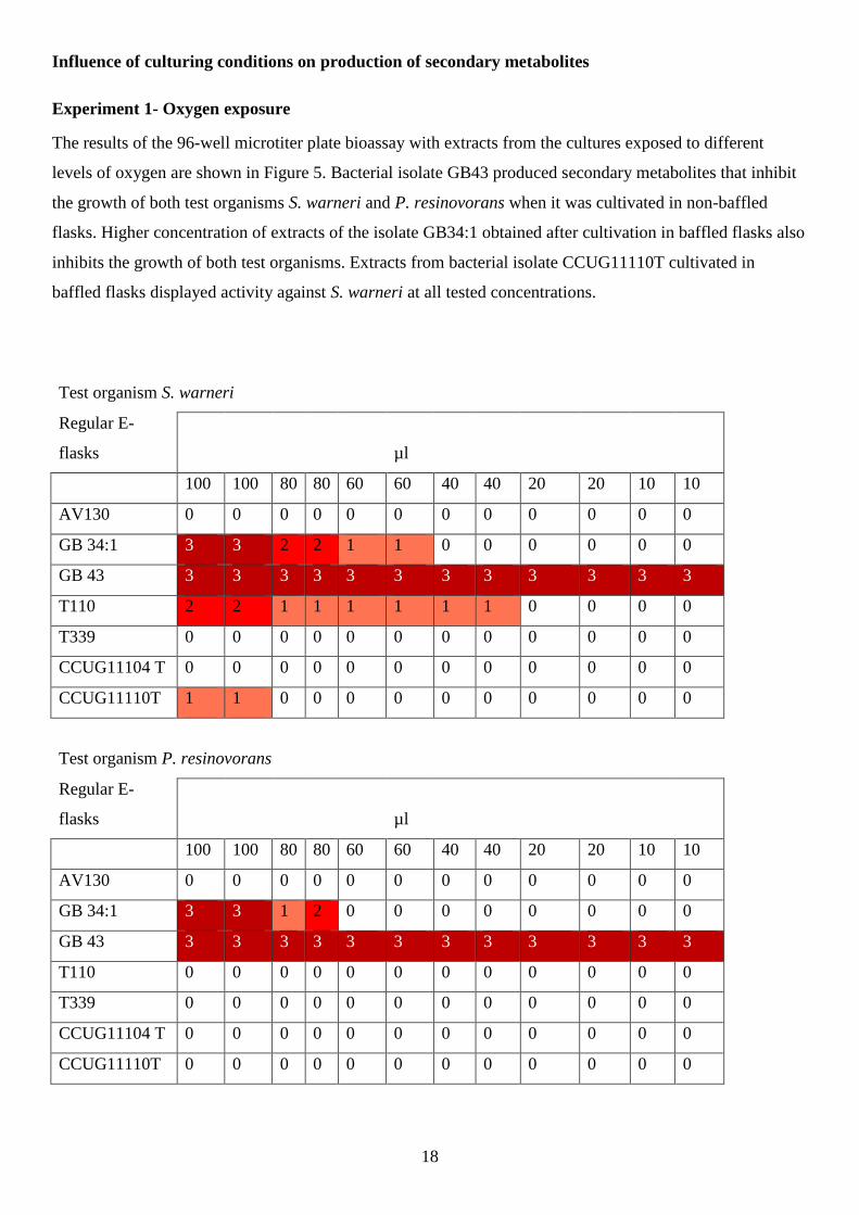
Influence of culturing conditions on production of secondary metabolites
Experiment 1- Oxygen exposure
The results of the 96-well microtiter plate bioassay with extracts from the cultures exposed to different
levels of oxygen are shown in Figure 5. Bacterial isolate GB43 produced secondary metabolites that inhibit
the growth of both test organisms S. warneri and P. resinovorans when it was cultivated in non-baffled
flasks. Higher concentration of extracts of the isolate GB34:1 obtained after cultivation in baffled flasks also
inhibits the growth of both test organisms. Extracts from bacterial isolate CCUG11110T cultivated in
baffled flasks displayed activity against S. warneri at all tested concentrations.
Test organism S. warneri
Regular E-
flasks µl
100 100 80 80 60 60 40 40 20 20 10 10
AV130 0 0 0 0 0 0 0 0 0 0 0 0
GB 34:1 3 3 2 2 1 1 0 0 0 0 0 0
GB 43 3 3 3 3 3 3 3 3 3 3 3 3
T110 2 2 1 1 1 1 1 1 0 0 0 0
T339 0 0 0 0 0 0 0 0 0 0 0 0
CCUG11104 T 0 0 0 0 0 0 0 0 0 0 0 0
CCUG11110T 1 1 0 0 0 0 0 0 0 0 0 0
Test organism P. resinovorans
Regular E-
flasks µl
100 100 80 80 60 60 40 40 20 20 10 10
AV130 0 0 0 0 0 0 0 0 0 0 0 0
GB 34:1 3 3 1 2 0 0 0 0 0 0 0 0
GB 43 3 3 3 3 3 3 3 3 3 3 3 3
T110 0 0 0 0 0 0 0 0 0 0 0 0
T339 0 0 0 0 0 0 0 0 0 0 0 0
CCUG11104 T 0 0 0 0 0 0 0 0 0 0 0 0
CCUG11110T 0 0 0 0 0 0 0 0 0 0 0 0

19
Test organism S. warneri
Baffled flasks µl
100 100 80 80 60 60 40 40 20 20 10 10
AV130 3 3 2 2 1 1 0 0 0 0 0 0
GB 34:1 3 3 1 1 0 0 0 0 0 0 0 0
GB 43 1 1 0 0 0 0 0 0 0 0 0 0
T110 3 3 3 3 2 2 2 2 0 0 0 0
T339 0 0 0 0 0 0 0 0 0 0 0 0
CCUG11104 T 0 0 0 0 0 0 0 0 0 0 0 0
CCUG11110T 3 3 3 3 3 3 3 3 2 2 1 1
Test organism P. resinovorans
Baffled flasks µl
100 100 80 80 60 60 40 40 20 20 10 10
AV130 0 0 0 0 0 0 0 0 0 0 0 0
GB 34:1 3 3 3 3 0 0 0 0 0 0 0 0
GB 43 0 0 0 0 0 0 0 0 0 0 0 0
T110 2 2 2 2 2 2 2 2 1 1 0 0
T339 0 0 0 0 0 0 0 0 0 0 0 0
11104 T 1 1 1 1 0 0 0 0 0 0 0 0
CCUG11110T 0 0 0 0 0 0 0 0 0 0 0 0
Figure 5: Inhibition of the S. warneri and P. resinovorans growth by extracts of isolates cultured with
varying oxygen access. The activity was graded with following scale: 0 – bacterial growth not inhibited at
all; 1 – little inhibition of bacterial growth; 2 – intermediate inhibition of bacterial growth; 3 – full
inhibition, no bacterial growth at all.
Experiment 2- Different growth media composition
In general, only the extracts of isolates CCUG11110T and GB43 inhibited the growth of both test
organisms. The bacterial isolate CCUG11110T produced metabolites that inhibited the growth of both test
organisms when cultivated in the medium AGS whereas metabolites from GB43 cultured in AGS and GS
correspondingly inhibited the growth of test organisms S. warneri and P. resinovorans (Figure 6).

20
Test organism S. warneri µl
100 100 80 80 60 60 40 40 20 20 10 10
AGS GB43 3 3 3 3 3 3 2 2 2 2 1 1
CCUG11110T 3 3 3 3 3 3 3 2 1 1 1 1
GB34:1 1 1 1 1 1 1 1 0 0 0 0 0
GB59 0 0 0 0 0 0 0 0 0 0 0 0
GS GB43 3 3 3 3 3 3 3 3 0 0 0 0
CCUG11110T 1 1 0 0 0 0 0 0 0 0 0 0
GB34:1 0 0 0 0 0 0 0 0 0 0 0 0
GB59 0 0 0 0 0 0 0 0 0 0 0 0
Test organism
P. resinovorans µl
100 100 80 80 60 60 40 40 20 20 10 10
AGS GB43 3 3 3 3 3 3 3 3 3 3 0 0
CCUG11110T 2 2 1 1 1 1 1 1 1 1 1 1
GB34:1 0 0 0 0 0 0 0 0 0 0 0 0
GB59 0 0 0 0 0 0 0 0 0 0 0 0
GS GB43 3 3 3 3 3 3 2 3 0 0 0 0
CCUG11110T 0 0 0 0 0 0 0 0 0 0 0 0
GB34:1 0 0 0 0 0 0 0 0 0 0 0 0
GB59 0 0 0 0 0 0 0 0 0 0 0 0
Figure 6: Inhibition of the S. warneri and P. resinovorans growth by extracts of isolates cultured in different
culture media. The activity was graded with following scale: 0 – bacterial growth not inhibited at all; 1 –
little inhibition of bacterial growth; 2 – intermediate inhibition of bacterial growth; 3 – full inhibition, no
bacterial growth at all.
Experiment 3- Culturing in the presence of various supplements
The bacterial isolate T110 produced metabolite(s) active against S. warneri after being exposed to the
supplements compared to control (Figure 7). The addition of ScCl3 down-regulated the activity of the
bacterial isolates GB34:1 against S. warneri (Figure 7), and the same observation was noticed for bacterial
isolate T110 against the other test organism P. resinovorans (Figure 7). Also GBL deactivated the
metabolites from T110 whereas Triclosan triggered the activity against P. resinovorans (Figure 7).

21
Test organism S. warneri µl
100 100 80 80 60 60 40 40 20 20 10 10
CCUG11110T Control 3 3 3 3 3 3 3 3 2 2 1 1
Triclosan 3 3 3 3 3 3 3 3 3 3 3 3
ScCl3 3 3 3 3 3 3 2 2 1 1 1 0
GBL 3 3 3 3 3 3 3 3 1 1 1 1
GB34:1 Control 3 3 3 3 2 2 2 2 0 0 0 0
Triclosan 3 3 3 3 3 3 3 3 3 3 3 3
ScCl3 0 0 0 0 0 0 0 0 0 0 0 0
GBL 3 3 3 3 3 3 3 2 0 0 0 0
T110 Control 2 2 1 1 1 0 0 0 0 0 0 0
Triclosan 3 3 3 3 3 3 3 3 3 3 2 2
ScCl3 3 3 3 3 3 3 3 3 3 3 3 3
GBL 3 3 3 3 3 3 3 3 3 3 3 2
GB43 Control 3 3 3 3 3 3 3 3 1 1 0 0
Triclosan 3 3 3 3 3 3 3 3 3 3 3 3
ScCl3 3 3 3 3 3 3 3 3 0 0 0 0
GBL 3 3 3 3 3 3 3 3 0 0 0 0
Test organism P. resinovorans µl
100 100 80 60 60 40 40 20 20 10 10
CCUG11110T Control 3 3 3 3 3 3 3 3 3 3 2 2
Triclosan 3 3 3 3 3 3 3 3 3 3 3 3
ScCl3 3 3 3 3 3 3 2 1 0 0 0 0
GBL 3 3 3 3 3 3 3 3 3 3 1 1
GB34:1 Control 3 3 3 3 3 3 3 3 1 1 0 0
Triclosan 3 3 2 2 2 2 2 1 0 0 0 0
ScCl3 1 1 0 0 0 0 0 0 0 0 0 0
GBL 3 3 3 3 3 3 3 3 3 3 0 0
T110 Control 1 1 0 0 0 0 0 0 0 0 0 0
Triclosan 3 3 3 3 3 3 3 3 0 0 0 0
ScCl3 0 0 0 0 0 0 0 0 0 0 0 0
GBL 0 0 0 0 0 0 0 0 0 0 0 0
GB43 Control 3 3 3 3 3 3 3 3 3 3 0 0
Triclosan 3 3 3 3 3 3 3 3 3 3 0 0
ScCl3 3 3 3 3 3 3 3 3 3 3 0 0
GBL 3 3 3 3 3 3 3 3 3 3 0 0
Figure 7: Inhibition of the S. warneri and P. resinovorans growth by extracts of isolates cultured on GPS
medium with different supplements. The activity was graded with following scale: 0 – bacterial growth not
inhibited at all; 1 – little inhibition of bacterial growth; 2 – intermediate inhibition of bacterial growth; 3 –
full inhibition, no bacterial growth at all.

22
Microtiter plate assay against pathogenic organisms
The HPLC separated fractions of extracts from the supplement experiment were tested against several
pathogenic microorganisms. The additions of Triclosan to the culture with bacterial isolate CCUG11110T
resulted in two new substances active against pathogen F (red encircled activity area Figure 8). The active
substance (blue encircled activity area Figure 8) was established to be Triclosan.
Separation of extracts produced by the bacterial isolate T110 and T110 supplemented with ScCl3 both
resulted in detecting antimicrobial affect. However, the retention time for active fractions was different for
these two samples suggesting production of two different active metabolites (orange and purple encircled
activity area Figure 9).
Figure 8: HPLC chromatograms of the isolate CCUG11110T extracts cultured without supplements (top)
and with addition of Triclosan (bottom) and corresponding activity against a panel of pathogenic
microorganisms. The green encircled fractions in the top chromatogram show activity against pathogen G.
The blue encircled fractions in bottom chromatogram were active against almost all of the test pathogens
and the two red encircled fractions were only active against pathogen F. The remaining detected activity was
most likely due to cyclic dipeptides derived from media components.

23
Figure 9: HPLC chromatograms of the isolate T110 extracts cultured without supplements (top) and with
addition of ScCl3 (bottom) and corresponding activity against a panel of pathogenic microorganisms. The
orange and purple circles show the activity against the test organism F at different retention time for top and
bottom chromatogram. The remaining detected activity was most likely due to cyclic dipeptides derived
from media components.

24
DISCUSSION
The aim of this study was to trigger several bacterial isolate to produce different secondary metabolites,
hopefully also including novel compounds that may be used as future antibiotics as a countermeasure to the
increasing antibiotic resistance among an alarmingly large proportion of microbial pathogens. Three
experiments were performed, in which the influence of different oxygen levels, different media composition
and adding different supplements to the cultures on the production of active metabolites was tested. The
hypothesis was that a change in activity against the test organisms should be distinguished if it is as agreed
with similar research from the literature (Bode et al. 2002, Craney et al. 2012, de Kievit and Iglewski 2000,
Inaoka and Ochi 2011). Overall it seemed that the experiment with varying supplements added to the
cultures generated the most powerful antimicrobial effect for the highest number of tested isolates.
Especially, addition of Triclosan yielded a number of active substances in the extracts of all bacterial
isolates. One of the detected substances was established as Triclosan (Figure 8) after LC- MS analysis and
molecular weight comparison with substances available for comparison in the database Antibase. Also the
determination of substances by LC-MS revealed the higher diversity of produced secondary metabolites. It
can confirm the approach referred to as OSMAC (Bode et al. 2002): that one microorganism is capable of to
generate several active metabolites under varying culture conditions.
The reference isolate CCUG11110T, denotation for Streptomyces coelicolor, showed its potential to produce
antibiotics, particularly in the experiments using supplements (Figure 7). Similar experiments in reviews
have shown the same outcome in successful activity against several test organisms (Craney et al. 2012,
Kawai et al. 2007 and Takano et al. 2000). Concerning non-Streptomyces and their optimistically capacity to
produce novel antibiotics (Tiwari and Gupta 2012), bacterial isolate GB34:1 (representing Nocardia sp)
displayed promising antibiotic production in the current research both in terms of the detected genes coupled
to the secondary metabolism (Figure 2) and activity against the test organisms (Figure 5 and 7). It must be
further investigated what kind of active metabolites were produced by this organism.
The initial test consisted of performing PCRs of four genes (PKS-1, PKS-2, NRPS and DTS) encoding for
enzymes that have been shown to be involved in the production of secondary metabolites. Isolate GB43,
belonging to the genus Pseudomonas, was tested positive for all four genes. Its potential was also reflected
in the bioassays. Metabolites produced by the isolate GB43 showed activity against both S. warneri and P.
resinovorans in all bioassays except in the inhibition zone test against P. resinovorans and also in the
microtiter plate inhibition assay cultured in baffled flasks (Figure 5). Since none of the isolates apart from
one (AV226:2, figure not shown) were inhibiting the growth of P. resinovorans in the inhibition zone test, it
can be concluded that the growth of the test organism may have been too strong or simply that no effective
antibiotics against P. resinovorans could be found. Based on the results from the bioassay of the extracts
produced by this isolate that originated from cultures grown at different oxygen level, one can conclude that

25
Pseudomonas sp. apparently grows well in the presence of high oxygen level but are not able to produce
antimicrobial metabolites.
GBL used in this report as a supplement has a similar core structure as the family of signal molecule AHL.
Mostly gram-negative bacteria use AHL in the quorum-sensing signaling (Yajima, A. 2014) whereas many
Gram-positive bacteria use GBLs as signaling molecules for regulation of antibiotic production (Takano et
al. 2000). It is consistent that isolate GB43, which is a Gram-negative bacterium, were activated in the
production of metabolites in the supplement experiment after DHM- furanone was added to the culture.
DHM- furanone activated the secondary metabolite production for all bacterial isolates except T110 against
P. resinovorans. Both that absence of activity for bacterial isolate T110 and the poor activity of all isolates
against P. resinovorans in general could possibly be explained by that P. resinovorans may have used a
quorum-sensing mechanism that protected its growth in the presence of active metabolites. Pseudomonas
species, like other Gram-negative bacteria, are also known to use efflux pumps to effectively reject the
added antimicrobial agents, which had led to the enormous problems with nosocomial infections caused by
antibiotic resistant bacteria (Köhler et al. 1997, Nikaido 1996).
CONCLUSION AND FUTURE PERSPECTIVES
Different culturing conditions and added supplements positively affected the cultivated bacterial isolates in a
way that the production of secondary metabolites with antimicrobial properties was in general enhanced.
However, the experiments have to be repeated in order to obtain more reliable result. Also, more various
culture media and culture conditions should be included to clearer detect differences. The substances that
were separated after the supplement experiments will be further investigated by Nuclear Magnetic
Resonance (NMR) to closer elucidate their chemical structures. If they prove to be novel they will undergo
additional tests such as for example Minimum Inhibitory Concentration (MIC) that allows to established the
minimal concentration of the compound, at which it totally inhibit the growth of tested pathogen.

26
ACKNOWLEDGMENT
I would like to thank my supervisor Joakim Bjerketorp and assistant supervisor Jolanta Levenfors at the
Department of Microbiology at Swedish University of Agriculture Sciences (SLU) for their most
appreciated help during this project. Also Pierre Andersson and Anders Broberg at the Department of
Chemistry at SLU will be thanked for their help to interpret the results from the chemical analysis. Lastly I
will also thank my examiner Bengt Guss and all of the other employees at the Department of Microbiology
at SLU.

27
REFERENCES
Ayuso-Sacido, A. and Genilloud, O. (2005) New PCR Primers for the Screening of NRPS and PKS-I
Systems in Actinomycetes: Detection and Distribution of These Biosynthetic Gene Sequences in Major
Taxonomic Groups. Microbial Ecology. 49(1):10-24
Baltz, R. H. (2008) Renaissance in antibacterial discovery from actinomycetes. Current opinion in
Pharmacology. 8(5): 557-563
Bentley, SD., Chater, KF., Cerdeño-Tárraga, AM., Challis, GL., Thomson, NR., James, KD., Harris, DE.,
Quail, MA., Kieser, H., Harper, D., Bateman, A., Brown, S., Chandra, G., Chen, CW., Collins, M., Cronin,
A., Fraser A, Goble A, Hidalgo J, Hornsby T, Howarth S, Huang CH, Kieser T, Larke L, Murphy L, Oliver
K, O'Neil S, Rabbinowitsch E, Rajandream MA, Rutherford K, Rutter S, Seeger K, Saunders D, Sharp S,
Squares R, Squares S, Taylor K, Warren T, Wietzorrek A, Woodward J, Barrell BG, Parkhill J, Hopwood
DA. (2002) Complete genome sequence of the model actinomycete Streptomyces coelicolor A3(2). Nature.
417(6885):141-147.
Bérdy, J. (2005) Bioactive Microbial Metabolites. Journal of Antibiotics. 58(1): 1–26
Bode, H. B., Bethe, B., Höfs, R. and Zeeck, A. (2002) Big Effects from Small Changes: Possible Ways to
Explore Nature's Chemical Diversity. Chembiochem. 3(7): 619-27.
Bush, K. (2010) The coming of age of antibiotics: discovery and therapeutic value. Annals of the New York
academy of sciences. 1213(1): 1-4
Craney, A., Ozimok, C., Pimentel-Elardo, SM., Capretta, A. and Nodwell, JR. (2012) Chemical perturbation
of secondary metabolism demonstrates important links to primary metabolism. Chemistry and Biology.
19(8): 1020-1027
Donadioa, S., Monciardini, P., Alduina, R., Mazzaa, P., Chiocchini, C., Cavaletti, L., Sosioa, M. and Puglia,
A. (2001) Microbial technologies for the discovery of novel bioactive metabolites. Journal of Biotechnology
99 (3): 187-198
de Kievit T. R. and Iglewski B. H. (2000) Bacterial Quorum Sensing in Pathogenic Relationships.
Infection and Immunity. 68(9): 4839-4849

28
Enright, M. C., Robinson, A. D., Randle, G., Feil, E. J., Grundmann, H. and Spratt, B. G. (2002) The
Evolutionary History of Methicillin-Resistant Staphylococcus aureus (MRSA). National Academy of
Sciences. 99(11): 7687-7692
Fleming, A. (1929) On the Antibacterial Action of Cultures of a Penicillium, with Special Reference to their
Use in the Isolation of B. influenzæ. The British Journal of Experimental Pathology. 10(3): 226–236.
Gajraj, S. B., Alpana, B., Vijay, K. & Omprakash, G. (2013) Isolation, purification and partial
characterization of an antifungal agent produced by salt-tolerant alkaliphilic Streptomyces violascens.
Proceedings of the National Academy of Sciences, India Section B: Biological Sciences. 83(1): 109-117
Genilloud, O., González, I., Salazar, O., Martín, J., Tormo, J. R. and Vicente, F. (2011) Current approaches
to exploit actinomycetes as a source of novel natural products. Journal of Industrial Microbiology &
Biotechnology. 38(3): 375-389
Inaoka, T. and Ochi, K. (2011) Scandium Stimulates the Production of Amylase and Bacilysin in Bacillus
subtilis. Applied and Environmental Microbiology. 77(22): 8181-8183
Kawai, K., Wang, G., Okamoto, S. and Ochi K. (2007) The rare earth, scandium, causes antibiotic
overproduction in Streptomyces spp. FEMS Microbiology Letters. 274(2): 311–315
Koehn, F. E. and Carter, G. T. (2005) The evolving role of natural products in drug discovery. Nature
Reviews Drug Discovery. 4(3): 206-220
Kumar, V., Bisht, G. S. and Gusain, O. (2013) Terrestrial actinomycetes from diverse locations of
Uttarakhnad, India: Isolation and screening for their antibacterial activity. Iran Journal of Microbiology.
5(3): 299–308.
Köhler, T., Michéa-Hamzehpour, M., Henze, U., Gotoh, N., Kocjancic Curty, L. and Pechère, J-C. (1997)
Characterization of MexE–MexF–OprN, a positively regulated multidrug efflux system of Pseudomonas
aeruginosa. Molecular Microbiology. 23(2): 345-354
Livermore, D. M. (2011) Discovery research: the scientific challenge of finding new antibiotics. Journal of
Antimicrobial Chemotherapy. 66 (9): 1941-1944.

29
Mahajan, GB. and Balachandran, L. (2012) Antibacterial agents from actinomycetes - a review. Frontier in
Bioscience (Elite Ed). 1(4): 240-53.
Manivasagana, P., Venkatesanb, J., Sivakumarc, K., Kima S-K. (2013) Marine Actinobacterial metabolites:
Current status and future perspectives. Microbiological Research. 168 (6): 311-32
Nikaido, H. (1996) Multidrug Efflux Pumps of Gram-Negative Bacteria. Journal of Bacteriology,
178(20):5853–5859
Neu, H. C. (1992) The Crisis in Antibiotic Resistance. Science. 257(5073): 1064-1073
Ochi, K., Hosaka, T. (2013) New strategies for drug discovery: activation of silent or weakly expressed
microbial gene clusters. Applied Microbiological and Biotechnology. 97(1): 87-98
Persidis, A. (1998) High-throughput screening. Nature biotechnology. 16(2): 488-489
Pohanka, A., Broberg, A., Johansson, M., Kenne, L., and Levenfors, J. (2005) Pseudotrienic Acids A and B,
Two Bioactive Metabolites from Pseudomonas sp. MF381-IODS. Journal of Natural Products. 68(9): 1380-
1385
Raaijmakers J. M., Weller D. M. and Thomashow L. S. (1997) Frequency of Antibiotic-Producing
Pseudomonas spp. in Natural Environments. Applied and Environmental Microbiology. 63(3): 881-887
Subramani, R., Aalbersberg, W. (2012) Marine actinomycetes: An ongoing source of novel bioactive
metabolites. Microbiological Research. 167(10): 571–580
Stainer, R. Y., Palleroni, N. J., Doudoroff, M. (1966) The aerobic pseudomonas: A taxonomic study.
Journal of General Microbiology. 43(2): 159-271
Takano, E., Nihira, T., Hara, Y., Jones, J. J., Gershater, C. J. L., Yamada, Y. and Bibb, M. (2000)
Purification and Structural Determination of SCB1, a γ-Butyrolactone That Elicits Antibiotic Production in
Streptomyces coelicolor A3(2). The Journal of Biological Chemistry. 275(15): 11010–11016
Tiwari, K. and Gupta, R. K. (2012) Rare actinomycetes: a potential storehouse for novel antibiotics. Critical
Reviews in Biotechnology. 32(2): 108–132

30
Xie, P., Ma, M., Rateb, M. E., Shaaban, K. A., Yu, Z, Huang, S., Zhao, L., Zhu, X., Yan, Y., Peterson, R.
M., Lohman, J. R., Yang, D.,Yin, M., Rudolf, J. D., Jiang, Y., Duan, Y., and Shen, B. (2014) Biosynthetic
Potential-Based Strain Prioritization for Natural Product Discovery: A Showcase for Diterpenoid-Producing
Actinomycetes. Journal of Natural Products. 77(2): 377-87
Yajima, A. (2014) Recent progress in the chemistry and chemical biology of microbial signaling molecules:
quorum-sensing pheromones and microbial hormones. Tetrahedron Letters. 55(17): 2773–2780
Zotchev, S. B. (2012) Marine actinomycetes as an emerging resource for the drug development
pipelines. Journal of Biotechnology. 158 (4): 168– 175