Antibacterial Activity of Silver Nanoparticles of Seaweeds...antibacterial activity of silver...
12
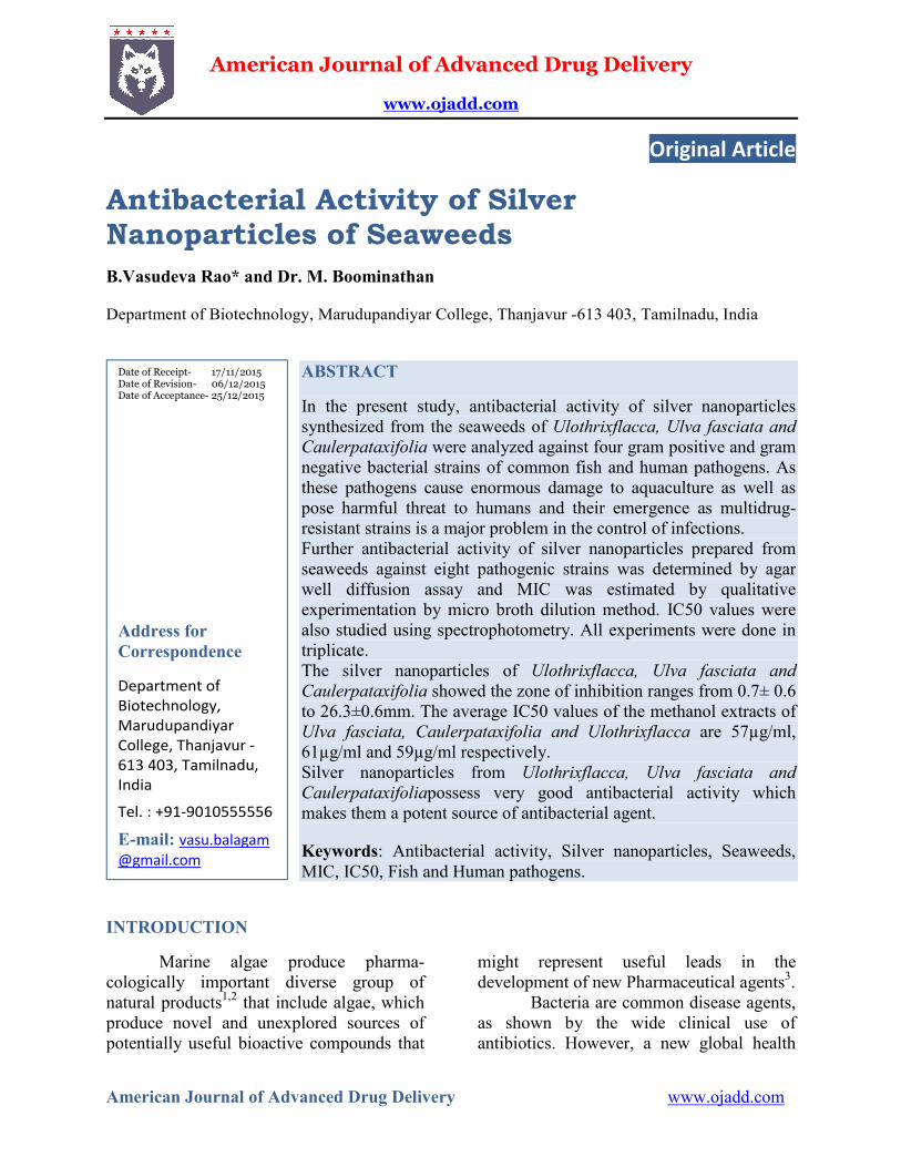
American Journal of Advanced Drug Delivery www.ojadd.com American Journal of Advanced Drug Delivery www.ojadd.com Original Article Antibacterial Activity of Silver Nanoparticles of Seaweeds B.Vasudeva Rao* and Dr. M. Boominathan Department of Biotechnology, Marudupandiyar College, Thanjavur -613 403, Tamilnadu, India ABSTRACT In the present study, antibacterial activity of silver nanoparticles synthesized from the seaweeds of Ulothrixflacca, Ulva fasciata and Caulerpataxifolia were analyzed against four gram positive and gram negative bacterial strains of common fish and human pathogens. As these pathogens cause enormous damage to aquaculture as well as pose harmful threat to humans and their emergence as multidrug- resistant strains is a major problem in the control of infections. Further antibacterial activity of silver nanoparticles prepared from seaweeds against eight pathogenic strains was determined by agar well diffusion assay and MIC was estimated by qualitative experimentation by micro broth dilution method. IC50 values were also studied using spectrophotometry. All experiments were done in triplicate. The silver nanoparticles of Ulothrixflacca, Ulva fasciata and Caulerpataxifolia showed the zone of inhibition ranges from 0.7± 0.6 to 26.3±0.6mm. The average IC50 values of the methanol extracts of Ulva fasciata, Caulerpataxifolia and Ulothrixflacca are 57µg/ml, 61µg/ml and 59µg/ml respectively. Silver nanoparticles from Ulothrixflacca, Ulva fasciata and Caulerpataxifoliapossess very good antibacterial activity which makes them a potent source of antibacterial agent. Keywords: Antibacterial activity, Silver nanoparticles, Seaweeds, MIC, IC50, Fish and Human pathogens. INTRODUCTION Marine algae produce pharma- cologically important diverse group of natural products 1,2 that include algae, which produce novel and unexplored sources of potentially useful bioactive compounds that might represent useful leads in the development of new Pharmaceutical agents 3 . Bacteria are common disease agents, as shown by the wide clinical use of antibiotics. However, a new global health Date of Receipt- 17/11/2015 Date of Revision- 06/12/2015 Date of Acceptance- 25/12/2015 Address for Correspondence Department of Biotechnology, Marudupandiyar College, Thanjavur - 613 403, Tamilnadu, India Tel. : +91-9010555556 E-mail: vasu.balagam @gmail.com
Transcript of Antibacterial Activity of Silver Nanoparticles of Seaweeds...antibacterial activity of silver...
American Journal of Advanced Drug Delivery
www.ojadd.com
American Journal of Advanced Drug Delivery www.ojadd.com
Original Article
Antibacterial Activity of Silver Nanoparticles of Seaweeds
B.Vasudeva Rao* and Dr. M. Boominathan
Department of Biotechnology, Marudupandiyar College, Thanjavur -613 403, Tamilnadu, India
ABSTRACT
In the present study, antibacterial activity of silver nanoparticles synthesized from the seaweeds of Ulothrixflacca, Ulva fasciata and Caulerpataxifolia were analyzed against four gram positive and gram negative bacterial strains of common fish and human pathogens. As these pathogens cause enormous damage to aquaculture as well as pose harmful threat to humans and their emergence as multidrug-resistant strains is a major problem in the control of infections. Further antibacterial activity of silver nanoparticles prepared from seaweeds against eight pathogenic strains was determined by agar well diffusion assay and MIC was estimated by qualitative experimentation by micro broth dilution method. IC50 values were also studied using spectrophotometry. All experiments were done in triplicate. The silver nanoparticles of Ulothrixflacca, Ulva fasciata and Caulerpataxifolia showed the zone of inhibition ranges from 0.7± 0.6 to 26.3±0.6mm. The average IC50 values of the methanol extracts of Ulva fasciata, Caulerpataxifolia and Ulothrixflacca are 57µg/ml, 61µg/ml and 59µg/ml respectively. Silver nanoparticles from Ulothrixflacca, Ulva fasciata and Caulerpataxifoliapossess very good antibacterial activity which makes them a potent source of antibacterial agent.
Keywords: Antibacterial activity, Silver nanoparticles, Seaweeds, MIC, IC50, Fish and Human pathogens.
INTRODUCTION
Marine algae produce pharma-cologically important diverse group of natural products1,2 that include algae, which produce novel and unexplored sources of potentially useful bioactive compounds that
might represent useful leads in the development of new Pharmaceutical agents3.
Bacteria are common disease agents, as shown by the wide clinical use of antibiotics. However, a new global health
Date of Receipt- 17/11/2015 Date of Revision- 06/12/2015 Date of Acceptance- 25/12/2015
Address for Correspondence
Department of Biotechnology, Marudupandiyar College, Thanjavur -613 403, Tamilnadu, India
Tel. : +91-9010555556
E-mail: vasu.balagam @gmail.com
Rao et al______________________________________________________ ISSN 2321-547X
AJADD[3][06][2015] 296-307
problem has arisen as indiscriminant antibiotic use and the remarkable ability of bacteria to acquire resistance (via genetic mutation or gene acquisition) lower these drugs’ effectiveness. Clearly, new classes of antibiotics with novel structures are needed to combat this trend.
The antimicrobial activity of microalgae extracts is generally assayed using various organicsolvents4. An organic solvent always provides a higher efficiency in extracting compounds for antimicrobial activity as compared to aqueous extract5,6 Marine algae are a rich source of novel bioactive compounds which may find several applications7,8. Secondary or primary metabolites from these organisms may be potential bioactive compounds of interest for the pharmacological industry. The cell extracts and active constituents of various algae have been shown to have antibacterial activity against Gram positive and Gram negative bacteria. The production of bioactive metabolites is considered to be a response to ecological pressures such as competition, predation deterrence, and reproduction. Such chemicals are often common in sessile eukaryotic organisms like marine sponges and corals, seaweeds, and terrestrial plants9,10. Silver nanoparticles (Ag-NPs) have been known for its inhibitory and bactericidal effects in the past decades11. Antibacterial activity of silver containing materials can be applied in medicine for reduction of infections on the burn treatment12,13. AgNPs have been synthesized using marine microalgae and screened for its antibacterial activity14. Noble metal nanoparticles show unique electronic, optical, magnetic and chemical properties, which differ considerably from those of the corresponding bulk materials15-17 and hence preparations of noble metal nanoparticles are of technological importance. Recently inorganic nanoparticles protected by organic
ligands have attracted much interest due to their diverse technological applications18-20.
Seaweeds contain various organic and inorganic substances which can benefit human health 21. Due to the high amount of vitamins and minerals present in the green seaweed it is used widely in agriculture, pharmaceutical, biomedical, and nutraceutical industries22. About 250 macro algal species have been commercially utilized worldwide and about 150 species are favorably consumed as human food23.
Seaweeds concentrate minerals and trace elements from marine water and convert them in organic forms as they grow in a mineral-rich medium24. The numerous elements coming from the sea are Ca, Cl, Cu, I, Mg, Mn, Na, P, S and Zn25. They selectively absorb elements like Na, K, Ca, Mg, I and Br from the seawater and accumulate them in their thalli. The accumulated elements vary from species to species. For example, large quantities of K and I are taken up by many brown seaweeds and Ca and Br by red algae. The possibility of free-radical involvement in the antibacterial activity of silver nanoparticles (Ag-NPs) has been previously reported26, but the underlying mechanism and characteristics remain unclear. Silver ions cause the release of K+ ions from bacteria; thus, the bacterial plasma or cytoplasmic membrane, which is associated with many important enzymes and DNA, is an important target site of silver ions27- 30. After all intensive studies on the antibacterial activities on various kinds of seaweeds, chlorophyceae members showed well antibacterial activity on both gram positive and gram negative bacteria.
In the present study the antibacterial activity of seaweeds (Ulothrixflacca, Ulva fasciata and Caulerpataxifolia) extracts was studied against eight common pathogens of fish and human.

Rao et al______________________________________________________ ISSN 2321-547X
AJADD[3][06][2015] 296-307
MATERIALS AND METHODS
Seaweed collection Ulothrixflacca, Ulva fasciata and
Caulerpataxifoliaseeweeds collected from the rocky platforms at tenneti park located at Vishakhapatnam coastal banks. Species identification was done by referring the printed journal (Y. Sarojini, P.SanthaRao, B. Sujatha K.Lakshminarayana Distribution and diversity of marine macro algae in relation to environmental factors at Visakhapatnam coast. Seaweed Res. Utiln., 35 (1&2): 55-64, 2013 and authentified at Department of Botany, Andhra University, Visakhapatnam, Andhra Pradesh.
The three seaweed species were collected during the months of July and August of the year 2013. They were carefully picked from the submerged rocks of Vishakhapatnam coastal banks and washed with fresh water thoroughly to remove the cell debris, sand and other particles. They were then kept in the ice bags and transferred to the laboratory. In laboratory, seaweeds were again washed with distilled water carefully and kept for shade dry. After drying, the seaweeds were stored in desiccator for further use.
Preparation of silver nanoparticles
1gm of each seaweed powder was extracted with methanol using soxhlet and filtered. 1mM Silver nitrate solution was added to the filtrates slowly under magnetic stirring conditions for even coating of silver and subjected to heating at 12°C for 10 min. The extracts are used as reducing and stabilizing agent for 1mM of Silver nitrate. This one pot green synthesis was the modified method followed by Vigneshwaran et al. 31.
Pathogen selection
The increasing resistivity of several pathogens against the present antibiotics insists the need for the new molecules to
fight against those pathogens. Especially in aquaculture, the pathogenic bacteria cause immense damage to the pond. Of all the aqua pathogenic bacteria, very few shows pathogenicity over humans.Such kind of pathogens which uses both aqua species and humans as their hosts were selected for the present study. Four gram positive and four gram negative pathogens were procured from MTCC Chandigarh.
Evaluation of antimicrobial activity of the bio-synthesized silver nanoparticles
The prepared algal extracts (Ulothrixflacca, Ulva fasciata and Caulerpataxifolia) were tested for Minimum Inhibitory Concentration (MIC), IC50 values and antibacterial activity by well diffusion method against pathogenic bacterial strains.
Test organisms
Sl. no
Organism Code Type
1 Staphylococcus aureus MTCC 3160 gm positive
2 Listeria monocytogenes MTCC 839 gm positive
3 Clostridium perfringens MTCC – 450 gm positive
4 Mycobacterium spp MTCC – 290 gm positive
5 Delftiaacidovorans MTCC- 104 gm negative
6 Edwardsiellatarda MTCC-2400 gm negative
7 Plesiomonasshigelloides MTCC-1737 gm negative
8 Vibrio vulnificus MTCC- 1145 gm negative
The pathogenic cultures were
subcultured into peptone broth and incubated at 37˚C to attain 105-106 CFU/ml using MacFarland’s standard and were used in further experiments.
Agar well-diffusion method
Muller Hinton Agar plates were swabbed with fresh cultures of pathogenic organisms using sterile cotton swab. Approximately 6-mm diameter of well was made with the help of sterile gel puncture.
Rao et al______________________________________________________ ISSN 2321-547X
AJADD[3][06][2015] 296-307
Using a micropipette, the prepared algal extracts were loaded at different concentrations. Ampicillin antibiotic (50µg/ml) was used as a positive control, followed by incubation at 37˚C for 18-24 h. After incubation effect of nanoparticles on tested bacterial culture were determined by measuring the zone of inhibition compared with control.
RESULTS
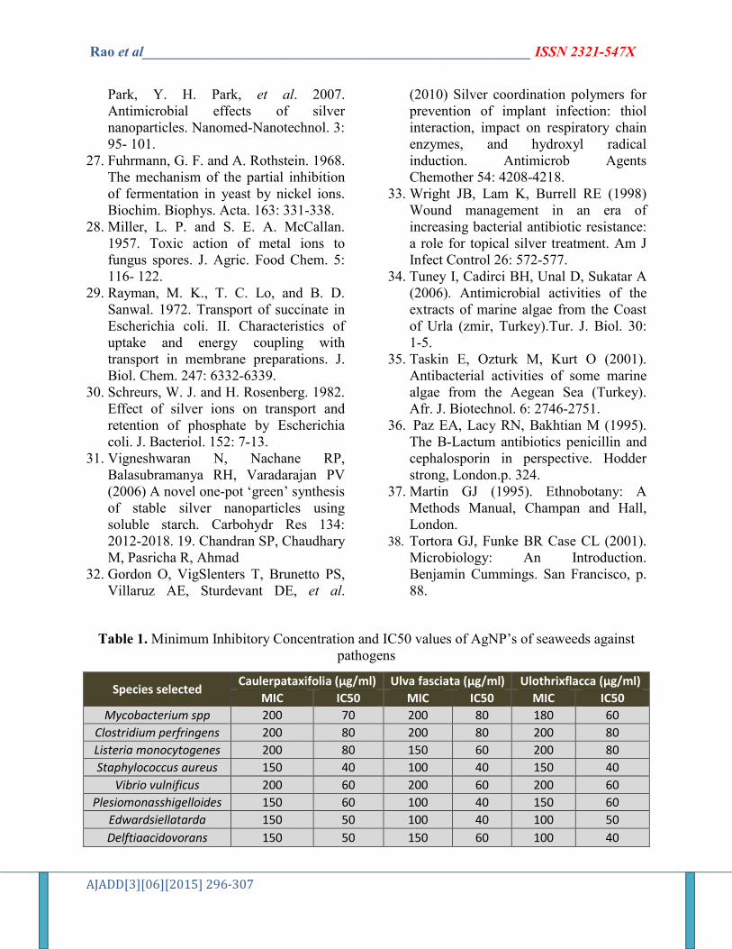
Minimum inhibitory concen-tration (MIC) is the lowest concentration of an antimicrobial that inhibits the visible growth of a microorganism after overnight incubation.IC50 value is the concentration of the extract required for 50% growth inhibition. The pathogenic cultures were subcultured into peptone broth and incubated at 37˚C to attain 105-106 CFU/ml using MacFarland’s standard and were used in further experiments.
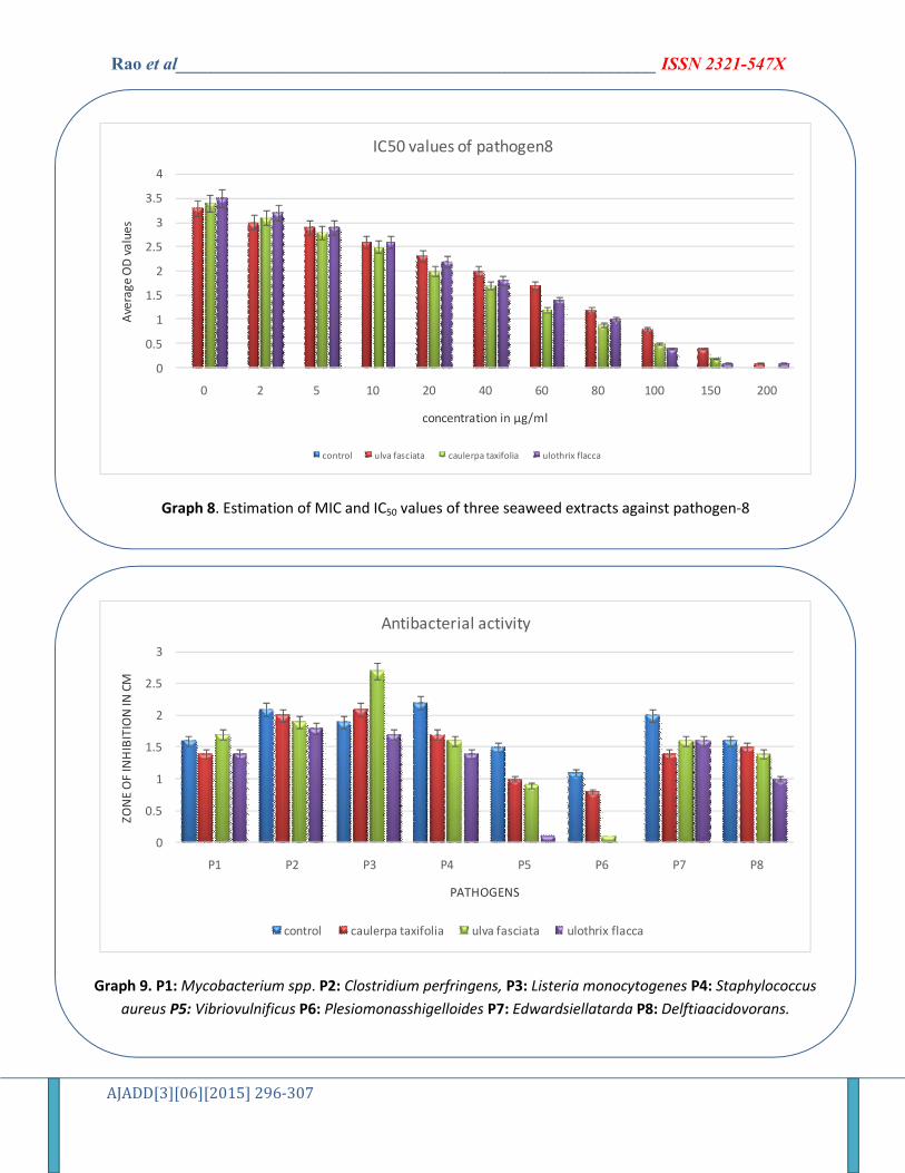
According to the IC50 values that are shown in the (Table-1), the average IC50 values of the methanol extracts of ulvafasciata, caulerpataxifolia and ulothrixflacca are 15µg/ml, 27µg/ml and 23µg/ml respectively. So these three respective concentrations were taken to study the zone of inhibition assay for all the eight pathogens (Table -2). Of all the three chlorophyta members, caulerpataxifolia showed maximum zone of inhibition activity when compared with the other two. On the whole the antibacterial activity is shown significantly more on the gram negative bacteria than the gram positive bacteria. Gram negative bacteria Vibriovulnificus and Plesiomonasshigelloides showed resistance against ulothrixflacca with no zone of inhibition (shown in graph-9).
DISCUSSION
The inhibitory action of silver compounds and silver ions had been
historically recognized and applied as a useful therapeutic agent for preventing wound infections. The inhibitory action of silver on bacterial cells is related to the strong interaction of silver with thiol groups present in key respiratory enzymes in bacteria32. Whereas, ano crystalline silver shows the most effective inhibitory action with a rapid inhibition rate33. The biosynthesized nanoparticles also exhibit well significant antibacterial activity. Ulva fasciata silver nanoparticles (57µg/ml) showed highest zone of inhibition(26.3±0.6mm) against Listeria monocytogenes. It was observed that Ulothrixflacca showed no activity on Plesiomonasshigelloides. Caulerpataxifolia showed significant activity on all the eight pathogens. The present study showed the gram positive microorganism was more susceptible to the extracts of the algae used. Tuney et al. (2006) also reported that Gram-positive bacteria were more effectively controlled by the extracts of algae used in their study compared to Gram-negative bacteria34. Similar observations were made by Taskin et al. (2001)35 and (Paz et al., 1995)36
indicating that the more susceptibility of Gram-positive bacteria to the algal extract was due to the differences in their cell wall structure and their composition. The presence of thick murine layer in the cell wall also prevents the entry of the inhibitors (Martin, 1995)37. In Gram-negative bacteria, the outer membrane acts as a barrier to many environmental substances including antibiotics (Tortora et al., 2001)38. The present study showed the use of biosynthesized nanoparticles to control several pathogens that affect the aquaculture economy. The active compounds in this activity need to be isolated and characterized to identify potential pharmaceutical applications for the bioactivity results reported in the present study. More studies regarding the application of these active compounds to the aquaculture gives better
Rao et al______________________________________________________ ISSN 2321-547X
AJADD[3][06][2015] 296-307
results in disease control and management. As the seaweeds are rich in various micro elements and minerals they can be administered to the aquatic environment. Among the three sea weeds Caulerpataxifolia exhibited better antibacterial activity when compared to the other two seaweed extracts.
CONCLUSION
From the present study we conclude that even at very small concentration (in µg/ml) AgNPs of three seaweeds (Ulva fasciata, Caulerpataxifolia and Ulothrixflacca) possess very good antibacterial activity which makes them a potent source of antibacterial agents against the common pathogens of fish and human. Of all three seaweeds Caulerpataxifolia exhibited significant activity when compared with the other two compounds. Also, synthesis of AgNPs can potentially eliminate the problem of chemical agents that may have adverse effects, thus making nanoparticles more compatible with the eco-friendly approach. Moreover the synthesized AgNPs enhance the therapeutic efficacy and strengthen the medicinal values of Ulva fasciata, Caulerpataxifolia and Ulothrixflacca. Hence, our results are promising and prove to be an important step in this direction as it decreases the load of multidrug resistant pathogens in aquaculture.
ACKNOWLEDGEMENT
We thanks to management of Department of Biotechnology, Marudupandiyar College, Thanjavur -613 403, Tamilnadu, India for providing experimental facilities.
REFERENCES
1. Ravikumar S, Krishnakumar S, Jacob Inbaneson S, Gnanadesigan M Antagonistic activity of marine
actinomycetes from Arabian Sea coast. Archives of Applied Science Research. 2010; 2(6):273-280.
2. Krishnakumar S, Premkumar J, Alexis Rajan R, Ravikumar S Optimization of potential antibiotic production by salt- tolerant actinomycetesStreptomyces sp. - MSU29 isolated from marine sponge. International J on Applied Bioengineering. 2011; 5(2):12-17.
3. Iwamoto C, YamadaT, Ito Y, Minoura K, Numata A Cytotoxic cytochalasans from a Penicilliumspecies separated from a marine alga. Tetrahedron. 2001; 57: 2904–2997.
4. Cordeiro RA, Gomes VM, Carvalho AFU, Melo VMM Effect of Proteins from the Red Seaweed Hypneamusciformis(Wulfen) Lamouroux on the Growth of Human Pathogen Yeasts. Brazilian Arch Boil Technol. 2006; 49(6): 915-921.
5. Masuda M, Abe T, Sato S, Suzuki T, Suzuki M Diversity of halogenated secondary metabolites in the red alga Laurencianipponica(Rhodomelaceae, Ceramiales). J of Phycology. 1997; 33: 196-208.
6. Lima-Filho JVM, Carvalho AFFU, Freitas SM, Melo VMM Antibacterial activity of extracts of six macroalgae from the northeastern Brazilian coast. Brazilian J of Microbiology. 2002; 33: 311-313.
7. Aziz A, Poinssot B, Daire X, Adrian M, Bezier A, Lambert B, Joubert JM, Pugin A (2003). Laminarin elicits defense responses in grapevine and induses protection against Botrytis cinerea and Plasmoparaviticola. Mol. Plant microbe interact. 16:1118-1128.
8. Delattre C, Michaud P, Courtois B, Courtois J, (2005). Oligosaccharides engineering from plants and algae applications in biotechnology and
Rao et al______________________________________________________ ISSN 2321-547X
AJADD[3][06][2015] 296-307
therapeutics. Minerva Biotec. 17:107-117.
9. Rosenthal GA & DH Janzen. 1979. Herbivores: Their interaction with secondary plant metabolites, 718 pp. Academic Press, Orlando.
10. Pawlik JR. 1993. Marine invertebrate chemical defenses. Chemical Reviews 93: 1911-1922.
11. Cho KH, Park JE, Osaka T, Park SG: The study of antimicrobial activity and preservative effects of nanosilver ingredient. ElectrochimActa 2005, 51:956–960.
12. Ulkur E, Oncul O, Karagoz H, Yeniz E, Celikoz B: Comparison of silvercoated dressing (Acticoat™), chlorhexidine acetate 0.5% (BactigrassW), and fusidic acid 2% (FucidinW) for topical antibacterial effect in methicillin-resistant staphylococci-contaminated, full-skin thickness rat burn wounds. Burns 2005, 31:874–877.
13. Parikh DV, Fink T, Rajasekharan K, Sachinvala ND, Sawhney APS, Calamari TA, Parikh AD: Antimicrobial silver/sodium carboxymethyl cotton dressings for burn wounds. Text Res J 2005, 75:134–138.
14. Merin DS, Prakash, Bhimba VB (2010) Antibacterial screening of silver nanoparticles synthesized by marine microalgae. Asian Pac J Trop Med 3: 797- 798. 10. Hameed SV, Sult.
15. D. L. Feldheim and C. A. Foss, “Metal nanoparticles: Synthesis, Characterization and Applications,” Marcel Dekker Inc., New York, 2002.
16. G. Cao, “Nanostructures and Nanomaterials,” Edited by Imperial College Press, London, 2004.
17. C. P. Poole and F. J. Owens, “Introduction to Nanotechnology,” Edited by Wiley Interscience Publication, New Jersey, 2005.
18. M. Brust, M. Walker, D. Bethell, D. J. Schiffrin and R. J. Whyman, “Synthesis of Thiol Derivatised Gold Nanoparticles in a Two Phase Liquid/Liquid System,” Journal of the Chemical Society, Chemical Communications, Vol. 7, No. 7, 1994, pp. 801-802. doi:10.1039/c39940000801.
19. A. S. Nair and T. Y. Pradeep, “Halocarbon Mineralization and Catalytic Destruction by Metal Nanoparticles,” Current Science, Vol. 84, No. 12, 2003, pp. 1560-1564.
20. Y. Fang, “Optical Absorption of Nanoscale Colloidal Silver: Aggregate Band and Adsorbate-Silver Surface Band,” Journal of Physical Chemistry, Vol. 108, No. 10, 1998, pp. 4315-4318. doi:10.1063/1.475831 [7] X. Wang, J. Zhuang, Q. Peng and Y.
21. S. Cox, N. Abu-Ghannam and S.Gupta, An assessment of the antioxidant and antimicrobial activity of six species of edible Irish seaweeds, Int. Food Res. Journal. 17 (2010) 205-220.
22. M. Padua, P. SegioGrowoskiFontoura and A. Luiz Mathias, Chemical Composition of Ulvariaoxysperma(Kützing) Bliding, Ulva lactuca(Linnaeus) and Ulva fascita(Delile), Braz. Arch. of Biol and Technol. 47 (1) (2004) 49-55.
23. P. Kumari, M. Kumar, V. Gupta, C.R.K. Reddy and B. Jha, Tropical marine macro algae as potential sources of nutritionally important PUFAs. Food Chem, 120 (2010) 749–757.
24. Chapman, V.J. and D.J. Chapman, 1980. Seaweeds and Their Uses, 3rd edn. Chapman and Hall, New York, pp: 62-96.
25. Jarvis, D.C., 1976. Folk Medicine: A Doctor's guide to good health. London, Pan Books, pp: 120-132.
26. Kim, J. S., E. Kuk, K. N. Yu, J.-H. Kim, S. J. Park, H. J. Lee, S. H. Kim, Y. K.
Rao et al______________________________________________________ ISSN 2321-547X
AJADD[3][06][2015] 296-307
Park, Y. H. Park, et al. 2007. Antimicrobial effects of silver nanoparticles. Nanomed-Nanotechnol. 3: 95- 101.
27. Fuhrmann, G. F. and A. Rothstein. 1968. The mechanism of the partial inhibition of fermentation in yeast by nickel ions. Biochim. Biophys. Acta. 163: 331-338.
28. Miller, L. P. and S. E. A. McCallan. 1957. Toxic action of metal ions to fungus spores. J. Agric. Food Chem. 5: 116- 122.
29. Rayman, M. K., T. C. Lo, and B. D. Sanwal. 1972. Transport of succinate in Escherichia coli. II. Characteristics of uptake and energy coupling with transport in membrane preparations. J. Biol. Chem. 247: 6332-6339.
30. Schreurs, W. J. and H. Rosenberg. 1982. Effect of silver ions on transport and retention of phosphate by Escherichia coli. J. Bacteriol. 152: 7-13.
31. Vigneshwaran N, Nachane RP, Balasubramanya RH, Varadarajan PV (2006) A novel one-pot ‘green’ synthesis of stable silver nanoparticles using soluble starch. Carbohydr Res 134: 2012-2018. 19. Chandran SP, Chaudhary M, Pasricha R, Ahmad
32. Gordon O, VigSlenters T, Brunetto PS, Villaruz AE, Sturdevant DE, et al.
(2010) Silver coordination polymers for prevention of implant infection: thiol interaction, impact on respiratory chain enzymes, and hydroxyl radical induction. Antimicrob Agents Chemother 54: 4208-4218.
33. Wright JB, Lam K, Burrell RE (1998) Wound management in an era of increasing bacterial antibiotic resistance: a role for topical silver treatment. Am J Infect Control 26: 572-577.
34. Tuney I, Cadirci BH, Unal D, Sukatar A (2006). Antimicrobial activities of the extracts of marine algae from the Coast of Urla (zmir, Turkey).Tur. J. Biol. 30: 1-5.
35. Taskin E, Ozturk M, Kurt O (2001). Antibacterial activities of some marine algae from the Aegean Sea (Turkey). Afr. J. Biotechnol. 6: 2746-2751.
36. Paz EA, Lacy RN, Bakhtian M (1995). The B-Lactum antibiotics penicillin and cephalosporin in perspective. Hodder strong, London.p. 324.
37. Martin GJ (1995). Ethnobotany: A Methods Manual, Champan and Hall, London.
38. Tortora GJ, Funke BR Case CL (2001). Microbiology: An Introduction. Benjamin Cummings. San Francisco, p. 88.
Table 1. Minimum Inhibitory Concentration and IC50 values of AgNP’s of seaweeds against pathogens
Species selected Caulerpataxifolia (µg/ml) Ulva fasciata (µg/ml) Ulothrixflacca (µg/ml)
MIC IC50 MIC IC50 MIC IC50
Mycobacterium spp 200 70 200 80 180 60
Clostridium perfringens 200 80 200 80 200 80
Listeria monocytogenes 200 80 150 60 200 80
Staphylococcus aureus 150 40 100 40 150 40
Vibrio vulnificus 200 60 200 60 200 60
Plesiomonasshigelloides 150 60 100 40 150 60
Edwardsiellatarda 150 50 100 40 100 50
Delftiaacidovorans 150 50 150 60 100 40
Rao et al______________________________________________________ ISSN 2321-547X
AJADD[3][06][2015] 296-307
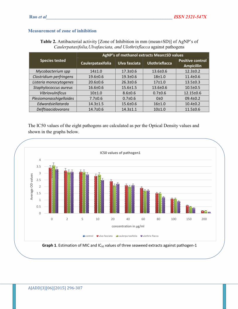
Measurement of zone of inhibition
Table 2. Antibacterial activity [Zone of Inhibition in mm (mean±SD)] of AgNP’s of Caulerpataxifolia,Ulvafasciata, and Ulothrixflacca against pathogens
Species tested AgNP’s of methanol extracts Mean±SD values
Caulerpataxifolia Ulva fasciata Ulothrixflacca Positive control
Ampicillin
Mycobacterium spp 14±1.0 17.3±0.6 13.6±0.6 12.3±0.2
Clostridium perfringens 19.6±0.6 19.3±0.6 18±1.0 11.4±0.6
Listeria monocytogenes 20.6±0.6 26.3±0.6 17±1.0 13.5±0.3
Staphylococcus aureus 16.6±0.6 15.6±1.5 13.6±0.6 10.5±0.5
Vibriovulnificus 10±1.0 8.6±0.6 0.7±0.6 12.15±0.6
Plesiomonasshigelloides 7.7±0.6 0.7±0.6 0±0 09.4±0.2
Edwardsiellatarda 14.3±1.5 15.6±0.6 16±1.0 10.4±0.2
Delftiaacidovorans 14.7±0.6 14.3±1.1 10±1.0 11.5±0.6
The IC50 values of the eight pathogens are calculated as per the Optical Density values and
shown in the graphs below.
0
0.5
1
1.5
2
2.5
3
3.5
4
0 2 5 10 20 40 60 80 100 150 200
Ave
rage
OD
val
ues
concentration in µg/ml
IC50 values of pathogen1
control ulva fasciata caulerpa taxifolia ulothrix flacca
Graph 1. Estimation of MIC and IC50 values of three seaweed extracts against pathogen-1
Rao et al______________________________________________________ ISSN 2321-547X
AJADD[3][06][2015] 296-307
0
0.5
1
1.5
2
2.5
3
3.5
4
4.5
0 2 5 10 20 40 60 80 100 150 200
Ave
rage
OD
val
ues
concentration in µg/ml
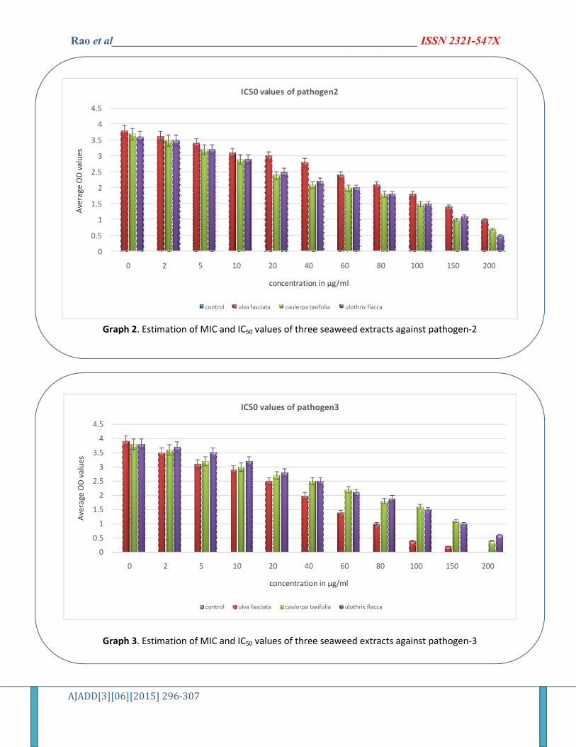
IC50 values of pathogen2
control ulva fasciata caulerpa taxifolia ulothrix flacca
Graph 2. Estimation of MIC and IC50 values of three seaweed extracts against pathogen-2
0
0.5
1
1.5
2
2.5
3
3.5
4
4.5
0 2 5 10 20 40 60 80 100 150 200
Ave
rage
OD
val
ues
concentration in µg/ml
IC50 values of pathogen3
control ulva fasciata caulerpa taxifolia ulothrix flacca
Graph 3. Estimation of MIC and IC50 values of three seaweed extracts against pathogen-3
Rao et al______________________________________________________ ISSN 2321-547X
AJADD[3][06][2015] 296-307
0
0.5
1
1.5
2
2.5
3
3.5
4
4.5
0 2 5 10 20 40 60 80 100 150 200
Ave
rage
OD
val
ues
concentration in µg/ml
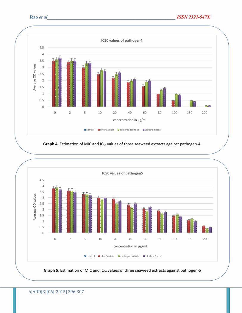
IC50 values of pathogen4
control ulva fasciata caulerpa taxifolia ulothrix flacca
Graph 4. Estimation of MIC and IC50 values of three seaweed extracts against pathogen-4
0
0.5
1
1.5
2
2.5
3
3.5
4
4.5
0 2 5 10 20 40 60 80 100 150 200
Ave
rage
OD
va
lue
s
concentration in µg/ml
IC50 values of pathogen5
control ulva fasciata caulerpa taxifolia ulothrix flacca
Graph 5. Estimation of MIC and IC50 values of three seaweed extracts against pathogen-5
Rao et al______________________________________________________ ISSN 2321-547X
AJADD[3][06][2015] 296-307
0
0.5
1
1.5
2
2.5
3
3.5
4
0 2 5 10 20 40 60 80 100 150 200
Ave
rage
OD
val
ues
concentration in µg/ml
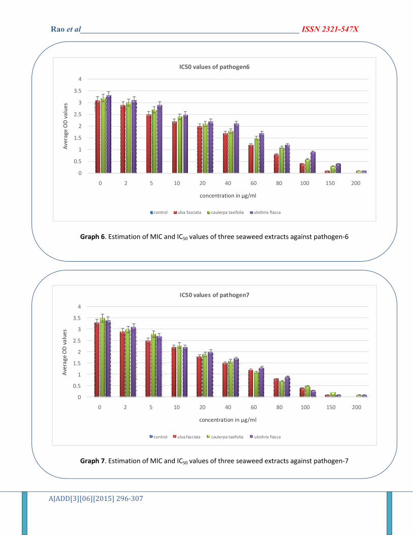
IC50 values of pathogen6
control ulva fasciata caulerpa taxifolia ulothrix flacca
Graph 6. Estimation of MIC and IC50 values of three seaweed extracts against pathogen-6
0
0.5
1
1.5
2
2.5
3
3.5
4
0 2 5 10 20 40 60 80 100 150 200
Ave
rage
OD
va
lue
s
concentration in µg/ml
IC50 values of pathogen7
control ulva fasciata caulerpa taxifolia ulothrix flacca
Graph 7. Estimation of MIC and IC50 values of three seaweed extracts against pathogen-7
Rao et al______________________________________________________ ISSN 2321-547X
AJADD[3][06][2015] 296-307
0
0.5
1
1.5
2
2.5
3
3.5
4
0 2 5 10 20 40 60 80 100 150 200
Ave
rag
e O
D v
alu
es
concentration in µg/ml
IC50 values of pathogen8
control ulva fasciata caulerpa taxifolia ulothrix flacca
Graph 8. Estimation of MIC and IC50 values of three seaweed extracts against pathogen-8
0
0.5
1
1.5
2
2.5
3
P1 P2 P3 P4 P5 P6 P7 P8
ZO
NE
OF
INH
IBIT
ION
IN C
M
PATHOGENS
Antibacterial activity
control caulerpa taxifolia ulva fasciata ulothrix flacca
Graph 9. P1: Mycobacterium spp. P2: Clostridium perfringens, P3: Listeria monocytogenes P4: Staphylococcus
aureus P5: Vibriovulnificus P6: Plesiomonasshigelloides P7: Edwardsiellatarda P8: Delftiaacidovorans.