Anti-GD1a antibodies activate complement and calpain … · Anti-GD1a antibodies activate...
74
Anti-GD1a antibodies activate complement and calpain to injure distal motor nodes of Ranvier in mice McGonigal R 1 , Rowan EG 2 , Greenshields KN 1 , Halstead SK 1 , Humphreys PD 1 , Rother RP 3 , Furukawa K 4 , Willison HJ 1 1 Division of Clinical Neurosciences, Glasgow Biomedical Research Centre, University of Glasgow, Glasgow G12 8TA 2 Strathclyde Institute of Pharmacy and Biomedical Sciences, Sir John Arbuthnott Building, University of Strathclyde, Glasgow G4 0NR 3 Alexion Pharmaceuticals, Cheshire, CT 06410, USA 4 Department of Biochemistry II, Nagoya University Graduate School of Medicine, Nagoya, Japan Address for correspondence: Professor Hugh J. Willison, University of Glasgow Division of Clinical Neurosciences, Glasgow Biomedical Research Centre, Room B330 120 University Place, Glasgow, G12 8TA, Scotland. Tel: 44 (0) 141 330 8384 e-mail: [email protected] Running head: Anti-GD1a antibody-mediated nodal injury (40 characters) Word count: (excludes refs and legends) 7633
-
Upload
vuongthien -
Category
Documents
-
view
220 -
download
0
Transcript of Anti-GD1a antibodies activate complement and calpain … · Anti-GD1a antibodies activate...
Anti-GD1a antibodies activate complement and calpain to injure distal
motor nodes of Ranvier in mice
McGonigal R1, Rowan EG2, Greenshields KN1, Halstead SK1,
Humphreys PD1, Rother RP3, Furukawa K4, Willison HJ1
1Division of Clinical Neurosciences, Glasgow Biomedical Research Centre,
University of Glasgow, Glasgow G12 8TA
2Strathclyde Institute of Pharmacy and Biomedical Sciences, Sir John Arbuthnott
Building, University of Strathclyde, Glasgow G4 0NR
3Alexion Pharmaceuticals, Cheshire, CT 06410, USA
4Department of Biochemistry II, Nagoya University Graduate School of Medicine,
Nagoya, Japan
Address for correspondence:
Professor Hugh J. Willison,
University of Glasgow Division of Clinical Neurosciences,
Glasgow Biomedical Research Centre, Room B330
120 University Place, Glasgow, G12 8TA, Scotland.
Tel: 44 (0) 141 330 8384
e-mail: [email protected]
Running head: Anti-GD1a antibody-mediated nodal injury (40 characters)
Word count: (excludes refs and legends) 7633

Abstract
The motor axonal variant of Guillain-Barré syndrome is associated with anti-
GD1a IgG antibodies which are believed to be the pathogenic factor. In previous
studies we have demonstrated the motor terminal to be a vulnerable site. Here
we show both in vivo and ex vivo that nodes of Ranvier in intramuscular motor
nerve bundles are also targeted by anti-GD1a antibody in a gradient-dependent
manner, with greatest vulnerability at distal nodes. Complement deposition is
associated with prominent nodal injury as monitored with electrophysiological
recordings and fluorescence microscopy. Complete loss of nodal protein staining,
including voltage-gated sodium channels and ankyrin G, occurs and is
completely protected by both complement and calpain inhibition, although the
latter provides no protection against electrophysiological dysfunction. In ex vivo
motor and sensory nerve trunk preparations, antibody deposits are only observed
in experimentally desheathed nerves, which are thereby rendered susceptible to
complement-dependent morphological disruption, nodal protein loss and reduced
electrical activity of the axon. These studies provide a detailed mechanism by
which loss of axonal conduction can occur in a distal dominant pattern as
observed in a proportion of motor axonal Guillain-Barré syndrome patients, and
also provide an explanation for the occurrence of rapid recovery from complete
paralysis and electrophysiological in-excitability. The studies also identify
therapeutic approaches in which nodal architecture can be preserved.
Keywords: Anti-GD1a antibody, GD1a ganglioside, node of Ranvier, acute motor
axonal neuropathy, complement, calpain.

Abbreviations: α-BTx, α-bungarotoxin; AMAN, acute motor axonal neuropathy;
BNB, blood nerve barrier; CAP, compound action potential; CFP, cyan
fluorescent protein, CFP; GBS, Guillain-Barré syndrome; GD3s, GD3 synthase;
LOS, lipo-oligosaccharides; LTx, alpha-latrotoxin; mAb, monoclonal antibody;
MAC, membrane attack complex; NF, neurofilament; NHS, normal human serum,
NoR; node of Ranvier; PBS, phosphate-buffered saline; TTx, tetrodotoxin; TS,
triangularis sternae; RT, room temperature; WT, wild type.

Introduction
The motor axonal variant of Guillain-Barré syndrome (GBS), termed acute motor
axonal neuropathy (AMAN) (Feasby et al., 1986;Hughes and Cornblath
2005;McKhann et al., 1993) characteristically follows Campylobacter jejuni
infection and is associated with serum anti-GM1, -GD1a and -GalNAc-GD1a
ganglioside antibodies (Ho et al., 1999;Lugaresi et al., 1997;Ogawara et al.,
2000). AMAN-associated Campylobacter jejuni strains have ganglioside-like
surface lipo-oligosaccharides (LOS) Aspinall et al., 1993) suggesting induction is
due to a mechanism of molecular mimicry, which has been proven
experimentally (Ang et al., 2004;Goodyear et al., 1999).
Gangliosides are sialic acid-containing glycosphingolipids expressed at
high levels in the nervous system in a range of cell-specific patterns (Ledeen
1978). Gangliosides have diverse functions related to neural development,
maintenance and regeneration, including stabilising the axo-glial junction at the
node of Ranvier (NoR) (Sheikh et al., 1999b;Susuki et al., 2007a;Silajdzic et al.,
2009). Although no specific neural function has been attributed to GD1a, it has
been identified in the motor nerve terminal and nodal axolemma (De Angelis et
al., 2001;Gong et al., 2002;Goodfellow et al., 2005;Sheikh et al., 1999a), sites
which correspond to those predicted from clinical, electrophysiological and
pathological data to be affected in motor axonal forms of GBS (Griffin et al.,
1996;Ho et al., 1997;Kuwabara et al., 2004).
The distal motor nerve, nerve terminal and ventral roots have relatively
higher permeability to circulating factors than nerve trunks, owing to local
variations in the protective properties of the blood nerve barrier (BNB) (Burkel
1967;Malmgren and Olsson 1980;Olsson 1990;Saito and Zacks 1969). These
BNB variations could allow circulating antibody access to either very distal or
very proximal motor axonal membranes and thereby account for more targeted
injury to these regions. Thus it has been proposed that one explanation for the
very rapid recovery from paralysis seen in some AMAN patients could be due to
axonal conduction block at the distal motor axon and nerve terminal, a site with
the capacity to regenerate rapidly (Goodfellow et al., 2005;Ho et al.,1997).
Conversely, severe proximal axonal injury resulting in widespread axonal
degeneration that overwhelmed the compensatory capacity of motor unit
remodelling would inevitably lead to permanent motor axonal deficits, as is seen
in some AMAN cases (Hiraga et al., 2005a;Hiraga et al., 2005b).
Several anti-GM1 and -GD1a ganglioside antibody-mediated mouse and
rabbit models of AMAN have been generated (Goodfellow et al., 2005;Sheikh et
al., 2004;Susuki et al., 2003). Models to date have focused on sciatic nerve and
ventral root axons, or on axonal components of neuromuscular junctions. In a
passive immunisation mouse model of AMAN mediated by anti-GD1a antibody
supplemented with guinea pig complement, axonal injury was observed in spinal
roots and sciatic nerve (Sheikh et al., 2004). Similarly in a rabbit model induced
by active immunisation with GM1, axonal injury was observed in spinal roots, in
which rabbit complement deposits were also evident (Susuki et al., 2003).
Extensions of this study focussing on the NoR revealed destabilisation of nodal
and paranodal structures, including loss of sodium (NaV) channels, findings
interpreted as the consequence of antibody and complement-mediated axo-glial
disruption(Susuki et al., 2007b), and their protection with a complement inhibitor
(Phongsisay et al., 2008).
These complement mediated effects at the NoR in the ventral root mirror
those demonstrated in patient autopsy tissue (Hafer-Macko et al., 1996). As the
NoR is vital for impulse propagation (Poliak and Peles 2003;Scherer 1996),
understanding AMAN immunopathology at this site in relation to function is both
critical and complex. The NoR is organised into 3 subdomains - the nodal gap,
the paranode and the juxtaparanode (Fig. S8). The voltage-gated sodium
channel isoform Nav1.6 is expressed at the NoR (Caldwell et al., 2000), along
with the cytoskeletal protein ankyrin G (Kordeli et al., 1990) and the cell adhesion
molecules neurofascin 186 and NrCAM (Davis et al., 1996). At the paranode, the
axo-glial junction is formed by the axolemmal proteins contactin and Caspr, while
neurofascin 155 is the glial receptor to this complex (Charles et al.,
2002;Einheber et al., 1997;Menegoz et al., 1997;Peles et al., 1997;Rios et al.,
2000;Tait et al., 2000). The axo-glial junction acts as a barrier to prevent lateral
movement of nodal constituents, thus organising the channel clustering required
for maintenance of membrane potentials (Bhat et al., 2001;Boyle et al., 2001). At
the juxtaparanode, voltage-gated potassium channels localised on the axon, in
complex with Caspr 2 and Tag1 (Arroyo et al., 1999;Wang et al., 1993), play a
role in repolarisation the resting membrane potential following an action potential

(Poliak and Peles2003;Rasband et al., 2002;Traka et al., 2002).
Glycosyltransferase knockout mouse studies indicate that GD1a or related
gangliosides clearly modulate the structural and functional integrity of this site,
although the precise mechanisms are poorly understood (Sheikh et
al.,1999b;Silajdzic et al., 2009;Susuki et al., 2007a).
In our ex vivo mouse model of AMAN, motor nerve terminals enriched in
GD1a develop severe functional and pathological injury when exposed to anti-
GD1a antibody with complement activation(Goodfellow et al., 2005). The pore
forming action of complement is critical to the development of this injury and that
mediated by other anti-ganglioside antibodies, in part through allowing
uncontrolled calcium influx into the nerve terminals, with subsequent Ca2+-
dependent protease, calpain, activation and cleavage of structural proteins in the
axon terminal (O'Hanlon et al., 2003).
This study set out to assess whether anti-GD1a-antibody mediated injury
could be observed to occur at NoR in the distal portions of the axon, upstream
from the motor nerve terminal. If present, we also intended to determine the
mechanism of action and functional effects of any observed injury that might lead
to therapeutic intervention, analogous to our previous approach to the
neuromuscular junction.
Materials and methods
Mice
Male GD3 synthase knockout mice (GD3s-/-) mice (Okada et al., 2002) were
crossed with B6/Cg-TgN(Thy1-CFP) x DBA mice that endogenously express
cyan fluorescent protein (CFP) in their axons (Feng et al., 2000, kindly provided
by Dr W. Thompson, Austin, Texas) to produce a doubly genetically modified
mouse referred to as GD3s-/-/CFP in this study. GD3s-/- mice were preferentially
used as they express greater amounts of axonal GD1a compared with their wild
type (WT) counterparts, owing to blockade of b-series biosynthesis and
consequent accumulation of a-series gangliosides including GD1a. The
ganglioside biosynthetic pathway illustrating this is shown in Fig. S1. Through
virtue of expressing high amounts of GD1a, GD3s-/- mice bind more anti-GD1a
antibody than their wildtype (WT) counterparts as previously reported at motor
nerve terminals (Goodfellow et al., 2005). In order to confirm that GD3s-/-/CFP
were an appropriate cross in which to model these experiments, binding of anti-
GD1a antibody was quantified in GD3s-/-/CFP mice in comparison with WT/CFP
controls (Fig. S2). Mice were killed by CO2 inhalation at 6-12 weeks of age and
experiments were carried out under licence (PPL60/3842) in accordance with UK
Home Office guidelines.
Antibodies and reagents

The IgG2b mAb to GD1a (herein termed anti-GD1a antibody, also known as
MOG-35) was produced by immunisation of GalNAcT-/- mice (lacking all complex
gangliosides (Takamiya et al., 1996), with the Campylobacter jejuni HS:19 LOS
strain that possesses structures identical to GD1a to which it raises a cross-
reactive immune response (Bowes et al., 2002)] and acts as an antecedent
infection in AMAN, as described (Boffey et al., 2005). Antibodies to channels,
other proteins and membrane attack complex (MAC), C5b-9 are detailed in Table
1. Eculizumab, a humanised anti-human C5 mAb that binds plasma C5 to
prevent MAC formation and ALXN3300 (the isotype-matched control mAb) were
supplied by Alexion Pharmaceuticals (Cheshire, USA). The synthetic peptide
AK295 binds calpain I, II and cathepsin B to prevent their activation and
proteolytic action (Li et al., 1996). Toxins were used as follows: g-bungarotoxin
(BTx, Molecular Probes, UK) Alexa Fluor 488 and 647 conjugates at 1:500; g-
latrotoxin (LTx, Alomone Labs, Israel) at 12nM; tetrodotoxin (TTX, Biotium Inc,
USA) at 5たM; vecuronium (Organon Laboratories Ltd, Cambridge, UK) at 5µM.
Ex vivo and in vivo muscle and nerve permeability studies
Triangularis sterni (TS) muscle, phrenic nerve, sural nerve and sciatic nerve were
dissected, mounted and maintained alive in Ringers medium (116mM NaCl,
4.5mM KCl, 1mM MgCl2, 2mM CaCl2, 1mM NaH2PO4, 23mM NaHCO3, 11mM
glucose, pH 7.4) pre-gassed with 95% O2/5% CO2 at room temperature (~20°C).
Muscle and nerve (desheathed by slitting and opening the epineurium with a fine
needle, or left intact) were incubated with 100たg/ml anti-GD1a antibody for 2h at
32°C, 30mins at 4°C and a final 10mins at RT, plus BTx to label NMJ. Antibody
control preparations were incubated with Ringers alone. Preparations were
rinsed in Ringers prior to fixation in 4% paraformaldehyde (20mins, RT). Tissue
was then rinsed in PBS, 0.1M glycine and PBS (10mins each, R.T.). Tissue was
incubated with anti-IgG2b-FITC (1:200) and the pan anti-neurofascin antibody
NFC2 (1:1000) with 0.5% Triton X-100 in blocking solution (1% goat serum and
1% L-lysine) overnight at 4°C. Intramuscular nerve bundles were divided into four
categories for quantification: single fibres, small bundles (<15たm), medium
bundles (15-35たm) and large bundles (>35たm). NoR were identified by
neurofascin staining and the anti-GD1a antibody immunofluorescence at this
region were measured within each category and compared to control tissue.
To study the binding of antibody in vivo, the same quantification was performed
on TS muscle removed from a mouse injected i.p. 16h previously with 3mg anti-
GD1a antibody. PBS was used for control groups.
For sciatic, sural and phrenic nerves, in order to assess antibody and
complement access through the relatively impermeable epineurium, and
therefore vulnerability to injury, isolated nerves were incubated ex vivo with anti-
GD1a antibody under intact and desheathed conditions. It was thereby
established that desheathing was essential for achieving anti-GD1a antibody
binding at NoR in nerve trunks, and that under these conditions antibody binding
levels were equivalent to intramuscular nerve NoR. Data for the phrenic nerve is
shown in Fig. S3. All studies on nerve trunks were thus conducted on
desheathed nerves.
Ex vivo preparations for complement activation and nodal protein disruption
Muscle and nerve preparations were subjected to the same protocols as used for
assessing permeability, with the additional step that tissue was incubated with
40% normal human serum (NHS) for 3h at RT prior to fixation. Muscle was
cryosectioned at 10たm and stained for MAC, nodal channels and other proteins
overnight at 4oC as listed in Table 1. In order to identify NoR, fluoromyelin green
(1:400) that labels lipids, or dystrophin (1:200) that labels the myelin sheath were
applied. Secondary antibodies were applied for 3 hrs at RT as follows: anti-rabbit
IgG-Cy5 (1:300) for Nav1.6, Caspr, NFC2, Kv1.1, neurofilament; anti-mouse
IgG1-Cy5 (1:300) for ankyrin G, moesin, NrCAM and dystrophin; anti-mouse
IgG2a-TRITC (1:200) for MAC. NoR with a normal immunostaining pattern for
nodal proteins were scored as present or absent/abnormal. To determine
whether any abnormal immunostaining was dependent on nodal MAC deposition,
or resultant from an upstream effect of massive synaptic injury, Nav1.6
immunostaining at NoR was compared between antibody treated and g-LTx
treated tissue. Our previous studies have shown that the nerve terminal effects of
antiganglioside antibodies mimic those of g-LTx (Plomp et al., 1999;Plomp and
Willison 2009) . g-LTx was added at 2nM in Ringers to the organ bath at the
same time NHS was added in a parallel preparation.
To assess the contribution of MAC to any observed injury, the C5 inhibitor
Eculizumab was added at 100たg/ml to NHS 10mins prior to incubation with the
muscle. To investigate the contribution of calpain, 100たM of the calpain inhibitor

AK295 (kindly provided by Dr J. Powers and J. Glass, Atlanta, Georgia), was
added concurrently with NHS. Eculizumab concentration had been previously
optimised (Halstead et al., 2008b). AK295 was optimised for concentration by
dose ranging studies from 25-200 micromolar concentrations and the lowest
concentration that fully protected protein cleavage was used. In Eculizumab-
treated and -unprotected intramuscular axons, the presence of axonal CFP was
used to monitor axonal integrity. After AK295 treatment, the intensity of
neurofilament immunoreactivity was quantified at the nerve terminal as
delineated by BTx staining and compared to AK295-unprotected tissue levels.
The efficacies of Eculizumab and AK295, as monitored by immunostaining
profiles, were expressed as the percentage of protected versus unprotected
signals at the relevant NoR sites.
Perineural and extracellular recordings
TS nerve-muscle preparations were freshly dissected and set up ex vivo for
electrophysiological recordings as for immunocytochemisrty studies. Experiments
were carried out at room temperature (20-22°C) using 2 M NaCl-filled
microlectrodes with a resistance of 25-45 MΩ in preparations bathed in Ringers.
Recordings were made from nerve terminals and small and large intramuscular
nerve bundles after anti-GD1a antibody incubation followed by NHS for 3hrs.
Perineural waveforms associated with nerve terminal action potentials were
made as previously described (Braga et al., 1991). Muscles were paralysed with
5 µM vecuronium to prevent twitching. In some experiments the same

microelectrode was used to measure muscle resting membrane potentials.
Signals were amplified, recorded and analysed as per the nerve extracellular
recordings below.
For extracellular recordings, nerves were mounted in a custom made
Perspex recording block across three chambers and sealed in with vacuum
grease. Nerves were stimulated with a Grass S88 stimulator delivering pulses at
a frequency of 1Hz, and at a supramaximal voltage. Signals were amplified via a
CED1902 and digitised by a NIDAQ-MX A/D converter (National Instruments,
Austin, Texas), then captured and analysed using WinWCP version 4.1.0
software. Phrenic nerves and sural nerves remained in the recording chamber
throughout the experiments and recordings were made for 2h on the application
of NHS. Recordings from the larger sciatic nerve were collected on transfer of the
nerve to the recording chamber following NHS treatment in a petri dish, as
penetration of NHS into the nerve was not uniformly maximal whilst in the
recording chamber. At the termination of all experiments 5µM TTX was added to
the recording chamber to confirm that the waveform being recorded was the
result of the opening of sodium channels. A representative graph of the positive
peak value of compound action potential (CAP) over time was plotted to convey
conduction. A minimum of 200 control waveforms were averaged prior to the
addition of NHS. Absolute CAP values were not presented as these varied
between experiments; instead the percentage of the starting CAP peak value
was calculated for each of 3-5 preparations and averaged for each treatment
group. A 2-sample t-test was used for comparison of phrenic and sural nerve
conduction, whilst a paired t-test was performed for comparison of sciatic nerve
CAP recordings.
Image acquisition and analysis
Fluorescent images were captured on both a Zeiss Axio Imager Z1 with
ApoTome attachment and a Zeiss Pascal confocal laser scanning microscope.
Image analysis was carried out using ImageJ software. For quantitative analysis
of antibody and MAC deposition, the fluorescence signal at the region of the NoR
was measured and any background fluorescence subtracted. Where relevant,
measurements were categorised by bundle size as described above. The same
procedure was carried out for the quantitation of neurofilament signal over the
motor endplate. Measurements were pooled from three experimental
preparations and presented as box and whisker plots to represent the spread of
the non-parametric data. Mann-Whitney mean rank test was used to compare
possible statistical differences between groups where the level of significance
was set at 1%. For comparison of nodal protein immunostaining, NoR positive for
individual markers were counted for each bundle category and the chi-squared
test used at a 1% level of significance.
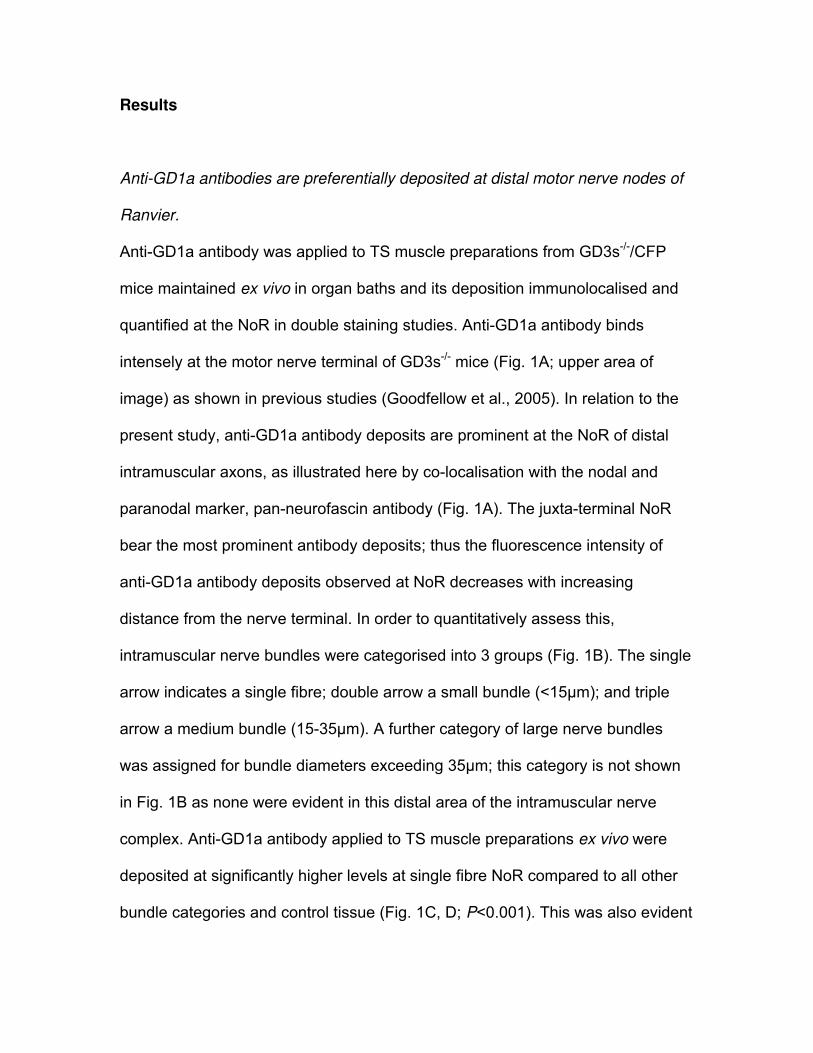
Results
Anti-GD1a antibodies are preferentially deposited at distal motor nerve nodes of
Ranvier.
Anti-GD1a antibody was applied to TS muscle preparations from GD3s-/-/CFP
mice maintained ex vivo in organ baths and its deposition immunolocalised and
quantified at the NoR in double staining studies. Anti-GD1a antibody binds
intensely at the motor nerve terminal of GD3s-/- mice (Fig. 1A; upper area of
image) as shown in previous studies (Goodfellow et al., 2005). In relation to the
present study, anti-GD1a antibody deposits are prominent at the NoR of distal
intramuscular axons, as illustrated here by co-localisation with the nodal and
paranodal marker, pan-neurofascin antibody (Fig. 1A). The juxta-terminal NoR
bear the most prominent antibody deposits; thus the fluorescence intensity of
anti-GD1a antibody deposits observed at NoR decreases with increasing
distance from the nerve terminal. In order to quantitatively assess this,
intramuscular nerve bundles were categorised into 3 groups (Fig. 1B). The single
arrow indicates a single fibre; double arrow a small bundle (<15µm); and triple
arrow a medium bundle (15-35µm). A further category of large nerve bundles
was assigned for bundle diameters exceeding 35µm; this category is not shown
in Fig. 1B as none were evident in this distal area of the intramuscular nerve
complex. Anti-GD1a antibody applied to TS muscle preparations ex vivo were
deposited at significantly higher levels at single fibre NoR compared to all other
bundle categories and control tissue (Fig. 1C, D; P<0.001). This was also evident

for small bundles (P<0.001) and medium bundles (P=0.0023). Large bundles had
an insignificant anti-GD1a antibody deposition level, comparable to control
tissue, suggesting anti-GD1a antibody was unable to gain access to bundles of
this size following topical application. In order to assess anti-GD1a antibody
penetration to these intramuscular nerve compartments when delivered through
the vascular bed (as opposed to organ bath incubation), anti-GD1a antibody was
injected intraperitoneally, and 16 hours later the TS muscle was removed for
antibody quantification, as for ex vivo preparations above. Equivalent results to
the ex vivo findings were observed, with antibody deposits being greatest in the
distal part of the nerve in a gradient-dependent manner when categorised by
bundle size (Fig. 1E). In order to establish that these differences were not due to
a proximal to distal gradient of GD1a expression at NoR in nerve, frozen sections
of permeabilised intramuscular nerve bundles in which antibody access is
expected to be uniform were stained with anti-GD1a antibody, and the signal
intensity was found to be the same, irrespective of the nerve bundle size (Fig.
S4). These ex vivo and in vivo findings demonstrate that anti-GD1a antibody is
able to bind to intramuscular nerve NoR in a distal to proximal downward
gradient, presumed due to the relatively increasing impermeability of the BNB to
antibody as bundle size increases.
Nodal proteins are disrupted and distal motor nerves are rendered inexcitable by
anti-GD1a antibody directed complement activation

In our previously reported model of anti-ganglioside mediated injury to the
nerve terminal, complement activation has been monitored by heterologous
(human) MAC deposition at motor nerve terminals (Plomp and Willison2009).
Similarly in this study, MAC deposition was demonstrated at NoR of the distal
intramuscular nerves in response to the addition of an exogenous source of
human complement in the form of NHS (Fig. 2A). As with anti-GD1a antibody
deposition, MAC deposition as assessed by fluorescence intensity for anti-MAC
antibody was gradient-dependent, with significantly higher levels of fluorescence
observed at single fibre NoR compared to all other categories (Fig. 2B; P<0.001)
and significantly higher MAC levels at small bundle NoR compared to larger
categories (Fig. 2B; P<0.001). Axonal injury was further characterised by the
complete loss of the endogenous axonal CFP signal, both at the nerve terminal
and along the distal axon as illustrated in Fig. 2C. Even in large bundles, the CFP
signal was relatively attenuated in antibody plus NHS treated preparations.
In order to assess the functional effect of MAC deposition to the distal
axonal region, electrophysiological assessment of local ion currents was
performed by recording perineural currents. End plate microelectrode recordings
would not be useful to assess this as all our previous electrophysiological studies
have shown that the motor nerve terminal is irreversibly paralysed in this model
(Plomp and Willison, 2009). Perineural recordings were made at the nerve
terminal, at small nerve bundles and at large nerve bundles. In control tissue
(anti-GD1a antibody without NHS), biphasic waveforms (see discussion for a
fuller account of the nature of the waveform) were observed that correspond to
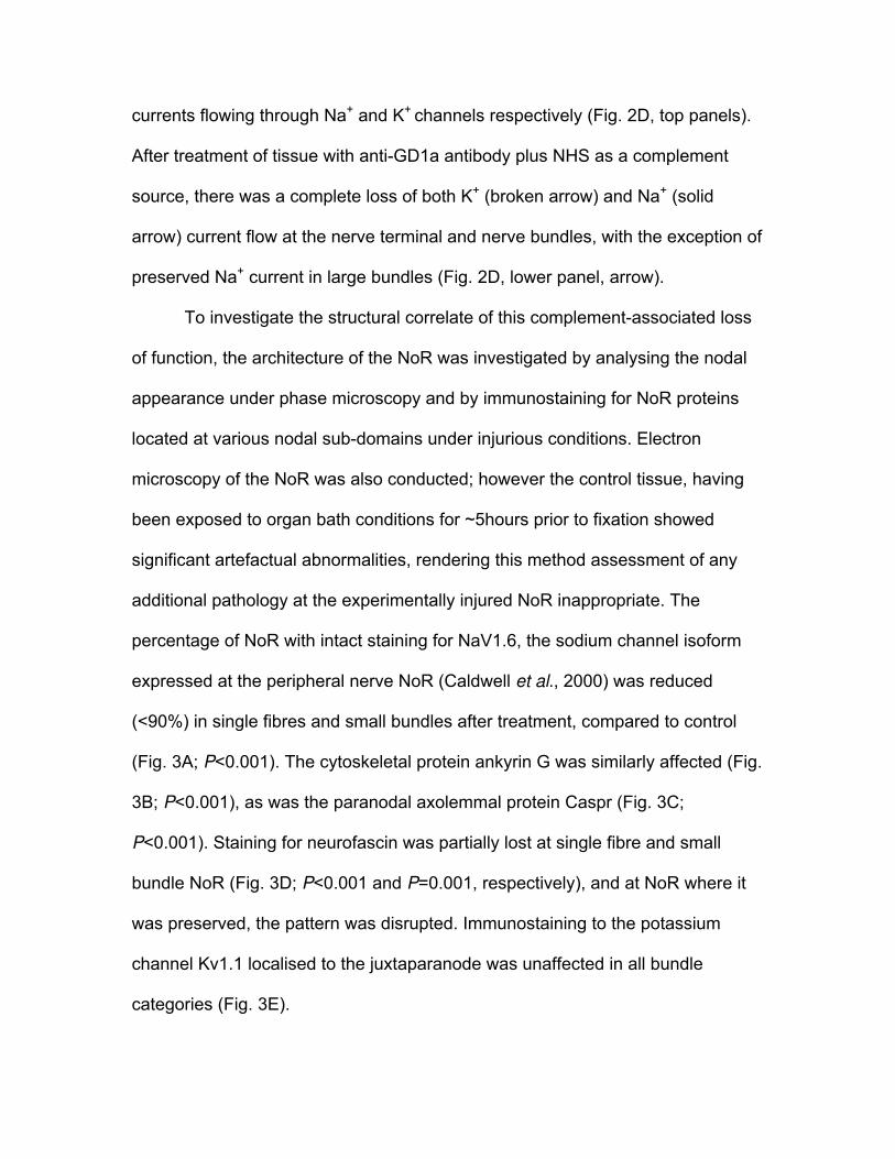
currents flowing through Na+ and K+ channels respectively (Fig. 2D, top panels).
After treatment of tissue with anti-GD1a antibody plus NHS as a complement
source, there was a complete loss of both K+ (broken arrow) and Na+ (solid
arrow) current flow at the nerve terminal and nerve bundles, with the exception of
preserved Na+ current in large bundles (Fig. 2D, lower panel, arrow).
To investigate the structural correlate of this complement-associated loss
of function, the architecture of the NoR was investigated by analysing the nodal
appearance under phase microscopy and by immunostaining for NoR proteins
located at various nodal sub-domains under injurious conditions. Electron
microscopy of the NoR was also conducted; however the control tissue, having
been exposed to organ bath conditions for ~5hours prior to fixation showed
significant artefactual abnormalities, rendering this method assessment of any
additional pathology at the experimentally injured NoR inappropriate. The
percentage of NoR with intact staining for NaV1.6, the sodium channel isoform
expressed at the peripheral nerve NoR (Caldwell et al., 2000) was reduced
(<90%) in single fibres and small bundles after treatment, compared to control
(Fig. 3A; P<0.001). The cytoskeletal protein ankyrin G was similarly affected (Fig.
3B; P<0.001), as was the paranodal axolemmal protein Caspr (Fig. 3C;
P<0.001). Staining for neurofascin was partially lost at single fibre and small
bundle NoR (Fig. 3D; P<0.001 and P=0.001, respectively), and at NoR where it
was preserved, the pattern was disrupted. Immunostaining to the potassium
channel Kv1.1 localised to the juxtaparanode was unaffected in all bundle
categories (Fig. 3E).

The complete and rapid disappearance of key NoR component proteins as
assessed by immunostaining following 3hrs of NHS exposure was striking. In
order to assess this in more detail for Nav1.6, intermediate stages of dissolution
of Nav1.6 immunostaining were qualitatively examined at 15mins and 30mins
after the addition of complement treatment. At 15min there was no alteration to
staining; however by 30mins a proportion of NoR developed punctuate and
dispersed Nav1.6 staining, indicating fragmentation and spread of Nav1.6
channel clusters bound by the anti-Nav1.6 antibody, examples for 3 separate
NoR being shown in Fig. S5.
In this model, the motor nerve terminal is also severely and concomitantly
injured such that it might conceivably have more proximal motor axonal
consequences. By way of control to ensure that the observed nodal protein
staining loss was associated with MAC deposition and injury directly at the NoR,
Nav1.6 staining at NoR was assessed after g-LTx-induced injury that creates a
nerve terminal lesion identical to that of anti-GD1a antibody directed MAC (Fig.
S6). Nav1.6 immunostaining was still intact after LTx treatment, thereby
confirming that its loss is due to anti-GD1a antibody with local complement
activation at NoR. Taken together these results suggest that anti-GD1a antibody
directed complement-mediated disruption to the nodal architecture of the distal
axons results in a block in nerve conduction.
Complement inhibition completely protects nodes of Ranvier from anti-GD1a
antibody-mediated injury

In order to demonstrate the role for the MAC component of complement
activation, the C5 complement inhibitor Eculizumab, that completely prevents
MAC assembly, was introduced to the organ bath model. Eculizumab protected
Nav1.6, ankyrin G and Caspr immunostaining at NoR from injury mediated by
complement activation, compared with the isotype control antibody ALXN3300. In
quantitative analysis, the percentage of NoR with intact Nav1.6 staining is
significantly greater on the addition of Eculizumab compared to the isotype-
matched control mAb ALXN3300 at single fibres and small bundles (Fig. 4A;
P<0.001). As demonstrated previously, there was no reduction in immunostaining
at medium and large bundles in response to complement and thus complement
inhibition could not further attenuate this. Single fibre and small bundle NoR also
had significantly preserved ankyrin G and Caspr staining with Eculizumab
protection compared to ALXN3300 application (Fig. 4B,C; P<0.001). Additionally,
endogenous CFP was maintained in axons and bundles with Eculizumab
treatment compared to ALXN3300, essentially maintaining the normal overall
architecture with a normal appearance (Fig. 4D).
Calpain inhibition protects Na channel and axonal protein integrity without
preserving nerve currents
A major consequence of MAC pore deposition in plasma membranes is the
formation of a bi-directional, non-specific ion and water pore. At the NoR, the
electrical function of the nodal axolemmal membrane is dependent upon tightly
regulated ion homeostasis and the consequences of this uncontrolled flux are
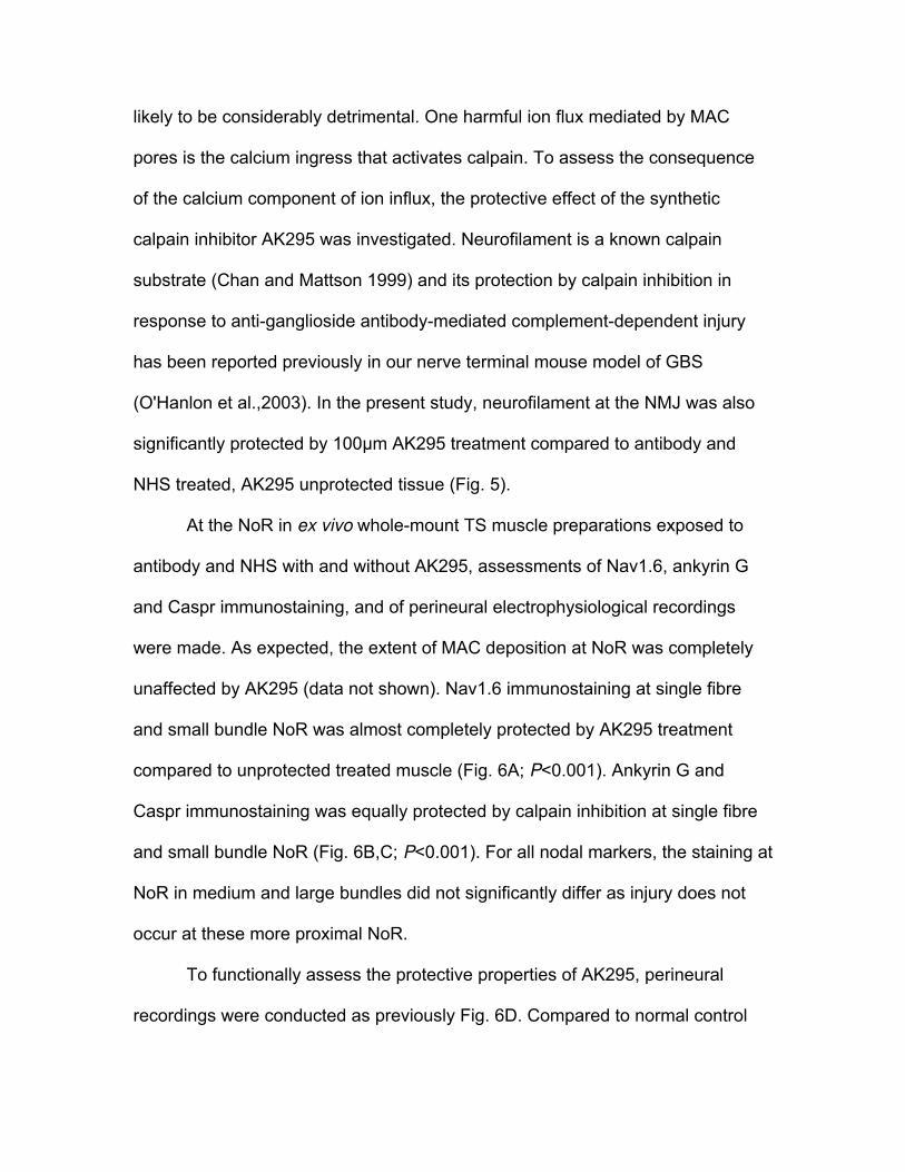
likely to be considerably detrimental. One harmful ion flux mediated by MAC
pores is the calcium ingress that activates calpain. To assess the consequence
of the calcium component of ion influx, the protective effect of the synthetic
calpain inhibitor AK295 was investigated. Neurofilament is a known calpain
substrate (Chan and Mattson 1999) and its protection by calpain inhibition in
response to anti-ganglioside antibody-mediated complement-dependent injury
has been reported previously in our nerve terminal mouse model of GBS
(O'Hanlon et al.,2003). In the present study, neurofilament at the NMJ was also
significantly protected by 100たm AK295 treatment compared to antibody and
NHS treated, AK295 unprotected tissue (Fig. 5).
At the NoR in ex vivo whole-mount TS muscle preparations exposed to
antibody and NHS with and without AK295, assessments of Nav1.6, ankyrin G
and Caspr immunostaining, and of perineural electrophysiological recordings
were made. As expected, the extent of MAC deposition at NoR was completely
unaffected by AK295 (data not shown). Nav1.6 immunostaining at single fibre
and small bundle NoR was almost completely protected by AK295 treatment
compared to unprotected treated muscle (Fig. 6A; P<0.001). Ankyrin G and
Caspr immunostaining was equally protected by calpain inhibition at single fibre
and small bundle NoR (Fig. 6B,C; P<0.001). For all nodal markers, the staining at
NoR in medium and large bundles did not significantly differ as injury does not
occur at these more proximal NoR.
To functionally assess the protective properties of AK295, perineural
recordings were conducted as previously Fig. 6D. Compared to normal control
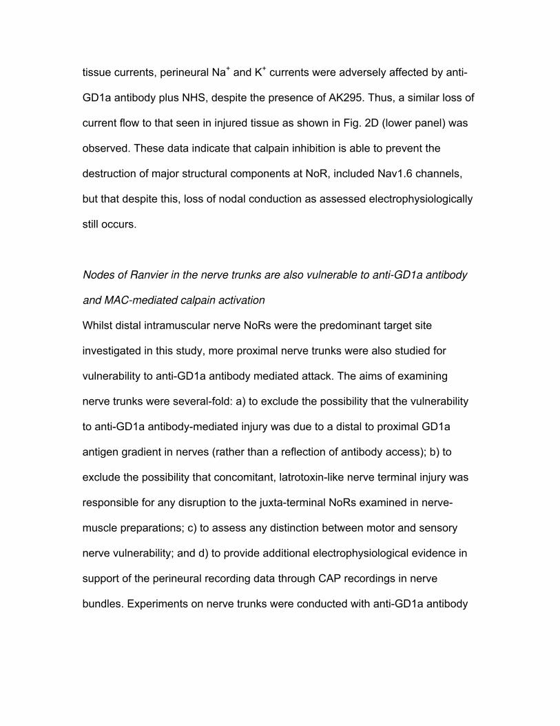
tissue currents, perineural Na+ and K+ currents were adversely affected by anti-
GD1a antibody plus NHS, despite the presence of AK295. Thus, a similar loss of
current flow to that seen in injured tissue as shown in Fig. 2D (lower panel) was
observed. These data indicate that calpain inhibition is able to prevent the
destruction of major structural components at NoR, included Nav1.6 channels,
but that despite this, loss of nodal conduction as assessed electrophysiologically
still occurs.
Nodes of Ranvier in the nerve trunks are also vulnerable to anti-GD1a antibody
and MAC-mediated calpain activation
Whilst distal intramuscular nerve NoRs were the predominant target site
investigated in this study, more proximal nerve trunks were also studied for
vulnerability to anti-GD1a antibody mediated attack. The aims of examining
nerve trunks were several-fold: a) to exclude the possibility that the vulnerability
to anti-GD1a antibody-mediated injury was due to a distal to proximal GD1a
antigen gradient in nerves (rather than a reflection of antibody access); b) to
exclude the possibility that concomitant, latrotoxin-like nerve terminal injury was
responsible for any disruption to the juxta-terminal NoRs examined in nerve-
muscle preparations; c) to assess any distinction between motor and sensory
nerve vulnerability; and d) to provide additional electrophysiological evidence in
support of the perineural recording data through CAP recordings in nerve
bundles. Experiments on nerve trunks were conducted with anti-GD1a antibody
and NHS as the complement source, in the presence and absence of calpain
inhibition with AK295.
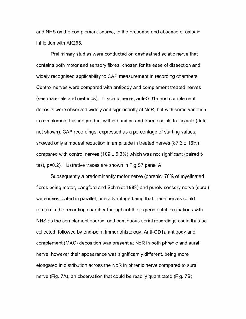
Preliminary studies were conducted on desheathed sciatic nerve that
contains both motor and sensory fibres, chosen for its ease of dissection and
widely recognised applicability to CAP measurement in recording chambers.
Control nerves were compared with antibody and complement treated nerves
(see materials and methods). In sciatic nerve, anti-GD1a and complement
deposits were observed widely and significantly at NoR, but with some variation
in complement fixation product within bundles and from fascicle to fascicle (data
not shown). CAP recordings, expressed as a percentage of starting values,
showed only a modest reduction in amplitude in treated nerves (87.3 ± 16%)
compared with control nerves (109 ± 5.3%) which was not significant (paired t-
test, p=0.2). Illustrative traces are shown in Fig S7 panel A.
Subsequently a predominantly motor nerve (phrenic; 70% of myelinated
fibres being motor, Langford and Schmidt 1983) and purely sensory nerve (sural)
were investigated in parallel, one advantage being that these nerves could
remain in the recording chamber throughout the experimental incubations with
NHS as the complement source, and continuous serial recordings could thus be
collected, followed by end-point immunohistology. Anti-GD1a antibody and
complement (MAC) deposition was present at NoR in both phrenic and sural
nerve; however their appearance was significantly different, being more
elongated in distribution across the NoR in phrenic nerve compared to sural
nerve (Fig. 7A), an observation that could be readily quantitated (Fig. 7B;

P<0.001) . Furthermore, sural nerve NoRs with antibody and MAC deposits and
yet intact Nav1.6 channel immunostaining was often observed (Fig. 7C), a finding
not seen in the phrenic nerve or its intramuscular branches. In terms of functional
effects on CAP amplitudes, sural nerve CAPs remained stable or only modestly
reduced over time (76.3 ± 9.6%; Fig S7 panel B), which was not significantly
different from controls.
In phrenic nerves, immunostaining of Nav1.6, ankyrin G and Caspr at NoR
was quantified in response to anti-GD1a and NHS exposure. Having
demonstrated nodal protein loss upon NHS exposure, experiments were also
conducted in the presence and absence of AK295 (calpain inhibition) as for the
ex vivo TS preparation. Immunostaining of the extracellular domain of NrCAM,
and moesin (a Schwann cell microvillal component) was also assessed, these
being molecules within the nodal complex but predicted to be unaffected by
calpain cleavage directly (Fig. S8). There was a significant loss of
immunostaining to Nav1.6, ankyrin G and Caspr in nerve exposed to antibody
and complement, compared to control (Fig. 8A, C; P<0.001). Unlike the reduction
in Nav1.6, ankyrin G and Caspr staining, the NrCAM and moesin staining was
retained but appeared mislocalised, being more diffusely spread throughout the
NoR area in comparison with the staining pattern in control tissue (Fig. 8A, C).
Nav1.6 channel and Caspr staining was significantly protected by AK295
(Fig 8B; P=0.01 for both proteins), although this was more modest when
compared with the levels of protection achieved at the distal nerve NoR.
Protection of ankyrin G staining followed the same trend but did not achieve
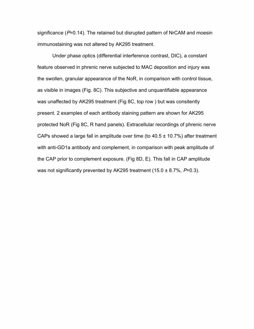
significance (P=0.14). The retained but disrupted pattern of NrCAM and moesin
immunostaining was not altered by AK295 treatment.
Under phase optics (differential interference contrast, DIC), a constant
feature observed in phrenic nerve subjected to MAC deposition and injury was
the swollen, granular appearance of the NoR, in comparison with control tissue,
as visible in images (Fig. 8C). This subjective and unquantifiable appearance
was unaffected by AK295 treatment (Fig 8C, top row ) but was consitently
present. 2 examples of each antibody staining pattern are shown for AK295
protected NoR (Fig 8C, R hand panels). Extracellular recordings of phrenic nerve
CAPs showed a large fall in amplitude over time (to 40.5 ± 10.7%) after treatment
with anti-GD1a antibody and complement, in comparison with peak amplitude of
the CAP prior to complement exposure. (Fig 8D, E). This fall in CAP amplitude
was not significantly prevented by AK295 treatment (15.0 ± 8.7%, P=0.3).

Discussion
This study presents 3 major findings that develop our knowledge of nerve injury
in anti-GD1a ganglioside antibody-mediated acute neuropathy models. Firstly,
we demonstrate the increased vulnerability of very distal intramuscular NoR to
antibody and complement mediated injury, in comparison with more proximal
nodes that are relatively protected by the blood nerve barrier. Secondly, we show
that axolemmal MAC pores at NoR result in calpain activation that in turn causes
a) immunodetectable loss of key protein components of the nodal complex
including Nav1.6 channels, most likely by protein cleavage leading to
fragmentation (Iwata et al., 2003, von Reyn et al., 2009), and b) loss of function
as demonstrated by the inability to record nerve terminal action potentials in
distal axons. Implicit in the inhibition studies that demonstrated the involvement
of complement and calpain activation, we infer that blockade of these
pathological processes could be exploited therapeutically. Thirdly, we show that
electrical inexcitability of the NoR induced by MAC pores can occur in the
presence of preserved gross structural integrity including that of key protein
components (Nav1.6, ankyrin, Caspr), suggesting that failure of the axolemmal
membrane to maintain ionic homeostasis when punctured by MAC pores is the
critical factor in mediating axonal conduction block in this model.
The study has been facilitated by new investigative approaches. Firstly,
we established that intramuscular motor nerve NoR provide a simple site

relatively devoid of blood nerve barrier restrictions for analysing the pathological
effects of locally or systemically delivered autoantibodies that might target this
site, both ex vivo and in vivo (Burkel1967; Malmgren and Olsson1980;
Olsson1990). As part of this we mapped the antibody access gradient in
intramuscular nerve bundles, allowing us to identify and focus attention on the
most vulnerable distal sites. We also demonstrated that any distal NoR effects
did not result from concomitant latrotoxin-like, pre-synaptic injury that occurs in
this model (Duchen et al., 1981;Plomp and Willison, 2009). A particular
advantage of this preparation is that it allows for concomitant experimentation on
neuromuscular junctions and NoR, although the former site was not assessed in
this study as it has been previously addressed (Goodfellow et al., 2005).
Secondly we have exploited genetically modified mice in which ganglioside
antigen (and consequentially antibody binding) levels can be controlled, crossed
with constitutive fluorescence for easy identification of intramuscular axons (Feng
et al., 2000; Okada et al., 2002). The development of mouse models that display
structural and functional similarities to the disease process in humans is an
important goal for understanding mechanism and therapies. Models have
limitations, in that only restricted elements of the human pathological cascade
are monitored under very controlled conditions; however this also provides
opportunities for unique insights into pathogenesis as highly selected events can
be tracked in their entirety. Thus in this study we have precisely established the
nature of acute pathological and electrophysiological events that result from MAC

injury to NoR and their therapeutic responsiveness, events that would never be
tractable either in man or in longer term in vivo animal studies.
The application of perineural recordings to electrophysiologically monitor
NoR in our studies was also critical as we know from extensive prior work that
the motor end plate is concomitantly paralysed in this model, and as a result
measurement of endplate potentials or muscle action potentials was not a viable
means to indirectly assess NoR function. The size and location of nerve
terminals at mammalian motor neuromuscular junctions has made it a huge
technical challenge to employ conventional intracellular or patch clamp recording
techniques to directly record the electrical activity of neurons. However, the
perineural recording technique allows for the recording of local electrical signals,
resulting from the opening of ion channel from the preterminal, terminal and
axonal regions of motor neurons (Brigant and Mallart, 1982 Mallart, 1985). When
an electrode is inserted through the perineural sheath of a motor nerve close to
nerve terminals a waveform composed of two negative spikes can be recorded
upon nerve stimulation. The first negative spike is attributed with inward Na+
current (sensitive to TTX) at the nodes of Ranvier in the axonal trunk, and the
second negative spike represents the net local circuit current generated by the
large outward current of K+ and a relatively small inward Ca2+ current at motor
nerve terminals. By convention the first negative deflection is referred to as a Na+
current (INa) and the second negative deflection is a K+ current (IK).
The loss of recordable perineural currents in a distal-dominant pattern
correlated well with our immunohistological findings. At the distal NoR, the

absence of recordable currents indicates a severe disruption of the ability of the
NoR and motor nerve terminal to generate Na+ and K+ currents respectively. This
may either be due to calpain cleavage of the channels directly, or due to the
inability of the injured axon to maintain a resting membrane potential in the
presence of MAC pores. The perineural current data obtained in the presence of
calpain, in which channel integrity is preserved, indicate the latter mechanism is
more likely, as discussed further below. In the large intramuscular nerve bundles
which are relatively resistant to injury, we found the Na+ current to be relatively
preserved whereas the K+ current was reduced or absent, and interpreted this as
an inability to generate or propagate an action potential in the severely affected
distal motor nerve that would be required to activate the terminals voltage
dependent K+ channels. Even though antibody and complement levels were
undetectable in large intramuscular bundles, the attenuation of the CFP signal in
these bundles (as seen in Figure 2C) suggests that some level of injury with
resultant CFP leakage is taking place, but at an insufficient level to ablate either
the Nav1.6 immunohistology signal, or the perineural Na+ current. Whilst these
explanations derive from current recording data that is indirectly linked to specific
channel function, they nevertheless offer an internally consistent interpretation of
our observations and are also consistent with our previous experiments in which
mono-phasic waveforms (Na+ current) result from loss of sodium channel
function with apparent block of the K+ current (Braga et al., 1992).
Our previous studies have shown that the nerve terminal in this mouse
model of anti-GD1a antibody-mediated AMAN is dependent upon MAC

deposition (Goodfellow et al., 2005; Willison et al., 2008), and can be completely
attenuated by the C5 neutralising antibody, Eculizumab (Halstead et al., 2008b) .
Here we also demonstrate the pivotal role for complement in mediating the
disorganisation of the NoR, and its inhibition by Eculizumab, thereby supporting
our previous work. This also supports data from an active immunisation model of
AMAN in rabbits in which ventral root NoR are targeted by anti-GM1 antibody
and complement that can be inhibited by Nefomstat mesilate, although the
precise mechanism(s) underlying this protection may be different (Phongsisay et
al., 2008). This raises the therapeutic prospect of using Eculizumab in AMAN and
GBS patients, as has been achieved in other MAC mediated disorders (Hillmen
et al., 2006).
The MAC pore, like many other pore forming toxins, comprises a
transmembrane doughnut-shaped channel of ~5nm pore size that allows
unselective, bidirectional flow of water, ions and soluble intracellular constituents
(Podack et al., 1982; Lacovache et al., 2008) including CFP. Thus in this model,
the outward flow of CFP and its subsequent dilution in the extracellular
environment (accounting for its disappearance; it is not a calpain substrate)
appears to be a very sensitive marker of pore formation. Even in the large
intramuscular nerve bundles in which MAC is undetectable, the CFP signal is
attenuated, although this may alternatively be due to diffusion down axon with
subsequent leakage in the more distally injured region.
At the NoR under physiological conditions in which it is bathed in
extracellular fluid, or exposed to Ringers (containing 2mM Ca2+ as present in our

ex vivo preparations), extracellular Ca2+ will flow intracellularly through MAC
pores where one effect will be to activate the ubiquitous family of calcium
activated cysteine proteases, or calpains, as we have previously shown for the
nerve terminal (O'Hanlon et al., 2003). Calpain-mediated proteolysis cleaves a
wide range cytoskeletal and membrane proteins (Vosler et al., 2008), and the
protective consequence of its pharmacological inhibition provides the evidence of
its activation, as demonstrated here. Ankyrin G and neurofilament proteins are
long known calpain substrates and more recent in vitro studies have also
identified the sodium channel as a calpain substrate (Iwata et al., 2004; von Reyn
et al., 2009). Our finding of Nav1.6 protection by calpain inhibition, as
demonstrated by preserved immunostaining, supports these reports. Both of the
Nav.1.6 antibodies we used (see Table 1) bind to peptide domains on calpain-
susceptible intracellular cytoplasmic loops between transmembrane channel
subunits. Thus, the apparent disappearance of Nav1.6 observed in this study
over such a short timeframe most likely equate to cleavage of the cytoplasmic
loop(s), rather than a more global disintegration, internalisation, or shedding of
Nav1.6. Moreover, proteolysis of Nav1.6 intracellular loops may not majorly affect
channel function, since activation is preserved, the dominant effect being a
failure of inactivation (von Reyn et al 2009). Since ankyrin G links Nav1.6 to the
cytoskeleton, it is equally possible that the un-tethered Nav1.6 becomes
mislocalised through lateral diffusion; indeed multiple effects of MAC-mediated
injury are likely. Our attempts to identify channel fragments by Western blotting
were unsuccessful, owing to the minute amounts of Nav1.6 protein fragments in

either phrenic nerve or neuromuscular preparations (data not shown). The
mislocalised but preserved immunostaining of Kv1.1 at the juxtaparanode,
moesin in the Schwann cell microvilli, and the extracellular domain of NrCAM at
the NoR, alongside the phase optics images of the NoR, strongly suggests that
highly selective injury to the NoR axolemma was accompanied by local swelling
and disorganisation, with grossly preserved structural integrity of the glial-axonal
unit over this timeframe. The mechanistic similarities between this model and the
Nav1.6 loss at ventral root NoR recently reported in the more chronic rabbit
model of anti-GM1 antibody-mediated AMAN are intriguing, but unknown (Susuki
et al., 2007b) .
Functional performance of the NoR under these injurious conditions was
assessed with particular attention to Nav1.6, owing to its central role in nodal
conduction. Injured NoR rapidly became electrically inactive, even when Nav1.6
and other calpain substrates were protected by AK295 treatment; indicating that
failure to maintain ionic and water homeostasis owing to the presence of MAC
pores, leading to membrane depolarization and inactivation of Nav1.6 channels,
was the most likely mechanism, rather than Nav1.6 disruption. Similar
conclusions were drawn following our studies at the nerve terminal in which the
calpain inhibitor calpeptin was ineffective at protecting function or ultrastructure,
although the neurofilament integrity was preserved (O'Hanlon et al., 2003).
Ideally, ultrastructural examination of NoR would inform this; however electron
micrographs of both control and affected NoR all showed fixation-related
artefacts owing to the extended periods of time the nerve was maintained ex

vivo, and were not suitable for analysis. In the previously reported rabbit model of
AMAN, ultrastructural examination of the NoR demonstrated the extension of the
nodal gap and the detachment of paranodal loops although these images were
collected after a more prolonged period of injury, as noted above (Susuki et al.,
2007b).
Our experimental transition in this study from intramuscular nerve bundles
to nerve trunks provided comparable and supportive immunohistological and
electrophysiological evidence. It also unearthed a differential sensitivity of motor
(phrenic) and sensory (sural) nerves to MAC mediated injury that remains
unexplained. Whether this quantitative or qualitative resistance of sural nerve to
MAC-mediated injury provides insights into human AMAN, in which motor nerves
are selectively affected, is unknown. In the currently used GD3s-/- mouse model,
the sural nerve contains GD1a that is sufficiently available for antibody binding
with complement activation, whereas in man the levels of GD1a available for
antibody targeting is most likely greater in human motor than sensory nerve (De
Angelis et al., 2001; Gong et al., 2002).
The above model describing very distal injury as a feature of AMAN
corresponds well with existing clinical data, notwithstanding the co-occurrence in
some cases of severe proximal injury (Ho et al., 1997; McKhann et al., 1991). In
terms of underlying molecular mechanisms, the acute and severe motor NoR
injury in this model may correspond to the initial phases of axonal conduction
block seen in human AMAN, in which rapid onset but potentially reversible
pathophysiology develops, prior to any cellular infiltration or axonal degeneration.
The extent to which such events occur in man cannot be readily determined at a
pathophysiological level as clinical and electrophysiological interrogation is very
limited; however recovery in AMAN may be either very rapid and complete, or
very prolonged with poor outcome, owing to extensive proximal axonal
degeneration (McKhann et al., 1993; Hiraga et al., 2005b). The events at the
NoR described here would correspond well with the early injury phase of a
dichotomous outcome model (Gabriel 2005). Critically, inhibition of the terminal
complement product, MAC, as an early intervention seems essential from these
data to limiting both acute injury, and the development of more destructive long
term pathology, whilst the inhibition of calpain activation downstream from MAC
may also offer some partially additive benefit. The expectation that clinical trials
of complement or calpain inhibition in GBS and its variants will inform this further
is considerable (Wang et al., 2004; Halstead et al., 2005; Halstead et al., 2008a;
Halstead et al., 2008b).

Figure legends
Fig.1
Anti-GD1a antibody is deposited at NoR in a gradient-dependent manner in distal
intramuscular nerves. TS muscle was treated ex vivo with anti-GD1a antibody
(100µg/ml for 160mins) and antibody deposits localised and quantified. A) Anti-
GD1a antibody (magenta) binds at the NoR of distal motor axons as determined
by co-localisation with neurofascin (green) and a narrowing of the endogenously
expressed axonal CFP (blue). B) Nerve fibres and bundles were categorised by
size for quantification. Single arrow, single fibre; double arrow, small bundle;
triple arrow, medium bundle. C) Intensity of anti-GD1a antibody binding was
assessed according to bundle size; image shows antibody at a single fibre NoR
compared to that seen at small bundle NoR. D) Single fibres showed significantly
higher fluorescence intensity at NoR compared to small bundles, small bundles
compared to medium bundles, and medium bundles compared to large bundles.
Single fibre, small bundle and large bundles NoR all had significantly increased
levels compared to control (no antibody) tissue. E) 16hrs following injection of
anti-GD1a antibody (i.p. total dose 3mg), fluorescence intensity at NoR showed
the same gradient-dependent binding pattern as that seen in ex vivo antibody
treated tissue compared to control PBS injected mice.
* p<0.05, compared to small, medium, large bundles and control; # p<0.05
compared to medium, large bundles and control; ** p<0.05, compared to large
bundles and control. Scale bar = 20µm.
Fig. 2
Complement activation at distal nerve NoR is associated with marked attenuation
of endogenous CFP and loss of perineural currents. Ex vivo TS preparations
exposed to anti-GD1a antibody or Ringers control, followed by 40% NHS as a
source of complement, were examined for MAC deposition at NoR, the
distribution of axonal CFP, and perineural current recordings. A) Illustrative
image of a NoR in a small nerve bundle coated with MAC deposits. B)
Quantification of MAC deposits demonstrated significantly higher levels at single
fibre NoR, and small bundle NoR, compared to all other categories. C) Illustrative
low power images of intramuscular CFP axon bundles (blue) terminating at BTx
delineated NMJ (magenta) in control tissue exposed to Ringers followed by NHS
(left panel), and anti-GD1a antibody followed by NHS, the latter showing marked
attenuation (right panel). D) Perineural recordings from control (Ringers followed
by NHS) and treated (anti-GD1a antibody followed by NHS) tissue demonstrate
intact Na+ (solid arrow) and K+ (broken arrow) currents at nerve terminals, small
bundles and large bundles in control nerves. These currents are completely
attenuated in treated nerves, with the exception of the Na+ currents in large
bundles.
* p<0.05, compared to small, medium, large bundles and control; # p<0.05
compared to medium, large bundles and control. Scale bar = 10µm (A) and 20µm
(C).

Fig. 3
Immunohistological appearance of nodal markers at the NoR of distal
intramuscular nerves following exposure to anti-GD1a antibody and NHS
(treated), compared with Ringers and NHS (control). Ex vivo TS muscles were
incubated with 100µg/ml anti-GD1a antibody and 40% NHS as a source of
complement. The percentages of NoR positive for immunostaining in each
bundle category for 5 nodal proteins were determined. A-D) Nav1.6, ankyrin G,
Caspr and neurofascin immunostaining was significantly reduced at single fibre
and small bundle NoR after treatment compared to controls. E) Kv1.1
immunostaining remained unchanged after treatment in all bundle categories.
Merged illustrations are shown for control tissue; and both single and merged
illustrations for treated tissue.
* p<0.05, compared to control counterpart. Scale bar = 10µm.
Fig. 4
The complement inhibitor, Eculizumab neuroprotects the distal nerve NoR on
treatment with anti-GD1a antibody and NHS. Eculizumab (100µg/ml) plus 40%
NHS were admixed 10mins prior to addition to ex vivo TS muscle preparations
and the protective effects on the immunostaining signal of Nav1.6 channel,
ankyrin G, Caspr and endogenous axonal CFP were compared to tissue treated
with anti-GD1a antibody and NHS admixed with the isotype matched control mAb
ALXN3300. A-C) Nav1.6, ankyrin G and Caspr immunostaining was significantly
preserved at single fibre and small bundle NoR following Eculizumab treatment;

illustrative images on right. * p<0.05, compared to control counterpart. Scale bar
= 10µm. D) Illustrative low power images of intramuscular CFP axon bundles
(white) in TS muscle terminating at BTx delineated NMJ (magenta) after
treatment with Eculizumab (left image) or ALXN3300 (right image). The CFP
signal is completely preserved by Eculizumab, but markedly attenuated with the
isotype control antibody. Scale bar = 100µm.
Fig. 5
The calpain inhibitor AK295 protects neurofilament at the nerve terminal from
degradation by anti-GD1a antibody and NHS exposure. Ex vivo TS muscle was
treated with anti-GD1a antibody and NHS with or without 100µM AK295.
Neurofilament immunostaining (red) intensity over the motor endplate (delineated
by BTx, green) was measured and expressed as a percentage of normal levels.
In the images, extensive pruning of the distal neurofilament arborisation can be
seen in AK295 unprotected tissue (right), compared with protected tissue (left).
* p<0.05, compared to AK295 treatment. Scale bar = 50µm.
Fig. 6
Calpain inhibition preserves immunostaining profiles of NoR proteins, without
protecting conduction of distal axons after treatment with anti-GD1a antibody and
NHS. Ex vivo TS preparations were incubated with anti-GD1a antibody and NHS
with or without 100µM AK295 and its protective effect on the immunostaining of
proteins quantified in different bundle categories. Perineural recordings were
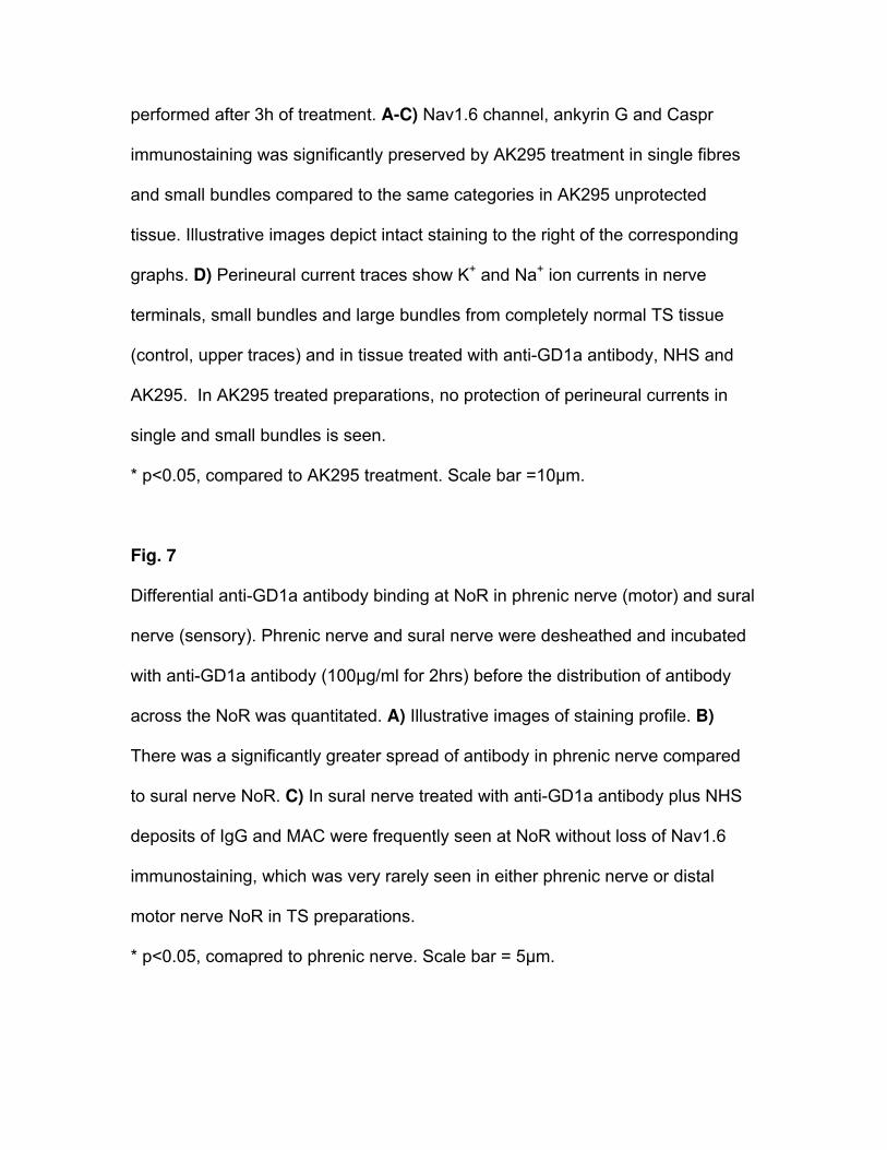
performed after 3h of treatment. A-C) Nav1.6 channel, ankyrin G and Caspr
immunostaining was significantly preserved by AK295 treatment in single fibres
and small bundles compared to the same categories in AK295 unprotected
tissue. Illustrative images depict intact staining to the right of the corresponding
graphs. D) Perineural current traces show K+ and Na+ ion currents in nerve
terminals, small bundles and large bundles from completely normal TS tissue
(control, upper traces) and in tissue treated with anti-GD1a antibody, NHS and
AK295. In AK295 treated preparations, no protection of perineural currents in
single and small bundles is seen.
* p<0.05, compared to AK295 treatment. Scale bar =10µm.
Fig. 7
Differential anti-GD1a antibody binding at NoR in phrenic nerve (motor) and sural
nerve (sensory). Phrenic nerve and sural nerve were desheathed and incubated
with anti-GD1a antibody (100µg/ml for 2hrs) before the distribution of antibody
across the NoR was quantitated. A) Illustrative images of staining profile. B)
There was a significantly greater spread of antibody in phrenic nerve compared
to sural nerve NoR. C) In sural nerve treated with anti-GD1a antibody plus NHS
deposits of IgG and MAC were frequently seen at NoR without loss of Nav1.6
immunostaining, which was very rarely seen in either phrenic nerve or distal
motor nerve NoR in TS preparations.
* p<0.05, comapred to phrenic nerve. Scale bar = 5µm.

Fig. 8
Phrenic nerve NoR immunostaining profiles of nodal proteins after exposure to
anti-GD1a antibody plus NHS are partially protected by AK295. Phrenic nerve
was desheathed and treated with anti-GD1a antibody or Ringers, plus NHS, with
and without AK295. and the effect on nodal protein immunostaining was
quantified. A) Anti-GD1a antibody treated phrenic nerve has significantly less
NoR that were immuno-positive for Nav1.6 channel, ankyrin G, Caspr, NrCAM
and moesin than control. B) AK295 does not protect ankyrin G, and only
modestly protects Nav1.6 channel and Caspr immunostaining from complement-
mediated injury. C) Moesin and NrCAM staining profiles are not significantly
altered in intensity after anti-GD1a antibody exposure; however they both show
an abnormal distribution as highlighted in the images. Examples of normal
control staining of all of the proteins (magenta) versus treated and AK295
protected nerve. Note occurrence of swollen morphology of treated nerves in DIC
that is not ameliorated by AK295 treatment. D) Extracellular recordings of anti-
GD1a antibody-treated phrenic nerve show a reduction in CAP amplitudes over
time that are unaffected by AK295. Anti-GD1a antibody was added for 2 hours;
subsequently NHS was added for 2 hours starting at 0 mins. Arrows indicate the
addition of 5µM TTX to terminate the experiment. E) CAP amplitudes are
expressed as the percentage of the starting value, there being no significant
difference between AK295 treated or untreated nerves.
* p<0.05, compared to control or AK295 counterpart. Scale bar = 5µm.
Supplementary figure legends
Fig. S1
Ganglioside biosynthesic pathway showing the structure of GD1a and the pattern
of deficiency seen in GD3 synthase-/- mice.
Fig. S2
Anti-GD1a antibody more effectively binds nerve terminals in transgenic mice
over-expressing a-series gangliosides (GD3-/-/CFP) compared to wild type mice,
as assessed by immunocytochemistry. TS muscle from both modified and WT
mice were incubated with anti-GD1a antibody (red) and the fluorescence
intensity of deposits measured over the NMJ, identified by BTx (green).
* p<0.05, compared to wild-type (WT). Scale bar = 20µm
Fig. S3
Nerve desheathment results in a significant increase in anti-GD1a antibody
deposition at NoR. Data shown for phrenic nerve.
* p<0.05, desheathed nerve compared with intact nerve.
Fig. S4
GD1a is uniformly present at intramuscular nerve NoR irrespective of bundle
size. In permeabilised TS preparations, anti-GD1a antibody binds equally to NoR

in single fibres, small, medium and large bundles, as assessed by
immunohistology.
Fig. S5
Three examples of Nav1.6 channel immunostaining at the phrenic nerve NoR
after anti-GD1a antibody exposure and 30mins treatment with NHS. Various
stages of dissolution of Nav1.6 immunostaining are evident (arrows), prior to its
subsequent complete disappearance.
Scale bar = 5µm
Fig. S6
Following g-LTx treatment that specifically disintegrates the nerve terminal
through pore formation in the pre-synaptic membrane, Nav1.6 channel
immunostaining is unaffected (arrow). * signifies the position of the now invisible
nerve terminal arborisation where endogenous CFP is lost due to the g-LTx-
evoked injury.
Scale bar = 20µm.
Fig. S7
Sciatic nerve and sural nerve CAP recordings show slight or no reduction
respectively after treatment with anti-GD1a antibody and NHS as a source of
complement. A) Extracellular recordings were obtained from control and treated
sciatic nerve and the peak CAP plotted over time. Sciatic nerves were exposed

to NHS as the complement source prior to transfer to the recording chamber. B)
For sural nerve, NHS was added to the recording chamber at 0 mins. Arrows
indicate the addition of 5µM TTX to terminate the experiment.
Fig S8.
Summary cartoon showing the principal features of the pathophysiological
casdade occurring at NoR exposed to anti-GD1a antibody and complement.
MAC pores form in the axolemmal membrane at the NoR that allow sufficient
uncontrolled ingress of calcium to activate calpain, resulting in degradation of
calpain substrates including Nav1.6, neurofilament, Caspr and Ankyrin G.
Acknowledgements
This work was supported by the Wellcome Trust (#077041/Z/05/Z, to HJW). RM
and KNG were supported by the MRC doctoral training account awarded to the
University of Glasgow. Dr Jaap Plomp is thanked for critical reading of the
manuscript.

References
Ang CW, Jacobs BC, Laman JD. The Guillain-Barré syndrome: a true case of
molecular mimicry. Trends Immunol 2004; 25: 61-66.
Arroyo EJ, Xu YT, Zhou L et al. Myelinating Schwann cells determine the
internodal localization of Kv1.1, Kv1.2, Kvbeta2, and Caspr. Journal of
Neurocytology1999; 28: 333-47.
Aspinall GO, McDonald AG, Raju TS, Pang H, Moran AP, Penner JL. Chemical
structures of the core regions of Campylobacter jejuni serotypes O:1, O:4, O:23,
and O:36 lipopolysaccharides. European Journal of Biochemistry 1993; 213:
1017-27.
Bhat MA, Rios JC, Lu Y et al. Axon-glia interactions and the domain organization
of myelinated axons requires neurexin IV/Caspr/Paranodin. Neuron.2001; 30:
369-8.
Boffey J, Odaka M, Nicoll D et al. Characterisation of the immunoglobulin
variable region gene usage encoding the murine anti-ganglioside antibody
repertoire. J.Neuroimmunol 2005; 165: 92-103.
Bowes T, Wagner ER, Boffey J et al. Tolerance to self gangliosides is the major
factor restricting the antibody response to lipopolysaccharide core
oligosaccharides in Campylobacter jejuni strains associated with Guillain-Barre
syndrome. Infection & Immunity 2002; 70 :5008-18.

Boyle ME, Berglund EO, Murai KK, Weber L, Peles E, Ranscht B. Contactin
orchestrates assembly of the septate-like junctions at the paranode in myelinated
peripheral nerve. Neuron 2001; 30: 385-97.
Braga MF, Harvey AL, Rowan EG. Effects of tacrine, velnacrine (HP029),
suronacrine (HP128), and 3,4-diaminopyridine on skeletal neuromuscular
transmission in vitro. Br.J.Pharmacol 1991; 102: 909-915.
Braga MF, Anderson AJ, Harvey AL, Rowan EG. Apparent block of K+ currents
in mouse motor nerve terminals by tetrodotoxin, mu-conotoxin and reduced
external sodium. Br J Pharmacol 1992; 106:91-4.
Brigant J and Mallart, A. Presynaptic currents in mouse motor endings. J.
Physiol., Lond 1982; 333: 619636.
Burkel WE. The histological fine structure of perineurium. Anatomical Record
1967; 158:177-89.
Caldwell JH, Schaller KL, Lasher RS, Peles E, Levinson SR. Sodium channel
Na(v)1.6 is localized at nodes of ranvier, dendrites, and synapses. Proceedings
of the National Academy of Sciences of the United States of America 2000;.97:
5616-20.
Chan SL, Mattson MP. Caspase and calpain substrates: roles in synaptic
plasticity and cell death. Journal of Neuroscience Research.1999; 58:167-90.

Charles P, Tait S, Faivre-Sarrailh C et al. Neurofascin is a glial receptor for the
paranodin/Caspr-contactin axonal complex at the axoglial junction. Current
Biology 2002; 12: 217-20.
Davis JQ, Lambert S, Bennett V. Molecular composition of the node of Ranvier:
identification of ankyrin-binding cell adhesion molecules neurofascin
(mucin+/third FNIII domain-) and NrCAM at nodal axon segments. Journal of Cell
Biology 1996; 135: 1355-67.
De Angelis MV, Di Muzio A, Lupo S, Gambi D, Uncini A, Lugaresi A. Anti-GD1a
antibodies from an acute motor axonal neuropathy patient selectively bind to
motor nerve fiber nodes of Ranvier. Journal of Neuroimmunology 2001; 121: 79-
82.
Duchen LW, Gomez S, Queiroz LS. The neuromuscular junction of the mouse
after black widow spider venom. Journal of Physiology 1981; 316: 279-91.
Einheber S, Zanazzi G, Ching W et al. The axonal membrane protein Caspr, a
homologue of neurexin IV, is a component of the septate-like paranodal junctions
that assemble during myelination. Journal of Cell Biology 1997;.139:1495-506.
Feasby TE, Gilbert JJ, Brown WF et al. An acute axonal form of Guillain-Barre
polyneuropathy. Brain 1986; 109: 1115-26.
Feng G, Mellor RH, Bernstein M et al. Imaging neuronal subsets in transgenic
mice expressing multiple spectral variants of GFP. Neuron 2000; 28: 41-51.

Gabriel CM. Prognosis in the acute motor axonal form of Guillain-Barre
syndrome. J.Neurol.Neurosurg.Psychiatry 2005; 76: 622.
Gong Y, Tagawa Y, Lunn MP et al. Localization of major gangliosides in the
PNS: implications for immune neuropathies. Brain 2002; 125: 2491-506.
Goodfellow JA, Bowes T, Sheikh K et al. Overexpression of GD1a ganglioside
sensitizes motor nerve terminals to anti-GD1a antibody-mediated injury in a
model of acute motor axonal neuropathy. Journal of Neuroscience 2005;
25:1620-8.
Goodyear CS, O'Hanlon GM, Plomp JJ et al. Monoclonal antibodies raised
against Guillain-Barre syndrome-associated Campylobacter jejuni
lipopolysaccharides react with neuronal gangliosides and paralyze muscle-nerve
preparations.[erratum appears in J Clin Invest 1999; 104:697-708.
Griffin JW, Li CY, Macko C et al. Early nodal changes in the acute motor axonal
neuropathy pattern of the Guillain-Barre syndrome. Journal of Neurocytology
1996; 25:33-51.
Hafer-Macko C, Hsieh ST, Li CY et al. Acute motor axonal neuropathy: an
antibody-mediated attack on axolemma. Annals of Neurology 1996; 40: 635-44.
Halstead SK, Humphreys PD, Goodfellow JA, Wagner ER, Smith RA, Willison
HJ. Complement inhibition abrogates nerve terminal injury in Miller Fisher
syndrome. Annals of Neurology 2005;.58: 203-10.

Halstead SK, Humphreys PD, Zitman FM, Hamer J, Plomp JJ, Willison HJ. C5
inhibitor rEV576 protects against neural injury in an in vitro mouse model of Miller
Fisher syndrome. Journal of the Peripheral Nervous System 2008a; 13: 228-35.
Halstead SK, Zitman FM, Humphreys PD et al. Eculizumab prevents anti-
ganglioside antibody-mediated neuropathy in a murine model. Brain 2008b.131:
1197-208.
Hillmen P, Young NS, Schubert J et al. The complement inhibitor eculizumab in
paroxysmal nocturnal hemoglobinuria. New England Journal of Medicine
2006;.355:1233-43.
Hiraga A, Kuwabara S, Ogawara K et al. Patterns and serial changes in
electrodiagnostic abnormalities of axonal Guillain-Barre syndrome. Neurology
2005a;.64: 856-60.
Hiraga A, Mori M, Ogawara K et al. Recovery patterns and long term prognosis
for axonal Guillain-Barre syndrome. J.Neurol.Neurosurg.Psychiatry 2005b; 76:
719-722.
Ho TW, Hsieh ST, Nachamkin I et al. Motor nerve terminal degeneration provides
a potential mechanism for rapid recovery in acute motor axonal neuropathy after
Campylobacter infection. Neurology 1997;.48: 717-24.
Ho TW, Willison HJ, Nachamkin I et al. Anti-GD1a antibody is associated with
axonal but not demyelinating forms of Guillain-Barre syndrome. Annals of
Neurology 1999;.45: 168-73.

Hughes RA, Cornblath DR. Guillain-Barre syndrome. Lancet 2005; .366: 1653-
66.
Iacovache I, van der Goot FG, Pernot L. Pore formation: an ancient yet complex
form of attack. Biochim.Biophys.Acta 2008; 1778: 1611-1623.
Iwata A, Stys PK, Wolf JA et al. Traumatic axonal injury induces proteolytic
cleavage of the voltage-gated sodium channels modulated by tetrodotoxin and
protease inhibitors. Journal of Neuroscience 2004;.24: 4605-13.
Kordeli E, Davis J, Trapp B, Bennett V. An isoform of ankyrin is localized at
nodes of Ranvier in myelinated axons of central and peripheral nerves. Journal of
Cell Biology 1990; 110: 1341-52.
Kuwabara S, Ogawara K, Misawa S et al. Does Campylobacter jejuni infection
elicit "demyelinating" Guillain-Barre syndrome. Neurology.2004; 63:529-33.
Langford LA, Schmidt RF. An electron microscopic analysis of the left phrenic
nerve in the rat. Anat Rec 2002; 205: 207-13.
Ledeen RW. Ganglioside structures and distribution: are they localized at the
nerve ending? Journal of Supramolecular Structure 1978; 8: 1-17.
Li Z, Ortega-Vilain AC, Patil GS et al. Novel peptidyl alpha-keto amide inhibitors
of calpains and other cysteine proteases. Journal of Medicinal Chemistry 1996;
39: 4089-98.

Lugaresi A, Ragno M, Torrieri F, Di Guglielmo G, Fermani P, Uncini A. Acute
motor axonal neuropathy with high titer IgG and IgA anti-GD1a antibodies
following Campylobacter enteritis. Journal of the Neurological Sciences
1997;.147: 193-200.
Mallart A. Electric current flow inside perineural sheaths of mouse motor nerves.
J. Physiol. Lond. 1985; 368: 565575.
Malmgren LT, Olsson Y. Differences between the peripheral and the central
nervous system in permeability to sodium fluorescein. Journal of Comparative
Neurology. 1980; 103-107.
McKhann GM, Cornblath DR, Griffin JW et al. Acute motor axonal neuropathy: a
frequent cause of acute flaccid paralysis in China. Annals of Neurology 1993;.33:
333-42.
McKhann GM, Cornblath DR, Ho T et al. Clinical and electrophysiological
aspects of acute paralytic disease of children and young adults in northern
China. Lancet 1991; 338: 593-7.
Menegoz M, Gaspar P, Le Bert M et al. Paranodin, a glycoprotein of neuronal
paranodal membranes. Neuron. 1997; 319-331.
O'Hanlon GM, Humphreys PD, Goldman RS et al. Calpain inhibitors protect
against axonal degeneration in a model of anti-ganglioside antibody-mediated
motor nerve terminal injury. Brain 2003; 126: 2497-2509.

Ogawara K, Kuwabara S, Mori M, Hattori T, Koga M, Yuki N. Axonal Guillain-
Barre syndrome: relation to anti-ganglioside antibodies and Campylobacter jejuni
infection in Japan. Ann.Neurol. 2000; 48: 624-631.
Okada M, Itoh MM, Haraguchi M et al. b-series Ganglioside deficiency exhibits
no definite changes in the neurogenesis and the sensitivity to Fas-mediated
apoptosis but impairs regeneration of the lesioned hypoglossal nerve. Journal of
Biological Chemistry 2002; 277: 1633-6.
Olsson Y. Microenvironment of the peripheral nervous system under normal and
pathological conditions. (Review). Critical Reviews in Neurobiology 1990; 5: 265-
311.
Peles E, Nativ M, Lustig M et al. Identification of a novel contactin-associated
transmembrane receptor with multiple domains implicated in protein-protein
interactions. EMBO Journal 1997;.16: 978-88.
Phongsisay V, Susuki K, Matsuno K et al. Complement inhibitor prevents
disruption of sodium channel clusters in a rabbit model of Guillain-Barre
syndrome. Journal of Neuroimmunology. 2008; 101-104.
Plomp JJ, Molenaar PC, O'Hanlon GM et al. Miller Fisher anti-GQ1b antibodies:
alpha-latrotoxin-like effects on motor end plates. Ann.Neurol. 1999; 45: 189-199.
Plomp JJ, Willison HJ. Pathophysiological actions of neuropathy-related anti-
ganglioside antibodies at the neuromuscular junction. J.Physiol 2009; 587: 3979-
3999.
Podack ER, Muller-Eberhard HJ, Horst H, Hoppe W. Membrane attack complex
of complement (MAC): three-dimensional analysis of MAC-phospholipid vesicle
recombinants. J Immunol 1982; 128:2353-2357.
Poliak S, Peles E. The local differentiation of myelinated axons at nodes of
Ranvier. Nature Reviews Neuroscience 2003; 4:968-80.
Rasband MN, Park EW, Zhen D et al. Clustering of neuronal potassium channels
is independent of their interaction with PSD-95. Journal of Cell Biology 2002;
159: 663-72.
Rios JC, Melendez-Vasquez CV, Einheber S et al. Contactin-associated protein
(Caspr) and contactin form a complex that is targeted to the paranodal junctions
during myelination. Journal of Neuroscience 2000; 8354-8364.
Saito A, Zacks SI. Ultrastructure of Schwann and perineural sheaths at the
mouse neuromuscular junction. Anatomical Record 1969; 164: 379-90.
Scherer SS. Molecular specializations at nodes and paranodes in peripheral
nerve. Microscopy Research & Technique 1996;.34:452-61.
Sheikh KA, Deerinck TJ, Ellisman MH, Griffin JW. The distribution of ganglioside-
like moieties in peripheral nerves. Brain 1999a; 122: 449-60.
Sheikh KA, Sun J, Liu Y et al. Mice lacking complex gangliosides develop
Wallerian degeneration and myelination defects. Proceedings of the National
Academy of Sciences of the United States of America 1999b;.96:7532-7.
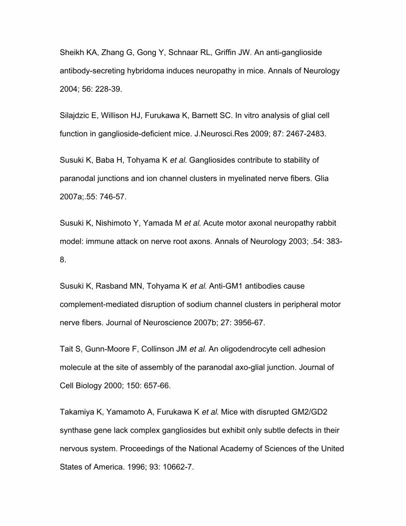
Sheikh KA, Zhang G, Gong Y, Schnaar RL, Griffin JW. An anti-ganglioside
antibody-secreting hybridoma induces neuropathy in mice. Annals of Neurology
2004; 56: 228-39.
Silajdzic E, Willison HJ, Furukawa K, Barnett SC. In vitro analysis of glial cell
function in ganglioside-deficient mice. J.Neurosci.Res 2009; 87: 2467-2483.
Susuki K, Baba H, Tohyama K et al. Gangliosides contribute to stability of
paranodal junctions and ion channel clusters in myelinated nerve fibers. Glia
2007a;.55: 746-57.
Susuki K, Nishimoto Y, Yamada M et al. Acute motor axonal neuropathy rabbit
model: immune attack on nerve root axons. Annals of Neurology 2003; .54: 383-
8.
Susuki K, Rasband MN, Tohyama K et al. Anti-GM1 antibodies cause
complement-mediated disruption of sodium channel clusters in peripheral motor
nerve fibers. Journal of Neuroscience 2007b; 27: 3956-67.
Tait S, Gunn-Moore F, Collinson JM et al. An oligodendrocyte cell adhesion
molecule at the site of assembly of the paranodal axo-glial junction. Journal of
Cell Biology 2000; 150: 657-66.
Takamiya K, Yamamoto A, Furukawa K et al. Mice with disrupted GM2/GD2
synthase gene lack complex gangliosides but exhibit only subtle defects in their
nervous system. Proceedings of the National Academy of Sciences of the United
States of America. 1996; 93: 10662-7.
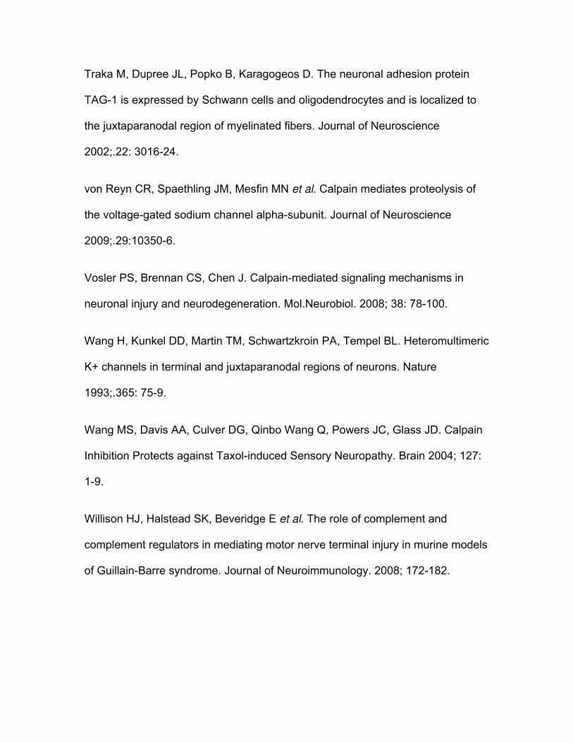
Traka M, Dupree JL, Popko B, Karagogeos D. The neuronal adhesion protein
TAG-1 is expressed by Schwann cells and oligodendrocytes and is localized to
the juxtaparanodal region of myelinated fibers. Journal of Neuroscience
2002;.22: 3016-24.
von Reyn CR, Spaethling JM, Mesfin MN et al. Calpain mediates proteolysis of
the voltage-gated sodium channel alpha-subunit. Journal of Neuroscience
2009;.29:10350-6.
Vosler PS, Brennan CS, Chen J. Calpain-mediated signaling mechanisms in
neuronal injury and neurodegeneration. Mol.Neurobiol. 2008; 38: 78-100.
Wang H, Kunkel DD, Martin TM, Schwartzkroin PA, Tempel BL. Heteromultimeric
K+ channels in terminal and juxtaparanodal regions of neurons. Nature
1993;.365: 75-9.
Wang MS, Davis AA, Culver DG, Qinbo Wang Q, Powers JC, Glass JD. Calpain
Inhibition Protects against Taxol-induced Sensory Neuropathy. Brain 2004; 127:
1-9.
Willison HJ, Halstead SK, Beveridge E et al. The role of complement and
complement regulators in mediating motor nerve terminal injury in murine models
of Guillain-Barre syndrome. Journal of Neuroimmunology. 2008; 172-182.

Merge CFP axon pNFasc anti-GD1a Ab
CFP axonsBTx
B
pNFascanti-GD1a Ab
C
anti-GD1a Ab
Figure 1
A
*
#
D
**
Large
Med
ium
Small
Sin
gle
Larg
e
Mediu
m
Smal
l
Sin
gle
120
100
80
60
40
20
0
IgG
Flu
ore
sc
en
ce
In
ten
sit
y
(arb
itra
ry u
nit
s)
E
*
#
**
Large
Med
ium
Small
Sin
gle
Larg
e
Mediu
m
Smal
l
Sin
gle
120
100
80
60
40
20
0IgG
Flu
ore
sc
en
ce
In
ten
sit
y
(arb
itra
ry
un
its
)
in vivoex vivo
CONTROL CONTROLAnti-GD1a Ab exposed Anti-GD1a Ab exposed
Single
Small
Mediu
mLarg
e
Single
Single
Small
Small
Small
Mediu
m
Mediu
m
Mediu
m
Single
Large
Large
Large
##
* *
B
*
#
Figure 2
1 ms
1 mV
0mv
Control Injured
C
CO
NT
RO
LIN
JU
RE
D
Nerve terminal Small bundle Large bundleD
Merge MAC DIC
A120
100
80
60
40
20
0
MA
C F
luo
res
ce
nc
e I
nte
ns
ity
(arb
itra
ry u
nit
s)
CONTROL INJURED
ex vivo
Single
Single
Small
Small
Mediu
m
Mediu
m
Large
Large
*
#

0
20
40
60
80
100
0
0
0
0
0
0
Control Injured
Kv1.1
Caspr
CFP
axons
Nav1.6
CFP axon
BTx
10um
pNFasc
CFP
axons
Merge Kv1.1
Nav1.6
DystrophinNav1.6
ankyrin G
Myelin
ankyrin G
Caspr
Myelin
Caspr
pNFasc
MyelinpNFasc
ankyrin G
CFP
axons
* *
*
*
**
*
*
A Nav1.6
D pan-Neurofascin
E Kv1.1
Figure 3%
im
mu
no
sta
ined
No
R
0
20
40
60
80
100
B Ankyrin G
0
20
40
60
80
100
0
20
40
60
80
100
0
20
40
60
80
100
single small medium large
C Caspr
single
single
single
single
small
small
small
small
medium
medium
medium
medium
large
large
large
large
Bundle size
Bundle size
Bundle size
Bundle size
Bundle size
Control
Injured
Control
Injured
Control
Injured
Control
Injured
Control
Injured
% i
mm
un
osta
ined
No
R%
im
mu
no
sta
ined
No
R%
im
mu
no
sta
ined
No
R%
im
mu
no
sta
ined
No
R

0
20
40
60
80
100
0
20
40
60
80
100
Nav1.6
CFP axons
ankyrin G
CFP axons
BTx
Merge
ankyrin G
Caspr
CFP axonsMerge
Caspr
*
*
*
*
A Nav1.6
B Ankyrin G
Figure 4
*
*
C Caspr
D
CFP axonsBTx
CFP axonsBTx
Eculizumab ALXN3300
Eculizumab
ALXN3300
Eculizumab
ALXN3300
Eculizumab
ALXN3300
single
single
small
small
medium
medium
large
large
Bundle size
Bundle size
% i
mm
un
osta
ined
No
R%
im
mu
no
sta
ined
No
R
single small medium large
Bundle size
% i
mm
un
osta
ined
No
R
100
80
60
40
20

Merge
Neurofilament
Merge
Neurofilament
No InhibitorAK295
100
80
60
40
20
0
Treatment
NF
sig
na
l (
as a
% o
f co
ntr
ol)
*
Figure 5
AK295 protected AK295 unprotected
InjuredAK295
100
80
60
40
20
0
Treatment
Ne
uro
fila
me
nt
sig
nal
( %
of
co
ntr
ol)
*

Figure 6
Figure 7
A
Merge Nav1.6MACIgG
C
Sural nervePhrenic nerve
12
10
8
6
4
2
Nerve
*
Merge IgG CFP axon
Merge
Su
ral
nerv
e
IgG CFP axon
Ph
ren
icn
erv
e
B
Sp
read
of
IgG
sta
inin
g (
µm
)
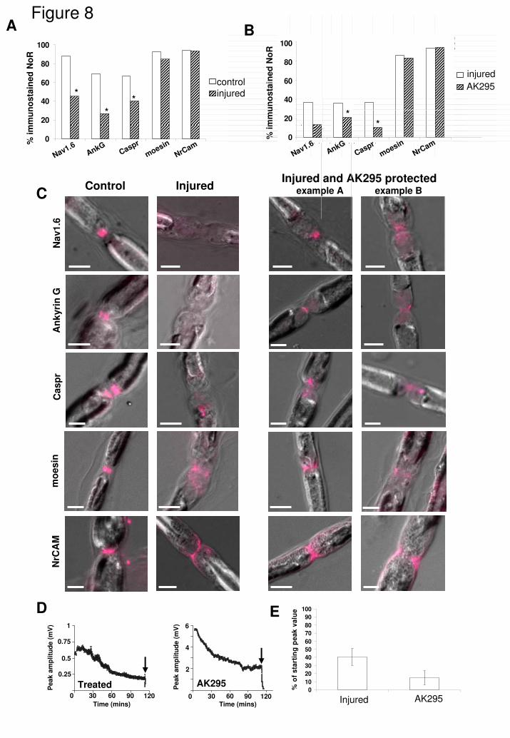
Control Injured
Na
v1
.6A
nk
yri
nG
Casp
r
Injured and AK295 protectedexample A example B
NrC
AM
mo
esin
0
20
40
60
80
100
0
10
20
30
40
50
60
70
80
90
100
% o
f s
tart
ing
pe
ak
va
lue
Figure 8A B
C
D E
Treated30 60 90
Time (mins)
1200
1
0.75
0.5
0.25
Peak a
mp
litu
de (
mV
)
AK295
30 60 90Time (mins)
1200
6
4
2
Peak a
mp
litu
de (
mV
)
*
*
*
% i
mm
un
osta
ined
No
R
controlinjured
injured
AK295
Injured AK295
AnkG
% i
mm
un
osta
ined
No
R
27 27
28 28
29 29
30 30
PN Ak295
AK295
Treated
AK295
Treated
AK29570
80
90
100
ng
70
80
90
100
ng0
20
40
60
80
100
Nav1.6Caspr
moesin
NrCam
Nav1.6AnkG
Caspr
moesin
NrCam
**
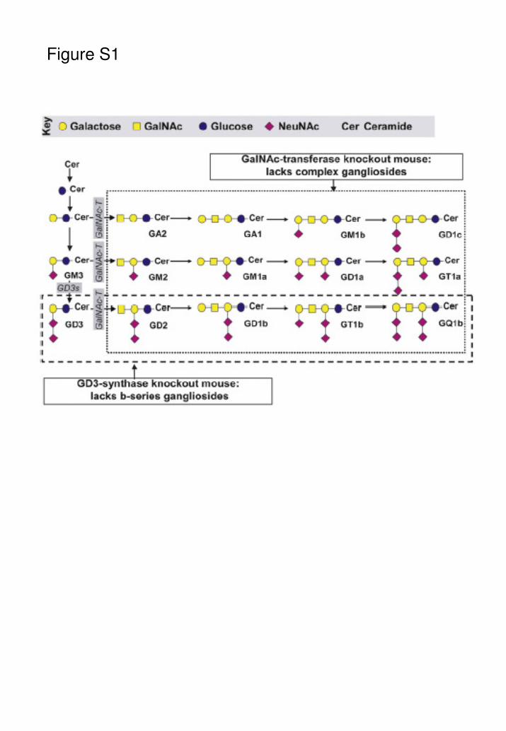
Figure S1

*
WT/CFP
IgG BTx
Merge
IgG BTx
Merge
GD3-/-/CFP
Figure S2
250
200
150
100
50
0
Mouse strain
GD3-/-/CFP WT/CFP
Flu
ore
scen
ce
in
ten
sit
y
(arb
itra
ry u
nit
s)
*
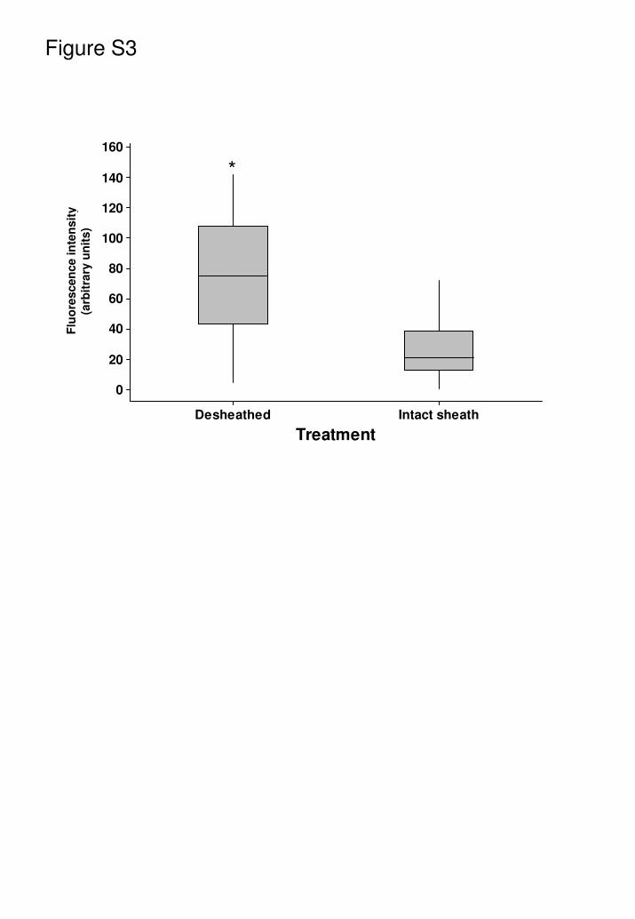
Figure S3
*
Intact sheathDesheathed
160
140
120
100
80
60
40
20
0
Treatment
Flu
ore
scen
ce
in
ten
sit
y
(arb
itra
ry u
nit
s)
*
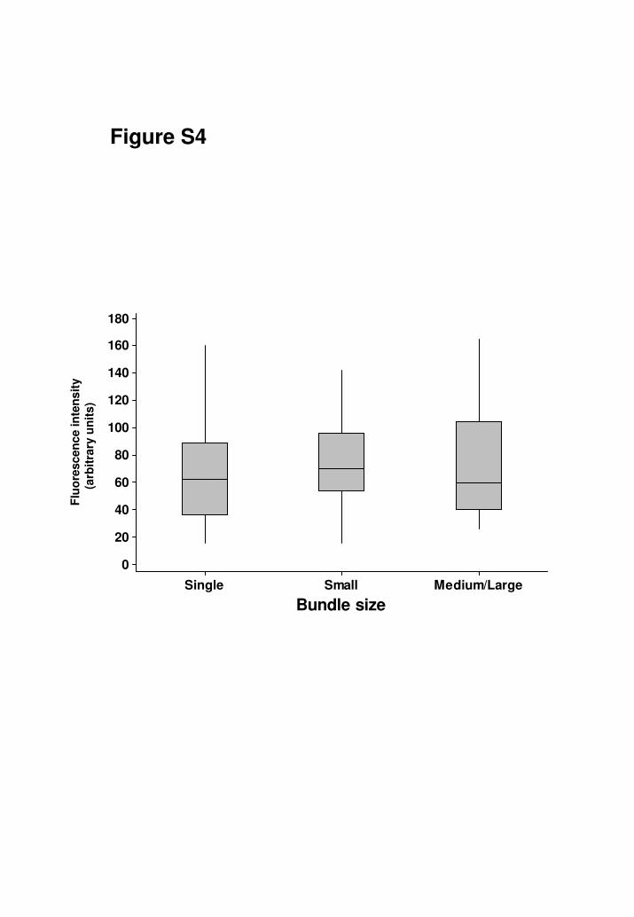
Figure S4
Flu
ore
scen
ce
in
ten
sit
y
(arb
itary
un
its)
Medium/LargeSmallSingle
180
160
140
120
100
80
60
40
20
0
Bundle size
Flu
ore
scen
ce
in
ten
sit
y
(arb
itra
ry u
nit
s)

Merge DIC
Merge DIC
Nav1.6
Nav1.6
Merge DIC Nav1.6
Figure S5
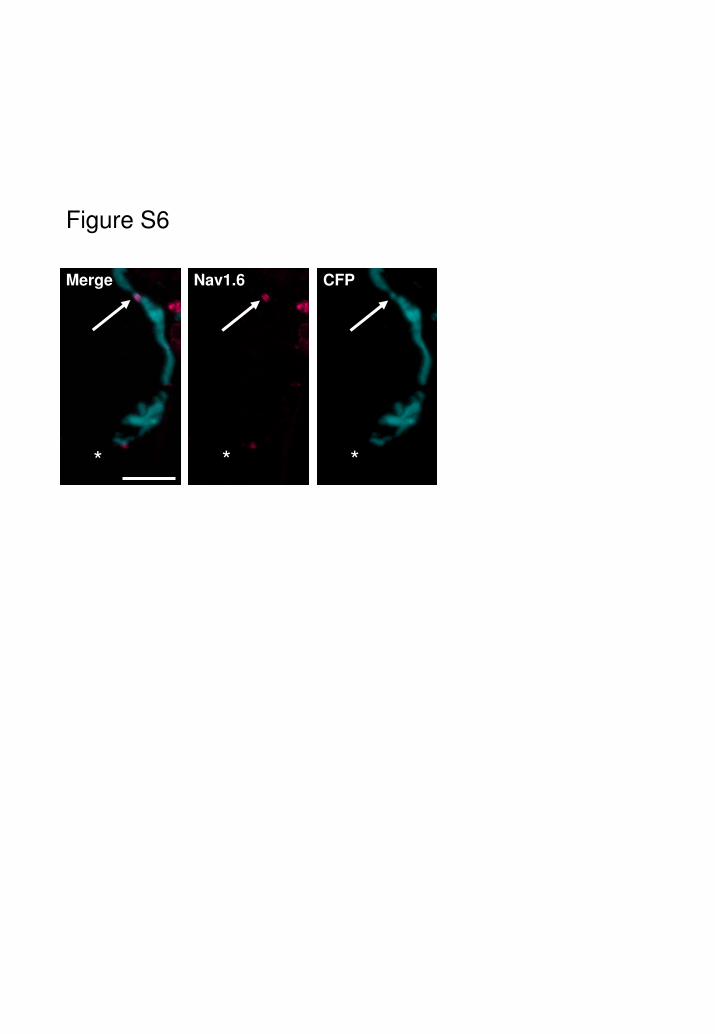
Figure S6
* * *
Merge Nav1.6 CFP

Treated Sciatic NerveControl Sciatic Nerve
15 30 45Time (mins)
0
4
3
2
1
Peak a
mp
litu
de (
mV
)
8 16 24Time (mins)
0
4
3
2
1
Peak a
mp
litu
de (
mV
)
32 40
A
Figure S7
Sural Nerve1
30 60 90Time (mins)
1200
0.75
0.5
0.25
Peak a
mp
litu
de (
mV
)
B
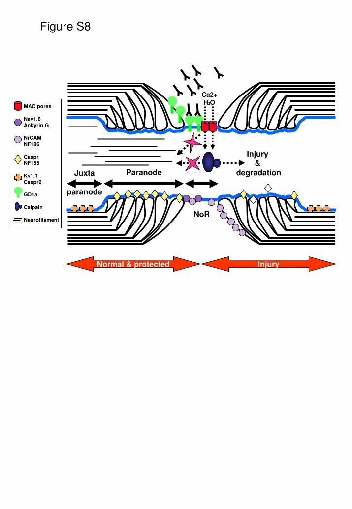
Figure S8
Ca2+H2O
Juxta
paranode
Paranode
NoR
Injury&
degradation
Normal & protected Injury
MAC pores
Nav1.6
Ankyrin G
NrCAM
NF186
Caspr
NF155
Kv1.1
Caspr2
GD1a
Calpain
Neurofilament

Table 1
P. Brophy, Edinburgh, UK1:400Ig
AbcamCambridge, UK
1:100IgG1MouseNrCAM*
Chemicon IntTemecula, CA
1:200IgGRabbitNeurofilament
P. Brophy, Edinburgh, UK1:1000IgGRabbitpan Neurofascin
SigmaMissouri, USA
1:100IgG1Mousepan Nav
SigmaMissouri, USA
1:100IgGRabbitNav1.6
BIOMOL IntEurope
1:100IgG1MouseMoesin
DakoGlostrup, Denmark
1:50IgG2aMouseHuman MAC (C5b-9)
Alomone LabsIsrael
1:200IgGRabbitKv1.1
Molecular ProbesPaisley, UK
1:400--FluoroMyelingreen
SigmaMissouri, USA
1:200IgG1MouseDystrophin
E. Peles, Israel1:1000IgGRabbitCaspr
Zymed LabsSan Francisco, CA
1:100IgG1MouseAnkyrin G
SourceDilutionIsotypeHostAntigen